Page 1
”WI
‘M"
’IH'IJJWH
H HH
“I “W
I”,! l I"!
*
'f"
H”Hi
I!”I,
M!“
W
«v.3 , «.5, m A. .
w J‘kfizcfitwfl'éga LN”;
CGNTENT GP
Twas for fine flag” 6? M S.
mama RATE causes
Sac-228M igémy Waliach
$133
Page 2
THESIS
This is to certify that the
thesis entitled
The Influence of the Thyroid Hormone on the
Ascorbic Acid Content of the Adrenal Cortex
presented by‘
Donald Pinny Wallach
‘5
has been accepted towards fulfillment
. of the requirements for
_Ml_fi.___degree inMLQLOgy
”WMajor professor
DateW_
“-795
Page 3
THE INFLUENCE OF THE THYROID HORMONE
ON THE ASCORBIC ACID CONTENT OF THE ADRENAL CORTEX
By
DONALD PINNY EflpLACH
A THESIS
Submitted to the school of Graduate Studies of Michigan
State College of Agriculture and Applied Science
in partial fulfillment of the requirements
for the degree of
MASTER OF SCIENCE
Department of Physiology and Pharmacology
1948
Page 5
ACKNOWLEDGEMENTS
The author wishes to express his gratitude to Professor
E. P. Reineke of the Department of Physiology and Pharmacol-
ogy, for the valuable advice and assistance rendered both
during the experimental work, and during the preparation of
this manuscript. Also to professor B. B. Roseboom for the
use of the facilities of the department. A great debt is
owed to Mr. Augustus Klock and Mr. Phillip Kotlar of the
Fieldston School, New York City, who provided the inspir—
ation in earlier years, which culminated in this work.
301335
Page 6
TABLE OF CONTENTS
Page
INTRODUCTION . . . . . . . . . . . . . . . . . . . . 1
REVIEW OF THE LITERATURE . . . . . . . . . . . . . . 3
Effect of Thyroxine on the Adrenals. . . . . . . 3
Hypertrophy not due to Raised Metabolism. . A
Effect of Hypothroidism on the Adrenals. . . . . 4
Effect of Total Adrenalectomy. . . . . . . . . . 6
Metabolic Depression and Adrenalin. . . . . 6
Effects of Adrenocortical Extracts in
Adrenalectomized Animals. . . . . . . . 8
Effects of Sublethal Adrenocortical Injury . . . 9
Inhibitory Effects of Adrenocortical Hormones on
the Thyroid. . . . . . . . . . . . . . . . . . 12
Effect of Adrenocortical Hormones on Metabolism. 13
Effects of Adrenocortical Hormones on the
Thyroid Gland. . . . . . . . . . . . . . . . . 1A
Summary of Evidence on the antagonistic Inter—
relationship of the Thyroid and Adrenal
Cortex . . . . . . . . . . . . . . . . . . . 15
Evidence of a Synergism Between the Thyroid
and Adrenal Cortex . . . . . . . . . 17
The Adrenal Medulla and the Thyroid. . . . . . . 17
Vitamin C and the Adrenal Cortex . . . . . . . . 18
Vitamin C and Adrenocortical Activity . . . 19
Evidence that Vitamin C is Not Essential to
Adrenocortical Activity . . . . . . . . . 21
Chemical Evidence that Vitamin C is a Part
of Adrenocortical Hormones. . . . . 22
Vitamin C and Adrenocortical Activity ina
Hyperthyroid State. . . . . . . . . . . . 24
Effects of Vitamin C Administration on
Metabolism. . . . . . . . . . . . 24
Relation of the Anterior Hypophysis to the
Adrenal Cortex. . . . . . . . . . . . . . 25
The Anterior Hypophysis and Adrenocortical
Vitamin C . . . . . . . . . . . . . . . . 26
Effects of Administration of Adrenocortical
Extracts on the Adrenal Cortex . . . . . . . . 29
Mechanism of Atrophy. . . . . . . . . . . . 20
A Sex Difference in Response. . . . . . . . 3
Negative Effects of Adrenocortical Hormones
on the Adrenal. . . . . . . . . . . . . . 3O
Adrenocortical Implants and the Adreno-
cortical Hormones . . . . . . . . . . . . 31
Effect of Adrenocortical Hormones on the
Adrenals in Conditions of Prolonged
Stress. . . . . . . . . . . . . . . . . . 31
Page 7
Page
A Contradiction to the Mechanism of
Atrophy Suggested Previously . . . . . . 32
Histological Changes in the Adrenals in Hyper-
and Hypothyroid States. . . . . . . . . . . . 32
MATERIALS AND METHODS . . . . . . . . . . . . . .
Extraction Procedure. . . . . . . . . . .
Method for Estimation of Ascorbic Acid.
Calculations . . . . . . . . . . .
Preparation of Reagents . . . . . . . . .
EXPERIMENT 1.. . . . . . . . . . . . . . . . . . 38
The Effect of Time on the Response of the
Adrenals to a Constant Level of Thyroidal
Stimulation . . . . . . . . . . . . . . . . . 38
EXPERIMENT II
Effect of Varying Levels of Thyroidal Stimula-
tion on Adrenal Weight and Ascorbic Acid
Content . . . . . . . . . . . . . . . . . . . Al
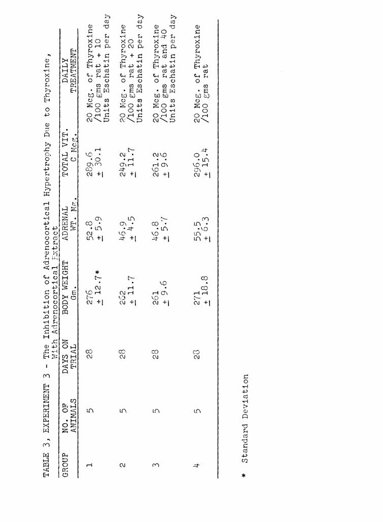
EXPERIMENT III. . . . . . . . . . . . . . . . 45
A Determination of the Secretion Rate of the
Adrenal Cortex in the Hyperthyroid Rat. . . . 45
EXPERIMENT IV . . . . . . . . . . . . . . . . . . 48
The Effect of Thyroxine on the Adrenals of
Pregnant Rats and on the Adrenals of their
Offspring . . . . . . . . . . . . . . . . . . 48
DISCUSSION. . . . . . . . . . . . . . . . . . . . . 51
SUMMARY AND CONCLUSIONS . . . . . . . . . . . . . . 54
BIBLIOGRAPHY. O O O O O O O O O O O O O O O O O O O 56
Page 8
INTRODUCTION
With the current interest in the physiology of the
thyroid gland, and the possible use, in certain phases of
animal production, of thyroidally active iodinated proteins,
and the goitrogenic thioureas, it becomes of interest to
know what the effects of the thyroid hormone, or a lack of
it, are on the animal body with respect to productive
processes, and specific organs affected.
One of the most important endocrine glands in the
animal body, indeed one essential to life, is the adrenal
cortex. This study is concerned with the effects of vary-
ing levels of thyroidal stimulation on the adrenal cortex.
There is extensive evidence in the literature suggesting
an interrelationship between the thyroid gland and the adrenal
cortex. The nature of this interrelationship is, however,
rather imperfectly understood. It has recently been demon-
strated that the cholesterol and ascorbic acid concentra-
tions in the adrenal cortex are reliable indicators of the
functional activity of this organ. Using the determination
of ascorbic acid in the adrenal cortex as a criterion of
activity, the following facets of the thyroid-adrenocortical
relationship have been investigated.
I. The effect of varying dosage levels of thyroidally
active iodinated casein (Protamone) administered in the diet
at levels of physiological stimulation.
II. The effects of varying dosage levels of thyroxine
administered parenterally above physiological levels of stim-
ulation.
Page 9
(2)
III. The concentration of ascorbic acid in the adrenals
at various time intervals during a six week regimen of thy—
roxine injections at a constant dose.
IV. The effect of concurrent dosage of varying levels
of adrenocortical extracts with administration of a constant
dosage level of thyroxine.
V. The influence of thyroidectomy.
VI. The effects of a constant dosage level of thyroxine
on the adrenal cortices of pregnant rats, and on the adrenal
cortices of the new born young of these mothers.
VII. The influence of sex on normal concentrations of
ascorbic acid found in the adrenals.
Page 10
REVIEW OF THE LITERATURE
Evidence of an interrelationship between the thyroid
gland and the adrenal cortex exists in abundance. For the
sake of convenience this has been divided into four prin-
cipal categories, the first of which includes the effects
of thyroxine administration on the adrenals.
Effect of Thyroxine on the Adrenals
The first work reported concerning the effects of
thyroid administration on the adrenals was that of Hoskins
(1). In this memorable but often overlooked paper, Hoskins
reported that feeding desiccated thyroid to guinea pigs
produced adrenal hypertrophy. Subsequently this work was
confirmed for the adrenals of the rat by Herring (2), Cam-
eron and Carmichael (3), Korenchevsky, et. a1. (4), Cohen
(5), Schmidt and Schmidt (6), DeNesselow and Griffiths (7),
Ingle and Kendall (8), Uotila (9), Korenchevsky and Hall (10),
and Lowenstein and Zwemmer (ll).
Ingle and Higgins (12), in a somewhat different approach,
enucleated one adrenal in a rat, and found that thyroxine
administration caused extensive hypertrophy in the intact
gland. In the enucleated gland, there was an increase in
size of the zona glomerulosa, but there was no significant
increase between the weight of this gland and an enucleated
adrenal in a control animal not receiving thyroxine injec-
tions. The enucleated gland of a thyroxine—treated rat will
regenerate faster, and to a greater extent, than will an
Page 11
untreated control.
Hypertrophy not due to Raised Metabolism.
The increase in metabolism following thyroxine admin—
istration is not the cause of adrenal hypertrophy. This was
demonstrated by DeNesselow and Griffith (7), when they ad-
ministered dinitrophenol to rats. This compound raised the
metabolic rate of the rats, but did not cause adrenal hyper-
trophy. Further evidence that this compound acts differently
from thyroxine was demonStrated by Cutting and Tainter (13).
They failed to note growth acceleration in tadpoles following
dinitrophenol adminstration. The metamorphosis acceler-
ating effect of thyroxine in this species is well known.
If a hyperthyroid condition produces adrenal hyper-
trophy, it seems logical to expect that the opposite condition
of hypothyroidism should produce adrenal atrophy. The second
category of evidence concerning the thyroid-adrenal relation-
ship is thus concerned with the effects of hypothyroid states
on the adrenal.
Effect of Hypothyroidism on the Adrenals
The literature concerning the effects of hypothyroid
conditions on the adrenal seems somewhat confusing. Gley (14)
as cited by Koelsche and Kendall (15), found that there was
an enlargement of the suprarenals after atrophy of the thyroid
gland or after thyroidectomy, but upon histological examina-
tion, this increase in size was attributed to an increase in
the lipoid content of the cortical cells. He concluded, that
Page 12
the enlargement could not be due to hyperfunction of the
adrenals. Cennedy and Purves (16), noted adrenal hyper-
trophy in rats fed goitrogenic seeds of the Brassicae family.
Their histological studies confirmed Gley's work, as they
found an increase in the fat content of the cortical cells.
In recent years, a number of the sulfa drugs have been
found to possess goitrogenic activity as well as the thioureas,
and thiouracil compounds. Leblond and Hoff (17), adminis-
tered sulfadiazine, sulfathiazole, and thiouracil to three
groups of rats and thyroidectomized a fourth group. The
animals were kept on these respective treatments for three
weeks, and all animals except those receiving sulfadiazine,
showed a decrease in the size of the adrenals. The most
marked decrease occurred in the thiouracil group, followed
by the thyroidectomized, and sulfathiazole groups. Sulfa-
diazine had no effect.
The influence of thiouracil on the adrenals was con-
firmed by Baumann and Marine (18). They administered
thiouracil to rats for four months, and noted an involution
of the adrenals to half their former size. Histological
examination of the adrenals of these animals, showed the
adrenals were still capable of hypertrophy, however, because
if these hypothyroid animals were subjected to stresses,
the atrophied adrenals would enlarge.
Glock (19), fed rats 0.5% thiourea in the diet, and
noted that the animals remained at constant weight for
four weeks. After this period death often ensued, but if
adrenocortical extracts were administered, the rats sometimes
Page 13
recovered. Histologically, the adrenals of the rats in
moribund condition, showed marked congestion, particularly
of the zona reticularis, depletion of the lipoid content of
the cortex, hemorrhages, and sometimes areas of necrosis. This
work possibly indicates a toxic action of the particular thio-
urea used, as these results have not been reported for other
goitrogens.
The third division of this discussion deals with the
effects of adrenalectomy and sublethal adrenocortical injury
on the thyroids.
Effect of Total Adrenalectomy
Most of the evidence concerned with the effects of adrenal
extirpation on the thyroids, is related to metabolism studies
and is therefore indirect. Gradinescu (20), as cited by Aub
et. a1. (21), noted a drop in the metabolism of three adrena-
1ectomized cats and one dog. He attributed this drop to "a
lack of regulatory mechanism for the peripheral vessels whose
movements are normally controlled by adrenin from the suprare-
nal capsules." This drop in metabolism has been confirmed
in cats by Webster et. a1. (22) and Aub et. a1. (21), in dogs
by Harrop et. a1. (23), and in rats by Carr and Beck (24).
It has been noted by Lerman (25), and Aub et. a1. (39), that
basal metabolism often is low in Addison's syndrome.
Metabolic Depression and Adrenalin.
Aub et. al. (21), noted that when the thyroid is re-
moved from animals, about three days elapse before a lowering
Page 14
(7)
in the metabolic rate is seen. When the hypophysis or the
gonads are removed, metabolic effects are not seen for even
longer periods of time. When the adrenals are removed, how-
ever, the decline in metabolic rate is abrupt, and prompt,
and the authors suggest from these facts, that the metabolic
depression following adrenalectomy is not related to other
organs of internal secretion. In support of this idea, the
following facts are cited:
1. Under anaesthesia, the lowered basal metabolic rate
due to adrenalectomy may be returned to normal by intravenous
injection of adrenalin, and they suggest that the metabolic
fall is due to a lack of epinephrine.
2. Adrenalectomy causes a drop in metabolism in a thy-
roidectomized animal, and thus the thyroid is not involved.
3. Adrenaline injections increase metabolism in thy-
roidectomized animals.
A. In thyrotoxic animals, adrenalectomy does not cause
a return to normal of the elevated metabolism.
5. Metabolism of resting frog muscle is greatly in-
creased by bathing in adrenaline solutions. The same holds
true for heart muscle.
Aub et. a1. (39), in a subsequent paper, reported that
under urethane anaesthesia there is a greater flow of adren—
aline than normal. If the adrenals are removed under this
anaesthetic, there is a prompt and progressive fall in
metabolism averaging 12%, and this drop occurs after removal
of the thyroid. Thus the thyroid is not essential to this
reaction. Adrenaline administered intravenously causes a
Page 15
(8)
distince rise in metabolism under urethane anaesthesia, and
the authors suggest as before, that this fall in metabolism'
is due to a lack of adrenaline.
From this work Aub et. a1. (39) conclude that there are
two physiological mechanisms for the elevation of basal metab-
olic rate. The adrenaline mechanism is a quick reaction
while that of thyroxine is for sustained periods of time.
Aub et. a1. (39) failed to note any metabolic effects fol-
lowing the injection of adrenocortical extracts into adren-
alectomized cats.
Effects of Adrenocortical Extracts in Adrenalectomized Animals.
The work of Webster et. a1. (22), contradicts some of
Aub's work. Webster et. a1. (22) noted a 50% drop in metab—
olism in bilaterally adrenalectomized cats, and administra-
tion of adrenocortical extracts could bring this back to normal
in 24 to 48 hours. They also noted a 15% to 30% increase in
metabolism in thyroidectomized cats following adminstration
of adrenocortical extracts, and this effect occurred in a short
period of time. They further observed that adrenocortical
extracts exerted no metabolic effects in normal animals.
Harrop et. a1. (23), confirmed some of Webster's work in
dogs. He observed that large amounts of adrenaline-free ad-
renocortical extracts had no metabolic effects on normal dogs.
This has been confirmed in human subjects by Hitchcock et. a1.
(26). The adrenocortical extracts could, however, prevent a
drop of 20%—25% in metabolism following adrenalectomy. Harrop
noted in contradiction to Aub et. a1. (21), that the metabolic
Page 16
(9)
decline following adrenalectomy does not occur in a dog
maintained with cortin for a "considerable interval" after
the withdrawal of adrenocortical extracts, and is more or
less coincident with a fall in body temperature. This paper
also reported the histological condition of the thyroids of
adrenalectomized dogs maintained with adrenocortical extracts.
An absence of hyperplasia was observed, as well as frequent
calcification of the colloid.
It has thus been adequately demonstrated that following
total extirpation of the adrenals, there is a fall in metab-
olism. This fall has not been observed following sublethal
adrenocortical injury, and in many cases a rise in metabolism
has been noted.
Effects of Sublethal Adrenocortical Injury
The first work reported on sublethal adrenocortical
injury and its effect on metabolism was that of Golyakowski
(27), as cited by Webster et. a1. (22). Golyakowski (27)
almost completely ligated the blood vessels supplying the
adrenals in dogs, and noted a 30% increase in heat produc-
tion in those dogs surviving six weeks or longer.
Similar work on other species has been reported by Marine
and Baumann (28), (29), Scott (30), Davis and Hastings (31),
and Carr and Beck (24). 'Marine and Baumann (28), noted in
rabbits when the adrenals were damaged by freezing with
ethylene chloride, or were removed, a disturbance in metabolism
characterized by increased heat production and carbon dioxide
output was seen. (As a good many rabbits have accessory
Page 17
(10)
adrenocortical tissue, adrenalectomy in those rabbits which
survive the operation, is really equivalent to sublethal
injury of the adrenal cortex.) This heat rise continued
for periods up to three weeks, after which there was a
gradual decline until the death of the animal. The symptom
complex suggested in these rabbits was that of exophthalmic
goiter. From this work Marine and Baumann (28), suggested
that there is an antagonism between the adrenal cortex and
the thyroid. They believe that the adrenal cortex exerts
an inhibiting effect over the thyroid secretion, and when
its influence is absent, the thyroid brings about the ob-
served metabolic changes. In addition to these changes in
metabolism, Marine and Baumann (28) also noted sleeker fur
in those rabbits with injured adrenocortical tissue, as well
as a vicious disposition which is characteristic of a hyper-
thyroid rabbit.
In a subsequent paper, Marine and Baumann (29), noted
that thyroidectomy preceding destruction of adrenocortical
tissue in the rabbit, prevented the previously observed heat
rise.
Scott (30) repeated this work in cats, and noted that
adrenal injury by partial ligation of the blood supply, or
freezing with ethylene chloride, caused a significant and
prolonged increase in heat production followed by a gradual
decline to the death. The injury to the adrenals must be
such that there is enough functional adrenocortical tissue
present to just maintain the animal. If there is too much
functional tissue present, the rise in heat production is
Page 18
(11)
not observed. Scott (30) observed in cats with the adrenals
injured to the optimum degree, that there was an unusual sleek-
ness of the fur, and an increase in appetite which suggested
increased thyroid activity.
The decline in heat production in the terminal stages
of life could be prevented by increasing the iodine content
of the diet. Scott suggests that the gradual decline in heat
production following the initial heat rise, is due to exhaus—
tion of the thyroid,because 30 days after the addition of
iodides to the diet, the thyroids were hypertrophied in those
cats which showed an increase in heat production after sup-
rarenal injury.
Barlow (32) noted in the cat after unilateral adrenalec-
tomy, an initial drop in total metabolism, which gradually
returned to normal or above. In bilaterally adrenalectomized
cats, there was an initial depression in metabolism followed
by a rise toward normal for the first six hours after the
operation, and then a gradual decline in metabolism until the
death of the animal at 40 hours.
Davis and Hastings (31), reported similar work in the mouse.
This animal like the rabbit, possesses accessory adrenocortical
tissue, and therefore adrenalectomy really amounts to sublethal
adrenocortical injury in those animals surviving the operation.
Davis and Hastings (31) noted that excised muscle from adren-
alectomized mice showed an increase in "aerobic" metabolism
40-70 days following adrenalectomy. If adrenalectomy was pre-
ceded by thyroidectomy, this increase in heat production was
not observed. Davis also noted that the activity of
Page 19
(l)R)
adrenalectomized mice was similar to intact mice receiving
thyroxine, and further, that adrenalectomized mice Show a
much greater sensitivity to thyroxine. This observation
has been made by Lerman (25), in human patients with Addisons
disease. He reports in such patients a lowering of the basal
metabolic rate, and if thyroid is administered to correct this
condition, the patient may be thrown into a state of Addisonian
crisis.
Davis and Hastings (31) from their work suggest that there
is an interrelationship between the thyroid and the adrenal
cortex such that when there is a deficiency of adrenocortical
hormones, there is an increased production of the thyroid
hormone, and this overproduction of the thyroid hormone con-
tinues until sufficient adrenocortical tissue has regenerated
to make up the deficiency.
Inhibitory Effects of Adrenocortical Hormones on the Thyroid.
Further evidence of a regulatory effect of the adrenocort-
ical hormones on the thyroid was presented in a review by
Marine (33). He points out that in Graves disease, the
adrenals are usually small, and he attributes the etiolog-
ical factor in Graves disease to adrenocortical insuffi—
ciency. He believes that the gonads may be involved, but the
thyroid changes in this disease are due to a compensatory
mechanism arising from the lack of adrenocortical hormones.
In addition to his rabbit work (previously cited) he points
out the fact that in the new born human infant, the adrenal
cortex begins to involute about the 8th day after birth,
Page 20
(13)
premature or otherwise. With this involution there is a
rapid rise in heat production. Marine believes that in
intrauterine life, the adrenal cortex controls tissue
oxidations, and that the adrenal cortex involutes after
birth to increase heat production.
From the evidence presented, it would appear that the
adrenocortical hormones have an inhibitory effect on the
thyroid secretion, as a lack of these hormones will cause
an increase in heat production which is not observed when
the thyroid is absent.
Effect of Adrenocortical Hormones on Metabolism
If the adrenocortical hormones have this inhibitory
effect on the thyroid, their administration should logically
depress metabolism in some way. The fourth category of ev-
idence concerning the adrenocortical—thyroid relationship is
thus concerned with the effects of adrenocortical extracts
on metabolism and on the thyroid gland itself.
Evidence concerning the effects of adrenocortical hormones
on metabolism is contradictory. Marine et. a1. (34), fed
rabbits glycerol emulsions of ex adrenal glands and noted a
depression in metabolism of 6%-27%. The fall in metabol-
ism began 5—7 days after beginning the treatments which is,
about the length of time required for thyroidectomy to show
its effects. He suggested that the metabolism depressing
substance exerts its effects either directly or indirectly
through the visceral nervous system on the thyroid cells,
either to inhibit the formation of thyroxine, or to prevent
Page 21
(14)
its secretion. In a subsequent paper Marine et. a1. (35),
reported that this same glycerol emulsion caused beneficial
effects in Graves disease, causing an increase in muscle
strength and body weight. Marine (33), also found that
feeding ovarian corpora lutea had some beneficial effects in
Graves disease.
These results have not been confirmed by Aub et. a1. (39),
Webster (22), or Hitchcock (26). Aub et. a1. (39), found no
metabolic effects following injection of adrenocortical ex-
tracts in the cat. Webster et. a1. (22), confirmed these
findings in the cat and rabbit. Hitchcock et. a1. (26),
injected adrenocortical extracts into humans and noted no
effects on Oxygen consumption.
The author had occasion to check oxygen consumption of
hyperthyroid rats receiving 20 gamma of thyroxine per 100
grams of body weight coincident with the administration of 1
unit of the commercial adrenocortical extract "Eschatin"
(Parke—Davis) per 100 grams of body weight, and noted no dif-
ference in oxygen consumption between these rats and some
receiving thyroxine at the aforementioned dosage. Thus the
majority of evidence would seem to point to the fact that adren-
ocortical extracts exert no direct effects on energy metabolism.
Effects of Adrenocorticel Hormones on the Thyroid Gland.
The evidence concerning the effects of adrenocortical
extracts on the thyroid itself is meagre and contradictory.
Black et. al. (36), fed an adrenal residue consisting of the
"final filtrate from an aqueous extract of the gland after
Page 22
(15)
the removal of the nucleoproteins with acetic acid, and then
the coaguable acid and alkali albumens. The resultant fil-
trate having been boiled at least twice, represents a slightly
hydrolyzed preparation". This was administered per—os to dogs
in experimental periods ranging from 1 to 6 months. Half of
the dog's thyroid was analyzed for iodine content at the start
of the experiment, and the other half analyzed at the con-
clusion of the experiment. With the extract used, an average
increase of 70.4% in the iodine content of the gland was
noted at the end of 45 days. Another adrenal extract con-
sisting of the adrenal nucleoproteins was also fed to dogs,
and this produced in the same length of time, an average
increase in the iodine content of the dog thyroid, of 50.7%.
Epinephrine had no effect.
Elmer et. a1. (37), did not confirm these findings in
guinea pigs. Using three groups of animals he injected the
first group with thyrotrophin, the second group received
cortin, and the third group received cortin and thyrotrophin.
Cortin had no effect on the normal thyroid, and was unable
to prevent the hypertrophy induced by thyrotrophin. The
authors point out that these results are opposed to Marine's
theory that Graves disease is the result of adrenocortical
insufficiency.
Summary of Eyidence on the antagonistic Interrelationship
of the Thyroid and Adrenal Cortex
To summarize the arguments in favor of an interrelation-
ship between the thyroid and the adrenal:
Page 23
(l6)
1. Thyroxine administration produces adrenal hypertrophy.
2. Administration of goitrogens or thyroidectomy, for the
most part produces adrenocortical atrophy. Where there is
hypertrophy, it is due to fatty infiltration of the cortex,
and is not considered to be due to hyperfunction.
3. Adrenalectomy in those animals without accessory
adrenocortical tissue like the dog and cat, will result in
a decrease in metabolism until the death of the animal. This
decline is sometimes preceded by a slight rise. The decline
in metabolism can be prevented with adrenocortical extracts,
and epinephrine.
4. In those animals with accessory adrenocortical tissue
such as the rat and the mouse, adrenalectomy is tantamount
to sublethal adrenocortical injury in those animals that
survive the operation. In animals without accessory adrenals
such as the dog and eat, if the adrenals are crippled by
freezing, or by partial ligation, these animals are then
equivalent to adrenalectomized animals with accessory
adrenocortical tissue. In this condition, a rise in metab-
olism is noted immediately following adrenocortical injury,
but is not seen where the thyroid is absent.
From these facts it is postulated that cortical hormones
in some way exert an inhibiting effect on the thyroid, and
when this secretion is lacking, the thyroid is free to exert
its well known metabolic effects.
Page 24
(17)
Evidence of a Synergism Between the Thyroid and Adrenal Cortex.
The evidence presented so far is conducive to the idea
that there is an antagonism between the thyroid and the
adrenal cortex. There is some evidence, however, that the
adrenocortical hormones have a synergistic effect with thy-
roxine. In an abstract of work by Bock (38), it was observed
that the addition of 5 cc of cortin to a bath of 500 cc of
water containing 0.2 cc of thyroxine, accelerated the meta-
morphosis of tadpoles and axolotls beyond the rate attained
by this concentration of thyroxine alone. This concentration
of cortin alone, did not influence metamorphosis.
The adrenal Medulla and the Thyroid
No discussion of an interrelationship between the thyroid
and the adrenals could be considered complete without some
reference to the relationship of the adrenal medulla to the
thyroid.
Levy (40), noted that the thyroid is induced to secrete
by stimulation of the cervical sympathetic ganglia, and the
thyroid secretion renders more excitable, the sympathetic
structures acted on by adrenalin in raising arterial pressure.
Cannon and Cattell (41), demonstrated that adrenalin causes
an action current in the thyroid, and in a subsequent paper
Cannon and Cattell (42), reported that there are non medul-
lated fibers distributed to the thyroid. These fibers were
found to be true secretory fibers, as they did not exert
their effects through alterations in the blood supply to
the thyroid. Cannon found that an action current is evoked
Page 25
in these fibers by adrenalin, and he suggests that this may
indicate secretory activity by the thyroid.
Metabolism studies by Marine and Lenhart (43), show that
there is an increased oxygen consumption following an injec-
tion of adrenalin in normal and thyroidectomized rabbits,
but the onset of increased oxygen consumption in a thyro-
idectomized rabbit is delayed and does not last as long, as
in a normal control animal.
The observation of Aub et. a1. (39) that there are two
mechanisms regulating metabolism in the body, one being
adrenaline which serves to raise metabolism quickly and
for short periods of time, while the thyroid exerts its
effects more slowly, and for longer periods of time, may be
worthy of repetition here.
Vitamin C and the Adrenal Cortex
Since the discovery of ascorbic acid in the adrenals by
Szent—Gyorgyi (44), there has been a lot of evidence brought
forth indicating that vitamin C is in some way concerned
with adrenocortical activity. The highest concentrations
of ascorbic acid found in the body are in the adrenals,
Yavorsky et. a1. (45). Harris and Ray (46), noted in guinea
pigs, that even though the adrenals are very rich in vitamin
C, there is only enough vitamin C present to supply the physi—
ological needs of the guinea pig for 24 hours. With vitamin
C administration, the adrenal ascorbic acid concentration
does not increase. In scorbutic guinea pigs even when teeth
lesions are evident, there are still considerable amounts of
Page 26
(19)
ascorbic acid present in the adrenals. During the progress
of avitaminosis C, the decline in adrenal ascorbic acid is
very slow, and at the death of the animal, there is still
present in the adrenal, small amounts of the vitamin. Harris
and Ray (46), concluded that vitamin C was necessary for the
normal functioning of the adrenals. Quick (47), confirmed this
work in guinea pigs. He observed a marked adrenal hypertrophy
in guinea pigs dead of scurvey, and using the silver nitrate
reduction method of Szent—Gyorgyi (44), for the determina-
tion of vitamin 0, he noted that the adrenals were depleted
of vitamin C. He also noted that there is a marked capil-
lary permeability in vitamin C deficiency which results in
diffuse hemorrhages. This condition is also seen in acute
adrenocortical insufficiency. It thus appeared that vitamin
C was not stored in the adrenals for the physiological needs
of the body.
Vitamin C and Adrenocortical Activity.
The first direct evidence that Vitamin C is in some way
concerned with adrenocortical activity was advanced by Kuehel
and Mitchell (48), in an important, but often overlooked,
piece of work. Kuchel and Mitchell (48) noted that when rats
were stimulated by fear, injections of acetyl choline, light
ether anaesthesia for 45 minutes, curare administration in
conjunction with light ether anaesthesia for 45 minutes,
electrical stimulation following ether anaesthesia, injections
of morphine, injections of eserine, and any situation causing
adrenal stimulation, there was a decline in the ascorbic acid
Page 27
(20)
content of the glands. Kuchel and Mitchell (48) further
noted that adrenaline content bore no constant relation-
ship to ascorbic acid and the ratio of glutathione to ascorbic
acid was not altered by adrenal stimulation. Bowman and
Muntwyler (49) confirmed some of Kuchel and Mitchell's work
in rats and guinea pigs. Euler and Klussman (50), as cited
by King (51), noted a 50% depletion in adrenal ascorbic acid
in guinea pigs injected with methylene blue. Flexner and
Grollman (52), in a series of experiments designed to augment
or inhibit adrenocortical activity, observed that when adren—
ocortical tissue was stimulated by unilateral adrenalectomy,
administration of thyrotrophic hormone, inanition due to
starvation, and injection of tetrahydro—beta-napthylamine, a
sympathetic stimulant, there was an increase in the osmic acid
reducing ability of the adrenocortical tissue. Conversely,
when the adrenocortical tissue was inhibited by administration
of cortical hormones as a purified concentrate, or as a char—
coal absorbate, there was a diminished reducing ability of
the adrenocortical zones of the adrenal. The reducing sub-
stances possibly acting on the osmic acid, were thought by
Flexner and Grollman (52), to be the "unsaturated constit-
uents of visible droplets of lipoid,” as well as ascorbic
acid, glutathione, and cortical hormones.
Torrance (53), noted that a dose of diptheria toxin which
will prove fatal to some of the test guinea pigs, will reduce
the ascorbic acid content of the adrenals. Long (54), in a
review article covering several years of work, noted that
injection of adrenotrophic hormone, epinephrine injections,
Page 28
(21)
hemorrhage, burns, muscle trauma, cold, painful stimuli of
peripheral nerves, and scalds caused depletion of ascorbic
acid. Sayers and Sayers (55) in another review article add
to this list of stimuli which cause adrenocortical depletion
of ascorbic acid, benzene injections, estrogens, chloroform,
insulin, tetanus toxin, anoxia, infectious disease, atropine,
nicotine, histamine, heat killed typhoid organisms, dibena-
mine, and intraperitoneal injection of glucose. They further
report that a relationship has been found between the inten-
sity of cold, histamine, and epinephrine stimuli, and the
absolute depletion of adrenocortical ascorbic acid.
Evidence that Vitamin C is Not Essential to Adrenocortical
Activity.
Some evidence that vitamin C is not involved in the
secretion of adrenocortical hormones may be gathered from
the work of Vars and Pfiffner (56), Svirbely and Kendall (57),
and Sure et. a1. (58). Vars and Pfiffner (56), maintained
dogs on scorbutic rations for periods of l to 4 years and
did not note signs of scurvey, or adrenal deficiency of vit-
amin C. Guinea pigs on scorbutic rations do show a dimuni—
tion of vitamin C in the adrenals and cortical hormones will
not prevent this loss. Vars concludes that there is no
evidence that the adrenals are concerned with synthesis or
metabolism of vitamin C, and if the adrenals play a part,
the action is conferred by the adrenocortical hormone.
Svirbely and Kendall (57) postulated that if ascorbic
acid was essential to the physiological response of cortin,
Page 29
(22)
the absence of ascorbic acid in the diet might be reflected
by the amount of cortin required to maintain a dog in normal
condition. Failure in physiologic response to cortin would
be demonstrated by a change in nitrogen metabolism if the
animal was kept on a constant amount of cortin. In his
experiment Svirbely adrenalectomized a male and a female
dog and placed them on a scorbutic diet. He then injected
them daily with ascorbic acid-free cortin and did not note
any changes in daily nitrogen balance. The dogs did not
show any signs of seurvey with these treatments, and it was
suggested by the authors that the dog has the ability to
synthesize vitamin C. In guinea pigs, cortin had no effect
on the onset of scurvey.
One of the stresses reported by Flexner and Grollman (52)
as causing an increase in the osmic acid reducing powers of
the adrenal cortex is inanition due to starvation. This caused
an increase in the reducing substances of the cortex, one of
which was thought to be ascorbic acid. This result was not
reported by Sure et. a1. (58). They fasted rats for periods
of 10-11 days and noted no changes in the concentration of
vitamin C in the endocrine organs of the rat. As starvation
is certainly a severe stress, it seems surprising that no
changes were noted.
Chemical Evidence that Vitamin C is a Part of Adrenocortical
Hormones.
The majority of evidence cited shows that vitamin C is
somehow concerned with the secretion of adrenocortical hormones.
Page 30
(23)
Some indication that Vitamin C is a side chain on some of
the adrenocortical steroid hormones may be gained from the
work of Zwemmer et. a1. (59). They noted that after crys-
talline steroid compounds from the adrenal cortex had been
crystallized from various solvents, the mother liquor was
lO-lOO times more potent with regard to adrenocortical activ-
ity than the best crystalline product. This proved that most
of the hormone activity was tied up in compounds which were
soluble not only in aqueous solutions, but also in other
solvents as well, Purely steroid compounds are not water
soluble, and as vitamin C is found in such large quantities
in the adrenals, it led to speculation on a possible linkage
between vitamin C and the steroid nuclei.
As Zwemmer et. a1. (59), point out, "Evidence for such a
combination would be, a) physiological activity of similar
substances; b) its isolation from the adrenal glands, and
c) enhancing the activity of cortically active steroids by
linking them with ascorbic acid or a sugar?
Some of the cardiac glucosides such as Strophanthin,
Quabain, Digitalis, and Digitora, are steroids and in
addition possess a reducing sugar side chain. It was found
that these substances, in common with some of the cortical
hormones, will lower blood potassium, increase blood sugar,
and decrease plasma protein. Strophanthin, would also
diminish the lipoid content and the size of the adrenals
as can be accomplished following administration of a wide
variety of adrenocortical hormones, and could also protect
rats and mice from an ordinarily lethal dose of potassium
Page 31
(21+)
chloride and insulin.
By analogy then, it would appear that the more active
water soluble adrenocortical steroids, have a reducing side
chain which is probably vitamin C.
Vitamin C and Adrenocortical Activity in a Hyperthyroid State.
Little work has been done on the relationship of vitamin
C to adrenocortical activity in the hyperthyroid state. Sure
and Theis (60), reported that in rats fed dosages of thy-
roxine up to 0.5 mg per day, there was a striking decrease in
the ascorbic acid content of the adrenals, the greatest decrease
being observed on the 18th day. They further noted that ad—
ministration of vitamin C and vitamin B1 counteracted these
losses. This work has been confirmed to some extent by Marine
et. a1. (61). They placed guinea pigs on a scorbutic diet
and noted that when they were injected with thyrotrophin,
they showed exophthalmous and thyroid hyperplasia more quickly
than when adequate vitamin C is present in the diet. Thyroids
of guinea pigs given thyrotrophin and vitamin C average 30%
smaller than those given thyrotrophin alone. Also vitamin C
prevented or lessened the adrenal hypertrophy seen in scurvey
or following administration of thyrotrophin. From this work
it was concluded that Vitamin C has an inhibiting effect on
the thyrotropic hormone of the anterior pituitary.
Effects of Vitamin C Administration on Metabolism.
Belasco and Murlin (62), reported a direct effect on me-
tabolism following administration of vitamin C to rats. They
Page 32
(25)
reported that vitamin C administration will reduce the metab—
olism from plus 59% to plus 35%, and from this they conclude
that there is an increased requirement for vitamin C in a
hyperthyroid animal. This work was confirmed by Lewis (63)
in humans. He studied the vitamin C excretion of five hyper-
thyroid human patients and noted that before thyroidectomy,
the vitamin C excretion was much lower, and following the
operation, the vitamin C values in the urine rose to normal,
in 4 out of the 5 patients. If there is a relation between
the vitamin C content of the adrenals and their secretory
activity, and if in a hyperthyroid state there is an increased
demand for cortical hormones, then it appears logical to expect
that the bodily requirements of vitamin C in a hyperthyroid
animal are going to increase. In animals which must get their
vitamin C from an exogenous source like the guinea pig, admin-
istration of vitamin C should prevent some of the strain on
the adrenal caused by an insufficient supply of vitamin C,
and thus to some extent prevent, hypertrophy as was shown
by Marine et. al. (61).
Further proof that Vitamin C is essential to the secretion
of adrenocortical hormones is intimately connected with the
relationship of the anterior pituitary to the adrenal.
Relation of the Anterior Hypophysis to the Adrenal Cortex.
The first work reported on the relation of the hypophysis
to the adrenal cortex was that of Smith (65). He reported a
method of hypophysectomy, and noted as a result of this oper-
ation, growth stasis and a regression in size of the adrenal
Page 33
(26)
cortex. Intramuscular injections of rat pituitaries into
hypophysectomized rats affected a "partial repair" of the
adrenals.
This work was confirmed by McQueen-Williams (64). He
implanted beef anterior pituitaries into intact rats and
noted that this produced adrenal hypertrophy which was largely
due to adrenocortical enlargement. He noted a very slight
response of the adrenals in a thyroidectomized animal, and
from this evidence he concluded that there are two adren—
ocorticotrophic hormones, one acting directly on the adrenals,
and one acting by way of the thyroid. What probably happened
here is that the thyrotrophic titer of the implants was higher
than the titer of adrenocorticotrophin, which was the cause
of the observed results.
The Anterior HypOphysis and Adrenocortical Vitamin C.
Tyslowitz (66), in further confirmation of Smith's work,
(65) noted that following hypophysectomy, there was a loss in
weight of the adrenals. This was accompanied by a coincident
decline in the content of adrenal vitamin C, and also a decline
in the vitamin C content of other organs. Tyslowitz (66) came
to the rather odd conclusion that the observed decline in
vitamin C content of the adrenals was not Specific for that
organ.
Bowman et. a1. (67), confirmed Tyslowitz' work in that he
also noted a decline of adrenocortical vitamin C following
hypophysectomy to the extent of 45%.
Page 34
(27)
The first anterior pituitary adrenocorticotrophic extracts
were prepared simultaneously by Evans (68), and Collip et. a1.
(69). Evans (68) reported atrOphy of the adrenals following
hypophysectomy in the rat, and observed that the atrophy was
largely restricted to the cortex. He further reported that
an alkaline extract of the anterior pituitary will cause re-
growth of this atrophied adrenocortical tissue. Collip et.
a1. (69), reported the first purified adrenotropic extracts. In
completely hypophysectomized rats the left adrenal was removed
11 to 148 days following hypophysectomy. This was not followed
by hypertrophy of the right adrenal normally seen in intact
animals. In 6 control rats, the right adrenal which was
removed 1 week after the left, was found to weigh the same
or less than the previously removed left gland. A large
number of different anterior pituitary extracts were injected
into those hypophysectomized animals with the right gland
intact, and adrenal repair was noted where the adrenal extract
contained the adrenotropic factor. When it did not, no adrenal
repair was seen. Collip et. al. (69) also noted that the
adrenotropic principle is heat stable as boiling it for
thirty minutes did not effect its potency.
In 1943 Li et. a1. (70) and Sayers et. a1. (71), presented
methods for preparing the adrenotropic principle from the
sheep and hog pituitary respectively, and also demonstrated
the chemical properties of this hormone. Sayers et. al. (71),
noted that 5 gamma of his hog pituitary preparation, could
maintain the adrenals of a hypophysectomized rat in a normal
condition.
Page 35
(28)
Following the preparation of pure adrenocorticotrophic
hormones, Sayers et. al. (72), observed that intraperitoneal
injections of these extracts will produce in 20 minutes a
diminution in the ascorbic acid of the rat adrenal gland to
2/3 of normal. A maximum decrease is seen in one hour and
the vitamin remains at this low level for 2 to 3 hours and
returns to the initial level nine hours later. Adrenal
cholesterol shows a maximum decrease 3 hours after injection,
and its restoration to normal levels is at a slower rate than
that of vitamin C.
Sayers and Sayers (55) in a review article, sum up the
evidence concerned with the regulation of adrenocortical vitamin
C by the anterior pituitary, and conclude that measurement
of adrenal vitamin C is an accurate measure of the activity
of this gland. This conclusion is justified by the following
facts, cited by Sayers and Sayers (55).
"a) Administration of purified A.C.T. and subjection to
stress produce a depletion of ascorbic acid within a period
of less than one hour.
b) Removal of the pituitary leaves the adrenal ascorbic
acid nonresponsive to stress.
0) Purified A.C.T. free from other pituitary activities
depletes ascorbic acid of the adrenal of the hypophysec-
tomized rat. A quantitative relationship exists between
the dose of A.C.T. injected and the amount of reduction of
ascorbic acid." 8
Long (54), emphasizes that in response to stress, the
first response of the organism is to secrete adrenotrophic
Page 36
(29)
hormone from the anterior pituitary, and this in turn, acting
on the adrenals produces the observed changes in adrenal
ascorbic acid. Presumably the effects of thyroxine would
be exerted by way of the anterior pituitary, as it has been
demonstrated that the adrenal cortex fails to respond in a
hypophysectomized animal. It should be noted, however, that
Miller and Riddle (73), reported maintenance of normal adrenal
size in hypophysectomized pigeons injected with thyroxine.
Effects of Administration of Adrenocortical Extracts
on the AdrenaI'Cortex
Mechanism of Atrophy.
There is extensive literature reporting that administration
of adrenocortical extracts, and hormones structurally related to
those secreted by the adrenal cortex, will produce adrenocortical
atrophy. This has been observed by Korenchevsky et. al. (74),
Ingle and Kendall (75), Ingle et. a1. (76), Ingle (77), Selye
(78), Selve and Dosne (79), Carnes et. a1. (81) and Lowenstein
(11). Ingle et. a1. (75) (76) suggested a mechanism for this
reaction. They observed that when adrenocorticotrophic
hormone from the anterior pituitary, was injected coincident
with large amounts of cortin, the otherwise observed atrophy
was prevented. They suggested that the anterior hypophysis
is sensitive to the amount of cortin, in the body fluids or
to the physiological responses produced by cortin, and
that changes in the adrenal cortex are influenced by the
secretion of adrenocorticotrophic hormone from the an-
terior pituitary. When there is an amount of cortin pre-
sent in the body above physiological requirements, the
Page 37
(30)
output of adrenocorticotrophic hormone is supressed. Ingle
(77), further observed in support of his theory, that when
the hypophysis is absent, and the size of the adrenals is
maintained with adrenocorticotrophic hormone (henceforth
referred to as A.C.T.) that cortin has no effect on the
adrenals.
A Sex Difference in Response.
A sex difference in the response of the adrenals to
adrenocortical and related hormones was reported by Ingle
(82). He noted that in the female rat the adrenal is bigger,
and does not Show as marked an atrophy following the admin-
istration of a given amount of cortin, as in the male. The
maximum extent of adrenocortical atrophy producable by Cortin
administration was found to be comparable to the atrophy
observed in the adrenals 1 week after hypophysectomy. Selye
(80), in contradiction to Ingle's work, noted that the female
adrenal was more sensitive to treatment with desocycorticos-
terone, and he suggested that the reason for this increased
sensitivity was due to the presence of more adrenocortical
tissue in the female. When this atrophied, it would produce
a more striking decrease in weight. He also noted that the
female adrenal was much more sensitive to the involuting
action of progesterone, than was the male adrenal.
Negative Effects of Adrenocortical Hormones on the Adrenal.
No effects of adrenocortical extracts on the adrenal have
been reported by Howard and Grollman (83), and King (84).
Page 38
(31)
Howard and Grollman (83), injected the equivalent of 10 to
40 gms of fresh beef adrenocortical tissue into rats and did
not note any changes in the adrenals. King (84), also failed
to note changes in the adrenals of rats fed a charcoal absor-
bate of beef adrenals.
Adrenocortical Implants and the Adrenocortical Hormones.
Some rather interesting indirect evidence that adrenocor-
tical hormones have an inhibiting effect on adrenocortical
tissue, may be gathered from the work of Wyman and Tum—
Suden (85). They found that autoplastic and homoplastic
transplants of adrenocortical tissue will not grow in the
presence of already functioning adrenocortical tissue. In
a subsequent paper, Wyman and Tum—Suden (86), concluded that
A.C.T. was necessary for growth of adrenocortical transplants.
The fact that transplants will not grow in the presence of
functioning adrenocortical tissue, is due to the inhibiting
effects of the hormones secreted by this tissue, on the
secretion of A.C.T. by the anterior pituitary.
Effect of Adrenocortical Hormones on the Adrenals in Condi-
tions of Prélonged Stress.
In conditions of prolonged stress, adrenocortical hor—
mones can prevent adrenocortical hypertrophy, as was demon-
strated by Selye et. a1. (87) (88). The stresses reported
on were surgical shock and injection of 4% formaldehyde
solution. It was noted that the adrenals of the female rat
are more sensitive to such stresses, and that administration
Page 39
(32)
of desoxycorticosterone could prevent adrenal enlargement due
to formaldehyde injections in this sex, but had no effect in
the male.
A Contradiction to the Mechanism of Atrophy Suggested Prev-
iouSly.
The theory that adrenocortical hormones have an inhibiting
effect on the secretion of A.C.T. would seem to explain these
results very satisfactorily were it not for some interesting
contradictory evidence reported by Selye (80). He noted
that following administration of desoxycorticosterone to the
rat, all three zones of the adrenal cortex atrophy. This is
not true of hypophysectomy as the zona reticularis undergoes
such rapid involution that numerous hemorrhages appear in
this region. If adrenocortical hormones do inhibit A.C.T.
secretion by the anterior hypophysis, then the histological
appearance of the adrenals should be identical with that seen
following hypophysectomy. No satisfactory explanation has
yet been offered for the differences noted here.
Histological Changes in the Adrenals
in Hyper- and Hypothyroid
States
In the previously cited literature passing reference has
been made to histological changes seen in the adrenals fol-
lowing different treatments. At this point it seems advis-
able to cite a reference concerning specific regions of the
adrenal affected by hyper- and hypothyroid states. Deane (89),
noted that in hypothyroidism induced either by thiouracil, or
thyroidectomy, the zona fasciculata of the adrenal cortex
Page 40
(33)
atrophies and its ketosteroid content is reduced. The zona
glomerulosa on the other hand, increases in activity, and
contains an abnormal amount of steroid material. Following
thiouracil administration, this zone becomes temporarily
exhausted. In a hyperthyroid state, the zona glomerulosa
is also active coincident with the zona fasciculata. This
latter zone in prolonged stages of hyperthyroidism begins to
show necrosis on its interior side. The zona glomerulosa
becomes exhausted of ketosteroids. Deane (89) believes that
A.C.T. has control only over the zona fasciculata, and in
hypothyroidism this zone atrophies due to a lessened secre—
tion of A.C.T. The zona glomerulosa, however, is not under
pituitary control, but responds to changes in salt and water
balance resulting from the different states.
MATERIALS AND METHODS
Male rats of the Michigan State College strain were used
in all the phases of this investigation except as otherwise
stated in the text.
Extraction Procedure
After a given experimental treatment, a uniform method
was followed in killing the animals, and taking the desired
observations. The animals were killed by decapitation and
immediately afterwards the adrenals were removed and placed
on a piece of moist filter paper. They were then carefully
trimmed of adipose connective tissue, and weighed on a Roller
Smith torsion balance to the nearest tenth of a milligram.
Page 41
(34)
Following this they were placed in previously prepared 50 cc
centrifuge tubes containing about 3.5 gms of course sand sat-
urated with 1 cc of 3% metaphosphoric acid. The glands were
macerated to as fine a degree as possible by gentle grinding
with a glass rod. To the resulting mass, 10 cc of metaphos—
phoric acid was added and the whole stirred vigorously. The
tubes were centrifuged at approximately 2500 revolutions per
minute for five minutes; the resulting supernatant liquid
was removed to a clean centrifuge tube, and the residue of
sand and tissue was washed and centrifuged twice more with
10 and 5 cc portions of metaphosphoric acid.
To the resulting slightly turbid extract containing small
amounts of suspended tissue, was added a small amount of
Celite, a filtering aid, which material was stirred into
the extract until it was milky white, and the resulting sus-
pension was centrifuged for fifteen minutes. The supernatant
liquid was completely freed of turbidity and floating pieces
of tissue by this process.
A twenty cc aliquote of this clear extract was then buffered
to a pH range of 3.0-3.5 with 10 cc of a sodium citrate buffer,
after which it was ready for the estimation of ascorbic acid.
Method for Estimation of Ascorbic Acid
The method used for the determination of ascorbic acid was
essentially that of Mindlin and Butler (90), as modified for
plant and animal tissues by Bessey (91). The method is based
on the ability of ascorbic acid to reduce a solution of the
indicator dye, 2,6 dichlorophenol indophenol, the extent of
Page 42
(35)
reduction as measured by increased light transmission in a
spectrophotometer, being an indication of the potency of the
sample.
For all ascorbic acid determinations in this study, a Cole-
man Universal Spectrophotometer with a filter setting of 520
was used.
Five cc of the aforementioned adrenal ascorbic acid
extract, was blown with an Ostwald pipette into a spectropho—
tometer cuvette containing 5 cc of the indicator solution
2,6 dichlorophenol indophenol and a reading was made 30
seconds afterward in comparison with a blank of distilled
water. Occasionally gas bubbles would be noted in the cuvette
immediately following transfer of the extract to the indicator
solution, but it was always possible to dislodge these by
gently jarring the cuvette against the side of a table. In
the method described by Mindlin and Butler (90), after the
final reading is made, a crystal of ascorbic acid is added
to the cuvette in the spectrophotometer to further reduce
any dye that remains, and the reading, if it is below 100,
is compensated for in the calculations for ascorbic acid.
In these determinations it was always possible to get read-
ings of 98 to 100 when an ascorbic acid crystal was added
and so this procedure was omitted.
Calculations.
The formula for the determination of ascorbic acid as
derived by Mindlin and Butler (90), is: C = K(1og Gs-long).
C is the ascorbic acid content of the unknown sample. Log Gs,
Page 43
(36)
is the log of the galvanometer reading of the sample at 30
seconds. Log Gb is the log of a blank of citrate-buffered
metaphosphoric acid and indicator dye solution read at 30
seconds. K is a constant and was calculated from ascorbic
acid solutions of known concentration by the formula
C
(log Gs-log Gb) = K. When the formula is applied, the result-
ing answer is the micrograms of ascorbic acid per cc of
extract added to the indicator solution. This answer, cor-
rected for the dilution to 36 cc of the total ascorbic acid
present in the adrenals, results in the total amount of ascorbic
acid present in the adrenals.
Preparation of Reagents
Musulin and King (92), found that metaphosphoric acid is
a superior extractive agent for ascorbic acid, and exerts a
positive protective action against oxidation of the ascorbic
acid. Thus for all determinations, metaphosphoric acid in 3%
solution was used.
The metaphosphoric acid was prepared by the method of Briggs
(93). In this method sodium dihydrogen phosphate (NaH2P04. H20)
is heated in a muffle furnace at 700 degrees Centigrade for
1 hour. This drives off two molecules of water and in addi-
tion polymerizes the sodium metaphosphate (NaP03) to a glass-
like compound of indefinite formula, (NaP03)X. This molten
glass-like material was poured onto an aluminum plate, and
following cooling was made up into a 3% solution. This solution
was stable indefinitely. Immediately before use 1 cc of glacial
Page 44
(37)
acetic acid was added per 100 cc of metaphOSphate solution,
to convert it to metaphosphoric acid.
The citrate buffer used in these determinations was made
by dissolving 21 gms of citric acid in 200 cc of water and
then adding enough sodium hydroxide pellets to raise the pH
to a range of 3.0-3.5. The pH was determined with a beckman
pH meter. The indicator solution of 2,6, dichlorobenzenone
indophenol (Eastman), was made by dissolving approximately
10-12 Mg. of this dye in 500 cc of water.
Page 45
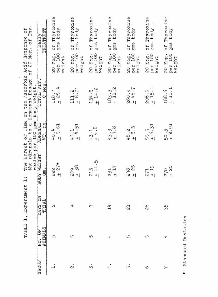
EXPERIMENT I
The Effect of Time on the Response of the Adrenals to a
Constant Level of Thyroidal Stimulation
It has been demonstrated by Long (54) and Sayers and Sayers
(55), that time relationships are very important in the short
time response of the adrenals to acute stress. This exper-
iment was designed to investigate the response of the adrenals
to the more prolonged stimulus of thyroxine injections.
Seven groups of 5 rats each, and 1 group of 8 rats were in—
jected daily with a solution of d,1 thyroxine at a dosage of 20
micrograms per 100 gm. of body weight. This dosage of thyroxine
is about 5 times the normal secretion rate of the strain of rats
used according to unpublished data of Dr. E. P. Reineke, who es—
timated this rate using the method of Dempsey and Astwood (94).
Four groups of controls were used. Group 1 was killed at
the start of the experiment, and the second, third, and fourth
groups were killed after 2, 4, and 6 weeks respectively. The
latter three groups received daily injections of 0.1 cc of
physiological saline solution per 100 gm of body weight.
Although there is some variation between the different
control groups, no consistent trends in adrenal weight, or
ascorbic acid content were noted. Consequently, the values
for the adrenal weight and ascorbic acid content of the 4
control groups were pooled, and are shown graphically in Figure
l, as the base line or 100 percent. The mean control adrenal
weights with standard deviation was 42.0 i 5.98 mg. The value
for ascorbic acid was 194.8 1 52.7 micrograms. All the values
for the experimental groups were calculated as percentages of
Page 46
/50
HO
IJO
IZO
//0
[00
.90
80
0- 7‘0 TAL v17: 6
A-ADRENAL anPERCENTOFNORMAL
70
5'0 1 1 1 I 1 1 1 l
2 4 7 l4 2/ 28 .35 42
THYROX/NE, DAYS
Fig. 1. Changes in the weight and ascorbic
acid content of the adrenals brought about
by a dosage of 20 micrograms of thyroxine
per 100 gm. body weight during a six week
experimental period.
Page 47
GROUP
*
TABLE
1,
Experiment
1:
NO.
OF
ANIMALS
1.
6
Standard
5 Ln
Deviation
DAYS
ON
TRIAL
2
14
21
28
The
Effect
of
Time
on
the
Ascorbic
Acid
Response
of
the
Adrenals
to
aConstant
Dosage_of
2O
Meg.
of
Thy-
roxineper
100
gms
body
Weight.
TOTAL
VIT.
CMeg.
BODYWEIGHT
Gm.
222
2:27*
209
2:38
213
i11.5
231
+17
238
+25
271
+19
270
+20
ADRENAL
WT.
Mg.
40.4
195.61
43.1
_-a_-_4.51
43.1
g4.8
50-5
.i2.91
118.7
.i25.4
188.6
+11.1
20
Meg.
per
100
weight
20
Meg.
per
100
weight
20
Meg.
per
100
weight
20
Meg.
per
100
weight
20
Meg.
per
100
weight
20
Meg.
per
100
weight
20
Meg.
per
100
weight
Daily
TREATMENT
of
Thyroxine
gms
body
of
Thyroxine
gms
body
of
Thyroxine
gms
body
of
Thyroxine
gms
body
of
Thyroxine
gms
body
of
Thyroxine
gms
body
of
Thyroxine
gms
body
Page 48
TABLE
1(Continued)
GROUP
Controls
9
Controls
10
Controls
11
Controls
12
Pooled
Controls
*Standard
Deviation
NO.
OF
ANIMALS
8
22
DAYS
ON
TRIAL
42
14
28
42
BODY
WEIGHT
Gm.
289
i46.0*
246
l6.62
240
.i15.7
ADRENAL
WT.
Mg.
53.9
g8.4
42.0
_:5.9
TOTAL
VIT.
CMeg.
228.4
.i39.8
192.7
146.8
190.7
i29.2
213.2
i21.8
200.5 I
.i17.4
Daily
TREATMENT
2O
Meg.
of
Thyroxine
per
100
gms
body
weight
No
treatment
0.1
cc.
Physiological
Saline
per
100
gms.
bodyweight
0.1
cc.
Physiological
Saline
per
100
gms.
body
weight
0.1
cc.
Physiological
Saline
per
100
gms.
body
weight
0.1
cc.
Physiological
Saline
per
100
gms.
bodyweight
Page 49
r-\
U)
\(
v
this base.
It can be readily seen (Fig. 1) that the initial response
of the adrenals to thyroidal stimulation was a sharp decline
in vitamin C content. This decline was greatest at about 4
days; the values then began to rise. At 2 weeks, the total
vitamin C in the adrenals was approximately normal, or a little
below, and the adrenals had not as yet, begun to show signs
of significant hypertrophy. At three weeks, the adrenals
were significantly hypertrophied, and the vitamin C content
was considerably increased. The zenith in response was reached
at 4 weeks, after which there was a decline in ascorbic acid
content at the fifth week to approximately normal values. The
adrenals also showed a slight decrease in size. At the sixth
week, there was again a slight increase in adrenal weight and
a marked increase in vitamin C content.
The results of this experiment demonstrate that time re-
lationships are very important in a study of the adrenal
response to a prolonged stimulus. If we accept the fact
that the vitamin C content of the adrenals is an index of
secretion, it would further appear from these results, that
the adrenals are discharging hormone into the blood stream
in the early stages of induced hyperthyroidism. This may
be deduced, from the immediate and abrupt decrease in adrenal
vitamin C. Concurrently, with the development of more secreting
units in the adrenal at the third week, as shown by increased
weight, there is an increase in ascorbic acid content above
normal values, which may mean that not only is there an in—
creased rate of secretion, but also an increased rate of
Page 50
(4O)
hormone synthesis. The supply of adrenocortical hormones
probably balances the demand at the fourth week, as the vita—
min C values and adrenal weights in response to this level of
thyroidal stimulation do not increase beyond this time.
At 4 weeks there may even be an over supply of adreno—
cortical hormones, and when these are discharged, they inhibit
further liberation of A.C.T. by the anterior pituitary. Thus
at the fifth week, there was a significant decline in adrenal
vitamin C, and a noticeable decrease in adrenal weight. At the
sixth week, a balance may be presumed to be established, in
which the size of the adrenals, and the rate of hormone syn-
thesis and secretion, is adequate for the degree of thyroidal
stimulation to which the adrenals are being subjected.
Page 51
(41)
EXPERIMENT II
Effect of Varying Levels of Thyroidal Stimulation on
Adrenal Weight and Ascorbic Acid Content
It has been demonstrated in Experiment I, that when a dosag
of thyroxine, approximately five times the normal secretion
rate of the strain of rats used, is injected daily, the great-
est response of the adrenals is seen at 4 weeks. This exper-
iment was designed to discover what the effects of varying
levels of thyroidal stimulation, are on the response of the
adrenals at 4 weeks.
In the first part of the experiment, 5 rats weighing
about 150 gm, were thyroidectomized. Four other rats the
same size were subjected to a sham operation in which the
thyroid was exposed, and each lobe was grasped with a pair
of forceps. As can be seen in Table 2, the thyroidectomized
group ceased growth. The controls gained nearly 100 gm. in
the 4 weeks following the sham operation. At the end of the
experimental period the adrenals were dissected out, weighed,
and analyzed for ascorbic acid. As may be observed in Table
2, there was a highly significant decrease in ascorbic acid
content and adrenal weight of the group subjected to thyro—
idectomy. Thus it might be said that normal adrenal size,
and presumably secretory activity are dependent to a great
degree on the normal secretion rate of the thyroid.
Koger and Turner (95) showed that dosages of thyroprotein
in the feed ranging from 0.01% to 0.16% produced noticeable
and largely adverse effects in male rats with regard to growth,
Page 52
TABLE
2,
EXPERIMENT
2,
PART
1.
Effect
of
Thyroidectomy.
GROUP
NO.
OF
ANIMALS
15
28
2'
wA
28
DAYS
ON
TRIAL
BODYWEIGHT
ADRENAL
Gm.
Mt.
fig.
152
27.4
i1700*
.1-205
241
37.6
.:25.0
.15.0
TOTAL
VIT.
CMeg.
125.6
i11.2
235.0
+47.0
TREATMENT
Thyroidectomy
Sham
Thyroidectomy
Part
2.
Effect
of
Feeding
19
28
Controls
928
*Standard
Deviation
rotamone
at
Physiological
Levels
238
38
_+_24.0
_—_'r_
231
35
.i22.0
i
231
41
_+_35.0
237
43
.:
of
Stimulation.
180.4
i22.6
142.3
+27.8
_+
2'5
0)‘l’
208
9
0.01%
Protamone
in
feed.
0.02%
Protamone
in
feed.
0.04%
Protamone
in
feed.
0.08%
Protamone
in
feed.
0.16%
Protamone
in
0.x
.1.663d
0
Page 53
TABLE
2(Continued),
Part
3.
Effect
of
Intections
of
Thyro
ine
at
Liwh
GROUP
110.
OF
ATTV“T
C‘
h;
_L.
F-‘J‘J
1A m
(1")
Pooled
22
Controls
Ex..
1
Normal
Females
CO
*Standard
Deviation
CO
(\1
C?
(\J 28
BODY Gm.
~57
_+_:12.0*
O O
\O 0\
ram r4H
Ln 1»
m +| m +|
C)O O
0 .)\
mm MH
'0: (\1
m +l N +I
OCH
0
O
\Q
m +| 220
_+_:19.0
{InIGE’IT
CO
o\3
Lfl +|
TOTAL
VIT.
CN03.
MC
/100
L0 10
N03
20
M03.
/100
gm.
A0
M03.
TREATF‘El\"11
gm.
Hat.
5.
of
Thyroxine
.0f
Thvroxine
/100
gm.
Rat.
Rat.
/100
grn.
Rat.
0MC
100
CQ\
a1
0(7)
0
U1.
Rat
0
COED O
.of
Thyrox
of
Thyroxine
of
Thyroxine
11’18
Ph;siological
me/100
gms.
Rat.
No
Treatment
Page 54
(42)
the higher levels showing a definite toxic effect. The effects
on the adrenals varied with 2 strains employed. One strain
showed a hypertrophy of the adrenals which "paralleled roughly
the level of thyroprotein fed." Females of the other strain
showed this hypertrophy, but surprisingly, the males did not
show any increase, and with some dosages used, the adrenal
weights showed a statistically significant decrease.
In the second part of this experiment the dosages used,
correspond to the first 5 dosage levels used by Koger and
Turner (95) and range from 0.01% to 0.16% thyroprotein
mixed in the feed. As can be seen in table 2, there is no
significant trend in either adrenal weight or ascorbic acid
content of any of the groups when compared with the controls.
The animals remained on the treatment for 4 weeks, and no
toxic symptoms were noted. It did appear, however, that as
the dosage of thyroprotein was increased, the rate of feed
consumption also increased. No figures are available for
this however. It might be deduced from this work that the
dosages of thyroprotein used were not sufficient to evoke any
additional response from the adrenals which their normal size,
and hormone producing ability could not take care of. Thus I
no significant hypertrOphy or increase in Vitamin C content
was noted.
The Protamone dosages used in the preceding work were com—
paratively low. To complete the picture of the effects of
varying dosages of thyroidal stimulation on the adrenals, it
was felt necessary to administer dosages of thyroxine at levels
considerably above those heretherto employed.
Page 55
(43)
Accordingly 5 groups of rats were placed on daily injec-
tions of thyroxine ranging from 5 micrograms to 80 micrograms
per 100 gm. body weight. As can be seen from Part 3, Table
2, 5 micrograms per 100 gm. body weight had no effect either
on adrenal weight or ascorbic acid content. One possible
reason for this is that this daily dosage is approximately
the same as the normal thyroid secretion rate of this strain
of rats. A great increase is observed with 10 micrograms per
100 gm. body weight, both in adrenal size and ascorbic acid
content. With 20 micrograms of thyroxine per 100 gm. body
weight, an even greater increase is seen. At dosages of 10
and 20 micrograms of thyroxine per 100 gm. body weight, it
appears that there is a progressive increase in size and
ascorbic acid content of the adrenals. This progressive
response is not seen when the dosage level is increased to
A0 micrograms per 100 gm. body weight. It might be post-
ulated that the adrenals in a rat receiving 20 micrograms of
thyroxine per 100 gm. body weight are of a size and degree of
secretory activity capable of coping with daily dosages of
thyroxine ranging from 20 to 40 micrograms per 100 gms of
body weight. When the dosage of thyroxine is again increased
to 80 micrograms per 100 gm. rat per day, there is again a great
increase in the size and ascorbic acid content of the adrenals.
It should be pointed out here that the influence of time
was not investigated for all these dosage ranges, and whether
on the higher dosages, the ascending or descending part of the
curve, was intercepted is not known. Also, it is not known
whether the effects of higher dosages would magnify the
Page 56
(44)
intensity of the decline and increase in the curve, or would
lengthen or shorten the curve.
It has been reported by numerous authors that the adrenals
of males are smaller than those of females. It was thought
that if this fact were true, there might also be an apprec-
iable difference in ascorbic acid content. Accordingly, in
this experiment, a group of 8 females approximately the same
age, were killed concurrently with the normal controls in
Part 2. It was found that while the female adrenals were
significantly heavier than the males, the ascorbic acid
content was not significantly different in either sex, even
though, as can be seen in table 2, the females did have a
higher content of ascorbic acid.
Page 57
(45)
EXPERIMENT III
A Determination of the Secretion Rate of the Adrenal
Cortex in the Hyperthyroid Rat
A method for the determination of the secretion rate of
the thyroid gland was proposed by Dempsey and Astwood (94),
and has been successfully used by others for the determina-
tion of the secretion rates of the thyroids in various species
of animals. The basis of this method is the inhibition of the
secretion of the animal's own thyroid with thiouracil. This
normally produces hypertrophy of the thyroid gland due to in-
creased thyrotrophic stimulation from the anterior pituitary.
If this increased thyrotrophin secretion is prevented by the
simultaneous administration of thyroxine, such that the weight
of the animal's own thyroid is maintained within normal limits,
the dosage of thyroxine necessary to accomplish this is taken
as the secretion rate of thyroxine for the species in question.
An analogous method for the determination of the secretion
of adrenocortical hormones in hyperthyroid rats was tried in
this experiment. As has been shown in the preceding experi-
ment, the maximum response of the adrenals to a dose of 20
micrograms per 100 gm. of body weight is seen at the fourth
week. It was thought that if this response could be wholly
or in part prevented, by the simultaneous injection of adren—
ocortical hormones, the dose of cortical hormones necessary
to accomplish this would be an indication of the increased
secretion rate of the adrenals in animals subjected to
thyroidal stimulation.
Page 58
(1+6)
A preliminary trial in hyperthyroid rats, showed that the
secretion rate expressed in dog units was going to be very
high. (According to Cartland and Kuizenga (96), the normal
secretion rate of adrenocortical hormones in the rat is about
300 times that of the dog.) Through the kindness of Dr. H. 0.
Von Rosenberg of Parke Davis 00., Detroit, Michigan, 3500 10g1
units of the suprarenal cortical extract "Eschatin" were sup—
plied for use in these experiments. This extract was injected
into 3 groups of 5 rats each which had been receiving thyroxine
injections of 20 micrograms per 100 of body weight for 20
days. Group 1 received 10 units of Eschatin per day. Groups
2 and 3 received 20 and 40 units per day respectively. The
animals were kept on the simultaneous injections of thyroxine
and cortical extracts for 8 days, after which they were sac—
rificed, and the adrenals removed and analyzed for ascorbic
acid.
Group 1, which received 10 units of Eschatin per day,
showed no statistically significant decrease in either adrenal
weight or ascorbic acid, when compared with the group receiving
the same amount of thyroxine alone for 28 days in experiment 2.
There is a noticeable decrease in adrenal weight, however,
but not in vitamin C.
The next two groups receiving 20 and 40 units of eschatin
per day respectively, in addition to thyroxine injections,
showed no significant differences, when compared to each other,
in either adrenal weight, or ascorbic acid. When these two
groups were compared with the group receiving thyroxine
alone for 28 days, a highly significant decrease in adrenal
Page 59
TABLE
3,
EXPERIMENT
3-The
Inhibition
of
Adrenocortical
Hypertrophy
Due
to
Thyroxine,
With
Adrenocortical
Extract
GROUP
NO.
OF
DAYS
ON
BODYWEIGHT
ADRENAL
TOTAL
VIT.
DAILY
ANIMALS
TRIAL
Gm.
WT.
Mg.
CMeg.
TREATMENT
l5
28
276
52.8
289.6
20
Meg.
of
Thyroxine
_t12.7*
+5.9
+30.1
/100
gms
rat
+10
Units
Eschatin
per
day
25
28
262
46.9
249.2
20
Meg.
of
Thyroxine
_+_;11.7
_i4.5
+11.7
/100
gms
rat
+20
Units
Eschatin
per
day
l2
20
Meg.
of
Thyroxine
+9.6
+5.7
.i9.6
/100
gms
rat
and
40
Units
Eschatin
per
day
45
28
271
55:5
296.0
20
Meg.
ofThyroxine
:0.3
2:15.4
/100
gms
rat
*Standard
Deviation
Page 60
(’47)
size was seen as well as a significant decrease in ascorbic
acid.
It appears from these data that the increase in secretion
rate of adrenocortical hormones in rats receiving 20 micro-
grams of thyroxine per 100 gm of body weight, is somewhere
between 10 and 20 dog units per day. fbove the dosage of 20
units, which produces a maximum effect, additional adren—
ocortical hormone has no effect.
Page 61
(48)
EXPERIMENT IV
The Effect of Thyroxine on the Adrenals of Pregnant Rats
and on the Adrenals of Their Offsgiing ‘
Evidence has been advanced in experiments 1 and 3 of this
report, that the adrenals in a hyperthyroid rat are in a state
of hypersecretion, and that the secretion rate is equivalent
to 10 to 20 dog units of adrenocortical hormones.
Some question arose as to the effects of thyroxine on the
adrenals of pregnant rats, and to its possible effects on
the adrenals of the offspring. Also, if the adrenals of a
hyperthyroid dam are in a state of hypersecretion, what is
the effect of this increased secretion on the fetal adrenals?
Only one piece of work on this question has been found by
the author. Hoskins (1), reported that when desiccated thyroid
was fed to pregnant guinea pigs, the adrenals of the mother
hypertrophied, and the adrenals of the offspring were atro-
phied. Hoskins (l) attributed this decrease in size of the
adrenals of the offspring, which reached 53% in some cases,
to "a reaction in the adrenals of the offspring to epine-
phrinemia caused by thyroidism in the mother". Hoskins also
thyroidectomized some pregnant guinea pigs and noted that the
adrenals of the offspring were hypertrophied, the average
increase being 20%. He surmised that this hypertrophy was
due to "a reaction to adrenal deficiency caused by hypo—
thyroidism in the mother”. The results of this paper seem
to indicate that the adrenals of the fetus are sensitive to
the secretory status of the dam's adrenals. When the dam
Page 62
(49)
is in a hyperthyroid condition the adrenals of the fetus
atrophy due to adrenocorticotrophin suppression, brought
about by the excessive adrenocortical secretion of the dam.
If these observations of Hoskins on guinea pigs were
valid for rats, presumably the ascorbic acid content of the
fetal adrenals should be appreciably altered if hyperthyroid-
ism was induced in the mother.
Four pregnant rats were selected and injected daily with
20 micrograms of thyroxine per 100 gm. body weight, for periods
ranging from 16 to 30 days, prior to parturition. Three rats
served as controls. Approximately 12 hours after parturition
in each case, the rats were killed and the adrenals weighed
and analyzed for ascorbic acid. The adrenals of the offspring
were treated in a somewhat different manner due to their small
size. Instead of each pair of adrenals being analyzed separ-
ately, the adrenals from a litter of rats were pooled, after
being individually weighed, and the total ascorbic acid content
was divided by the number of baby rats which had contributed
their adrenals to the sample in order to obtain the total
adrenal vitamin C per rat.
It can be seen from Table 4 that there is no difference in
either the weights or the vitamin C contents of the adrenals
of the control litters, and the litters from hyperthyroid
mothers, even though the adrenals of the hyperthyroid mothers
reached tremendous weights, in one case a total of 106.1 mg.
Thus it may be deduced from this work that either the
adrenals of the fetal rat are not responsive to thyroidal
and adrenocorticotrophic stimulation, and adrenocortical
Page 63
TABLE
4,
EXPERIMENT
4,
PART
1.
The
Effect
of
aConstant
Dosage
of
Thyroxine
on
the
Weight
and
Vitamin
CContent
of
the
Adrenals
of
Pregnant
Rats.
FEMALE
DAYS
ON
BODYwEIGHT
ADRENAL
TOTAL
VIT.
Daily
NO.
TRIAL
Gm.
wT.
Mg.
0Meg.
TREATMENT
116
260
73.7
233.4
20
Meg.
of
Thyroxine
/1oo
gms.
Rat.
230
272
94.4
369.7
20
Meg.
of
Thyroxine
/100
gms.
Hat.
317
260
78.4
298.9
20
Meg.
of
Thyroxine
/100
gms.
Rat.
417
300
106.1
427.2
20
Meg.
Of
Thyroxine
/lOO
gms.
Rat.
Page 64
TABLE
4,
EXPERIMENT
4,
PART
2.
The
Response
of
the
Fetal
Adrenals
to
aConstant
Dosage
Level
of
Thyroidal
Stimulation
in
the
Mother.
GROUP
NO
OF
DAYS
0N
BODYWEIGHT
ADRENA
Mcg.
VIT.
C
A1
'LS
TRIA
Gm.
JT.
Mg.
Per
Rat
Thyroxine:
816
5.8
Litter
1.i'O
Thyroxine:
Litter
210
30
5.6
2.4
7.9
i0
:,0
Thyroxine:
Litter
34
17
5.9
.320
(\J
0:)
mac
m +|
Thyroxine:
Litter
4ll
17
6.0
2.06
7.1
.i
.:
5
Control:
Litter
55
—
Control:
Litter
68
—7.3
2.9
8.3
i0
10
Control:
Litter
7l2
—5-5
i1
0’3
(3
NO
m +|
*Standard
Deviation
Page 65
(50)
suppression, from the mother, or that the placenta is not
permeable to these hormones.
It should be pointed out however, that this work does not
refute the work of Hoskins (1), because he used guinea pigs,
and the reactions he observed may be a species peculiarity.
Page 66
(51)
DISCUSSION
Claude Bernard has observed, as have others, that "Vital
mechanisms have one object, preserving constant the internal
environment". Numerous other workers have observed that bio-
logical organisms, like the societies of today, must achieve
a state of balance, and that when, one part of the organism
is subjected to a stress, the other parts of the organism
attempt to compensate in one way or another for this stress.
The degree of success attained, is measured by the ability
of the individual to survive.
With these ideas in mind, one can proceed to the idea of
balance between two individual organs, in particular the
thyroid gland, and the adrenal cortex.
It has been shown in the experimental work of this report,
that the level of thyroidal stimulation determines the size
and secretory rate of the adrenal cortex to a large extent.
Thus when the thyroid is removed, the adrenals atrophy and
the ascorbic acid content decreases. There is a rather narrow
range in which thyroidal stimulation has no appreciable effect
on the adrenals, and this range is approximately the secre-
tion rate of the normal animal. When the level of thyroidal
stimulation is boosted above the "physiological" range, the
adrenals respond with increased secretion, and this increase
is for the most part progressive with increased dosage.
When measuring the "hormone content" of the adrenal, it
might remain a question in the minds of some, whether an
increased content of vitamin C meant increased synthesis
Page 67
(52)
and subsequent liberation of the adrenocortical hormones,
or whether the increase in ascorbic acid is indicative of
increased storage. In favor of the former interpretation
is the fact that the initial response of the adrenals to
adequate thyroidal stimulation, is a decrease in ascorbic
acid. This decrease in ascorbic acid content has been shown
to be due to liberation of adrenocortical hormones. Only
when enough secretory units are laid down, so that the adrenals
can balance the demand for their hormones, does the content
of ascorbic acid show an increase.
Another piece of evidence that the adrenals in a hyper-
thyroid animal are in a state of hypersecretion, may be
gathered from Experiment 3. Here it was demonstrated that
the increase in adrenocortical secretion rate of a hyper-
thyroid rat on a dosage of 20 micrograms of thyroxine per
100 gm. body weight, is between 10 and 20 dog units of adren-
ocortical hormones, this dosage being necessary to cause
atrophy of the adrenals. This is prima-facie evidence that
the adrenals are in a state of active, if not hyper-secretion.
In experiment 4, some interesting questions are raised for
further study. In this experiment it was demonstrated that
thyroxine dosage will superimpose its effects on the adren-
ocortical hypertrophy produced by pregnancy. In addition to
this, it was observed that thyroxine dosage has no appre-
ciable effects on the adrenals of the offspring of hyper-
thyroid mothers. It is not clear at this time whether the
reason for the negative response of the fetal adrenals is
due to impermeability of the placenta to maternal
Page 68
adrenocorticotrophin, alrenocortical hormones, and exogenous
thyroxine, or whether it is due to a lack of responsiveness
of the fetal endocrine system.|—]
:3‘
(D literature previously cited, has amply demonstrated
that the mechanism of adrenocortical response to stimuli is
mediated through the anterior pituitary. It has been demon—
strated for at least one species, however, that thyroxine
alone will maintain the normal size of the adrenals. (Miller
and Riddle, (73), in pigeons.)
Page 69
SUMMARY AND CONCLUSIONS
1. It has been demonstrated that time is a very im—
portant factor in the response of the adrenals to thyroxine
stimulation. The initial adrenal response is a sharp decline
in ascorbic acid followed by increases both in adrenal size
and ascorbic acid content. The maximum response of the ad-
renals on a thyroxine dosage of 20 micrograms per 100 gm.
body weight, is seen at the fourth week, followed by a slight
decline in ascorbic acid and adrenal weight, at the fifth
week and then a slight increase again at the sixth week.
2. In thyroidectomized rats, great decreases in adrenal
weight and ascorbic acid are seen at 4 weeks. Thus the normal
size and secretory activity of the adrenals are dependent to
a great extent upon a normal rate of thyroid secretion.
3. Within "physiological" limits, thyroxine administration
has no significant effect on the adrenals.
4. Above the "physiological" range, increased dosages of
thyroxine will cause for the most part, progressive increases
in adrenal size and ascorbic acid content at 4 weeks.
5. There is a significant difference in adrenal weight
between male and female rats, the females having the heavier
glands. There is an appreciable but not statistically
greater content of ascorbic acid in the female adrenal,
when compared with the male.
6. The adrenal hypertrophy induced by thyroxine admin-
istration at a dose of 20 micrograms per 100 gm. body weight,
may be prevented by simultaneous administration of
Page 70
(55)
adrenocortical extracts. The dose of adrenocortical extracts
necessary to accomplish this is between 10 and 20 dog units.
Thus the secretion rate of the adrenals of the male rat with
this degree of hyperthyroidism is somewhere between 10 and
20 dog units.
7. Thyroxine administration to pregnant rats will super-
impose its hypertrophic effects on the adrenal hypertrophy
normally seen in pregnancy.
8. Thyroxine administration to pregnant rats will not
cause any changes in size or ascorbic acid content of the
fetal adrenals.
Page 71
O\
10.
ll.
BIBLIOGRAPHY
Hoskins, R. G. Thyroid Secretion as a Factor in Adrenal
Activity. Jour. Amer. Med. Assn. 55: 1724. 1910.
Herring, P. T. The Action of Thyroid Upon the Growth of
the Body and Organs of the White Rat. Cuart. Jour. Exp.
Physiol. 11: 47. 1917.
Cameron, A. T. & Carmichael J. Contributions to the Bio-
chemistry of Iodine. IV. The Effect of Thyroxine on
Growth in White Rats and Rabbits. Jour. Biol. Chem.
46: 35. 1921.
Korenchevsky V., Dennison M., and Kohn-Speyer A. Simul-
taneous Administration of Testicular Hormone with Ant-
uitrin and Prolan or with Desiccated Thyroid. Biochem.
Jour. 27: 1513. 1933.
Cohen, R. S. Effect of Experimentally Produced Hyper-
thyroidism Upon the Reproductive and Associated Organs
of the Male Rat. Amer. Jour. Anat. 56: 143. 1935.
Schmidt, I. G. and Schmidt, L. H. Variations in the
Structure of Adrenals and Thyroids Produced by Thyroxine
and High Environmental Temperatures. Endocrin. 23:
559. 1938.
DeWesselow, O.L.V.S., and Griffiths, W. J. The Role of
the Adrenal Gland and of the Raised Metabolism in the
Production of Organ Hypertrophy in the Thyroid Fed Rat.
Brit. Jour. Exp. Path. 19: 347. 1938.
Ingle, D. J. and Kendall, E. C. Weights of Adrenal Glands
in Rats Fed Different Amounts of Sodium and Potassium.
Amer. Jour. Physiol. 122: 585. 1938.
Uotila, U. U. The Regulation of Thyrotropic Function by
Thyroxine After Pituitary Stalk Section. Endocrin. 26:
129. 1940.
Korenchevsky, V. and Hall, K. Correlation Between Sex
Hormones, Thyroid Hormones, and Desoxcorticosterone as
Judged by Their Effects on the Weights of Organs of
Gonadectomized Rats. Biochem. Jour. 35: 726. 1941.
Lowenstein, B. E. and Zwemmer, R. L. A Thyroid Adrenal
Relation in Resistance to Potassium Endocrin. 30: 1035.
1942.
Page 72
12.
13.
14.
H C\
17.
19.
20.
22.
24.
25.
(57)
Ingle, D. J. and Higgins, G. M. The Effect of Thy—
roxine on the Extent of Regeneration in the Enucleated
Adrenal Gland of the Rat. Endocrin. 23: 419. 1938.
Cutting, C. C., and Tainer, M. C. Comparative Effects of
Dinitrophenol and Thyroxine on Tadpole Metamorphosis.
Proc. Soc. Exp. Biol. Med. 31: 97. 1933.
Gley, E. Acad. Roy. de Belg. Bull. de la Cl. de Sci.
Brux. 911. 1913. Cited by: (15) Koelsche, G. A.,
and Kendall, E. C. The Relation of the Suprarenal
Cortical Hormone to Nitrogen Metabolism in Experimental
Hyperthyroidism. Amer. Jour. Physiol. 113: 335. 1935.
Kennedy, T. H., and Purves, H. D. Studies on Experimental
Goitre. I. The Effect of Brassicae Seed Diets on Rats.
Brit. Jour. Exp. Path. 22: 245. 1941.
Leblond, C. P., and Hoff, H. E. Effects of Sulfonamides
and Thiourea Derivatives on Heart Rate and Organ Morph-
ology. Endocrin. 34: 229. 1944.
Baumann, E. J., and Marine, D. Involution of the Adrenal
Cortex in Rats Fed Thiouracil. Endocrin. 36: 400. 1945.
Glock, G. E. Thiourea and the Suprarenal Cortex. Nature.
156: 508. 1945.
Gradinescu. Arch. f. d. Gesammt. Physiol. 152: 186.
1913. Cited by: 21. Aub, J. C., Foreman, J., and Bright,
E. M. The Effect of Adrenalectomy Upon the Total Met-
abolism of the Cat. Amer. Jour. Physiol. 61: 326. 1922.
Webster, B., Pfiffner, J. J., and Swingle, W. W. The
Effect of Adrenal Cortical Hormone Upon the Respiratory
Metabolism of the Cat. Amer. Jour. Physiol. 99: 710.
1932.
Harrop, G. A., Weinstein, A., Soffer, L. J., and Trescher,
J. H. Studies on the Suprarenal Cortex. II Metabolism,
Circulation, and Blood Concentration During Suprarenal
Insufficiency in the Dog. Jour. Exp. Med. 58: l. 1933.
Carr, C. F., and Beck, F. F. The Metabolism of Adren-
alectomized Rats. Amer. Jour. Physiol. 119: 580. 1937.
Lerman, J. The Physiology of the Thyroid Gland. Chapt.
24. Glandular Physiology and Therapy. Amer. Med. Assn.
Chicago. 1942.
Page 73
26.
27.
30.
LO
LO
35-
36.
37.
Hitchcock, F. A., Grubbs, R. C., and Hartman, F. A.
The Effect of Idrenal Cortical Extract Upon Oxygen
Consumption in Normal Human Beings. Amer. Jour. Physiol.
121: 542. 1938.
Golyakowski. Vrach. St. Petersburg, 20: 1017. 1899.
Cited by: (22) Webster, B., Pfiffner, J. J., and Swingle,
W. W. The Effect of Adrenal Cortical Hormone Upon the
Respiratory Metabolism of the Cat. Amer. Jour. Physiol.
99: 710. 1932.
Marine, D. and Baumann, E. J. Effect of Suprarenal
Insufficiency. Amer. Jour. Physiol. 57: 135. 1921.
Marine, D. and Baumann, E. J. Suprarenal Insufficiency in
Thyroidectomized Rabbits. Amer. Jour. Physiol. 59: 353.
1922.
Scott, W. J. M. Influence of the Glands with Internal
Secretion on the Respiratory Exchange. IV Effect of
Suprarenal Insufficiency in Cats. Jour. Exp. ied. 36:
199. 1922.
Davis, J. E. and Hastings, A. B. Relationship of Adrenal
and Thyroid Glands to Excised Muscle Metabolism. Amer.
Jour. Physiol. 105: 110. 1933.
Barlow, O. W. Adrenalectomy and its Relation to Metabol-
ism. Jour. Physiol. 70: 453. 1924.
Marine, D. Remarks on the Pathogenesis of Graves Disease.
Amer. Jour. Med. Sci. 180: 767. 1930.
Marine, D., Baumann, E. J., and Cipra, A. Influence of
Glands With Internal Secretions on the Respiratory
Exchange. II The Effect of Feeding Emulsions of the
Interrenal Gland to Rabbits. Amer. Jour. Physiol. 72:
248. 1925.
Marine, D. Baumann, E. J., and Webster, B. Value of
Haxuronic Acid in the Treatment of Graves Disease with
Suprarenal Cortex. Proc. Soc. Exp. Biol. Med. 28: 327.
1930.
Black, E. M., Hupper, M. and Rogers, J. The Effects of
Adrenal Feeding Upon the Iodine Content of the Thyroid
Gland. Amer. Jour. Physiol. 59: 222. 1922.
Elmer, A. W., Giedosz, G., and Scheps, M. L'Action Chez
1e Cobaye de la Cortine sur, L'Hyperactivite de la Thyroide
Provoquee par 1e Thyreostimuline Prehypophysaire. Compt.
Rend. Soc. de Biol. 118: 1373. 1935.
Page 74
38.
39.
40.
41.
42.
43.
2+4.
45.
46.
47.
48.
49.
50.
(59)
Bock, K. A. Die Einwerkung Von Nebennierenrindenextrakt
auf den Ablauf der Thyroxinmetamorphose bei Froschlarven
und bein Axolotl. Klin. Wehnschr. 17: 1311. 1938.
Cited in: Abstracts of Endocrine Literature. Endocrin.
26: 359. 1940.
Aub, J. C., Bright, E. M. and Forman, J. The Metabolic
Effect of Adrenalectomy Upon the Urethanized Cat. Amer.
Jour. Physiol. 61: 349. 1922.
Levy, R. L. Observations on the Synergism of Thyroxine
and Adrenaline, and the Sympathetic Innervation of the
Thyroid. Amer. Jour. Physiol. 41: 492. 1916.
Cannon, W. B., and Cattel, M. Studies on the Conditions
of fictivity in Endocrine Glands. III The Influence of
the Adrenal Secretion on the Thyroid. Amer. Jour. Physiol.
41: 74. 1916.
Cannon, W. B., and Cattel, M. Studies on the Conditions of
Activity in Endocrine Glands. II The Secretory Inner-
vation of the Thyroid Gland. Amer. Jour. Physiol. 41:
58. 1916.
Marine, D., and Lenhart, C. H. Effect of the Subcutaneous
Injection of Adrenalin on Normal and Thyroidectomized
Rabbits. Amer. Jour. Physiol. 54: 248. 1920.
Szent-Gyorgyi, A. Observations on the Functions of Per-
oxidase Systems and the Chemistry of the Adrenal Cortex.
Biochem. Jour. 22: 1387. 1928.
Yavorsky, M. Almaden, P., and King, C. G. The Vitamin C
Content of Human Tissues. Jour. Biol. Chem. 106: 525.
1934.
Harris, L. J., and Ray, S. N. Vitamin C in the Suprarenal
Medulla. Biochem. Jour. 27: 2006. 1933.
Quick, A. J. Hypertrophy of the Adrenals in Scurvey. Proc.
Soc. Exp. Biol. Med. 30: 753. 1933.
Kuchel, C. C., and Mitchell, M. L. The Effects of Stim-
ulation of the Adrenal Gland Upon its Content of Ascorbic
Acid, Adrenalin, and Glutathione. Australian Jour. Exp.
Biol. Med. Sci. 14: 51. 1936.
Bowman, D. E., and Muntwyler, E. Ascorbic Acid Content of
Tissues Following Ether Anaesthesia. Proc. Soc. Exp. Biol.
Med. 35: 557. 1936.
Euler, H. V. and Klussman, E. Arkiv. Kemi. Mineral. Geol.
11B: 13. 1933. Cited by: (51) King, C. G. Vitamin C.
Ascorbic Acid. Physiol. Rev. 16: 238. 1936.
Page 75
52.
U1
U.)
54.
56.
57-
58.
59-
63.
64.
(6O)
Flexner, L. B., and Grollman, A. The Reduction of Osmic
Acid as an Indicator of Adrenal Cortical Activity in the
Rat. Anat. Rec. 75: 207. 1939.
Torrance, C. C. Diptheria Intoxication and Vitamin C Content
of the Suprarenals of Guinea Pigs. Jour. Biol. Chem. 132:
575. 1940.
Long, C.N.H. The Relation of Cholesterol and Ascorbic Acid
to the Secretion of the Adrenal Cortex. Recent Progress
in Hormone Research. Vol. 1:99. Academic Press Inc. New
York City, New York. 1947.
Sayers G., and Sayers, M. A. The Pituitary Adrenal System.
Recent Progress in Hormone Research. Vol. 11:81 Academic
Press Inc. New York City, New York. 1948.
Vars, H. M., and Pfiffner, J. J. Vitamin C and the Adrenal
Gland in the Dog. Proc. Soc. Exp. Biol. Med. 31: 839. 1934.
Svirbely, J. L., and Kendall, E. C. Vitamin C. and the
Adrenal Cortical Hormone. Amer. Jour. Physiol. 116: 187.
1936.
Sure, B., Theis, R. M., and Harrelson, R. T. Vitamin
Interrelationships. I Influence of Avitaminosis on
Ascorbic Acid Content of Vgrious Tissues and Endocrines.
Jour. Biol. Chem. 129: 245. 1939.
Zwemmer, R. L., Lowenstein, B. E., and Pines, K. L. Exper—
imental Studies of Similarities Between Cortin and Some
Cardiac Glucosides. Endocrin. 27: 945. 1940.
Sure, B., and Theis, R. M. Influence of Hyperthyroidism
on Vitamin C Content of Various Tissues and Endocrines.
Endocrin. 24: 672. 1939.
Marine, D., Baumann, E. J., and Rosen, H. Effect of Ascor—
bic Acid 0n Thyroid and Suprarenals of Guinea Pigs. Proc.
Soc. Exp. Biol. Med. 31: 870. 1934.
Belasco, I. J., and Murlin, J. R. The Effect of Vitamins
A and C in Experimental Hyperthyroidism. Jour. Nutrit.
20: 577. 1940.
Lewis, R. A. The Effect of Hyperthyroidism Upon Metabl-
ism of Vitamin C. Bull. Johns Hopkins Hosp. 63: 31. 1938.
McQueen—Williams, M. Necessary Concurrence of Thyroid in
the Marked Adrenal Cortical Hypertrophy Following Beef
Anterior Pituitary Implants. Proc. Soc. Exper. Biol.
fled. 32: 296. 1934.
Page 76
76.
77-
78.
(61)
Smith, P. E. Ablation and Transplantation of the Hypophysis
in the Rat. Anat. Rec. 32: 221. 1926.
Tyslowitz, R. Effect of Hypophysectomy on Concentrations
of Ascorbic Acid in the Adrenals of the Rat. Endocrin.
32: 103. 1943.
Bowman, D. E., Morris, L. E., and Stacey, J. R. Tissue
Ascorbic Acid in Hypophysectomized Rats. Proc. Soc. Exp.
Biol. Med. 45: 784. 1940.
Evans, H. M. Present Position of Our Knowledge of Anterior
Pituitary Function. Jour. Amer. Med. Assn. 101: 425.
1933-
Collip, J. B., Anderson, E. M., and Thomson, D. L. The
Adrenotropic Principle of the Anterior Pituitary Lobe.
Lancet. 225: 347. 1933.
Li, C. H., Evans, E. M., and Simpson, M. E. Adrenocorti-
cotropic Hormone. Jour. Biol. Chem. 149: 413. 1943.
Sayers, G., White, A., and Long, C.N.H. Preparation and
Properties of Pituitary Adrenotropic Hormone. Jour. Biol.
Chem. 149: 425. 1943.
Sayers, G. Sayers, M. A., Lewis, H. L., and Long, C.N.H.
Effect of Adrenotropic Hormone on Ascorbic Acid and
Cholesterol Content of the Adrenals. Proc. Soc. Exp.
Biol. Med. 55: 238. 1944.
Miller, R. A., and Riddle, 0. Stimulation of Adrenal Cortex
of Pigeons by Anterior Pituitary Hormones, and by their
Secondary Products. Proc. Soc. xp. Biol. Med. 41: 518.
1939.
Korenchevsky, V. Dennison, M., and Hall, K. The Action
of Testosterone Propionate on Normal Adult Female Rats.
Biochem. Jour. 31: 780. 1937.
Ingle, D. J., and Kendall, E. C. Atrophy of the Adrenal
Cortex of the Rat Produced by the Administration of Large
Amounts of Cortin. Science. 86: 245. 1937.
Ingle, D. J., Higgins, G. M., and Kendall, E. C. Atrophy
of the Adrenal Cortex in the Rat Produced by Administration
of Large Amounts of Cortin. Anat. Rec. 71: 363. 1938.
Ingle, D. J. The Effects of Administering Large Amounts of
Cortin on the Adrenal Cortices of Normal and Hypophysec-
tomized Rats. Amer. Jour. Physiol. 124: 369. 1938.
Selye, H. Postnatal Masculinization of the Female Rat by
Means of Testosterone Profionate. Anat. Rec. 76: 145.
1940.
Page 77
81.
82.
84.
86.
87.
88.
89.
91.
(62)
Selye, H., and Dosne, C. Changes Produced by Desoxycorti—
costerone Overdosage in the Rgt. Proc. Soc. Exp. Biol.
Med. 44: 165. 1940.
Selye, H. Compensatory Atrophy of the Adrenals. Jour. Amer.
M:d. Assn. 115: 2246. 1904.
Carnes, W. H., Ragan, C., Ferrebee, J. W. and O'Neill J.
Effects of Desoxycorticosterone Acetate in the Albino Rat.
Endocrin 29: 144. 1941.
Iggle, D. J. A Comparison of the Resistance of Male and
F male Rmts to Cortin. Endocrin. 24: 194. 1939.
Howard, E. and Grollman, A. The Effect of Extracts of the
Adrenal Cortex on Growth and The Reproductive System of
Normal Rats with Particular R ference to Intersexuality.
Amer. Jour. Physiol. 107: 480. 1934.
King, J. S. Effect of Massive Doses of Adrenal Cortical
Hormone on the Albino Rat. Proc. Soc. Exp. Biol. Med.
35: 579- 1937-
Wyman, L. C., and Tum-Suden C. Studies on Suprarenal
Insufficiency. XI The Growth of Transplanted Cortical
Tissue in the Rat. Amer. Jour. Physiol. 101: 662. 1932.
Wyman, L. C., and Tum—Sudan, C. Factors Determining<»w(
Limiting the Growth of Transplanted Suprarenal Cortical
Tissue. Endocrin. 21: 523. 1937.
Selye, H. Interactions Between Various Steroid Hormones.
Candian Med. Assn. Jour. 42: 113. 1940.
Selye, H., Dosne, C., Basset, L., and Whittaker, J. On the
Therapeutic Value of Adrenal Cortical Hormones in Traumatic
Shock and Allied Conditions. Canadian Med. Assn. Jour.
43: l. 1940.
Deane, H. E., and Greep, R.O. A Cytochemical Study of the
Adrenal Cortex in Hypo— and Hyperthyroidism. Endocrin.
41: 243. 1947.
Mindlin, R. L. and Butler, A. M. The Determination of
Ascorbic Acid in Plasma; a Macromethod, and a Micromethod.
Jour. Biochem. 122: 673. 1937.
Bessey, O. A. A Method for the Determination of Small
Cuantities of Ascorbic Acid and Dehydroascorbic Acid in
Turbid and Colored Solutions in The Presence of Other
Reducing Substances. Jour. Biol. Chem. 103: 687. 1933.
Page 78
KO
0\
(63)
Musulin, R. R., and King, C. G. Metaphosphoric Acid in the
Extraction and Titration of Vitamin C. Jour. Biol. Chem.
116: 409. 1936.
Briggs, D. R. A Stable Metaphosphate Preparation for Use
as a Protein Precipitant. Proc. Soc. Exp. Biol. Med. 37:
634. 1938.
Dempsey, E. W., and Astwood, E. B. Determination of the
Rate of Thyroid Hormone Secretion at Various Environmental
Temperatures. Endocrin. 23: 509. 1934.
Koger, M., and Turner, C. W. The Effects of Mild Hyperthy-
roidism of Growing Animals of Four Species. Univ. Mo.
Res. Bull. 377: Sept. 1943.
Cartland, G. F., and Kuizenga, M. H. The Biosassay of
Adrenocortical Extracts. fmer. Jour. Physiol. 117: 678.
1936.
Page 79
A Q 1:7.
{EEC L .
Page 81
mu(I(1M(lymluulyyunn