Page 1
Wound healing in Mac-1 deficient mice
Lin Chen, MD, PhD1; Sridevi Nagaraja, PhD
2; Jian Zhou, BS
1; Yan Zhao, BS
1; David Fine, BS
1;
Alexander Y. Mitrophanov, PhD2; Jaques Reifman, PhD
2; Luisa A. DiPietro, DDS, PhD
1
1Center for Wound Healing and Tissue Regeneration, College of Dentistry, University of Illinois at
Chicago, Chicago, IL, USA. 2Department of Defense Biotechnology High Performance Computing
Software Applications Institute, Telemedicine and Advanced Technology Research Center, U.S.
Army Medical Research and Materiel Command, Ft. Detrick, MD, USA
Running title: Collagen synthesis in wounds of Mac-1 null mice
Key words: skin, wound healing, Mac-1, macrophage, collagen, TGF-β1
Reprint requests:
Drs. Lin Chen and Luisa A. DiPietro, Center for Wound Healing and Tissue Regeneration,
College of Dentistry, University of Illinois at Chicago, 801 S. Paulina St., Chicago, IL 60612. E-
mail address: [email protected] (LC) and [email protected] (LAD), Phone: (312) 413-5387 (LC)
and (312) 355-0432 (LAD), Fax: (312) 996-0943
Page 2
2
ABSTRACT
Mac-1 (CD11b/CD18) is a macrophage receptor that plays several critical roles in macrophage
recruitment and activation. Because macrophages are essential for proper wound healing, the
impact of Mac-1 deficiency on wound healing is of significant interest. Prior studies have
suggested that Mac-1-/- mice exhibit deficits in healing, including delayed wound closure in
head and ear wounds. The current study examined whether Mac-1 deficiency influences wound
healing in small excisional and incisional skin wounds. Three millimeter diameter full thickness
excisional wounds and incisional wounds were prepared on the dorsal skin of Mac-1 deficient
(Mac-1 -/-) and wild-type (WT) mice, and wound healing outcomes were examined. Mac-1
deficient mice exhibited a normal rate of wound closure, generally normal levels of total
collagen, and nearly normal synthesis and distribution of collagens I and III. In incisional
wounds, wound breaking strength was similar for Mac-1 -/- and WT mice. Wounds of Mac-1
deficient mice displayed normal total macrophage content, although macrophage phenotype
markers were skewed as compared to WT. Interestingly, amounts of TGF-β1 and its
downstream signaling molecules, SMAD2 and SMAD3, were significantly decreased in the
wounds of Mac-1 deficient mice compared to WT. The results suggest that Mac-1 deficiency
has little impact on the healing of small excisional and incisional wounds. Moreover, the
findings demonstrate that the effect of single genetic deficiencies on wound healing can
markedly depend upon the wound model. These conclusions have implications for the
interpretation of the many prior studies that utilize a single model system to examine wound
healing outcomes in genetically deficient mice.
Page 3
3
INTRODUCTION
Wound healing is a complex yet well-regulated process in which multiple resident cells,
recruited inflammatory cells, and stem cells interact to create an environment that supports the
healing process. An optimal inflammatory response is a normal and important part of the healing
process that helps to eliminate contaminating microorganisms and dead or injured cells.1, 2
Multiple prior studies have demonstrated that appropriate inflammation is critical to healing
outcomes, and several specific immune cell types have been shown to play critical roles.
Mac-1 (Mβ2 integrin, CD11b/CD18, or CR3) is a β2 integrin cell-surface receptor composed of
αM- and β2-subunits. Mac-1 is primarily found on myeloid cells including monocytes,
macrophages, neutrophils, and some lymphocytes. It binds to a variety of soluble matrix-bound
or cell-bound ligands, such as complement iC3b, fibrinogen, factor X, heparin/heparin sulfate,
and intercellular adhesion molecule 1. Via these ligands, Mac-1 mediates multiple leukocyte
functions, including activation, adhesion, diapedesis, cell motility, cell-to-cell interaction, and
phagocytosis.3-5
Two specific Mac-1 positive cell types, neutrophils and macrophages, are known to be important
to wound healing. Neutrophils are among the first leukocytes to infiltrate the wound, entering
the site within minutes to hours after injury.1 Neutrophils are known to be important in
eliminating microbes and engulfing dead or damaged cells in the wound. However, neutrophils
may also damage normal tissue through the release of reactive oxygen species and proteases.6
Neutrophil depletion has been shown to accelerate wound reepithelialization in mice, yet to have
Page 4
4
little effect on collagen deposition, wound breaking strength and macrophage infiltration.7 In
contrast to neutrophils, which function primarily at early time points after injury, macrophages
play a pivotal role in all stages of skin wound healing.8, 9
Wound macrophages exhibit
extraordinary phenotypic plasticity that is essential to their function.10-13
In the early stage of
wound healing, macrophages are in a primarily pro-inflammatory state (often termed M1 or
classically activated macrophages), producing IL-1β, IL-6, and TNF-.11, 12
As the wound heals
and enters the stages of cellular proliferation and remodeling, macrophages adopt a reparative
phenotype (often termed M2 or alternatively activated macrophages), producing factors that
support tissue growth and reduce inflammation.11, 12
Several studies have demonstrated that
macrophage function is essential for healing, and some have suggested that the reparative
phenotype macrophage is particularly critical for adequate repair.10-12
More recent studies have
established that macrophages are in fact a heterogeneous cell population at all stages of the
wound healing process,10-12
raising questions about what phenotypes are most beneficial.
Although several prior studies suggest that the addition of activated macrophages can improve
healing outcomes,14
other studies have demonstrated that the treatment of wounds with M2
macrophages does not benefit wound healing.15
Given the importance of neutrophils and macrophages to the healing process, and the critical role
that Mac-1 plays in in the activation, adhesion, and migration of these cells, Mac-1 might be
expected to play an important role in wound repair. Indeed, two prior studies, one using ear and
head wounds, and the other examining 5mm excisional skin wounds, have demonstrated that
specific strains of Mac-1 deficient mice exhibit delayed wound healing.16, 17
In the current
study, we used a commercially available Mac-1 deficient strain to examine whether this deficit
Page 5
5
extends to slightly smaller wounds and incisional wounds produced on the dorsal skin. The
current results show that although small excisional wounds of Mac-1 mice exhibit some
functional changes related to macrophages and decreased levels of TGF-β1, wounds on these
mice heal nearly normally. The results also demonstrate that incisional wounds heal with normal
breaking strength in Mac-1 deficient mice. The data has important implications for the
interpretation of wound healing outcomes in genetically deficient mouse strains.
MATERIALS AND METHODS
Animals
Nine to ten-week old female Mac-1 -/- (CD11b null) mice on a C57BL/6 background and WT
C57BL/6 were used in the current study. All mice were purchased from the Jackson Laboratory
(Bar Harbor, Maine). The absence of CD11b/Mac-1 in Mac-1 -/- mice was confirmed by
immunohistochemistry of sections from day 1 wounds of WT and Mac-1 -/- mice with CD68
(macrophages) or Gr-1 (neutrophils) and CD11b antibodies as described below (see Indirect
immunofluorescence). CD11b was absent on both neutrophils and macrophages in Mac-1-/-
mice (Fig.1 A and B).
Wound models
Six 3mm full thickness excisional wounds were made on dorsal skin using a standard biopsy
punch under ketamine (100mg/Kg) and xylazine (5mg/Kg) anesthesia. The wounds of 10 mice
from each group were photographed at time zero and then every day post wounding until the
wounds healed. Wound sizes were determined by software AxioVision (Zeiss, Oberkochen,
Germany). The percent of wound closure was calculated as previously described.18, 19
Out of six
Page 6
6
wounds on each mouse, three wounds were frozen and stored at -80oC for ELISA, one wound
was stored in RNAlater (Sigma Aldrich, St. Louis, MO) for total RNA extraction and real-time
PCR, 1 wound was embedded in OCT compound and frozen for immunostaining, and one
wound was fixed in formalin for histologic analysis. To evaluate wound breaking strength, a
single 2cm incisional wound was made on the dorsal skin of Mac-1 -/- and WT mice while under
anesthesia. Incisional wounds were closed using two surgical clips that were removed at day 5
post-wounding. All studies were approved by the University of Illinois at Chicago Institutional
Animal Care and Use Committee and the U.S. Army Animal Care and Use Review Office.
Wound breaking strength
Skin strips that spanned the 2cm incisional wounds were prepared at days 7, 14, 21, and 28 post-
wounding. Breaking strength (in lb) was recorded as weight load at the point of wound breakage
using a motorized tensiometer (Mark-10, Copiague, NY) as described previously.19, 20
Two
wound strips per mouse were subjected to analysis and the average was recorded as the wound
breaking strength for that individual animal. Normal skin from unwounded mice was also tested.
N (number of mice) = 6 at each time point and in each group.
Indirect immunofluorescence
For immunofluorescent staining of macrophages, neutrophils, and myofibroblasts, 8m frozen
tissue sections were air-dried, fixed in cold acetone for 10 min, and blocked with 10% goat
serum for 30 min. Sections were then incubated with rat anti-mouse CD68 (Abcam, Cambridge,
MA), rat anti-mouse Gr-1 (BD Biosciences, San Jose, CA), rabbit anti-mouse -smooth muscle
actin (-SMA) (for myofibroblast staining, Abcam, Cambridge MA) or rabbit anti-mouse
Page 7
7
CD11b (Abcam, Cambridge, MA) for 45 min followed by incubation with Alexa fluor 594 goat
anti-rat IgG, Alexa fluor goat anti-rat or rabbit IgG 488 (Invitrogen, Carlsbad, CA), respectively.
The staining procedures were all performed at room temperature. Stained sections were
evaluated using a fluorescence microscope, Axioskop 40 (Zeiss, Oberkochen, Germany) and
recorded with a digital camera, AxioCam MRc (Zeiss, Oberkochen, Germany). Gr-1 positive
cells in the wounds and wound margins were counted and the average number per 20x field was
calculated. The density (% positive staining in wound margin and wound bed) of CD68 and -
SMA was quantified using Image J.21
N (number of mice) = 6 at each time point and in each
group.
Hematoxylin/eosin (HE) staining
Five μm thick paraffin-embedded skin tissues were stained with HE using a standard protocol.19
The sections were examined with an Axioskop 40 microscope (Zeiss, Oberkochen, Germany).
Images were captured with 5x objective lenses. The rate of re-epithelization based on HE stained
wound sections was calculated according to our previously described method.18, 19
For re-
epithelialization measurements, N (number of mice) = 6 at each time point and in each group
except for n=5 for WT at day 3. The volume of granulation tissue in wound bed between wound
edges on days 5 and 7 post wounding was also measured using AxioVision software (Zeiss,
Oberkochen, Germany). For the measurement of granulation tissue, N (number of mice) = 6 at
each time point and in each group except for n=5 for Mac-1-/- at day 5.
Page 8
8
Real time PCR
Total RNA was extracted from normal skin (unwounded skin) and wounds of Mac-1 -/- and WT
mice using TriZol (Invitrogen, Carlsbad CA). One μg of each sample was treated with DNAse I
(Invitrogen, Carlsbad CA) to remove any contaminating DNA, and then subjected to reverse
transcription using a Retro-script kit (Invitrogen, Carlsbad CA). Relative mRNA expression of
collagen I, collagen III, YM1, iNOS, SMAD2, and SMAD3 was examined using a SYBR green
PCR mix (Roche, Basel, Switzerland) and gene specific primers by a real-time PCR system
(StepOne Plus, Applied Biosystems, Carlsbad, CA). GAPDH was used as a housekeeping gene
for calibration. Primer sequences are listed in Table 1. N (number of mice) = 6 at each time point
and in each group.
ELISA and Multiplex ELISA
Skin wounds tissue from WT and Mac-1 -/- mice was homogenized in ice cold PBS with a
protease inhibitor cocktail for mammalian cells (Sigma Aldrich, St. Louis MO) followed by
sonication. The samples were centrifuged at 16,000xg for 15 minutes. The supernatants were
collected and stored at -80oC until use. Protein expression of CCL-2, CCL-3, CXCL1, IL-1β, IL-
6, and TNF- was determined using a multiplex ELISA kit (eBioscience, San Diego, CA). TGF-
1 protein expression was also examined by an ELISA kit (eBioscience, San Diego, CA). Assays
were performed according to the manufacturers’ instructions. N (number of mice) = 6 at each
time point and in each group.
Page 9
9
Hydroxyproline analysis
Wounds from WT and Mac-1 -/- mice were excised on days 7, 14, 21 and 28 after injury,
weighed, and stored at -80°C until analysis. Both wounded and unwounded skin tissues were
hydrolyzed in 1 ml of 6 N HCl overnight at 95°C for 20 hours. The hydroxyproline content was
then analyzed using a hydroxyproline assay kit (QuickZyme Biosciences, Leiden, Netherlands).
Results are expressed as nM/mg tissue.
Collagen analyses with picrosirius red and Masson’s trichrome
Five μm thick paraffin-embedded skin tissues were stained with Masson’s trichrome and
picrosirius red using standard protocols.19
The relative density of blue-stained collagen under
Masson’s trichrome staining was quantified in the wound bed at days 7, 14, 21, and 28 using
Image J. Mature collagen (collagen I, red) and immature collagen (collagen III, green) were
quantified using Image J analysis of picrosirius red stained sections. The percent of mature or
immature collagen was calculated as follows: pixels of mature or immature collagen/total pixels
of mature and immature collagen x100. N (number of mice) = 5 at each time point and in each
group.
Statistical analyses
Data are expressed as mean + SEM. Two-way ANOVA followed by Bonferroni's multiple
comparisons test or t test was used for statistical analysis using software GraphPad Prism
(GraphPad Software, Inc. La Jolla, CA). P values less than 0.05 were considered statistically
significant.
Page 10
10
RESULTS
Mac-1 deficiency does not affect wound closure but reduces granulation tissue formation.
To compare wound healing in small excisional wounds in Mac-1 -/- and WT mice, wound size
was measured every day after wounding until fully closed. Wounds in Mac-1 deficient mice
closed at the same rate as WT mice (Fig. 1C&E). Histological analysis confirmed that there was
no significant change in wound re-epithelization between these two groups of mice (p>0.05, Fig.
1D&F). When granulation tissue was measured, the volume of granulation tissue in the wound
bed of Mac-1 deficient mice was about half that of WT mice at day 7(p<0.05, Fig. 1G).
Changes in macrophage phenotype but not quantity occur in the wounds of Mac-1 deficient
mice.
We next examined possible changes in macrophage infiltration and function in excisional skin
wounds of Mac-1 deficient mice. In wounds of both WT and Mac-1 -/- mice, the number of
macrophages markedly increased from days 3 to 7 post-wounding and then slowly returned to
baseline (Fig. 2A). No significant difference in macrophage content was observed between the
two groups at any time point. This observation was paralleled by the minimal changes in the
level of the macrophage chemoattractant CCL-2 when compared between the two strains. A
single significant difference was found; CCL-2 was markedly decreased in Mac-1 -/- mice at day
1 post-wounding (p<0.001), a time point well in advance of peak macrophage content (Fig. 2B).
Next we examined whether the generation of macrophage phenotypes might be altered in
wounds of Mac 1-/- mice. To assess and compare the macrophage phenotypic assortment in
Page 11
11
wounds, we first measured the expression of a frequently used M2 marker, YM1. As compared
to WT mice, YM1 expression was significantly decreased at days 1, 3, and 7 post injury in the
wounds of Mac-1 -/- mice, reaching statistical significance at days 1 and 7 (p<0.001) (Fig. 2C).
Interestingly, the pro-inflammatory (i.e., M1) macrophage marker iNOS was also significantly
downregulated on days 3, 7, and 14 post-wounding in wounds of Mac-1-/- mice compared to WT
mice (Fig. 2D, p<0.001). Taken together, these data suggest that when compared to WT,
macrophage phenotypes and the phenotypic transition may be skewed in the wounds of Mac-1 -/-
mice.
Neutrophil content and proinflammatory mediators in wounds of Mac-1 -/- mice
The number of neutrophils in the wounds of Mac-1 -/- and WT mice were compared by
quantifying Gr-1 positive cells in the wound bed. The number of neutrophils was slightly but
significantly greater in the wounds of Mac-1 -/- mice than those of WT mice at the single time
point of 12-hours (Fig. 3A, p<0.05), but this difference quickly resolved. Wounds of WT and
Mac-1 -/- mice showed no differences in the amount of the key neutrophil chemoattractant
CXCL-1 (Fig. 3B).
The amounts of three other well-described inflammatory cytokines, IL-1β, IL-6, and TNF-,
were also compared in wounds from the two strains. Each of these cytokines showed an
expected increase and decrease as wound healing progressed in both strains. Only a single
significant difference was seen between Mac-1 -/- and WT over the time course of wound
healing for these three cytokines (Fig. 3, C, D & E). IL-6 was significantly lower in wounds of
Mac-1 -/- mice than in WT mice at day 1 post-wounding (p<0.01) (Fig. 3D).
Page 12
12
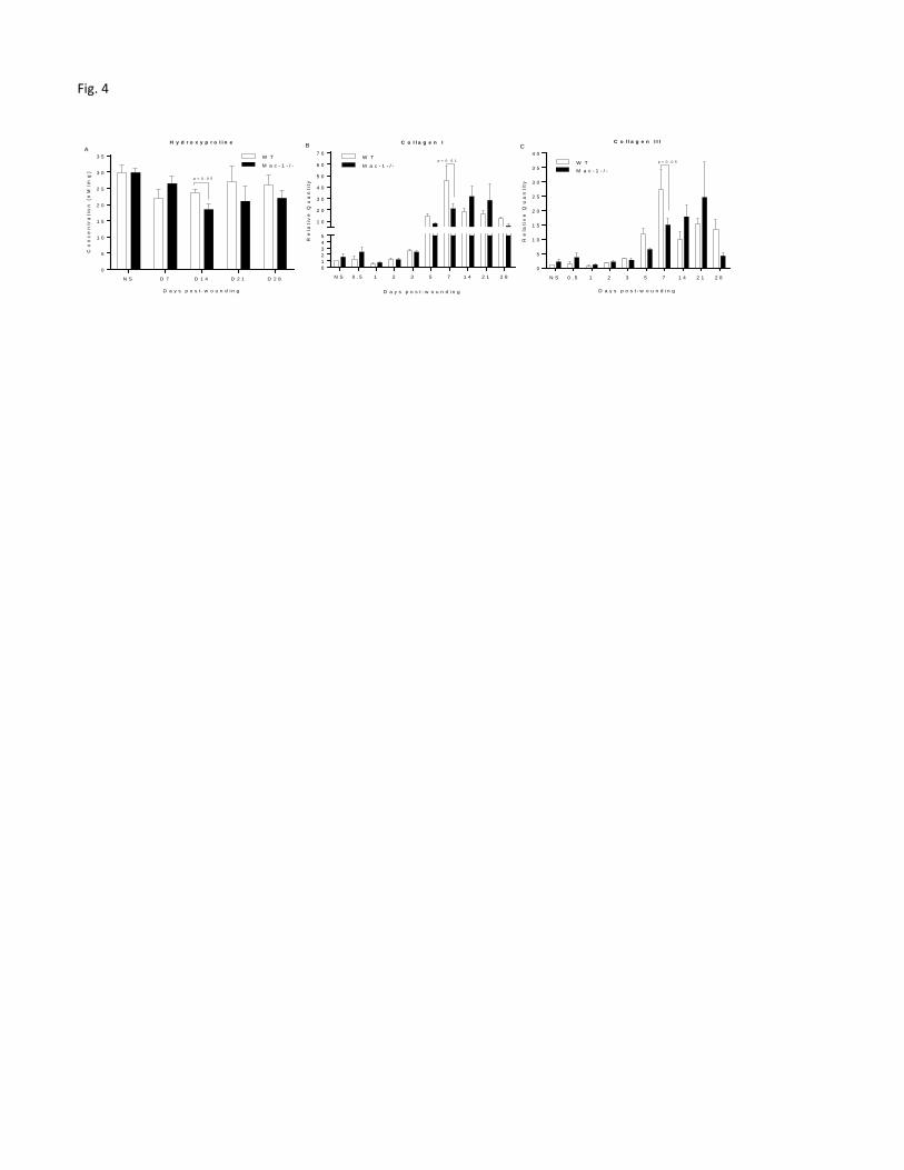
Mac-1 deficient mice exhibit minimal changes in wound collagen synthesis and
architecture.
To determine if Mac-1 deficiency might affect collagen synthesis in wounds, we measured
hydroxyproline as a marker of total collagen content. The concentration of hydroxyproline in the
wounds was slightly lower in Mac-1 -/- mice than that of WT mice at days 14, 21 and 28.
However, this difference reached statistical significance only at day 14 (p<0.05, Fig. 4A).
To further understand if the production of major collagens (i.e., collagen I and III) involved in
the wound healing process were altered by Mac-1 deficiency, collagen mRNA expression was
measured by quantitative RT-PCR. The expression of collagen I mRNA was low in the early
wounds of both strains of mice, reaching a peak at day 7 in WT mice and day 14 in Mac-1 -/-
mice, respectively (Figure 4B). Collagen III expression was also low at early time points,
peaking on day 7 in WT mice and on day 21 in Mac-1 -/- mice, respectively (Fig. 4C). At the
single time point of day 7, the expression of both collagens I and III was significantly higher in
WT mice than in Mac-1 -/- mice (p<0.01 and 0.05 for collagen I and collagen III, respectively,
Fig. 4B&C). This difference was not sustained, as the expression of both collagens I and III
showed no statistically significant difference between Mac-1 -/- and WT mice at days 14 and 21
(p<0.05) (Fig. 4 B&C).
Histologic analysis of collagen showed no differences between WT and Mac-1 deficient
mice.
Page 13
13
To further assess whether any substantive differences in collagen production occurred in the
wounds of Mac-1 -/- and WT mice, collagen content was evaluated histologically using
Masson’s trichrome and picrosirius red staining. Quantification of trichrome staining in wounds
of Mac-1 -/- and WT mice showed no significant differences (Fig. 5A&B). Picrosirius red
staining was also examined as this stain allows for the quantification of the relative amounts of
collagen I and collagen III. The picrosirius staining revealed no statistically significant
difference in the relative levels of Collagen I and Collagen III in wounds from the two strains of
mice at any time point. Unwounded skin from both WT and Mac-1 -/- mice contained
approximately 90% collagen I (red-orange) and 10% collagen III (green) (Fig. 5C&D). By day 7
after wound placement, collagen III was predominant in both strains. After day 14, the level of
collagen I gradually increased as collagen III gradually decreased in both strains. By day 28,
wounds of both strains contained about 80% collagen I and 20% collagen III. Therefore, the
collagen in wounds from WT and Mac-1 -/- mice underwent the same temporal transition from
primarily immature type III collagen to mature type I collagen fibers.
Myofibroblast levels and wound breaking strength are similar in Mac-1 deficient and WT
mice.
Because myofibroblasts are involved in wound contraction, collagen synthesis, and scar
formation, the density of these cells was compared in wounds of Mac-1 -/- and WT mice using
α-SMA as a marker. When compared to WT, wounds of Mac 1 -/- mice exhibited a noticeable
but insignificant decrease in α-SMA+ myofibroblasts than Mac-1 -/- mice on day 7 (p=0.3, Fig.
6A).
Page 14
14
To further examine dermal repair, we tested wound breaking strength in Mac-1 -/- and WT mice
using an incisional wound model. In keeping with the minimal changes in collagen content that
were observed in excisional wounds (Fig. 4&5), no difference in incisional wound breaking
strength was observed between the two mouse strains on days 7, 14, 21, or 28 post-wounding
(Fig. 6B).
TGF- β1 production was significantly impaired in Mac-1 deficient mice
Given the critical role of TGF-β1 in wound repair, we next compared TGF-β1 production in the
wounds of Mac-1 -/- and WT mice. TGF-β1 concentration was more than fourfold lower in
wounds of Mac-1 -/- versus WT mice (day 7, 121.7+18.8 pg/mg vs. 25.9+10.7 pg/mg, p<0.001,
Fig. 7A). Furthermore, mRNA analysis showed that the expression of SMAD2 and SMAD3, two
molecules in the TGF-β1 signaling pathway, was also significantly lower in wounds of Mac-1 -/-
mice (Fig. 7B).
DISCUSSION
In the current study, we sought to investigate the impact of Mac-1 deficiency on mouse skin
wound healing using small excisional and incisional skin wound models. Rather surprisingly, the
results of this study demonstrate that small excisional wounds on Mac-1 deficient mice exhibit
no substantial impairment in wound closure or collagen synthesis. The few changes that were
significantly different in the wounds of Mac-1 -/- mice were often seen only at single time
points. For example, at limited and specific time points, amounts of collagen I and collagen III
mRNA as well as total collagen content were significantly decreased in wounds of Mac-1 -/-
mice compared to WT mice. One interesting difference was that the production of TGF-β1 was
Page 15
15
significantly decreased in the wounds of Mac-1 -/- mice; a similar decrease was seen in two
TGF-β1 downstream signaling pathway molecules, SMAD2 and SMAD3. TGF-β1 contributes to
the recruitment of fibroblasts and stimulates the synthesis of collagens I, III, proteoglycans,
fibronectin, and other extracellular matrix components through the SMAD2 and 3 signaling
pathway.22, 23
The observation of generally normal wound closure and collagen synthesis in the
face of TGF-β1 deficiency demonstrates that the Mac-1-/- mice were capable of compensating
for this deficit in the wound models employed in this study.
One of the more striking observations of our study was a significant change in macrophage
phenotype in the wounds of Mac-1 -/- mice. Using the markers YM1 (for M2 like macrophages)
and iNOS (for M1 like macrophages), wounds of Mac-1 -/- mice were shown to have
significantly lower levels of macrophages of both phenotypes. With the caveat that our studies
examined only single markers, these results suggest that the wounds of Mac-1 -/- mice are
characterized by an unusual pattern of macrophage phenotypes. The concept that macrophages
in the wounds of Mac-1 -/- mice exist primarily in non-traditional phenotypes can be explained
by emerging studies on the complexity of this cell type. Recent studies have demonstrated that
macrophage phenotypes might be best considered in a multi-dimensional model.24
In particular,
the analysis of genome-wide transcriptional profiling of macrophages now suggests a very
complex macrophage response to stress signals.25
What is most interesting in the current study is
that despite apparent changes in macrophage functionality, small excisional wounds healed quite
normally in Mac-1 deficient mice. This result provides supporting evidence that our current
understanding of importance of specific macrophage phenotypes in wound healing is incomplete.
Page 16
16
Our study demonstrates that infiltration of macrophages into wounds, in terms of total numbers,
was unaffected in Mac-1 -/- mice compared to wild type mice. Thus macrophage recruitment to
wounds, and perhaps emigration from the wound site, appear to be independent of Mac-1 for
skin wounds. This finding is similar to prior wound healing studies in Mac-1 deficient strains.
Those previous investigations also reported minimal changes in macrophage content in skin
injury sites.16, 17
In contrast, in an induced peritoneal inflammation model, Mac-1 -/- mice
exhibited enhanced macrophage accumulation as activated macrophages failed to emigrate to
local draining lymph nodes through lymphatics.26
This reduced emigration does not seem to take
place in skin injury, however, suggesting that Mac-1 is either not critical to macrophage influx or
efflux in wounds, or that another molecule compensates. Interestingly, we detected a short term
but significant increase in the number of neutrophils in wounds of Mac-1-/- mice at 12 hours
after injury. These findings parallel what has been observed in a corneal wounds27
and peritoneal
inflammation 28
in Mac-1 -/- mice; this increase in neutrophil content has been suggested to
derive from a decrease in neutrophil apoptosis.27, 28
Our finding that small excisional skin wounds of Mac-1 -/- mice heal very normally differs from
a previous report that described delayed wound closure in partial-thickness ear wounds and full-
thickness skin head wounds of Mac-1 -/- (CD11b null) mice.17
The differences between these
two sets of results indicate that in the face of a genetic perturbation (such as a gene deletion), the
wound model, including size and location, impacts healing outcomes. More significantly, the
results suggest that small excisional wounds and incisional wounds, as used here, may be more
resilient in overcoming genetic deficits. The potential for differing wound models to exhibit
dissimilar outcomes in genetically deficient mice has been demonstrated at least once before, as
Page 17
17
previous studies showed differential delays in healing between different wound models in TGF-
deficient mice.29-31
In TGF- deficient mice, wounds produced by either tail amputation or on
the head were found to close normally, while ear wounds exhibited a delay in reepithelialization.
Beyond the wound model, another difference between the current and prior studies in Mac-1 -/-
mice is the strain background of the Mac-1 -/- mice. Here we used a commercially available
CD11b -/- congenic C57BL/6 strain, while in the prior work the CD11b -/- phenotype was on a
mixed strain background. Strain background is infrequently considered as an important variable,
despite published evidence for strain variation in healing rates and outcomes.32
In addition to the
current and prior study of wound healing in CD11b-/- mice, another previous study has described
a wound healing deficit in a Mac-1 deficient mouse strain generated via CD18 deletion.16
In the
CD18-/- strain of Mac-1 deficiency, wound healing was severely delayed. Macrophage
recruitment was not affected, but neutrophils were completely absent in wounds. In addition,
TGF-β1, TGF-β receptor type-II, -SMA (myofibroblast), and ED-A fibronectin were
substantially reduced in the wounds of CD18 null mice. However, this particular CD18
homozygous null mouse strain spontaneously developed severe chronic dermatitis with very few
neutrophils. These mice also had elevated circulating neutrophil counts, increased
immunoglobulin levels, a severe defect in T cell proliferation, lymphadenopathy, splenomegaly,
and abundant plasma cells in skin, lymph nodes, gut, and kidney.33
The obvious changes in the
function of multiple organs and within the immune system suggests that caution is needed in
drawing conclusions regarding the impact of Mac-1 deficiency on wound healing using the
CD18-/- mouse model. A comparison of the three wound healing studies (Table 2) in Mac-1
deficient mice suggests that the results in genetically deficient mice can be highly variable and
Page 18
18
may be dependent upon wound model, genetic background, and other physiologic abnormalities
of the strain.
In the current study, the wounds of Mac-1 deficient mice did exhibit some changes from WT
patterns. Mac-1-/- wounds showed significantly downregulated TGF-β1 production, decreased
SMAD2 and SMAD3 expression, decreased granulation tissue, minor decreases in collagen
synthesis, and alterations of macrophage phenotype markers. Importantly, though, in
assessments of functional healing, small 3 mm diameter excisional and incisional wounds healed
nearly normally in Mac-1-/- mice, with normal wound closure, and normal collagen content and
architecture. The small excisional skin wounds utilized here do heal with substantial contraction,
although reepithelialization does contributes to wound closure in this model.34
Considered in
context of prior studies, our results suggest that wounds that heal with contraction, such as the
3mm excisional wounds, and wounds that heal by primary intention, such as incisional wounds,
may more easily overcome genetic or other wound healing deficits. These results point to the
idea that the relative importance of any single factor in healing wounds may be quite distinct in
different types of injury. The concept that only certain types of wounds might manifest delayed
healing in the face of genetic deficiency is something that is rarely discussed in studies of
genetically deficient strains. Approximately 400 different genetically deficient (often-called
knockout) mouse strains have now been examined for wound healing phenotypes. Our data
suggests that the conclusions reached from observations in knockout mice should be limited to
the specific wound models that were examined, and should not be assumed to be a global
observation that is applicable to all wounds. In particular, the absence of any meaningful wound
Page 19
19
healing deficiency in a single model may provide incomplete information and should be
cautiously interpreted.
ACKNOWLEDGEMENTS
Source of Funding: This work was supported by the Clinical and Rehabilitative Medicine
Research Program of the U.S. Army Medical Research and Materiel Command, Ft. Detrick,
Maryland (award USAMRAA W81XWH-14-C-0152) and National Institutes of Health Grant
R01GM50875. The opinions and assertions contained herein are private views of the authors and
are not to be construed as official or as reflecting the views of the U.S. Army, the U.S.
Department of Defense, or the National Institutes of Health. This article has been approved for
public release with unlimited distribution. We are grateful to Dr. Wendy Cerny and Ms.
Elizabeth Michalczyk for critical reading of this manuscript.
Conflict of interest: None.
Disclosure Statement: The authors have nothing to disclose.
Page 20
20
Table 2. Wound healing in Mac-1 deficient mice (CD11b or CD18 null)
CD11b -/-
Chen, et al, current study
CD11b -/-
Sisco, et al, 2007
CD18 -/-
Peters, et al, 2005
Viability Viable, fertile, normal in size, Viable, fertile, normal in size, Neonatal death of 10–40%
Immunologic
abnormalities
Reduced mast cells in skin;
deficient neutrophil function;
decreased response to LPS
Reduced mast cells in skin;
deficient neutrophil function;
decreased response to LPS
Chronic dermatitis; elevated
circulating neutrophils and
immunoglobulin; T lymphocyte
and lymphoid organ abnormalities
Mouse strain C57BL/6 129SvxC57BL/6 129/SvxC57BL/6
Source of mice Jackson Laboratory Coxon, et al, 1996 In house
Sex of mice Female ? ?
Wound model 3 mm full-thickness dorsal
skin wounds
3 mm partial-thickness ear wounds
6 mm full- thickness head wounds
5 mm full-thickness dorsal skin
wounds
Wound closure
Re-epithelialization
Inflammation
Neutrophils at 12h Absent
Macrophages
IL-1β
IL-6 at day 1
TNF-
TGF-β1 at day 7 at day 5
Page 21
21
CXCL-1 (KC)
CCL-2 (MCP-1) at day 1
Granulation
Collagen
-SMA
References 26, 27, 35
and 36
26, 27, 35, and 36 16 and 33
No change , decreased , increased . Blank cells indicate that this parameter was not measured.
References
1. Eming SA, Krieg T, Davidson JM. Inflammation in wound repair: molecular and cellular
mechanisms. J Invest Dermatol 2007;127:514-25.
2. Guo S, DiPietro LA. Factors affecting wound healing. J Dent Res 2010;89:219-29.
3. Harris ES, McIntyre TM, Prescott SM, Zimmerman GA. The leukocyte integrins. J Biol
Chem 2000;275:23409-12.
4. Plow EF, Haas TA, Zhang L, Loftus J, Smith JW. Ligand binding to integrins. J Biol
Chem 2000;275:21785-8.
5. Rosetti F, Mayadas TN. The many faces of Mac-1 in autoimmune disease. Immunol Rev
2016;269:175-93.
6. Dovi JV, Szpaderska AM, DiPietro LA. Neutrophil function in the healing wound:
adding insult to injury? Thromb Haemost 2004;92:275-80.
7. Dovi JV, He LK, DiPietro LA. Accelerated wound closure in neutrophil-depleted mice. J
Leukoc Biol 2003;73:448-55.
8. Goren I, Allmann N, Yogev N, Schurmann C, Linke A, Holdener M, Waisman A,
Pfeilschifter J, Frank S. A transgenic mouse model of inducible macrophage depletion: effects of
diphtheria toxin-driven lysozyme M-specific cell lineage ablation on wound inflammatory,
angiogenic, and contractive processes. Am J Pathol 2009;175:132-47.
9. Mirza R, DiPietro LA, Koh TJ. Selective and specific macrophage ablation is detrimental
to wound healing in mice. Am J Pathol 2009;175:2454-62.
10. Daley JM, Brancato SK, Thomay AA, Reichner JS, Albina JE. The phenotype of murine
wound macrophages. J Leukoc Biol 2010;87:59-67.
11. Novak ML, Koh TJ. Macrophage phenotypes during tissue repair. J Leukoc Biol
2013;93:875-81.
12. Das A, Sinha M, Datta S, Abas M, Chaffee S, Sen CK, Roy S. Monocyte and
macrophage plasticity in tissue repair and regeneration. Am J Pathol 2015;185:2596-606.
13. Sica A, Erreni M, Allavena P, Porta C. Macrophage polarization in pathology. Cell Mol
Life Sci 2015;72:4111-26.
Page 22
22
14. Zuloff-Shani A, Adunsky A, Even-Zahav A, Semo H, Orenstein A, Tamir J, Regev E,
Shinar E, Danon D. Hard to heal pressure ulcers (stage III-IV): efficacy of injected activated
macrophage suspension (AMS) as compared with standard of care (SOC) treatment controlled
trial. Arch Gerontol Geriatr 2010;51:268-72.
15. Jetten N, Roumans N, Gijbels MJ, Romano A, Post MJ, de Winther MP, van der Hulst
RR, Xanthoulea S. Wound administration of M2-polarized macrophages does not improve
murine cutaneous healing responses. PLoS One 2014;9:e102994.
16. Peters T, Sindrilaru A, Hinz B, Hinrichs R, Menke A, Al-Azzeh EA, Holzwarth K,
Oreshkova T, Wang H, Kess D, Walzog B, Sulyok S, Sunderkotter C, Friedrich W, Wlaschek M,
Krieg T, Scharffetter-Kochanek K. Wound-healing defect of CD18(-/-) mice due to a decrease in
TGF-beta1 and myofibroblast differentiation. EMBO J 2005;24:3400-10.
17. Sisco M, Chao JD, Kim I, Mogford JE, Mayadas TN, Mustoe TA. Delayed wound
healing in Mac-1-deficient mice is associated with normal monocyte recruitment. Wound Repair
Regen 2007;15:566-71.
18. Chen L, Guo S, Ranzer MJ, DiPietro LA. Toll-like receptor 4 has an essential role in
early skin wound healing. J Invest Dermatol 2013;133:258-67.
19. Zhao Y, Bao L, Chan LS, DiPietro LA, Chen L. Aberrant Wound Healing in an
Epidermal Interleukin-4 Transgenic Mouse Model of Atopic Dermatitis. PLoS One
2016;11:e0146451.
20. Ranzer MJ, Chen L, DiPietro LA. Fibroblast function and wound breaking strength is
impaired by acute ethanol intoxication. Alcohol Clin Exp Res 2011;35:83-90.
21. Schneider CA, Rasband WS, Eliceiri KW. NIH Image to ImageJ: 25 years of image
analysis. Nat Methods 2012;9:671-5.
22. Roberts AB, Sporn MB, Assoian RK, Smith JM, Roche NS, Wakefield LM, Heine UI,
Liotta LA, Falanga V, Kehrl JH, et al. Transforming growth factor type beta: rapid induction of
fibrosis and angiogenesis in vivo and stimulation of collagen formation in vitro. Proc Natl Acad
Sci U S A 1986;83:4167-71.
23. Leask A, Abraham DJ. TGF-beta signaling and the fibrotic response. FASEB J
2004;18:816-27.
24. Schultze JL. Reprogramming of macrophages--new opportunities for therapeutic
targeting. Curr Opin Pharmacol 2016;26:10-5.
25. Xue J, Schmidt SV, Sander J, Draffehn A, Krebs W, Quester I, De Nardo D, Gohel TD,
Emde M, Schmidleithner L, Ganesan H, Nino-Castro A, Mallmann MR, Labzin L, Theis H,
Kraut M, Beyer M, Latz E, Freeman TC, Ulas T, Schultze JL. Transcriptome-based network
analysis reveals a spectrum model of human macrophage activation. Immunity 2014;40:274-88.
26. Cao C, Lawrence DA, Strickland DK, Zhang L. A specific role of integrin Mac-1 in
accelerated macrophage efflux to the lymphatics. Blood 2005;106:3234-41.
27. Li Z, Burns AR, Smith CW. Lymphocyte function-associated antigen-1-dependent
inhibition of corneal wound healing. Am J Pathol 2006;169:1590-600.
28. Coxon A, Rieu P, Barkalow FJ, Askari S, Sharpe AH, von Andrian UH, Arnaout MA,
Mayadas TN. A novel role for the beta 2 integrin CD11b/CD18 in neutrophil apoptosis: a
homeostatic mechanism in inflammation. Immunity 1996;5:653-66.
29. Kim I, Mogford JE, Chao JD, Mustoe TA. Wound epithelialization deficits in the
transforming growth factor-alpha knockout mouse. Wound Repair Regen 2001;9:386-90.
30. Luetteke NC, Qiu TH, Peiffer RL, Oliver P, Smithies O, Lee DC. TGF alpha deficiency
results in hair follicle and eye abnormalities in targeted and waved-1 mice. Cell 1993;73:263-78.
Page 23
23
31. Mann GB, Fowler KJ, Gabriel A, Nice EC, Williams RL, Dunn AR. Mice with a null
mutation of the TGF alpha gene have abnormal skin architecture, wavy hair, and curly whiskers
and often develop corneal inflammation. Cell 1993;73:249-61.
32. Li X, Gu W, Masinde G, Hamilton-Ulland M, Xu S, Mohan S, Baylink DJ. Genetic
control of the rate of wound healing in mice. Heredity (Edinb) 2001;86:668-74.
33. Scharffetter-Kochanek K, Lu H, Norman K, van Nood N, Munoz F, Grabbe S, McArthur
M, Lorenzo I, Kaplan S, Ley K, Smith CW, Montgomery CA, Rich S, Beaudet AL. Spontaneous
skin ulceration and defective T cell function in CD18 null mice. J Exp Med 1998;188:119-31.
34. Cheng TL, Lai CH, Chen PK, Cho CF, Hsu YY, Wang KC, Lin WL, Chang BI, Liu SK,
Wu YT, Hsu CK, Shi GY, Wu HL. Thrombomodulin promotes diabetic wound healing by
regulating toll-like receptor 4 expression. J Invest Dermatol 2015;135:1668-75.
35. Perera PY, Mayadas TN, Takeuchi O, Akira S, Zaks-Zilberman M, Goyert SM, Vogel
SN. CD11b/CD18 acts in concert with CD14 and Toll-like receptor (TLR) 4 to elicit full
lipopolysaccharide and taxol-inducible gene expression. J Immunol 2001;166:574-81.
36. Rosenkranz AR, Coxon A, Maurer M, Gurish MF, Austen KF, Friend DS, Galli SJ,
Mayadas TN. Impaired mast cell development and innate immunity in Mac-1 (CD11b/CD18,
CR3)-deficient mice. J Immunol 1998;161:6463-7.
Page 24
24
Figure legends
Figure 1. Wound closure in Mac-1 deficient and WT mice. A) CD11b expression on neutrophils
in day 1 wounds of WT and Mac-1-/- mice. B) CD11b expression on macrophages in day 1
wounds of WT and Mac-1-/- mice. Bar=20m C) Representative photomicrographs of wounds
from days 0 to 8 post-wounding. Six 3mm full thickness excisional wounds were made on the
dorsal skin of WT C57BL/6 and Mac-1 -/- mice. Bar=3mm. D) Representative photomicrographs
of HE stained histologic sections of day 1, 3 and 7 wounds. Bar=200m. Arrows indicate wound
edges. E) Wound closure expressed as percent of closure as calculated from photomicrographs of
wounds. N=10 at each time point and in each group. F) Wound re-epithelialization at days 1 and
3 using histological analysis. N=6 at each time point and in each group except that n=5 in WT
group at day 3. G). Volume of granulation tissue at days 5 and 7. N=6 at each time point and in
each group except that n=5 in Mac-1 -/- group at day 5. [Color figure can be viewed in the online
issue, which is available at wileyonlinelibrary.com.]
Figure 2. Macrophage content, CCL2 levels, and expression of macrophage phenotype markers
in wounds of Mac-1 deficient and WT mice. A) Time course of macrophage density in wounds.
B Protein levels of CCL-2 determined by multiplex ELISA, expressed as pg per mg of
homogenized tissue. C) mRNA expression of the anti-inflammatory macrophage marker YM1
during the course of wound healing. D) mRNA expression of the pro-inflammatory macrophage
marker, iNOS during the course of wound healing. NS: normal or unwounded skin, N=6 at each
time point and in each group.
Page 25
25
Figure 3. Neutrophil content and levels of CXCL-1, IL-1β, IL-6, and TNF- in wounds of Mac-
1 deficient and WT mice. A) Time course of the number of neutrophils in wounds. B, C, D, and
E) Protein levels of CXCL-1, IL-1β, IL-6, and TNF- determined by multiplex ELISA,
expressed as pg per mg of homogenized tissue. NS: normal or unwounded skin. N=6 at each time
point and group.
Figure 4. Biochemical and mRNA analysis of wound collagen content in Mac1 deficient and
WT mice. A) Levels of hydroxyproline in wounds over time. Concentrations of hydroxyproline
were determined using a hydroxyproline assay kit. B, C) mRNA expression of collagen I and
collagen III over the time course of the healing. NS: normal or unwounded skin. N=6 at each
time point and in each group.
Figure 5. Histologic assessment of collagen content in wounds of Mac-1 deficient and WT mice.
A) Representative photomicrographs of Masson’s Trichrome stained sections of the wound bed
at days 7, 14, 21, and 28. Bar=200µm. B) Quantification of Masson’s Trichrome collagen
staining in wounds. The density of blue-stained collagen in wound bed was quantified using
Image J. C) Representative photomicrographs of picrosirius red staining sections of normal skin
(NS), days 7 and 28 wounds. Bar=100µm. D) Summary of quantification of picrosirius red
staining. The relative density of collagen I (red-orange) and collagen III (green) in wound bed
was quantified using Image J. NS: normal or unwounded skin. N=5 at each time point and in
each group. [Color figure can be viewed in the online issue, which is available at
wileyonlinelibrary.com.]
Page 26
26
Figure 6. Myofibroblast content and wound breaking strength in wounds of Mac-1 deficient and
WT mice. A) Myofibroblast content in day 7 wound bed, assessed as the % of wound area. With
-SMA+ staining. B) Wound breaking strength, in lbs, over the time course of healing. NS:
normal or unwounded skin. N=6 at each time point and in each group.
Figure 7. Amount of activated TGF- β1 protein and mRNA expression of SMAD2&3 in
wounds of Mac-1 deficient and WT mice. A) Protein concentrations of activated TGF-β1 in day
7 wounds as determined by ELISA. B) mRNA expression of SMAD2 and SMAD3 in day 7
wounds as determined by real time PCR. N=6 at each time point and in each group.
Page 27
Fig. 1
1 3
0
1 0
2 0
3 0
4 0
5 0
6 0
D a y s p o s t - w o u n d i n g
%re
-ep
ith
eli
ali
za
tio
n
W TM a c - 1 - / -
F
WT Mac-1-/-
C
D0 D1 D3 D5
D8
D7
0 0 . 5 1 2 3 4 5 6 7 8
0
2 0
4 0
6 0
8 0
1 0 0
D a y s p o s t - w o u n d i n g
% c
los
ure
W T
M a c - 1 - / -
E
5 7
0 . 0
0 . 2
0 . 4
0 . 6
0 . 8
1 . 0
1 . 2
D a y s p o s t - w o u n d i n g
Gra
nu
lati
on
tis
su
e (
mm
2)
W TM a c - 1 - / -
Gp < 0 . 0 5
D WT Mac-1-/-
D1 D3 D7
Gr-1 CD11b DAPI GR-1/CD11b/DAPI
WT
Mac-1-/-
A B CD68 CD11b DAPI CD68/CD11b/DAPI
Page 28
Fig. 2
0
N S 0 . 5 1 2 3 5 7 1 4 2 1 2 8
0
5
1 0
1 5
2 0
2 5
3 0
3 5
4 0
D a y s p o s t w o u n d i n g
% W
ou
nd
Are
a p
os
itiv
e f
or
CD
68
W T
M a c - 1 - / -
M a c r o p h a g e sA
N S 0 . 5 1 2 3 5 7 1 4 2 1 2 8
0
5 0
1 0 0
1 5 0
2 0 0
2 5 0C C L - 2 ( M C P - 1 )
D a y s p o s t - w o u n d i n g
CC
L-2
(p
g/m
g)
W TM a c - 1 - / -P < 0 . 0 0 1
B
N S 1 3 7 1 4 2 1
0
1
2
1 0
2 0
3 0
4 0
5 0
6 0
iN O S
D a y s p o s t - w o u n d i n g
Re
lati
ve
Qu
an
tity
W T
M a c - 1 - / -D
p < 0 . 0 1
p < 0 . 0 1
p < 0 . 0 1
N S 1 3 7 1 4 2 1
0
2 5
1 0 0
2 0 0
3 0 0
4 0 0
5 0 0
6 0 0
7 0 0
8 0 0
Y M 1
Re
lati
ve
Qu
an
tity
W T
D a y s p o s t - w o u n d i n g
M a c - 1 - / -
p < 0 . 0 0 1
p < 0 . 0 0 1
C
Page 29
Fig. 3
N S 0 . 5 1 2 3 5 7 1 4 2 1 2 8
0
5
1 0
1 0 0
2 0 0
3 0 0
4 0 0
5 0 0
6 0 0 C X C L - 1 ( K C )
D a y s p o s t - w o u n d i n g
CX
CL
-1(p
g/m
g)
W T
M a c - 1 - / -
B
N S 0 . 5 1 2 3 5 7 1 4 2 1 2 8
0
1
2
3
4
5
6
7
8
9
1 0
1 1 I L - 1 β
D a y s p o s t - w o u n d i n g
IL-1
β (
pg
/mg
)
W T
M a c - 1 - / -
C
N S 0 . 5 1 2 3 5 7 1 4 2 1 2 8
0
5 0
1 0 0
1 5 0
2 0 0
2 5 0
3 0 0
3 5 0
4 0 0 I L - 6
D a y s p o s t - w o u n d i n g
IL-6
(p
g/m
g)
W T
M a c - 1 - / -
p < 0 . 0 1
D
N S 0 . 5 1 2 3 5 7 1 4 2 1 2 8
0
5
1 0
1 5
2 0
2 5
3 0
3 5 T N F α
D a y s p o s t - w o u n d i n g
TN
Fα
(p
g/m
g)
W T
M a c - 1 - / -
E
N S 0 . 5 1 2 3 5 7 1 4 2 1 2 8
0
2 0
4 0
6 0
8 0
1 0 0
1 2 0
1 4 0
1 6 0
1 8 0
D a y s p o s t - w o u n d i n g
No
. o
f n
eu
tro
ph
ils
(2
0x
/fie
ld)
W T
M a c - 1 - / -p < 0 . 0 5
N e u t r o p h i l sA
Page 30
Fig. 4
N S D 7 D 1 4 D 2 1 D 2 8
0
5
1 0
1 5
2 0
2 5
3 0
3 5
D a y s p o s t - w o u n d i n g
Co
nc
en
tra
tio
n (
nM
/mg
)
W T
M a c - 1 - / -
H y d r o x y p r o l i n eA
p < 0 . 0 5
N S 0 . 5 1 2 3 5 7 1 4 2 1 2 8
012345
1 0
2 0
3 0
4 0
5 0
6 0
7 0
C o l l a g e n I
D a y s p o s t - w o u n d i n g
Re
lati
ve
Qu
an
tity
W T
M a c - 1 - / -p < 0 . 0 1
B
N S 0 . 5 1 2 3 5 7 1 4 2 1 2 8
0
5
1 0
1 5
2 0
2 5
3 0
3 5
4 0
C o l l a g e n I I I
D a y s p o s t - w o u n d i n g
Re
lati
ve
Qu
an
tity
W TM a c - 1 - / -
p < 0 . 0 5
C
Page 31
Fig. 5
N S 7 1 4 2 1 2 8
0
2 0
4 0
6 0
8 0
1 0 0
D a y s p o s t - w o u n d i n g
% o
f to
tal
co
lla
ge
n
W T
M a c - 1 - / -
W T
M a c - 1 - / -
C o l l a g e n I
C o l l a g e n I I I
D
NS Day 7 Day 28
WT Mac-1 -/-
C
7 1 4 2 1 2 8
0
1 0
2 0
3 0
4 0
5 0
6 0
7 0
8 0
D a y s p o s t - w o u n d i n g
Re
lati
ve
De
ns
ity
W TM a c - 1 - / -
B
WT Mac-1-/-
D7 D14 D21 D24 A
Page 32
Fig. 6
W T M a c - 1 - / -
0
5
1 0
1 5
2 0α - S M A
% w
ou
nd
are
a p
os
itiv
e p = 0 . 3
A
N S 7 1 4 2 1 2 8
0 . 0 0
0 . 2 5
0 . 5 0
0 . 7 5
1 . 0 0
1 . 2 5
1 . 5 0
D a y s p o s t - w o u n d i n gB
rea
kin
g s
tre
ng
th (
Ib)
W T
M a c - 1 - / -
B
Page 33
Fig. 7
0
3 0
6 0
9 0
1 2 0
1 5 0
TG
F-1
β (
pg
/mg
)
P < 0 . 0 0 1
A
W T M a c - 1 - / - S M A D - 2 S M A D - 3
0 . 0
0 . 2
0 . 4
0 . 6
0 . 8
1 . 0
1 . 2
Re
lati
ve
Qu
an
tity
W T
M a c - 1 - / -
B
p < 0 . 0 0 1
p < 0 . 0 0 1