Gholami et al. Annals of Surgical Innovation and Research (2015) 9:1 DOI 10.1186/s13022-015-0010-5
CASE REPORT Open Access
Bone-added periodontal plastic surgery: a newapproach in esthetic dentistryGholam Ali Gholami1*, Hadi Gholami2*, Reza Amid1, Mahdi Kadkhodazadeh1, Amir Reza Mehdizadeh3
and Navid Youssefi3
Abstract
This article proposes a combined technique including bone grafting, connective tissue graft, and coronallyadvanced flap to create some space for simultaneous bone regrowth and root coverage.A 23 year-old female was referred to our private clinic with a severe class II Miller recession and lack of attachedgingiva. The suggested treatment plan comprised of root coverage combined with xenograft bone particles.The grafted area healed well and full coverage was achieved at 12-month follow-up visit. Bone-added periodontalplastic surgery can be considered as a practical procedure for management of deep gingival recession withoutbuccal bone plate.
Keywords: Plastic surgery, Root coverage, Periodontal regeneration, Bone graft
IntroductionMarginal tissue recession is a mucogingival problem thatis considered a major challenge for clinicians and pa-tients. It is frequentlyassociated with esthetic concerns,fear of tooth loss, root caries, and dentin hypersensitiv-ity. Manyprotocols are available for the managementofsuch defects including different types of soft tissuegrafts. Several studies have confirmed thatthese reces-sions can be predictably covered by various surgical pro-cedures like as pedicle flaps, subepithelial connectivetissue grafts (CTG) with or without coronally positionedflap (CPF), and guided tissue regeneration (GTR), if theinterdental papilla is not affected [1-5].In spite of predictable clinical outcomes by using
CTGs, its healingprocess and histological outcome stillremain controversial. Evidence data of human histologyafter the use of these techniques are scarce. The histo-logic evidenceshave been mostly derived from animalstudies or some case reportsconducted by the extractionof the treated teeth. It seems that CPF and CTG are as-sociated with somedegrees of periodontal regeneration[6-8]. However, some authorshave reported that healing
* Correspondence: [email protected]; [email protected] of Periodontics, Dental School, Shahid Beheshti University ofMedical Sciences, Evin, Tehran, Iran2Department of Prosthodontics, Faculty of Dental Medicine, University ofBern, Bern, SwitzerlandFull list of author information is available at the end of the article
occurs primarily by a long junctional epithelium or to alimited extent by connective tissueadhesion of the graft[9,10]. The concern about the nature of the grafted tis-sue attachment is based on the concept that the ultimategoal of periodontal treatment is to fully restore the at-tachment apparatus. Current available therapies haveshown limited and rather unpredictable results. The na-ture of connective tissue attachment seems to be stableover time, although, ultimate goal of a root coverageprocedure should be new bone formation overthe de-nuded roots.To our knowledge, there is no report of bone-added
periodontal plastic surgery for root coverage proceduresin humans. The aim of the present investigation was topresent a technique with a combination of bone substi-tute, CTG, and CPF thatwas used to create some spacefor osteoconduction and soft tissue coverage over de-nuded roots.
Case presentationA 23 year-old female was referred to our private clinicwith a chief complaint of hypersensitivity, fear of toothloss and gingival recession in the mandibular anteriortooth. She was in good general health and non smoker.Intraoral examination showed a good oral hygiene statuswith a full-moth plaque score equal to 17% [11]. A deepclass II Miller recession with the lack of attached
l. This is an Open Access article distributed under the terms of the Creativeommons.org/licenses/by/4.0), which permits unrestricted use, distribution, andiginal work is properly credited. The Creative Commons Public Domaing/publicdomain/zero/1.0/) applies to the data made available in this article,
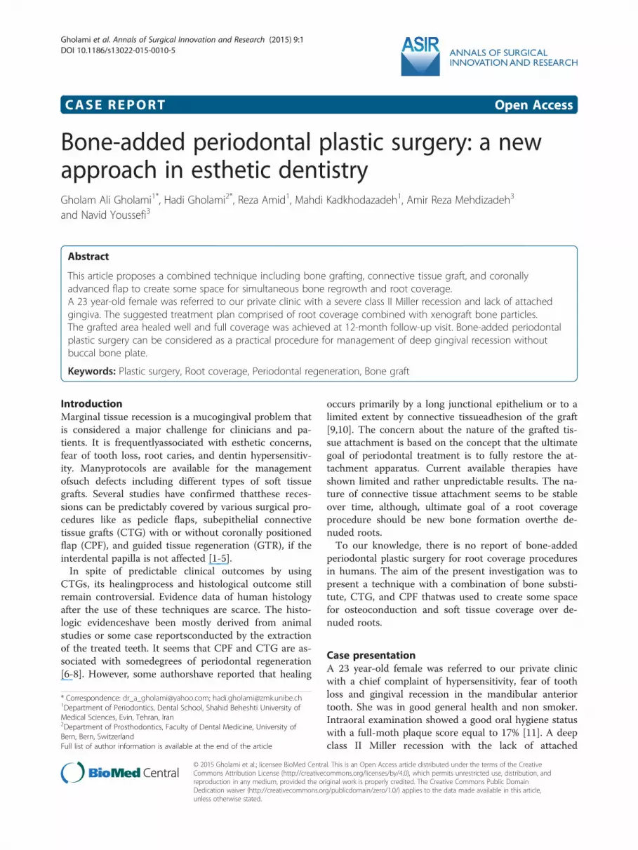
Figure 1 A severe deep class II Miller gingival recessions in anterior mandibular tooth.
Gholami et al. Annals of Surgical Innovation and Research (2015) 9:1 Page 2 of 6
gingiva, and narrow band of keratinized tissue was ob-served (Figure 1). Probing depths (PD) and clinical at-tachment level (CAL) measurements and registrations ofmarginal gingival recession (MGR) were obtained usinga periodontal probe (UNC 15, Hu-Friedy Mfg. Inc, Chi-cago, IL, USA). The measurements were rounded up tothe nearest millimeter. Tooth mobility was assessed andgraded 0-2 and tooth hypersensitivity calculated via vis-ual analysis scale (VAS). Measurements were done by anexaminer with more than 10 years of clinical experience.Bone mapping revealed that it was complete lack of buc-cal bone plate over involved tooth. The suggested treat-ment plan comprised root coverage combined withxenograft bone particles. Initial therapies, includingsupra-gingival plaque removal, polishing, occlusal adjust-ment, and oral hygiene instruction with proper toothbrushing method were performed. Surgical procedureswere performed by one of the authors who was notinvolved with clinical measurements. The clinical mea-surements were done by other experienced and cali-brated investigators who were not informed about thesurgical method. In case of controversy in the measure-ments by the examiners, they were asked to repeat theevaluation to reach a consensus. Another experienced andblinded operator was responsible for the radiographic
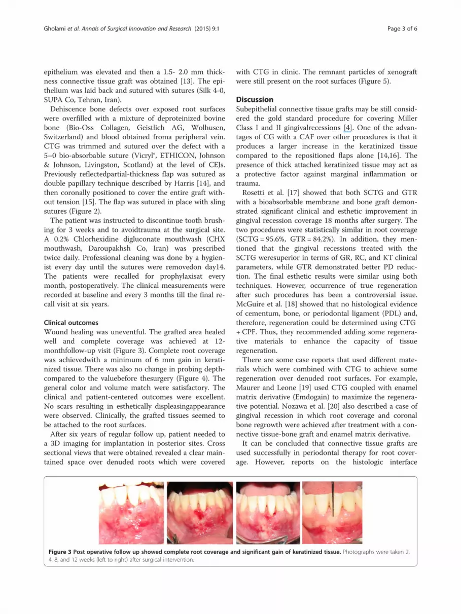
Figure 2 Surgical procedure: bed preparation, root surface preparatiomixed with blood, stabilization of connective tissue graft over bone g
examination. Written informed consent was obtainedfrom the patient for publication of this case report andany accompanying images. A copy of the written consentis available for review by the Editor-in-Chief of thisjournal.
Surgical procedureThe design of CPF was similar to that described previ-ously by Langer and Langer [12]. After local anesthesiaof the recipient site using 2% articainewithepinephr-ine1:100 000 (Septanest®, Septodont, Spain), an intra cre-vicular incision was made from right to left mandibularcanines. Two verticalreleasing incisions were made alongneighboring teeth. A partial thickness flap was elevatedwith a No.15c surgical blade beyond the mucogingivaljunction. Thus, it was extended until it could be pas-sively positioned coronally over the defect without ten-sion (Figure 2). The exposedroot surface was debridedcompletely with a curette (3/4 Gracey curette, Hu-FriedyMfg. Inc, Chicago, IL, USA) and conditioned with tetra-cycline powder. Pedicle flaps were sutured to make asingle flap. The connective tissue was harvested frompalate by trap door technique. In the palate, the distancebetween the horizontal incision and gingival margin hadto be more than 2 mm. By using no.15 scalpel, the
n, covering bone dehiscence with xenograft bone particlesraft, double papilla and coronally positioned flap.
Gholami et al. Annals of Surgical Innovation and Research (2015) 9:1 Page 3 of 6
epithelium was elevated and then a 1.5- 2.0 mm thick-ness connective tissue graft was obtained [13]. The epi-thelium was laid back and sutured with sutures (Silk 4-0,SUPA Co, Tehran, Iran).Dehiscence bone defects over exposed root surfaces
were overfilled with a mixture of deproteinized bovinebone (Bio-Oss Collagen, Geistlich AG, Wolhusen,Switzerland) and blood obtained froma peripheral vein.CTG was trimmed and sutured over the defect with a5–0 bio-absorbable suture (Vicryl®, ETHICON, Johnson& Johnson, Livingston, Scotland) at the level of CEJs.Previously reflectedpartial-thickness flap was sutured asdouble papillary technique described by Harris [14], andthen coronally positioned to cover the entire graft with-out tension [15]. The flap was sutured in place with slingsutures (Figure 2).The patient was instructed to discontinue tooth brush-
ing for 3 weeks and to avoidtrauma at the surgical site.A 0.2% Chlorhexidine digluconate mouthwash (CHXmouthwash, Daroupakhsh Co, Iran) was prescribedtwice daily. Professional cleaning was done by a hygien-ist every day until the sutures were removedon day14.The patients were recalled for prophylaxisat everymonth, postoperatively. The clinical measurements wererecorded at baseline and every 3 months till the final re-call visit at six years.
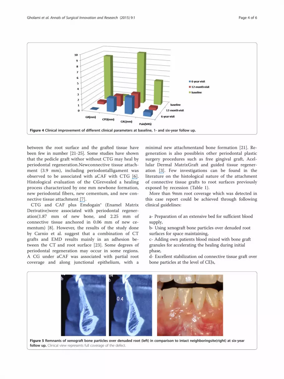
Clinical outcomesWound healing was uneventful. The grafted area healedwell and complete coverage was achieved at 12-monthfollow-up visit (Figure 3). Complete root coveragewas achievedwith a minimum of 6 mm gain in kerati-nized tissue. There was also no change in probing depth-compared to the valuebefore thesurgery (Figure 4). Thegeneral color and volume match were satisfactory. Theclinical and patient-centered outcomes were excellent.No scars resulting in esthetically displeasingappearancewere observed. Clinically, the grafted tissues seemed tobe attached to the root surfaces.After six years of regular follow up, patient needed to
a 3D imaging for implantation in posterior sites. Crosssectional views that were obtained revealed a clear main-tained space over denuded roots which were covered
Figure 3 Post operative follow up showed complete root coverage an4, 8, and 12 weeks (left to right) after surgical intervention.
with CTG in clinic. The remnant particles of xenograftwere still present on the root surfaces (Figure 5).
DiscussionSubepithelial connective tissue grafts may be still consid-ered the gold standard procedure for covering MillerClass I and II gingivalrecessions [4]. One of the advan-tages of CG with a CAF over other procedures is that itproduces a larger increase in the keratinized tissuecompared to the repositioned flaps alone [14,16]. Thepresence of thick attached keratinized tissue may act asa protective factor against marginal inflammation ortrauma.Rosetti et al. [17] showed that both SCTG and GTR
with a bioabsorbable membrane and bone graft demon-strated significant clinical and esthetic improvement ingingival recession coverage 18 months after surgery. Thetwo procedures were statistically similar in root coverage(SCTG = 95.6%, GTR = 84.2%). In addition, they men-tioned that the gingival recessions treated with theSCTG weresuperior in terms of GR, RC, and KT clinicalparameters, while GTR demonstrated better PD reduc-tion. The final esthetic results were similar using bothtechniques. However, occurrence of true regenerationafter such procedures has been a controversial issue.McGuire et al. [18] showed that no histological evidenceof cementum, bone, or periodontal ligament (PDL) and,therefore, regeneration could be determined using CTG+ CPF. Thus, they recommended adding some regenera-tive materials to enhance the capacity of tissueregeneration.There are some case reports that used different mate-
rials which were combined with CTG to achieve someregeneration over denuded root surfaces. For example,Maurer and Leone [19] used CTG coupled with enamelmatrix derivative (Emdogain) to maximize the regenera-tive potential. Nozawa et al. [20] also described a case ofgingival recession in which root coverage and coronalbone regrowth were achieved after treatment with a con-nective tissue-bone graft and enamel matrix derivative.It can be concluded that connective tissue grafts are
used successfully in periodontal therapy for root cover-age. However, reports on the histologic interface
d significant gain of keratinized tissue. Photographs were taken 2,
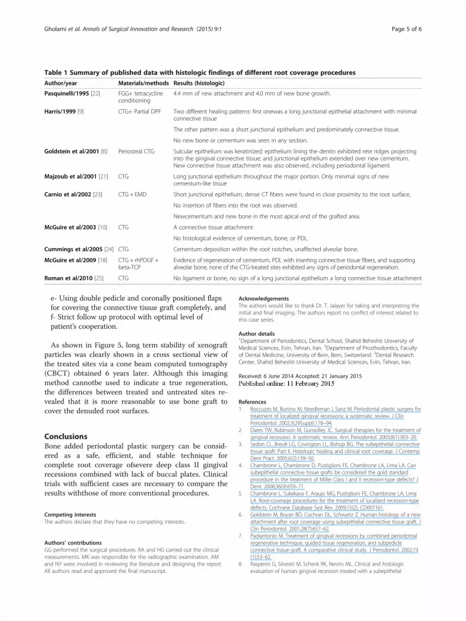
Figure 4 Clinical improvement of different clinical parameters at baseline, 1- and six-year follow up.
Gholami et al. Annals of Surgical Innovation and Research (2015) 9:1 Page 4 of 6
between the root surface and the grafted tissue havebeen few in number [21-25]. Some studies have shownthat the pedicle graft withor without CTG may heal byperiodontal regeneration.Newconnective tissue attach-ment (3.9 mm), including periodontalligament wasobserved to be associated with aCAF with CTG [6].Histological evaluation of the CGrevealed a healingprocess characterized by one mm newbone formation,new periodontal fibers, new cementum, and new con-nective tissue attachment [7].CTG and CAF plus Emdogain® (Enamel Matrix
Derivative)were associated with periodontal regener-ation(1.87 mm of new bone, and 2.25 mm ofconnective tissue anchored in 0.06 mm of new ce-mentum) [8]. However, the results of the study doneby Carnio et al. suggest that a combination of CTgrafts and EMD results mainly in an adhesion be-tween the CT and root surface [23]. Some degrees ofperiodontal regeneration may occur in some regions.A CG under aCAF was associated with partial rootcoverage and along junctional epithelium, with a
Figure 5 Remnants of xenograft bone particles over denuded root (lefollow up. Clinical view represents full coverage of the defect.
minimal new attachmentand bone formation [21]. Re-generation is also possiblein other periodontal plasticsurgery procedures such as free gingival graft, Acel-lular Dermal MatrixGraft and guided tissue regener-ation [3]. Few investigations can be found in theliterature on the histological nature of the attachmentof connective tissue grafts to root surfaces previouslyexposed by recession (Table 1).More than 9mm root coverage which was detected in
this case report could be achieved through followingclinical guidelines:
a- Preparation of an extensive bed for sufficient bloodsupply,b- Using xenograft bone particles over denuded rootsurfaces for space maintaining,c- Adding own patients blood mixed with bone graftgranules for accelerating the healing during initialphase,d- Excellent stabilization od connective tissue graft overbone particles at the level of CEJs,
ft) in comparison to intact neighboringsite(right) at six-year
4.4 mm of new attachment and 4.0 mm of new bone growth.
Harris/1999 [9] CTG+ Partial DPF Two different healing patterns: first onewas a long junctional epithelial attachment with minimalconnective tissue
The other pattern was a short junctional epithelium and predominately connective tissue.
No new bone or cementum was seen in any section.
Goldstein et al/2001 [6] Periosteal CTG Sulcular epithelium was keratinized; epithelium lining the dentin exhibited rete ridges projectinginto the gingival connective tissue; and junctional epithelium extended over new cementum.New connective tissue attachment was also observed, including periodontal ligament.
Majzoub et al/2001 [21] CTG Long junctional epithelium throughout the major portion. Only minimal signs of newcementum-like tissue
Carnio et al/2002 [23] CTG + EMD Short junctional epithelium, dense CT fibers were found in close proximity to the root surface,
No insertion of fibers into the root was observed.
Newcementum and new bone in the most apical end of the grafted area.
McGuire et al/2003 [10] CTG A connective tissue attachment
No histological evidence of cementum, bone, or PDL.
Cummings et al/2005 [24] CTG Cementum deposition within the root notches, unaffected alveolar bone.
McGuire et al/2009 [18] CTG + rhPDGF +beta-TCP
Evidence of regeneration of cementum, PDL with inserting connective tissue fibers, and supportingalveolar bone, none of the CTG-treated sites exhibited any signs of periodontal regeneration.
Roman et al/2010 [25] CTG No ligament or bone, no sign of a long junctional epithelium a long connective tissue attachment
Gholami et al. Annals of Surgical Innovation and Research (2015) 9:1 Page 5 of 6
e- Using double pedicle and coronally positioned flapsfor covering the connective tissue graft completely, andf- Strict follow up protocol with optimal level ofpatient’s cooperation.
As shown in Figure 5, long term stability of xenograftparticles was clearly shown in a cross sectional view ofthe treated sites via a cone beam computed tomography(CBCT) obtained 6 years later. Although this imagingmethod cannotbe used to indicate a true regeneration,the differences between treated and untreated sites re-vealed that it is more reasonable to use bone graft tocover the denuded root surfaces.
ConclusionsBone added periodontal plastic surgery can be consid-ered as a safe, efficient, and stable technique forcomplete root coverage ofsevere deep class II gingivalrecessions combined with lack of buccal plates. Clinicaltrials with sufficient cases are necessary to compare theresults withthose of more conventional procedures.
Competing interestsThe authors declare that they have no competing interests.
Authors’ contributionsGG performed the surgical procedures. RA and HG carried out the clinicalmeasurements. MK was responsible for the radiographic examination. AMand NY were involved in reviewing the literature and designing the report.All authors read and approved the final manuscript.
AcknowledgementsThe authors would like to thank Dr. T. Jalayer for taking and interpreting theinitial and final imaging. The authors report no conflict of interest related tothis case series.
Author details1Department of Periodontics, Dental School, Shahid Beheshti University ofMedical Sciences, Evin, Tehran, Iran. 2Department of Prosthodontics, Facultyof Dental Medicine, University of Bern, Bern, Switzerland. 3Dental ResearchCenter, Shahid Beheshti University of Medical Sciences, Evin, Tehran, Iran.
Received: 6 June 2014 Accepted: 21 January 2015
References1. Roccuzzo M, Bunino M, Needleman I, Sanz M. Periodontal plastic surgery for
treatment of localized gingival recessions: a systematic review. J ClinPeriodontol. 2002;3(29Suppl):178–94.
2. Oates TW, Robinson M, Gunsolley JC. Surgical therapies for the treatment ofgingival recession. A systematic review. Ann Periodontol. 2003;8(1):303–20.
3. Sedon CL, Breult LG, Covington LL, Bishop BG. The subepithelial connectivetissue graft: Part II. Histologic healing and clinical root coverage. J ContempDent Pract. 2005;6(2):139–50.
4. Chambrone L, Chambrone D, Pustiglioni FE, Chambrone LA, Lima LA. Cansubepithelial connective tissue grafts be considered the gold standardprocedure in the treatment of Miller Class I and II recession-type defects? JDent. 2008;36(9):659–71.
5. Chambrone L, Sukekava F, Araujo MG, Pustiglioni FE, Chambrone LA, LimaLA. Root-coverage procedures for the treatment of localized recession-typedefects. Cochrane Database Syst Rev. 2009;15(2), CD007161.
6. Goldstein M, Boyan BD, Cochran DL, Schwartz Z. Human histology of a newattachment after root coverage using subepithelial connective tissue graft. JClin Periodontol. 2001;28(7):657–62.
7. Paolantonio M. Treatment of gingival recessions by combined periodontalregenerative technique, guided tissue regeneration, and subpedicleconnective tissue graft. A comparative clinical study. J Periodontol. 2002;73(1):53–62.
8. Rasperini G, Silvestri M, Schenk RK, Nevins ML. Clinical and histologicevaluation of human gingival recession treated with a subepithelial
Gholami et al. Annals of Surgical Innovation and Research (2015) 9:1 Page 6 of 6
connective tissue graft and enamel matrix derivative (Emdogain): a casereport. Int J Periodontics Restorative Dent. 2000;20(3):269–75.
9. Harris RJ. Human histologic evaluation of root coverage obtained with aconnective tissue with partial thickness double pedicle graft. A case report.J Periodontol. 1999;70(7):813–21.
10. McGuire MK, Cochran DL. Evaluation of human recession defects treatedwith coronally advanced flaps and either enamel matrix derivative orconnective tissue. Part 2: Histological evaluation. J Periodontol. 2003;74(8):1126–35.
11. O’leary TJ, Drake RB, Nayor JE. The plaque control record. J Periodontol.1972;43(1):38.
12. Langer B, Langer L. Subepithelial connective tissue graft technique for rootcoverage. J Periodontol. 1985;56(12):715–20.
13. Nemcovsky CE, Artzi Z, Tal H, Kozlovsky A, Moses O. A multicentercomparative study of two root coverage procedures: coronally advancedflap with addition of enamel matrix proteins and subpedicle connectivetissue graft. J Periodontol. 2004;75(4):600–7.
14. Harris RJ. Connective tissue grafts combined with either double pediclegrafts or coronally positioned pedicle grafts: results of 266 consecutivelytreated defects in 200 patients. Int J Periodontics Restorative Dent. 2002;22(5):663–71.
15. Greenstein G, Greenstein B, Cavallaro J, Elian N, Tarnow D. Flapadvancement: practical techniques to attain tension-free primary closure. JPeriodontol. 2009;80(1):4–15.
16. Sugarman EF. A clinical and histological study of the attachment of graftedtissue to bone and teeth. J Periodontol. 1969;40(7):381–7.
17. Rosetti EP, Marcantonio RA, Rossa Jr C, Chaves ES, Goissis G, Marcantonio JrE. Treatment of gingival recession: comparative study between subepithelialconnective tissue graft and guided tissue regeneration. J Periodontol.2000;71(9):1441–7.
18. McGuire MK, Scheyer ET, Schupbach P. Growth factor-mediated treatmentof recession defects: a randomized controlled trial and histologic and micro-computed tomography examination. J Periodontol. 2009;80(4):550–64.
19. Maurer S, Leone CW. Use of a serially layered, double connective tissuegraft approach to enhance maxillary anterior esthetics. Int J PeriodonticsRestorative Dent. 2001;21(5):497–503.
20. Nozawa T, Sugiyama T, Satoh T, Tanaka K, Enomoto H, Ito K. Connectivetissue-bone onlay graft with enamel matrix derivative for treatment of gin-gival recession: a case report. Int J Periodontics Restorative Dent. 2002;22(6):559–65.
21. Majzoub Z, Landi L, Grusovin MG, Cordioli G. Histology of connective tissuegraft. A case report. J Periodontol. 2001;72(11):1607–15.
22. Pasquinelli KL. The histology of new attachment utilizing a thickautogenous soft tissue graft in an area of deep recession: a case report. IntJ Periodontics Restorative Dent. 1995;15(3):248–57.
23. Carnio J, Camargo PM, Kenney EB, Schenk RK. Histological evaluation of 4cases of root coverage following a connective tissue graft combined withan enamel matrix derivative preparation. J Periodontol. 2002;73(12):1534–43.
24. Cummings LC, Kaldahl WB, Allen EP. Histologic evaluation of autogenousconnective tissue and acellular dermal matrix grafts in humans. JPeriodontol. 2005;76(2):178–86.
25. Roman A, Câmpian R, Domşa I, Soancă A, Gocan H. Subepithelialconnective tissue graft for root coverage: clinical case reports and histologicevaluation. Rom J Morphol Embryol. 2010;51(4):793–7.
Submit your next manuscript to BioMed Centraland take full advantage of:
• Convenient online submission
• Thorough peer review
• No space constraints or color figure charges
• Immediate publication on acceptance
• Inclusion in PubMed, CAS, Scopus and Google Scholar
• Research which is freely available for redistribution
Submit your manuscript at www.biomedcentral.com/submit