ORIGINAL INVESTIGATION Clinical utility of chromosomal microarray analysis in invasive prenatal diagnosis Lluı ´s Armengol • Julia ´n Nevado • Clara Serra-Juhe ´ • Alberto Plaja • Carmen Mediano • Fe Amalia Garcı ´a-Santiago • Manel Garcı ´a-Aragone ´s • Olaya Villa • Elena Mansilla • Cristina Preciado • Luis Ferna ´ndez • Marı ´a A ´ ngeles Mori • Lidia Garcı ´a-Pe ´rez • Pablo Daniel Lapunzina • Luis Alberto Pe ´rez-Jurado Received: 13 August 2011 / Accepted: 16 September 2011 / Published online: 6 October 2011 Ó The Author(s) 2011. This article is published with open access at Springerlink.com Abstract Novel methodologies for detection of chromo- somal abnormalities have been made available in the recent years but their clinical utility in prenatal settings is still unknown. We have conducted a comparative study of cur- rently available methodologies for detection of chromosomal abnormalities after invasive prenatal sampling. A multicentric collection of a 1-year series of fetal samples with indication for prenatal invasive sampling was simultaneously evaluated using three screening methodologies: (1) karyotype and quantitative fluorescent polymerase chain reaction (QF-PCR), (2) two panels of multiplex ligation-dependent probe ampli- fication (MLPA), and (3) chromosomal microarray-based analysis (CMA) with a targeted BAC microarray. A total of 900 pregnant women provided informed consent to participate (94% acceptance rate). Technical performance was excellent for karyotype, QF-PCR, and CMA (*1% failure rate), but relatively poor for MLPA (10% failure). Mean turn-around time (TAT) was 7 days for CMA or MLPA, 25 for karyotype, and two for QF-PCR, with similar combined costs for the different approaches. A total of 57 clinically significant chromosomal aberrations were found (6.3%), with CMA yielding the highest detection rate (32% above other meth- ods). The identification of variants of uncertain clinical sig- nificance by CMA (17, 1.9%) tripled that of karyotype and MLPA, but most alterations could be classified as likely benign after proving they all were inherited. High accept- ability, significantly higher detection rate and lower TAT, could justify the higher cost of CMA and favor targeted CMA as the best method for detection of chromosomal abnormali- ties in at-risk pregnancies after invasive prenatal sampling. Electronic supplementary material The online version of this article (doi:10.1007/s00439-011-1095-5) contains supplementary material, which is available to authorized users. L. Armengol Á M. Garcı ´a-Aragone ´s qGenomics Laboratory, Doctor Aiguader, 88, 08003 Barcelona, Spain J. Nevado Á F. A. Garcı ´a-Santiago Á E. Mansilla Á L. Ferna ´ndez Á M. A ´ . Mori Á P. D. Lapunzina Instituto de Gene ´tica Me ´dica y Molecular, Hospital Universitario La Paz, Paseo de la Castellana, 261, 28046 Madrid, Spain J. Nevado Á C. Serra-Juhe ´ Á F. A. Garcı ´a-Santiago Á O. Villa Á E. Mansilla Á C. Preciado Á L. Ferna ´ndez Á M. A ´ . Mori Á P. D. Lapunzina Á L. A. Pe ´rez-Jurado Centro de Investigacio ´n en Red en Enfermedades Raras (CIBERER), Spain http://www.ciberer.es C. Serra-Juhe ´ Á O. Villa Á C. Preciado Á L. A. Pe ´rez-Jurado (&) Unitat de Gene `tica, Universitat Pompeu Fabra, Parc de Recerca Biome `dica de Barcelona (PRBB), Doctor Aiguader, 88, 08003 Barcelona, Spain e-mail: [email protected]A. Plaja Á C. Mediano Á L. A. Pe ´rez-Jurado Programa de Medicina Molecular i Gene `tica, Hospital Universitari Vall d’Hebron, Passeig de la Vall d’Hebron, 119-129, 08035 Barcelona, Spain L. Garcı ´a-Pe ´rez Fundacio ´n Canaria de Investigacio ´n y Salud (FUNCIS), Unidad Central de Coordinacio ´n de Ensayos Clı ´nicos, Servicio Canario de la Salud, Pe ´rez de Rozas, 5, 4 a Planta, 38004 Santa Cruz de Tenerife, Spain L. Garcı ´a-Pe ´rez CIBER Epidemiologı ´a y Salud Pu ´blica (CIBERESP), Barcelona, Spain 123 Hum Genet (2012) 131:513–523 DOI 10.1007/s00439-011-1095-5
Transcript
ORIGINAL INVESTIGATION
Clinical utility of chromosomal microarray analysis in invasiveprenatal diagnosis
Lluıs Armengol • Julian Nevado • Clara Serra-Juhe • Alberto Plaja • Carmen Mediano •
Fe Amalia Garcıa-Santiago • Manel Garcıa-Aragones • Olaya Villa • Elena Mansilla •
Cristina Preciado • Luis Fernandez • Marıa Angeles Mori • Lidia Garcıa-Perez •
Pablo Daniel Lapunzina • Luis Alberto Perez-Jurado
Received: 13 August 2011 / Accepted: 16 September 2011 / Published online: 6 October 2011
� The Author(s) 2011. This article is published with open access at Springerlink.com
Abstract Novel methodologies for detection of chromo-
somal abnormalities have been made available in the recent
years but their clinical utility in prenatal settings is still
unknown. We have conducted a comparative study of cur-
rently available methodologies for detection of chromosomal
abnormalities after invasive prenatal sampling. A multicentric
collection of a 1-year series of fetal samples with indication
for prenatal invasive sampling was simultaneously evaluated
using three screening methodologies: (1) karyotype and
(2) two panels of multiplex ligation-dependent probe ampli-
fication (MLPA), and (3) chromosomal microarray-based
analysis (CMA) with a targeted BAC microarray. A total of
900 pregnant women provided informed consent to participate
(94% acceptance rate). Technical performance was excellent
for karyotype, QF-PCR, and CMA (*1% failure rate), but
relatively poor for MLPA (10% failure). Mean turn-around
time (TAT) was 7 days for CMA or MLPA, 25 for karyotype,
and two for QF-PCR, with similar combined costs for the
different approaches. A total of 57 clinically significant
chromosomal aberrations were found (6.3%), with CMA
yielding the highest detection rate (32% above other meth-
ods). The identification of variants of uncertain clinical sig-
nificance by CMA (17, 1.9%) tripled that of karyotype and
MLPA, but most alterations could be classified as likely
benign after proving they all were inherited. High accept-
ability, significantly higher detection rate and lower TAT,
could justify the higher cost of CMA and favor targeted CMA
as the best method for detection of chromosomal abnormali-
ties in at-risk pregnancies after invasive prenatal sampling.
Electronic supplementary material The online version of thisarticle (doi:10.1007/s00439-011-1095-5) contains supplementarymaterial, which is available to authorized users.
L. Armengol � M. Garcıa-Aragones
qGenomics Laboratory, Doctor Aiguader, 88,
08003 Barcelona, Spain
J. Nevado � F. A. Garcıa-Santiago � E. Mansilla �L. Fernandez � M. A. Mori � P. D. Lapunzina
Instituto de Genetica Medica y Molecular, Hospital Universitario
La Paz, Paseo de la Castellana, 261, 28046 Madrid, Spain
J. Nevado � C. Serra-Juhe � F. A. Garcıa-Santiago � O. Villa �E. Mansilla � C. Preciado � L. Fernandez �M. A. Mori � P. D. Lapunzina � L. A. Perez-Jurado
Centro de Investigacion en Red en Enfermedades Raras
(CIBERER), Spain
http://www.ciberer.es
C. Serra-Juhe � O. Villa � C. Preciado � L. A. Perez-Jurado (&)
Unitat de Genetica, Universitat Pompeu Fabra, Parc de Recerca
Biomedica de Barcelona (PRBB), Doctor Aiguader, 88,
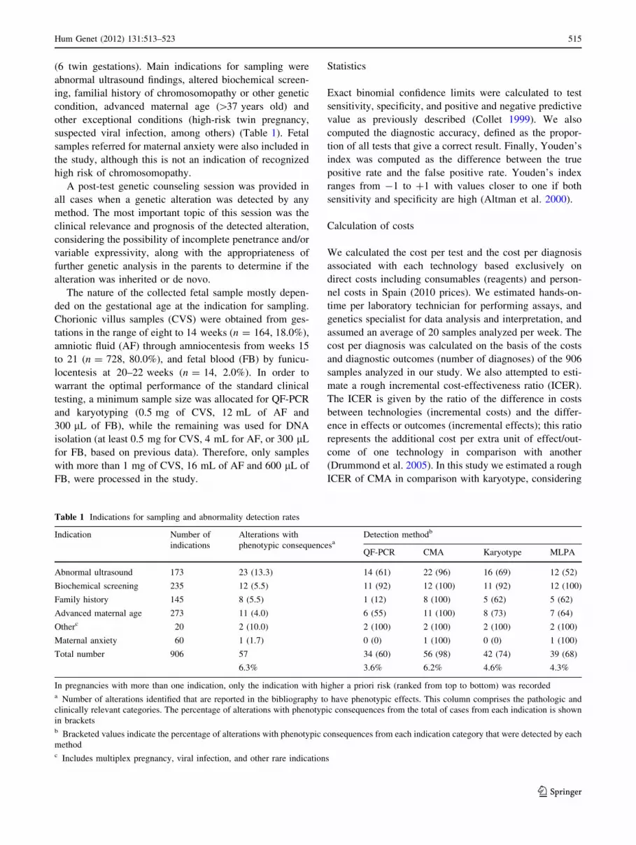
Total number 906 57 34 (60) 56 (98) 42 (74) 39 (68)
6.3% 3.6% 6.2% 4.6% 4.3%
In pregnancies with more than one indication, only the indication with higher a priori risk (ranked from top to bottom) was recordeda Number of alterations identified that are reported in the bibliography to have phenotypic effects. This column comprises the pathologic and
clinically relevant categories. The percentage of alterations with phenotypic consequences from the total of cases from each indication is shown
in bracketsb Bracketed values indicate the percentage of alterations with phenotypic consequences from each indication category that were detected by each
methodc Includes multiplex pregnancy, viral infection, and other rare indications
Hum Genet (2012) 131:513–523 515
123
the number of diagnoses as a measure of effects, although
we are aware that this is an intermediate outcome.
Results
Acceptance of novel prenatal testing procedures
All women who answered the questionnaire after the pre-
test counseling session (402/402) considered to have
received enough information of the ongoing study in order
to make a decision about participation. Among the 94%
who decided to participate, the main motivations were to
obtain more information (45%), to contribute to scientific
progress (48%), to decrease anxiety (5%), and in gratitude
for the professional kindness (1%). Fifty-six women (6%)
declined to join the study but continued with standard
prenatal testing; 60% of them argued more anxiety due to
extra testing. The median level of perceived anxiety prior
to testing was three on a scale between one (very low) and
five (very high), mostly due to the reason for referral for
prenatal testing. The fact of entering the study did not
represent any additional source of stress in those who
accepted participating.
Technical performance and turn-around-time
Good quality DNA for the different analyses was obtained
from 95% of CVS, 100% from FB, and 56% from AF
uncultured samples; thus it was necessary to obtain DNA
from cultured chorionic villi and amniotic fluid in 5% and
in 44% of the cases, respectively (Supplementary Table 1).
One advantage of performing multiple techniques on the
same sample was that failures could be attributed to either
a single technology or the common manipulation in most
cases. We considered a technical failure when it was not
possible to provide a definitive result with that technology
for any reason. Karyotype was the most robust technique
with only eight failures (0.9%), all cases due to cell culture
failure. CMA showed a failure rate of 1.1% (10/906), the
same as QF-PCR, but seven out of the ten failing samples
had been extracted the same day. MLPA was the less
robust technique, with a failure rate of 10.1% (183/1812),
61 with the subtelomeric set of probes and 122 to the
genomic disorders (RGD) set (see Supplementary Meth-
ods). In most cases, MLPA failure was attributable to
uncertainty in the interpretation of noisy electropherograms
with variable peak heights.
TAT was measured since the arrival of the biological
sample to the laboratory and until the results of the main
test were obtained. Time for downstream analysis of the
findings (parental testing, validation by an alternative
genetic test, etc.) was not computed to determine the TAT
mainly because additional samples were not readily
available (parental samples were not collected on a regular
basis), and because the approaches required for validations
were different for each case and technology. Overall,
QF-PCR was the fastest technique generating results with
an average TAT of two working days, while the average
TAT for CMA and MLPA from uncultured specimens was
7 days. For cultured samples, including G-banding karyo-
type, average TAT ranged between 4 and 27 days
(Supplementary Table 1).
Chromosomal aberrations detected
A total of 100 chromosomal aberrations were identified in
95 different samples and were classified into different
categories according to their predicted clinical significance
(Tables 2, 3, and Supplementary Table 2 for detailed
description). In the Pathologic category we detected 26
6 segmental aneuploidies and 2 fully penetrant microde-
letion disorders. Seventeen fetuses were observed to carry a
clinically relevant alteration, including 11 sex chromosome
aneuploidies and six recurrent microduplication syn-
dromes. Twenty-one aberrations corresponded to the
Uncertain Relevance category, four cytogenetically bal-
anced rearrangements and 16 copy number alterations,
three of them in malformed fetuses. The Benign category
was composed of 22 variants. Although cross-validation
was provided by the simultaneous use of multiple tech-
nologies in most cases, we used additional molecular
techniques in the follow-up of some of the alterations
identified by CMA, including the analysis of parental
samples to define whether the rearrangements were de
novo or inherited (Table 3 and Supplementary Table 2). As
an example, FISH was used for confirmation of the carrier
status for a balanced rearrangement in the mother of a fetus
with an unbalanced alteration (Supplementary Figure 1).
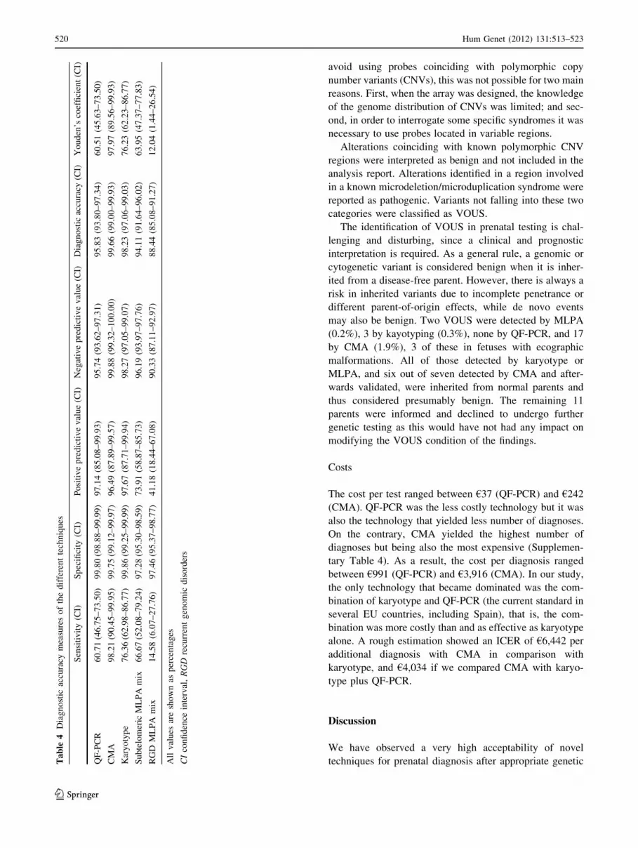
Sensitivity, specificity and detection rates
The nature and resolution of the assessed technologies
necessarily impact on the final detection rate achieved by
each of them, with intrinsic a priori limitations. In order to
perform a comparative evaluation of the different methods
by means of specificity and sensitivity, we used all chro-
mosomal abnormalities identified with predictable clinical
outcome, regardless of its size, as the one-for-all measure
unit. Specificity was found to be very high in all cases,
above 99% for QF-PCR, karyotype, and CMA, and 97%
for MLPA. However, sensitivity was significantly higher
for CMA (98.2%) than for other technologies (Table 4).
Youden’s index also revealed that CMA combines the
highest true to false positive ratio.
516 Hum Genet (2012) 131:513–523
123
The overall detection rate of pathological and clinically
relevant alterations was 6.3% (57/906 samples) with dif-
ferent detection capabilities depending on the technology
(Table 1). CMA yielded a superior detection rate in fetuses
with abnormal ultrasound (13.3%), but it was also signifi-
cant in pregnancies with a priori low risk (1.7 and 4.0%,
anxiety and advanced maternal age with normal screening,
respectively) (Table 1), far above the risk of pregnancy
loss by invasive sampling (Driscoll and Gross 2009).
Overall, CMA detected 32% more alterations than QF-PCR
and karyotype, including eight conditions with rather poor
prognosis for postnatal development (Table 3). This per-
cent increase in detection rates was equally high among
low-risk pregnancies and on those with an ultrasound
anomaly (Supplementary Table 3).
Another advantage of CMA was the ability to better
characterize two supernumerary markers (sSMC) and a
derivative chromosome identified by karyotyping (Table 3).
Interpretation and reporting criteria
Interpretation of results in studies using targeted CMA is
conceptually more straightforward than in studies using
whole-genome microarrays, although no increase of unclear
results has been reported in previous comparative studies
(Coppinger et al. 2009). Although a great effort was made to
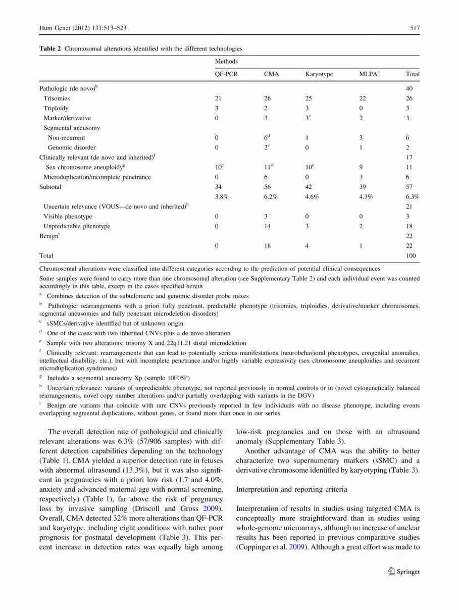
Table 2 Chromosomal alterations identified with the different technologies
Methods
QF-PCR CMA Karyotype MLPAa Total
Pathologic (de novo)b 40
Trisomies 21 26 25 22 26
Triploidy 3 2 3 0 3
Marker/derivative 0 3 3c 2 3
Segmental aneusomy
Non-recurrent 0 6d 1 3 6
Genomic disorder 0 2e 0 1 2
Clinically relevant (de novo and inherited)f 17
Sex chromosome aneuploidyg 10e 11e 10e 9 11
Microduplication/incomplete penetrance 0 6 0 3 6
Subtotal 34
3.8%
56
6.2%
42
4.6%
39
4.3%
57
6.3%
Uncertain relevance (VOUS—de novo and inherited)h 21
Visible phenotype 0 3 0 0 3
Unpredictable phenotype 0 14 3 2 18
Benigni 22
0 18 4 1 22
Total 100
Chromosomal alterations were classified into different categories according to the prediction of potential clinical consequences
Some samples were found to carry more than one chromosomal alteration (see Supplementary Table 2) and each individual event was counted
accordingly in this table, except in the cases specified hereina Combines detection of the subtelomeric and genomic disorder probe mixesb Pathologic: rearrangements with a priori fully penetrant, predictable phenotype (trisomies, triploidies, derivative/marker chromosomes,
segmental aneusomies and fully penetrant microdeletion disorders)c sSMCs/derivative identified but of unknown origind One of the cases with two inherited CNVs plus a de novo alteratione Sample with two alterations: trisomy X and 22q11.21 distal microdeletionf Clinically relevant: rearrangements that can lead to potentially serious manifestations (neurobehavioral phenotypes, congenital anomalies,
intellectual disability, etc.), but with incomplete penetrance and/or highly variable expressivity (sex chromosome aneuploidies and recurrent
microduplication syndromes)g Includes a segmental aneusomy Xp (sample 10F05P)h Uncertain relevance: variants of unpredictable phenotype, not reported previously in normal controls or in (novel cytogenetically balanced
rearrangements, novel copy number alterations and/or partially overlapping with variants in the DGV)i Benign are variants that coincide with rare CNVs previously reported in few individuals with no disease phenotype, including events
overlapping segmental duplications, without genes, or found more than once in our series
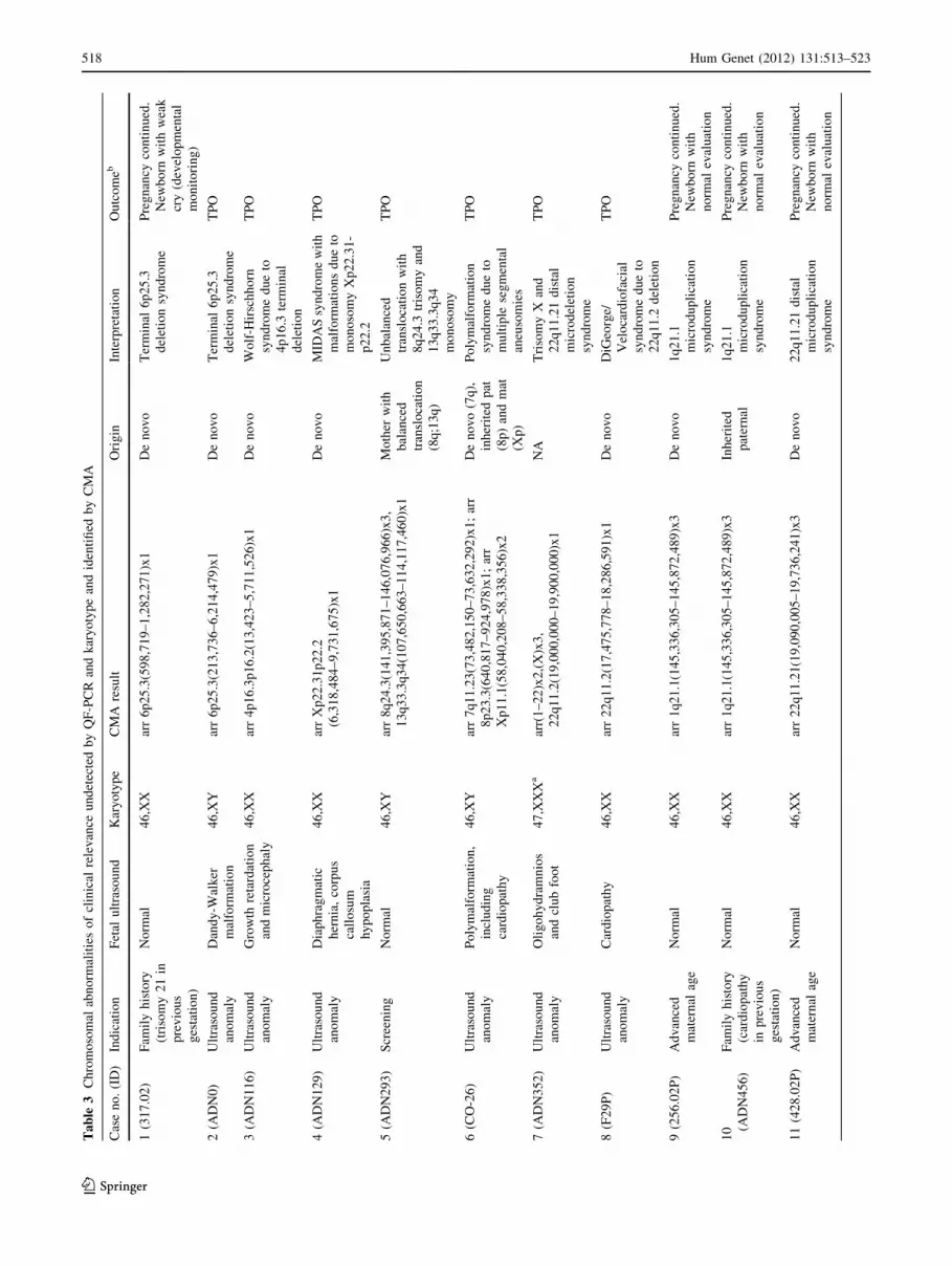
Hum Genet (2012) 131:513–523 517
123
Ta
ble
3C
hro
mo
som
alab
no
rmal
itie
so
fcl
inic
alre
lev
ance
un
det
ecte
db
yQ
F-P
CR
and
kar
yo
typ
ean
did
enti
fied
by
CM
A
Cas
en
o.
(ID
)In
dic
atio
nF
etal
ult
raso
un
dK
ary
oty
pe
CM
Are
sult
Ori
gin
Inte
rpre
tati
on
Ou
tco
meb
1(3
17
.02
)F
amil
yh
isto
ry
(tri
som
y2
1in
pre
vio
us
ges
tati
on
)
No
rmal
46
,XX
arr
6p
25
.3(5
98
,71
9–
1,2
82
,27
1)x
1D
en
ov
oT
erm
inal
6p
25
.3
del
etio
nsy
nd
rom
e
Pre
gn
ancy
con
tin
ued
.
New
bo
rnw
ith
wea
k
cry
(dev
elo
pm
enta
l
mo
nit
ori
ng
)
2(A
DN
0)
Ult
raso
un
d
ano
mal
y
Dan
dy
-Wal
ker
mal
form
atio
n
46
,XY
arr
6p
25
.3(2
13
,73
6–
6,2
14
,47
9)x
1D
en
ov
oT
erm
inal
6p
25
.3
del
etio
nsy
nd
rom
e
TP
O
3(A
DN
11
6)
Ult
raso
un
d
ano
mal
y
Gro
wth
reta
rdat
ion
and
mic
roce
ph
aly
46
,XX
arr
4p
16
.3p
16
.2(1
3,4
23
–5
,71
1,5
26
)x1
De
no
vo
Wo
lf-H
irsc
hh
orn
syn
dro
me
du
eto
4p
16
.3te
rmin
al
del
etio
n
TP
O
4(A
DN
12
9)
Ult
raso
un
d
ano
mal
y
Dia
ph
rag
mat
ic
her
nia
,co
rpu
s
call
osu
m
hy
po
pla
sia
46
,XX
arr
Xp
22
.31
p2
2.2
(6,3
18
,48
4–
9,7
31
,67
5)x
1
De
no
vo
MID
AS
syn
dro
me
wit
h
mal
form
atio
ns
du
eto
mo
no
som
yX
p2
2.3
1-
p2
2.2
TP
O
5(A
DN
29
3)
Scr
een
ing
No
rmal
46
,XY
arr
8q
24
.3(1
41
,39
5,8
71
–1
46
,07
6,9
66
)x3
,
13
q3
3.3
q3
4(1
07
,65
0,6
63
–1
14
,11
7,4
60
)x1
Mo
ther
wit
h
bal
ance
d
tran
slo
cati
on
(8q
;13
q)
Un
bal
ance
d
tran
slo
cati
on
wit
h
8q
24
.3tr
iso
my
and
13
q3
3.3
q3
4
mo
no
som
y
TP
O
6(C
O-2
6)
Ult
raso
un
d
ano
mal
y
Po
lym
alfo
rmat
ion
,
incl
ud
ing
card
iop
ath
y
46
,XY
arr
7q
11
.23
(73
,48
2,1
50
–7
3,6
32
,29
2)x
1;
arr
8p
23
.3(6
40
,81
7–
92
4,9
78
)x1
;ar
r
Xp
11
.1(5
8,0
40
,20
8–
58
,33
8,3
56
)x2
De
no
vo
(7q
),
inh
erit
edp
at
(8p
)an
dm
at
(Xp
)
Po
lym
alfo
rmat
ion
syn
dro
me
du
eto
mu
ltip
lese
gm
enta
l
aneu
som
ies
TP
O
7(A
DN
35
2)
Ult
raso
un
d
ano
mal
y
Oli
go
hy
dra
mn
ios
and
clu
bfo
ot
47
,XX
Xa
arr(
1–
22
)x2
,(X
)x3
,
22
q1
1.2
(19
,00
0,0
00
–1
9,9
00
,00
0)x
1
NA
Tri
som
yX
and
22
q1
1.2
1d
ista
l
mic
rod
elet
ion
syn
dro
me
TP
O
8(F
29
P)
Ult
raso
un
d
ano
mal
y
Car
dio
pat
hy
46
,XX
arr
22
q1
1.2
(17
,47
5,7
78
–1
8,2
86
,59
1)x
1D
en
ov
oD
iGeo
rge/
Vel
oca
rdio
faci
al
syn
dro
me
du
eto
22
q1
1.2
del
etio
n
TP
O
9(2
56
.02
P)
Ad
van
ced
mat
ern
alag
e
No
rmal
46
,XX
arr
1q
21
.1(1
45
,33
6,3
05
–1
45
,87
2,4
89
)x3
De
no
vo
1q
21
.1
mic
rod
up
lica
tio
n
syn
dro
me
Pre
gn
ancy
con
tin
ued
.
New
bo
rnw
ith
no
rmal
eval
uat
ion
10 (A
DN
45
6)
Fam
ily
his
tory
(car
dio
pat
hy
inp
rev
iou
s
ges
tati
on
)
No
rmal
46
,XX
arr
1q
21
.1(1
45
,33
6,3
05
–1
45
,87
2,4
89
)x3
Inh
erit
ed
pat
ern
al
1q
21
.1
mic
rod
up
lica
tio
n
syn
dro
me
Pre
gn
ancy
con
tin
ued
.
New
bo
rnw
ith
no
rmal
eval
uat
ion
11
(42
8.0
2P
)A
dv
ance
d
mat
ern
alag
e
No
rmal
46
,XX
arr
22
q1
1.2
1(1
9,0
90
,00
5–
19
,73
6,2
41
)x3
De
no
vo
22
q1
1.2
1d
ista
l
mic
rod
up
lica
tio
n
syn
dro
me
Pre
gn
ancy
con
tin
ued
.
New
bo
rnw
ith
no
rmal
eval
uat
ion
518 Hum Genet (2012) 131:513–523
123
Ta
ble
3co
nti
nu
ed
Cas
en
o.
(ID
)In
dic
atio
nF
etal
ult
raso
un
dK
ary
oty
pe
CM
Are
sult
Ori
gin
Inte
rpre
tati
on
Ou
tco
meb
12
(50
6P
)F
amil
yh
isto
ry
(pre
vio
us
neo
nat
al
dea
th)
No
rmal
46
,XY
arr
22
q1
1.2
1(1
9,0
90
,00
5–
19
,73
6,2
41
)x3
;
arr
Yp
11
.2(4
,91
7,1
08
–5
,07
7,7
86
)x2
,
Yp
11
.2(6
,67
1,4
14
–6
,85
2,9
46
)x2
Inh
erit
ed
pat
ern
al
22
q1
1.2
1d
ista
l
mic
rod
up
lica
tio
n
syn
dro
me
and
Yp
du
pli
cati
on
Pre
gn
ancy
con
tin
ued
.
New
bo
rnw
ith
no
rmal
eval
uat
ion
13 (A
DN
19
2)
Ult
raso
un
d
ano
mal
y
(In
crea
sed
nu
chal
tran
slu
cen
cy)
No
rmal
at
20
wee
ks
ges
tati
on
46
,XY
arr
22
q1
1.2
1(1
7,4
75
,77
8–
19
,73
6,2
41
)x3
Inh
erit
ed
mat
ern
al
22
q1
1.2
1
mic
rod
up
lica
tio
n
syn
dro
me
Pre
gn
ancy
con
tin
ued
.
New
bo
rnw
ith
no
rmal
eval
uat
ion
14 (A
DN
28
7)
An
xie
tyN
orm
al4
6,X
Yar
r1
6p
11
.2(2
9,6
15
,67
5–
29
,99
7,9
92
)x3
De
no
vo
16
p1
1.2
mic
rod
up
lica
tio
n
syn
dro
me
Pre
gn
ancy
con
tin
ued
.
New
bo
rnw
ith
no
rmal
eval
uat
ion
15 (A
DN
15
8)
Ad
van
ced
mat
ern
alag
e
No
rmal
Fai
lure
arr(
16
)x3
De
no
vo
(CV
S)—
AF
no
rmal
Pla
cen
tal
con
fin
ed
mo
saic
ism
for
tris
om
y1
6
Pre
gn
ancy
con
tin
ued
.
New
bo
rnw
ith
no
rmal
eval
uat
ion
16 (A
DN
18
1c)
Fam
ily
his
tory
(tri
som
y1
3in
pre
vio
us
ges
tati
on
)
No
rmal
46
,XY
/
47
,XY
?m
ar
arr
7p
22
.3p
22
.1(1
19
,39
6–
6,3
83
,91
0)x
3*
4D
en
ov
o
(CV
S)—
AF
no
rmal
/no
UP
D
Pla
cen
tal
con
fin
ed
mo
saic
ism
for
iso
(7p
)
Pre
gn
ancy
con
tin
ued
.
New
bo
rnw
ith
no
rmal
eval
uat
ion
17 (A
DN
66
.2)
Ult
raso
un
d
ano
mal
y
Car
dio
pat
hy
,
corp
us
call
osu
m
agen
esis
47
,XY
,
add
(8p
)
arr
8p
23
.3(3
04
,17
6–
3,6
34
,21
6)x
1,
8p
11
.23
(39
,05
1,9
24
–4
3,8
95
,57
3)x
3
De
no
vo
Po
lym
alfo
rmat
ion
syn
dro
me
du
eto
inv
du
pd
el(8
p)
TP
O
18 (A
DN
18
07
)
Fam
ily
his
tory
(car
rier
so
f
Gau
cher
dis
ease
)
No
rmal
47
,XY
,?
mar
arr 22
q1
1.1
q1
1.2
1(1
5,6
57
,18
1–
17
,02
6,8
97
)x3*
4
De
no
vo
Cat
-ey
esy
nd
rom
ed
ue
tote
tras
om
y2
2q
11
TP
O
Alt
erat
ion
sli
sted
inth
ista
ble
wer
ed
etec
ted
by
CM
Aan
dre
mai
ned
un
det
ecte
d(1
–1
5)
or
un
pro
per
lyd
efin
ed(1
6–
18
)b
yk
ary
oty
pe
or
QF
-PC
R.
So
me
of
the
var
ian
tsw
ere
det
ecte
db
yM
LP
A
NA
no
tav
aila
ble
,M
IDA
Sm
icro
ph
thal
mia
,d
erm
alap
lasi
a,an
dsc
lero
der
ma,
CV
Sch
ori
on
icv
illu
ssa
mp
le,
AF
amn
ioti
cfl
uid
,U
PD
un
ipar
enta
ld
iso
my
,T
OP
term
inat
ion
of
pre
gn
ancy
aT
riso
my
Xd
etec
ted
by
kar
yo
typ
e,2
2q
11
.21
del
etio
nu
nd
etec
ted
bP
ost
nat
alfo
llo
w-u
po
fle
ssth
an6
mo
nth
sin
all
case
s
Hum Genet (2012) 131:513–523 519
123
avoid using probes coinciding with polymorphic copy
number variants (CNVs), this was not possible for two main
reasons. First, when the array was designed, the knowledge
of the genome distribution of CNVs was limited; and sec-
ond, in order to interrogate some specific syndromes it was
necessary to use probes located in variable regions.
Alterations coinciding with known polymorphic CNV
regions were interpreted as benign and not included in the
analysis report. Alterations identified in a region involved
in a known microdeletion/microduplication syndrome were
reported as pathogenic. Variants not falling into these two
categories were classified as VOUS.
The identification of VOUS in prenatal testing is chal-
lenging and disturbing, since a clinical and prognostic
interpretation is required. As a general rule, a genomic or
cytogenetic variant is considered benign when it is inher-
ited from a disease-free parent. However, there is always a
risk in inherited variants due to incomplete penetrance or
different parent-of-origin effects, while de novo events
may also be benign. Two VOUS were detected by MLPA
(0.2%), 3 by kayotyping (0.3%), none by QF-PCR, and 17
by CMA (1.9%), 3 of these in fetuses with ecographic
malformations. All of those detected by karyotype or
MLPA, and six out of seven detected by CMA and after-
wards validated, were inherited from normal parents and
thus considered presumably benign. The remaining 11
parents were informed and declined to undergo further
genetic testing as this would have not had any impact on
modifying the VOUS condition of the findings.
Costs
The cost per test ranged between €37 (QF-PCR) and €242
(CMA). QF-PCR was the less costly technology but it was
also the technology that yielded less number of diagnoses.
On the contrary, CMA yielded the highest number of
diagnoses but being also the most expensive (Supplemen-
tary Table 4). As a result, the cost per diagnosis ranged
between €991 (QF-PCR) and €3,916 (CMA). In our study,
the only technology that became dominated was the com-
bination of karyotype and QF-PCR (the current standard in
several EU countries, including Spain), that is, the com-
bination was more costly than and as effective as karyotype
alone. A rough estimation showed an ICER of €6,442 per
additional diagnosis with CMA in comparison with
karyotype, and €4,034 if we compared CMA with karyo-
type plus QF-PCR.
Discussion
We have observed a very high acceptability of novel
techniques for prenatal diagnosis after appropriate geneticTa
ble
4D
iag
no
stic
accu
racy
mea
sure
so
fth
ed
iffe
ren
tte
chn
iqu
es
Sen
siti
vit
y(C
I)S
pec
ifici
ty(C
I)P
osi
tiv
ep
red
icti
ve
val
ue
(CI)
Neg
ativ
ep
red
icti
ve
val
ue
(CI)
Dia
gn
ost
icac
cura
cy(C
I)Y
ou
den
’sco
effi
cien
t(C
I)
QF
-PC
R6
0.7
1(4
6.7
5–
73
.50
)9
9.8
0(9
8.8
8–
99
.99
)9
7.1
4(8
5.0
8–
99
.93
)9
5.7
4(9
3.6
2–
97
.31
)9
5.8
3(9
3.8
0–
97
.34
)6
0.5
1(4
5.6
3–
73
.50
)
CM
A9
8.2
1(9
0.4
5–
99
.95
)9
9.7
5(9
9.1
2–
99
.97
)9
6.4
9(8
7.8
9–
99
.57
)9
9.8
8(9
9.3
2–
10
0.0
0)
99
.66
(99
.00
–9
9.9
3)
97
.97
(89
.56
–9
9.9
3)
Kar
yo
typ
e7
6.3
6(6
2.9
8–
86
.77
)9
9.8
6(9
9.2
5–
99
.99
)9
7.6
7(8
7.7
1–
99
.94
)9
8.2
7(9
7.0
5–
99
.07
)9
8.2
3(9
7.0
6–
99
.03
)7
6.2
3(6
2.2
3–
86
.77
)
Su
bte
lom
eric
ML
PA
mix
66
.67
(52
.08
–7
9.2
4)
97
.28
(95
.30
–9
8.5
9)
73
.91
(58
.87
–8
5.7
3)
96
.19
(93
.97
–9
7.7
6)
94
.11
(91
.64
–9
6.0
2)
63
.95
(47
.37
–7
7.8
3)
RG
DM
LP
Am
ix1
4.5
8(6
.07
–2
7.7
6)
97
.46
(95
.37
–9
8.7
7)
41
.18
(18
.44
–6
7.0
8)
90
.33
(87
.11
–9
2.9
7)
88
.44
(85
.08
–9
1.2
7)
12
.04
(1.4
4–
26
.54
)
All
val
ues
are
sho
wn
asp
erce
nta
ges
CI
con
fid
ence
inte
rval
,R
GD
recu
rren
tg
eno
mic
dis
ord
ers
520 Hum Genet (2012) 131:513–523
123
counseling, with only 6% of women declining to enter the
study due to increased anxiety. Technical performance was
excellent for CMA, and similar to QF-PCR or karyotype
under standard procedures. However, despite our extensive
experience with the technology, MLPA showed a high rate
of technical failure in uncultured AF samples, likely due to
the low purity of the DNA obtained (salt and/or protein
contamination). Finally, CMA revealed to be the most
sensitive technique for diagnosing chromosomal alterations
associated with medical conditions, being able to detect all
but one clinically relevant alterations (56/57), followed by
G-banding karyotype (42/57), MLPA (39/57) and QF-PCR
(34/57, all detected by karyotype as well). In other words,
CMA increased *32% the detection rate of any other
method. Conversely, the only clinically relevant alteration
missed by CMA was a triploidy with karyotype 69, XXX,
associated with a usually lethal condition in utero. This
limitation could be overcome using microarrays that also
interrogate nucleotide variation (SNPs), that can also detect
uniparental disomies, but their clinical utility in prenatal
setting remains to be proven. There was also a qualitative
advantage of CMA, as the origin of small supernumerary
marker chromosomes and derivatives was readily deter-
mined. Maybe the most relevant implication of our data is
that 14 relevant fetal conditions (*1.6% of the entire
study) would have remained undiagnosed using only the
currently implemented detection methods in the clinic. The
use of CMA resulted in an increased detection rate
regardless of the indication for study. This becomes espe-
cially evident in the high-risk group (ultrasound findings),
in which the percentage of detection was elevated to
13.3%; but also in groups with a priori low risk, which
showed a detection rate far above 1/100 and a relative
increase over 45% when compared to currently imple-
mented methods (Supplementary Table 3).
Since a decision on the continuation of a pregnancy
might follow the diagnostic findings of a prenatal test, it is
not desirable to identify VOUS and there are some diffi-
culties dealing with some clinically relevant conditions
with variable expressivity or incomplete penetrance. We
tried to minimize VOUS by designing a targeted micro-
array, that interrogates only regions of known clinical
relevance and using large segments of DNA as probes, and
MLPA panels were also selected with similar criteria.
Overall, CMA detected 17 of the total 21 VOUS identified,
although 3 of them might be causative as they were found
in fetuses with malformations. Most VOUS could be
classified as likely benign after proving they all were
inherited from a parent with no-disease phenotype. For the
management of such findings, it is of extraordinary help the
development of public and trustable databases of variation
on normal individuals, such as the database of genome
variants (DGV) and the initiatives to catalogue normal and
pathological variation from the ISCA and DECIPHER
consortia. The minimization of VOUS is just a matter of
time and of better describing structural variation in larger
cohorts of cases and controls by means of different -omic
approaches, which would allow establishing a solid statis-
tical framework for assigning (or not) pathogenicity to each
specific copy number variant. In such a future scenario, the
use of higher genome coverage approaches might be
preferred.
Interestingly, we detected six cases of recurrent micro-
duplication syndromes (0.7% of our series), three inherited
from a phenotypically normal parent and three de novo. We
faced the difficult counseling of these genomic imbalances
associated with variable phenotypes and incomplete pene-
trance with still scarce literature, although preliminary
guidelines for clinical evaluation and anticipatory guidance
have been published (Berg et al. 2010). Following a
20-week normal ultrasound evaluation, the parental deci-
sion in all cases was continuation of the pregnancy. The
situation, however, is comparable to the counseling of sex
chromosome aneuploidies, where the phenotype can be
very mild with incompletely penetrant features.
In order to provide further objective assessment tools,
we also estimated the costs of the different technologies.
Although CMA is still the most expensive technology, it is
also the one that yields a higher number of diagnoses. Our
cost analysis study had some limitations related to the non-
inclusion of some relevant costs like equipment deprecia-
tion and maintenance, as well as costs of downstream
analyses required for confirming findings. Our rough esti-
mation indicates that expenses not included in our cost
analysis (mainly capital expenses to set up a clinical lab-
oratory and direct maintenance costs) are similar for the
different platforms. Thus, the most important direct costs
were included and the figures reported herein can show the
relative differences between technologies. The other limi-
tation was the use of number of diagnoses as outcome
measure instead of a health-related outcome (Grosse et al.
2008). Therefore, the estimated ICER is a tentative figure
reported here with the aim of promoting the debate about
the willingness to pay for new technologies and to show the
need of economic evaluations in the field of genetic
screening and diagnosis (Carlson et al. 2005). A full eco-
nomic evaluation will be addressed in the future.
In summary, our data indicate that from the perspective
of diagnostic capacity, sensitivity, and specificity, CMA is
the most reliable technology. According to women’s
acceptance, the diagnostic yield increase that CMA brings
into prenatal genetic testing of risk pregnancies, the
extraordinary medical and social cost of birth defects
associated to chromosomal disorders, and until non-inva-
sive methods are able to provide a similar sensitivity, we
consider that CMA should already be a first-tier option for
Hum Genet (2012) 131:513–523 521
123
invasive prenatal diagnosis of at-risk pregnancies instead
of the current combination of RAD (QF-PCR or FISH) and
karyotype.
Although non-invasive assays for fetal diagnosis are an
intense field of research, at present these are only experi-
mental approaches available for specific chromosomal or
single gene disorders (Chiu et al. 2011). Thus, nowadays,
invasive fetal sampling is still the common practice, indi-
cated in those cases where the risk of a detectable abnor-
mality in the fetus is above the risk of a procedure-related
pregnancy loss, *1/200 (Driscoll and Gross 2009). While
the evaluation of larger series is granted, the much higher
detection rate of CMA, even in a priori low-risk groups
([1/100), should open the door to consider even deeper
changes in currently established screening policies in pre-
natal care.
Acknowledgments We would like to thank the support of the
medical geneticists and obstetricians in both hospitals, Drs. M. del
Campo, T. Vendrell, A. Cueto, S. Garcıa-Minaur, M.A. Sanchez, J.
Sagala, M.T. Higueras, E. Carreras, R. Rodrıguez, A. Gonzalez, F.
Herrero, as well as all participants. Thanks to Dr. J.R. Gonzalez for
statistical support and critical review and A. Fernandez, M. Arranz
and R. Sansegundo for technical assistance. We thank P. Serrano-
Aguilar for assistance with economical aspects. This work was sup-
ported by a grant from the Spanish Agency for Evaluation of Novel
Sanitary Technologies (AETS-PI08/90056).
Conflict of interest Lluıs Armengol and Luis Perez-Jurado are
executive director and scientific advisor, respectively, of qGenomics,
a privately held company that provide genomics services to the sci-
entific and medical community.
Open Access This article is distributed under the terms of the
Creative Commons Attribution Noncommercial License which per-
mits any noncommercial use, distribution, and reproduction in any
medium, provided the original author(s) and source are credited.