[ Color index : Important | Notes | Extra ] Editing file link Common adult fractures Objectives: ➢ CLAVICLE FRACTURE ➢ Humerus (proximal and distal) ➢ Both bone and foramen fractures ➢ Distal radius fracture ➢ Hip fracture ➢ Femur shaft fracture ➢ Tibial shaft fracture ➢ Ankle fracture Done by: Ibrahim Alshayea Edited By: Saleh Al Khalifa Revised by: Adel F. Al Shihri Resources:435 Slide+Notes+Appley’s+Toronto+433 Team.
Transcript
[ Color index : Important | Notes | Extra ] Editing file link
Common adult fractures
Objectives: ➢ CLAVICLE FRACTURE
➢ Humerus (proximal and distal)
➢ Both bone and foramen fractures
➢ Distal radius fracture
➢ Hip fracture
➢ Femur shaft fracture
➢ Tibial shaft fracture
➢ Ankle fracture
Done by: Ibrahim Alshayea Edited By: Saleh Al Khalifa Revised by: Adel F. Al Shihri Resources:435 Slide+Notes+Appley’s+Toronto+433 Team.
Before you study this lecture we suggest to read how to manage open fracture
How to do physical examination for any fracture?
1st step: Expose the area and check for any deformity or skin changes
2nd step: N/V examination: ask the patient to move distal parts from fracture and check sensation then check temperature, color, pulses and capillary refill distally to fracture
3rd step: Examine the joint above and joint below
4th step: Check for other complications
In fractures, we don’t examine range of motions passively because of pain.
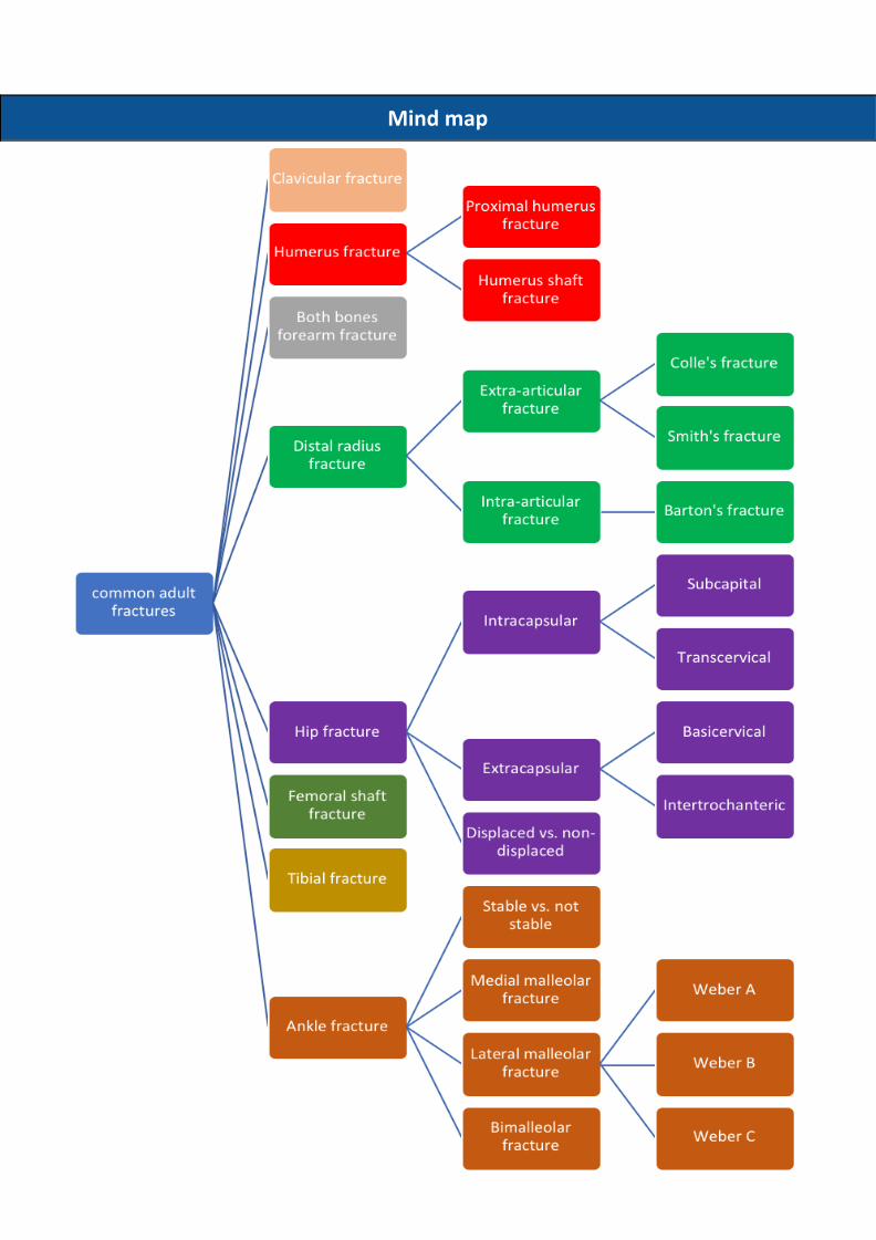
Mind map
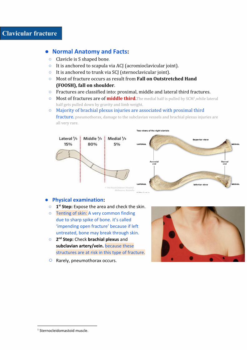
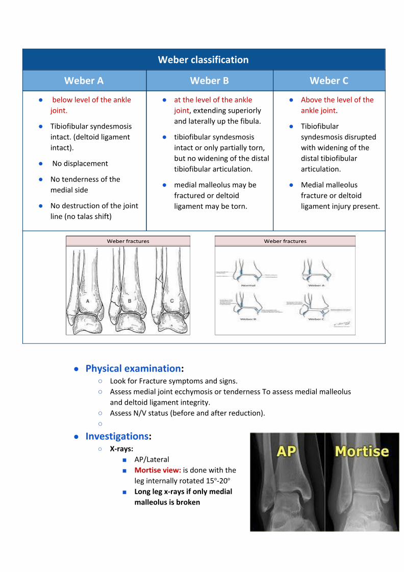
Clavicular fracture
● Normal Anatomy and Facts: ○ Clavicle is S shaped bone. ○ It is anchored to scapula via ACJ (acromioclavicular joint). ○ It is anchored to trunk via SCJ (sternoclavicular joint). ○ Most of fracture occurs as result from Fall on Outstretched Hand
(FOOSH), fall on shoulder. ○ Fractures are classified into: proximal, middle and lateral third fractures. ○ Most of fractures are of middle third.The medial half is pulled by SCM ,while lateral 1
half gets pulled down by gravity and limb weight.
○ Majority of brachial plexus injuries are associated with proximal third fracture. pneumothorax, damage to the subclavian vessels and brachial plexus injuries are
all very rare.
● Physical examination:
○ 1st Step: Expose the area and check the skin.
○ Tenting of skin: A very common finding
due to sharp spike of bone. it’s called
‘impending open fracture’ because if left
untreated, bone may break through skin.
○ 2nd Step: Check brachial plexus and
subclavian artery/vein. because these
structures are at risk in this type of fracture.
○ Rarely, pneumothorax occurs.
1 Sternocleidomastoid muscle.
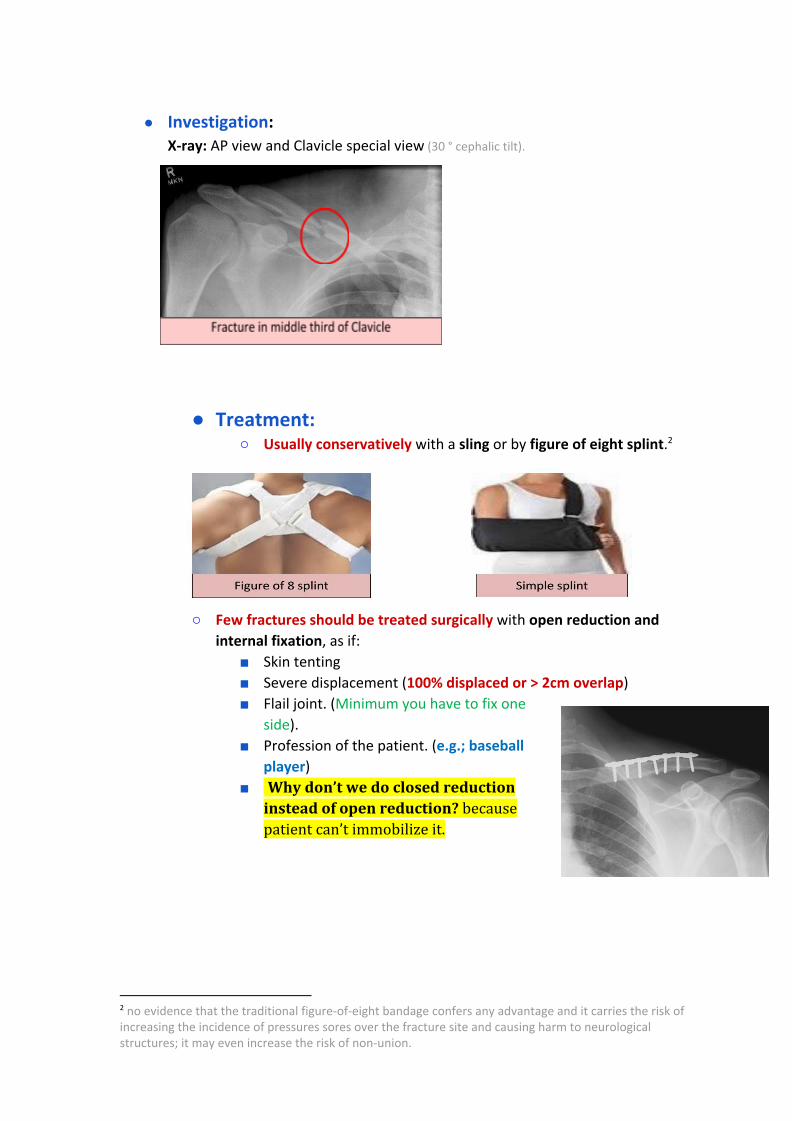
● Investigation: X-ray: AP view and Clavicle special view (30 ° cephalic tilt).
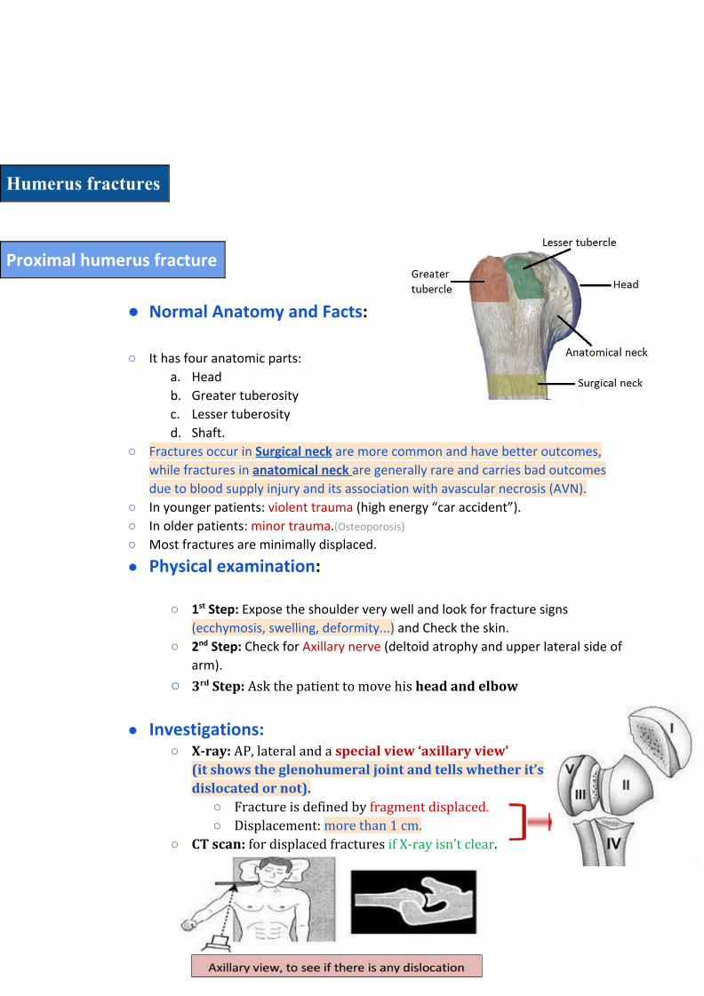
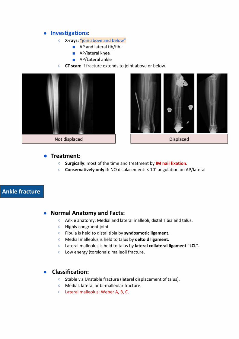
● Treatment: ○ Usually conservatively with a sling or by figure of eight splint. 2
○ Few fractures should be treated surgically with open reduction and
internal fixation, as if:
■ Skin tenting
■ Severe displacement (100% displaced or > 2cm overlap) ■ Flail joint. (Minimum you have to fix one
side). ■ Profession of the patient. (e.g.; baseball
player) ■ Why don’t we do closed reduction
instead of open reduction? because patient can’t immobilize it.
2 no evidence that the traditional figure-of-eight bandage confers any advantage and it carries the risk of increasing the incidence of pressures sores over the fracture site and causing harm to neurological structures; it may even increase the risk of non-union.
Humerus fractures
Proximal humerus fracture
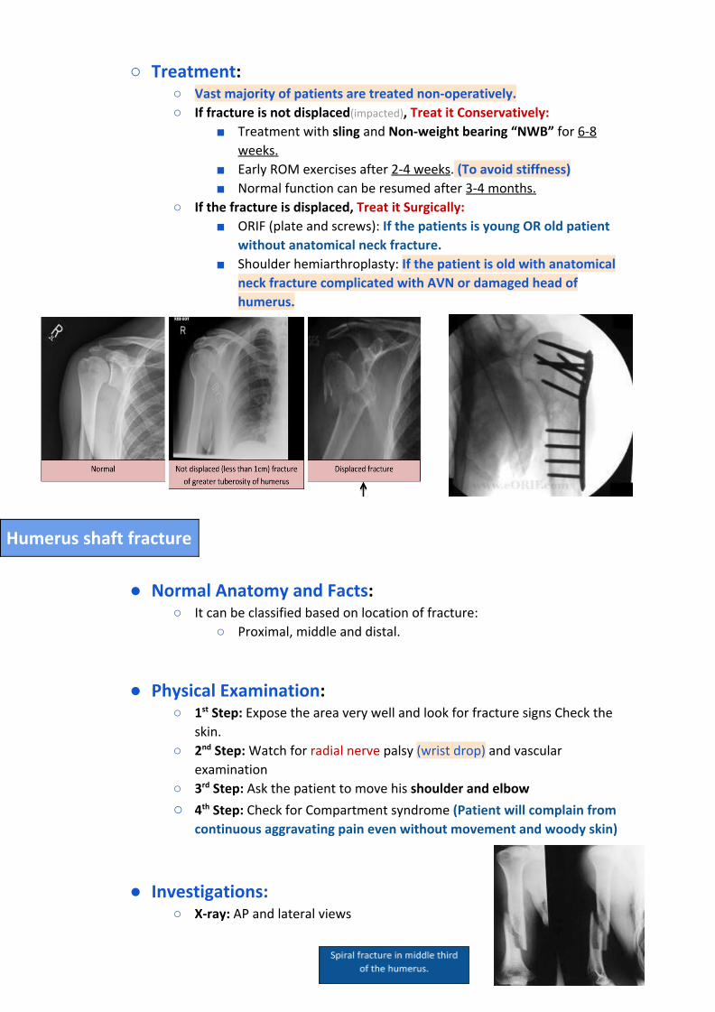
● Normal Anatomy and Facts:
○ It has four anatomic parts:
a. Head
b. Greater tuberosity
c. Lesser tuberosity
d. Shaft.
○ Fractures occur in Surgical neck are more common and have better outcomes,
while fractures in anatomical neck are generally rare and carries bad outcomes
due to blood supply injury and its association with avascular necrosis (AVN).
○ In younger patients: violent trauma (high energy “car accident”).
○ In older patients: minor trauma.(Osteoporosis)
○ Most fractures are minimally displaced.
● Physical examination:
○ 1st Step: Expose the shoulder very well and look for fracture signs
(ecchymosis, swelling, deformity...) and Check the skin.
○ 2nd Step: Check for Axillary nerve (deltoid atrophy and upper lateral side of
arm).
○ 3rd Step: Ask the patient to move his head and elbow
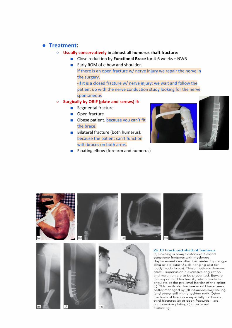
● Investigations: ○ X-ray: AP, lateral and a special view ‘axillary view’
(it shows the glenohumeral joint and tells whether it’s dislocated or not).
○ Fracture is defined by fragment displaced. ○ Displacement: more than 1 cm.
○ CT scan: for displaced fractures if X-ray isn’t clear.
○ Treatment: ○ Vast majority of patients are treated non-operatively.
○ If fracture is not displaced(impacted), Treat it Conservatively: ■ Treatment with sling and Non-weight bearing “NWB” for 6-8
weeks.
■ Early ROM exercises after 2-4 weeks. (To avoid stiffness)
■ Normal function can be resumed after 3-4 months.
○ If the fracture is displaced, Treat it Surgically:
■ ORIF (plate and screws): If the patients is young OR old patient
without anatomical neck fracture.
■ Shoulder hemiarthroplasty: If the patient is old with anatomical
neck fracture complicated with AVN or damaged head of
humerus.
Humerus shaft fracture
● Normal Anatomy and Facts:
○ It can be classified based on location of fracture:
○ Proximal, middle and distal.
● Physical Examination: ○ 1st Step: Expose the area very well and look for fracture signs Check the
skin.
○ 2nd Step: Watch for radial nerve palsy (wrist drop) and vascular
examination
○ 3rd Step: Ask the patient to move his shoulder and elbow
○ 4th Step: Check for Compartment syndrome (Patient will complain from
continuous aggravating pain even without movement and woody skin)
● Investigations: ○ X-ray: AP and lateral views
● Treatment:
○ Usually conservatively in almost all humerus shaft fracture:
■ Close reduction by Functional Brace for 4-6 weeks + NWB
■ Early ROM of elbow and shoulder.
if there is an open fracture w/ nerve injury we repair the nerve in
the surgery.
-if it is a closed fracture w/ nerve injury: we wait and follow the
patient up with the nerve conduction study looking for the nerve
spontaneous
○ Surgically by ORIF (plate and screws) if:
■ Segmental fracture
■ Open fracture
■ Obese patient. because you can’t fit
the brace.
■ Bilateral fracture (both humerus).
because the patient can’t function
with braces on both arms.
■ Floating elbow (forearm and humerus)
Both bone forearm fractures
● Normal Anatomy and Facts:
○ Forearm is complex with two mobile parallel bones.
○ Radius and ulna articulate proximally and distally.
○ Fractures are often from fall or direct blow.
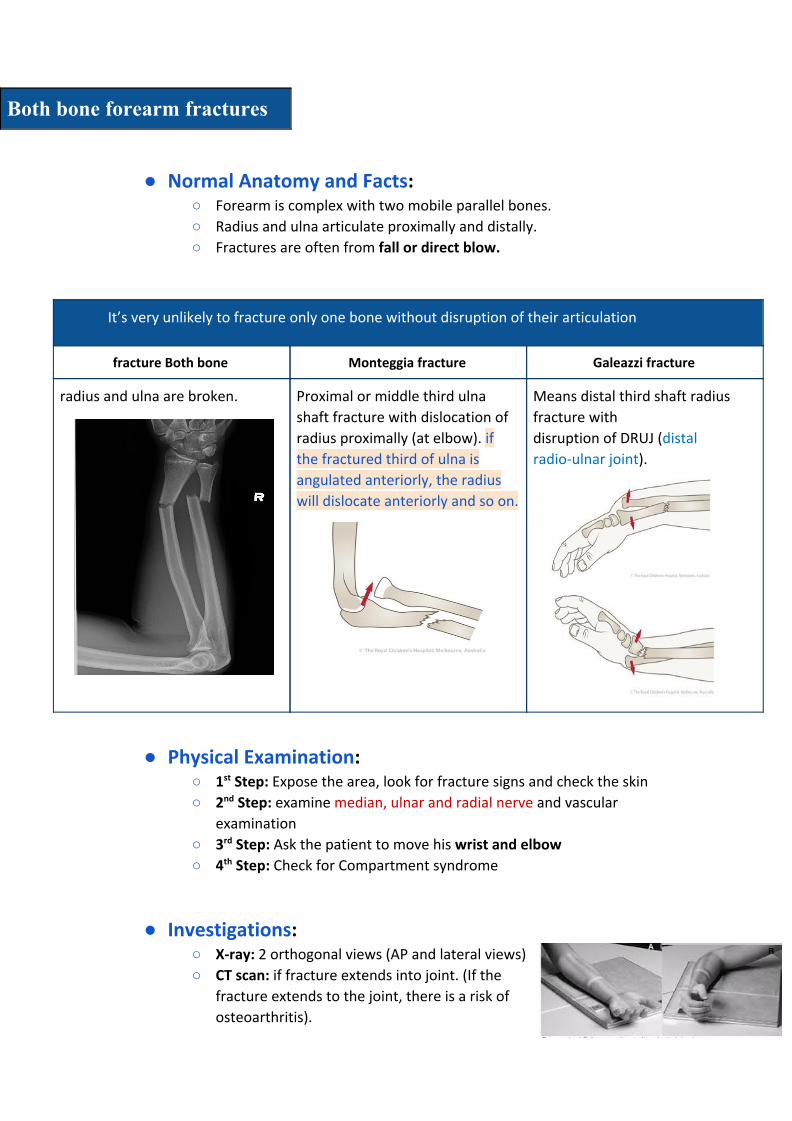
It’s very unlikely to fracture only one bone without disruption of their articulation
fracture Both bone Monteggia fracture Galeazzi fracture
radius and ulna are broken.
Proximal or middle third ulna
shaft fracture with dislocation of
radius proximally (at elbow). if the fractured third of ulna is
angulated anteriorly, the radius
will dislocate anteriorly and so on.
Means distal third shaft radius
fracture with
disruption of DRUJ (distal
radio-ulnar joint).
● Physical Examination: ○ 1st Step: Expose the area, look for fracture signs and check the skin
○ 2nd Step: examine median, ulnar and radial nerve and vascular
examination
○ 3rd Step: Ask the patient to move his wrist and elbow
○ 4th Step: Check for Compartment syndrome
● Investigations: ○ X-ray: 2 orthogonal views (AP and lateral views)
○ CT scan: if fracture extends into joint. (If the
fracture extends to the joint, there is a risk of
osteoarthritis).
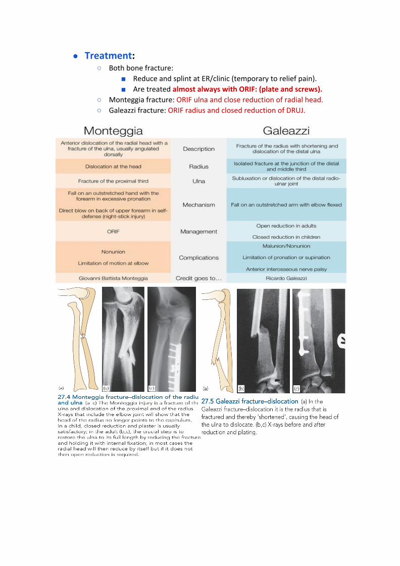
● Treatment: ○ Both bone fracture:
■ Reduce and splint at ER/clinic (temporary to relief pain).
■ Are treated almost always with ORIF: (plate and screws). ○ Monteggia fracture: ORIF ulna and close reduction of radial head.
○ Galeazzi fracture: ORIF radius and closed reduction of DRUJ.
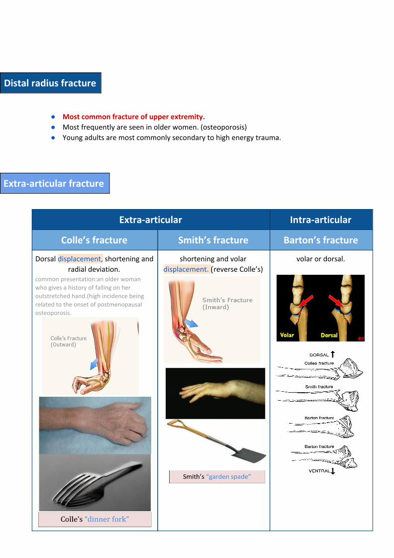
Distal radius fracture
● Most common fracture of upper extremity.
● Most frequently are seen in older women. (osteoporosis)
● Young adults are most commonly secondary to high energy trauma.
radial deviation. common presentation:an older woman who gives a history of falling on her outstretched hand.(high incidence being related to the onset of postmenopausal osteoporosis.
shortening and volar
displacement. (reverse Colle’s)
volar or dorsal.
● Investigations: ○ AP and lateral X-ray views
○ CT scan because it extends to joint “Barton’s”
● Treatment: ○ For Extra-articular fractures:
■ If stable, do close reduction under conscious sedation and cast
application (below elbow cast). If reduction is successful, send
patient home. Then wait for 1 week if the swelling decreased and
not painful. Continue Immobilization for 6-8 weeks. If not make
split to decrease the pressure and wait for 15 minutes, if pain
continues remove it completely.
■ Why Below elbow cast? In adults below elbow cast used to prevent
stiffness but we can use above elbow cast in children
■ ROM exercises after cast removal.
■ If not stable, treat it surgically
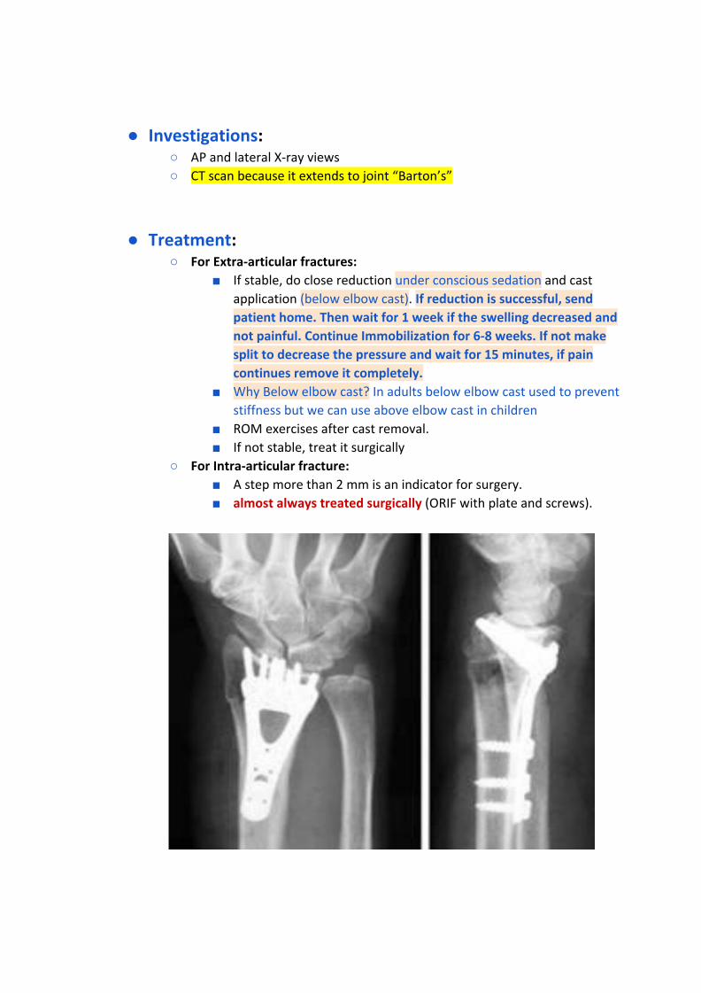
○ For Intra-articular fracture:
■ A step more than 2 mm is an indicator for surgery.
■ almost always treated surgically (ORIF with plate and screws).
Lower extremity fractures
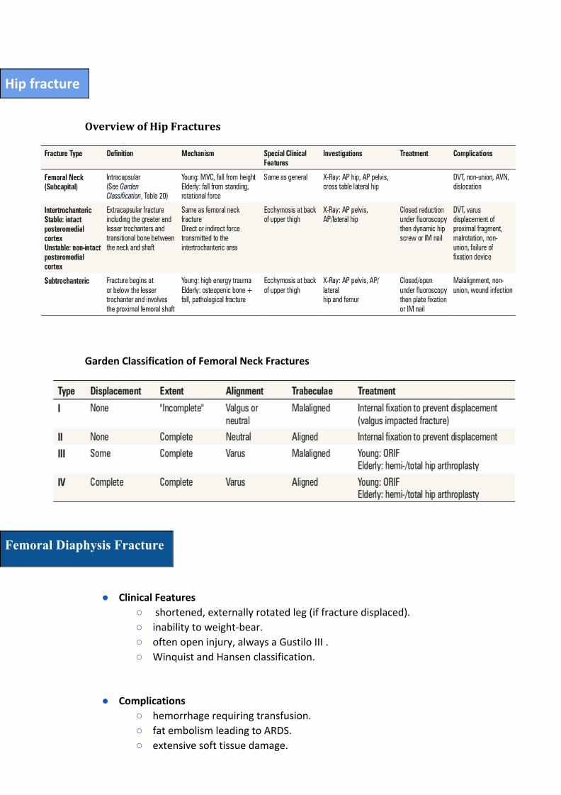
Hip fracture
● Normal Anatomy and Facts:
○ It is the most common fracture in lower limb.
○ It is associated with osteoporosis.
○ Most common mechanism is a fall from standing height (mechanical). ○ Other causes of fall (stroke, MI, hypoglycemic attack) should be rolled out
during clinical evaluation.
○ Common associated injuries (fragility fractures):
■ Distal radius fracture
■ Proximal humerus fracture
■ Subdural hematoma.
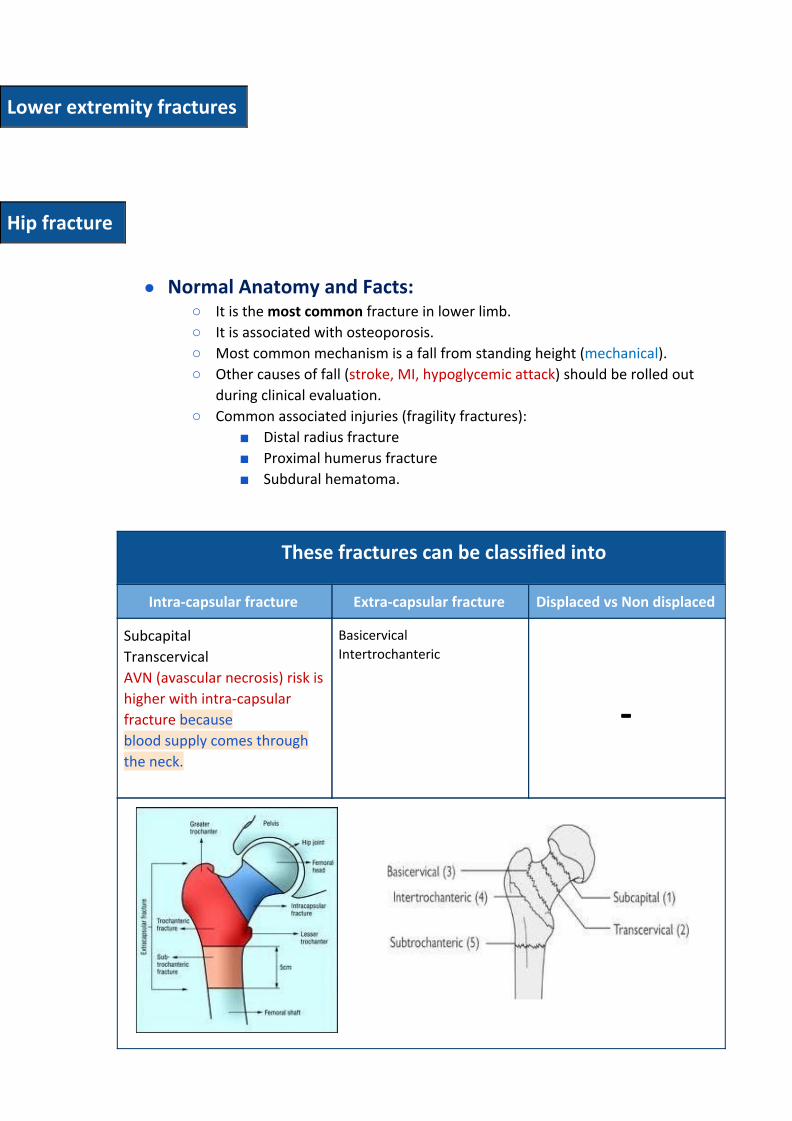
These fractures can be classified into
Intra-capsular fracture Extra-capsular fracture Displaced vs Non displaced
Subcapital
Transcervical
AVN (avascular necrosis) risk is
higher with intra-capsular
fracture because
blood supply comes through
the neck.
Basicervical Intertrochanteric
-
● Physical Examination: ○ Full detailed history of mechanism of injury.
○ R/O syncope, chest pain, weakness etc.
○ A detailed systematic review.
○ Deformity: Abduction, External rotation and shortening.
○ Assess distal N/V status.
○ Avoid ROM if fracture is expected.
● Investigations: ○ X-ray: AP and lateral Hip – AP Pelvis – AP Femur
○ MRI: is sensitive for occult fracture (rarely done only if you can’t decide
whether a fracture is present or not).
● Treatment: (IMPORTANT)
○ No close reduction is needed.
○ No traction is needed.
○ Patient needs surgery ideally within 48 hrs.
○ The goal is to ambulate patient as soon as possible.
○ Be sure that DVT prophylaxis is started. ○ Be sure that patient will be evaluated for osteoporosis after discharge.
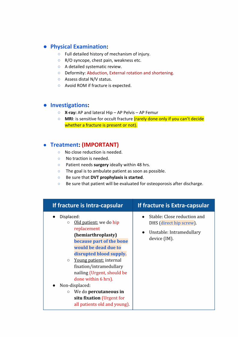
If fracture is Intra-capsular If fracture is Extra-capsular
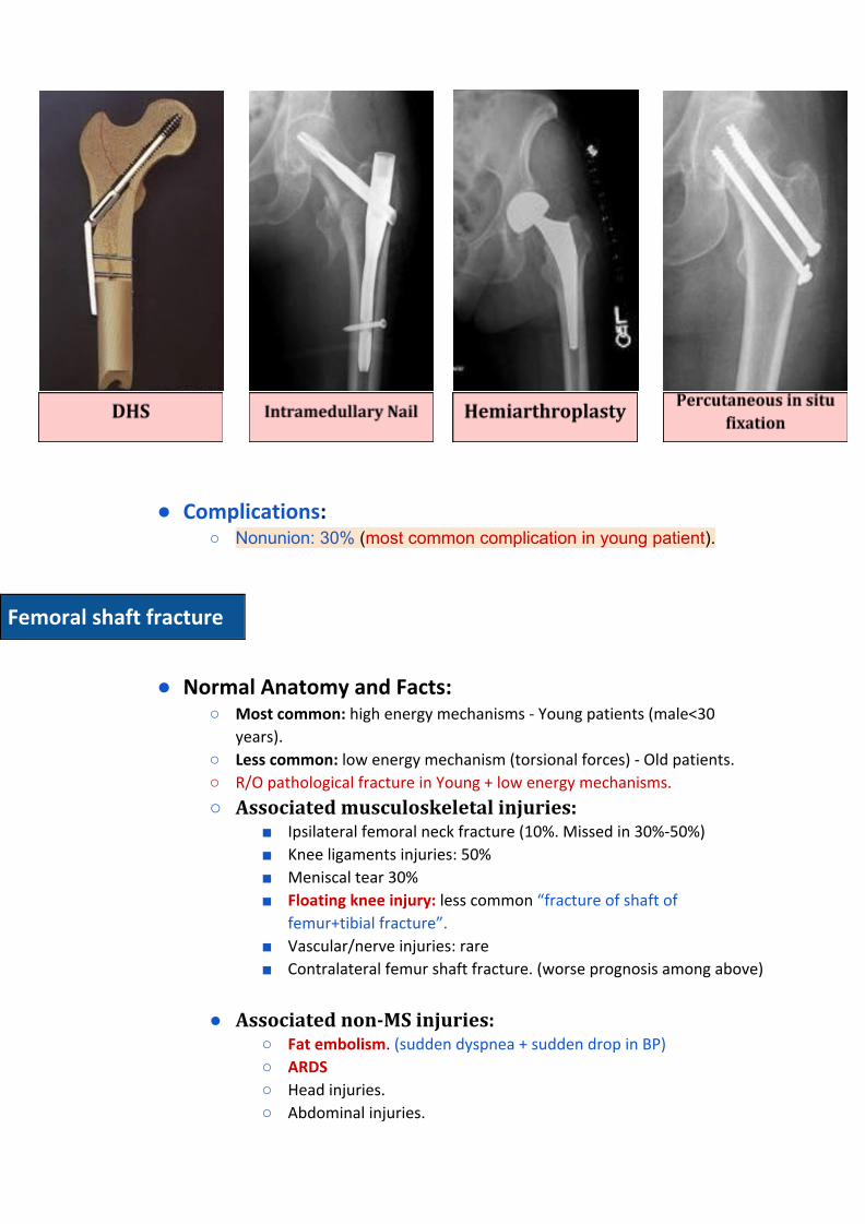
● Displaced: ○ Old patient: we do hip
replacement (hemiarthroplasty) because part of the bone would be dead due to disrupted blood supply.
○ Young patient: internal fixation/intramedullary nailing (Urgent, should be done within 6 hrs).
● Non-displaced: ○ We do percutaneous in
situ fixation (Urgent for all patients old and young).
● Stable: Close reduction and DHS (direct hip screw).
● Unstable: Intramedullary device (IM).
● Complications: ○ Nonunion: 30% (most common complication in young patient).
Femoral shaft fracture
● Normal Anatomy and Facts:
○ Most common: high energy mechanisms - Young patients (male<30
years).
○ Less common: low energy mechanism (torsional forces) - Old patients.
○ R/O pathological fracture in Young + low energy mechanisms.