Abstract: Sheath blight of rice is a destructive disease that could be calamitous to rice cultivation. Thesignificant objective of this study is to contemplate the proteomic analysis of the high virulent andless virulent isolate of Rhizoctonia solani using a quantitative LC-MS/MS-based proteomic approach toidentify the differentially expressed proteins promoting higher virulence. Across several rice-growingregions in Odisha, Eastern India, 58 Rhizoctonia isolates were obtained. All the isolates varied in theirpathogenicity. The isolate RS15 was found to be the most virulent and RS22 was identified as theleast virulent. The PCR amplification confirmed that the RS15 and RS22 belonged to the Rhizoctoniasubgroup of AG1-IA with a specific primer. The proteomic information generated has been depositedin the PRIDE database with PXD023430. The virulent isolate consisted of 48 differentially abundantproteins, out of which 27 proteins had higher abundance, while 21 proteins had lower abundance.The analyzed proteins acquired functionality in fungal development, sporulation, morphology,pathogenicity, detoxification, antifungal activity, essential metabolism and transcriptional activities,protein biosynthesis, glycolysis, phosphorylation and catalytic activities in fungi. A QuantitativeReal-Time PCR (qRT-PCR) was used to validate changes in differentially expressed proteins at themRNA level for selected genes. The abundances of proteins and transcripts were positively correlated.This study provides the role of the proteome in the pathogenicity of R. solani AG1-IA in rice andunderpins the mechanism behind the pathogen’s virulence in causing sheath blight disease.
Rhizoctonia solani Kuhn (Teleomorph: Thanatephorus cucumeris (Frank) Donk) is a soil-borne necrotrophic fungal pathogen that affects many food crops that are important tohumankind. Fourteen anastomosis groups (AG1–AG13 and AGBI) have been assignedbased on hyphal fusion [1]. The rice sheath blight causal organism, R. solani subgroupof AG1-IA, leads to severe economic losses to rice growers worldwide. Under favorableconditions, the pathogen could cause significant yield losses, affecting as much as 50% ofthe entire yield [2]. The severity of the disease depends on the variety, plant age, climaticfactors and excessive use of nitrogenous fertilizers. R. solani AG1-IA produces necrotic, darkto reddish-brown color, oval or elliptical-shaped lesions on the leaf sheath, blade and culmof rice [3,4]. Unavailability of resistant donors, pathogen variability, prolonged existenceas sclerotia in soil and lack of promising crop protection strategies make it challenging tomanage sheath blight disease [5].
J. Fungi 2022, 8, 370. https://doi.org/10.3390/jof8040370 https://www.mdpi.com/journal/jof
The Rhizoctonia population is diverse, considering its discrete cultural, morphological,pathological and physiological characteristics [6]. R. solani AG1-IA isolates are geneti-cally highly variable. Variability is a vital factor that degenerates the static mechanismof host resistance. Different molecular approaches, such as DNA-based sequence homol-ogy restriction analysis of ribosomal DNA (RAPD), have confirmed that R. solani isolatesare genetically more diverse [7,8]. Principal factors affecting the virulence of R. solaniinclude cell-wall-degrading enzymes and necrosis-inducing factors. Fungal-secreted pro-teins, known as effectors, are critical for host cell infection, as they impede their immunesystem and are consequently responsible for the pathogenicity of the necrotrophs [9].Whole-genome sequencing of R. solani AG1-IA manifests three genes that encode effectorproteins, including peptidase inhibitor I9 domain, cytochrome C oxidase assembly proteinCtaG/cox11 domain, and glycosyltransferase GT family 2 domain; they are involved innecrosis-inducing activities in rice and maize crop [10]. Nagarajkumar et al. [11] reportedthe production of higher amounts of oxalic acid by a virulent isolate of R. solani AG1-IAthan the less virulent isolate. A significant amount of oxalate increases the receptivityof plant tissues to pathogen infection. Recently, the polygalacturonase (AG1IA_04727)gene was substantial in R. solani pathogenesis [12]. Hence, the knowledge of the virulencespectrum of Rhizoctonia isolates and their discrepancy is crucial for effective disease man-agement strategies, including the development of sheath blight-resistant cultivars throughavailable breeding programs [13] or genetic engineering or fungicides as well as biologicalcontrol [14–16].
Proteomics was proven a significant tool to interpret the molecular mechanism be-hind biological systems. Proteomics is the study of the whole set of proteins expressedin each given condition in a particular cell. Unlike the static genome, the proteome ishighly dynamic. The last two decades provided considerable knowledge on functionalinformation of abiotic and biotic stress responses [17]. Proteomics allows us to accuratelypredict the expression profile of the final gene product, which is generally not offered bytranscriptomics or genomics. Gel-based and gel-free proteomics approaches were usedto determine differentially expressed proteins in plant and fungal species [18–21]. Theircomparative proteome analysis could comprehend the pathogenicity, metabolism, infectionprocess, stress response, broad host range, signal transduction and candidate virulenceprotein identification of fungal species and related strains [22]. Using a two-dimensionalpolyacrylamide gel electrophoresis analysis (2-DE), Manikandan et al. [20] reported thatthe F. oxysporum f.sp. lycopersici virulent isolate (FOL-8) had 14 differentially expressedproteins implicated in pathogenicity, infection and disease development in tomato roottissue. The analysis of 1-DE and 2-DE gel electrophoresis demonstrated about 75 proteinsexpressed differentially during the sclerotial maturation of R. solani AG-1. The identi-fied proteins were involved in the essential fungal metabolism and pathogenicity of R.solani [23]. Anderson et al. [24] applied mass spectrometry-based proteomics to identifythe pathogenesis-related protein in R. solani AG8. R. solani AG 4 isolate Rs23A revealedproteins with a possible role in fungal virulence, pathogenicity and cellular processes, aswas demonstrated by proteomic profiles of its extracellular and mycelial proteins [25].Comparative secretome analysis demonstrated that I9 domain-containing proteins wereabundant in diverse R. solani isolates that can cause plant cell death [26]. Proteomic re-sponses to R. solani were investigated using an eight-plexiTRAQ technique (isobaric tagsfor relative and absolute quantitation) in resistant (Teqing) and susceptible (Lemont) ricecultivars. In total, 755 proteins were differentially expressed, with the majority of theminvolved in plant metabolism and defense [27].
However, knowledge about differentially expressed proteins promoting virulencebetween different R. solani AG1-IA isolates is limited. Although the molecular basis of R.solani-Rice interaction may be well presumed, the mechanism that underpins differences inthe virulence factors between various R. solani AG1-IA isolates was not conclusive. Proteinprofiling of virulent and less virulent isolates provides an opportunity to identify thevirulence factors. Label-free proteomics has certain advantages, such as reliability, accuracy,
J. Fungi 2022, 8, 370 3 of 18
fewer experimental errors and a convenient approach in proteomics studies [28,29]. Withthis background information, the present study is undertaken to identify the differen-tially expressed proteins among R. solani isolates using a quantitative LC-MS/MS-basedproteomic approach.
2. Materials and Methods2.1. Isolation of Sheath Blight Pathogen
Infected rice plant sheaths and leaves exhibiting typical symptoms of sheath blightdisease were gathered across various rice-growing areas of Odisha (40 locations from sevendistricts), Eastern India, during 2019–2020 and brought to the laboratory to isolate thecausal organism. From each location 3–4 rice fields were selected randomly when thecrop was at tillering to maturity stage. The specimens were gently rinsed in tap water,cut into small pieces (approximately 1 cm) and then sterilized for 1 min using 1% sodiumhypochlorite solution, followed immediately by three sterile distilled water washes andblotted out between sterilized filter papers. The dried tissue bits were placed on a wateragar medium and incubated at 27 ± 1 ◦C for 1–2 days. The emerged hyphal tips wereexamined under a microscope and then transferred to a Petri dish containing a medium ofpotato dextrose agar (PDA). The pure cultures in PDA slants were later stored at 4 ◦C.
2.2. Pathogenicity and Disease Assessment
At the ICAR-National Rice Research Institute, Cuttack, India (85◦55′48” E longitudesand 20◦26′35” N latitude), the experiment was implemented. The sheath blight susceptiblecultivar, Tapaswini, was used for the experiment. Seeds were sterilized with a 2% sodiumhypochlorite solution and washed with sterile distilled water before sowing. Then, seedswere sown in a pot (45× 60 cm) with autoclaved pot mixture. The 25-day-old rice seedlingswere transplanted at two seedlings per hill with three hills per pot (45 × 60 cm). The 58 R.solani isolates were grown on PDA medium with seven-day-old mycelial plugs embeddedbeneath the leaf sheath of 60-day-old plants. The inoculated sheaths were covered withabsorbent cotton to maintain humidity. The plants without pathogen inoculation weremaintained as healthy controls. The experiment employed a completely randomized blockdesign. Every treatment had three replications with three pots in each and the experimentwas repeated twice. Disease progress was documented on the 7th, 14th, 21st and 28thday after pathogen inoculation. The relative lesion height (RLH = lesion height/Plantheight × 100) was recorded as described by Sharma et al. [30]. The disease severity wascalculated based on the RLH percentage. The data of RLH was converted into a diseaseindex based on a disease score with a 0 to 9 rating scale [31], where 0 represented noinfection; 1, lesion was limited to 20%; 3, 21–30%; 5, 31–45%; 7, 46–65%; 9, more than66–100% of plant height [32]. Based on disease reaction, isolates were categorized into fourgroups: highly virulent (8–9), moderately virulent (4–7.9), less virulent (1–3.9) and avirulent(0). The virulence index was calculated by averaging the disease score. The percentagedisease index (PDI) was calculated following Wheeler [33]. PDI = Sum of all rating/(Totalno. of observations ×Maximum rating scale) × 100.
2.3. Virulence Test on Other Rice Genotypes
The selected highly virulent (RS15) and less virulent (RS22) isolates were inoculated onother genotypes viz., Pusa Basmati-1, Vanaprabha, Hazaridhan, Swarna, Lunishree, Savitri,Sadabahar and TN1. The preparation of inoculums, method of artificial inoculation anddisease assessments were similar to those in Section 2.2. The experiment was conductedwith three replications in a completely randomized manner. Each replication had five potswith three hills (two seedlings/hill) in each pot. The experiment was repeated twice.
2.4. Molecular Characterization
The highly virulent and less virulent isolate of R. solani was characterized at themolecular level using ITS and AG1-1A primers. The fungal culture was grown on potato
J. Fungi 2022, 8, 370 4 of 18
dextrose broth (PDB) for three days at 27 ± 1 ◦C. The fungal mycelium was harvestedand finely ground with liquid nitrogen. The fungal genomic DNA was extracted as perthe procedure by the manufacturer (Qiagen fungal DNA extraction kit). A thermal cycler(Eppendorf Master Cycler Gradient, (St. Louis, MO, USA) was used to amplify the ITS byapplying the primer pair ITS1:CCTGTGCACCTGTGAGACAC and ITS4: TGTCCAAGT-CAATGGACTAT with the following PCR conditions: 94 ◦C for 5 min; 35 cycles of 94 ◦C for1 min; 56 ◦C for 1 min and 72 ◦C extension for 1.5 min; and a final extension at 72 ◦C for10 min [34]. Furthermore, the AG1-1A specific primers 5′-CTCAAACAGGCATGCTC-3′
and 5′-CAGCAATAGTTGGTGGA-3′ were used to identify R. solani as the subgroup ofAG1-1A. A total of 25 µL PCR reaction mixture contained 12.5 µL Mix (Bangalore Genei,India), 0.5 µL of forward primer, 0.5 µL of reverse primer, 0.5 µL Taq enzyme, 1 µL offungal DNA and 10 µL Nanopure water. The PCR reaction was executed with the followingprogram: 94 ◦C for 4 min; 30 cycles of 94 ◦C for 1 min and 54 ◦C for 2 min; 72 ◦C for 3 min;and the final extension at 72 ◦C for 7 min [35]. The 25 µL of PCR products were analyzedon 1.5% agarose gel (Sigma-Aldrich, St. Louis, MO, USA), and a gel documentation sys-tem (Biorad, Berkley, CA, USA) was used to analyze the ethidium bromide-stained DNAbands. The PCR product was purified using a QIA quick gel extraction kit (Qiagen, Inc.,Chatsworth, CA, USA) as per the manufacturer’s instruction. The purified PCR productsof ITS and AG1-1A were sent for DNA sequencing at AgriGenome Labs Pvt Ltd., Kochi,India. The DNA sequences were compared with existing R. solani sequences available inthe GenBank database (www.ncbi.nlm.nih.gov), and submitted to GenBank applying theBankit sequence submission tool.
2.5. Protein Extraction
R. solani isolates, RS15 and RS22, were cultured on PDB for three days at 27 ± 1 ◦C.Sterile filter paper was used to collect the mycelial mat, washed thrice with sterile distilledwater and liquid nitrogen was used to obtain a fine powder. The TCA (sigma-76-05-1)-acetone (Merck-100014) method was employed to precipitate the proteins. The sampleswere brought up to 10% TCA and incubated at −20 ◦C overnight. The samples werecentrifuged for 10 min at 13,000 rpm to obtain an intact pellet and the supernatant wasseparated. Acetone wash (added 500 µL of acetone to the pellet and incubated for 10 minat −20 ◦C and again centrifuged for 10 min at 13,000 rpm) of the pellet was performedthrice. Finally, the air-dried pellet was dissolved in a 50 mM ammonium bicarbonate(Fluka analytical FL40867) buffer. The samples were solubilized in 100 µL of 50 mMammonium bicarbonate (NH4HCO3) with 1% SDS, mixed well by vortexing followed by20 min sonication and centrifuged for 10 min at 10,000 rpm. The supernatant was quantifiedusing the Bicinchoninic Acid assay, in which proteins were reduced from Cu+2 to Cu+1 inan alkaline solution, resulting in the formation of the bicinchoninic acid-induced purplecolor. The protein samples were incubated along with the standard at 37 ◦C for 60 minand the spectrophotometer reading was noted at 562 nm. From all the samples, an equalamount of protein was loaded on a 12% 1D SDS-PAGE, silver-stained gels were observedfor bands and an Epson Expression 11000XL Scanner was used to scan them.
2.6. In Solution Protein Digestion
The extracted protein (100 µg) was digested, treated for 1 h at 95 ◦C with 100 mMdithiothreitol (DTT), and exposed for 45 min at room temperature in the dark to 250 mMiodoacetamide. With the addition of 4 µg of trypsin, digestion of the protein samples wasachieved at 37 ◦C overnight. The peptides were resuspended in 0.1% formic acid (50 µL)and incubated for 45 min at 37 ◦C. The supernatants were transferred into a separate tubeafter centrifugation at 10,000× g. The speed vacuum concentrator (Basic Concentraor,Eppendorf, Hamburg, Germany) was used to vacuum dry (45 ◦C for 6 h at 800 Pa in VAQmode) the resulting samples and was dissolved in 0.1% formic acid in water (20 µL) forLC-MS/MS.
An ACQUITY UPLC system (Waters, UK) was used to perform liquid chromatographyby injecting 10 µL of the protein samples. The ACQUITY UPLC BEH C18 column (150 mm× 2.1 mm × 1.7 µm) (Waters, UK) separated the samples. Three runs will be performedon each sample to achieve label-free quantification. A gradient elution program wasperformed for chromatographic separation with mobile phase A (0.1% formic acid in water)and mobile phase B (0.1% formic acid in acetonitrile). Mass spectrometric detection wasperformed with an electrospray ionization (ESI) source equipped with an SYNAPT G2QTOF (Waters, UK). Sample analysis was performed in a positive mode.
2.8. Peptide Identification and Data Analysis
MassLynx 4.1 WATERS was used to process raw data. The protein identification onPLGS (Protein Lynx Global Server) software 3.0.2, WATERS involved matching MSMSspectra of individual peptides to the database sequence. The sample runs were processedfollowing specific search parameters in the software: Peptide tolerance (ppm): 50; min no.of fragment matches for peptides: 2; fragment tolerance (ppm): 100; min no. of peptidematches for proteins: 2; min no. of fragment matches for proteins: 5; missed cleavages:1. The sample cysteine sites were modified during processing to carbamidomethylatedcysteine, and the methionine sites were considered as a variable modification to the mass,which was prone to oxidation. The Swissprot protein database for R. solani was used tosearch for proteins present in the sample, and a 5% false discovery rate was used. Theexpression score was normalized using PLGS software, and the mean abundance of aparticular protein in two samples was determined by ratio calculation. A t-test determinedthe differences between isolates RS15 and RS22. Protein abundance RS15 to RS22 withat least two-fold differences was considered for up-regulation and 0.5-fold changes fordown-regulation. The differentially abundant proteins were classified according to theirmolecular functions, biological processes and protein class using the Panther online tool(http://www.pantherdb.org/pathway/ (accessed on 10 January2022)).
2.9. Quantitative Real-Time PCR Analysis
R. solani isolates RS15 and RS22 were cultured on PDB for 3 days at 27 ± 1 ◦C, andthe mycelial mat was collected from three biological replicates and immediately groundwith liquid N2 for RNA extraction. Three technical replicates per biological replicateswere analyzed during qRT-PCR. Total RNA was extracted using the RNeasy Mini kit (QIA-GEN, Hilden, Germany). The isolated RNA quality was checked in 1.5% agarose gel andquantified by a NanoDrop® ND-1000 UV-Vis spectrophotometer (Thermo SCIENTIFIC,Walthem, MA, USA). The residual DNA was removed with an RNase-free DNase I enzyme(QIAGEN, Hilden, Germany). According to the manufacturer’s instructions, the comple-mentary DNAs were synthesized from 1 µg of total RNA using the QuantiTect® ReverseTranscription Kit (QIAGEN) in a 20 µL reaction mixture. The qRT-PCR specific primerswere designed using the Primer QuestTMtool (Integrated DNA Technologies, Coralville,IA, USA) and all primer information is listed in Supplementary Table S1. The RT-PCRwas performed on a BIORAD CFX96 Real-Time system, with a total of 10 µL PCR reactionmixture containing 1.0 µL of diluted template cDNA, 5.0 µL of 2× buffer SYBR Green(QIAGEN, Germany), 0.5 µL of each forward and reverse primer and 3.0 µL of sterilizednanopure water. The RT-PCR amplification conditions were as follows: pre-incubationfor 95.0 ◦C for 15 min, and three-step amplification at 94.0 ◦C for 0.15 s, 60 ◦C for 0.30 sand 72 ◦C for 0.30 s and 39 cycles, followed by 95.0 ◦C for 0.05 s, 65 ◦C for 0.05 s and95 ◦C for 0.5 s. The Rhi-18S rRNA (Forward: ATGATAACTCGACGGATCGC; Reverse:CTTGGATGTGGTAGCCGT) was used as the internal control to normalize the expressionof each gene, and the specificity of amplicons was verified by a melting curve analysis usingthe peak values [36]. Additionally, specificity was confirmed by running a 2.5% agarose gel.Fluorescence was measured at the end of every 72 ◦C extension phase. The final thresholdcycles (Ct) values were the means of three values, including three biological replicates
and three technical replicates per biological replicates. The relative expression level ofeach gene was calculated using the 2-∆∆Ct method. The Ct values for the housekeepinggene was subtracted from the gene of interest to obtain a Ct value. The Ct value of themock-inoculated control sample was subtracted from the ∆Ct value to obtain the ∆∆Ctvalue = Ct Target-Ct Reference. Each fold change in expression level relative to that of thecontrol was expressed as 2-∆∆Ct and all experiments were repeated thrice.
2.10. Statistical Analysis
The IRRISTAT v.92-1 program (Biometric Unit, International Rice Research Institute,Los Baños, Laguna, Philippines) was utilized for the data analysis. Data were subject toan analysis of variance (ANOVA, New Providence, NJ, USA), and the data in percentageswere arcsine transformed before the analysis. The treatment means were compared by theDuncan’s multiple range test (DMRT) [37].
3. Results3.1. Isolation, Pathogenicity and Molecular Characterization
Overall, 58 isolates were identified from various rice-growing areas of Odisha, EasternIndia. Artificial inoculation on the rice cultivar Tapaswini demonstrated that typical symp-toms of sheath blight disease were produced in all isolates, which displayed differences invirulence, disease index and disease progress. RS15 recorded 100% PDI among the isolates,followed by RS34 (92.59%). The isolates RS16, RS17, RS22, RS26 and RS27 had the lowestPDI of 11.11%. Based on disease reaction, the 58 isolates were categorized into highlyvirulent (2), moderately virulent (31) and less virulent (25). The virulent index varied from1 to 9 (Table 1).
Table 1. Pathogenicity of R. solani isolates on Tapaswini.
Isolates PDI * Average VirulentIndex Disease Reaction
RS1 25.93 (30.61) k 2.33 Less virulentRS2 25.93 (30.61) k 2.33 Less virulentRS3 33.33 (35.26) j 3.00 Less virulentRS4 25.93 (30.61) k 2.33 Less virulentRS5 33.33 (35.26) j 3.00 Less virulentRS6 18.52 (25.49) l 1.67 Less virulentRS7 25.93 (30.61) k 2.33 Less virulentRS8 18.52 (25.49) l 1.67 Less virulentRS9 77.78 (61.93) d 7.00 Moderately virulent
RS10 77.78 (61.93) d 7.00 Moderately virulentRS11 85.19 (67.51) c 7.67 Moderately virulentRS12 48.15 (43.94) h 4.33 Moderately virulentRS13 33.33 (35.26) j 3.00 Less virulentRS14 77.78 (61.93) d 7.00 Moderately virulentRS15 100.00 (88.19) a 9.00 Highly virulentRS16 11.11 (19.47) m 1.00 Less virulentRS17 11.11 (19.47) m 1.00 Less virulentRS18 55.56 (48.20) g 5.00 Moderately virulentRS19 33.33 (35.26) j 3.00 Less virulentRS20 18.52 (25.49) l 1.67 Less virulentRS21 18.52 (25.49) l 1.67 Less virulentRS22 11.11 (19.47) m 1.00 Less virulentRS23 77.78 (61.93) d 7.00 Moderately virulentRS24 40.74 (39.66) i 3.67 Less virulentRS25 40.74 (39.66) i 3.67 Less virulentRS26 11.11 (19.47) m 1.00 Less virulentRS27 11.11 (19.47) m 1.00 Less virulentRS28 77.78 (61.93) d 7.00 Moderately virulent
J. Fungi 2022, 8, 370 7 of 18
Table 1. Cont.
Isolates PDI * Average VirulentIndex Disease Reaction
RS29 25.93 (30.61) k 2.33 Less virulentRS30 25.93 (30.61) k 2.33 Less virulentRS31 55.56 (48.20) g 5.00 Moderately virulentRS32 48.15 (43.94) h 4.33 Moderately virulentRS33 18.52 (25.49) l 1.67 Less virulentRS34 92.59 (74.77) b 8.33 Highly virulentRS35 25.93 (30.61) k 2.33 Less virulentRS36 25.93 (30.61) k 2.33 Less virulentRS37 70.37 (57.05) e 6.33 Moderately virulentRS38 33.33 (35.26) j 3.00 Less virulentRS39 33.33 (35.26) j 3.00 Less virulentRS40 33.33 (35.26) j 3.00 Less virulentRS41 33.33 (35.26) j 3.00 Less virulentRS42 33.33 (35.26) j 3.00 Less virulentRS43 33.33 (35.26) j 3.00 Less virulentRS44 62.96 (52.52) f 5.67 Moderately virulentRS45 62.96 (52.52) f 5.67 Moderately virulentRS46 33.33 (35.26) j 3.00 Less virulentRS47 40.74 (39.66) i 3.67 Less virulentRS48 55.56 (48.20) g 5.00 Moderately virulentRS49 77.78 (61.93) d 7.00 Moderately virulentRS50 33.33 (35.26) j 3.00 Less virulentRS51 11.11 (19.47) m 1.00 Less virulentRS52 11.11 (19.47) m 1.00 Less virulentRS53 11.11 (19.47) m 1.00 Less virulentRS54 18.52 (25.49) l 1.67 Less virulentRS55 48.15 (43.94) h 4.33 Moderately virulentRS56 40.74 (39.66) i 3.67 Less virulentRS57 11.11 (19.47) m 1.00 Less virulentRS58 11.11 (19.47) m 1.00 Less virulent
* PDI recorded on 28th days after pathogen inoculation. Values are the mean of three replications. Values inthe parenthesis are arcsine-transformed values. Means in a column followed by same superscript letter are notsignificantly different at the 5% level by DMRT.
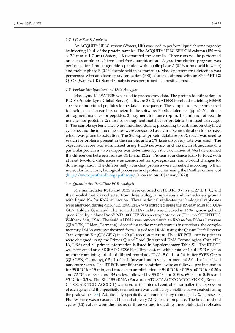
The RLH was recorded on the 7th, 14th, 21st and 28th days after pathogen inoculation.Among the isolates, RS15 demonstrated the maximum percentage of RLH, and RS22demonstrated the most negligible percentage of RLH at different time intervals (Figure 1).
Isolates RS15 and RS22 were characterized at the molecular level by sequencing therDNA internal transcribed spacer region using ITS and AG1-1A specific primers. TheDNA sequences were matched with the NCBI GenBank BLAST analysis (http://www.ncbi.nlm.nih.gov (accessed on 5 March 2021)). The resultant sequences were submitted toGenBank, and the accession numbers of MW762960, MW757241 for RS15 and MW757249and MW757239 for RS22 were obtained, respectively.
3.2. Determination of Virulence on Other Rice Genotypes
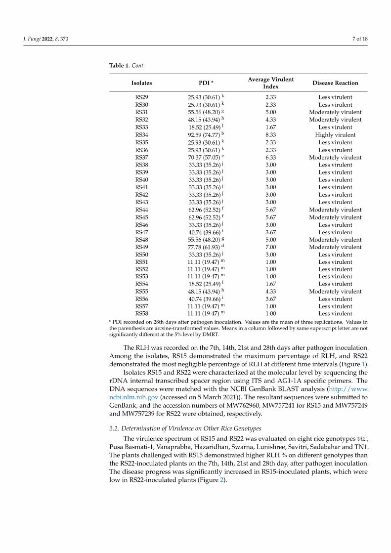
The virulence spectrum of RS15 and RS22 was evaluated on eight rice genotypes viz.,Pusa Basmati-1, Vanaprabha, Hazaridhan, Swarna, Lunishree, Savitri, Sadabahar and TN1.The plants challenged with RS15 demonstrated higher RLH % on different genotypes thanthe RS22-inoculated plants on the 7th, 14th, 21st and 28th day, after pathogen inoculation.The disease progress was significantly increased in RS15-inoculated plants, which werelow in RS22-inoculated plants (Figure 2).
Figure 1. Relative Lesion Length (RLH) % of different R. solani isolates on different time intervals.Vertical bars indicate the standard error of three replications. Analysis of variance was performedthrough DMRT with IRRISTAT.
Figure 2. Relative Lesion Length % of RS22 and RS15 on different rice genotypes. Vertical barsindicate the standard error of three replications. Analysis of variance was performed through DMRTwith IRRISTAT.
3.3. Proteomic Analysis
A comparative proteomic analysis was performed for virulent (RS15) and less virulent(RS22) isolates using the LC-MS/MS analysis to identify the proteins related to virulence.The results demonstrated that the virulent isolate comprised 48 disparately abundantproteins, out of which 27 proteins had higher abundance and 21 had lower abundancein the virulent isolate. The proteomic information generated has been archived in thePRIDE database and can be accessed with PXD023430 [38,39]. Among the differentiallyabundant proteins, JmjC domain-containing histone demethylation protein 1, sorting nexin-
J. Fungi 2022, 8, 370 9 of 18
4, rRNA biogenesis protein RRP36, Exportin-T, Heat shock 70, topoisomerase 1, cyanatehydratase, pyranose 2-oxidase, actin-like protein ARP6, phenylalanine ammonia-lyase andsqualene synthase were the most predominant with an increased abundance in the virulentisolate. Similarly, GMP synthase, elongation factor G, glycylpeptide N-tetradecanoyl trans-ferase, sulphate adenylyltransferase, 5′-3′ exoribonuclease 2, glyceraldehyde-3-phosphatedehydrogenase, protein phosphatase methylesterase and protein CFT1 were found to bedepleted (Table 2).
Table 2. List of differentially expressed proteins among virulent (RS15) and less virulent (RS22) isolates.
Accessions Description Fold Change ** Category
P0CN36 Protein EFR3 2.034 Up-regulatedQ4P3U5 Protein EFR3 2.034 Up-regulated* P10248 Phenylalanine ammonia-lyase 2.054 Up-regulatedQ4PB37 Pre-mRNA-splicing factor 2.181 Up-regulated
Q4P0P3 Mediator of RNA polymerase IItranscription subunit 14 2.270 Up-regulated
* Proteins selected for qPCR analysis. ** Fold change >2 is considered for up-regulated and <0.5 is down-regulated.
3.4. Pathway Analysis
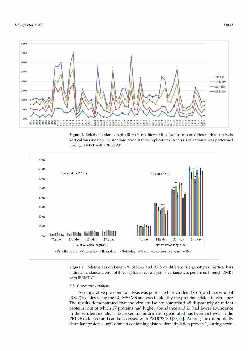
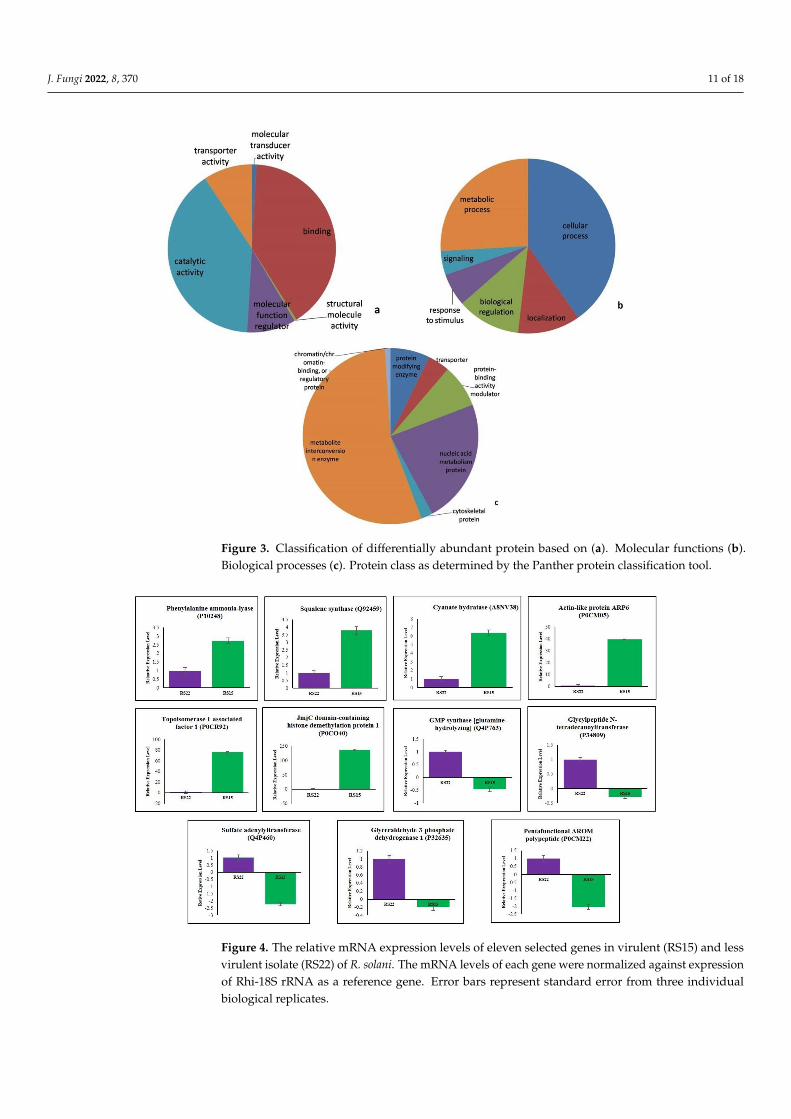
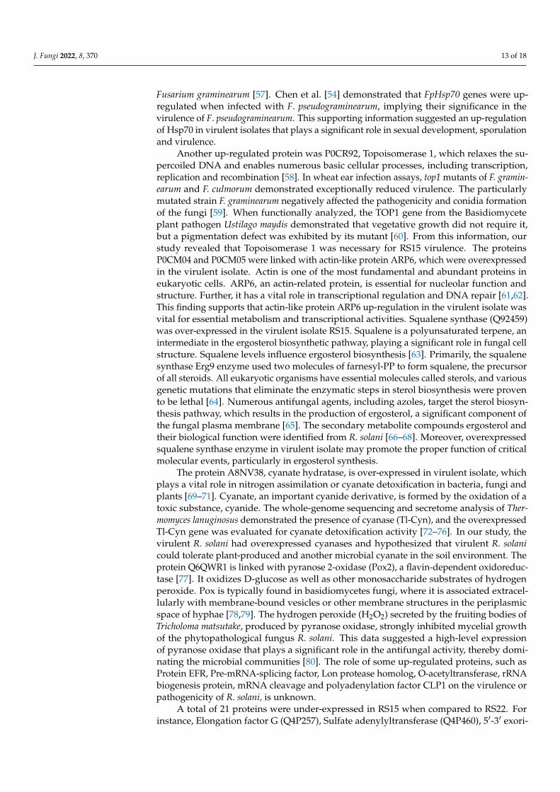
A pathway analysis using the Panther online tool revealed the molecular functions,biological processes and protein class in which differently abundant proteins were involved,as depicted in Figure 3.
Catalytic activity (GO: 0003824) and binding (GO: 0005488) were the most predom-inantly represented molecular functions. Similarly, the cellular process (GO:0009987),metabolic process (GO: 0008152), biological regulation (GO: 0065007) and localization (GO:0051179) were the primarily represented biological processes. The predominant proteinclasses represented by differentially abundant proteins were the metabolite inter-conversionenzyme (PC00262), nucleic acid metabolism protein (PC00171) and protein-binding activitymodulator (PC00095).
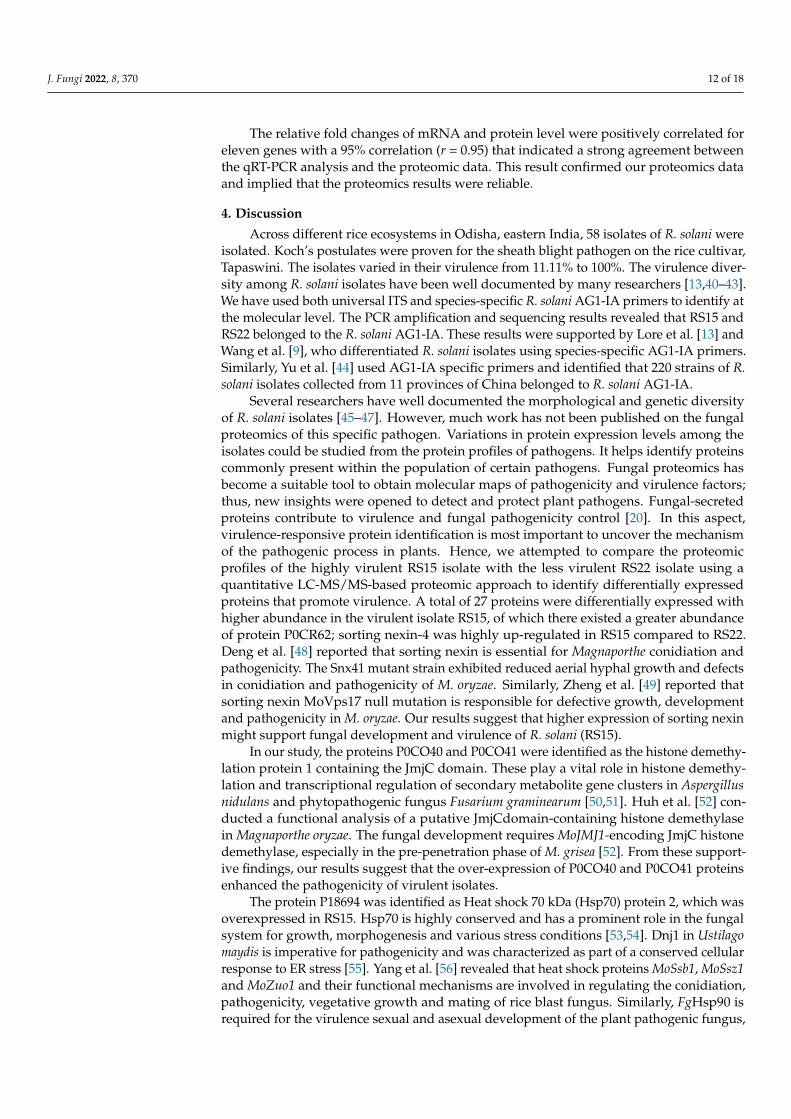
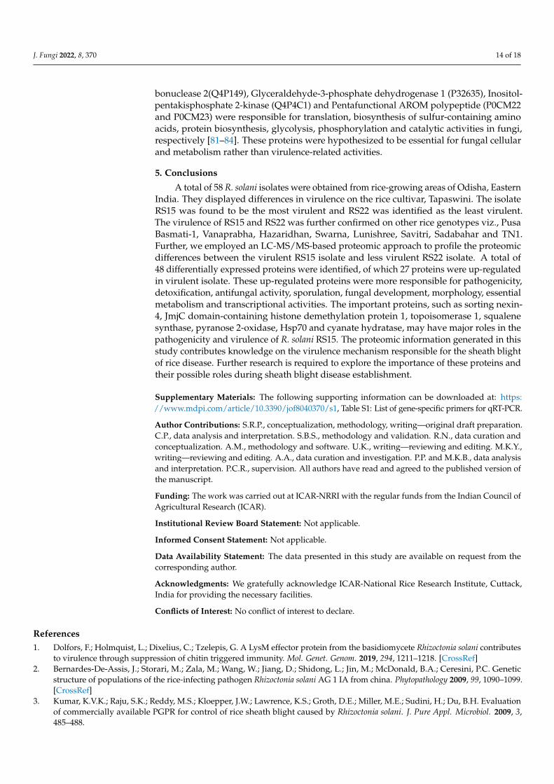
3.5. Validation of Differentially Expressed Proteins by qRT–PCR
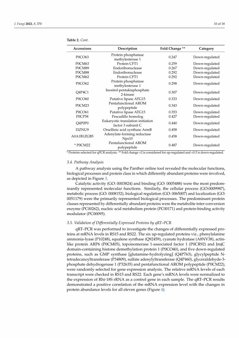
qRT–PCR was performed to investigate the changes of differentially expressed pro-teins at mRNA levels in RS15 and RS22. The six up-regulated proteins viz., phenylalanineammonia-lyase (P10248), squalene synthase (Q92459), cyanate hydratase (A8NV38), actin-like protein ARP6 (P0CM05), topoisomerase 1-associated factor 1 (P0CR92) and JmjCdomain-containing histone demethylation protein 1 (P0CO40), and five down-regulatedproteins, such as GMP synthase [glutamine-hydrolyzing] (Q4P763), glycylpeptide N-tetradecanoyltransferase (P34809), sulfate adenylyltransferase (Q4P460), glyceraldehyde-3-phosphate dehydrogenase 1 (P32635) and pentafunctional AROM polypeptide (P0CM22),were randomly selected for gene expression analysis. The relative mRNA levels of eachtranscript were checked in RS15 and RS22. Each gene’s mRNA levels were normalized tothe expression of Rhi-18S rRNA as a control gene in each sample. The qRT–PCR resultsdemonstrated a positive correlation of the mRNA expression level with the changes inprotein abundance levels for all eleven genes (Figure 4).
J. Fungi 2022, 8, 370 11 of 18
Figure 3. Classification of differentially abundant protein based on (a). Molecular functions (b).Biological processes (c). Protein class as determined by the Panther protein classification tool.
Figure 4. The relative mRNA expression levels of eleven selected genes in virulent (RS15) and lessvirulent isolate (RS22) of R. solani. The mRNA levels of each gene were normalized against expressionof Rhi-18S rRNA as a reference gene. Error bars represent standard error from three individualbiological replicates.
J. Fungi 2022, 8, 370 12 of 18
The relative fold changes of mRNA and protein level were positively correlated foreleven genes with a 95% correlation (r = 0.95) that indicated a strong agreement betweenthe qRT-PCR analysis and the proteomic data. This result confirmed our proteomics dataand implied that the proteomics results were reliable.
4. Discussion
Across different rice ecosystems in Odisha, eastern India, 58 isolates of R. solani wereisolated. Koch’s postulates were proven for the sheath blight pathogen on the rice cultivar,Tapaswini. The isolates varied in their virulence from 11.11% to 100%. The virulence diver-sity among R. solani isolates have been well documented by many researchers [13,40–43].We have used both universal ITS and species-specific R. solani AG1-IA primers to identify atthe molecular level. The PCR amplification and sequencing results revealed that RS15 andRS22 belonged to the R. solani AG1-IA. These results were supported by Lore et al. [13] andWang et al. [9], who differentiated R. solani isolates using species-specific AG1-IA primers.Similarly, Yu et al. [44] used AG1-IA specific primers and identified that 220 strains of R.solani isolates collected from 11 provinces of China belonged to R. solani AG1-IA.
Several researchers have well documented the morphological and genetic diversityof R. solani isolates [45–47]. However, much work has not been published on the fungalproteomics of this specific pathogen. Variations in protein expression levels among theisolates could be studied from the protein profiles of pathogens. It helps identify proteinscommonly present within the population of certain pathogens. Fungal proteomics hasbecome a suitable tool to obtain molecular maps of pathogenicity and virulence factors;thus, new insights were opened to detect and protect plant pathogens. Fungal-secretedproteins contribute to virulence and fungal pathogenicity control [20]. In this aspect,virulence-responsive protein identification is most important to uncover the mechanismof the pathogenic process in plants. Hence, we attempted to compare the proteomicprofiles of the highly virulent RS15 isolate with the less virulent RS22 isolate using aquantitative LC-MS/MS-based proteomic approach to identify differentially expressedproteins that promote virulence. A total of 27 proteins were differentially expressed withhigher abundance in the virulent isolate RS15, of which there existed a greater abundanceof protein P0CR62; sorting nexin-4 was highly up-regulated in RS15 compared to RS22.Deng et al. [48] reported that sorting nexin is essential for Magnaporthe conidiation andpathogenicity. The Snx41 mutant strain exhibited reduced aerial hyphal growth and defectsin conidiation and pathogenicity of M. oryzae. Similarly, Zheng et al. [49] reported thatsorting nexin MoVps17 null mutation is responsible for defective growth, developmentand pathogenicity in M. oryzae. Our results suggest that higher expression of sorting nexinmight support fungal development and virulence of R. solani (RS15).
In our study, the proteins P0CO40 and P0CO41 were identified as the histone demethy-lation protein 1 containing the JmjC domain. These play a vital role in histone demethy-lation and transcriptional regulation of secondary metabolite gene clusters in Aspergillusnidulans and phytopathogenic fungus Fusarium graminearum [50,51]. Huh et al. [52] con-ducted a functional analysis of a putative JmjCdomain-containing histone demethylasein Magnaporthe oryzae. The fungal development requires MoJMJ1-encoding JmjC histonedemethylase, especially in the pre-penetration phase of M. grisea [52]. From these support-ive findings, our results suggest that the over-expression of P0CO40 and P0CO41 proteinsenhanced the pathogenicity of virulent isolates.
The protein P18694 was identified as Heat shock 70 kDa (Hsp70) protein 2, which wasoverexpressed in RS15. Hsp70 is highly conserved and has a prominent role in the fungalsystem for growth, morphogenesis and various stress conditions [53,54]. Dnj1 in Ustilagomaydis is imperative for pathogenicity and was characterized as part of a conserved cellularresponse to ER stress [55]. Yang et al. [56] revealed that heat shock proteins MoSsb1, MoSsz1and MoZuo1 and their functional mechanisms are involved in regulating the conidiation,pathogenicity, vegetative growth and mating of rice blast fungus. Similarly, FgHsp90 isrequired for the virulence sexual and asexual development of the plant pathogenic fungus,
J. Fungi 2022, 8, 370 13 of 18
Fusarium graminearum [57]. Chen et al. [54] demonstrated that FpHsp70 genes were up-regulated when infected with F. pseudograminearum, implying their significance in thevirulence of F. pseudograminearum. This supporting information suggested an up-regulationof Hsp70 in virulent isolates that plays a significant role in sexual development, sporulationand virulence.
Another up-regulated protein was P0CR92, Topoisomerase 1, which relaxes the su-percoiled DNA and enables numerous basic cellular processes, including transcription,replication and recombination [58]. In wheat ear infection assays, top1 mutants of F. gramin-earum and F. culmorum demonstrated exceptionally reduced virulence. The particularlymutated strain F. graminearum negatively affected the pathogenicity and conidia formationof the fungi [59]. When functionally analyzed, the TOP1 gene from the Basidiomyceteplant pathogen Ustilago maydis demonstrated that vegetative growth did not require it,but a pigmentation defect was exhibited by its mutant [60]. From this information, ourstudy revealed that Topoisomerase 1 was necessary for RS15 virulence. The proteinsP0CM04 and P0CM05 were linked with actin-like protein ARP6, which were overexpressedin the virulent isolate. Actin is one of the most fundamental and abundant proteins ineukaryotic cells. ARP6, an actin-related protein, is essential for nucleolar function andstructure. Further, it has a vital role in transcriptional regulation and DNA repair [61,62].This finding supports that actin-like protein ARP6 up-regulation in the virulent isolate wasvital for essential metabolism and transcriptional activities. Squalene synthase (Q92459)was over-expressed in the virulent isolate RS15. Squalene is a polyunsaturated terpene, anintermediate in the ergosterol biosynthetic pathway, playing a significant role in fungal cellstructure. Squalene levels influence ergosterol biosynthesis [63]. Primarily, the squalenesynthase Erg9 enzyme used two molecules of farnesyl-PP to form squalene, the precursorof all steroids. All eukaryotic organisms have essential molecules called sterols, and variousgenetic mutations that eliminate the enzymatic steps in sterol biosynthesis were provento be lethal [64]. Numerous antifungal agents, including azoles, target the sterol biosyn-thesis pathway, which results in the production of ergosterol, a significant component ofthe fungal plasma membrane [65]. The secondary metabolite compounds ergosterol andtheir biological function were identified from R. solani [66–68]. Moreover, overexpressedsqualene synthase enzyme in virulent isolate may promote the proper function of criticalmolecular events, particularly in ergosterol synthesis.
The protein A8NV38, cyanate hydratase, is over-expressed in virulent isolate, whichplays a vital role in nitrogen assimilation or cyanate detoxification in bacteria, fungi andplants [69–71]. Cyanate, an important cyanide derivative, is formed by the oxidation of atoxic substance, cyanide. The whole-genome sequencing and secretome analysis of Ther-momyces lanuginosus demonstrated the presence of cyanase (Tl-Cyn), and the overexpressedTl-Cyn gene was evaluated for cyanate detoxification activity [72–76]. In our study, thevirulent R. solani had overexpressed cyanases and hypothesized that virulent R. solanicould tolerate plant-produced and another microbial cyanate in the soil environment. Theprotein Q6QWR1 is linked with pyranose 2-oxidase (Pox2), a flavin-dependent oxidoreduc-tase [77]. It oxidizes D-glucose as well as other monosaccharide substrates of hydrogenperoxide. Pox is typically found in basidiomycetes fungi, where it is associated extracel-lularly with membrane-bound vesicles or other membrane structures in the periplasmicspace of hyphae [78,79]. The hydrogen peroxide (H2O2) secreted by the fruiting bodies ofTricholoma matsutake, produced by pyranose oxidase, strongly inhibited mycelial growthof the phytopathological fungus R. solani. This data suggested a high-level expressionof pyranose oxidase that plays a significant role in the antifungal activity, thereby domi-nating the microbial communities [80]. The role of some up-regulated proteins, such asProtein EFR, Pre-mRNA-splicing factor, Lon protease homolog, O-acetyltransferase, rRNAbiogenesis protein, mRNA cleavage and polyadenylation factor CLP1 on the virulence orpathogenicity of R. solani, is unknown.
A total of 21 proteins were under-expressed in RS15 when compared to RS22. Forinstance, Elongation factor G (Q4P257), Sulfate adenylyltransferase (Q4P460), 5′-3′ exori-
J. Fungi 2022, 8, 370 14 of 18
bonuclease 2(Q4P149), Glyceraldehyde-3-phosphate dehydrogenase 1 (P32635), Inositol-pentakisphosphate 2-kinase (Q4P4C1) and Pentafunctional AROM polypeptide (P0CM22and P0CM23) were responsible for translation, biosynthesis of sulfur-containing aminoacids, protein biosynthesis, glycolysis, phosphorylation and catalytic activities in fungi,respectively [81–84]. These proteins were hypothesized to be essential for fungal cellularand metabolism rather than virulence-related activities.
5. Conclusions
A total of 58 R. solani isolates were obtained from rice-growing areas of Odisha, EasternIndia. They displayed differences in virulence on the rice cultivar, Tapaswini. The isolateRS15 was found to be the most virulent and RS22 was identified as the least virulent.The virulence of RS15 and RS22 was further confirmed on other rice genotypes viz., PusaBasmati-1, Vanaprabha, Hazaridhan, Swarna, Lunishree, Savitri, Sadabahar and TN1.Further, we employed an LC-MS/MS-based proteomic approach to profile the proteomicdifferences between the virulent RS15 isolate and less virulent RS22 isolate. A total of48 differentially expressed proteins were identified, of which 27 proteins were up-regulatedin virulent isolate. These up-regulated proteins were more responsible for pathogenicity,detoxification, antifungal activity, sporulation, fungal development, morphology, essentialmetabolism and transcriptional activities. The important proteins, such as sorting nexin-4, JmjC domain-containing histone demethylation protein 1, topoisomerase 1, squalenesynthase, pyranose 2-oxidase, Hsp70 and cyanate hydratase, may have major roles in thepathogenicity and virulence of R. solani RS15. The proteomic information generated in thisstudy contributes knowledge on the virulence mechanism responsible for the sheath blightof rice disease. Further research is required to explore the importance of these proteins andtheir possible roles during sheath blight disease establishment.
Supplementary Materials: The following supporting information can be downloaded at: https://www.mdpi.com/article/10.3390/jof8040370/s1, Table S1: List of gene-specific primers for qRT-PCR.
Author Contributions: S.R.P., conceptualization, methodology, writing—original draft preparation.C.P., data analysis and interpretation. S.B.S., methodology and validation. R.N., data curation andconceptualization. A.M., methodology and software. U.K., writing—reviewing and editing. M.K.Y.,writing—reviewing and editing. A.A., data curation and investigation. P.P. and M.K.B., data analysisand interpretation. P.C.R., supervision. All authors have read and agreed to the published version ofthe manuscript.
Funding: The work was carried out at ICAR-NRRI with the regular funds from the Indian Council ofAgricultural Research (ICAR).
Institutional Review Board Statement: Not applicable.
Informed Consent Statement: Not applicable.
Data Availability Statement: The data presented in this study are available on request from thecorresponding author.
Acknowledgments: We gratefully acknowledge ICAR-National Rice Research Institute, Cuttack,India for providing the necessary facilities.
Conflicts of Interest: No conflict of interest to declare.
References1. Dolfors, F.; Holmquist, L.; Dixelius, C.; Tzelepis, G. A LysM effector protein from the basidiomycete Rhizoctonia solani contributes
to virulence through suppression of chitin triggered immunity. Mol. Genet. Genom. 2019, 294, 1211–1218. [CrossRef]2. Bernardes-De-Assis, J.; Storari, M.; Zala, M.; Wang, W.; Jiang, D.; Shidong, L.; Jin, M.; McDonald, B.A.; Ceresini, P.C. Genetic
structure of populations of the rice-infecting pathogen Rhizoctonia solani AG 1 IA from china. Phytopathology 2009, 99, 1090–1099.[CrossRef]
3. Kumar, K.V.K.; Raju, S.K.; Reddy, M.S.; Kloepper, J.W.; Lawrence, K.S.; Groth, D.E.; Miller, M.E.; Sudini, H.; Du, B.H. Evaluationof commercially available PGPR for control of rice sheath blight caused by Rhizoctonia solani. J. Pure Appl. Microbiol. 2009, 3,485–488.
4. Ghosh, S.; Gupta, S.K.; Jha, G. Identification and functional analysis of AG1-IA specific genes of Rhizoctonia solani. Curr. Genet.2014, 60, 327–341.
5. Taheri, P.H.M. Riboflavin-induced resistance against rice sheath blight functions through the potentiation of lignin formation andjasmonic acid signalling pathway. Commun. Agric. Appl. Biol. Sci. 2007, 72, 309–313.
6. Ou, S.H. Rice Diseases; Common Wealth Mycology Institute: Richmond, Surrey, UK, 1985; pp. 3–10.7. Rauf, C.; Ahmad, I.; Ashraf, M. Anastomosis groups of Rhizoctonia solani Kuhn isolates from potato in Pakistan. Pak. J. Bot. 2007,
39, 1335–1340.8. Sharon, M.; Sneh, B.; Kuninaga, S.; Hyakumachi, M.; Naito, S. Classification of Rhizoctonia spp. using rDNA-ITS sequence analysis
supports the genetic basis of the classical anastomosis grouping. Mycoscience 2008, 49, 93–114. [CrossRef]9. Wang, X.; Jiang, N.; Liu, J.; Liu, W.; Wang, G.L. The role of effectors and host immunity in plant-necrotrophic fungal interactions.
mechanisms of the rice sheath blight pathogen. Nat. Commun. 2013, 4, 1424. [CrossRef]11. Nagarajkumar, M.; Jayaraj, J.; Muthukrishnan, S.; Bhaskaran, R.; Velazhahan, R. Detoxification of oxalic acid by Pseudomonas
fluorescens strain PfMDU2: Implications for the biological control of rice sheath blight caused by Rhizoctonia solani. Microbiol. Res.2005, 160, 291–298.
12. Rao, T.B.; Chopperla, R.; Methre, R.; Punniakotti, E.; Venkatesh, V.; Sailaja, B.; Sundaram, R.M. Pectin induced transcriptome of aRhizoctonia solani strain causing sheath blight disease in rice reveals insights on key genes and RNAi machinery for developmentof pathogen derived resistance. Plant Mol. Biol. 2019, 100, 59–71.
13. Lore, J.S.; Jain, J.; Hunjan, M.S.; Gargas, G.; Mangat, G.S.; Sandhu, J.S. Virulence spectrum and genetic structure of Rhizoctoniaisolates associated with rice sheath blight in the northern region of India. Eur. J. Plant Pathol. 2015, 143, 847–860.
14. Kim, B.S.; Hwang, B.K. Microbial fungicides in the control of plant diseases. J. Phytopathol. 2007, 155, 641–653.15. Yang, Y.; Zhang, H.; Li, G.; Li, W.; Wang, X.; Song, F. Ectopic expression of MgSM1, a Cerato-platanin family protein from
Magnaporthe grisea, confers broad-spectrum disease resistance in Arabidopsis. Plant Biotechnol. J. 2009, 7, 763–777.16. Berg, G. Plant-microbe interactions promoting plant growth and health: Perspectives for controlled use of microorganisms in
agriculture. Appl. Microbiol. Biotechnol. 2009, 84, 11–18.17. Komatsu, S.; Tanaka, N. Rice proteome analysis: A step toward functional analysis of the rice genome. Proteomics 2005, 5, 938–949.
[CrossRef]18. Wang, Y.; Wu, J.; Park, Z.Y.; Kim, S.G.; Rakwal, R.; Agrawal, G.K. Comparative secretome investigation of Magnaporthe oryzae
proteins responsive to nitrogen starvation. J. Proteome Res. 2011, 10, 3136–3148. [CrossRef]19. Prabhukarthikeyan, S.R.; Manikandan, R.; Durgadevi, D.; Keerthana, U.; Harish, S.; Karthikeyan, G.; Raguchander, T. Bio-
suppression of turmeric rhizome rot disease and understanding the molecular basis of tripartite interaction among Curcuma longa,Pythium aphanidermatum and Pseudomonas fluorescens. Biol. Control 2017, 111, 23–31. [CrossRef]
20. Manikandan, R.; Harish, S.; Karthikeyan, G.; Raguchander, T. Comparative proteomic analysis of different isolates of Fusariumoxysporum f.sp. lycopersici to exploit the differentially expressed proteins responsible for virulence on tomato plants. Front.Microbiol. 2018, 9, 420. [CrossRef]
21. Prabhukarthikeyan, S.R.; Yadav, M.K.; Anandan, A.; Aravindan, S.; Keerthana, U.; Raghu, S.; Baite, M.S.; Parameswaran, M.S.;Panneerselvam, M.S.; Rath, P.C. Bio-protection of brown spot disease of rice and insight into the molecular basis of interactionbetween Oryza sativa, Bipolaris oryzae and Bacillus amyloliquefaciens. Biol. Control 2019, 137, 104018. [CrossRef]
22. Xu, S.; Chen, J.; Liu, L.; Wang, X. Proteomics associated with virulence differentiation of Curvularia lunata in Maize in China. J.Integr. Plant Biol. 2007, 49, 487–496. [CrossRef]
23. Kwon, Y.S.; Kim, S.G.; Chung, W.S.; Bae, H.; Jeong, S.W.; Shin, S.C.; Jeong, M.J.; Park, S.C.; Kwak, Y.S.; Bae, D.W.; et al. Proteomicanalysis of Rhizoctonia solani AG-1 sclerotia maturation. Fungal Biol. 2014, 118, 433–443.
24. Anderson, J.P.; Sperschneider, J.; Win, J.; Kidd, B.; Yoshida, K.; Hane, J.; Diane, G.O.; Saunders, D.G.O.; Singh, K.B. Comparativesecretome analysis of Rhizoctonia solani isolates with different host ranges reveals unique secretomes and cell death inducingeffectors. Sci. Rep. 2017, 7, 10410.
25. Lakshman, D.; Roberts, D.P.; Garrett, W.M.; Natarajan, S.S.; Omar, D.; Alkharouf, N.; Pain, A.; Khan, F.; Jambhulkar, P.P.; Mitra,A. Proteomic Investigation of Rhizoctonia solani AG 4 Identifies Secretome and Mycelial Proteins with roles in Plant Cell WallDegradation and Virulence. J. Agric. Food Chem. 2016, 64, 3101–3110. [CrossRef]
27. Ma, H.; Sheng, C.; Qiao, C.; Zhao, H.; Niu, D. A comparative proteomic approach to identify defence-related proteins betweenresistant and susceptible rice cultivars challenged with the fungal pathogen Rhizoctonia solani. Plant Growth Regul. 2019, 90, 73–88.[CrossRef]
28. Mora, L.; Bramley, P.M.; Fraser, P.D. Development and optimisation of a label-free quantitative proteomic procedure and itsapplication in the assessment of genetically modified tomato fruit. Proteomics 2013, 13, 2016–2030. [CrossRef]
29. Li, L.; Luo, Z.; Huang, X.; Zhang, L.; Zhao, P.; Ma, H.; Li, X.; Ban, Z.; Liu, X. Label-free quantitative proteomics to investigatestrawberry fruit proteome changes under controlled atmosphere and low temperature storage. J. Proteom. 2015, 120, 44–57.[CrossRef]
30. Sharma, N.R.; Teng, P.S.; Oliver, F.M. Comparisons of assessment methods for rice sheath blight disease. Philipp. Phytopathol.1990, 26, 20–24.
31. IRRI. Standard Evaluation System (SES) of Rice (Revised), 5th ed.; International Rice Research Institute: Manila, Philippines, 2013.32. Prasad, B.; Eizenga, G.C. Rice sheath blight disease resistance identified in Oryza species accessions. Plant Dis. 2008, 92, 1503–1509.33. Wheeler, B.E.J. An Introduction to Plant Diseases; John Wiley and Sons Limited: London, UK, 1969; p. 301.34. White, T.J.; Bruns, T.; Lee, S.; Taylor, J. Amplification and direct sequencing of fungalribosomal RNA genes for phylogenetics. In
PCR Protocols. A Guide to Methods and Applications; Academic Press: San Diego, CA, USA, 1990; pp. 315–322. [CrossRef]35. Matsumoto, M. Trials of direct detection and identification of Rhizoctonia solani AG1 and AG 2 subgroups using specifically
primed PCR analysis. Mycoscience 2002, 43, 185–189.36. Meng, H.; Wang, S.; Yang, W.; Ding, X.; Li, N.; Chu, Z.; Li, X. Identification of virulence associated milRNAs and their bidirectional
targets in Rhizoctonia solani and maize during infection. BMC Plant Biol. 2021, 21, 155. [CrossRef]37. Gomez, K.A.; Gomez, A.A. Statistical Procedure for Agricultural Research; John Wiley and Sons: New York, NY, USA, 1984.38. Perez-Riverol, Y.; Csordas; Bai, A.; Bernal-Llinares, J.; Hewapathirana, M.; Kundu, S.; Inuganti, D.J.; Griss, A.; Mayer, J.; Eisenacher,
G.; et al. The PRIDE database and related tools and resources in 2019: Improving support for quantification data. Nucleic AcidsRes. 2019, 47, 442–450.
39. Deutsch, E.W.; Bandeira, N.; Sharma, V.; Perez-Riverol, Y.; Carver, J.J.; Kundu, D.J.; Garcia-Seisdedos, D.; Jarnuczak, A.F.;Hewapathirana, S.; Pullman, B.S.; et al. The ProteomeXchange consortium in 2020: Enabling ‘big data’ approaches in proteomics.Nucleic Acids Res. 2020, 48, 1145–1152.
40. Adhipathi, P.; Singh, V.; Meena, S.C. Virulence diversity of Rhizoctonia solani causing sheath blight disease in rice and its hostpathogen interaction. Bioscan 2013, 8, 949–952.
41. Singh, R.; Murti, S.; Mehilalm, T.A.; Prasad, D. Virulence Diversity in Rhizoctonia solani Causing Sheath Blight in Rice. J. PlantPathol. Microb. 2015, 6, 296. [CrossRef]
42. Pavani, S.L.; Singh, V. Assessment of virulence diversity of Rhizoctonia solani causing sheath blight disease in rice from EasternUp. Curr. J. Appl. Sci. Technol. 2018, 26, 1–10.
43. El-Shafey, R.A.S.; Elamawi, R.M.; Saleh, M.M.; Tahoon, A.M.; Emeran, A.A. Morphological, pathological and molecular character-isation of rice sheath blight disease causal organism Rhizoctonia solani AG-1 IA in Egypt. Arch. Phytopathol. Plant Prot. 2019, 52,507–529.
44. Yu, Y.; Chun-Hao, J.; Chao, W.; Liu-Jun, C.; Li, H.; Xu, Q.; Jian-Hua, G. An improved strategy for stable biocontrol agents selectingto control rice sheath blight caused by Rhizoctonia solani. Microbiol. Res. 2017, 203, 1–9.
45. Shu, C.; Zou, C.; Chen, J.L.; Fang, T.; Run, H.Y.; Er-Xun, Z. Genetic diversity and population structure of Rhizoctonia solani AG-1IA, the causal agent of rice sheath blight, in South China. Can. J. Plant Pathol. 2014, 36, 179–186. [CrossRef]
46. Moni, Z.R.; Ali, M.A.; Alam, M.S.; Rahman, M.A.; Bhuiyan, M.R.; Mian, M.S.; Iftekharuddaula, K.M.; Latif, M.A.; Khan, M.A.I.Morphological and Genetical Variability among Rhizoctonia solani isolates causing sheath blight disease of rice. Rice Sci. 2016, 23,42–50.
47. Nagaraj, B.T.; Sunkad, G.; Pramesh, D.; Manjunath, K.N.; Patil, M.B.; Yadav, M.K.; Patil, N.B. Morphological, genetic and virulencediversity of Rhizoctonia solani isolates from different rice growing regions of Southern India. Res. J. Biotechnol. 2019, 14, 16–23.
48. Deng, Y.Z.; Qu, Z.; He, Y.; Naqvi, N.I. Sorting nexinSnx41 is essential for conidiation and mediates glutathione-based antioxidantdefense during invasive growth in Magnaporthe oryzae. Autophagy 2012, 8, 1058–1070. [CrossRef]
49. Zheng, H.; Guo, Z.; Xi, Y.; Yuan, M.; Lin, Y.; Wu, C.; Abubakar, Y.S.; Dou, X.; Li, G.; Wang, Z.; et al. Sorting nexin (MoVps17)is required for fungal development and plant infection by regulating endosome dynamics in the rice blast fungus. Environ.Microbiol. 2017, 19, 4301–4317. [CrossRef]
50. Gacek-Matthews, A.; Berger, H.; Sasaki, T.; Wittstein, T.; Gruber, C.; Lewis, Z.A.; Strauss, J. KdmB, a Jumonji Histone H3Demethylase, Regulates Genome-Wide H3K4 Trimethylation and Is Required for Normal Induction of Secondary Metabolism inAspergillus nidulans. PLoS Genet. 2016, 12, e1006222. [CrossRef]
51. Bachleitner, S.; Sorensen, J.L.; Gacek-Matthews, A.; Sulyok, M.; Studt, L.; Strauss, J. Evidence of a Demethylase-Independent Rolefor the H3K4-Specific Histone Demethylases in Aspergillus nidulans and Fusarium graminearum Secondary Metabolism. Front.Microbiol. 2019, 10, 1759. [CrossRef]
52. Huh, A.; Dubey, A.; Kim, S.; Jeon, J.; Lee, Y. MoJMJ1, encoding a histone demethylase containing JmjC domain, is required forpathogenic development of the rice blast fungus, Magnaporthe oryzae. Plant Pathol. J. 2017, 33, 193–205.
53. Li, Z.; Srivastava, P. Heat-shock proteins. Curr. Protoc. Immunol. 2004, 1, Appendix 1T. [CrossRef]54. Chen, L.; Geng, X.; Ma, Y.; Zhao, J.; Che, W.; Xing, X.; Shi, Y.; Sun, B.; Li, H. The ER Lumenal Hsp70 Protein FpLhs1 Is Important
for Conidiation and Plant Infection in Fusarium pseudograminearum. Front. Microbiol. 2019, 10, 1401. [CrossRef]55. Presti, L.L.; LopezDiaz, C.; Turra, D.; di Pietro, A.; Hampel, M.; Heimel, K.; Kahmann, R. A conserved co-chaperone is required
for virulence in fungal plant pathogens. New Phytol. 2016, 209, 1135–1148.
56. Yang, J.; Liu, M.; Liu, X.; Yin, Z.; Sun, Y.; Zhang, H.; Zheng, X.; Wang, P.; Zhang, Z. Heat shock proteins MoSsb1, MoSsz1, andMoZuo1 attenuate MoMkk1-mediated CWI signaling and are important for growth and pathogenicity of Magnaporthe oryzae. Mol.Plant-Microbe Interact. 2018, 31, 1211–1221. [CrossRef]
57. Bui, D.; Lee, Y.; Lim, J.Y.; Fu, M.; Kim, J.; Choi, G.J.; Son, H.; Lee, Y.W. Heat shock protein 90 is required for sexual and asexualdevelopment, virulence, and heat shock response in Fusarium graminearum. Sci. Rep. 2016, 6, 28154. [CrossRef]
58. Wang, J.C. Cellular roles of DNA topoisomerases: A molecular perspective. Nat. Rev. Mol. Cell Biol. 2002, 3, 430–440.59. Baldwin, T.K.; Urban, M.; Brown, N.; Hammond-Kosack, K.E. A role for topoisomerase in Fusarium graminearum and F. culmorum
pathogenesis and sporulation. Mol. Plant Microbe Interact. 2010, 23, 566–577.60. Gerhold, D.; Thiyagarajan, M.; Kmiec, E.B. The topoisomerase I gene from Ustilago maydis: Sequence, disruption and mutant
phenotype. Nucleic Acids Res. 1994, 22, 3773–3778. [CrossRef]61. Klages-Mundt, N.L.; Kumar, A.; Zhang, Y.; Kapoor, P.; Shen, X. The nature of actin family proteins in chromatin-modifying
complexes. Front. Genet. 2018, 9, 398. [CrossRef]62. Kitamura, H.; Matsumori, H.; Kalendova, A.; Hozak, P.; Goldberg, I.G.; Nakao, M.; Saitoh, N.; Harata, M. The actin family protein
ARP6 contributes to the structure and the function of the nucleolus. Biochem. Biophys. Res. Commun. 2015, 464, 554–560. [CrossRef]63. Garaiova, M.; Zambojova, V.; Simova, Z.; Griac, P.; Hapala, I. Squalene epoxidase as a target for manipulation of squalene levels
in the yeast Saccharomyces cerevisiae. FEMS Yeast Res. 2013, 14, 310–323. [CrossRef]64. Karst, F.; Lacroute, F. Ergosterol biosynthesis in Saccharomyces cerevisiae: Mutants deficient in the early steps of the pathway. Mol.
Gen. Genet. 1977, 154, 269–277.65. Zhang, Y.; Yang, H.; Turra, D.; Zhou, S.; HazalAyhan, D.; DeIulio, G.A.; Guo, L.; Broz, K.; Wiederhold, N.; Coleman, J.J.; et al. The
genome of opportunistic fungal pathogen Fusarium oxysporum carries a unique set of lineage-specific chromosomes. Commun.Biol. 2020, 3, 50. [CrossRef]
66. Ma, Y.M.; Li, Y.; Liu, J.Y.; Song, Y.C.; Tan, R.X. Anti-Helicobacter pylori metabolites from Rhizoctonia sp. Cy064, an endophyticfungus in Cynodondactylon. Fitoterapia 2004, 75, 451–456.
67. Aliferis, K.A.; Jabaji, S. 1H NMR and GC-MS metabolic fingerprinting of developmental stages of Rhizoctonia solani sclerotia.Metabolomics 2010, 6, 96–108.
68. Liang, X.; Wang, X.-H.; Luo, R.-Y.; Lu, S.-Q.; Guo, Z.-J.; Wang, M.-A.; Liu, Y.; Zhou, L.-G. Secondary metabolites of rice sheathblight pathogen Rhizoctonia solani Kuhn and their biological activities. J. Integr. Agric. 2015, 14, 80–87.
69. Elleuche, S.; Poggeler, S.A. Cyanase is transcriptionally regulated by arginine and involved in cyanate decomposition in Sordariamacrospora. Fungal Genet. Biol. 2008, 45, 1458–1469.
70. Marsalova, L.; Vitamvas, P.; Hynek, R.; Prasil, I.T.; Kosova, K. Proteomic Response of Hordeum vulgare cv. Tadmor and Hordeummarinum to Salinity Stress: Similarities and Differences between a Glycophyte and a Halophyte. Front. Plant Sci. 2016, 7, 1154.[CrossRef]
71. Linder, T. Cyanase-independent utilization of cyanate as a nitrogen source in ascomycete yeasts. World J. Microb. Biot. 2019, 35, 3.[CrossRef]
72. Mchunu, N.P.; Permaul, K.; Abdul Rahman, A.Y.; Saito, J.A.; Singh, S.; Alam, M. Xylanase Super producer: Genome sequence of acompost-loving thermophilic fungus, Thermomyces lanuginosus Strain SSBP. Genome Announc. 2013, 1, 4–5.
73. Winger, A.M.; Heazlewood, J.L.; Chan, L.J.G.; Petzold, C.J.; Permaul, K.; Singh, S. Secretome analysis of the thermophilic xylanasehyper-producer Thermomyces lanuginosus SSBP cultivated on corn cobs. J. Ind. Microbiol. Biotechnol. 2014, 41, 1687–1696.
74. Ranjan, B.; Pillai, S.; Permaul, K.; Singh, S. Expression of a novel recombinant cyanate hydratase (rTl-Cyn) in Pichia pastoris,characteristics and applicability in the detoxification of cyanate. Bioresour. Technol. 2017, 238, 582–588.
75. Ranjan, B.; Pillai, S.; Permaul, K.; Singh, S. A novel strategy for the efficient removal of toxic cyanate by the combinatorial use ofrecombinant enzymes immobilized on aminosilane modified magnetic nanoparticles. Bioresour. Technol. 2018, 253, 105–111.
76. Ranjan, B.; Pillai, S.; Singh, K.P.S. Simultaneous removal of heavy metals and cyanate in a wastewater sample using immobilizedcyanate hydratase on magnetic-multiwall carbon nanotubes. J. Hazard. Mater. 2019, 363, 73–80.
77. Sutzl, L.; Laurent, C.V.F.P.; Abrera, A.T.; Schutz, G.; Ludwig, R.; Haltrich, D. Multiplicity of enzymatic functions in the CAZy AA3family. Appl. Microbiol. Biotechnol. 2018, 102, 2477–2492.
78. Daniel, G.; Volc, J.; Kubatova, E.; Nilsson, T. Ultrastructural and immunocytochemical studies on the H2O2-producing enzymepyranose oxidase in Phanerochaete chrysosporium grown under liquid culture conditions. Appl. Environ. Microbiol. 1992, 58,3667–3676.
79. Abrera, A.T.; Sutzl, L.; Haltrich, D. Pyranose oxidase: A versatile sugar oxidoreductase for bioelectrochemical applications.Bioelectrochemistry 2020, 132, 107409. [CrossRef]
80. Takakura, Y. Tricholoma matsutake fruit bodies secrete hydrogen peroxide as a potent inhibitor of fungal growth. Can. J. Microbiol.2015, 61, 6. [CrossRef]
81. Marzluf, G.A. Molecular genetics of sulfur assimilation in filamentous fungi and yeast. Annu. Rev. Microbiol. 1997, 51, 73–96.[CrossRef]
82. Lima, J.O.; Pereira, J.F.; Rincones, J.; Barau, J.G.; Araujo, E.F.; Pereira, G.A.G.; Queiroz, M.V. The glyceraldehyde-3-phosphatedehydrogenase gene of Moniliophthora perniciosa, the causal agent of witches’ broom disease of The obroma cacao Genet. Mol.Biol. 2009, 32, 362–366. [CrossRef]