Dumbbell-Shaped Ossicles Discovered in Pedicellaria of Flower Sea Urchins Authors: Tamori, Masaki, Koki, Jun, and Motokawa, Tatsuo Source: Zoological Science, 35(1) : 92-98 Published By: Zoological Society of Japan URL: https://doi.org/10.2108/zs170109 BioOne Complete (complete.BioOne.org) is a full-text database of 200 subscribed and open-access titles in the biological, ecological, and environmental sciences published by nonprofit societies, associations, museums, institutions, and presses. Your use of this PDF, the BioOne Complete website, and all posted and associated content indicates your acceptance of BioOne’s Terms of Use, available at www.bioone.org/terms-of-use. Usage of BioOne Complete content is strictly limited to personal, educational, and non - commercial use. Commercial inquiries or rights and permissions requests should be directed to the individual publisher as copyright holder. BioOne sees sustainable scholarly publishing as an inherently collaborative enterprise connecting authors, nonprofit publishers, academic institutions, research libraries, and research funders in the common goal of maximizing access to critical research. Downloaded From: https://bioone.org/journals/Zoological-Science on 10 Jan 2022 Terms of Use: https://bioone.org/terms-of-use
Transcript
Dumbbell-Shaped Ossicles Discovered in Pedicellaria ofFlower Sea Urchins
Authors: Tamori, Masaki, Koki, Jun, and Motokawa, Tatsuo
Source: Zoological Science, 35(1) : 92-98
Published By: Zoological Society of Japan
URL: https://doi.org/10.2108/zs170109
BioOne Complete (complete.BioOne.org) is a full-text database of 200 subscribed and open-access titlesin the biological, ecological, and environmental sciences published by nonprofit societies, associations,museums, institutions, and presses.
Your use of this PDF, the BioOne Complete website, and all posted and associated content indicates youracceptance of BioOne’s Terms of Use, available at www.bioone.org/terms-of-use.
Usage of BioOne Complete content is strictly limited to personal, educational, and non - commercial use.Commercial inquiries or rights and permissions requests should be directed to the individual publisher ascopyright holder.
BioOne sees sustainable scholarly publishing as an inherently collaborative enterprise connecting authors, nonprofitpublishers, academic institutions, research libraries, and research funders in the common goal of maximizing access tocritical research.
Downloaded From: https://bioone.org/journals/Zoological-Science on 10 Jan 2022Terms of Use: https://bioone.org/terms-of-use
The pedicellariae are minute appendages about a few millimeters long peculiar to sea urchins and starfish and are best developed in the former (Hyman, 1955). Echinoid pedi-cellariae have a stalk on which a head with movable jaws were borne. They distribute abundantly on the test between spines. There are several kinds of pedicellariae with specific roles (Campbell, 1983). Some deport unwanted particles off the test and some beat back intruders, such as predators and larvae searching for a rigid surface for settlement. The globiferous pedicellaria (GP) is a weapon of defense. It is used in biting enemies and injecting venom. It is most devel-oped in toxopneustids, especially in epifaunal species dwell-ing in shallow warm water, such as Toxopneustes pileolus (Coppard et al., 2010).
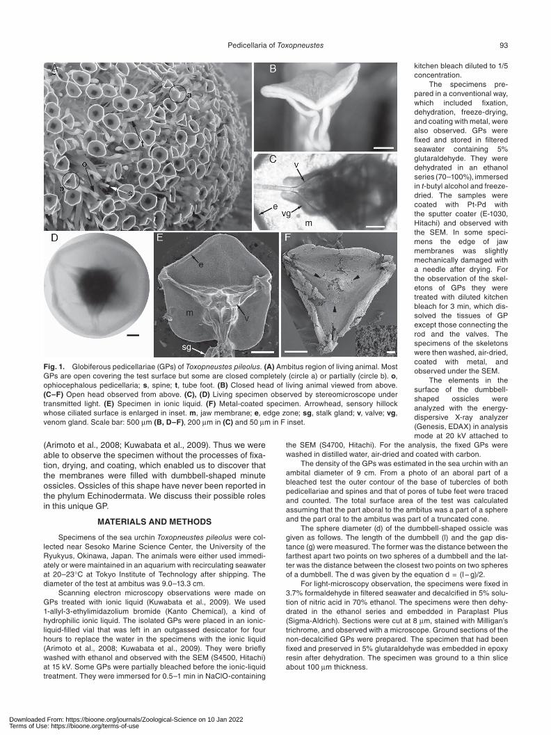
The GP is the most outstanding organ on the body sur-face of T. pileolus. It has a membranous tissue between jaws (Fujiwara, 1935). To our knowledge, there has been no report of such a membrane in any type of pedicellariae in other sea urchins. When a GP opens, it looks like an umbrella turned inside out, or a flower, or a trumpet; hence the com-
Dumbbell-Shaped Ossicles Discovered in Pedicellaria of Flower Sea Urchins
Masaki Tamori1*, Jun Koki2, and Tatsuo Motokawa1
1School of Life Science and Technology, W3-42, Tokyo Institute of Technology, O-okayama 2-12-1, Meguro-ku, Tokyo 152-8551, Japan
2Center for Advanced Materials Analysis, S7-18, Tokyo Institute of Technology, O-okayama 2-12-1, Meguro-ku, Tokyo 152-8551, Japan
Sea urchins have a globiferous pedicellaria that stands from a test with a stalk on which lies a head made of three movable jaws with venom-injecting teeth. The globiferous pedicellariae of the flower sea urchin Toxopneustes pileolus, one of the most developed among sea urchins, are unique in that the jaws are provided with a jaw membrane that gives the pedicellaria an appearance of a flower when the jaws are open. We observed this membrane in an ionic liquid that does not require pro-cesses, such as fixation, dehydration, or coating with conductive materials, for observation with a scanning electron microscope. Using this technique, we discovered dumbbell-shaped ossicles, which consist of two spheres of similar size connected by a cylinder. The diameter of the sphere is 4–8 μm, and the total length of the ossicle is 10–20 μm. The jaw membrane is trimmed with an edge zone. The ossicles were found sparsely in the connective tissue of general part of the membrane, but in the edge zone their density was so high that neighboring ossicles were in close contact with each other. Some neighboring ossicles crossed their cylinders and some inserted one of their spheres to snugly fit in the gap between the spheres of neighboring ossicles. Their structural role is very likely in strengthening the jaw membrane, probably serving as fillers in the general part of the membrane; in the edge zone the interlocking of adjacent ossicles forms a loose network pro-viding a firm frame for the head of the globiferous pedicellaria. When opened, the stiff frame pre-vents the membrane from sagging. When clasped, it works as a closed door, firmly keeping prey trapped.
mon names for this species are the flower sea urchin in English and the trumpet sea urchin in Japanese. Here we call this membrane the “jaw membrane.” In the daytime, most of GPs are open, covering the entire test surface facing the open water. Spines and other types of pedicellariae are hidden under the canopy of the “flowers” (Fig. 1A). The GPs are reddish brown, but the periphery of the membranes are white; thus, the open GPs make conspicuous white circles, which might function as a warning coloration to potential predators and intruders. The membranes are folded when the jaws are closed (Fig. 1B). Because this GP is notoriously toxic, a number of studies have focused on the chemical nature of its toxins (Fujiwara, 1935; Takei et al., 1993; Kuwabara, 1994; Zhang et al., 1999, 2001; Nakagawa et al., 2003; Hatakeyama et al., 2015). Morphological studies, how-ever, have been limited. Fujiwara (1935) published the gross morphology and histology, mainly focusing on the poison glands, which were more fully described in the related spe-cies Sphaerechinus granularis (Ghyoot et al., 1994). Skeletal elements were described briefly in a review article on the evolution of pedicellariae in echinoids (Coppard et al., 2010).
In the present study, we performed a morphological study of this unique GP. Attention was given to the jaw mem-branes. We observed the specimen with a scanning electron microscope (SEM) in ionic liquid that prevents charging
Downloaded From: https://bioone.org/journals/Zoological-Science on 10 Jan 2022Terms of Use: https://bioone.org/terms-of-use
93Pedicellaria of Toxopneustes
(Arimoto et al., 2008; Kuwabata et al., 2009). Thus we were able to observe the specimen without the processes of fixa-tion, drying, and coating, which enabled us to discover that the membranes were filled with dumbbell-shaped minute ossicles. Ossicles of this shape have never been reported in the phylum Echinodermata. We discuss their possible roles in this unique GP.
MATERIALS AND METHODS
Specimens of the sea urchin Toxopneustes pileolus were col-lected near Sesoko Marine Science Center, the University of the Ryukyus, Okinawa, Japan. The animals were either used immedi-ately or were maintained in an aquarium with recirculating seawater at 20–23°C at Tokyo Institute of Technology after shipping. The diameter of the test at ambitus was 9.0–13.3 cm.
Scanning electron microscopy observations were made on GPs treated with ionic liquid (Kuwabata et al., 2009). We used 1-allyl-3-ethylimidazolium bromide (Kanto Chemical), a kind of hydrophilic ionic liquid. The isolated GPs were placed in an ionic-liquid-filled vial that was left in an outgassed desiccator for four hours to replace the water in the specimens with the ionic liquid (Arimoto et al., 2008; Kuwabata et al., 2009). They were briefly washed with ethanol and observed with the SEM (S4500, Hitachi) at 15 kV. Some GPs were partially bleached before the ionic-liquid treatment. They were immersed for 0.5–1 min in NaClO-containing
kitchen bleach diluted to 1/5 concentration.
The specimens pre-pared in a conventional way, which included fixation, dehydration, freeze-drying, and coating with metal, were also observed. GPs were fixed and stored in filtered seawater containing 5% glutaraldehyde. They were dehydrated in an ethanol series (70–100%), immersed in t-butyl alcohol and freeze-dried. The samples were coated with Pt-Pd with the sputter coater (E-1030, Hitachi) and observed with the SEM. In some speci-mens the edge of jaw membranes was slightly mechanically damaged with a needle after drying. For the observation of the skel-etons of GPs they were treated with diluted kitchen bleach for 3 min, which dis-solved the tissues of GP except those connecting the rod and the valves. The specimens of the skeletons were then washed, air-dried, coated with metal, and observed under the SEM.
The elements in the surface of the dumbbell-shaped ossicles were analyzed with the energy-dispersive X-ray analyzer (Genesis, EDAX) in analysis mode at 20 kV attached to
the SEM (S4700, Hitachi). For the analysis, the fixed GPs were washed in distilled water, air-dried and coated with carbon.
The density of the GPs was estimated in the sea urchin with an ambital diameter of 9 cm. From a photo of an aboral part of a bleached test the outer contour of the base of tubercles of both pedicellariae and spines and that of pores of tube feet were traced and counted. The total surface area of the test was calculated assuming that the part aboral to the ambitus was a part of a sphere and the part oral to the ambitus was part of a truncated cone.
The sphere diameter (d) of the dumbbell-shaped ossicle was given as follows. The length of the dumbbell (l) and the gap dis-tance (g) were measured. The former was the distance between the farthest apart two points on two spheres of a dumbbell and the lat-ter was the distance between the closest two points on two spheres of a dumbbell. The d was given by the equation d = (l − g)/2.
For light-microscopy observation, the specimens were fixed in 3.7% formaldehyde in filtered seawater and decalcified in 5% solu-tion of nitric acid in 70% ethanol. The specimens were then dehy-drated in the ethanol series and embedded in Paraplast Plus (Sigma-Aldrich). Sections were cut at 8 μm, stained with Milligan’s trichrome, and observed with a microscope. Ground sections of the non-decalcified GPs were prepared. The specimen that had been fixed and preserved in 5% glutaraldehyde was embedded in epoxy resin after dehydration. The specimen was ground to a thin slice about 100 μm thickness.
Fig. 1. Globiferous pedicellariae (GPs) of Toxopneustes pileolus. (A) Ambitus region of living animal. Most GPs are open covering the test surface but some are closed completely (circle a) or partially (circle b). o, ophiocephalous pedicellaria; s, spine; t, tube foot. (B) Closed head of living animal viewed from above. (C–F) Open head observed from above. (C), (D) Living specimen observed by stereomicroscope under transmitted light. (E) Specimen in ionic liquid. (F) Metal-coated specimen. Arrowhead, sensory hillock whose ciliated surface is enlarged in inset. m, jaw membrane; e, edge zone; sg, stalk gland; v, valve; vg, venom gland. Scale bar: 500 μm (B, D–F), 200 μm in (C) and 50 μm in F inset.
Downloaded From: https://bioone.org/journals/Zoological-Science on 10 Jan 2022Terms of Use: https://bioone.org/terms-of-use
94 M. Tamori et al.
RESULTS
General morphology and density of globiferous pedi-cellariae
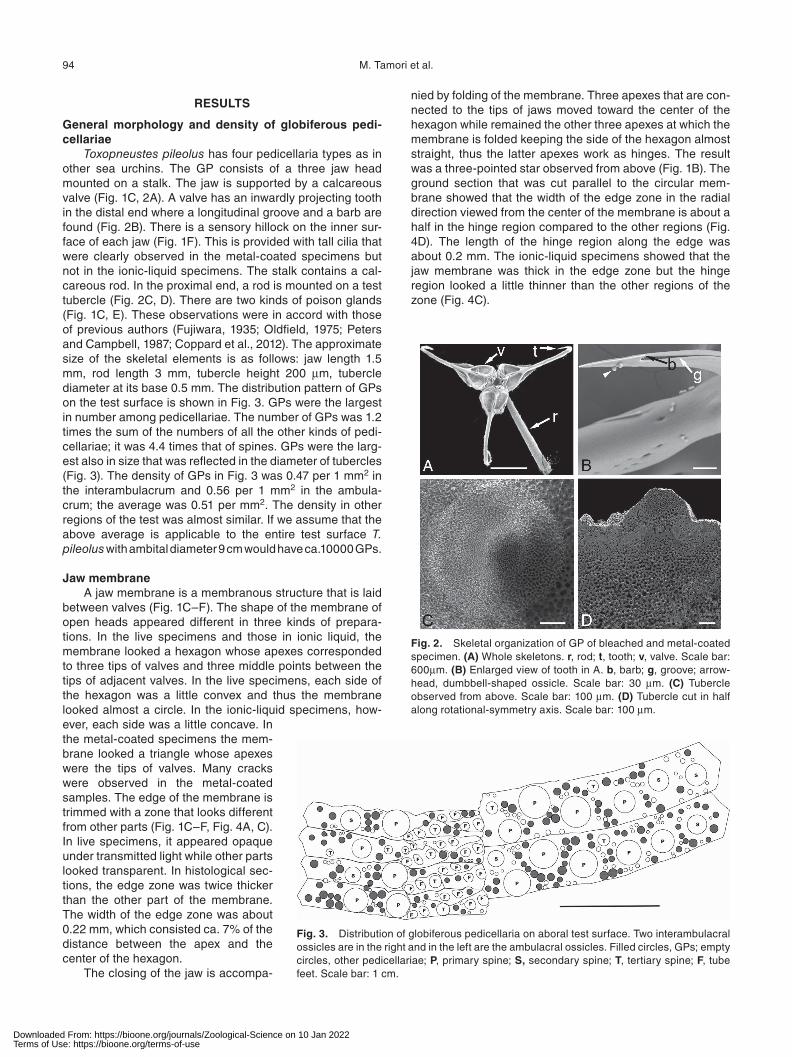
Toxopneustes pileolus has four pedicellaria types as in other sea urchins. The GP consists of a three jaw head mounted on a stalk. The jaw is supported by a calcareous valve (Fig. 1C, 2A). A valve has an inwardly projecting tooth in the distal end where a longitudinal groove and a barb are found (Fig. 2B). There is a sensory hillock on the inner sur-face of each jaw (Fig. 1F). This is provided with tall cilia that were clearly observed in the metal-coated specimens but not in the ionic-liquid specimens. The stalk contains a cal-careous rod. In the proximal end, a rod is mounted on a test tubercle (Fig. 2C, D). There are two kinds of poison glands (Fig. 1C, E). These observations were in accord with those of previous authors (Fujiwara, 1935; Oldfield, 1975; Peters and Campbell, 1987; Coppard et al., 2012). The approximate size of the skeletal elements is as follows: jaw length 1.5 mm, rod length 3 mm, tubercle height 200 μm, tubercle diameter at its base 0.5 mm. The distribution pattern of GPs on the test surface is shown in Fig. 3. GPs were the largest in number among pedicellariae. The number of GPs was 1.2 times the sum of the numbers of all the other kinds of pedi-cellariae; it was 4.4 times that of spines. GPs were the larg-est also in size that was reflected in the diameter of tubercles (Fig. 3). The density of GPs in Fig. 3 was 0.47 per 1 mm2 in the interambulacrum and 0.56 per 1 mm2 in the ambula-crum; the average was 0.51 per mm2. The density in other regions of the test was almost similar. If we assume that the above average is applicable to the entire test surface T. pileolus with ambital diameter 9 cm would have ca.10000 GPs.
Jaw membraneA jaw membrane is a membranous structure that is laid
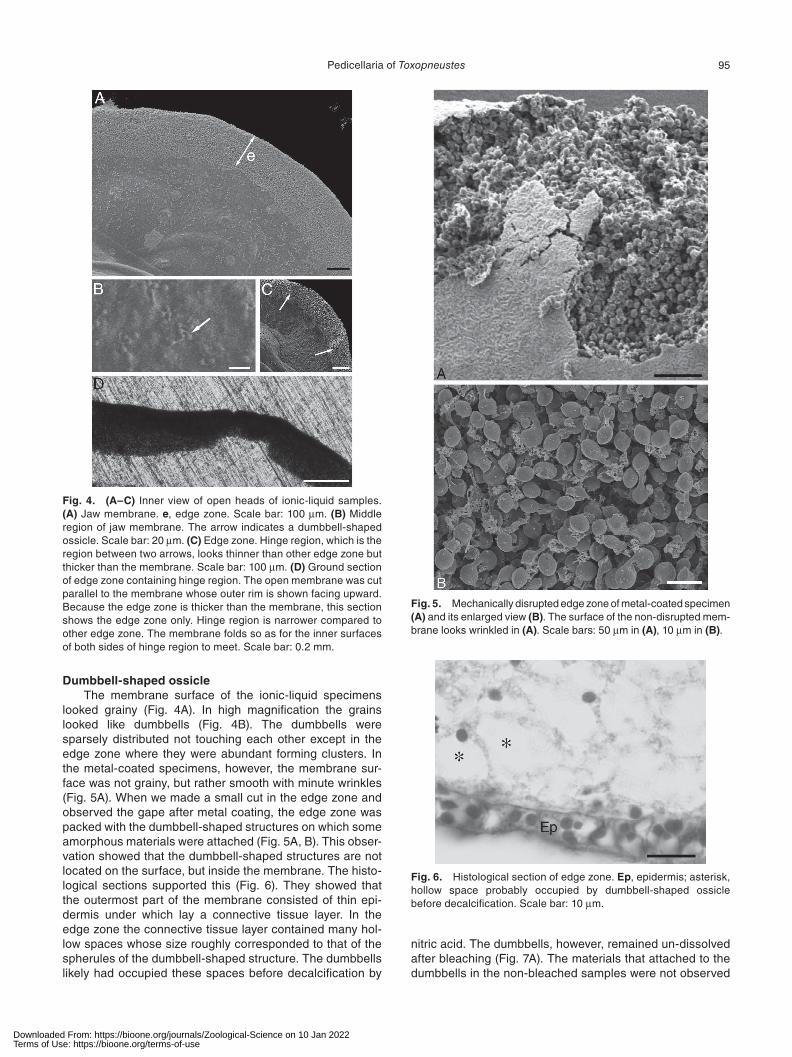
between valves (Fig. 1C–F). The shape of the membrane of open heads appeared different in three kinds of prepara-tions. In the live specimens and those in ionic liquid, the membrane looked a hexagon whose apexes corresponded to three tips of valves and three middle points between the tips of adjacent valves. In the live specimens, each side of the hexagon was a little convex and thus the membrane looked almost a circle. In the ionic-liquid specimens, how-ever, each side was a little concave. In the metal-coated specimens the mem-brane looked a triangle whose apexes were the tips of valves. Many cracks were observed in the metal-coated samples. The edge of the membrane is trimmed with a zone that looks different from other parts (Fig. 1C–F, Fig. 4A, C). In live specimens, it appeared opaque under transmitted light while other parts looked transparent. In histological sec-tions, the edge zone was twice thicker than the other part of the membrane. The width of the edge zone was about 0.22 mm, which consisted ca. 7% of the distance between the apex and the center of the hexagon.
The closing of the jaw is accompa-
nied by folding of the membrane. Three apexes that are con-nected to the tips of jaws moved toward the center of the hexagon while remained the other three apexes at which the membrane is folded keeping the side of the hexagon almost straight, thus the latter apexes work as hinges. The result was a three-pointed star observed from above (Fig. 1B). The ground section that was cut parallel to the circular mem-brane showed that the width of the edge zone in the radial direction viewed from the center of the membrane is about a half in the hinge region compared to the other regions (Fig. 4D). The length of the hinge region along the edge was about 0.2 mm. The ionic-liquid specimens showed that the jaw membrane was thick in the edge zone but the hinge region looked a little thinner than the other regions of the zone (Fig. 4C).
Fig. 2. Skeletal organization of GP of bleached and metal-coated specimen. (A) Whole skeletons. r, rod; t, tooth; v, valve. Scale bar: 600μm. (B) Enlarged view of tooth in A. b, barb; g, groove; arrow-head, dumbbell-shaped ossicle. Scale bar: 30 μm. (C) Tubercle observed from above. Scale bar: 100 μm. (D) Tubercle cut in half along rotational-symmetry axis. Scale bar: 100 μm.
Fig. 3. Distribution of globiferous pedicellaria on aboral test surface. Two interambulacral ossicles are in the right and in the left are the ambulacral ossicles. Filled circles, GPs; empty circles, other pedicellariae; P, primary spine; S, secondary spine; T, tertiary spine; F, tube feet. Scale bar: 1 cm.
Downloaded From: https://bioone.org/journals/Zoological-Science on 10 Jan 2022Terms of Use: https://bioone.org/terms-of-use
95Pedicellaria of Toxopneustes
Dumbbell-shaped ossicleThe membrane surface of the ionic-liquid specimens
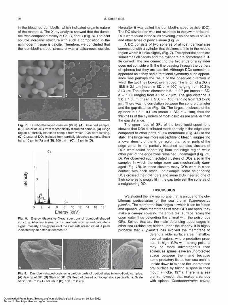
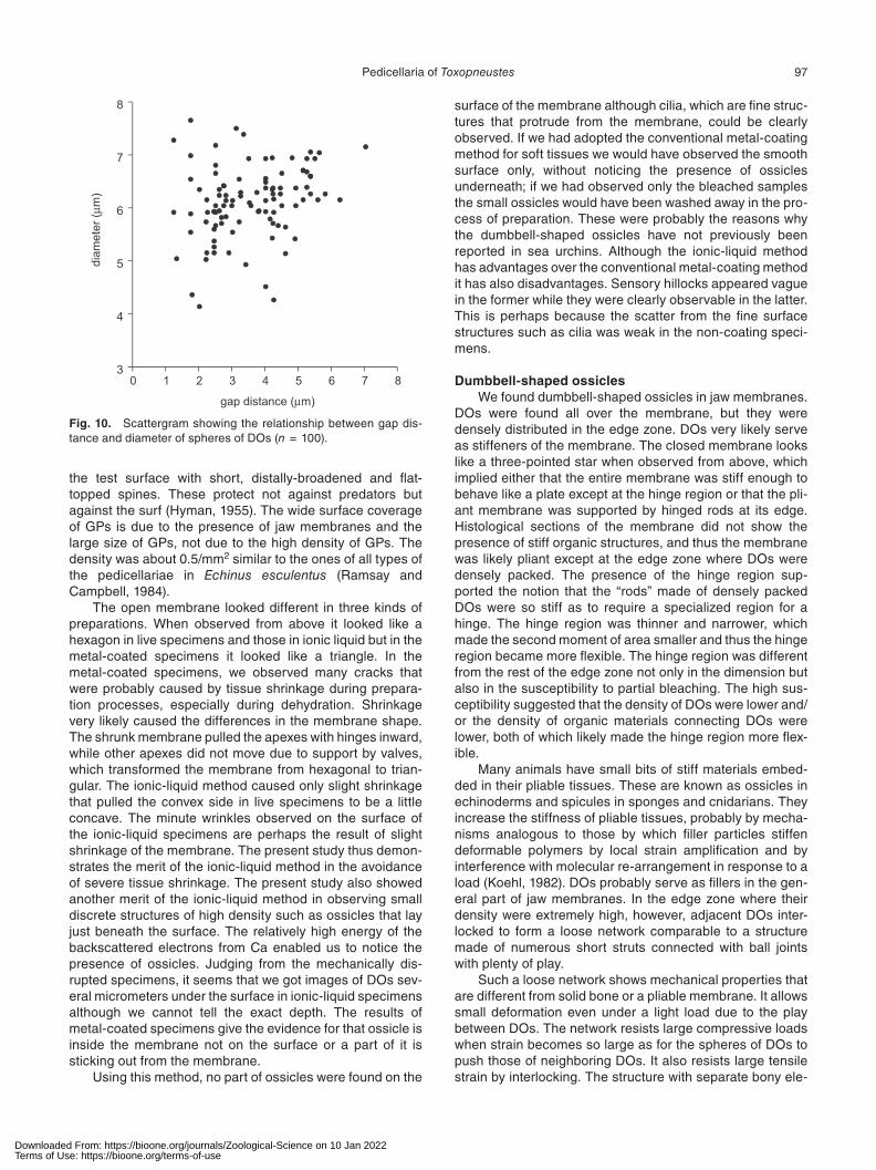
looked grainy (Fig. 4A). In high magnification the grains looked like dumbbells (Fig. 4B). The dumbbells were sparsely distributed not touching each other except in the edge zone where they were abundant forming clusters. In the metal-coated specimens, however, the membrane sur-face was not grainy, but rather smooth with minute wrinkles (Fig. 5A). When we made a small cut in the edge zone and observed the gape after metal coating, the edge zone was packed with the dumbbell-shaped structures on which some amorphous materials were attached (Fig. 5A, B). This obser-vation showed that the dumbbell-shaped structures are not located on the surface, but inside the membrane. The histo-logical sections supported this (Fig. 6). They showed that the outermost part of the membrane consisted of thin epi-dermis under which lay a connective tissue layer. In the edge zone the connective tissue layer contained many hol-low spaces whose size roughly corresponded to that of the spherules of the dumbbell-shaped structure. The dumbbells likely had occupied these spaces before decalcification by
Fig. 4. (A–C) Inner view of open heads of ionic-liquid samples. (A) Jaw membrane. e, edge zone. Scale bar: 100 μm. (B) Middle region of jaw membrane. The arrow indicates a dumbbell-shaped ossicle. Scale bar: 20 μm. (C) Edge zone. Hinge region, which is the region between two arrows, looks thinner than other edge zone but thicker than the membrane. Scale bar: 100 μm. (D) Ground section of edge zone containing hinge region. The open membrane was cut parallel to the membrane whose outer rim is shown facing upward. Because the edge zone is thicker than the membrane, this section shows the edge zone only. Hinge region is narrower compared to other edge zone. The membrane folds so as for the inner surfaces of both sides of hinge region to meet. Scale bar: 0.2 mm.
Fig. 5. Mechanically disrupted edge zone of metal-coated specimen (A) and its enlarged view (B). The surface of the non-disrupted mem-brane looks wrinkled in (A). Scale bars: 50 μm in (A), 10 μm in (B).
Fig. 6. Histological section of edge zone. Ep, epidermis; asterisk, hollow space probably occupied by dumbbell-shaped ossicle before decalcification. Scale bar: 10 μm.
nitric acid. The dumbbells, however, remained un-dissolved after bleaching (Fig. 7A). The materials that attached to the dumbbells in the non-bleached samples were not observed
Downloaded From: https://bioone.org/journals/Zoological-Science on 10 Jan 2022Terms of Use: https://bioone.org/terms-of-use
96 M. Tamori et al.
in the bleached dumbbells, which indicated organic nature of the materials. The X-ray analysis showed that the dumb-bell was composed mainly of Ca, C, and O (Fig. 8). The acid soluble inorganic structure with such a composition in the echinoderm tissue is calcite. Therefore, we concluded that the dumbbell-shaped structure was a calcareous ossicle.
Hereafter it was called the dumbbell-shaped ossicle (DO). The DO distribution was not restricted to the jaw membrane. DOs were found in the skins covering jaws and stalks of GPs and other types of pedicellariae (Fig. 9).
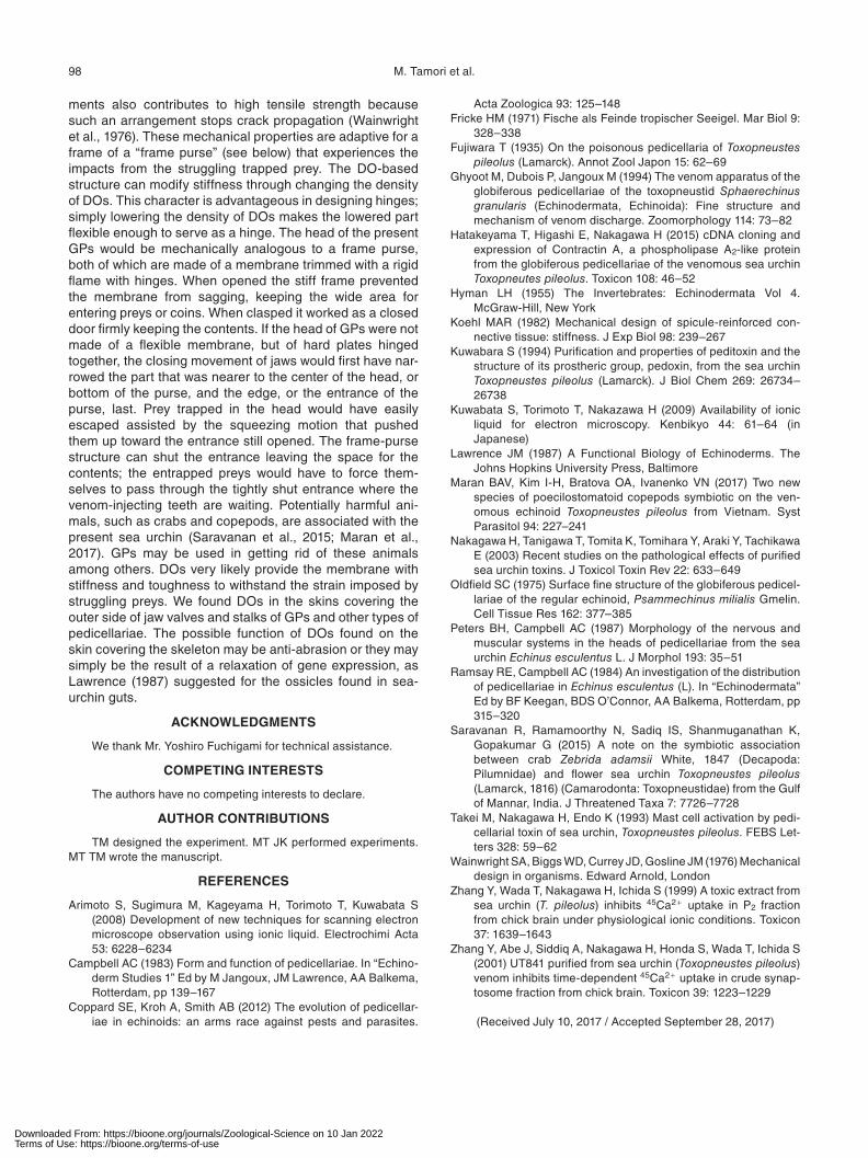
A DO consists of two spheres of almost identical size connected with a cylinder that thickens a little in the middle region where it kinks slightly (Fig. 7). The spherical parts are sometimes ellipsoids and the cylinders are sometimes a lit-tle curved. The line connecting the two ends of a cylinder does not coincide with the line passing through the centers of spheres but they are parallel. Although DOs sometimes appeared as it they had a rotational symmetry such appear-ance was perhaps the result of the observed direction in which the two lines looked overlapped. The length of a DO is 15.8 ± 2.1 μm (mean ± SD; n = 100) ranging from 10.3 to 21.3 μm. The sphere diameter is 6.1 ± 0.7 μm (mean ± SD; n = 100) ranging from 4.1 to 7.7 μm. The gap distance is 3.6 ± 1.3 μm (mean ± SD; n = 100) ranging from 1.3 to 7.0 μm. There was no correlation between the sphere diameter and the gap distance (Fig. 10). The largest thickness of the cylinder is 1.5 ± 0.1 μm (mean ± SD; n = 100), thus the thickness of the cylinders of most ossicles are smaller than the gap distance.
The open head of GPs of the ionic-liquid specimens showed that DOs distributed more densely in the edge zone compared to other parts of jaw membrane (Fig. 4A) or the stalk. The hinge was more susceptible to bleach, suggesting a lower density of the hinge region than other parts of the edge zone. In the partially bleached samples clusters of DOs were found separating from the hinge region while other part of the edge zone remained undamaged (Fig. 7C, D). We observed such isolated clusters of DOs also in the samples in which the edge zone was mechanically dam-aged (Fig. 7B). In those clusters many DOs were in close contact with each other. For example some neighboring DOs crossed their cylinders and some DOs inserted one of their spheres to snugly fit in the gap between the spheres of a neighboring DO.
DISCUSSION
We studied the jaw membrane that is unique to the glo-biferous pedicellariae of the sea urchin Toxopneustes pileolus. The membrane has hinges at which it can be folded and opened. When membranes of most GPs are open, they make a canopy covering the entire test surface facing the open water thus defending the animal with the poisonous GPs. Spines that are the main defending appendages in other sea urchins are hidden under the canopy. It is highly probable that T. pileolus has evolved the membrane to
defend a wider surface area in shallow tropical waters, where predation pres-sure is high. GPs with strong poisons may be more advantageous than spines, as spines leave an unprotected space between them and because some predatory fishes turn sea urchins upside down to expose the unprotected oral surface by taking a spine in their mouth (Fricke, 1971). There is a sea urchin, however, that makes a canopy with spines; Colobocentrotus covers
Fig. 7. Dumbbell-shaped ossicles (DOs). (A) Bleached sample. (B) Cluster of DOs from mechanically disrupted sample. (C) Hinge region of partially bleached sample from which DOs were leaving. (D) Cluster of DOs isolated from partially bleached sample. Scale bars: 10 μm in (A) and (B), 200 μm in (C), 15 μm in (D).
Fig. 8. Energy dispersive X-ray spectrum of dumbbell-shaped structure. Abscissa is energy of characteristic X-ray and ordinate is signal intensity. Energy peaks of the elements are indicated. A peak indicated by an asterisk denotes Na.
Fig. 9. Dumbbell-shaped ossicles in various parts of pedicellariae in ionic-liquid samples. (A) Jaw tip of GP. (B) Stalk of GP. (C) Head of closed ophiocephalous pedicellaria. Scale bars: 300 μm in (A), 50 μm in (B), 100 μm in (C).
Downloaded From: https://bioone.org/journals/Zoological-Science on 10 Jan 2022Terms of Use: https://bioone.org/terms-of-use
97Pedicellaria of Toxopneustes
the test surface with short, distally-broadened and flat-topped spines. These protect not against predators but against the surf (Hyman, 1955). The wide surface coverage of GPs is due to the presence of jaw membranes and the large size of GPs, not due to the high density of GPs. The density was about 0.5/mm2 similar to the ones of all types of the pedicellariae in Echinus esculentus (Ramsay and Campbell, 1984).
The open membrane looked different in three kinds of preparations. When observed from above it looked like a hexagon in live specimens and those in ionic liquid but in the metal-coated specimens it looked like a triangle. In the metal-coated specimens, we observed many cracks that were probably caused by tissue shrinkage during prepara-tion processes, especially during dehydration. Shrinkage very likely caused the differences in the membrane shape. The shrunk membrane pulled the apexes with hinges inward, while other apexes did not move due to support by valves, which transformed the membrane from hexagonal to trian-gular. The ionic-liquid method caused only slight shrinkage that pulled the convex side in live specimens to be a little concave. The minute wrinkles observed on the surface of the ionic-liquid specimens are perhaps the result of slight shrinkage of the membrane. The present study thus demon-strates the merit of the ionic-liquid method in the avoidance of severe tissue shrinkage. The present study also showed another merit of the ionic-liquid method in observing small discrete structures of high density such as ossicles that lay just beneath the surface. The relatively high energy of the backscattered electrons from Ca enabled us to notice the presence of ossicles. Judging from the mechanically dis-rupted specimens, it seems that we got images of DOs sev-eral micrometers under the surface in ionic-liquid specimens although we cannot tell the exact depth. The results of metal-coated specimens give the evidence for that ossicle is inside the membrane not on the surface or a part of it is sticking out from the membrane.
Using this method, no part of ossicles were found on the
surface of the membrane although cilia, which are fine struc-tures that protrude from the membrane, could be clearly observed. If we had adopted the conventional metal-coating method for soft tissues we would have observed the smooth surface only, without noticing the presence of ossicles underneath; if we had observed only the bleached samples the small ossicles would have been washed away in the pro-cess of preparation. These were probably the reasons why the dumbbell-shaped ossicles have not previously been reported in sea urchins. Although the ionic-liquid method has advantages over the conventional metal-coating method it has also disadvantages. Sensory hillocks appeared vague in the former while they were clearly observable in the latter. This is perhaps because the scatter from the fine surface structures such as cilia was weak in the non-coating speci-mens.
Dumbbell-shaped ossiclesWe found dumbbell-shaped ossicles in jaw membranes.
DOs were found all over the membrane, but they were densely distributed in the edge zone. DOs very likely serve as stiffeners of the membrane. The closed membrane looks like a three-pointed star when observed from above, which implied either that the entire membrane was stiff enough to behave like a plate except at the hinge region or that the pli-ant membrane was supported by hinged rods at its edge. Histological sections of the membrane did not show the presence of stiff organic structures, and thus the membrane was likely pliant except at the edge zone where DOs were densely packed. The presence of the hinge region sup-ported the notion that the “rods” made of densely packed DOs were so stiff as to require a specialized region for a hinge. The hinge region was thinner and narrower, which made the second moment of area smaller and thus the hinge region became more flexible. The hinge region was different from the rest of the edge zone not only in the dimension but also in the susceptibility to partial bleaching. The high sus-ceptibility suggested that the density of DOs were lower and/or the density of organic materials connecting DOs were lower, both of which likely made the hinge region more flex-ible.
Many animals have small bits of stiff materials embed-ded in their pliable tissues. These are known as ossicles in echinoderms and spicules in sponges and cnidarians. They increase the stiffness of pliable tissues, probably by mecha-nisms analogous to those by which filler particles stiffen deformable polymers by local strain amplification and by interference with molecular re-arrangement in response to a load (Koehl, 1982). DOs probably serve as fillers in the gen-eral part of jaw membranes. In the edge zone where their density were extremely high, however, adjacent DOs inter-locked to form a loose network comparable to a structure made of numerous short struts connected with ball joints with plenty of play.
Such a loose network shows mechanical properties that are different from solid bone or a pliable membrane. It allows small deformation even under a light load due to the play between DOs. The network resists large compressive loads when strain becomes so large as for the spheres of DOs to push those of neighboring DOs. It also resists large tensile strain by interlocking. The structure with separate bony ele-
Fig. 10. Scattergram showing the relationship between gap dis-tance and diameter of spheres of DOs (n = 100).
Downloaded From: https://bioone.org/journals/Zoological-Science on 10 Jan 2022Terms of Use: https://bioone.org/terms-of-use
98 M. Tamori et al.
ments also contributes to high tensile strength because such an arrangement stops crack propagation (Wainwright et al., 1976). These mechanical properties are adaptive for a frame of a “frame purse” (see below) that experiences the impacts from the struggling trapped prey. The DO-based structure can modify stiffness through changing the density of DOs. This character is advantageous in designing hinges; simply lowering the density of DOs makes the lowered part flexible enough to serve as a hinge. The head of the present GPs would be mechanically analogous to a frame purse, both of which are made of a membrane trimmed with a rigid flame with hinges. When opened the stiff frame prevented the membrane from sagging, keeping the wide area for entering preys or coins. When clasped it worked as a closed door firmly keeping the contents. If the head of GPs were not made of a flexible membrane, but of hard plates hinged together, the closing movement of jaws would first have nar-rowed the part that was nearer to the center of the head, or bottom of the purse, and the edge, or the entrance of the purse, last. Prey trapped in the head would have easily escaped assisted by the squeezing motion that pushed them up toward the entrance still opened. The frame-purse structure can shut the entrance leaving the space for the contents; the entrapped preys would have to force them-selves to pass through the tightly shut entrance where the venom-injecting teeth are waiting. Potentially harmful ani-mals, such as crabs and copepods, are associated with the present sea urchin (Saravanan et al., 2015; Maran et al., 2017). GPs may be used in getting rid of these animals among others. DOs very likely provide the membrane with stiffness and toughness to withstand the strain imposed by struggling preys. We found DOs in the skins covering the outer side of jaw valves and stalks of GPs and other types of pedicellariae. The possible function of DOs found on the skin covering the skeleton may be anti-abrasion or they may simply be the result of a relaxation of gene expression, as Lawrence (1987) suggested for the ossicles found in sea-urchin guts.
ACKNOWLEDGMENTS
We thank Mr. Yoshiro Fuchigami for technical assistance.
COMPETING INTERESTS
The authors have no competing interests to declare.
AUTHOR CONTRIBUTIONS
TM designed the experiment. MT JK performed experiments. MT TM wrote the manuscript.
REFERENCES
Arimoto S, Sugimura M, Kageyama H, Torimoto T, Kuwabata S (2008) Development of new techniques for scanning electron microscope observation using ionic liquid. Electrochimi Acta 53: 6228–6234
Campbell AC (1983) Form and function of pedicellariae. In “Echino-derm Studies 1” Ed by M Jangoux, JM Lawrence, AA Balkema, Rotterdam, pp 139–167
Coppard SE, Kroh A, Smith AB (2012) The evolution of pedicellar-iae in echinoids: an arms race against pests and parasites.
Acta Zoologica 93: 125–148Fricke HM (1971) Fische als Feinde tropischer Seeigel. Mar Biol 9:
328–338Fujiwara T (1935) On the poisonous pedicellaria of Toxopneustes
pileolus (Lamarck). Annot Zool Japon 15: 62–69Ghyoot M, Dubois P, Jangoux M (1994) The venom apparatus of the
globiferous pedicellariae of the toxopneustid Sphaerechinus granularis (Echinodermata, Echinoida): Fine structure and mechanism of venom discharge. Zoomorphology 114: 73–82
Hatakeyama T, Higashi E, Nakagawa H (2015) cDNA cloning and expression of Contractin A, a phospholipase A2-like protein from the globiferous pedicellariae of the venomous sea urchin Toxopneutes pileolus. Toxicon 108: 46–52
Hyman LH (1955) The Invertebrates: Echinodermata Vol 4. McGraw-Hill, New York
Koehl MAR (1982) Mechanical design of spicule-reinforced con-nective tissue: stiffness. J Exp Biol 98: 239–267
Kuwabara S (1994) Purification and properties of peditoxin and the structure of its prostheric group, pedoxin, from the sea urchin Toxopneustes pileolus (Lamarck). J Biol Chem 269: 26734–26738
Kuwabata S, Torimoto T, Nakazawa H (2009) Availability of ionic liquid for electron microscopy. Kenbikyo 44: 61–64 (in Japanese)
Lawrence JM (1987) A Functional Biology of Echinoderms. The Johns Hopkins University Press, Baltimore
Maran BAV, Kim I-H, Bratova OA, Ivanenko VN (2017) Two new species of poecilostomatoid copepods symbiotic on the ven-omous echinoid Toxopneustes pileolus from Vietnam. Syst Parasitol 94: 227–241
Nakagawa H, Tanigawa T, Tomita K, Tomihara Y, Araki Y, Tachikawa E (2003) Recent studies on the pathological effects of purified sea urchin toxins. J Toxicol Toxin Rev 22: 633–649
Oldfield SC (1975) Surface fine structure of the globiferous pedicel-lariae of the regular echinoid, Psammechinus milialis Gmelin. Cell Tissue Res 162: 377–385
Peters BH, Campbell AC (1987) Morphology of the nervous and muscular systems in the heads of pedicellariae from the sea urchin Echinus esculentus L. J Morphol 193: 35–51
Ramsay RE, Campbell AC (1984) An investigation of the distribution of pedicellariae in Echinus esculentus (L). In “Echinodermata” Ed by BF Keegan, BDS O’Connor, AA Balkema, Rotterdam, pp 315–320
Saravanan R, Ramamoorthy N, Sadiq IS, Shanmuganathan K, Gopakumar G (2015) A note on the symbiotic association between crab Zebrida adamsii White, 1847 (Decapoda: Pilumnidae) and flower sea urchin Toxopneustes pileolus (Lamarck, 1816) (Camarodonta: Toxopneustidae) from the Gulf of Mannar, India. J Threatened Taxa 7: 7726–7728
Takei M, Nakagawa H, Endo K (1993) Mast cell activation by pedi-cellarial toxin of sea urchin, Toxopneustes pileolus. FEBS Let-ters 328: 59–62
Wainwright SA, Biggs WD, Currey JD, Gosline JM (1976) Mechanical design in organisms. Edward Arnold, London
Zhang Y, Wada T, Nakagawa H, Ichida S (1999) A toxic extract from sea urchin (T. pileolus) inhibits 45Ca2+ uptake in P2 fraction from chick brain under physiological ionic conditions. Toxicon 37: 1639–1643
Zhang Y, Abe J, Siddiq A, Nakagawa H, Honda S, Wada T, Ichida S (2001) UT841 purified from sea urchin (Toxopneustes pileolus) venom inhibits time-dependent 45Ca2+ uptake in crude synap-tosome fraction from chick brain. Toxicon 39: 1223–1229
(Received July 10, 2017 / Accepted September 28, 2017)
Downloaded From: https://bioone.org/journals/Zoological-Science on 10 Jan 2022Terms of Use: https://bioone.org/terms-of-use