CHAPTER 2 Invertebrate Iridescent Viruses TREVOR WILLIAMS I. INTRODUCTION Invertebrate iridescent viruses IIV s) belong to the family Iridoviridae, which are large icosahedral viruses with a large genome of double-stranded DNA. These viruses are not occluded in a protective protein matrix, unlike the well-known nucleopolyhedroviruses INPVs), granuloviruses IGVs), cytoplasmic polyhedro- sis viruses ICPVs), and entomopoxviruses IEPVs). Members of the Iridoviridae, are structurally complex, however, and show a number of unique characteris- tics in terms of their genomic organization, structure, and mechanisms of replication, which unite the family. A common feature is that IV hosts generally occur in aquatic or damp habitats such as the soil or leaf litter. Geographically, IVs appear widely distrib- uted. The first IV was discovered in larvae of the crane fly, Tipula paludosa, in Great Britain !Xeros, 1954); since then, an additional 73 species of hosts have been reported with naturally occurring IV infections. Of these, the majority are insects from the orders Diptera 140 spp.), Coleoptera 18 spp.), and Lepidoptera 17 spp.). Most of the dipteran records are mosquitoes. A number of noninsect species are also infected by IVs, particularly terrestrial isopods 18 spp.) and daphnids. The IVs have attracted interest as pathogens of insect vectors of medical importance, such as mosquitoes and Simulium spp. Few of the viruses isolated from infected hosts, however, have been characterized, probably because of the low incidence of lethal infections in host populations and a resultant decline of interest in IVs as biocontrol agents. In spite of that, the past 5 years have seen initial changes in our perception of these viruses in terms of their relationships TREVOR WILLIAMS • ECOSUR, Tapachula 30700, Chiapas, Mexico. The Insect Viruses, edited by Lois K. Miller and 1. Andrew Ball. Plenum Publishing Corporation, New York, 1998. 31
Transcript
CHAPTER 2
Invertebrate Iridescent Viruses TREVOR WILLIAMS
I. INTRODUCTION
Invertebrate iridescent viruses IIV s) belong to the family Iridoviridae, which are large icosahedral viruses with a large genome of double-stranded DNA. These viruses are not occluded in a protective protein matrix, unlike the well-known nucleopolyhedroviruses INPVs), granuloviruses IGVs), cytoplasmic polyhedrosis viruses ICPVs), and entomopoxviruses IEPVs). Members of the Iridoviridae, are structurally complex, however, and show a number of unique characteristics in terms of their genomic organization, structure, and mechanisms of replication, which unite the family.
A common feature is that IV hosts generally occur in aquatic or damp habitats such as the soil or leaf litter. Geographically, IVs appear widely distributed. The first IV was discovered in larvae of the crane fly, Tipula paludosa, in Great Britain !Xeros, 1954); since then, an additional 73 species of hosts have been reported with naturally occurring IV infections. Of these, the majority are insects from the orders Diptera 140 spp.), Coleoptera 18 spp.), and Lepidoptera 17 spp.). Most of the dipteran records are mosquitoes. A number of noninsect species are also infected by IVs, particularly terrestrial isopods 18 spp.) and daphnids.
The IVs have attracted interest as pathogens of insect vectors of medical importance, such as mosquitoes and Simulium spp. Few of the viruses isolated from infected hosts, however, have been characterized, probably because of the low incidence of lethal infections in host populations and a resultant decline of interest in IVs as biocontrol agents. In spite of that, the past 5 years have seen initial changes in our perception of these viruses in terms of their relationships
TREVOR WILLIAMS • ECOSUR, Tapachula 30700, Chiapas, Mexico.
The Insect Viruses, edited by Lois K. Miller and 1. Andrew Ball. Plenum Publishing Corporation, New York, 1998.
31
32 TREVOR WILLIAMS
with their hosts, their molecular biology, and the taxonomic relationships within the family.
II. CLASSIFICATION
The family Iridoviridae was created in 1976 to encompass a growing number of icosahedral DNA viruses that had been reported from both invertebrate and vertebrate animals and that characteristically assembled in the cytoplasm of host cells (Fenner, 1976). The term iridovirus was then adopted to describe isolates from vertebrates; inasmuch as it is a coverall term, it may be applied equally to invertebrate isolates, although for historical reasons the term iridescent virus continues to be used.
The invertebrate isolates were divided between two genera based principally on particle size. The small IVs, with a diameter of approximately 120 nm, were assigned to the genus Iridovirus, whereas the larger IV s, with a diameter of approximately 180 nm, reported only from mosquitoes, midges, and a Simulium sp. were assigned to the genus Chloriridovirus (although strangely, this genus also contained members that should have been assigned to the Iridovirus genus on grounds of particle size) (Mathews, 1982). The prefix chlor was taken from the Greek chloros (green), reflecting the yellow-green coloration that the large IVs display in infected host tissues and as purified pellets of virus.
Isolates from vertebrates were assigned to three genera: Ranavirus, representing viruses from amphibians; Lymphocystivirus, for isolates from flat fish (dab and flounder); and African swine fever virus, which was initially incorporated into the family as a separate genus but was later removed in light of genomic organization data. Following this, the taxonomy of the family remained little changed, with 22 recognized small IVs (Iridovirus), 10 recognized large IVs (Chloriridovirus), and 50 isolates distributed among the vertebrate genera, mostly in the Ranavirus genus. The creation of an additional vertebrate genus was also sanctioned to accommodate two viruses isolated from a goldfish cell line (Francki et a1., 1991).
The type species for the whole Iridoviridae family is frog virus 3 (FV3) of the Ranavirus genus. This virus is highly amenable to cell culture and more is known about its composition, structure, replication, and molecular biology than any other iridovirus. Consequently, FV3 is considered to be the model virus for this family, and I will make reference to studies of FV3 periodically throughout this chapter.
A. Problems with the Established Classification of Iridescent Viruses
There are three principal problems with the classification of IVs devised in the 1970s: (1) the criteria used to recognize isolates as separate virus entities; (2) a grave lack of information on the interrelationships among IVs from differ-
INVERTEBRATE IRIDESCENT VIRUSES 33
ent hosts; and 13) the nomenclature system employed to name IVs. Let us consider each problem in turn.
As the number of reports of arthropod species with patent IV infections grew, lists were constructed of those hosts from which IV infections had been reported in the literature or were known to the authors from personal communications ITinsley and Kelly, 1970; Tinsley and Harrap, 1978). The issue of whether or not the virus had been isolated and had undergone some degree of characterization was not taken into account when producing these lists. However, the lists were accepted by the International Committee on Taxonomy of Viruses IICTV), presumably because of the desire to give some structure and order to the classification of the group and on the assumption that the status of each isolate would become apparent with the passage of time. Thus, viruses that lacked any degree of characterization achieved undeserved taxonomic status by publication in the ICTV reports IMathews, 1982; Murphy et a1., 1995).
Within the IVs, the data concerning taxonomic interrelationships among the 32 recognized IV types were extremely limited. What data existed were primarily serological in nature. This was useful in confirming that most of the small IVs IIridovirus genus) were interrelated IKelly et a1., 1979). However, the absence of comparative genetic data prevented any real advance in understanding IV interrelationships, a situation that remained unchanged throughout the 1980s.
Under an interim system of nomenclature for IVs, each record of a virus isolated from a different host was given the name of the host followed by the words "iridescent virus," and a type number was assigned that indicated the sequence of discovery IKelly and Tinsley, 1970). Hence, the first IV reported was named Tipula paludosa iridescent virus IIV type 1). The type number system was not applied universally, however, and after 1980, the assigning of type numbers ceased altogether, although applying the host name to the isolate continued. This scheme of nomenclature has one primary failing: It ties the identity of the virus to a particular host. This becomes a problem when viruses are not highly host specific.
B. New Changes in the Classification of Iridescent Viruses
Today, the picture is different. Recommendations were accepted by the ICTV regarding the status of previously recognized IV types and a new list of tentative IV species was established IWilliams et a1., 1996). In summary, 16 of the 32 previously recognized IV types were removed from the classification, as no characterization data were available and stocks of the viruses did not exist. Additional IV types were identified as strains of recognized IVs bearing different type numbers, for example, IV9, IVIO, and IV18 are actually variants of a single virus Isee Section VI).
Consequently, following the inclusion of one new tentative species, an IV from the lepidopteran Anticarsia gemmatalis IWilliams, 1994; Kinard et a1.,
34 TREVOR WILLIAMS
TABLE I. Iridescent Virus Classification following Recent Changes Approved by the ICTV
Particle Tentative species identified Genus diameter by type number(sl Host(sl
1995), the revised list of tentative IV species now includes just 13 viruses in the Iridovirus genus and one virus, IV3, from Aedes taeniorhynchus (Clark et a1., 1965) as the sole member and type species of the Chloriridovirus genus (Table I). This species list is tentative because of a continuing lack of detailed information for the majority of member viruses. Arguments supporting the classification changes have already been described in detail and were applied as much to the vertebrate genera as to the invertebrate genera (Williams, 1996).
C. The Nomenclature Problem
The existing nomenclature of IV s is no longer useful and should be replaced with a more appropriate system (Ward and Kalmakoff, 1991; Williams and Cory, 1994). In particular, the name of the virus should not depend on the host, because the same virus many naturally infect more than one host species; likewise, a single host species may be infected by several different IVs.
Thus, an alternative nomenclature system for IVs has recently been put forward in which the viruses are given geographical descriptors; the name of a town or city close to the site of discovery of the virus (Williams and Cory, 1994). Under this system, the name of the IV9/IVlO/IVI8 example given above would be changed to "Nelson iridescent virus"; Nelson being the town in New Zealand close to the site of isolation of IV9. Similarly, IV31/IV32 becomes "River-
INVERTEBRATE IRIDESCENT VIRUSES 35
side IV"; this being the point of discovery of the virus. This system is subject to criticism on the grounds that viruses may be assigned names that are difficult to pronounce by English users, although with careful selection of names from the local options, this should not be a major problem.
However, nomenclature changes need to be taken up by the majority of scientists working in the field, and changes should be accepted only cautiously as they can be difficult to rectify if subsequently found to be unworkable. Consequently, this nomenclature has not yet been adopted by the ICTV; and in this chapter I will continue to use the established system of type numbers and host names.
III. STRUCTURE AND COMPOSITION
IV particles comprise an electron-dense core of DNA and associated proteins, surrounded by a lipid membrane, which in turn is closely associated with the exterior protein capsid (Fig. 1). IVs released by budding may have an additional outer envelope, most commonly observed in virus grown in cell culture (Hukuhara and Hashimoto, 1967; Yule and Lee, 1973; Webb et a1., 1976). The structure and composition of each component are considered in turn.
A. Capsid
The IV capsid comprises a lattice of identical hexagonally packed subunits of a single polypeptide of 48-55 kDa, the major capsid protein (MCP). The MCP comprises about 470 amino acids and represents 40-45% of the total particle polypeptide (Moore and Kelly, 1980; Black et a1., 1981; Davison et a1., 1992; Stohwasser et a1., 1993). This protein is highly conserved and is distantly related to the MCP of icosahedral viruses from other virus families, that is, African swine fever virus and an algal virus from the PhycodnaviIidae (Stohwasser et a1., 1993; Mao et a1., 1996). Sodium dodecyl sulfate-polyacrylamide gel electrophoresis (SDS-PAGE) and surface labeling studies on IV6 have indicated that the MCP is the basis for two different structures: a weakly united trimeric form held together seemingly by hydrogen bonding located on the exterior surface of the capsid, whereas a covalently bonded MCP trimer lies beneath the surface layer (Cerutti and Devauchelle, 1990).
The capsid subunits are tubular in shape, 7-9 nm in diameter, and 7-9 nm in length, with a central hole. The subunits are the building blocks for 20 trisymetrons (each comprising 55 subunits) and 12 pentasymetrons (each of 31 subunits) that are needed to form the icosahedral particle. Thus, each IV particle probably comprises a total of 1472 subunits (Wrigley, 1969, 1970; Stoltz, 1971, 1973). Five trisymetrons are attached to the sides of each pentasymetron but with the corners out of alignment with the pentasymetron by three subunits, shown clearly in IVI (Manyakov, 1977). Larger IVs, such as IV3 and an isolate from Chironomus plumosus, have larger trisymetrons, probably com-
36 TREVOR WILLIAMS
FIGURE 1. Particles of IV3 in a mosquito cell showing regular paracrystalline packing and the presence of virogenic stroma. Scale bar, 1 jJ.m. (Photo courtesy of J. Becnel.)
prising 78 subunits, giving a likely 1560 subunits per particle (Stoltz, 1971, 1973).
An external fringe of small fibrils about 2.5 nm in length has been reported in certain IVs (Cole and Morris, 1980; Black et a1., 1981; Devauchelle et a1., 1985b). Fibrils with terminal knobs have also been observed (Stoltz, 1971, 1973). Some isolates have extremely long fibrils; these are most obvious in an isolate from the midge, Chironomus plumosus (Stoltz et a1., 1968) and in an iridovirus from fish (Zwillenberg and Wolf, 1968). In such cases, each capsid subunit appears to bear a single fibril.
B. Lipid Layer
The lipid layer is about 4 nm thick (Kelly, 1985), is intimately associated with the icosahedral capsid, and has been reported to make up 5.2-9% of the particle dry weight. Much of the lipid is phospholipid (Kalmakoff and Tremaine, 1968; Kelly and Vance, 1973; Balange-Orange and Devauchelle, 1982). The fatty acid and phospholipid composition of IVs differ from that of the host cell for all IVs assayed to date (Balange-Orange and Devauchelle, 1982; Williams and Thompson, 1995).
The lipid layer is essential for infectivity of vertebrate iridoviruses (Willis
INVERTEBRATE IRIDESCENT VIRUSES 37
and Granoff, 1974). In contrast, IVs are recognized to be resistant to ether treatment (Murphy et a1., 1995), although the evidence for this is somewhat anecdotal; ether-treated IV3 produced patent infections in mosquito larvae (Matta and Lowe, 1970). Likewise, Day and Mercer (1964) mentioned that patent infections were observed following injection of Galleria mellonella larvae with ether- or chloroform-treated IV2. However, lipid membrane extracts show biological activity of importance during the initial stages of infection. This property of IVs requires confirmation.
Differences in lipid composition between host and virus, and the observation that IV s do not appear to obtain their lipid component by budding through a cell organelle membrane, have led to the assumption that the lipid layer is acquired de novo during particle maturation; this situation also is believed to occur in poxviruses. However, recent work has shown that vaccinia virus particles acquire their internal lipid membrane from an intracellular compartment between the endoplasmic reticulum and the Golgi apparatus. Virally encoded proteins appear to facilitate this process (Schmelz et a1., 1994). The relevance of these findings to IV s is not known.
C. Core
The core is a highly hydrated electron dense entity. The core components appear to be uniformly distributed through the core but show a degree of structural organization. There is some evidence for the interaction of core structures with the lipid membrane, and it is suspected that core and capsid polypeptides are interconnected by way of complexes of protein passing through the lipid layer (Cuillel et a1., 1979; Cerutti and Devauchelle, 1982; Klump et a1., 1983; Robach et a1., 1983). In FV3, freeze etching revealed the presence of randomly oriented rods, some 10 nm in diameter, which are possibly part of the DNA-protein complex arranged in a long coiled filament (Tripier-Darcy and Nermut, 1983). Core structures also have been likened to bundles of parallel threads in an isolate from the mosquito Culiseta annulata (Buchatsky, 1977).
Much of the polypeptide diversity of IVs appears to be associated with the core and lipid membrane. At least six polypeptide species are associated with the DNA within the core, the major component being a 12.5-kDa species in IV6 (Cerutti and Devauchelle, 1985). Lipid-enveloped cores of IV3 were not found to be infectious to mosquito cells or larvae, possibly because of the failure of cores to attach to cells (Wagner et a1., 1973).
D. Physicochemical Characteristics and Composition
Size measurements among IVs tend to vary because of side-to-side or vertex-to-vertex measurements being reported. Measurements of dehydrated particles in ultrathin sections are most common and range from about 120 to 130 nm for the small IVs and 180 to 200 nm for large IVs, although several isolates have been reported with intermediate sizes (Stoltz et a1., 1968; Popel-
38 TREVOR WILLIAMS
kova, 1982), indicating that size alone is not an infallible criterion by which isolates can be assigned to genera. Measurements from negatively stained material give particle diameters some 20% larger than dehydrated sections (Wagner et a1., 1973; Fukaya and Nasu, 1966; Cole and Morris, 1980; Poprawski and Yule, 1990; Lacey and Adams, 1994). A smaller variant of IV3 showing turquoise iridescence has been detected in laboratory cultures (Wagner et a1., 1973) and was termed "turquoise mosquito iridescent virus./I However, all the studies described in this chapter refer to the original "regular" strain, unless otherwise stated.
The weight of intact virions is 1.28 x 10⁶ kDa for small IVs and 2.49 x 10⁶
kDa for the larger chloriridovirus IV3 (Glitz et a1., 1968; Lowe et a1., 1970) Particles of IV3 have a density of 1.35 g cm -3 compared to 1.30 g cm -3 or slightly more for IV1 and IV2 (Matta, 1970; Robertson and Longworth, 1973; Glitz et a1., 1968). Small IVs have an S20 w of approximately 2200 compared to up to 4458 for IV3 (Kelly and Robertson, 1973; Matta, 1970; Wagner et a1., 1973; Kinard et a1., 1995). The IV particle contains a linear dsDNA molecule representing 12-17% of the particle weight (Bellett and Inman, 1967; Kalmakoff and Tremaine, 1968; Matta, 1970; Stadelbacher et a1., 1978). Reported genome sizes for small IVs range from 140 kilobasepair (kbp) in IV24 to 209 kbp in IV6 (Williams and Cory, 1994; Fisher et a1., 1990). There are conflicting reports of the genome size of IV3, which was calculated to be 135 kbp by restriction endonuclease analysis (Williams and Cory, 1994) compared to a remarkable 383 kbp by sucrose gradient centrifugation (Wagner and Paschke, 1977). The genomes of small IVs (Iridovirus genus) have a characteristic GC content of 29-32% compared to 54% for IV3 (Glitz et a1., 1968; Black et a1., 1981; Wagner and Paschke, 1977).
One-dimensional PAGE analysis of intact virions usually resolves 20-32 polypeptides, although two-dimensional techniques have indicated greater diversity indicating several viral proteins to be oligomeric structures composed of subunits linked by disulfide bridges (Cerutti and Devauchelle, 1985). Polypeptide weights typically range from 11 to 200 kDa, although more extreme values have also been reported (Krell and Lee, 1974; Moore and Kelly, 1980; Cerutti and Devauchelle, 1990; Tajbakhsh and Seligy, 1990). Despite their larger size, IVs from mosquitoes have been reported to be structurally less complex than the small IVs with just 9 or 12 polypeptides detected by SDS-PAGE (Wagner et a1., 1974; Buchatsky and Sherban, 1976).
A number of virion-associated enzymes have been detected in IV6, including a nucleotide phosphohydrolase that hydrolyzed ATP to ADP; a protein kinase possibly responsible for phosphorylation of a DNA-binding protein; an alkaline protease possibly involved in virus uncoating; and a nonspecific DNase (Monnier and Devauchelle, 1976, 1980; Farara and Attias, 1983, 1986; Devauchelle et a1., 1985a).
IV. REPLICATION AND MOLECULAR BIOLOGY
Replication of IVs is sensitive to cell type (Tajbakhsh et a1., 1990b) and temperature. Replication of IV22 in mosquito cells, measured by the produc-
INVERTEBRATE IRIDESCENT VIRUSES 39
tion of viral antigen, was abundant at 20°C, minimal at 28°C, and absent at >30°C (Tesh and Andreadis, 1992). The same trend is observed in vivo; most IVs do not replicate at high temperatures (Tanada and Tanabe, 1965; Carter, 1975; Witt and Stairs, 1976). In contrast, in mosquito cells infected with IV3, cytopathic effects were observed only at >30°C (Webb et a1., 1974).
Replication of iridoviruses at the cellular and molecular level has been most extensively studied in FV3 and to a lesser extent in IV6. Consequently, the following account focuses mainly on FV3, with additional information from IVs where applicable.
A. Penetration and Shutdown of Macromolecular Synthesis
IV particles appear to be adsorbed onto the cell membrane and enter cells by pinocytosis. In the cytoplasm, particles may be surrounded by a membrane or may appear as free virions that undergo uncoating. Vesicles containing IVI were reported to aggregate and fuse into lysosomes in which virus uncoating takes place (Kelly and Tinsley, 1974; Webb et a1., 1976; Mathiesen and Lee, 1981). Uncoating of FV3 does not require protein synthesis.
There follows a rapid shutdown in host cell macromolecular synthesis, a characteristic of the infection process in all iridoviruses studies to date. FV3 particles can produce this effect in permissive and nonpermissive cells and the effect is not destroyed by treatment with heat or UV light (Willis and Granoff, 1976). This shutdown property is very potent; a single particle or possibly a single protein molecule may be able to elicit shutdown (Drillien et a1., 1977). A heat-stable protein that affects ribosomal transcription activity is believed to be responsible (Elharrar and Kim, 1977).
Host cell shutdown in mosquito cells was seen within 1 hr of infection with IV6, and the rate at which this occurred was dependent on the multiplicity of infection. IV6 also provoked shutdown in permissive and nonpermissive cells including vertebrate cells. Lipid vesicles extracted from IV6 particles made up 86% of the viral phospholipid and 4% of total viral protein and caused cell-cell fusion and shutdown in the same fashion as intact particles. Five protein components were detected, the major component being 11 kDa (Cerutti and Devauchelle, 1980, 1982, 1990; Cerutti et a1., 1989). Production of viral macromolecules is not affected by the shutdown process.
B. DNA Replication
IridoviIuses are unusual in that replication occurs in both the nucleus and the cytoplasm, although virus particle assembly is strictly cytoplasmic. During the first 3 hr postinfection, FV3 DNA is synthesized only in the nucleus in fragments of genomic or up to twice the genomic size. DNA then moves to the cytoplasm where it exists as concatamers of more than 10 times the genomic length (Goorha, 1982). Formation of concatamers probably occurs by direct joining of the DNA lengths present in the nucleus (Goorha and Dixit, 1984;
40 TREVOR WILLIAMS
Martin et al., 1984). It is assumed that concatameric DNA is packaged into virions using a "headfull" mechanism.
The iridovirus genome is circularly permuted and terminally redundant. This means that within each particle the complete genome is present plus a little extra DNA from the next genome copy in the concatamer (terminal redundancy), and this leads to a population of particles with different terminal sequences (circular permution) (Goorha and Murti, 1982j Willis et al., 1985). Consequently, although each genome is a linear DNA molecule, when restriction maps are made, they appear circular for all IVs mapped to date (Ward and Kalmakoff, 1987j Soltau et al., 1987j Schnitzler et al., 1987j Davison et al., 1992). In IV6, the degree of terminal redundancy has been estimated as 12 % and a total of six origins of replication have been detected (Delius et al., 1984j Handermann et al., 1992). The IV genome is not methylated, whereas high levels of methylation of cytosine are seen in vertebrate iridoviruses (Willis and Granoff, 1980j
Essani and Granoff, 1989j Eaton et al., 1991). . Extensive regions of repetitive DNA have been mapped from the genomes
of IVs. In IV9, the repetitive sequences were found to make up over 25% of the genome. These sequences are distinct from the DNA that accounts for the terminal redundancy of the genome. The coding function of these regions is unknown, although transcription of these regions was detected but only late in the infection cycle (Ward and Kalmakoff, 1987j Kalmakoff et ai., 1990j McMillan and Kalmakoff, 1994). The pattern of repetitive DNA in the genome of IV6 is complex and involves boxes of tandem repeat sequences and others with a number of different interdigitated repeat sequences of variable size and homology. Within this region a number of open reading frames (ORFs) of unknown function were detected (Fischer et al., 1988a,b, 1990).
The only exception to the genomic organization described above is a North American isolate of IVI in which the genome was reported to comprise two components, one of approximately 200 kbp and one of 10.8 kbp. The relative abundance of the two components was dependent on the stage of particle assembly and on the host (Tajbakhsh et al., 1990bj Czuba et al., 1994). Other isolates of IVI show only one component (Tajbakhsh and Seligy, 1990, personal observation).
C. Transcription, Translation, and IV Genes
Transcription and transcriptional control have been studied only in FV3, wherein it resembles a coordinated sequential process that is not dependent on DNA synthesis for any but the last 15 % of the viral genome. Three classes of mRNAs have been recognized, depending on the sequence in which they appear. There are ten "immediate-early" mRNAs representing about one third of the FV3 genome, or possibly more (Goorha and Granoff, 1979j Willis and Granoff, 1978j Mesnard et al., 1988).
A virus-associated protein appears to be the first in a series of trans-acting factors that facilitate the temporal sequence of viral transcription. This protein
INVERTEBRATE IRIDESCENT VIRUSES 41
is suspected to interact with the host RNA polymerase to facilitate virus transcription, possibly by binding to viral template DNA to enhance attachment or processing of immediate-early sequences by the host polymerase (Willis and Granoff, 1985). The promoter sequence that regulates transcription of a major immediate-early gene was identified as a 23-bp region 5' to a TATAlike box that signaled the start of transcription (Willis, 1987). Regulation of transcription of other immediate-early mRNAs appears to depend on inhibition by an early synthesized gene product(s) (Beckman et al., 1988), whereas synthesis of another trans-acting factor is required for the switch on of the next class of transcription products-" delayed-early mRNAs"-of which there are three in FV3 (Willis and Granoff, 1978; Goorha et al., 1979).
Transcription of late genes usually occurs after the nuclear phase of DNA replication, and again viral-induced trans-acting factors are required (Willis et al., 1979, 1990). There is a rapid turnover of FV3 mRNAs and continual transcription is required to maintain messages at a steady state (Chinchar et al., 1994).
Early genes appear to be transcribed in the nucleus, whereas late genes may be transcribed in the cytoplasm, possibly from the concatameric DNA, by way of a virally encoded DNA-dependent RNA polymerase. The gene for such an enzyme has been reported in IV6, although the C-terminal domain was missing from the ORF detected by Schnitzler et al. (1994b).
The mechanisms that control translation are poorly understood. Certain FV3 mRNAs continue to be produced after production of the relevant protein has finished. The production of antisense RNA that binds to mRNAs from early genes to prevent translation has been proposed as a way of limiting the production of early proteins (Mesnard et al., 1988; Willis et al., 1990). Similarly, modification of 5' termini of early mRNAs has been detected to occur later in the replication cycle. Such modifications prevented subsequent translation (Tondre et al., 1988; Aubertin et al., 1990). The number and context of AUG codons that signal the initiation of translation also differ among early and late mRNAs and are likely to playa role in translational control (Aubertin et a1., 1990).
In IV9, many immediate-early transcripts were detected originating from seven discrete locations scattered over the genome. Certain proteins were produced for the entire replication cycle; among these was probably the major capsid protein, which appeared at 4 hr postinfection together with two other possibly structural proteins. Three suspected structural proteins appeared as late as 36 hr postinfection. Overall, translation of 20 proteins was detected, of which 4 appeared and were shut off within 18 hr postinfection and 16 were still being produced at 48 hr postinfection (McMillan and Kalmakoff, 1994).
A putative apoptosis-inhibiting gene has been identified in IV6, which may be necessary to inhibit programmed cell death following viral infection. In baculoviruses, a similar gene produced a product of 30 kDa with a central zinc fingerlike structure composed of cysteines and histidine, probably with nucleic acid-binding properties. This protein effectively blocked host cell apoptosis. The homologous protein ofIV6lacked certain repeat motifs, but it has not
42 TREVOR WILLIAMS
been tested for biological activity (Birnbaum et a1., 1994; Handermann et a1., 1992; Sonntag et a1., 1994).
Additional ORFs with high homology to genes with established functions have been detected in IV6, including a putative antimutator enzyme GTP phosphohydrolase, a nonhistone DNA-binding protein, another DNA-binding protein possibly involved with transcription or DNA repair, and a putative DNA or RNA helicase (Schnitzler et a1., 1994a; Sonntag et a1., 1994). A gene (L96) for a large, basic, and mostly hydrophilic protein has been identified in IVI. The protein is possibly involved in DNA packaging in the core. As in FV3, putative TATA and CAAT boxes were detected upstream of ATG start co dons of this gene and are suspected to be involved in gene expression (Home et a1., 1990).
D. Particle Assembly
The sequence of particle formation has been subject to different interpretations, some stepwise, involving the sequential assembly of individual components, and others a coordinated and simultaneous process of particle organization and construction. In the stepwise process, viral DNA and nucleoprotein condensed to form the core, which was then enveloped by the capsid (Bird, 1961, 1962; Kelly and Tinsley, 1974). Others have reported face-by-face assembly of the capsid followed by entry of the core components through a hole left in the shell that was subsequently capped (Yule and Lee, 1973; Hess and Poinar, 1985). In contrast, the coordinated process involves the concurrent assembly of capsid and internal lipid membrane and the sequestering of core material from the surrounding area. Once the capsid is complete, DNA condenses to form the core proper (Devauchelle, 1977; Federici, 1980; Devauchelle et a1., 1985b). It seems likely that the pattern of particle assembly will be affected by a number of host- and virus-related factors, including the type, age, and condition of cell infected; stage of infection; temperature; permissiveness of host; and so forth.
Following infection by FV3, the host cytoskeleton undergoes dramatic changes in which microtubules (22-26 nm in diameter) breakdown concomitant with the appearance of large areas of granular, organelle-free, virus assembly sites referred to as virogenic stroma or viroplasmic centers. The structural integrity of these sites is maintained by intermediate filaments (7-11 nm in diameter) that reorganize from their normal position, radiating from the nucleus, and cluster around virogenic stroma. Fine microfilaments (4-8 nm in diameter) composed of actin break up and reform at the cell surface where they assist in budding and release of virus (Murti et a1., 1985; Murti and Goorha, 1990).
The role of the cytoskeleton in IV replication in insect cells is less clear. Cytoskeleton-disrupting drugs did not prevent the formation of virogenic stroma following infection by IVI and immunofluorescent studies directed at microtubules and microfilaments have not indicated that these elements are associated with virogenic centers (Bertin et a1., 1987; Seagull et a1., 1985). However, nuclear matrix or associated proteins have been detected serologi-
INVERTEBRATE IRIDESCENT VIRUSES 43
cally in assembly sites in vivo in cells infected by IVI and in assembly sites extracted and fractionated in vitro (Bladon et al., 1986).
V. SIGNS AND CHARACTERISTICS OF PATENT DISEASE
The principal sign of patent IV infection is the iridescent hue that arises from the paracrystalline arrangement of virus particles in host cells. Light is reflected from the surface of close-packed particles and causes interference with incident light (so-called "Bragg reflections"), resulting in a range of colors, most commonly lavender, blue, turquoise, and green. The viruses from mosquitoes and chironomids commonly display colors such as yellow-green, orange, and red by virtue of their larger particle size and greater interparticle spacing (Klug et al., 1959; Hemsley et al., 1994). In fact, the family name, Iridoviridae has its origin in the Greek iridos, meaning "shining like a rainbow," which is an appropriate way to describe the striking appearance of patently infected individuals. Purified pellets of IV also iridesce.
An isolate from the midge, Chironomus plumosus, did not show iridescence in host tissues, possibly because of the presence of fibrillar structures attached to the virus capsid that may increase interparticular spacing and prevent the occurrence of Bragg reflections (Stoltz et al., 1968). Iridescence has been dismissed as a trivial characteristic due solely to the physical structure of IVs (Stoltz, 1971; Kelly, 1985). There is no known adaptive advantage arising from iridescence in terms of enhanced virus transmission, for example. Iridoviruses from vertebrates do not iridesce.
Replication of IVs following injection of inoculum is not sensitive to the host life stage (larva, pupa, adult), unlike baculoviruses, for example, although natural patent infections are mainly observed in juvenile stages. However, adults are prone to lethal infections in bees, isopods, and Tenebrio beetles. In bees, IV infection appears to be the cause of "clustering disease" in Apis cerana from Kashmir and northern India. Diseased colonies become uncharacteristically inactive and form small detached groups of flightless individuals that crawl on the ground. Infected colonies perish rapidly (Bailey et al., 1976; Bailey and Ball, 1978; Singh, 1979; Mishra, et al.,1980).
A. Pathology
Most IVs are catholic in their tissue tropisms. Almost invariably, extensive replication occurs in the fat body and epidermis. For example, IV31 replicates in most isopod tissues, especially the epidermis, muscles and fat body, nerves, hemocytes, and in patches along the gut (Federici, 1980; Cole and Morris, 1980). The same virus appears to infect extensive tissues, including the gonads of Thaumamermis cosgrovei, a mermithid nematode parasite of the isopod (Poinar et al., 1980). IVl provoked the formation of epidermal tumors in Bombyx mori, but such pathology is not seen in other IV infections of other hosts (Hukuhara, 1964). In Aedes taeniorhychus infected by IV3, the fat body, epi-
44 TREVOR WILLIAMS
dermis, imaginal disks, hemocytes, trachea, muscle, visceral nerves, gonads, and esophagus were infected but not the remaining gut or Malpighian tubules (Hall and Anthony, 1971), A red IV from Aedes puntor caused the fat body to take on the appearance of grape clusters and was easily broken apart (Weiser and Zizka, 1985). Individuals with patent infections that survive to pupate characteristically show marked deformations of the pupa, particularly of the wing buds (Smith et a1., 1961; Carter, 1974; Stadelbacher et a1., 1978).
The principal pathological changes at the cellular level include cell rounding and the appearance of extensive areas of finely granulated material devoid of cell organelles-the virogenic stroma. Large numbers of particles form in close association but not generally within this zone. Most studies of the cellular pathology of IVs have focused on cell culture systems where the pathology of infection may be different from the equivalent in vivo changes.
Infection of mosquito and lepidopteran cells with IV2 or IV6 resulted in marked contraction of the cells after 72 hr followed by cell detachment at 96 hr, effects that were not sensitive to the multiplicity of infection and that may be caused by toxic viral proteins (Kelly and Tinsley, 1974). Rapid cell-cell fusion, however, was observed to be a function of the multiplicity of infection in cells infected by IV6. A virus protein inactivated by heat or antiserum (described in Section IVa) appears to be responsible (Cerutti and Devauchelle, 1979). The formation of numerous vesicles arising from blebbing of cell membranes followed by loss of cell adhesion and cell-cell fusion was reported in Spodoptera cells infected by IV1. Changes in the position and morphology of mitochondria were noted in this system and in infected isopods (Czuba et a1., 1994; Hess and Poinar, 1985).
B. IV Production and Quantification
Purely in terms of numbers, IVs are among the most efficient insect viruses in turning host resources into virus particles. Around 25% of the dry weight of a dead insect may be virus (Williams and Smith, 1957). With a particle weight of approximately 2 x 10-15 g (Thomas, 1961; Glitz et a1., 1968), predicted yields are 2.5 x 10¹² particles of IV1 in a Tipula oleracea larva and 2 x 10¹² particles of IV2 in a Galleria mellonella pupa (Carter, 1973b; Day and Mercer, 1964). Following the same reasoning, a Costelytra zealandica third ins tar larvae may yield 5.1 x 10¹² particles of IV16, a Phyllophaga amda larva may yield 2.5 x 10¹²
particles, a Heteronychus arator larva may yield 1. 75 x 1012 particles, a Tenebrio molitor pupa may yield 1.5 x 10¹⁰ particles, and field-collected Armadillidium vulgare and Porcellio scaber are predicted to yield 2.6 x 10¹⁰ particles and 3.1 x 10¹⁰ particles, respectively (estimates from data in Moore, 1973; Poprawski and Yule, 1990; Longworth et a1., 1979; Black et a1., 1981; Cole and Morris, 1980). Obviously these calculations make unsupported assumptions about the efficiency of virus replication and the composition of insect and virus samples. These estimates, however, do emphasize the great productivity of IVs within invertebrate hosts.
INVERTEBRATE IRIDESCENT VIRUSES 45
IVs may be quantified by plaque assay, insect bioassay, optical density, direct counting, and, to a degree, by enzyme-linked immunosorbent assay (ELISA). Czuba et a1. (1994) reported reliable quantification of IVI by plaque assay in Spodoptera cells. There was a linear relationship between the production of plaque-forming units (PFU) and the DNA content of viral inoculum at multiplicities of infection between 0.1 and 1000. Optical density measurements at 260 nm indicated that from one A260 unit it was possible to obtain 105 PFU or 11 JJ.g of DNA which would be equivalent to approximately 4.6 x 10⁶ IV particles (assuming a mean genome size of 200 kbp). Direct counting using an electron microscope indicated that four to six times more particles were actually present per ~60' probably because many particles in these preparations contained little or no DNA. The efficiency of infection was low; 0.1-0.3 % of IV1 particles produced virogenic stroma in host cells, and of these less than 1 % went on to produce a visible plaque. From these data, the formation of one PFU requires 4 x 10⁵ filled particles of IV1. However, values as low as 75 particles per PFU were reported for IV22 in Spodoptera cells using virus freshly extracted .from G. mellonella (Brown et a1., 1977). The reason for such differences may reside in the different isolates used, or in the passage history of each virus.
IV2 was quantified by optical density and direct counting was used to calculate the number of particles required to produce a patent infection in G. mellonella larvae. One ~60 unit of IV2 was equivalent to 1.8 x 10⁹ particles by direct counting. Lethal infections were achieved by injection of between 1.7 and 6.9 particles into final instar G. mellonella. In these studies, empty particles lacking DNA were rare (Day and Mercer, 1964; Day and Gilbert, 1967). The insect bioassay, involving calculation of a median lethal dose or infective dose value (LDso or IDso) following injection of G. mellonella larvae, may be one of the most sensitive methods of IV quantification (Carter, 1973c; Witt and Stairs, 1976; Ohba and Aizawa, 1978).
Ward and Kalmakoff (1991) suggested that ELISA could be used to give a semiquantitative estimate of the amount of virus present in a sample. Purified virus was used to raise polyvalent antiserum in rabbits. Field-collected mosquitoes were individually homogenized, fixed into microtiter plate wells, reacted with rabbit antiserum followed by horseradish peroxidase-labeled sheep antirabbit antibody, and read in an ELISA reader. The virus concentration was then estimated by reference to a standard curve.
Virus purification by sucrose gradient centrifugation resulted in disruption of virion structure and subsequent degradation of an orthopteran IV. Use of Picoll in place of sucrose avoided this problem (Boucias et a1., 1987).
C. Dose-Response Relationships and Survival Time
Study of the dose-response relationships of IVs generally has not been performed in a highly quantitative manner, and the "response" criterion has always been patent iridescence or mortality of the host. Many studies report that large doses are required to achieve patent infection by per os inoculation
46 TREVOR WILLIAMS
(Carter, 1973a; Linley and Nielsen, 1968a; Federici, 1980; Sieburth and Carner, 1987) although high doses may also result in rapid paralysis and death prior to extensive virus proliferation (Stadelbacher et a1., 1978), probably due to the action of cytotoxic viral proteins (Aubertin et a1., 1977; Lorbacher de Ruiz, 1990).
The dose-response relationship has been shown to be sensitive to temperature: The LDso value of IVI injected into G. mellonella larvae was three orders of magnitude higher at 30°C compared to the LDso value at 20°C (Witt and Stairs, 1976). The oral LDso of IV6 in the boll weevil Anthonomus grandis was 3.61 x 10⁻⁶ larval equivalents of G. mellonella (McLaughlin et a1., 1972), which, assuming a similar productivity to IV2 in the same host (Day and Mercer, 1964), would be roughly 7 x 10⁶ particles. The LD63 of IVI and IV2 by injection into lepidopteran and dipteran larvae ranged between 6.0 and 5 x 10⁷ particles, depending on virus and host species (Glitz et a1., 1968).
Survival times reported following per os doses range from about 45 days for IVI in Tipula oleracae, about 35 days for IV31/IV32 in isopods, about 28 days for IV30 in Helicoverpa zea, about 21 days for an IV of the mole cricket Scapteriscus borellii, and as little as 6 days for an IV of Anticarsia gemmatalis (Carter, 1973a; Grosholz, 1992; Sikorowski and Tyson, 1984; Fowler, 1989; Sieburth and Carner, 1987). Late ins tar larvae developed disease faster than younger conspecifics (Carter, 1974; Sieburth and Carner, 1987), which contrasts with the pattern shown by other insect viruses that tend to kill younger, smaller hosts more quickly than older, larger ones. Compared to feeding, injection of inoculum may result in a 30-50% reduction in survival time and a reduction in the variability of survival times (Carter, 1973a,b; Federici, 1980; Cole and Morris, 1980).
Infected hosts are reported to be more sluggish than healthy con specifics but may continue to live several weeks after developing iridescent coloration (Linley and Nielsen, 1968a; Poprawski and Yule, 1990). IV-infected soil-dwelling insects have been reported to move to near the surface prior to death where the probability of transmission by cannibalism or predation may be enhanced (Fowler and Robertson, 1972).
VI. RELATIONSHIPS AMONG IVs
Relationships among the members of the Iridotriridae are not well understood currently and the need for comparative studies of these viruses has been stated repeatedly (Hall, 1985; Kelly, 1985; Willis, 1990; Ward and Kalmakoff, 1991; Schnitzler and Darai, 1993; Stohwasser et a1., 1993).
A. Serology
The principal tools for comparative studies in the 1960s and 1970s were serologically based analyses involving a range of different IVs, although DNA renaturation and SDS-PAGE also were used occasionally (Kelly and Avery,
INVERTEBRATE IRIDESCENT VIRUSES 47
1974; Carey et a1., 1978). Serological relationships were summarized by Kelly et a1. (1979), who classified isolates into two main groups. One large group comprised the majority of interrelated isolates (IVl, IV2, IV9, IVlO, IV16, IV18, IV21, IV22, IV23, IV25, IV28), some of which were almost indistinguishable from one another by serology (e.g., IV9 andIV18, IV21 andIV28). In contrast, IV6 andIV24 were classified as serologically unrelated to other IVs or to one another. IV29 was placed in between these groups as being related to some but not all of the principal serogroup. The isopod virus IV31 showed a low cross-reactivity to the antiserum of IVI and was considered to be distantly related to the main serogroup (Cole and Morris, 1980). Viruses from other genera, IV3, FV3, and lymphocystis disease virus type 1 (LCDV-l ), showed no serological relatedness to IV s in the Iridovirus genus or to one another (Cunningham and Tinsley, 1968; McAuslan and Armentrout, 1974).
B. Genetic
Fueled by the need to clarify the interrelationships among these viruses, genetic analyses have recently been published. Comparative studies were performed involving 14 IV isolates plus FV3 and LCDV-l, the type species from the two vertebrate genera (Williams and Cory, 1994; Williams, 1994). Each isolate was subject to restriction enzyme analysis and the resulting gels were Southern blotted and probed with a fragment of the MCP gene of IV22 under different stringencies of hybridization.
From the restriction profiles it was immediately apparent that several isolates were strains of the same virus (Fig. 2a). Coefficients of similarity were calculated by pairwise comparison of the proportion of similar-sized restriction fragments shared by isolates. Coefficient values were particularly high among IV9, IVlO, and IV18 (up to 91.5%) and for comparison of IV21 and IV28 (up to 94.3%). In contrast, IV3, FV3, and LCDV-l showed no restriction profile similarities to any other isolates or to each other.
By Southern blot analysis at high stringency, the IV22 MCP gene probe consistently hybridized to a subset of the isolates, namely IVl, IV2, IV9, IVlO, IV18, IV22, IV24, IV29, and IV30 (Fig; 2b). When blots were probed at lower stringency, hybridization to comigrating fragments was evident for three additional isolates: IV6, IV21, and IV28(Fig. 2c). Hybridization of the probe to IV31 was weak but fairly consistent at low stringency. Only in one SaIl blot did the probe show affinity to IV3. Probe hybridization to FV3 or LCDV-l was not detected. Polymerase chain reaction (PCR) amplification of an MCP gene fragment using primers derived from the IV22 MCP gene sequence provided additional support for the results of the Southern blot analysis.
DNA-DNA dot-blot hybridization measurements were also consistent with the results of restriction endonuclease and Southern blot analyses. One large group of interrelated isolates was detected showing relative hybridization values of between 10 and 91 % at the intermediate stringency conditions employed (Fig. 3). The highest levels of hybridization were detected among IV9, IVlO, and IV18; so clearly they are strains of the same virus. A second, smaller
48 TREVOR WILLIAMS
a ~ 1 2 3 8 9 10 18 21 22 24 28 29 30 31 ~ ~
kbp kbp
23.1- -23.1
14.1- -14.1
9.4- -9.4
6.5- ~.5
5.1- -5.1 4.4- -4.4
2.8- -2.8
2.3- -2.3
2.0- -2.0
1.7- -1.7
1.1- -1.1
0.8- ~.8
0.5- ~.5
0.3- ~.3
FIGURE 2. (a) HindIII restriction endonuclease profiles of DNA from IVs identified by type number and two vertebrate isolates, FV3 and LCDV-l, showing clear similarities among IV9, IVIO, and IV18 and between IV21 and IV28. (b) Autoradiograph following hybridization of an MCP gene probe from IV22 to a Southern blot of the HindIII gel under conditions of high stringency. Hybridization of the probe was only detected to one large group of the IVs tested. (c) Autoradiograph following hybridization of the IV22 gene probe under conditions of low stringency, showing detectable hybridization to comigrating fragments of IV6, IV21, and IV28 and weak hybridization to IV31. No hybridization was detected between the probe and IV3 or either of the vertebrate viruses tested.
group of related isolates was also apparent; these are IV6, IV21, and IV28, with extremely high hybridization between IV21 and IV28 (also strains of a single virus). IV31 appeared to be only distantly related to the other IVs by hybridization and IV3 even more so. The vertebrate viruses, FV3 and LCDV-l, consistently failed to hybridize to any of the invertebrate isolates or to one another.
From these results a clear pattern emerged involving three discrete complexes of interrelated isolates. This genetic analysis was in close agreement with previous serological findings with the exception of the placement of IV24 and IV21/IV28. Consequently, the largest complex representing a total of
5 6 FIGURE 3. Autoradiograph of DNA-DNA dot-blot hybridization of 32P-Iabeled genomic DNA of IV22 to four replicate dots of IVlla-ld; IV2 le-Ih; IV3 li-ll; IV6 2a-2d; IV9 2e-2h; IVlO 2i-21; IVl8 3a-3d; IV213e-3h; IV22 (homologous) 3i-31; IV24 4a-4d; IV28 4e-4h; IV29 4i-41; IV30 Sa-Sd; IV31 Se-Sh; FV3 Si-Sl; LCDV-16a-6d; Salmon sperm DNA 6e-6h. Hybridization was only detected toward members of the Polyiridovirus complex.
nine viruses (13 isolates) was named the Polyiridovirus complex. The second smaller group of isolates comprising IV6 and IV21/IV28 were assigned to a separate complex, the Oligoiridovirus complex. Finally, IV31 from terrestrial isopods was assigned as the sole member of a third complex: the Crustaceoiridovirus complex (Fig. 4). Subsequent characterization of Anticarsia gemmatalis IV indicated it was an additional member of the Polyiridovirus complex (Williams, 1994; Kinard et a1., 1995). Serological and some genetic data suggest that IV16 and IV23 merit inclusion as probable members of the Polyiridovirus complex (Kalmakoff, et a1., 1972; Longworth et a1., 1979; McMillan et a1., 1990). IV32 was previously demonstrated to be the same virus as IV31 by restriction endonuclease profile (Cole and Morris, 1980).
The results indicated that no change was necessary to the status of the genus Chloriridovirus (type species IV3) or with respect to the vertebrate viruses, FV3 and LCDV-l, as the type species of two distinct vertebrate genera. Previous DNA studies supported the concept that the vertebrate viruses were not closely related to their invertebrate counterparts (Bellett and Fenner, 1968; Darai et a1., 1983).
c. DNA Sequence Analysis
Stohwasser et a1. (1993) used PCR primers targeted at highly conserved sequences of the MCP gene to amplify a fragment (506 bpI of the gene of IV6.
INVERTEBRATE IRIDESCENT VIRUSES
Polyiridovirus Complex
IV1
IV2
IV9/1V1011V18
1V1B
1V22
IV23
1V24
IV29
1V3D
Anticarsia gemmata/is IV
Genus: /ridovirus
Oligoiridovirus Complex
IVB
1V21/1V28
Crustaceoiridovirus Complex
1V3111V32
51
I Genus: Chloriridovirus I
1V3
FIGURE 4. Hybridization complexes within the Iridovirus genus reported by Williams and Cory (1994).
This product was labeled and used to probe a genomic library. Fragments showing hybridization to the probe were sequenced to find the ORF for the MCP gene, and thereby obtain a putative amino acid sequence for the capsid protein. Amino acid sequence alignments reveal that the MCP of IVl and IV22 is identical in all but a short section of the C-terminus. The amino acid sequence identity between IVI or IV22 and IV6 is 64.7% (Tajbakhsh et a1., 1990a; Cameron, 1990; Stohwasser et a1., 1993).
Kinard et a1. (1995) used PCR to amplify a 500-bp fragment of the MCP gene from IV2l and Anticarsia gemmatalis IV (AgIV). Direct sequencing of the PCR product followed by alignment of the deduced amino acid sequences revealed 100% sequence identity between IV21 and IV6. Amino acid sequence identities were 97.3% between AgIV and IVl and 58.0% between AgIV and IV21 or IV6.
Apparently, the MCP of IV16 from Costelytra zealandica is also quite similar to those of IVl and IV22, although details have not yet been published (Davison et a1., 1992). All sequence comparisons performed to date agree with the pattern of interrelationships described previously (Williams and Cory, 1994; Williams, 1994) in that AgIV and IV16 are related to IVI and IV22 (Polyiridovirus complex) and IV21 and IV6 are closely interrelated but are distinct from other IVs (Oligoiridovirus complex).
The MCP of FV3 showed 47-49% amino acid sequence identity to lVI, IV6, and IV22. Similarly, these IVs showed a 49-53% amino acid sequence identity to the fish iridovirus LCDV-l (Mao et a1., 1996; Schnitzler and Darai, 1993). Such comparisons support the concept that the vertebrate isolates are distinct from the invertebrate IVs and should be assigned to separate genera within the family.
52 TREVOR WILLIAMS
D. Biochemical
Williams and Thompson (1995) reasoned that the lipid composition of IVs may be a useful indicator in comparative studies. Eight IVs grown in the same host, G. mellonella, were purified and analyzed for their fatty acid composition by high-pressure liquid chromatography (HPLC). Viruses fell into two main groups: one comprisirig IV1, IV2, IV9, and IV31; the other comprising IV21, IV22, IV30, and Anticarsia gemmatalis IV. Analysis of fatty acid composition was not consistent with genetic or serological findings and seems to be of little use for comparative studies.
VII. ECOLOGY
Ecological studies of IVs are sparse, probably because the incidence of patent disease is typically very low. Even fundamental aspects of the virus-host relationship, such as the route of infection, remain poorly understood. Hopefully, this may change following new information on patterns of IV infection in certain host populations and the gradual adoption of molecular techniques to study invertebrate pathogens.
Ecological studies on all entomopathogenic viruses are faced with two problems: detection and identification of the pathogen. However, the majority of studies on IVs use only iridescence as the criteria for diagnosing infection; inapparent infections go undetected. Some have used, or proposed to use, serological or nucleic acid hybridization techniques to detect inapparent infections, with generally favorable results (Carter, 1973c, 1974; Kelly et a1., 1978; Ward and Kalmakoff, 1991; Tesh and Andreadis, 1992), while insect bioassay and PCR have also been employed successfully (Williams, 1993, 1995).
A. Transmission
1. Route of Infection
The route of infection is unknown or uncertain for most IV -host systems. Moreover, the often low incidence of patent disease following ingestion of inoculum has led to speculation that IVs depend on other mechanisms for their transmission. Cannibalism or predation has been highlighted as a probable route by which massive doses of IVs could be ingested, and indeed it appears to be the principal mechanism of transmission in populations of mosquitoes, isopods, tipulids, and mole crickets (Linley and Nielsen, 1968b; Federici, 1984; Carter, 1973b; Fowler, 1989). Carter (1973a) detected the presence of IV1 in the feces of patently infected Tipula larvae, but at a concentration insufficient to cause patent infection in conspecific larvae following a per os challenge. Similarly, infective IV6 was detected in the meconium of adult Bombyx mori that had been infected by injection as larvae (Ohba, 1975). Per as inoculation of
INVERTEBRATE IRIDESCENT VIRUSES 53
Helicoverpa zea and Anticarsia gemmatalis larvae or mole cricket nymphs, however, resulted in numerous patent infections (Sikorowski and Tyson, 1984; Sieburth and Carner, 1987; Boucias et a1., 1987).
The fact that IVs are highly infective when injected may mean that parasites or parasitoids could potentially vector the virus between hosts. Hess and Poinar (1985) mentioned two unpublished reports in which IV31/IV32-infected isopods appeared more prone to infection by nematodes. Particles also may gain entry to the host via cuticular abrasions or when the host integument is vulnerable, for example, at molting.
Electron microscope studies have observed that IV3 particles are degraded in mosquito larvae shortly after entering the midgut; intimate contact between virus and midgut cells was prevented by the peritrophic membrane. This led to speculation that the virus relies on physical breaks in the peritrophic membrane or that viral DNA released from degraded virions is itself infective (Stoltz and Summers, 1971; Hall and Anthony, 1971). Virus particles have been observed in a midgut epithelial cell following consumption of IV3 by mosquito larvae. The cell was located at the very anterior of the midgut where the peritrophic membrane is produced and is relatively thin (Hembree and Anthony, 1980).
Undeen and Fukuda (1994) reasoned that damage to the peritrophic membrane should enhance the probability of infection in mosquitoes. To test this, Ae. taeniorbyncbus larvae were exposed to IV3 in the presence or absence of silicon carbide fibers. The incidence of the resulting patent disease rose from 4.8 % with virus alone to 17.5 % in the presence of fibers. Moreover, when larvae were shaken in the presence of fibers to provoke external damage to the cuticle, no discernible effect on the probability of transmission was detected, suggesting that the gut rather than the external integument is indeed the principal route of entry of the virus.
2. Transmission
Alternating horizontal and vertical transmission has been proposed as the mechanism by which IV3 persists in Ae. taeniorbyncbus populations. Larvae develop patent disease and usually die in the fourth instar just prior to pupation. Infected cadavers often remain suspended from the water surface and are prone to cannibalism. This results in horizontal transmission to early and late instars alike, although their respective fates are stage-dependent: early ins tar larvae go on to develop patent infections and die in the fourth ins tar, whereas when larvae are infected as late ins tars, pupation and adult eclosion proceed as normal (Linley and Nielsen, 1968a,b). Female mosquitoes infected in this manner appear capable of transmitting the virus to between 19 and 46% of their progeny (Woodard and Chapman, 1968). Progeny larvae develop normally until the third or fourth ins tar, when the symptoms of disease become manifest and the larvae die, leading to a further cycle of horizontal transmission to early and late instar conspecifics.
The pattern of vertical transmission has been observed to be highly aggregated; patently infected progeny appeared in the broods of just 8 % of the fe-
54 TREVOR WILLIAMS
males that had been infected as late instar larvae. However, 100% of the larvae from these broods showed patent infections (Linley and Nielsen, 1968a), although others have indicated that vertical transmission of IV3 may not be so efficient (Fukuda and Clark, 1975). Clearly inapparent infections are common in this system, although the incidence of vertical transmission resulting in covert infection of progeny larvae is not known.
No evidence for intrinsic (genetic) resistance to IV3 was found when the progeny of individual Ae. taeniorhynchus females were challenged with IV3 and compared, in terms of variation in susceptibility, to an experimental population (Undeen and Fukuda, 1994). There also was no evidence that serial passage of IV3 in 30 generations of cultured mosquitoes increased the virulence of the pathogen (Woodard and Chapman, 1968).
One report notes that the transmission of IV3 may be sensitive to the presence of other host pathogens. The incidence of infection of Ae. taeniorhynchus by IV3 was between two and three times higher when a mosquito picornavirus was present in the inoculum. Moreover, the titer of each virus recovered from infected larvae was higher when the other virus was also present. The picornavirus was vertically transmitted and persisted as a low-level asymptomatic infection in the insect culture. It appears that the picornavirus enhanced both the transmission and replication of the iridescent virus (Wagner et al., 1974).
In Porcellio scaber (Isopoda) populations infected with IV31/IV32, increasing intraspecific competition by doubling the host density resulted in an insignificant increase in the incidence of disease (from approximately 7.5 to 11%). However, interspecific competition arising from the introduction of an equal density of Porcellio laevis caused a significant increase in percentage infection (to approximately 22%). P. scaber mortality was highest in treatments where P. laevis was present and the availability of food was restricted. This may have provoked high levels of interspecific aggression (wounding) and predationcannibalism, resulting in enhanced virus transmission. Vertical (transovarial) transmission in this system is not suspected to occur, as virtually all infections are patent and lethal and infected gravid females abort their offspring. Nematodes were absent or extremely rare in this study (Grosholz, 1992).
B. Persistence
l. Outside the Host
The ability of IVs to persist outside of a host is not well understood. It has been stated that IVs show marked stability in water and that virus particle structure may be responsible for this, particularly the internal lipid membrane (Kelly, 1985). However, there are few data that support this assertion. The ability of IV2 to produce patent infections in G. mellon ella fell by 50% after 32 days at 4°C (Day and Gilbert, 1967).
IVs may decay rapidly in the environment. Using patent infections as the indicator, the infectivity of IV3 to mosquito larvae fell markedly after 2 days in
INVERTEBRATE IRIDESCENT VIRUSES 55
fresh or brackish water at 27°C and was generally undetectable after 5 days, although some infectivity was detected after 20 days in one trial. When inoculated onto damp soil, the virus was almost completely inactivated within 24 hr (Linley and Nielsen, 1968b). Virus in the cadavers of isopods remained infective for up to 5 days at ambient laboratory temperatures (Grosholz, 1993). IV6 incorporated into a bait gradually lost infectivity when placed outside over 14 days and was inactivated more rapidly when sprayed on cotton plants (McLaughlin et a1., 1972).
2. In Host Populations: How Virulent AIe IVs?
The persistence of pathogens in host populations is intimately related to the degree of virulence and the strategies of transmission adopted (Ewald, 1995 j
Lipsitch et a1., 1995). It has recently become apparent that IVs can differ in their degree of virulence. ELISA tests on isopods indicated that >99% of infections were patent and lethal (Grosholz, 1992). Likewise, Helicoverpa zea larvae inoculated per os with IV30 only developed patent infectionsj larval homogenates bioassayed in G. mellonella gave no evidence of sublethal infection (T. Williams, unpublished data). Certain IVs have been reported to cause epizootics of disease, including IV30 in H. zea, the IV from Anticarsia gemma talis, and an IV from Scapteriscus borellii. In northwest Russia, 130 ponds were studied over a 2-year period. Patently infected mosquito and dixid larvae were found in just one, but the incidence of infection was 100% (Fedorova, 1986).
In contrast, inapparent and nonlethal infections have been detected in a population of the blackfly Simulium variegatum. Homogenates of blackfly larvae caused patent disease when injected into G. mellonella. This bioassay technique was used to reveal the presence of abundant covert infections in the springtime population in the River Ystwyth, Wales. The incidence of covert infection at three sites varied between 17 and 37% in March and fell to 0-20% in ApriL In May, a small number of patently infected larvae were found. Monitoring of this population continued for the summer months, but the incidence of covert and patent infection remained at virtually undetectable levels. In September, covert infections reappeared at levels of up to 20%. Laboratory rearing of larvae confirmed that the covert infections detected by bioassay were indeed inapparent and nonlethal, and not simply an early stage of what would later develop into overt lethal disease (Williams, 1995).
Covert infection was confirmed using a nested PCR technique with two sets of primers targeted at G/C-rich sequences of the major capsid protein gene. Treatment with XhoI was used to verify the product identity by demonstrating the presence of a restriction site reported by Cameron (1990) one third the way along this amplicon (Williams, 1993).
The covert infections of blackflies did not appear to be latent in the sense generally used for insect viruses (e.g., Hughes et a1., 1993), because the infection could be experimentally transmitted to another insect by injection. Probably the infection persists as particles within certain host cells but at a low density.
The blackfly-IV story is also intriguing from the standpoint of virus identity. Restriction endonuclease and Southern blot techniques were used to ana-
56 TREVOR WILLIAMS
lyze the genetic relationships among the various isolates detected in blackfly larvae. Three types of virus could be distinguished genetically, two of which occurred in both patently and covertly infected larvae and a third that was observed only as a covert infection. Such was the magnitude of the differences in the restriction profiles and the degree of hybridization to an MCP gene probe that the three types were tentatively suggested to be three distinct IV species. The isolates detected in the spring were clearly strains of IV22, the virus reported from blackfly larvae in the same river two decades earlier /Batson et al., 1976). The isolates detected in the autumn were all strains of a different virus that had been isolated from a patently infected larva in September of the previous year. The third virus appeared to be unlike any other isolate.
The fact that strains of the same virus were found producing covert and patent disease indicates that these viruses may show different grades of virulence in different individuals. Moreover, in all cases a marked degree of genetic heterogeneity was observed among the various isolates analyzed; identical isolates were never recovered from two different host larvae /Williams and Cory, 1993).
Recently, Marina et al. (1998) reported the replication of IV6 in Aedes aegypti larvae in the absence of patent disease. Exposure to virus inoculum resulted in extended juvenile development times; adult female fecundity was also reduced and showed increased variability. Males appeared capable of passing virus to uninfected females during the mating process, in contrast to previous observations based on patent infection /Hembree, 1979). After death, female mosquitoes were bioassayed and classified as "covertly infected" or as "uninfected survivors" of an inoculum challenge. Covertly infected females were smaller and had shorter lifespans than control or virus-challenged females. A conservative estimate for the reduction in the net reproductive rate (Raj of such insects was calculated at slightly more than 20% relative to controls.
IV infections of isopods were endemic at population densities of over 2000 individuals/m2, but were absent in low-density populations nearby (Grosholz, 1992). Host density and spatial heterogeneity in host populations were seasonally influenced. During the dry months of late summer in California, isopod populations were highly aggregated; the within-patch density was high and the distance between patches was large. The incidence of disease during this period was 1-2 %. Within-patch density fell and the spatial structure of the host populations broke down somewhat during the wetter months of winter and spring. These conditions appeared more suitable for virus transmission and the prevalence of disease rose to 13%. The probability of infection was not dependent on the sex or size of hosts, but species was Significant. The incidence of infection was highest in Porcellio laevis /5.7% overall) followed by P. dilatatus /4.6%), P. scaber /2.7%), and Armadillidium vulgare /1.6%). Experiments in which the spacing between patches was manipulated showed that the probability of isopod dispersal was negatively correlated with interpatch distance. The prevalence of disease was highest when interpatch distance was minimal. These observations indicate that IV disease was more frequent when the isopods could disperse freely among available patches but fell when the population
INVERTEBRATE IRIDESCENT VIRUSES 57
structure became highly aggregated and isopods tended to stay within their patch (Grosholz, 1993).
The incidence of infection in Tipula populations also appeared to be correlated with host density and spatial heterogeneity. Damp soil attracted host densities twice as high as dry soils. An epizootic of IV infection developed over the course of a 2-year study until the incidence of infection reached 90% and the host population crashed. The number of patently infected tipulid larvae was ten times higher in moist compared to dry habitats (Ricou, 1975).
C. Host Range
1. Laboratory Studies
The range of host species in which IVs may replicate depends very much on the route of infection. Most of the IVs tested have shown remarkably broad host ranges when the inoculum is administered by intrahemocelomic injection compared to a reduced host range following a per os challenge.
IV6 has been most studied in this respect and has been shown to cause patent infections in numerous species from the major insect orders, including species of agricultural and medical importance (Fukuda, 1971; Ohba, 1975) and a number of other arthropods, including terrestrial isopods (woodlice) and a centipede (Ohba and Aizawa, 1979). Experiments wherein the inoculum is injected tell us little, apart from the fact that once inside a host, many IVs can replicate and cause lethal infections. Moreover, such studies can be misleading when emphasis is placed on biocontrol of pest species that have been infected in this manner (Mitsuhashi, 1967; Jensen et a1., 1972; McLaughlin et a1., 1972), leading to the interpretation that IVs have a potential for control in situations where they do not.
Occasionally, accidental transmission of IVs during laboratory experiments has been observed between overtly different hosts. For example, Federici (1984) reported the transmission of IV31/IV32 from isopods to Acrolophus sp. (Lepidoptera) breeding in the detritus of isopod-rearing jars. Fowler (1989) observed devastating epizootics of IV disease in termite colonies that had inadvertently fed on filter paper contaminated by an IV from the mole cricket Scapteriscus borellii.
Most IVs can be grown in the standard laboratory host, Galleria mellon ella (Lep.: Pyralidae). However, IV16 from the scarabid Costelytra zealandica is highly specific in its host requirements. For this virus, one additional natural host species is suspected to exist, but attempts to infect other insects or to infect insect cell lines have all failed (N. McMillan, personal communication). Likewise, of the ten mosquito species challenged by immersion in a suspension of IV3 from Ae. taeniorhynchus, only Ae. sollicitans, Ae. vexans, and Ae. nigromaculis developed patent infections, usually at very low frequencies (Woodard and Chapman, 1968; Fukuda and Chapman, 1973). IV3 and the bee virus, IV24, also fail to replicate in G. mellonella.
58 TREVOR WILLIAMS
A few studies have attempted to determine host range given asymptomatic infection. Ward and Kalmakoff (1991) reported that of 11 insect species challenged by injection of 1V9 from Wiseana spp. (Lepidoptera), dot-blot DNA hybridization could be used to detect viral replication in seven species prior to or in the absence of iridescence of host tissues. Of these seven, virus was reisolated from just five, suggesting that DNA hybridization is a sensitive indicator of virus replication.
Serological evidence was presented for asymptomatic infection of mosquitoes, sandflies, and a triatomid bug following injection of 1V22 from Simulium variegatum. Indirect fluorescent antibody technique demonstrated the presence of virus antigen in head and abdominal squashes. The quantity of antigen was proportional to the interval between inoculation and testing. Electron microscopy of one mosquito species showed particles scattered irregularly throughout the cytoplasm of host cells, not in the crystalline arrays required to induce iridescence of host tissues (Tesh and Andreadis, 1992). It is clear that accurate appraisal of IV host range requires diagnosis of infection on grounds other than patent infection alone.
2. Host Range in Nature
Certain IVs appear to exploit multiple hosts in their natural habitat. Soildwelling insects in New Zealand appear to share a common IV pathogen in the example presented earlier of 1V9/1V1O/1V18 from two lepidopteran and one coleopteran species. Similarly, relationships have been inferred for 1V16 from the scarabids Costelytra zealandica and Odontria striata, a mosquito IV infecting Ae. antipodeus and Ae. subalbirostris, and probably for 1V22 from Simulium variegatum and a tipulid in the United Kingdom. These host ranges are based on the tentative identification of strains of a single virus in different hosts (N. McMillan personal communication; Elliott et a1., 1977; Anderson, 1983; Williams and Cory, 1994).
It has also been suggested that at least eight North American woodlice (Isopoda) species, representing six different genera, and the nematode Thaumamermis cosgrovei are host to 1V31/1V32 (Cole and Morris, 1980; Poinar et a1., 1980; Schultz et a1., 1982; Grosholz, 1993). Alternative hosts have also been suspected as providing a source of infection in Japanese beetle, Popillia japonica, populations. About 1 in 10,000 beetle larvae were patently infected, an incidence at which, the authors argued, the disease could not persist unless it originated from another soil arthropod (Lacey and Adams, 1994).
VIII. USE OF IRIDESCENT VIRUSES IN BIOCONTROL
Virtually every biocontrol-orientated research article or review of IVs arrives at the conclusion that they have little real potential as biocontrol agents. Field trials have confirmed laboratory and field observations of the low infectivity and slow speed of kill of IVs.
INVERTEBRATE IRIDESCENT VIRUSES 59
For control of Tipula spp., four treatments of IVI were applied to soil plots in northern England: virus formulated as a spray or a bran bait and 50 infected Tipula larvae either living or dead. The resulting incidence of infection was highest in the sprayed plots (4.4%) and lowest in the baited plots (2.1 %), with release of infected larvae giving intermediate values (Carter, 1978).
Trials with IV3 for control of Ae.taeniorhynchus were performed using pans to isolate experimental larvae from the natural population. Each pan received between 1 and 100 patently infected mosquito larvae either living or macerated. Experimental larvae were challenged as fourth instars and collected after pupation and allowed to emerge and mate. The incidence of patent infection in the progeny of these larvae was related to the concentration of the inoculum, but never exceeded 10% (Linley and Nielsen, 1968b).
The results of plastic tub trials of IV9 for control of Wiseana spp. were so disappointing that subsequent field trials were not performed. Artificial infestation of the tubs with Wiseana eggs followed by the application of virus sprays to grass tufts at three concentrations (up to 109 particles/m2) resulted in no patent infected larvae. The possibility of inapparent infections was investigated by electron microscopy and serological testing, with no positive results (Moore et a1., 1974).
Moderately high levels of infection have been achieved in boll weevils, Anthonomus grandis, following the application of IV6 as a bait spray to cotton plants in field cages. When leaves were collected and bioassayed by feeding to laboratory weevils, 47.5% became patently infected from the leaves collected after 1 day, falling to 11.8 % infected from the leaves collected after 5 days. After 19 days, a collection of weevils from the field showed no patent infections, but a blue pellet was obtained following trituration and centrifugation, suggesting a sublethal infection in apparently all the test insects. In a repeat experiment, the incidence of infection was about half that observed in the initial trial. The bait formulation appears to have provided considerable protection against inactivation of the virus by desiccation and UV irradiation (McLaughlin et a1., 1972).
Perhaps the only real hope for the successful use of IVs in insect biocontrol lies in the development of formulations that augment the infectivity of the virus (e.g., by interacting with other pathogens), through the detection of marked sublethal effects arising from covert infections, or via the discovery of factors that trigger covert infections to become patent and lethal. If covert infections by IVs are common, then these viruses are likely be more infective than is currently believed. Given such a scenario, it may be possible to insert a host-killing gene into the virus genome to produce an IV bioinsecticide, particularly for the control of insect vectors with aquatic stages and possibly soil pests. However, the true host range, persistence, and transmission mechanisms of IVs need to be clearly defined before the feasibility of using modified IV s could be determined.
ACKNOWLEDGMENTS. This work was supported by CONACyT-2280PN. Helpful comments were provided by Jim Kalmakoff, Richard Webby, Peter Christian,
60 TREVOR WILLIAMS
Max Day, and Vern Seligy. Mikhail Yu. Sokolov kindly translated papers in Russian and Jimmy Becnel generously provided photos.
REFERENCES
Anderson, J. P. T., 1983, The biology and host range of a mosquito iridescent virus, Diploma of Science thesis, University of Otago, NZ.
Aubertin, A. M., Anton, M., Bingen, A., Elharrar, M., and Kim, A., 1977, Solubilized viral proteins produce fatal hepatitis in mice, Nature 265:456.
Aubertin, A. M., Tham, T. N., and Tondre, 1., 1990, Regulation of protein synthesis in frog virus 3-infected cells, in: Molecular Biology of Iridoviruses (G. Darai, ed.), pp. 187-202, Kluwer Academic Publications, Boston.
Bailey, 1., and Ball, B. V., 1978, Apis iridescent virus and "clustering disease" of Apis cerana, J. Invertebr. Pathol. 31:368.
Bailey, 1., Ball, B. v., and Woods, R. D., 1976, An iridovirus from bees, J. Gen. Virol. 31:459. Balange-Orange, N., and Devauchelle, G., 1982, Lipid composition of an iridescent virus type 6
(CIY), Arch. Virol. 73:363. Batson, B. S., Johnston, M. R. 1., Arnold, M. K., and Kelly, D. C., 1976, An iridescent virus from
Simulium sp. (Dipterai Simuliidae) in Wales, J. Invertebr. Pathol. 27:133. Beckman, W., Tham, T. N., Aubertin, A. M., and Willis, D. B., 1988, Structure and regulation of the
immediate-early frog virus 3 gene that encodes ICR489, ,. Virol. 62:127l. Bellet, A. J. D., and Fenner, F., 1968, Base sequence homology among some cytoplasmic deoxyribo
viruses of vertebrate and invertebrate animals, J. Virol. 2:1374. Bellet, A. J. D., and Inman, R. B., 1967, Some properties of deoxyribonucleic acid preparations from
Chilo, Sericesthis and Tipula iridescent viruses, ,. Mol. Biol. 25:424. Bertin, J., Frosch, M., and Lee, P., 1987, Further studies on Tipula iridescent virus and the cyto
skeleton of virus-infected cells, Bur. J. Cell Biol. 43:215. Bird, F. T., 1961, The development of Tipula iridescent virus in the crane fly Tipula paludosa Meig.
and the wax moth, Galleria mellonella, Can. J. Microbiol. 7:827. Bird, F. T., 1962, On the development of the Tipula iridescent virus particle, Can. J. Microbiol. 8:533. Birnbaum, M. J., Clem, R. J., and Miller, 1. K., 1994, An apoptosis inhibiting gene from a nuclear
polyhedrosis virus encoding a polypeptide with cys/his sequence motifs, J. Virol. 68:252l. Black, P. N., Blair, C. D., Butcher, A., Capinera, J. 1., and Happ, G. M., 1981, Biochemistry and
ultrastructure of iridescent virus type 29, ,. Invertebr. Pathol. 38:12. Bladon, T., Frosch, M., Sabour, P. M., and Lee, P. E., 1986, Association of nuclear matrix proteins
with cytoplasmic assembly sites of Tipula iridescent virus, Virology 155:524. Boucias, D. G., Maruniak, J. E., and Pendland, J. C., 1987, Characterization of an iridovirus isolated
from the southern mole cricket, Scapteriscus vicinus, ,. Invertebr. Pathol. 50:238. Brown, D. A., Lescott, T., Harrap, K. A., and Kelly, D. c., 1977, The replication and titration of
iridescent virus type 22 in Spodoptera frugiperda cells, ,. Gen. Virol. 38:175. Buchatsky, 1. P., 1977, An iridovirus from larvae of Culiseta annulata and Culex territans, Acta
Virol. 21:85. Buchatsky, 1. P., and Sherban, S. D., 1976, Some biological properties of Aedes can tans iridescence . virus, Vopr. Virusol. 4:432. [In Russian]
Cameron, I. R., 1990, Identification and characterization of the gene encoding the major structural protein of insect iridescent virus type 22, Virology 178:35.
Carey G. P., Lescott T., Robertson J. S., Spencer 1. K., Kelly D. c., 1978, Three African isolates of small iridescent viruses, Virology 85:307.
Carter, J. B., 1973a, The mode of transmission of Tipula iridescent virus. I Source of infection, ,. Invertebr. Pathol. 21:123.
Carter, J. B., 1973b, The mode of transmission of Tipula iridescent virus. II Route of infection, ,. Invertebr. Pathol. 21:136.
INVERTEBRATE IRIDESCENT VIRUSES 61
Carter, J. B., 1973c, Detection and assay of Tipula iridescent virus by the latex agglutination test, J. Gen. Virol. 21:18l.
Carter, J. B., 1974, Tipula iridescent virus infection in the developmental stages of Tipula oleracea, J. Invertebr. Pathol. 24:27l.
Carter, J. B., 1975, The effect of temperature upon Tipula iridescent virus infection in Tipula oleracea, J. Invertebr. Pathol. 25:115.
Carter, J. B., 1978, Field trials with Tipula iridescent virus against Tipula spp. larvae in grassland, Entomophaga 23:169.
Cerutti, M., and Devauchelle, G., 1979, Cell fusion induced by an invertebrate virus, Arch. Virol. 61:149.
Cerutti, M., and Devauchelle, G., 1980, Inhibition of macromolecular synthesis in cells infected with an invertebrate virus (iridovirus type 6 or CIY), Arch. Virol. 63:297.
Cerutti, M., and Devauchelle, G., 1982, Isolation and reconstitution of Chilo iridescent virus membrane, Arch. Virol. 74:145.
Cerutti, M., and Devauchelle, G., 1985, Characterization and localization of CIY polypeptides, Virology 145:123.
Cerutti, M., and Devauchelle, G., 1990, Protein composition of chilo iridescent virus, in: Molecular Biology of Iridoviruses (G. Darai, ed.), pp. 81-112, Kluwer Academic Publications, Boston.
Cerutti, M., Cerutti, P., and Devauchelle, G., 1989, Infectivity of vesicles prepared from Chilo iridescent virus inner membrane: Evidence for recombination between associated DNA fragments, Virus Res. 12:299.
Chinchar, V. G., Han, J., Mao, J., Brooks, I., and Srivastava, K., 1994, Instability of frog virus 3 mRNA in productively infected cells, Virology 203:187.
Clark, T. B., Kellen, W. R., and Lum, P. T. M., 1965, A mosquito iridescent virus (MIY) from Aedes taeniorhynchus (Wiedemann), J. Invertebr. Pathol. 7:519.
Cole, A., and Morris, T. J., 1980, A new iridovirus of two species of terrestrial isopods, Armadillidium vulgare and Porcellio scaber, Intervirology 14:2l.
Cuillel, M., Tripper, F., Braunwald. J., and Jacrot, B., 1979, A low resolution structure of frog virus 3, Virology 99:277.
Cunningham, J. c., and Tinsley, T. W., 1968, A serological comparison of some iridescent nonoccluded insect viruses, J. Gen. Viral. 3:l.
Czuba, M., Tajbakhsh, S., Walker, T., Dove, M. J., Johnson, B. F., and Seligy, V. L., 1994, Plaque assay and replication of Tipula iridescent virus in Spodoptera frugiperda ovarian cells, Res. Virol. 145:319.
Darai, G., Anders, K., Koch, H., Delius, H., Gelderblom, H., Samalecos, C., and Flugel, R.M., 1983, Analysis of the genome of fish 1ymphocystis disease virus isolated directly from epidermal tumors of pleuronectes, Virology 126:466.
Davison, S., Carne, A., McMillan, N. A. J., and Kalmakoff, J., 1992, A comparison of the structural polypeptides of three iridescent viruses (types 6, 9, and 16) and the mapping of the DNA region coding for their major capsid polypeptides, Arch. Virol. 123:229.
Day, M. F., and Gilbert, N., 1967, The number of particles of Sericesthis iridescent virus required to produce infections of Galleria larvae, Aust. J. Biol. Sci. 20:69l.
Day, M. F., and Mercer, E. H., 1964, Properties of an iridescent virus from the beetle Sericesthis pruinosa, Aust. J. Bioi. Sci. 17:892.
Delius, H., Darai, G., and Fliigel, R. M., 1984, DNA analysis of insect iridescent virus 6: Evidence for circular permutation and terminal redundancy, J. Virol. 49:609.
Devauchelle, G., 1977, Ultrastructural characterization of an iridovirus from the marine worm, Nereis diversicolor (0. F. Miiller), Virology 81:237.
Devauchelle, G., Stoltz, D. B., and Darcy-Tripier, F., 1985b, Comparative ultrastructure of Iridoviridae, CUII. Top. Microbiol. Immunol. 116:l.
Drillien, R., Spehner, D., and Kim, A., 1977, Cell killing by FV3: Evidence for cell killing by single viral particles or single viral subunits, Biochem. Biophys. Res. Commun. 79:105.
62 TREVOR WILLIAMS
Eaton, B. T., Hyatt, A. D., and Hengstberger, S., 1991, Epizootic haematopoietic necrosis virus: Purification and classification, f. Fish Dis. 14:157.
Elharrar, M., and Kim, A, 1977, Effect of frog virus 3 infection on protein synthesis activity of mouse liver ribosomes, FEMS Lett. 1:13.
Elliott, R. M., Lescott, T., and Kelly, D. C., 1977, Serological relationships of iridescent virus type 25, Virology 81:309.
Essani, K., and Granoff, A., 1989, Amphibian and piscine iridoviruses proposal for nomenclature and taxonomy based on molecular and biological properties, Intervirology 30:187.
Ewald, P. W., 1995, The evolution of virulence: A unifying link between parasitology and ecology, f. Parasitol. 81:659.
Farara, T., and Attias, J., 1983, Alkaline protease activity associated with iridescent virus type 6, FEMS Microbiol. Lett. 20:351.
Farara, T., and Attias, J., 1986, Further characterisation of an alkaline protease activity associated with iridescent virus type 6, Arch Virol. 87:307.
Federici, B. A, 1980, Isolation of an iridovirus from two terrestrial isopods, the pill bug, Armadillidium vulgare and the sow bug, Porcellio dilatatus, f. Invertebr. Pathol. 36:373.
Federici, B. A, 1984, Diseases of terrestrial isopods, Symp. Zool. Soc. Lond. 53:233. Fedorova, V. G., 1986, On finding larvae of Culex terri tans Walk. and Dixidae infected with
iridovirus in the forest zone of Novgorod region, Med. Parazitol. Mosk. 3:86. [In Russian] Fenner, F., 1976, Second report of the International Committee on Taxonomy of Viruses IICTV),
Intervirology 7:1. Fischer, M., Schnitzler, P., Delius, H., and Darai, G., 1988a, Identification and characterization of
the repetitive DNA elements in the genome of insect iridescent virus type 6, Virology 167:485. Fischer, M., Schnitzler, P., Scholz, J., Rosen-Wolff, A., Delius, H., and Darai, G., 1988b, DNA
nucleotide sequence analysis of the Pvull DNA fragment L of the genome of insect iridescent virus type 6 reveals a complex cluster of multiple tandem, overlapping and interdigitated repetitive DNA elements, Virology 167:497.
Fischer, M., Schnitzler, P., Delius, H., Rosen-Wolff, A., and Darai, G., 1990, Molecular biology of insect iridescent virus type 6, in: Molecular Biology of Iridoviruses IG. Darai, ed.), pp. 47-80, Kluwer Academic Publications, Boston.
Fowler, H.G., 1989, An epizootic iridovirus of Orthoptera IGryllotalpidae: Scaptericus borellii) and its pathogenicity to termites IIsoptera: Cryptotermes), Revista Microbiol. 20:115.
Fowler, M., and Robertson, J. S., 1972, Iridescent virus infection in field populations of Wiseana cervinata ILepidoptera: Hepialidae) and Witlesia sp. ILepidoptera: Pyralidae) in New Zealand, . f. Invertebr. Pathol. 19:154.
Francki, R. I. B., Fauquet, C. M., Knudson, D. 1., and Brown, F., 1991, Classification and nomenclature of viruses: Fifth report of the International Committee on Taxonomy of Viruses, Arch Virol. 2 ISuppl.):132.
Fukaya, M. and Nasu, S., 1966, A Chilo iridescent virus from the rice stem borer, Chilo suppressalis Walker ILepidoptera: Pyralidae), Appl. Ent. Zool. 1:69.
Fukuda, T., 1971, Per os transmission of Chilo iridescent virus to mosquitoes, f. Invertebr. Pathol. 18:152.
Fukuda, T., and Chapman, H. c., 1973, Miscellaneous studies with a mosquito iridescent virus, in Proceedings of the 5th International Colloquium on Insect Pathology, Soc. Invert. Path, p. 26. Oxford.
Fukuda, T., and Clark, T. B., 1975, Transmission of the mosquito iridescent virus IRMIV) by adult mosquitoes of Aedes taeniorhynchus to their progeny, f. Invertebr. Pathol. 25:275.
Glitz, D. G., Hills, G. J., and Rivers, C. F., 1968, A comparison of the Tipula and Sericesthis iridescent viruses, f. Gen. Virol. 3:209.
Goorha, R., 1982, Frog virus 3 replication occurs in two stages, f. Virol. 43:519. Goorha, R., and Dixit, P., 1984, A temperature-sensitive Its) mutant of frog virus 31FV3) is defective
in second stage DNA replication, Virology 136:186. Goorha, R., and Granoff, A, 1979, Icosahedral cytoplasmic deoxyriboviruses, in: Comprehensive
Virology IR Fraenkel-Conrat and R. R. Wagner, eds.), pp. 347-399, Plenum, New York.
INVERTEBRATE IRIDESCENT VIRUSES 63
Goorha, R., and Murti, K. G., 1982, The genome of frog virus 3, an animal DNA virus, is circularly permuted and terminally redundant, Proc. Natl. Acad. Sci. USA 79:248.
Goorha, R., Willis, D .. B., and Granoff, A., 1979, Macromolecular synthesis in cells infected by frog virus 3. XII. Viral regulating proteins in transcriptional and post-transcriptional controls, f. Virol. 32:442.
Grosholz, E. D., 1992, Interactions of intraspecific, interspecific and apparent competition with host-pathogen population dynamics, Ecology 73:507.
Grosholz, E. D., 1993, The influence of habitat heterogeneity on host-pathogen population dynamics, Oecologia 96:347.
Hall, D. W., 1985, Pathobiology of invertebrate icosahedral cytoplasmic deoxyriboviruses (Iridoviridae), in: Viral Insecticides for Biological Control(K. Maramorosh and K. E. Sherman, eds.), pp. 163-196, Academic Press, New York.
Hall, D. w., and Anthony, D. w., 1971, Pathology of a mosquito iridescent virus (MIVI infecting Aedes taeniorhynchus, T. Invertebr. Pathol. 18:6l.
Handermann, M., Schnitzler, P., Rosen-Wolff, A., Raab, K., Sonntag, K. C., and Darai, G., 1992, Identification and mapping of origins of DNA replication within the DNA sequences of the genome of insect iridescent virus type 6, Virus Genes 6:19.
Hembree, S. C., 1979, Non-participation of male Aedes taeniorhynchus (Wiedemann) in vertical transmission of regular mosquito iridescent virus. Mosq. News 39:672-673.
Hembree, S. C., and Anthony, D. W., 1980, Possible site of entry of the regular mosquito iridescent virus, Mosq. News 40:449.
Hemsley, A. R., Collinson, M. E., Kovach, W. 1., Vincent, B., and Williams, T., 1994, The role of selfassembly in biological systems: Evidence from iridescent colloidal sporopollenin in Selaginella megaspore walls, Philos. Trans. R. Soc. Lond. Biol. 345:163.
Hess, R. T., and Poinar, G. o. Jr., 1985, Iridoviruses infecting terrestrial isopods and nematodes, Curro Top. Microbiol. Immunol. 116:49.
Home, W. A., Tajbakhsh, S., and Seligy, V. 1., 1990 Molecular cloning and characterization of a late Tipula iridescent virus gene, Gene 94:243.
Hughes, D. S., Possee, R. D., and King, 1. A., 1993, Activation and detection of a latent baculovirus resembling Mamestra brassicae nuclear polyhedrosis virus in M. brassicae insects, Virology 194:608.
Hukuhara, T., 1964, Induction of an epidermal tumour in Bombyx mori (1.1 with Tipula iridescent virus, f. Insect Pathol. 6:246.
Hukuhara, T., and Hashimoto, Y., 1967, Multiplication of Tipula and Chilo iridescent viruses in cells of Antheraea eucalypti, T. Invertebr. Pathol. 9:278.
Jensen, D. D., Hukuhara, T., and Tanada, Y., 1972, Lethality of Chilo iridescent virus to Colladonus montana leafhoppers, f. Invertebr. Pathol. 19:276.
Kalmakoff, J., and Tremaine, J. H., 1968, Physicochemical properties of Tipula iridescent virus, T. Virol. 2:738.
Kalmakoff, J., Moore, S., and Pottinger, R. P., 1972, An iridescent virus from the grass grub Costelytra zealandica: Serological study, T. Invertebr. Pathol. 20:70.
Kalmakoff, J., McMillan, N., and Davison, S., 1990, Insect iridescent virus type 9 and type 16, in: Molecular Biology of Iridoviruses (G. Darai, ed.l, pp. 113-136, Kluwer Academic Publications, Boston.
Kelly, D. C., 1985, Insect iridescent viruses, Curro Top. Microbiol. Immunol. 116:23. Kelly, D. C., and Avery, R. J., 1974, The DNA content of four small iridescent viruses: Genome size,
redundancy and homology determined by renaturation kinetics, Virology 57:425. Kelly, D. C., and Robertson, J. S., 1974, Icosahedral cytoplasmic deoxyriboviruses, r. Gen. Virol. 20
(suppl.):17. Kelly, D. C., and Tinsley, T. w., 1974, Iridescent virus replication: A microscope study of Aedes
aegypti and Antherea eucalypti cells in culture infected with iridescent virus types 2 and 6, Microbios 9:75.
Kelly, D. c., and Vance, D. E., 1973, The lipid content of two iridescent viruses, r. Gen. Virol. 21:417. Kelly, D. c., Edwards, M. 1., and Robertson, J. S., 1978, The use of enzyme-linked immunosorbent
64 TREVOR WILLIAMS
assay to detect and discriminate between small insect iridescent viruses, Ann. Appl. Biol. 90:369.
Kelly, D. C., Ayres, M. D., Lescott, T., Robertson, J. S., and Happ, G. M., 1979, A small iridescent virus (type 29) isolated from Tenebrio molitor: A comparison of its proteins and antigens with six other iridescent viruses, /. Gen. Virol. 42:95.
Kinard, G. R., Barnett, O. W., and Carner, G. R., 1995, Characterization of an iridescent virus isolated from the velvetbean caterpillar, Anticarsia gemma talis, /. Invertebr. Pathol.66:258.
Klug, A., Franklin, R. E., and Humphreys-Owen, S. P. F., 1959, The crystal structure of Tipula iridescent virus as determined by Bragg reflection of visible light, Biochim. Biophys. Acta 32:203.
Klump, H., Beaumais, J., and Devauchelle, G., 1983, Structural and thermodynamic investigation of the Chilo iridescent virus (iridovirus type 6), Arch. Virol. 75:269.
Krell, P., and Lee, P. E., 1974, Polypeptides in Tipula iridescent virus (TIV) and in TIV-infected hemocytes of Galleria mellonella (1.) larvae, Virology 60:315.
Lacey, 1. A., and Adams, J. R., 1994, An iridescent virus from Popillia iaponica (Col., Scarabaeidae), Entomophaga 39:131.
Linley, J. R., and Nielsen, H. T., 1968a, Transmission of a mosquito iridescent virus in Aedes taeniorhynchus. I. Laboratory experiments, ,. Invertebr. Pathol. 12:7.
Linley, J. R., and Nielsen, H. T., 1968b, Transmission of a mosquito iridescent virus in Aedes taeniorhynchus. II. Experiments related to transmission in nature, ,. Invertebr. Pathol. 12:17.
Lipsitch, M., Herre, E. A, and Novak, M. A., 1995, Host population structure and the evolution of virulence: A "law of diminishing returns," Evolution 49:743.
Longworth, J. F., Scotti, P. D., and Archibald, R. D., 1979, An iridovirus from the black beetle, Heteronychus arator (Coleoptera: Scarabaeidae), N.z. f. Zool. 6:637.
Lorbacher de Ruiz, H., 1990, Hepatotoxicity of iridoviruses, in: Molecular Biology of Iridoviruses (G. Darai, ed.), pp. 235-246, Kluwer Academic Publications, Boston.
Lowe, R. E., Hall, D. W., and Matta, J. F., 1970, Comparison of the mosquito iridescent viruses (MIV) with other iridescent viruses, in: Proceedings of the 4th International Colloquium on Insect Pathology, pp. 163-170, Toulouse, France: Society for Invert. Pathology.
Manyakov, V. F., 1977, Fine structure of the iridescent virus type 1 capsid, f. Gen. Virol. 36:73. Mao, J., Tham, T. N., Gentry, G. A., Aubertin, A M., and Chinchar, V. G., 1996, Cloning, sequence
analysis and expression of the major capsid protein of the iridovirus frog virus 3, Virology 216:431.
Marina, C., Arredondo, J., Castillo, A, and Williams, T., 1998, Effects of covert infection by invertebrate iridescent virus 6 in a heterologous mosquito host, Appl. Environ. Microbiol. (submitted).
Martin, J. P., Aubertin, A. M., Londre, 1., and Kim, A., 1984, Fate of frog virus 3 DNA replicated in the nucleus of argenine-deprived CHO cells, f. Gen. Virol. 65:721. .
Mathews, R. E. F., 1982, Classification and nomenclature of viruses: Fourth report of the international committee on taxonomy of viruses, Intervirology 17:55.
Mathieson, W. B., and Lee, P. E., 1981, Cytology and autoradiography of Tipula iridescent virus infection of insect suspension cell cultures, /. Ultrastruct. Res. 74:59.
Matta, J. F., 1970, The characterization of a mosquito iridescent virus (MIV) II. Physicochemical characterization, ,. Invertebr. Pathol. 16:157.
Matta, 1. F., and Lowe, R. E., 1970, The characterization of a mosquito iridescent virus (MIV) I. Biological characteristics, infectivity, and pathology, f. Invertebr. Pathol. 16:38.
McAuslan, B. R., and Armentrout, R. w., 1974, The biochemistry of icosahedral cytoplasmic deoxyviruses, CUII. Top. Microbiol. Immunol. 68:77.
McLaughlin, R. E., Scott, H. A., and Bell, M. R., 1972, Infection of the boll weevil by Chilo iridescent virus, f. Invertebr. Pathol. 19:285.
McMillan, N. A J., and Kalmakoff, J., 1994, RNA transcript mapping of the Wiseana iridescent virus genome, Virus Res. 32:343.
McMillan, N. A 1-, Davison, S., and Kalmakoff, J., 1990, A comparison of the genomes of two sympatric iridescent viruses, Arch. Virol. 114:277.
Mesnard, J. M., Tham, T. N., Tondre, 1., Aubertin, A M., and Kim, A, 1988, Organization of RNA transcripts from a 7.8-kb region of the frog virus 3 genome, Virology 165:122.
INVERTEBRATE IRIDESCENT VIRUSES 65
Mishra, R. C., Dogra, G. S., and Gupta, P. R., 1980, Some observations on iridovirus of bees, Indian Bee f. 42:9.
Mitsuhashi, J., 1967, Infection of the green rice hopper, Nephotetix cincticeps by Chilo iridescent virus, J. Invertebr. Pathol. 9:432.
Monnier, C., and Devauchelle, G., 1976, Enzyme activities associated with an invertebrate iridovirus: Nucleotide phosphohydrolase activity associated with iridescent virus type 6 (CIV), J. Virol. 19:180.
Monnier, C., and Devauchelle, G., 1980, Enzyme activities associated with an invertebrate iridovirus: Protein kinase activity associated with iridescent virus type 6 (Chilo iridescent virus), f. Virol. 35:444.
Moore, N. F., and Kelly, D. C., 1980, A comparative study of the polypeptides of three iridescent viruses by N terminal analysis and surface labeling, J. Invertebr. Pathol. 36:415.
Moore, S. G., 1973, Virus diseases of two insect pests of pastures: Potential for biological control, PhD thesis, University of Otago, NZ.
Moore, S. G., Kalmakoff, J., and Miles, 'J. A. R., 1974, Virus diseases of porina (Wiseana spp.; Lepidoptera: Hepialidae) 2. Transmission trials, N.z. J. Zool. 1:85.
Murphy, F. A., Fauquet, C. M., Bishop, D. H. 1., Ghabrial, S. A., Jarvis, A. W., Martelli, G. P., Mayo, M. A., and Summers, M. D., 1995, Virus Taxonomy. 6th Report of the International Committee on Taxonomy of Viruses, Springer-Verlag, Wein, Germany.
Murti, K. G., Goorha, R., and Chen, M., 1985, Interaction of frog virus 3 with the cytoskeleton, CUII. Top. Microbiol. Immunol. 116:107.
Murti, K. G., and Goorha, R., 1990, Virus-cytoskeleton interaction during replication of frog virus 3, in: Molecular Biology of Iridoviruses (G. Darai, ed.), pp. 137-162, Kluwer Academic Publications, Boston.
Ohba, M., 1975, Studies on the pathogenesis of Chilo iridescent virus 3. Multiplication of CIV in the silkworm Bombyx mon 1. and field insects, Sci. Bull. Fac. Agr. Kyushu Univ. 30:71. [In Japanese)
Ohba, M., and Aizawa, K., 1978, Comparative titration of Chilo iridescent virus in vivo and in vitro, J. Invertebr. Pathol. 32:394.
Ohba, M. and Aizawa, K., 1979, Multiplication of Chilo iridescent virus in noninsect arthropods, J. Invertebr. Pathol. 33:278.
Poinar, G. O. Jr., Hess, R., and Cole, A., 1980, Replication of an iridovirus in a nematode (Mermithidae), Intervirology 14:316.
Popelkova, Y., 1982, Coelomomyces from Aedes cinereus and a mosquito iridescent virus of Aedes can tans in Sweden, f. Invertebr. Pathol. 40:148.
Poprawski, T. J., and Yule, W. N., 1990, A new small iridescent virus from grubs of Phyllophaga anxia (LeConte) (Col.: Scarabidae), f. Appl. Entomol. 110:63.
Ricou, G., 1975, Production de Tipula paludosa Meig. en prarie en function de l'humiditie du sol, Rev. Ecol. Biol. Sol. 12:69.
Robach, Y., Michels, B., Cerf, F., Braunwald, J., and Tripier-Darcy, F., 1983, Ultrasonic absorption evidence for structural fluctuations in frog virus 3 and its subparticles, Proc. Natl. Acad. Sci. USA 80:398l.
Robertson, J. S., and Longworth, J. F., 1973, A comparison of iridescent virus types 1, 2 and 9, f. Invertebr. Pathol. 22:219.
Schmelz, M., Sodeik, B., Ericsson, M., Wolffe, E. J., Shida, H., Hiller, G., and Griffiths, G., 1994, Assembly of vaccinia virus: The second wrapping cisterna is derived from the trans Golgi network, f. Virol. 68:130.
Schnitzler, P., and Darai, G., 1993, Identification of the gene encoding the major capsid protein of fish lymphocystis disease virus, J. Gen. Virol. 74:2143.
Schnitzler, P., Soltau, J. B., Fischer, M., Reisner, H., Scholz, J., Delius, H., and Darai, G., 1987, Molecular cloning of the genome of insect iridescent virus type 6: Further support for circular permutation of the viral genome, Virology 160:66.
Schnitzler, P., Hug, M., Handermann, M., Janssen, W., Koonin, E. V., Delius, H., and Darai, G., 1994a, Identification of genes encoding zinc finger proteins, non-histone chromosomal HMG protein homologue, and a putative GTP phosphohydrolase in the genome of Chilo iridescent virus, Nucleic Acids Res. 22:158.
66 TREVOR WILLIAMS
Schnitzler, P., Sonntag, K. C., Muller, M., Janssen, W., Bugert, J. J., Koonin, E.V., and Darai, G., 1994b, Insect iridescent virus type 6 encodes a polypeptide related to the largest subunit of eukaryotic RNA polymerase II, J. Gen. Viro1. 75:1557.
Schultz, G. A., Garthwaite, R. 1., and Sassaman, c., 1982, A new family placement for Mauritaniseus littorinus (Miller I N. Comb. from the west coast of North America with ecological notes (Crustacea: Isopoda: Oniscoidea: Bathytropidael, Wasmann J. Bio1. 40:77.
Seagull, R., Lee, P. E., and Frosch, M., 1985, Comparison of microtubules and microfilaments in Tipula iridescent virus infected and uninfected cells, Can. J. Bioehem. Cell Bio1. 63:543.
Sieburth, P. J., and Carner, G. R., 1987, Infectivity of an iridescent virus for larvae of Antiearsia gemmatalis (Lepidoptera: Noctuidael, J. lnvertebr. Patho1. 49:49.
Sikorowski, P. P., and Tyson, G. E., 1984, Per os transmission of iridescent virus of Heliothis zea (Lepidoptera: Noctuidael, J. lnvertebr. Pathol. 44:97.
Singh, Y., 1979, Iridescent virus in the Indian honey bee Apis eerana F., Am. Bee J. 119:398. Smith, K. M., Hill, G. J., and Rivers, C. F., 1961, Studies on the cross-inoculation of the Tipula
iridescent virus, Virology 13:233. Soltau, J. B., Fischer, M., Schnitzler, P., Scholz, J., and Darai, G., 1987, Characterization of insect
iridescent virus type 6 by physical mapping, T. Gen. Viro1. 68:2717. Sonntag, K. C., Schnitzler, P., Koonin, E. v., and Darai, G., 1994, Chilo iridescent virus encodes a
putative helicase belonging to a distinct family within the "DEAD/H" superfamily: Implications for the evolution of large DNA viruses, Virus Genes 8:151.
Stadelbacher, E. A., Adams, J. R., Faust, R. M., and Tompkins, G. J., 1978, An iridescent virus of the bollworm Heliothis zea (Lepidoptera: Noctuidael, J. lnvertebr. Patho1. 32:71.
Stohwasser, R., Raab, K., Schnitzler, P., Janssen, W., and Darai, G., 1993, Identification of the gene encoding the major capsid protein of insect iridescent virus type 6 by polymerase chain reaction, J. Gen. Viro1. 74:873.
Stoltz, D. B., 1971, The structure of icosahedral cytoplasmic deoxyriboviruses, J. Ultrastruc. Res. 37:219.
Stoltz, D. B., 1973, The structure of icosahedral cytoplasmic deoxyriboviruses II. An alternative model, J. Ultrastruc. Res. 43:58.
Stoltz, D. B., and Summers, M. D., 1971, Pathway of infection of mosquito iridescent virus. I. Preliminary observations on the fate of ingested virus, J. Viro1. 8:900.
Stoltz, D. B., Hilsenhoff, W. 1., and Stich, H. F., 1968, A virus disease of Chironomus plumosus, J. Invertebr. Patho1. 12:118.
Tajbakhsh, S., and Seligy, V. 1., 1990, Molecular biology of Tipula iridescent virus, in: Molecular Biology of Iridoviruses (G. Darai, ed.I, pp. 13-46, Kluwer Academic Publications, Boston.
Tajbakhsh, S., Lee, P. E., Watson, D. C., and Seligy, V. 1., 1990a, Molecular cloning and expression of the Tipula iridescent virus capsid gene, J. Viro1. 64:125.
Tajbakhsh, S., Kiss, G., Lee, P. E., and Seligy, V. 1., 1990b, Semipermissive replication of Tipula iridescent virus in Aedes albopictus C6/36 cells, Virology 174:264.
Tanada, Y., and Tanabe, A. M., 1965, Resistance of Galleria mellonella (Linnaeusl to the Tipula iridescent virus at high temperatures, J. Invertebr. Patho1. 7:184.
Tesh, R. B., and Andreadis, T. G., 1992, Infectivity and pathogenesis of iridescent virus type 22 in various insect hosts, Arch. Virol. 126:57.
Thomas, R. S., 1961, The chemical composition and particle weight of the Tipula iridescent virus, Virology 14:240.
Tinsley, T. W., and Harrap, J. A., 1978, Viruses of invertebrates, in: Comprehensive Virology, vol. 12 (H. Fraenkel-Conrat and R. R. Wagner, eds.I, pp. 1-101, Plenum, New York.
Tinsley, T. W., and Kelly, D. c., 1970, An interim nomenclature system for the iridescent group of viruses, f. Invertebr. Pathol. 12:66.
Tondre, 1., Tham, T. N., Mutin, P. H., and Aubertin, A. M., 1988, Molecular cloning and physical and translational mapping of the frog virus 3 genome, Virology 162:108.
Tripier-Darcy, F., and Nermut, M. v., 1983, Cryodecapage d'un virus, Ie FV3 (frog virus 31: Structure fine de la capside et organisation interne, Biol. Cell. 48:17a.
Undeen, A. H., and Fukuda, T., 1994, Effects of host resistance and injury on the susceptibility of Aedes taeniorhynchus to mosquito iridescent virus, f. Am. Mosq. Assoc. 10:64.
INVERTEBRATE IRIDESCENT VIRUSES 67
Wagner, G. W., and Paschke, J. D., 1977, A comparison of the DNA of Rand T strains of mosquito iridescent virus, Virology 81:298.
Wagner, G. W, Paschke, J. D., Campbell, W R., and Webb, S. R., 1973, Biochemical and biophysical properties of two strains of mosquito iridescent virus, Virology 52:72.
Wagner, G. W., Paschke, J. D., Campbell, W. R., and Webb, S. R., 1974, Proteins of two strains of mosquito iridescent virus, Intervirology 3:97.
Ward, V. K., and Kalmakoff, J., 1987, Physical mapping of the DNA genome of insect iridescent virus type 9 from Wiseana spp. larvae, Virology 160:507.
Ward, V. K., and Kalmakoff, J., 1991, Invertebrate lridoviridae, in: Viruses of Invertebrates (E. Kurstak, ed.I, pp. 197-226, Marcel Dekker, New York.
Webb, S. R., Paschke, J. D., Wagner, G. W, and Campbell, W R., 1974, Infection of Aedes aegypti cells with mosquito iridescent virus, J. Invertebr. Pathol. 23:255.
Webb, S. R., Paschke, J. D., Wagner, G. W., and Campbell, W R., 1976, Pathology of mosquito iridescent virus of Aedes taeniorhynchus in cell cultures of Aedes aegypti, J. Invertebr. Pathol. 27:27.
Weiser, J., and Zizka, Z., 1985, A red mosquito iridescent virus in Aedes punctor in Czechoslovakia, Folia Parasitol. 32:285.
Williams, R. C., and Smith, K. M., 1957, A crystallizable insect virus, Nature 179:119. Williams, T., 1993, Covert iridovirus infection of blackfly larvae, Proc. R. Soc. Lond. Biol. 251:225. Williams, T., 1994, Comparative studies of iridoviruses: Further support for a new classification,
Virus Res. 33:99. Williams, T., 1995, Patterns of covert infection by invertebrate pathogens: Iridescent viruses of
blackflies, Mol. Ecol. 4:447. Williams, T., 1996, The Iridoviruses, Adv. Virus Res. 46:347. Williams, T., and Cory, J. S., 1993, DNA restriction fragment polymorphism in iridovirus isolates
from individual blackflies (Diptera: Simuliidael, Med. Vet. Entomol. 7:199. Williams, T., and Cory, J. S., 1994, Proposals for a new classification of iridescent viruses, J. Gen.
Virol. 75:1291. Williams, T., and Thompson, I. P., 1995, Fatty acid profiles of iridescent viruses, Arch. Virol.
140:975. Williams, T., Chinchar V. G., Darai, G., Kalmakoff, J. and Seligy, V., 1996, Report of the ICTV study
group for the Iridoviridae, ICTV meeting, Jerusalem, August 1996. Willis, D. B., 1987, DNA sequences required for trans-activation of an immediate-early frog virus 3
gene, Virology 161:1. Willis, D. B., 1990, Taxonomy of iridoviruses, in: Molecular Biology of Iridoviruses (G. Darai, ed.I,
pp. 1-12, Kluwer Academic Publications, Boston. Willis, D. B., and Granoff, A., 1974, Lipid composition of frog virus 3, Virology 61:256. Willis, D. B., and Granoff, A., 1976, Macromolecular synthesis in cells infected by frog virus 3. IV.
Regulation of virus-specific RNA synthesis, Virology 70:397. Willis, D. B., and Granoff, A., 1978, Macromolecular synthesis in cells infected by frog virus 3. IX.
Two temporal classes of early viral RNA, Virology 86:443. Willis, D. B., and Granoff, A., 1980, Frog virus 3 is heavily methylated at CpG sequences, Virology
107:250. Willis, D. B., and Granoff, A., 1985, 1rans-activation of an immediate-early frog virus 3 gene by a
virion protein, f. Virol. 56:495. Willis, D. B., Goorha, R., and Granoff, A., 1979, Macromolecular synthesis in cells infected by frog
virus 3. XI. A ts mutant of frog virus 3 that is defective in late transcription, Virology 98:328. Willis, D. B., Goorha, R., and Chinchar, V. G., 1985, Macromolecular synthesis in cells infected by
frog virus 3, Curro Top. Microbiol. Immunol. 116:77. Willis, D. B., Thompson, J. P., and Beckman, W., 1990, Transcription of frog virus 3, in: Molecular
Biology of Iridoviruses (G. Darai, ed.I, pp. 173-186, Kluwer Academic Publications, Boston. Witt, D. J., and Stairs, G. R., 1976, Effects of different temperatures on Tipula iridescent virus
infection in Galleria mellonella larvae, f. Invertebr. Pathol. 28:151. Woodard, D. B., and Chapman, H. C., 1968, Laboratory studies with the mosquito iridescent virus
(MIVI, T. Invertebr. Pathol. 11:296.
68 TREVOR WILLIAMS
Wrigley, N. G., 1969, An electron microscope study of the structure of Sericesthis iridescent virus, f. Gen. Viral. 5:123.
Wrigley, N. G., 1970, An electron microscope study of the structure of TIpula iridescent virus, f. Gen. Virol. 6:169.
Xeros, N., 1954, A second virus disease of the leather jacket, TIpula paludosa, Nature 174:562. Yule, G. B., and Lee, P. E., 1973, A cytological and immunological study of Tipula iridescent virus
infected Galleria mellonella larval hemocytes, Virology 51:409. Zwillenberg, L. 0., and Wolf, K., 1968, Ultrastructure of lymphocystis virus, ,. Virol. 2:393.
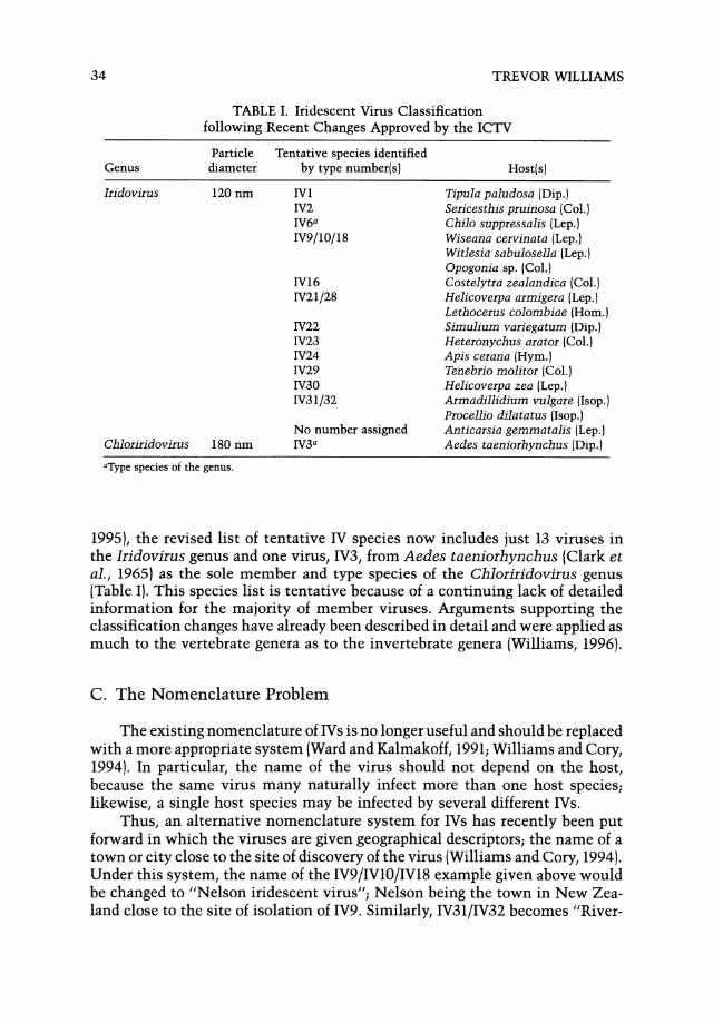
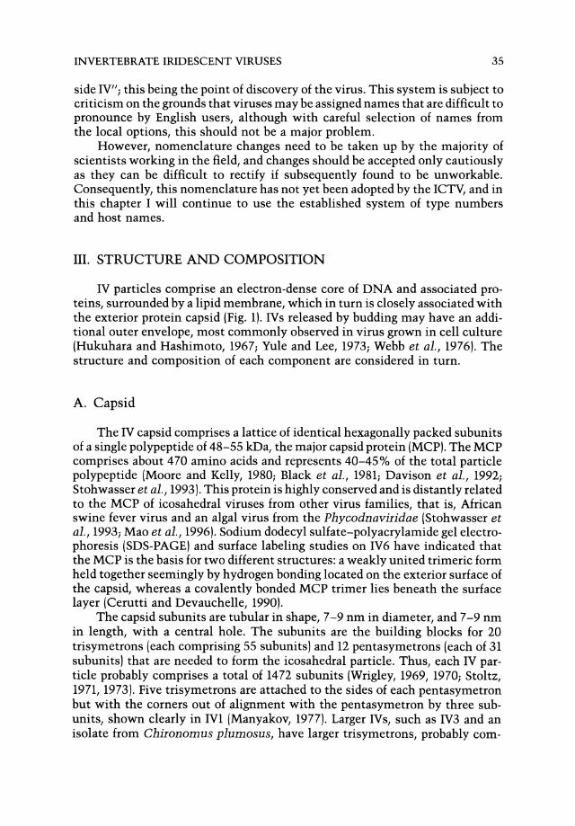
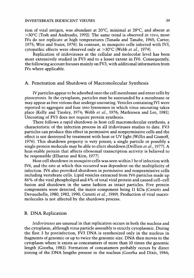

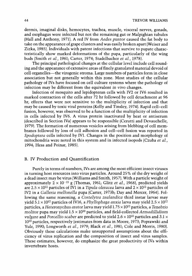
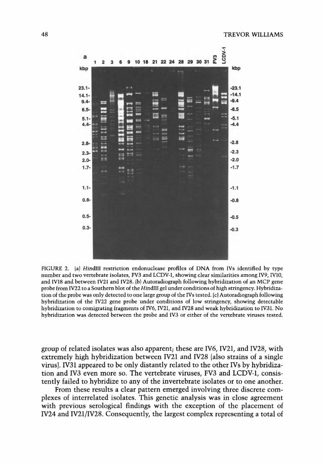
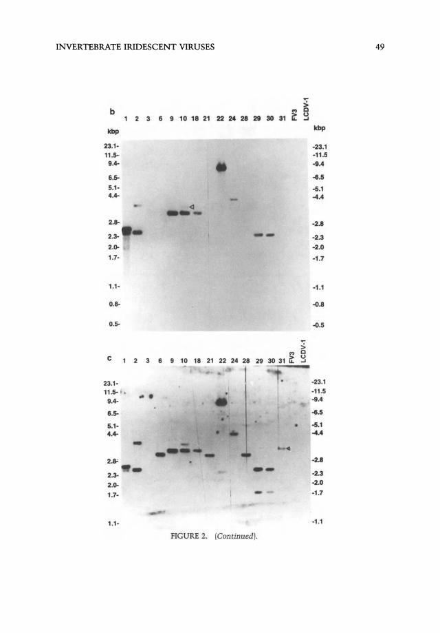
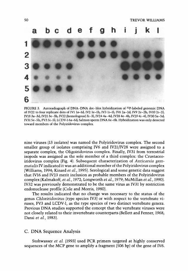
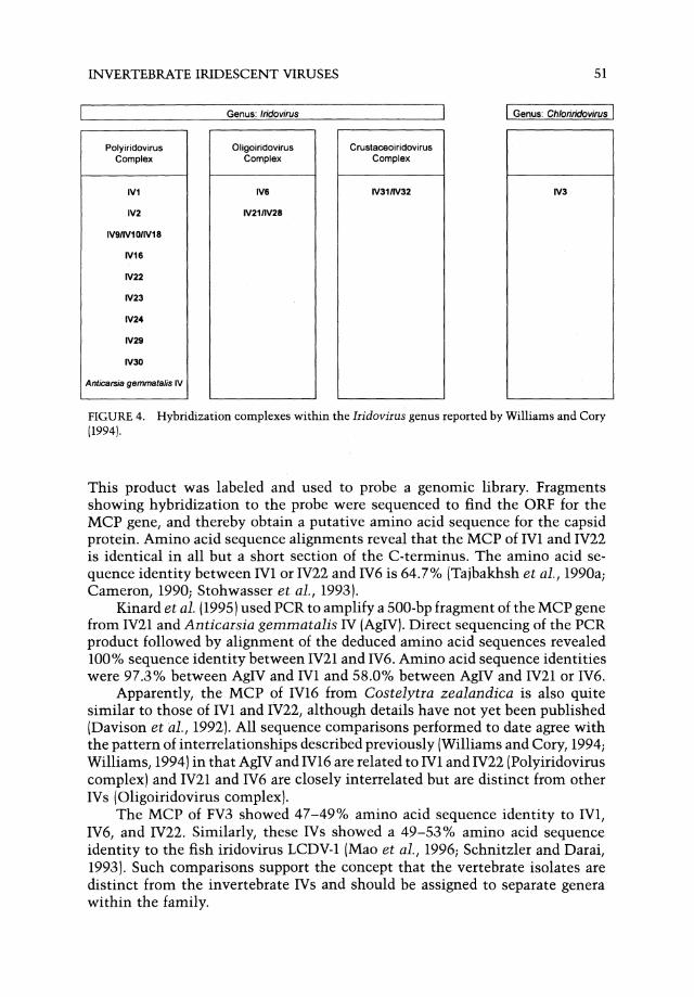

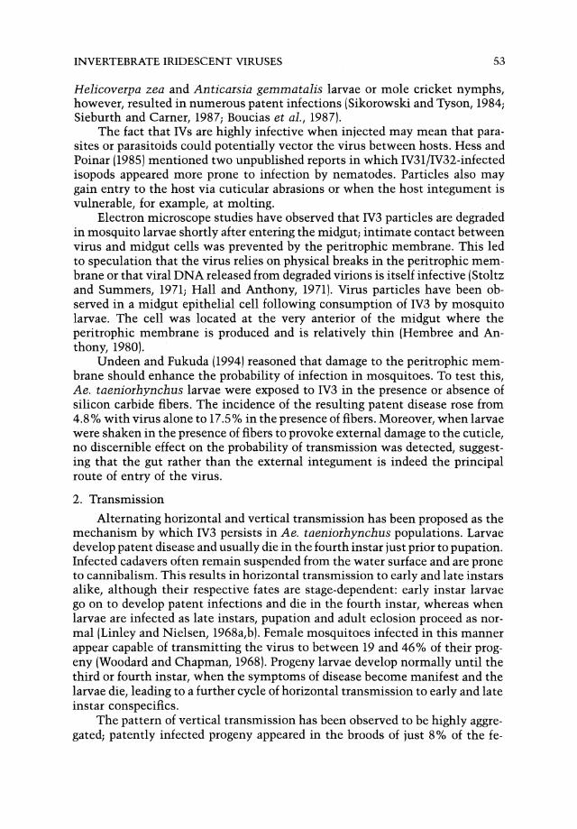
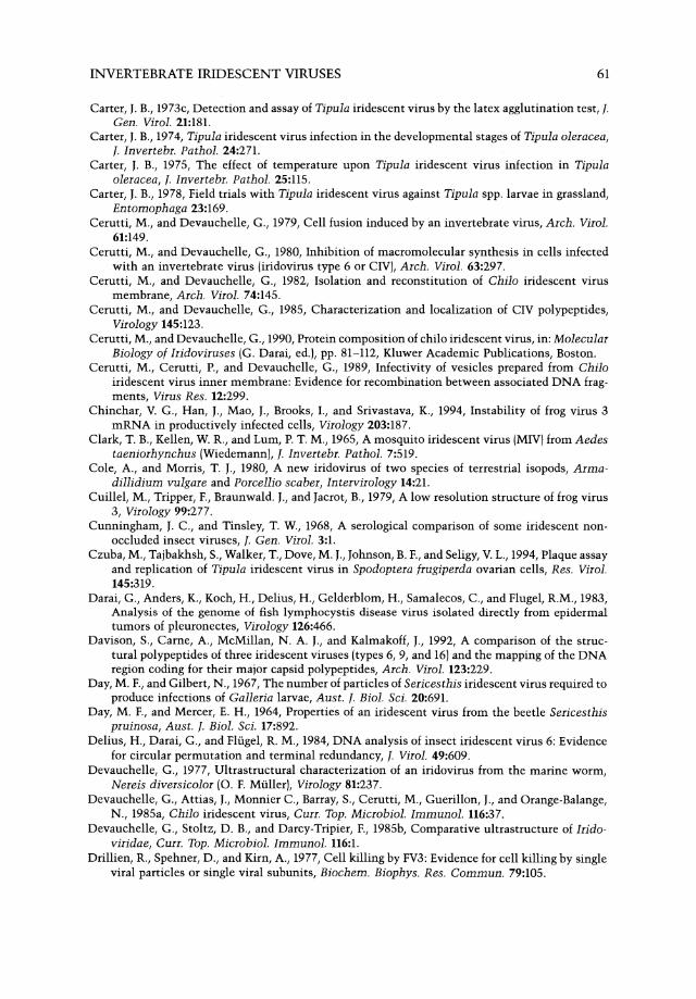
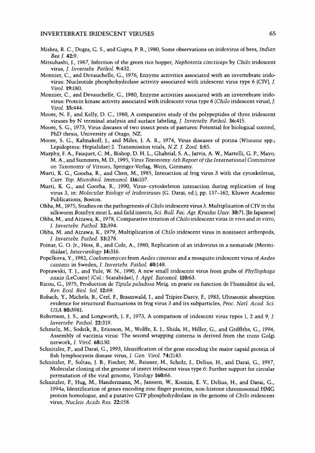
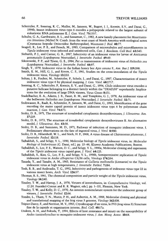
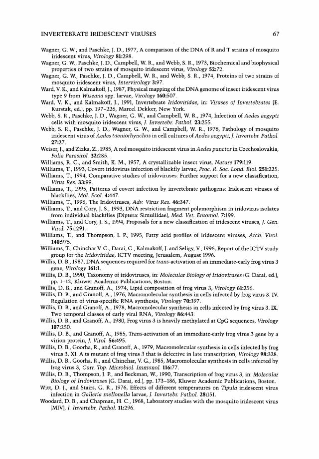
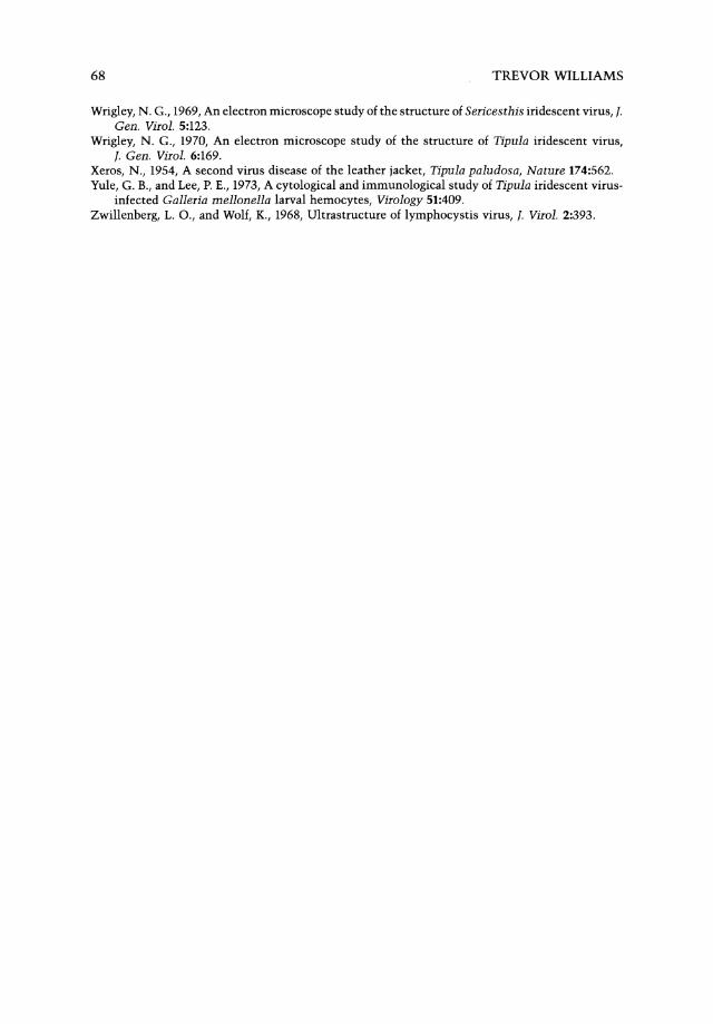


![Peter Abel Presentation für homepage [Kompatibilitätsmodus]](https://static.documents.pub/doc/80x56/631e0dcb1aedb9cd850f93c5/peter-abel-presentation-fuer-homepage-kompatibilitaetsmodus.jpg)


