Accepted Manuscript Title: Mammalian Epididymal Proteome Authors: Jean-Louis Dacheux, Cl´ emence Belleann´ ee, Russell Jones, Val´ erie Labas, Maya Belghazi, Benoˆ ıt Guyonnet, Xavier Druart, Jean Luc Gatti, Franc ¸oise Dacheux PII: S0303-7207(09)00180-4 DOI: doi:10.1016/j.mce.2009.03.007 Reference: MCE 7178 To appear in: Molecular and Cellular Endocrinology Received date: 15-11-2008 Revised date: 10-3-2009 Accepted date: 11-3-2009 Please cite this article as: Dacheux, J.-L., Belleann´ ee, C., Jones, R., Labas, V., Belghazi, M., Guyonnet, B., Druart, X., Gatti, J.L., Dacheux, F., Mammalian Epididymal Proteome, Molecular and Cellular Endocrinology (2008), doi:10.1016/j.mce.2009.03.007 This is a PDF file of an unedited manuscript that has been accepted for publication. As a service to our customers we are providing this early version of the manuscript. The manuscript will undergo copyediting, typesetting, and review of the resulting proof before it is published in its final form. Please note that during the production process errors may be discovered which could affect the content, and all legal disclaimers that apply to the journal pertain.
To appear in: Molecular and Cellular Endocrinology
Received date: 15-11-2008Revised date: 10-3-2009Accepted date: 11-3-2009
Please cite this article as: Dacheux, J.-L., Belleannee, C., Jones, R., Labas, V.,Belghazi, M., Guyonnet, B., Druart, X., Gatti, J.L., Dacheux, F., MammalianEpididymal Proteome, Molecular and Cellular Endocrinology (2008),doi:10.1016/j.mce.2009.03.007
This is a PDF file of an unedited manuscript that has been accepted for publication.As a service to our customers we are providing this early version of the manuscript.The manuscript will undergo copyediting, typesetting, and review of the resulting proofbefore it is published in its final form. Please note that during the production processerrors may be discovered which could affect the content, and all legal disclaimers thatapply to the journal pertain.
(Prostaglandin D2 synthase), clusterin, CRISP (Cystein-rich secretory protein) and E-RAPB
(epididymal retinoic acid-binding protein).
The protein composition changes continuously throughout the duct, independently of the
protein concentration in the fluid. The concentrations of the major common proteins cited
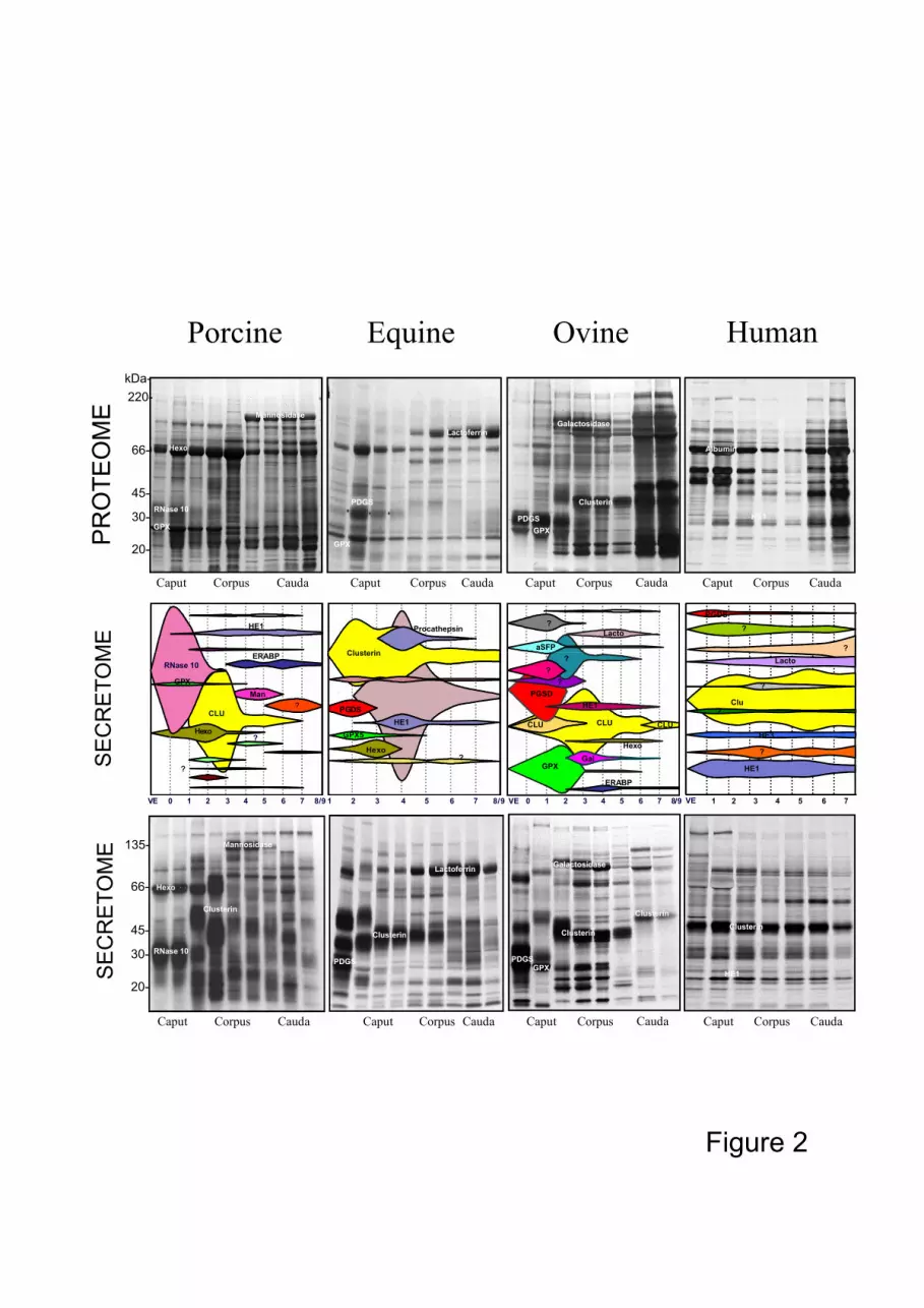
above vary between species (Fig. 2). Lactoferrin, mannosidase, PGDS and albumin are
present in high concentrations in the stallion, boar, ram and human, respectively, but GPX
and PGDS are virtually absent in humans and boar, respectively.
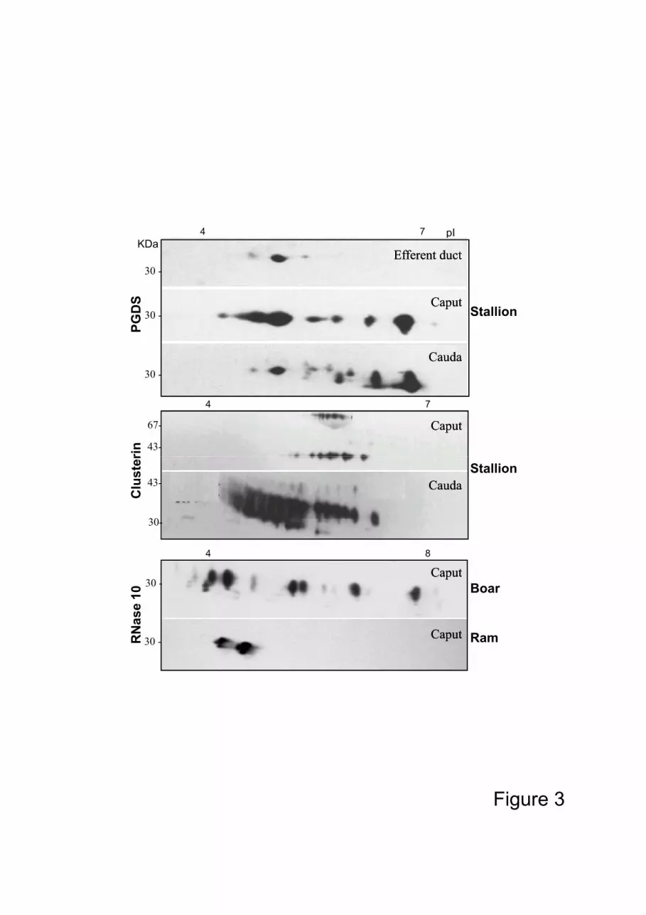
Most of the epididymal proteins are characterized by their numerous isoforms which
result from their high degree of glycosylation. The pI of these multi isoforms can vary
widely, ranging from pH 3 to 8 for the same protein (i.e. RNase 10 in the boar, Fig. 3). The 80
degree of glycosylation for the same protein can be different according to the epididymal
region eg. clusterin and PGDS in the horse, where the number of isoforms is different
between the anterior and the posterior part of the organ (Fig 3.), or RNase 10 (Train A)
which is different between species for the boar and the ram (Fig 3).
Dynamics of epididymal fluid proteomes
The spatial changes in the composition of luminal proteins are the result of two opposite
activities of the epithelium: protein secretion and protein absorption throughout the
epididymal duct. In the anterior part of the epididymis, the epididymal fluid is composed of
a mixture of testicular and epididymal proteins. Most of the proteins originating from the 90
testis, such as albumin, transferrin, testicular clusterin and PGDS (Fouchecourt et al., 2000)
are reabsorbed in the efferent ducts (Clulow et al., 1994) The rapidity of their absorption is
species-specific and generally almost all are absent in the posterior part of the epididymis,
except in humans in which albumin and transferrin are still present in large quantities.
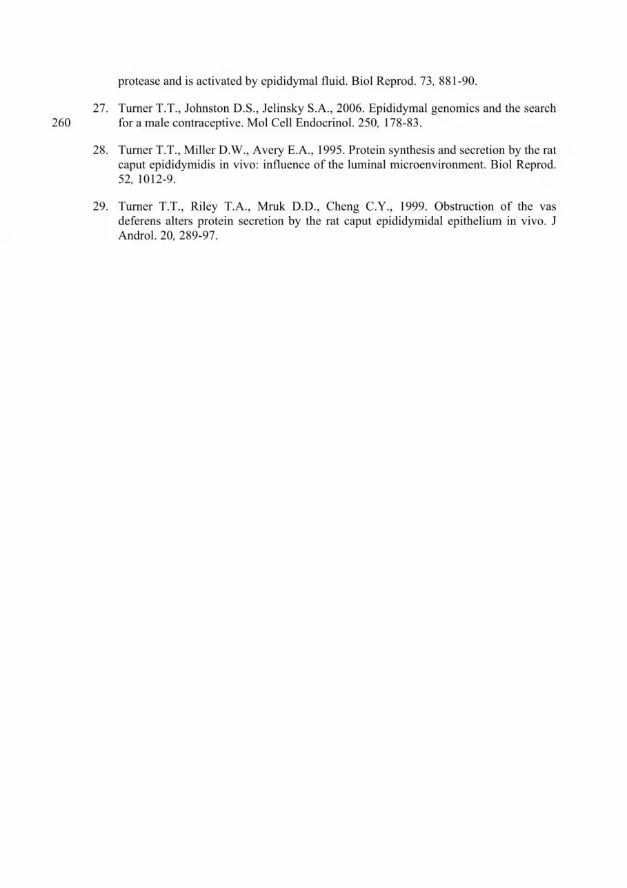
The epididymal epithelium has high protein synthesis and secretion activity, activity
being high both in the rates of protein synthesis and secretion and in the variety of proteins
secreted. The anterior part of the epididymis is the most active (Figs. 1B, 2). As for the
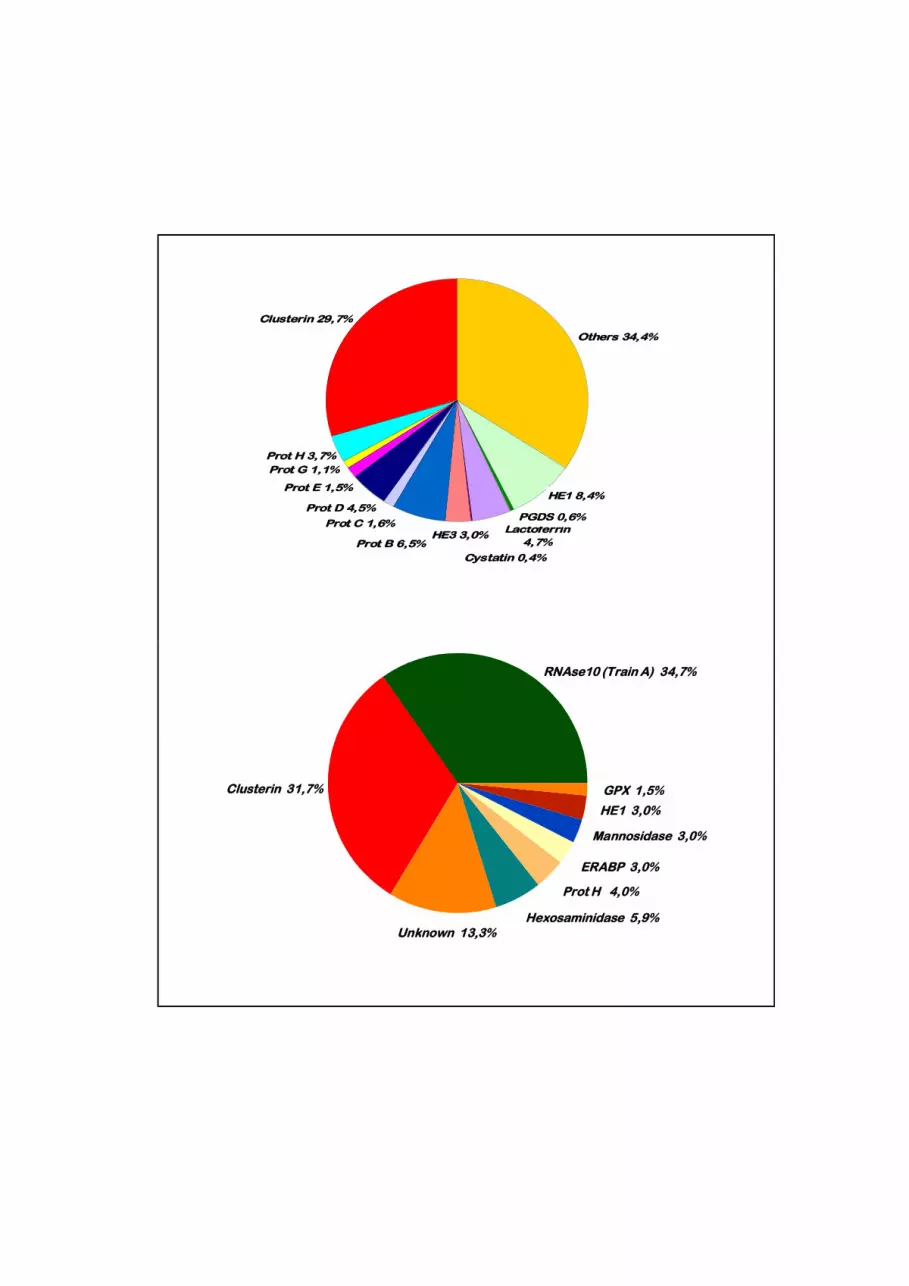
protein concentration, from 70 to 80% of the secretome is composed of 10 to 20 of. the
major secreted proteins present in the luminal fluid (Fig. 4). Most of the luminal proteins
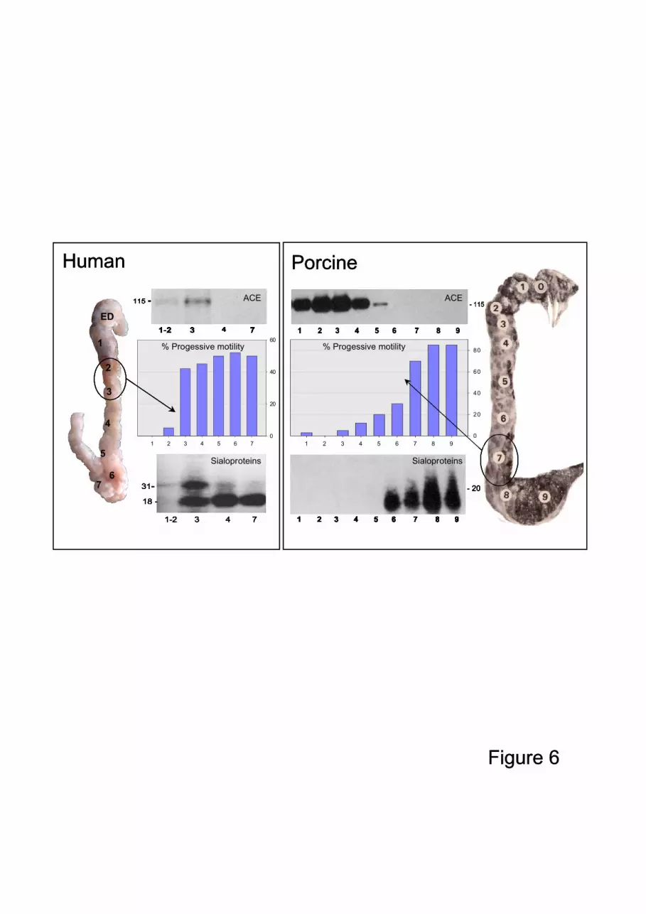
are secreted by the epithelium, but some, such as ACE, are released from the sperm surface100
by an unknown sheddase in an anterior part of the epididymis (Fig. 6) (Gatti et al.,
1999;Metayer et al., 2002; Thimon et al., 2005).
Among the different proteins secreted, the same protein can be secreted in the same
region of the epididymis in different species, for example PGDS, GPX and clusterin in the
anterior part, and glucosidases in the middle part. Clusterin is secreted at a greater rate than
the other proteins and can represent 30-40% of the all the proteins secreted. This clusterin
can be sequentially secreted under different isoforms in different parts of the epididymis as
in the stallion and the ram (Figs. 2, 3). Some highly secreted proteins are characteristic of a
species, for example, lactoferrin in the stallion, PGDS and GPXin the ram and RNase 10,
mannosidase and hexosaminidase in the boar (Fig. 2).110
In humans, in contrast to other species, few changes in pattern of protein secretion occur
throughout the epididymis, a finding which correlates with the low degree of structural
differentiation of the epididymal epithelium along the duct (Holstein, 1969).
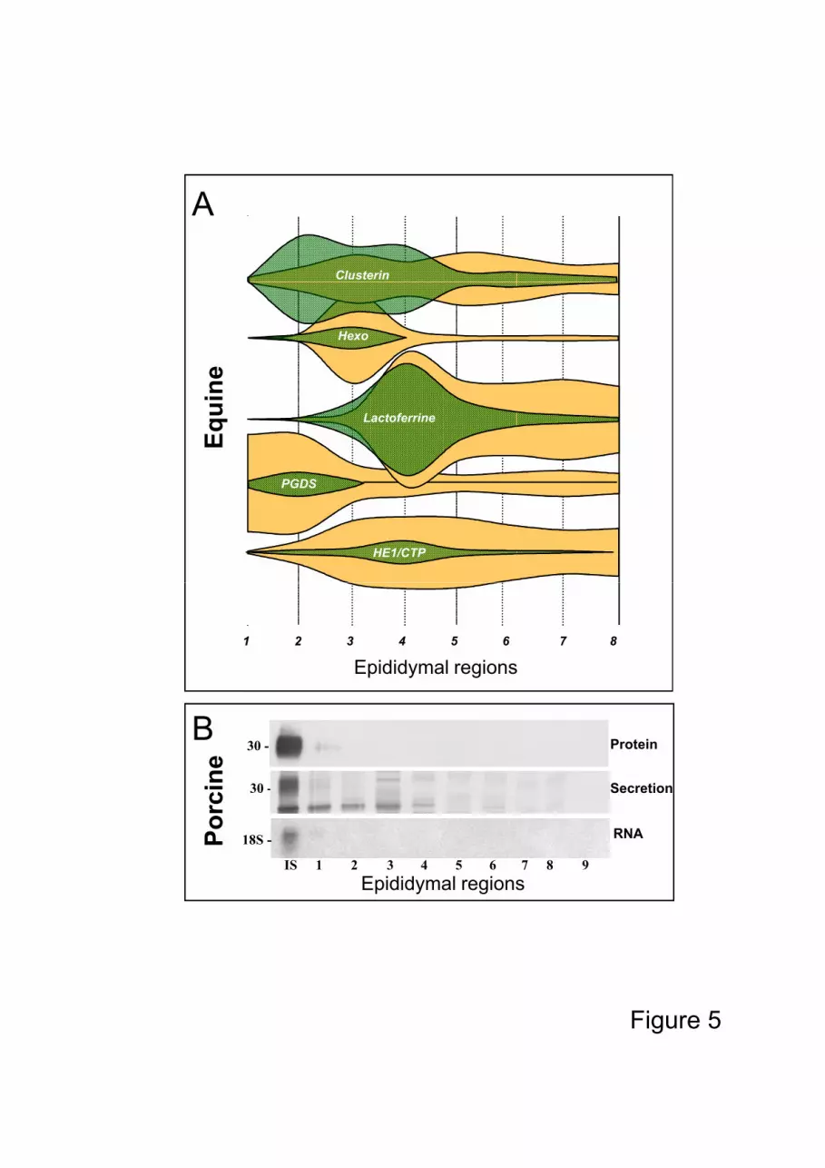
Variations in luminal protein concentrations, controlled by secretory and absorption
activites, are modulated for each species by the length of the epididymal duct involved in
the two activities, i.e. the flow rate of the luminal fluid, enzyme degradation or the binding
of protein on the sperm membrane. All these parameters differentially affect the
concentrations of almost all proteins. Some are reabsorbed as soon as they are secreted and
never reach high luminal concentrations, for example RNase 10 in the boar (Fig 5B)
(Castella et al., 2004), and several isoforms of clusterin in the stallion (Fig.3) (Fouchecourt 120
et al., 2000). For most other proteins, concentrations are high in the region where they are
secreted and gradually decrease in the following region, for example GPX and PGDS in
different species (Figs.2, 5A). The concentrations of some other proteins increase
throughout the epididymal duct, as for example lactoferrin in the stallion and
hexosaminidase and mannosidase in the boar (Fig.2).
Relationships between sperm maturation and epididymal proteome among species
The most obvious and easily observed change in sperm directly related to acquisition of
fertility is the activation of their flagella machinery. A gradual increase in the coordination
of propagation of flagellar bending makes the gamete motile and progressive. In species
that have been studied, this activation occurs in sperm in the corpus epididymides, after a 130
transitory phase of uncoordinated beating. In humans, this activation occurs in sperm from
the distal caput of the epididymis (Fig 6).
Transit of the sperm through the epididymis must be necessary for sperm maturation
since sperm maturation has not been achieved in vitro to date. Sperm maturation is probably
related to sequential modifications which occur mainly on the sperm surface. During the
maturation process, the immature gametes progressively lose or modify most of their
testicular surface proteins and gain new transient or permanent proteins in well organized
membrane protein domains. The pattern of sperm surface modification is species-specific,
related to the nature of the surface proteins involved or the epididymal regions where these
modifications occur. However, common changes in several identified sperm surface 140
proteins have been described, e.g. such as fertilin (Primakoff et al., 1988), CRISP1 (Roberts
et al., 2006), P34H (Sullivan et al., 2006) and ACE (Gatti et al., 2002), which represent the
most important sperm surface changes (Fig 6). Such common changes can also be
illustrated by the global surface changes of sialoglycoproteins on the sperm surface in the
last part of the epididymis (Dacheux et al., 1989; Dacheux et al., 1987).
Few relationships have been observed between the major sequential modifications of the
epididymal proteome and the sequential changes on the sperm surface. Furthermore, there
is currently no direct evidence of a specific role for these major proteins, although several
of them are known to be enzymes, inhibitors or binding proteins. For several of them, such
as clusterin which is the most common epididymal protein found, gene KO in mice does not 150
change the fertility of the animals (McLaughlin et al., 2000) .
It is probable that these major epididymal proteins which surround the gametes are more
involved in sperm preservation than in inducing specific and localized modifications on the
sperm surface. The high concentrations of proteins involved in the protection of gametes
from oxidative stress, such as GPX5, thioredoxin peroxidase, glutathione S-transferase P,
thioredoxin peroxidase and superoxide dismutase, probably contribute to sperm survival
during epididymal storage.
However, no more than 10% of all the proteins present in the epididymal fluid have been
identified. The wide range of protein concentrations makes identification of the less highly
represented proteins by mass spectrometry more difficult. Furthermore, these unidentified 160
proteins contain the most hydrophobic proteins. These proteins are almost never present in
a soluble form in the epididymal luminal fluid but partly associated with membrane
structures such as epididymosomes (Frenette et al., 2002; Gatti et al., 2005), or directly
transferred to the sperm surface or associated with several binding proteins such as clusterin
and several lipocalins (Ecroyd et al., 2005).
Conclusion
New approaches, including large scale analysis, are now being applied to the study of
epididymis physiology and the post-testicular differentiation of spermatozoa. Both
transcriptomic (Turner et al., 2006) and proteomic approaches provide a dramatic advance
to our understanding of male reproduction in different species. From the first results of 170
these global approaches, it is evident that significant differences exist between species
either in the sequential changes in the luminal proteome or sperm surface proteins. Each
species appears to have developed its own strategy for sperm maturation and preservation.
A general feature of sperm maturation probably exists between mammalians but it probably
involves different protein combinations. However, there is a need to identify more
epididymal and sperm proteins in order to obtain greater understanding of this aspect of the
male reproductive system which is fundamental for the survival of all species.
Reference List180
1. Castella S., Benedetti H., de Llorens R., Dacheux J.L., Dacheux F., 2004. Train A, an RNase A-like protein without RNase activity, is secreted and reabsorbed by the same epididymal cells under testicular control. Biol Reprod. 71, 1677-87.
2. Clulow J., Jones R.C., Hansen L.A., 1994. Micropuncture and cannulation studies of fluid composition and transport in the ductuli efferentes testis of the rat: comparisons with the homologous metanephric proximal tubule. Exp Physiol. 79, 915-28.
3. Dacheux J.L., Belghazi M., Lanson Y., Dacheux F., 2006. Human epididymal secretome and proteome. Mol Cell Endocrinol. 250, 36-42 .
4. Dacheux J.L., Chevrier C., Lanson Y., 1987. Motility and surface transformations of 190human spermatozoa during epididymal transit. Annals of New York Academy of Sciences. 513, 560-563.
5. Dacheux J.L., Dacheux F., Paquignon M., 1989. Changes in sperm surface membrane and luminal protein fluid content during epididymal transit in the boar. Biol Reprod. 40, 635-51.
6. Dacheux J.L., Gatti J.L., Dacheux F., 2003. Contribution of epididymal secretory proteins for spermatozoa maturation. Microsc Res Tech. 61, 7-17.
7. Dacheux J.L., Paquignon M., Lanneau M., 1984. Sequential analysis of the epididymal sperm maturation process in the boar. Ann N Y Acad Sci. 438, 526-29.
9. Ecroyd H., Belghazi M., Dacheux J.L., Gatti J.L., 2005. The epididymal soluble prion protein forms a high-molecular-mass complex in association with hydrophobic proteins. Biochem J. 392, 211-19.
10. Fouchecourt S., Metayer S., Locatelli A., Dacheux F., Dacheux J.L., 2000. Stallion epididymal fluid proteome: qualitative and quantitative characterization; secretion and dynamic changes of major proteins. Biol Reprod. 62, 1790-803.
11. Frenette G., Lessard C., Sullivan R., 2002. Selected proteins of "prostasome-like particles" from epididymal cauda fluid are transferred to epididymal caput 210spermatozoa in bull. Biol Reprod. 67, 308-13.
12. Gatti J.L., Druart X., Guerin Y., Dacheux F., Dacheux J.L., 1999. A 105- to 94-kilodalton protein in the epididymal fluids of domestic mammals is angiotensin I-converting enzyme (ACE); evidence that sperm are the source of this ACE. Biol Reprod. 60, 937-45.
13. Gatti J.L., Metayer S., Belghazi M., Dacheux F., Dacheux J.L., 2005. Identification, proteomic profiling, and origin of ram epididymal fluid exosome-like vesicles. Biol
Reprod. 72, 1452-65.
14. Gatti J.L., Metayer S., Moudjou M., Andreoletti O., Lantier F., Dacheux J.L., Sarradin P., 2002. Prion protein is secreted in soluble forms in the epididymal fluid and 220proteolytically processed and transported in seminal plasma. Biol Reprod. 67, 393-400.
15. Guérin, Y., Locatelli, Y., Comizolli, P., Mauget, R., Mermillod, P., Legendre, X., Gatti, J.L., Dacheux, J.L.2003. Conservation et utilisation du sperme épididymaire d’ovins et de cervidés en insémination artificielle et fécondation in vitro. In: Les Actes du BRG, BRG Publishers, Paris, vol 4, pp. 173-183.
16. Holstein A-F. Morphologische Studien am Nebenhoden des Menschen. Stuttgart: Geortge Thieme Verlag; 1969.
17. Jones RC. 2002. Evolution of the epididymis. In: Robaire B, Hinton BT (eds.), The epididymis from molecules to clinical practice. A comprehensive survey of the 230efferent ducts, the epididymis and the vas deferens. Kluwer Academic-Plenum Publishers, New York, London: pp. 11-33.
18. Jones R.C., 1999. To store or mature spermatozoa? The primary role of the epididymis. Int J Androl. 22, 57-67.
19. Jones R.C., Dacheux J.L., Nixon B., Ecroyd H.W., 2007. Role of the epididymis in sperm competition. Asian J Androl. 9, 493-99.
20. McLaughlin L., Zhu G., Mistry M., Ley-Ebert C., Stuart W.D., Florio C.J., Groen P.A., Witt S.A., Kimball T.R., Witte D.P., Harmony J.A., Aronow B.J., 2000. Apolipoprotein J/clusterin limits the severity of murine autoimmune myocarditis. J Clin Invest. 106, 1105-13.240
21. Metayer S., Dacheux F., Dacheux J.L., Gatti J.L., 2002. Germinal angiotensin I-converting enzyme is totally shed from the rodent sperm membrane during epididymal maturation. Biol Reprod. 67, 1763-67.
22. Primakoff P., Lathrop W., Woolman L., Cowan A., Myles D., 1988. Fully effective contraception in male and female guinea pigs immunized with the sperm protein PH-20. Nature. 335, 543-6.
23. Roberts K.P., Ensrud K.M., Wooters J.L., Nolan M.A., Johnston D.S., Hamilton D.W., 2006. Epididymal secreted protein Crisp-1 and sperm function. Mol Cell Endocrinol. 250, 122-7.
24. Sullivan R., Legare C., Villeneuve M., Foliguet B., Bissonnette F., 2006 . Levels of 250P34H, a sperm protein of epididymal origin as a predictor of conventional in vitro fertilization outcome. Fertil Steril.85, 1557-59
25. Syntin P., Dacheux F., Druart X., Gatti J.L., Okamura N., Dacheux J.L., 1996. Characterization and identification of proteins secreted in the various regions of the adult boar epididymis. Biol Reprod. 55, 956-74.
26. Thimon V., Metayer S., Belghazi M., Dacheux F., Dacheux J.L., Gatti J.L., 2005. Shedding of the germinal angiotensin I-converting enzyme (gACE) involves a serine
protease and is activated by epididymal fluid. Biol Reprod. 73, 881-90.
27. Turner T.T., Johnston D.S., Jelinsky S.A., 2006. Epididymal genomics and the search for a male contraceptive. Mol Cell Endocrinol. 250, 178-83.260
28. Turner T.T., Miller D.W., Avery E.A., 1995. Protein synthesis and secretion by the rat caput epididymidis in vivo: influence of the luminal microenvironment. Biol Reprod. 52, 1012-9.
29. Turner T.T., Riley T.A., Mruk D.D., Cheng C.Y., 1999. Obstruction of the vas deferens alters protein secretion by the rat caput epididymidal epithelium in vivo. J Androl. 20, 289-97.
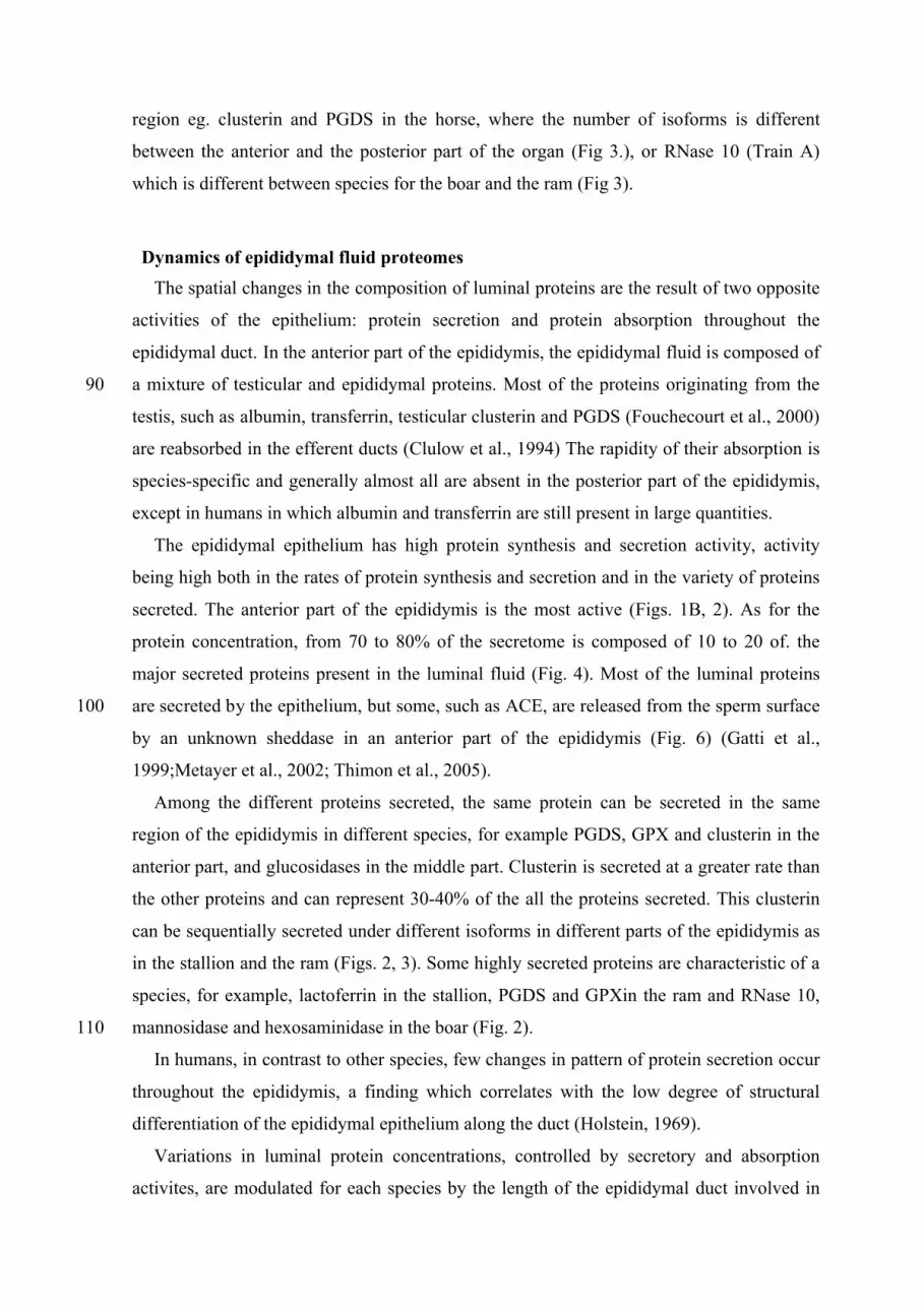
Legends of the figures:
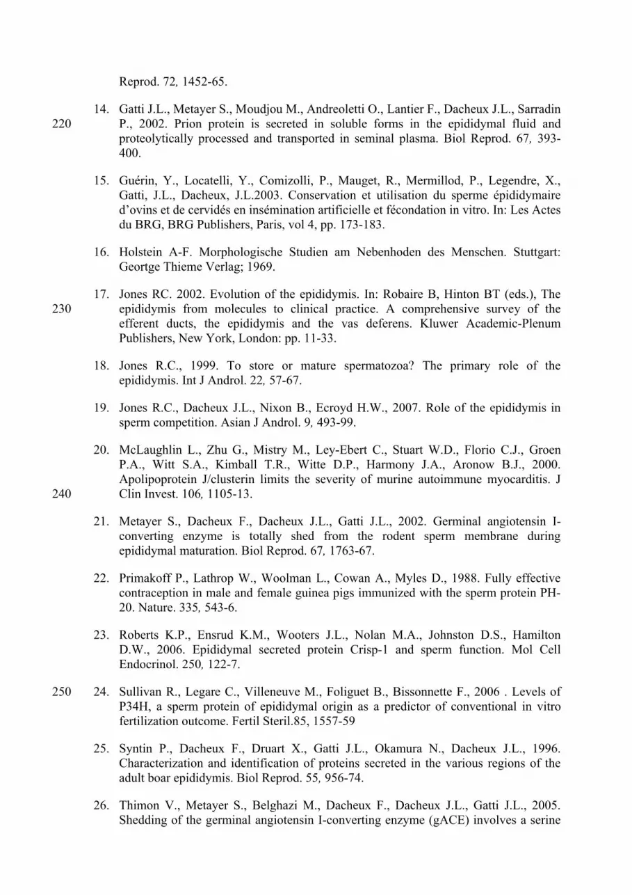
Figure 1: Spermatocrit (A), protein secretion (B) and concentrations of luminal proteins (C) 270
in the epididymal fluid from 9 regions of the epididymis (1-4: caput; 5-6:
corpus; 7-9: cauda) for three species (ram, boar, stallion) from (Syntin et al.,
1996; Fouchecourt et al., 2000) and unpublished data.
Figure 2: Epididymal secretomes and proteomes of four species. For proteomes, each plate
corresponds to 1D gel electrophoresis separation of about the same quantity of
epididymal protein from each region of the epididymis. The secretome diagrams
represent each secreted protein from the different epididymal regions and are
expressed as the percentage of total secretion of the whole organ. The plates
illustrating the secretory activities of the different epididymal regions for the
four animal species correspond to the autoradiograms of the same four 1D gel 280
separations presented above for the proteome (Syntin et al., 1996; Fouchecourt
et al., 2000 ; Druart et al., 1994 ; Druart, unpublished data ; Dacheux et al.,
2006).
Figure 3: Immunodetection of 2D electrophoresis gels of isoforms of three proteins (PGDS,
clusterin and RNase 10) according to their epididymal and species origins
(stallion: Fouchecourt et al., 2000; boar: Castella et al., 2004 and ram:
unpublished data).
Figure 4: Distribution of the major proteins secreted by the human and boar epididymis
(Dacheux et al., 2006; Syntin et al., 1996)
Figure 5: Relationship between secretome and proteome. A) Secretion and protein 290
concentrations for five major proteins present in the epididymal fluid of the
stallion. B) Epididymal localisation of the secretion, luminal protein and
corresponding RNA of RNase 10 (Train A) from the initial segment (IS), caput
(1-4), corpus (5-6), cauda (7-9) in the boar (Castella et al., 2004).
Figure 6: Sperm epididymal maturation according to epididymal region in the human
(Dacheux et al., 1987) and porcine (Dacheux et al., 1984) related to ACE,
sialoprotein modifications on the sperm surface and percentage of progressive
motility along the epididymis.
A
B
Figure 1
C
Figure
Equine OvinePorcine HumankDa-
PR
OT
EO
ME
66-
45-
30-
20-
220-
SE
CR
ET
OM
E
Clusterin?
?
PGDS
Caput Corpus Cauda Caput Corpus CaudaCaput Corpus Cauda CaudaCaput Corpus
HE1
ERABP
Lactoferrin
Mannosidase
PDGS
PDGS
Hexo
RNase 10
GPX
GPX GPX
Clusterin
Albumin
Galactosidase
HE1
ProcathepsinLacto
aSFP
?
Figure 2
SE
CR
ET
OM
E
66-
45-
30-
20-
Caput Corpus Cauda Caput Corpus CaudaCaput Corpus Cauda CaudaCaput Corpus