Vol. 14: 195-205, 1992 DISEASES OF AQUATIC ORGANISMS Dis. aquat. Org. Published December 15 Effects of cortisol implants on the susceptibility and the histopathology of the responses of naive coho salmon Oncorhynchus kisutch to experimental infection with Lepeophtheirus salmonis (Copepoda: Caligidae) S. C. Johnson'.', L. J. ~lbright~ ' Department of Fisheries and Oceans, Biological Sciences Branch, Pacific Biological Station, Nanaimo, British Columbia, Canada V9R 5K6 ZInstitute for Aquaculture Research, Simon Fraser University, Burnaby, British Columbia, Canada V5A IS6 ABSTRACT. The effects of hydrocortisol implants on the susceptibility of naive coho salmon Oncorhynchus kisutch to infection with the economically important marine ectoparasitlc copepod Lepeophtheirus salmonis was investigated under laboratory conditions. Cortisol-implanted coho salmon were more susceptible to infection than the control coho salmon. Copepods were lost from the gills of the control coho salmon by 10 d post-infection, and only a few remained on the body and fins at 20 d post-infection. Copepods were retained on the gills, fins and body of the cortisol-implanted coho salmon over the 20 d studied. Histological sections of control coho gills and fins revealed well devel- oped epithelial hyperplasias and inflammatory responses to the presence of L. salmonis. The magni- tude of the inflammatory response and the development of epithelial hyperplasia was suppressed in the cortisol-implanted coho. These data support the hypothesis that non-specific host defence mechanisms are important in resistance of coho salmon to infection with L, salmonis. INTRODUCTION Lepeophtheirus salmonis is a common marine ecto- parasitic copepod of wild and pen-reared salmonids throughout the northern hemisphere (Kabata 1979, 1988, Wootten et al. 1982). It has a direct life cycle consisting of 5 phases and 10 stages. These include 2 free-swimming naupliar stages, 1 free-swimming infectious copepodid stage, 4 attached chalimus stages, 2 preadult stages and an adult stage (Johnson & Albright 1991a). Attached copepodids, chalimus larvae, preadults and adults feed on host mucus, skin and blood (Kabata 1974, Brandal et al. 1976). When abundant, L. salmonis causes serious disease which is characterized by extensive areas of skin erosion and hemorrhaging on the head and back, and a distinct 'Addressee for correspondence 0 Inter-Research 1992 area of erosion and sub-epidermal hemorrhages in the perianal region (Brandal & Egidius 1979, Wootten et al. 1982). Johnson & Albright (1992) demonstrated that naive coho salmon Oncorhynchus kisutch are more resis- tant than naive chinook Oncorhynchus tshawytscha or naive Atlantic salmon Salmo salar to experimental infection with Lepeophtheirus salmonis. Histological examination of attachment and feeding sites on the gills of coho salmon revealed a well developed inflammatory response as early as 1 d post-infection. Copepods were eliminated from the gills of coho salmon from 10 d post-infection onwards. Histological examination of infected fins from coho salmon revealed variable epidermal erosion and inflamma- tion of the dermis up to 5 d post-infection. At 10, 15 and 20 d post-infection the lesions were characterized by well developed epithelial hyperplasias and intense inflammatory responses.
Transcript
Vol. 14: 195-205, 1992 DISEASES OF AQUATIC ORGANISMS
Dis. aquat. Org. Published December 15
Effects of cortisol implants on the susceptibility and the histopathology of the responses of
naive coho salmon Oncorhynchus kisutch to experimental infection with Lepeophtheirus
salmonis (Copepoda: Caligidae)
S. C. Johnson'.', L. J. ~ l b r i g h t ~
' Department of Fisheries and Oceans, Biological Sciences Branch, Pacific Biological Station, Nanaimo, British Columbia, Canada V9R 5K6
ZInstitute for Aquaculture Research, Simon Fraser University, Burnaby, British Columbia, Canada V5A IS6
ABSTRACT. The effects of hydrocortisol implants on the susceptibility of naive coho salmon Oncorhynchus kisutch to infection with the economically important marine ectoparasitlc copepod Lepeophtheirus salmonis was investigated under laboratory conditions. Cortisol-implanted coho salmon were more susceptible to infection than the control coho salmon. Copepods were lost from the gills of the control coho salmon by 10 d post-infection, and only a few remained on the body and fins at 20 d post-infection. Copepods were retained on the gills, fins and body of the cortisol-implanted coho salmon over the 20 d studied. Histological sections of control coho gills and fins revealed well devel- oped epithelial hyperplasias and inflammatory responses to the presence of L. salmonis. The magni- tude of the inflammatory response and the development of epithelial hyperplasia was suppressed in the cortisol-implanted coho. These data support the hypothesis that non-specific host defence mechanisms are important in resistance of coho salmon to infection with L, salmonis.
INTRODUCTION
Lepeophtheirus salmonis is a common marine ecto- parasitic copepod of wild and pen-reared salmonids throughout the northern hemisphere (Kabata 1979, 1988, Wootten et al. 1982). It has a direct life cycle consisting of 5 phases and 10 stages. These include 2 free-swimming naupliar stages, 1 free-swimming infectious copepodid stage, 4 attached chalimus stages, 2 preadult stages and an adult stage (Johnson & Albright 1991a). Attached copepodids, chalimus larvae, preadults and adults feed on host mucus, skin and blood (Kabata 1974, Brandal et al. 1976). When abundant, L. salmonis causes serious disease which is characterized by extensive areas of skin erosion and hemorrhaging on the head and back, and a distinct
'Addressee for correspondence
0 Inter-Research 1992
area of erosion and sub-epidermal hemorrhages in the perianal region (Brandal & Egidius 1979, Wootten et al. 1982).
Johnson & Albright (1992) demonstrated that naive coho salmon Oncorhynchus kisutch are more resis- tant than naive chinook Oncorhynchus tshawytscha or naive Atlantic salmon Salmo salar to experimental infection with Lepeophtheirus salmonis. Histological examination of attachment and feeding sites on the gills of coho salmon revealed a well developed inflammatory response as early as 1 d post-infection. Copepods were eliminated from the gills of coho salmon from 10 d post-infection onwards. Histological examination of infected fins from coho salmon revealed variable epidermal erosion and inflamma- tion of the dermis up to 5 d post-infection. At 10, 15 and 20 d post-infection the lesions were characterized by well developed epithelial hyperplasias and intense inflammatory responses.
196 Dis. aquat. Org. 14: 195-205, 1992
In the initial phases of the infection, reduction in the intensity of Lepeophtheirus salmonis on coho salmon is thought to be caused by non-specific host responses, including epithelia1 hyperplasia, soluble or cellular factors of the inflammatory response, and/or possibly serum enzymes or other proteins. Specific humoral or cellular factors may be important in later stages of the infection.
Implantation of corticosteroids in fish has been shown to increase their susceptibility to a wide variety of parasitic diseases (reviewed in Pickering 1987; Kent & Hedrick 1987, Nazrul Islam & Woo 1991). In fish, the implantation of hydrocortisol has been demonstrated to inhibit inflammatory responses, inhibit phagocytosis, suppress both humoral and cellular immune responses, and retard the wound healing process (Kent & Hedrick 1987, Pickerlng 1987, Roubal & Bullock 1988, Saad 1988, Nazrul Islam & Woo 1991).
In this study, naive coho salmon were treated with cortisol implants to determine the effects of immuno- suppression on their resistance to experimental infec- tion with Lepeophtheirus salmonis. The effects of cortisol implantation on the histopathology of attach- ment and feeding sites of L. salmonis on the gills and fins are also described.
MATERIALS AND METHODS
Parasite exposure, fish maintenance, and examina- tion. Sample fish (45 cortisol-implanted and 45 control) were introduced into separate 500 1 fiberglass tanks and allowed to acclirnate for 1 wk. The cortisol- implanted and adipose clipped fish were then trans- ferred into the tank containing the control fish and both groups of fish were exposed for 24 h to ca 4000 newly molted copepodid larvae. The methods for obtaining the copepodids and for carrying out the infections are given in Johnson & Albright (1991b). Prior to copepodid exposure and at 10 and 20 d post- infection, plasma samples were collected from 5 fish from each of the control and cortisol-implanted groups for cortisol analysis. At sampling, the fish were anes- thetized with a high dose of MS-222 (tricaine methane- sulfonate), had their caudal fins severed, and their blood was collected in 5 m1 heparinized tubes for plasma cortisol analysis.
Fish were maintained in flowing sea water with a temperature of 10.7 to 12.6 'C (mean: 11.5 "C) and ambient salinity (29 to 31 %A). All fish were fed a com- mercial dry pellet feed at 1% body wt d-'. At 1, 3, 5, 10, 15 and 20 d post-infection, 6 of each of the cortisol- implanted and control fish were rapidly killed with MS-222. The fork length and wet weight was deter- mined for each fish. Both the anesthetic bath and the
body surfaces were examined for copepods and the distribution of the copepods on the fish was noted. The number of copepods present was corrected to a stan- dard wet body wt to compensate for differences in size among hosts.
Intensity data were log (X + 1) transformed and dif- ferences in copepod intensity (number of parasites per infected host) investigated by analysis of variance (ANOVA) procedures. Comparisons of copepod inten- sity for each treatment over time were made using Scheffe's tests (Zar 1984). Comparisons of copepod intensity between treatments at each sampling period were made using t-tests.
Cortisol administration and analysis. Coconut oil was pasteurized at 70 'C, cooled to 30 "C, and mixed with hydrocortisol (Sigma No. H-4001) to yield a final concentration of 127 mg hydrocortisol ml- ' oil. A group of 45 naive coho were anesthetized with MS-222, adipose fin-clipped, and injected intraperitoneally with 0.2 m1 of cortisol solution per fish. This resulted in an implant of ca 0.5 mg hydrocortisol of fish based on an average fish wt of 51.0 g. A second group of 45 naive coho of the same size were selected as controls. These fish were anesthetized but not injected.
To determine plasma cortisol levels blood samples were spun at 6500 rpm (1900 X g ) for 5 min, and the resulting plasma was transferred to sterile cryotubes. Plasma samples were stored frozen at -70 "C and their cortisol levels determined using a radioimmuno- assay technique (Sumpter & Donaldson 1986).
Histology. Tissues for examination by light rnicro- scopy were fixed in Davidson's solution and dehy- drated through to 100 O/o alcohol. Tissues were either wax-embedded, cut to a thickness of 5 pm, and stained with hematoxlylin and eosin, or they were embedded in JB4 plastic resin, cut to a thickness of l to 2 p.m, and stained with Lee's stain (methylene blue and basic fuschin).
RESULTS
Intensity of infection
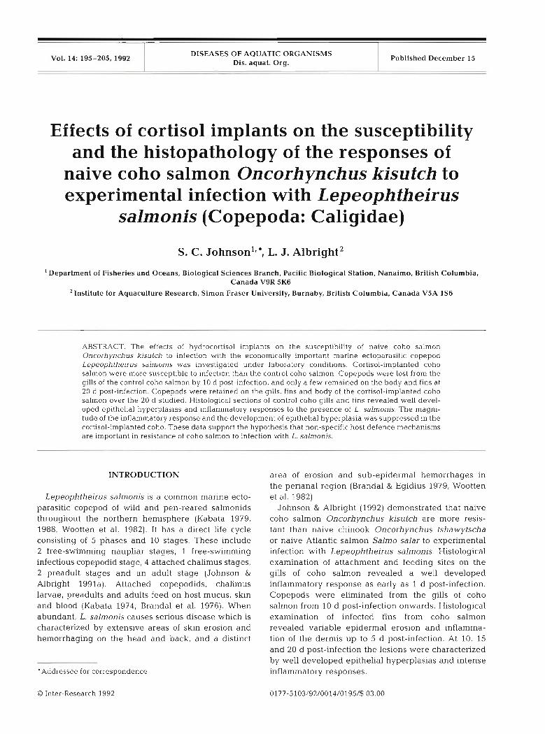
The intensity of Lepeophtheirus salmonis on the cortisol-implanted and control groups over time is presented in Fig. 1. The intensities of infection for both the cortisol-implanted and control groups were signifi- cantly different over time (l-way ANOVA; cortlsol- implanted. p<0.001; control: p<0.001). The results of multiple range tests (Scheffk's test; p<0.05) over time showed the control group to have significantly fewer copepods at 20 d post-infection when compared to 1, 3, 5 and 10 d post-infection, and the cortisol-implanted group to have significantly fewer parasites at 20 d post-
Johnson & Albright: Cortisol and susceptibility of coho salmon to Lepeophtheirus salmonis 197
EB Cortlsol
m Control
u
1 3 5 1 0 1 5 20 DAYS POST- INFECTION
Fig. 1 Lepeophtheirus salrnonis ~nfecting Oncorhynchus kisutch. Mean (+ SE) intensity on naive cortisol-implanted and naive control coho salmon at vanous times post-infection. F ~ s h were maintained at 10.7 to 12 6 ' C and ambient salinity
(29 to 31 960)
infection when compared to 1, 3, 5, 10 and 15 d post- infection (Fig. 1). At each sampling time, significantly fewer copepods were present on the control group than on the cortisol-implanted group (t-test; ~ ~ 0 . 0 5 ) .
Distribution
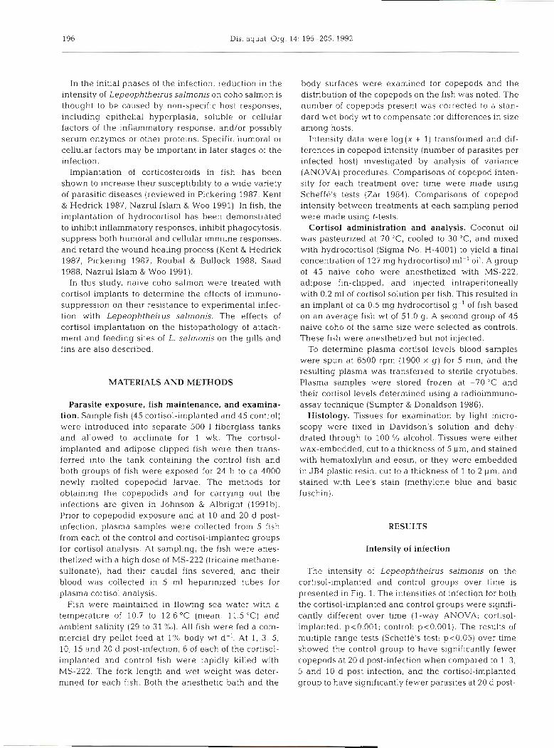
The highest percentage of copepods recovered from the control group at all sampling penods except at 20 d post-infection was from the fins (Fig. 2). The percent-
Bath Fins 46
I Body m GIIIS 59
1 3 5 1 0 15 20 DAYS POST-INFECTION
Fig. 2. Lepeophtheirus salmonls infecting Oncorhynchus kisutch Distribution on naive control coho salmon; conditions as In Fig. 1 Values above bars are the total number of
copepods collected
age of copepods recovered from the anesthetic bath decreased from ca 21 % to 0 % by 10 d post-infection. Of the copepods attached to the host at 1 and 3 d post- infection, ca 30 % were collected from the gills. The percentage of copepods on the gills declined to 0 O/o by 10 d post-infection. Small numbers of copepods were recovered from the body surfaces at 1, 3 and 5 d post- infection. At 15 and 20 d post-infection the percent- ages on the body surfaces increased due to molting to the preadult stage and shifting of their attachment sites.
The highest percentages of copepods recovered from the cortisol-implanted group were from the gills and fins at all sampling periods except 20 d post- infection (Fig. 3). Small percentages of copepods were recovered from the anesthetic bath up to 5 d post- infection, and from the body surfaces from 1 to 10 d post-infection. As with the control fish the percentages on the body surface increased at 15 and 20 d post- infection due to molting to the preadult stage and shift- ing of their attachment sites.
Mortality and gross morphology of the lesions
Of the cortisol-implanted fish, 16 % died between 9 and 17 d post-infection. These fish showed severe ero- sion of the snout and fins caused by heavy infections of filamentous bacteria (seawater 'myxobacteriosis'). From 5 d post-infection onwards all of the cortisol- implanted fish were lethargic and showed varying amounts of erosion of the snout and/or fins. No mor- tality occurred in the control group and no body and/or fin erosion was evident in the control fish sampled.
1 3 5 1 0 15 20 DAYS POST-INFECTION
Fig. 3. Lepeophtheirus salrnonis infecting Oncorhynchus kjsutch. Distribution on naive cortisol-implanted coho salmon; conditions as in Fig. 1. Values above bars are the
total number of copepods collected
198 Dis. aquat. Org. 14: 195-205. 1992
Table 1. Lepeophtheirus salmonis infect~ng Oncorhynchus kisutch. Ranges of plasma cort~sol levels (ng ml-') In naive coho salmon given intraperitoneal implants of hydrocortisol. pre-infection and after infection. All ranges are based on
dValues above the detection limit of the radioimmunoassay
Cortisol-treated fish had much higher plasma cortisol levels than the control fish in all samples (Table 1).
Grossly, the gills of both the control and cortisol- implanted groups at 1, 3 and 5 d post-infection showed variable amounts of erosion and clubbing of the filament tips. From 10 to 20 d post-infection the gills oi the cortisol-implanted group showed progressive ero- sion of the filament tips, clubbing, and fusion of the secondary lamellae.
Gross examination of 45 lesions on the fins of the control group over the period of 10 to 20 d post- infection revealed areas of proliferating host tissue immediately adjacent to or surrounding the copepods. Gross examination of 45 lesions on the fins of the corti- sol-implanted group over the same period revealed no tissue response to the presence of the copepods.
Gill histology
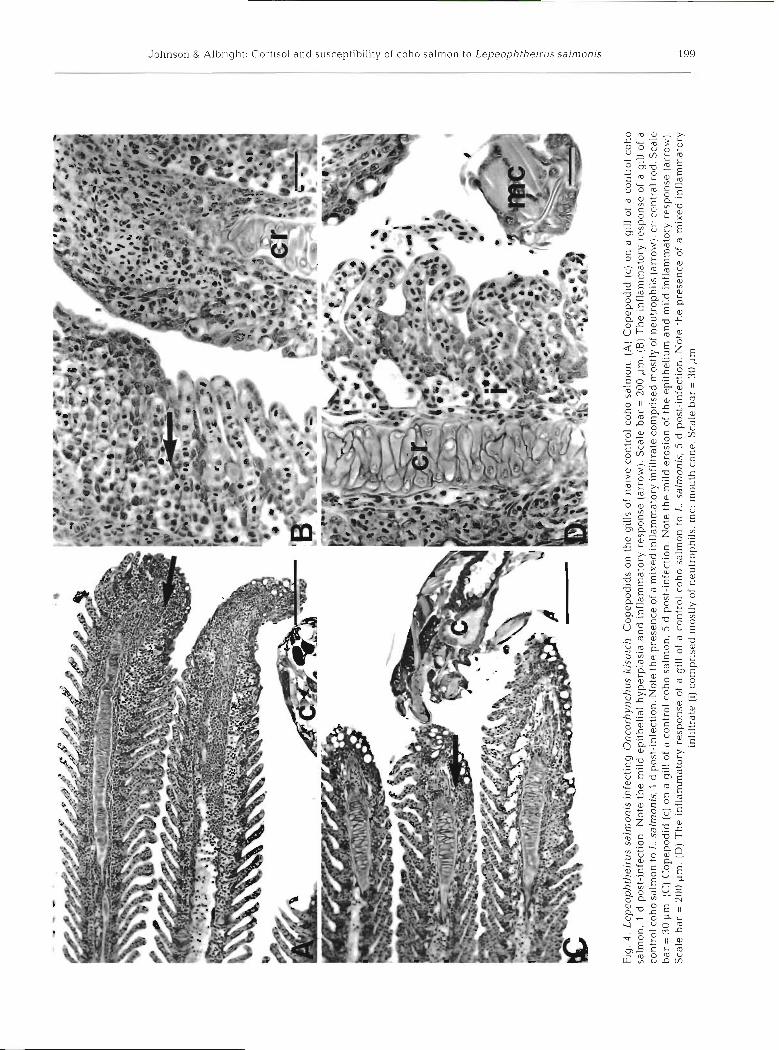
At 1 and 5 d post-infection, attachment and feeding sites on the gills of control coho salmon were charac- terized by variable amounts of erosion of the epithe- lium, hemorrhage, and inflammation (Fig. 4A to D). In some cases minor epithelial hyperplasia resulted in fusion of the secondary lamellae near the tips of the primary lamellae. In severe cases the central rods of the primary lamellae were exposed. The inflammatory infiltrate consisted of abundant neutrophils and a few lymphocytes (Fig. 4B, D). No secondary infection of the gills was observed over this period.
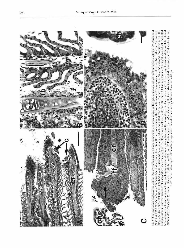
At l , 5 and 10 d post-infection, attachment and feed- ing sites on the gills of cortlsol-implanted coho salmon were characterized by variable amounts of erosion of the epithelium and underlying tissues, hemorrhage, and mild inflammatory responses (Fig. 5A, B). In most instances the inflammatory response was limited to the presence of scattered inflammatory cells (mostly neutrophils). In some sections a s~nal l amount of epithel~al hyperplasia and fusion of the secondary lamellae was evident. At 1 and 5 d post-infection, both the intensities of the inflammatory responses and the
extent of epithelial hyperplasias appeared to be less than those seen in the control group. Examination of attachment and feeding sites at 15 and 20 d post- infection revealed increased numbers of primary lamel- lae affected, well developed inflammatory responses, and increased levels of epithelial hyperplasia and fusion of the secondary gill lamellae (Fig. 5C, D). Hemorrhage in the tissues at the site of parasite attachment was a common feature of the lesions over this period. The inflammatory infiltrate consisted primarily of neutro- phils, but lymphocytes were also present (Fig. 5D). Secondary infection of the gill lesions by fllamentous bacteria occurred in some sections.
Fin histology
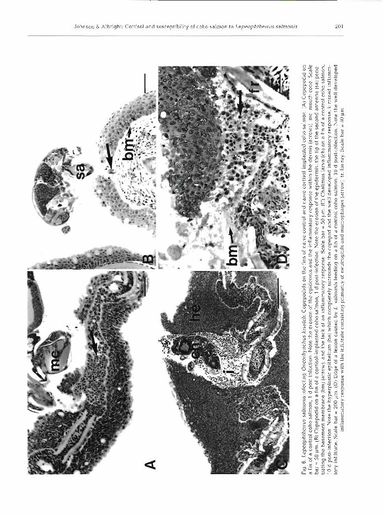
Over the period of 1 to 5 d post-infection, attachment and feeding sites on fins of both the control and cortl- sol-implanted groups were characterized by variable amounts of epidermal erosion and necrosis (Fig. 6A, B). Over this period the severity of the lesions was highly variable within treatment groups with later lesions not necessarily more severe than earlier lesions. Mild inflammation of the dermis occurred in the control group as early as 1 d post-infection (Fig. 6A). Neutro- phils were the predominant cells at these sites of inflammation. No inflammation was observed in the cortisol-implanted group over this period.
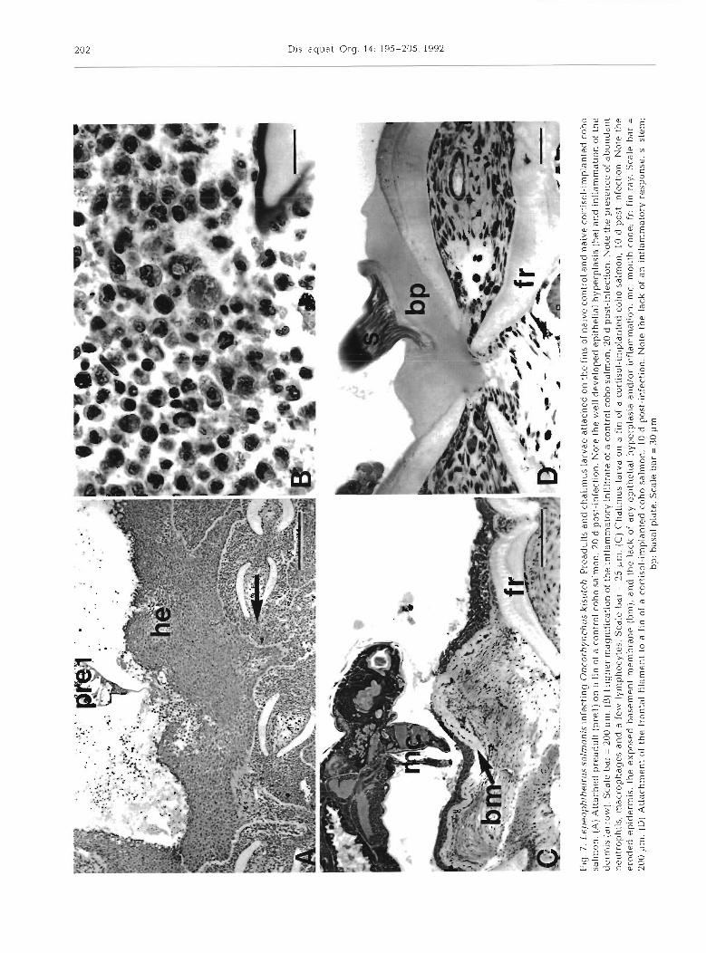
At 10.15 and 20 d post infection, attachment and feed- ing sites on the fins of the control group were character- ized by well developed epithelial hyperplasias (Figs. 6C to ?B), which in severe cases resulted in the complete encapsulation of the copepod. In cases of encapsulation the spaces surrounding the copepod were filled with a mixed inflammatory infiltrate (neutrophils, macro- phages, and a few lymphocytes), and necrotic tissue. Intense inflammation of the dermis occurred in all con- trol samples over tl-us penod. The inflammatory infiltrate consisted of abundant neutrophils and macrophages and a few lymphocytes (Figs. 6D & ?B).
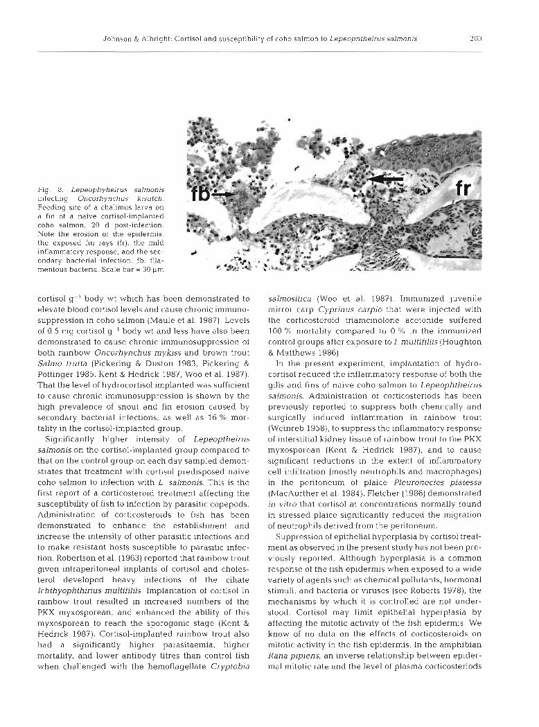
With exception of 2 samples that showed mild epithelial hyperplasia, hyperplasia was not seen in the cortisol-implanted group over the period of 10 to 20 d post-infection (Fig 7C, D). Mild inflammation of the dermis occurred in all samples collected at 15 and 20 d post-infection. The inflammatory infiltrate consisted of neutrophils, some macrophages, and some lympho- cytes (Fig. 8).
DISCUSSION
The level of hydrocortisol implanted in coho salmon in this investigation is higher than the level of 0.01 mg
url of,- =
1eq ap
s 'auos qlnour :
m .s[!qdo~
lnau jo Allsour p
as!~d
uo
s (!) a~
el~[!ju
! A
~oiem
ure[j~r! paxrm
e jo asu
asa
~d
aql aloN 'u
oI
~~
~J
u!
-~
SO
~
p S 'sruornps .7 01 uourles oqos loi1uos e jo 1116 e jo asuodsai Aioleunue~
ju! aqL
(a) .u
rl 00~
= req aless
.(
M~
JJ
~)
asu
od
sa~ ~ro
leuru
re~ju
! pl!m
pue urn!laql!da
aql jo uolsola p11~1.1 aql aloN
~uo!l3aju1-lsod p (; 'uouqes o
qo
~
[oiluo3 e jo [l16 e uo (5) p!podado3 (3
) .url oc =
~e
q
aleIS 'p
o~
[eJluas .J
J
.(MO
IIP) sl!qdo~
lnau JO A
llsour pas!~duro3 alellllju! d~
olemure[ju! pax!ur e jo a
sua
sa~
d ac(l aloN
.uorlsaju!-lsod p 's!aorules .7 01 uom
les oqo2 lo~
luo
s e JO
1116 e jo asuodsai Il~oleurure[ju! aq
l (g) 'urrl 0
0~
= 1eq aless '(~
orle
) asu
od
sa~ Il~
oleurure[ju~
pu
e e!se[d~aclAq [erlaqllda p[!u
aql aloN ~
uo
I~
~~
Ju
!-
~s
o~
p 1
'uour[es o
qo
~
[oiluos e jo 11!6 e uo (3) p!podado3
(Q) ,uom
[es oqo2 lo~
luo
3 ah!eu jo slll6 aq
l uo sp!podado3 .q~
lns!y snq
~u
Xq
ro3
uo
6u!l3aju! s!uoru[es sn~!aylydoada7 ,p
&,I
Fig
. 5
. L
epeo
phlh
eiri
ls s
alm
onis
infe
ctin
g O
ncor
hync
hus
kisu
tch.
Cop
epod
ids
and
chal
imus
lar
vae
on t
he g
ills
of
naiv
e co
rtis
ol-i
mpl
ante
d co
ho s
alm
on. (
A)
Cop
epod
id
(C)
on a
gil
l of
a c
orti
sol-
impl
ante
d co
ho s
alm
on, 5
d p
ost-
infe
ctio
n. N
ote
the
seve
re e
rosi
on o
f th
e ep
ithe
lium
res
ult
~n
g in e
xpos
ure
of t
he c
entr
al r
od (
cr) o
f th
e pr
imar
y la
mel
lde.
Sca
le b
ar =
200
pm. (B
] Fee
d~
ng
site
of
L, s
alm
onis
on
a gi
ll o
f a
cort
isol
-im
plan
ted
coho
sal
mon
, 5 d
pos
t-in
fect
ion.
Not
e th
e ex
pose
d ce
ntra
l ro
d (c
r) of
the
pn
mar
y la
mel
la, a
nd
the
pre
senc
e of
a s
econ
dary
bac
teri
al i
nlec
tion
. fb:
fil
amen
tous
bac
teri
a. S
cale
bar
= 3
0 pm
. (C
) Cha
lim
us la
rva
(ch)
on a
gil
l of
a co
rtis
ol-i
mpl
ante
d co
ho s
alm
on, 1
5 d
post
-inf
ecti
on. N
ote
the
hype
rpla
stic
epi
thel
ium
cau
sing
fus
ion
of t
he s
econ
dary
lam
ella
e, th
e ex
tens
ive
hem
orrh
age
(arr
ow) a
nd
the
wel
l de
velo
ped
~nf
larn
mat
ory r
espo
nse.
ff:
fro
ntal
fil
amen
t. S
cale
bar
= 2
00 p
m.
(D)
Fee
ding
sit
e of
L
. sa
lmon
is o
n a
gill
of
a co
rtis
ol-i
mpl
ante
d co
ho s
alm
on,
20 d
pos
t-in
fect
~on
. N
ote
the
wel
l de
velo
ped
infl
amm
ator
y re
spon
se.
i: m
ixed
inf
lam
mat
ory
infi
ltra
te.
Sca
le b
ar =
30
pm
Fig
. 6. L
epeo
phlh
elru
s sa
lmon
is in
fect
ing
Onc
orhy
ncl~
us ki
sulc
h. C
opep
odid
s on
the
fin
s of
nai
ve c
ontr
ol a
nd n
aive
cor
tiso
l-im
plan
ted
coho
sal
mon
. (A
) Cop
epod
id o
n a
fin
of a
con
trol
coh
o sa
lmon
, 1 d
pos
t-in
fect
ion.
Not
e th
e er
osio
n of
the
epi
derm
is a
nd t
he i
nfla
mm
ator
y re
spon
se w
ithi
n th
e de
rmis
(ar
row
s), m
c: m
outh
con
e. S
cale
ba
r =
50
pm
. (B
) Cop
epod
id o
n a
fin
of a
cor
tiso
l-~
mpl
ante
d coh
o sa
lmon
, 1 d
po
st-i
nfe
ctio
n.
Not
e th
e er
osio
n of
the
ep
~d
erm
is, th
e ti
p of
the
sec
ond
ante
nna
(sa)
pen
e-
trat
ing
the
base
men
t m
embr
ane
(bm
] (ar
row
), an
d th
e la
ck o
f an
infl
amm
ator
y re
spon
se. S
cale
bar
= 5
0 p
m.
(C) C
hali
mus
lar
va (
ch) o
n a
fin
of a
con
trol
coh
o sa
lmon
, 10
d p
ost
-~n
fect
~o
n.
Not
e th
e hy
perp
last
ic e
p~
the
l~u
m
(he)
whi
ch c
ompl
etel
y su
rrou
nds
the
cope
pod
and
the
wel
l de
velo
ped
infl
amm
ator
y re
spon
se.
i: m
ixed
inf
lam
m-
tory
inf
iltr
ate.
Sca
le b
ar =
200
pm
. (D
) Edg
e of
a l
esio
n ca
used
by
L. s
alm
onis
feed
ing
on a
fin
of
a co
ntro
l co
ho s
alm
on.
10
d p
ost-
infe
ctio
n. N
ote
the
wel
l de
velo
ped
infl
amm
ator
y re
spon
se w
ith
the
infi
ltra
te c
onsi
stin
g pr
imar
ily
of n
eutr
ophi
ls a
nd
mac
roph
ages
(ar
row
). fr
: fin
ray
. Sca
le b
ar =
30
pm
t- $i % z 3 'b.
c
cn
Fig
. 7
. Lep
eop
hlh
eir~
rs sa
ln~
on
rs
~n
fec
t~n
g
On
corh
yn
chu
s k
isu
tch
. Pre
adu
lts
and
ch
al~
mu
s larv
ae a
ttac
hed
on
th
e l~
ns
of
nal
ve
cont
rol
and
nai
ve
cort
~so
l-~
mp
lan
led
co
ho
sa
lmo
n.
(A) A
ttd
ched
pre
ad
~~
lt.
(pre
l) o
n a
fin
of
a co
ntr
ol c
oh
o s
alm
on
, 20
d p
ost
-~n
fect
~o
n.
No
te t
he
wel
l d
evel
op
ed e
pit
hel
~al
hy
per
pla
sia
(he)
and
~n
fla
mm
at~
on
of
th
e d
erm
is (
arro
w)
Sca
le b
ar =
200
pm
. (B
) H~
gh
e~
m
agni
fica
tion
of
the
~n
flam
mat
ory
Infi
ltra
te o
l a
cont
rol
coh
o s
alm
on
, 20
d p
ost
-~n
fect
ion
. No
te t
he
pre
sen
ce o
f ab
un
dan
t n
eutr
op
hil
s, r
nac
rop
hd
ges
an
d a
few
ly
mp
ho
cyte
s. S
cale
bar
= 2
5 p
m
(C) C
hal
lmu
s la
rva
on
a f
in o
f a
cort
iso
l-~
mp
lan
ted
coh
o s
alm
on
, 1
0 d
post
-infe
ctio
n
No
te t
he
ero
ded
ep
ider
mis
, th
e ex
po
sed
bas
emen
t m
emb
ran
e (b
m),
an
d t
he
lack
of
dn
y e
p~
the
l~a
l h
yp
erp
lasi
a an
d/o
r in
flam
mat
ion
, m
c, m
ou
th c
on
e; f
r. f
in r
ay.
Sca
le b
ar =
200
pm
(D
) Att
ach
men
t of
th
e fr
onta
l f~
ldn
ien
t to a
fin
of
a co
rtis
ol-
imp
lan
ted
co
ho
sal
mo
n,
10 d
pos
t-in
fect
ion.
No
te t
he
lack
of
an
~n
flam
mat
ory
resp
on
se
S.
stem
; b
p:
bas
al p
late
. Sca
le b
ar =
30
pm
Johnson & Albrlght: Cort~sol and susceptibility of coho salmon to Lepeophthe~l-tls salmonis 203
Fig. 8. Lepeophyheirus salmonis infecting Oncorhynchus kisutch. Feeding site of a chalimus larva on a fin of a naive cortisol-implanted coho salmon, 20 d post-infection Note the erosion of the epidermis, the exposed f ~ n rays (fr), the mild inflammatory response, and the sec- ondary bactcr~al infection. Ib: fila- mentous bacteria. Scale bar = 30 unl
cortisol g- ' body wt which has been demonstrated to elevate blood cortisol levels and cause chronic imrnuno- suppression in coho salmon (Maule et al. 1987). Levels of 0.5 mg cortisol g.' body wt and less have also been demonstrated to cause chronic immunosuppression of both rainbow Oncorhynchus mykiss and brown trout Salmo trutta (Pickering & Duston 1983, Pickering & Pottinger 1985, Kent & Hedrick 1987. Woo et al. 1987). That the level of hydrocortisol implanted was sufficient to cause chronic immunosuppression is shown by the high prevalence of snout and fin erosion caused by secondary bacterial infections, a s well a s 16 % mor- tality in the cortisol-implanted group.
Significantly higher intensity of Lepeoptheirus salmonis on the cortisol-implanted group compared to that on the control group on each day sampled demon- strates that treatment with cortisol predisposed naive coho salmon to infection with L, salrnonis. This is the first report of a corticosteroid treatment affecting the susceptibility of fish to infection by parasitic copepods. Administration of corticosteroids to fish has been demonstrated to enhance the establishment and increase the intensity of other parasitic infections and to make resistant hosts susceptible to parasitic infec- tion. Robertson et al. (1963) reported that rainbow trout given intraperitoneal implants of cortisol and choles- terol developed heavy infections of the ciliate Ichthyophthirius rnultifiliis. Implantation of cortisol in rainbow trout resulted in increased numbers of the PKX myxosporean, and enhanced the ability of this myxosporean to reach the sporogonic stage (Kent & Hedrick 1987). Cortisol-implanted rainbow trout also had a significantly higher parasitaemia, higher mortality, and lower antibody titres than control fish when challenged with the hemoflagellate Cryptobia
salmositica (Woo et al. 1987). Immunized juvenile mirror carp Cyprinus carpio that were injected with the corticosteroid triamcinolone acetonide suffered 100 % mortality compared to 0 % in the immunized control groups after exposure to I. multifiliis (Houghton & Matthews 1986).
In the present experiment, implantation of hydro- cortisol reduced the inflammatory response of both the gills and fins of naive coho salmon to Lepeophtheirus salmonis. Administration of corticosteriods has been previously reported to suppress both chemically and surgically induced inflammation in rainbow trout (Weinreb 1958), to suppress the inflammatory response of interstitial kidney tissue of rainbow trout to the PKX myxosporean (Kent & Hedrick 1987), and to cause significant reductions in the extent of inflammatory cell infiltration (mostly neutrophils and macrophages) in the peritoneum of plaice Pleuronectes platessa (MacAurther et al. 1984). Fletcher (1986) demonstrated in vitro that cortisol at concentrations normally found in stressed plaice significantly reduced the migration of neutrophils derived from the peritoneum.
Suppression of epithelial hyperplasia by cortisol treat- ment as observed in the present study has not been pre- viously reported. Although hyperplasia is a common response of the fish epidermis when exposed to a wide variety of agents such as chemical pollutants, hormonal stimuli, and bacteria or viruses (see Roberts 1978), the mechanisms by which it is controlled are not under- stood. Cortisol may limit epithelial hyperplasia by affecting the mitotic activity of the fish epidermis. We know of no data on the effects of corticosteroids on mitotic activity in the fish epidermis. In the amphibian Rana pipiens, an inverse relationship between epider- mal mitotic rate and the level of plasma corticosteriods
204 Dis. aquat. Org. 14: 195-205, 1992
has been reported (Garcia-Arce & Mizell 1972). Hydro- cortisone is also known to inhibit mammalian epidermal healing by retarding mitotic activity (Pickering 1987).
Epithelia1 hyperplasia is commonly reported in asso- ciation with sites of chronic inflammation in fish. This association suggests that components of the non- specific and/or specific humoral immune system may be important in the initiation and/or maintenance of hyperplasia. If this is the case cortisol may limit epithe- lial hyperplasia through its suppression of the non- specific and specific humoral systems.
In the cortisol-implanted group the percentage of copepods on the gills remained approximately constant while the percentage on the fins declined and the per- centage on the body increased towards the end of the experiment. These results suggest that Lepeophtheirus salmonis developed to the preadult stage faster on the fins than on the gills. Johnson & Albright (1992) reported a slower development rate of L. salmonis on the gills than on the fins of Atlantic salmon.
The level of cortisol implanted in this study elevated plasma cortisol levels beyond the normal physiological range for coho salmon (Advella et al. 1991). Therefore, the results cannot be taken as proof that stress or disease-induced elevations of plasma cortisol would lead to increased susceptibility of naive coho to infec- tion with Lepeophtheirus salmonis. Further study is required to determine if similar results can be obtained with plasma cortisol levels that mimic those seen in stressed or diseased coho salmon.
In conclusion, we have shown that implantation of hydrocortisol increased the susceptibility of naive coho salmon to infection with Lepeophtheirus salrnonis. Non-specific host defence mechanisms, including the magnitude of the inflammatory response and the de- velopment of epithelial hyperplasia, were suppressed in the cortisol-implanted group. These mechanisms appear to be responsible for the low susceptibility of naive coho salmon to infection with L. salmonis. The mechanism by which cortisol suppresses epithelial hyperplasia remains to be elucidated.
Acknowledgernenls. We thank Dr Leo Margolis for critically reviewing this manuscript and Helen Dye for conducting the cortisol analysis. This research was funded by the Department of Fisheries and Ocean's Biological Sciences Branch. Pacific Region, and a by a Natural Sciences and Engineering Research Council of Canada Operating Grant to L.J.A. S.C.J. was supported by a British Columbia Science Council GREAT scholarship
LITERATURE CITED
Advella, M. A., Schreck, C. B. , Prunet, P. (1991). Plasma pro- lactin and cortisol concentrations of stressed coho salmon.
Oncorhynchus kisutch, in fresh water or salt water. Gen. comp. Endocrinol. 81. 21-27
Brandal, P. O., Egidius, E. (1979). Treatment of salmon lice (Lepeophtheirus salmonis Kroyer, 1838) with NeguvonR - description of method and equipment Aquaculture 18 183-188
Brandal, P. O., Egidius, E., Romslo, 1. (1976). Host blood: a major food component for the parasitic copepod Lepeophtheirus salrnonis Kruyer, 1838 (Crustacea: Caligidae). Norw. J . Zool. 24: 341-343
Fletcher, T C. (1986). Modulation of nonspecific host defenses in fish. Vet. Immunol. Immunopathol. 12: 59-67
Garcia-Arce, H.. Mizell, S. (1972). Mitotic activity in dorsal epidermis of Rana pipiens. Comp. Biochem. Physiol. 42A: 501-509
Houghton, G.. Matthews, R. A. (1986). Immunosuppression of carp (Cyprinus carpio L ) to ichthyophthir~asis using the corticosteroid triamcinolone acetonide. Vet. Immunol. Immunopathol. 12: 413-419
Johnson, S. C., Albright. L. J . (1991a). The developmental stages of Lepeophtheirus salmonis (Kreryer. 1837) (Cope- poda: Caligidae). Can. J . Zool. 69: 929-950
Johnson, S C., Albright, L J (1991b) Development, growth, and surv~val of Lepeophtheirus salmonis (Copepoda: Caligidae) under laboratory conditions. J. mar. biol. Ass. U.K. 71: 425-436
Johnson, S. C., Albright, L. J. (1992). Comparative suscepti- bility and histopathology of the host response of naive Atlantic, chinook, and coho salmon to experimental infec- tion with Lepeophtheirus salmonis (Copepoda: Caligidae) Dis. aquat. Org. 14: 179-193
Kabata, Z. (1974). Mouth and mode of feeding of Caligidae (Copepoda), parasites of fishes, as determined by light and scanning electron microscopy. J . Fish. Res. Bd Can. 31. 1583-1588
Kabata, Z. (1979). P a r a s ~ t ~ c Copepoda of Bntish fishes. The Ray Society, London
Kabata, Z. (1988). Copepoda and Branchiura. In: Margolis, L., Kabata, Z. (eds.) Guide to the parasites of fishes of Canada. Part I1 - Crustacea. Can. Spec. Pub. Fish. Aquat. Sci. 101: 3-127
Kent, M L., Hedrick, R. P (1987). Effects of cortisol implants on the PKX myxosporean causing proliferative kidney disease in rainbow trout. Salrno gairdneri. J . Parasitol. 73: 455-461
MacAurthur, J . L., Fletcher, T C., Pirie, B. J S., Davidson, R. J. L., Thompson, A W (1984). Perltoneal inflammatory cells In plaice, Pleuronectes platessa L.: effects of stress and endotoxin. J . Fish Biol. 25: 69-81
Maule, A. G., Schreck, C. B., Kaattari, S. L. (1987). Changes in the immune system of coho salmon (Oncorhynchus kisutch) during the parr-to-smolt transformation and after implantation of cortisol Can. J . Flsh Aquat. Sci. 44: 161-166
Nazrul Islam, A. K. M., Woo, P. T K. (1991). Trypano- soma danilewskyi in Carassius auratus: the nature of protective immunity in recovered goldfish. J. Parasitol. 77: 258-262
Pickering. A. D. (1987). Stress responses and disease resistance in farmed flsh. In: Aqua Nor 87 Trondheim International Conference. Norske Fiskeoppdretternes Forening - Fiskeoppdrettones Salgslay A/L, Trondheim, Norway. p. 36-49
Pickering, A. D., Duston. J. (1983). Administration of cortisol to brown trout, Salrno trutta L., and its effects on the susceptibility to Saprolegnia infection and frunculosis. J. Fish Biol. 23: 163-175
Johnson & Albright: Cortisol and susceptibility of coho salmon to Lepeophtheirus salmonis
Pickering, A. D., Pottinger, T. G. (1985). Cortisol can increase the susceptibility of brown trout, Salrno trutta L., to disease without reducing the white blood cell count. J. Fish Biol. 27: 611-619
Roberts, R. J. (1978). Fish pathology. University Press. Aberdeen
Robertson, 0. H., Hane, S. , Wexler, B. C. , Rinfret, A . P. (1963) The effect of hydrocortisone on immature rainbow trout (Salmo gairdneri). Gen. comp. Endocrinol. 3: 422-436
Roubal, F. R., Bullock, A. M. (1988). The mechanism of wound repalr in the skin of juvenile Atlantic salmon, Salmo salar L.. following hydrocortisone implantation. J . Fish Biol. 32: 545-555
Saad, A. H. (1988). Corticosteroids and immune systems of non-mammalian vertebrates: a review. Dev. comp. Immunol. 12: 481-494
Sumpter, J . P.. Donaldson, E. M. (1986). The development and
Responsible Subject Editor: W. Korting, Hannover, Germany
validation of a radiolmmunoassay to measure plasma ACTH levels in salmonid flshes. Gen. comp. Endocrinol. 63: 367-376
We~nreb , E. L. (1958). Studies on the histology and hlstopathology of the ralnbow trout, Salrno gairdneri irideus l Hematology. under normal and experimental condltlons of ~nflammation. Zoologica, N.Y. 43: 145-154
Woo, P. T K. , Leatherland, J L , Lee, M. S. (1987). Cryptobia salmositica: cortisol ~ncreases the susceptibility of Salnio gairdneriRichardson to experimental cryptobiosis. J . Fish Dis. 10: 75-83
Wootten, R., Smith. J W , Needham. E. A. (1982). Aspects of the biology of the paras i t~c copepods Lepeophtheirus salmonis and Caligus elongatus on farmed salmonids, and their treatment. Proc. R. Soc. Edinb. (Sect. B) 81: 185-197
Zar, J. H. (1984). Biostatistical analysis. Prentice Hall, Englewood Cliffs
Manuscript first received: May 12, 1992 Revised version accepted: August 8, 1992