permission from Dove Medical Press Limited, provided the work is properly attributed. Permissions beyond the scope of the License are administered by Dove Medical Press Limited. Information on how to request permission may be found at: http://www.dovepress.com/permissions.php
International Journal of Nanomedicine 2015:10 5561–5569
International Journal of Nanomedicine Dovepress
submit your manuscript | www.dovepress.com
Dovepress 5561
O r I g I N a l r e s e a r c h
open access to scientific and medical research
Open access Full Text article
http://dx.doi.org/10.2147/IJN.S88059
cross talk between poly(aDP-ribose) polymerase 1 methylation and oxidative stress involved in the toxic effect of anatase titanium dioxide nanoparticles
Wenlin Bai1,2
Yujiao chen1,2
ai gao1,2
1Department of Occupational health and environmental health, school of Public health, 2Beijing Key laboratory of environmental Toxicology, capital Medical University, Beijing, People’s republic of china
Abstract: Given the tremendous growth in the application of titanium dioxide nanoparticles
(TNPs), concerns about the potential health hazards of TNPs to humans have been raised.
Poly(ADP-ribose) polymerase 1 (PARP-1), a highly conserved DNA-binding protein, is involved
in many molecular and cellular processes. Limited data demonstrated that certain nanomateri-
als induced the aberrant hypermethylation of PARP-1. However, the mechanism involved in
TNP-induced PARP-1 abnormal methylation has not been studied. A549 cells were incubated
with anatase TNPs (22.1 nm) for 24 hours pretreatment with or without methyltransferase
inhibitor 5-aza-2′-deoxycytidine and the reactive oxygen species (ROS) scavenger α-lipoic
acid to assess the possible role of methylation and ROS in the toxic effect of TNPs. After TNPs
characterization, a battery of assays was performed to evaluate the toxic effect of TNPs, PARP-1
methylation status, and oxidative damage. Results showed that TNPs decreased the cell viability
in a dose-dependent manner, in accordance with the increase of lactate dehydrogenase activity,
which indicated membrane damage of cells. Similar to the high level of PARP-1 methylation,
the generation of ROS was significantly increased after exposure to TNPs for 24 hours. Further-
more, α-lipoic acid decreased TNP-induced ROS generation and then attenuated TNP-triggered
PARP-1 hypermethylation. Meanwhile, 5-aza-2′-deoxycytidine simultaneously decreased the
ROS generation induced by TNPs, resulting in the decline of PARP-1 methylation. In summary,
TNPs triggered the aberrant hypermethylation of the PARP-1 promoter and there was a cross
talk between oxidative stress and PARP-1 methylation in the toxic effect of TNPs.
Keywords: titanium dioxide nanoparticles, PARP-1, oxidative stress, DNA methylation
IntroductionTitanium dioxide (TiO
2) is a natural mineral used broadly in domestic and cosmetic
products, including antifouling paints, coatings, ceramics, and additives in pharma-
ceuticals, food colorants, and sunscreen owing to its typical characteristics such as
surface adsorption, photo-catalysis, and ultraviolet (UV) absorption. Titanium, either
pure or in alloys, is also extensively used for implanted medical devices, such as
dental implants, joint replacements, cardiovascular stents, and spinal fixation devices.
Titanium dioxide nanoparticles (TNPs) are absorbed through inhalation, ingestion, and
dermal penetration into the body, and distributed in important organs such as lungs,1,2
lymph nodes,3 brain, liver, and kidneys.1
There are growing concerns about the possible influence of TNPs on human health.
It has been shown that ultrafine TiO2 particles could induce impairment of macrophage
function, persistently high inflammatory reactions, and increased pulmonary retention,
correspondence: ai gaoDepartment of Occupational health and environmental health, school of Public health, capital Medical University, 10 Xitoutiao, You an Men, Beijing 100069, People’s republic of chinaTel +86 10 8391 1509Fax +86 10 8391 1506email [email protected]
Journal name: International Journal of NanomedicineArticle Designation: Original ResearchYear: 2015Volume: 10Running head verso: Bai et alRunning head recto: Oxidative stress and PARP-1 methylation in anatase TNPsDOI: http://dx.doi.org/10.2147/IJN.S88059
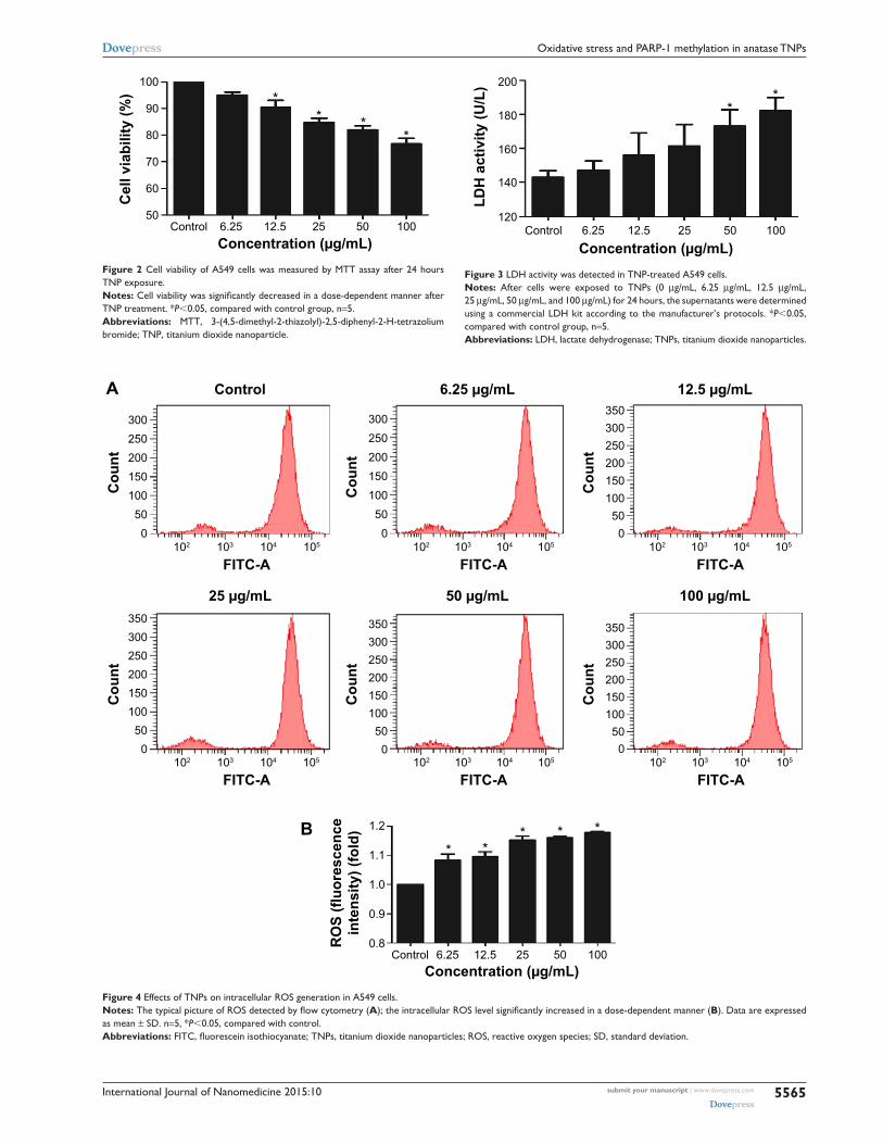
and 100 μg/mL) for 24 hours. As shown in Figure 2, with the
dosages increasing, viability of A549 cells induced by TNPs
was significantly decreased than control. In addition, the
MTT results were strongly in accordance with the increased
membrane damage measured by LDH release (Figure 3).
Our results suggested that TNPs induced cytotoxicity in a
dose-dependent manner.
Intracellular rOs generation induced by TNPsTo get a closer insight into the possible mechanism of TNP-
induced cellular toxicity, the intracellular ROS levels were
determined by using the DCFH-DA probe. From Figure 4A
and B, we observed the gradually elevated ROS levels in
A549 cells in a dose-dependent manner. The generation of
ROS in all TNP-treated groups was significantly different
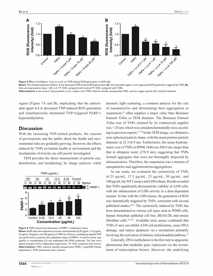
with control group. Compared to the group of treatment with
TNPs only, the fluorescence intensity of ROS in the group
of pretreatment with methyltransferase inhibitor 5-aza was
relatively weak (Figure 5A), in line with the effect of ROS
scavenger α-LA pretreatment (Figure 5B). It suggested
that the methyltransferase inhibitor markedly decreased the
generation of ROS induced by TNPs.
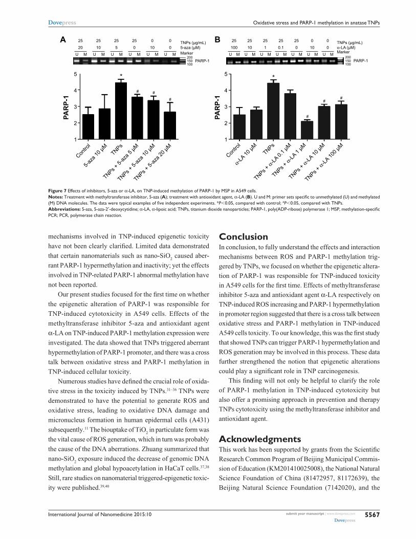
ParP-1 methylation alteration induced by TNPsMSP, a common detection method of methylation, was
performed to analyze the methylation status of PARP-1.
Our results revealed that TNPs notably elevated the level of
PARP-1 methylation in a dose-dependent way (Figure 6).
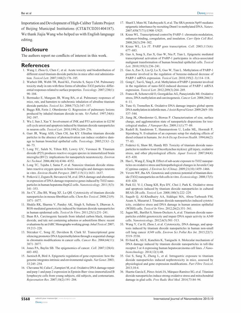
However, pretreatment of both methyltransferase inhibi-
tor 5-aza and ROS scavenger α-LA inversely altered the
TNP-induced hypermethylation of PARP-1 promoter
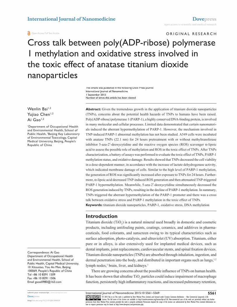
Figure 1 characterization of TNPs by TeM.Notes: Particle shape was analyzed by TeM (A) and the size distribution in the test media were evaluated by ImageJ software (B).Abbreviations: TNPs, titanium dioxide nanoparticles; TeM, transmission electron microscope.
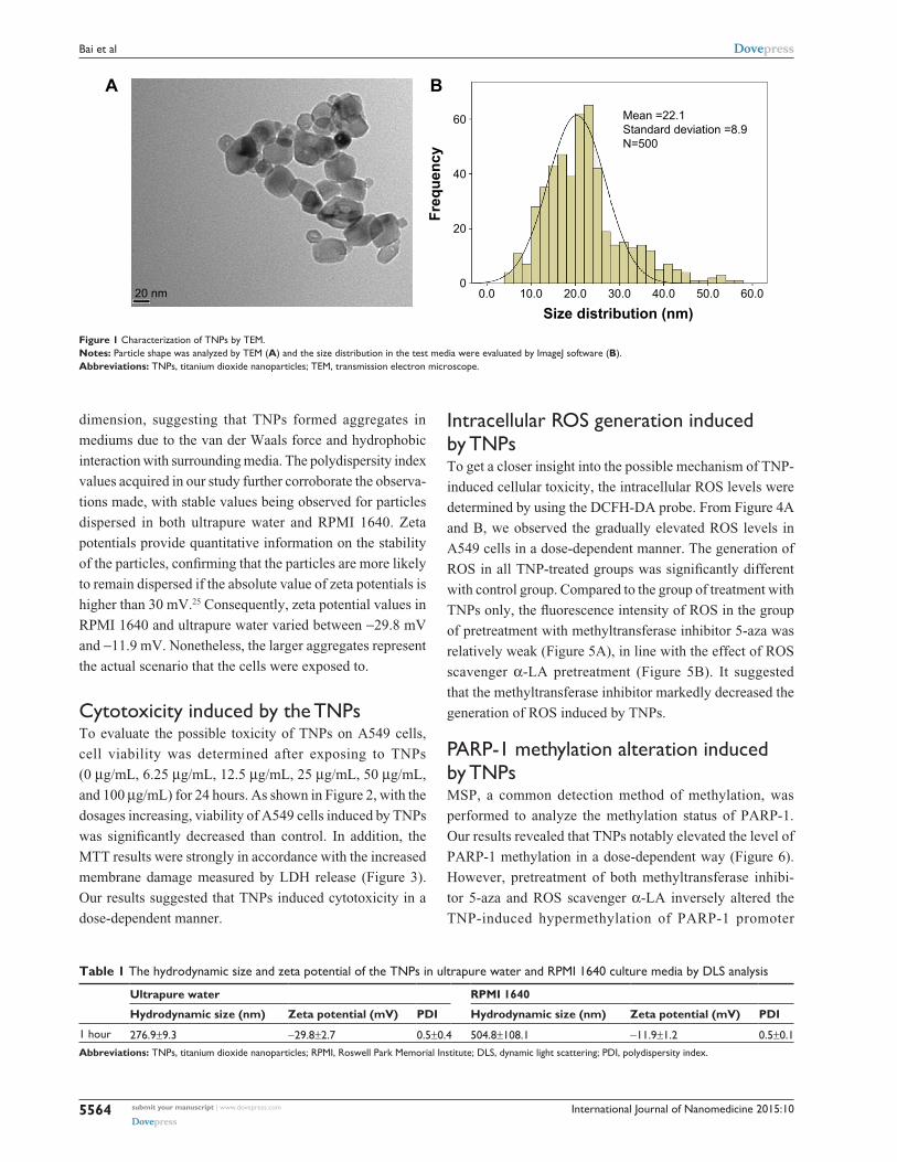
Table 1 The hydrodynamic size and zeta potential of the TNPs in ultrapure water and rPMI 1640 culture media by Dls analysis
International Journal of Nanomedicine 2015:10 submit your manuscript | www.dovepress.com
Dovepress
Dovepress
5565
Oxidative stress and ParP-1 methylation in anatase TNPs
Figure 2 cell viability of a549 cells was measured by MTT assay after 24 hours TNP exposure.Notes: Cell viability was significantly decreased in a dose-dependent manner after TNP treatment. *P,0.05, compared with control group, n=5.Abbreviations: MTT, 3-(4,5-dimethyl-2-thiazolyl)-2,5-diphenyl-2-h-tetrazolium bromide; TNP, titanium dioxide nanoparticle.
Figure 3 lDh activity was detected in TNP-treated a549 cells.Notes: after cells were exposed to TNPs (0 μg/ml, 6.25 μg/ml, 12.5 μg/ml, 25 μg/ml, 50 μg/ml, and 100 μg/ml) for 24 hours, the supernatants were determined using a commercial lDh kit according to the manufacturer’s protocols. *P,0.05, compared with control group, n=5.Abbreviations: lDh, lactate dehydrogenase; TNPs, titanium dioxide nanoparticles.
0102 103
ControlA
B
FITC-A
Cou
nt
104 105
50100150200250300
0102 103
6.25 µg/mL
FITC-A
Cou
nt
104 105
50100150200250300
0102 103
12.5 µg/mL
FITC-AC
ount
104 105
50100150200250
350300
0102 103
100 µg/mL
FITC-A
Cou
nt
104 105
50100150200250
350300
0102 103
50 µg/mL
FITC-A
Cou
nt
104 105
50100150200250
350300
0102 103
25 µg/mL
FITC-A
Cou
nt
104 105
50100150200250
350300
1.2
1.0
1.1
0.8RO
S (fl
uore
scen
cein
tens
ity) (
fold
)
0.9
Control 6.25 12.5Concentration (µg/mL)
25 50 100
*****
Figure 4 effects of TNPs on intracellular rOs generation in a549 cells.Notes: The typical picture of ROS detected by flow cytometry (A); the intracellular ROS level significantly increased in a dose-dependent manner (B). Data are expressed as mean ± sD. n=5, *P,0.05, compared with control.Abbreviations: FITC, fluorescein isothiocyanate; TNPs, titanium dioxide nanoparticles; ROS, reactive oxygen species; SD, standard deviation.
International Journal of Nanomedicine 2015:10submit your manuscript | www.dovepress.com
Dovepress
Dovepress
5566
Bai et al
α α α
α
Figure 5 effects of inhibitors, 5-aza or α-la, on TNP-induced rOs generation in a549 cells.Notes: The methyltransferase inhibitor, 5-aza decreased TNPs-induced rOs generation (A); the antioxidant agent, α-la suppressed rOs generation triggered by TNPs (B). Data are expressed as mean ± sD; n=5, *P,0.05, compared with control; #P,0.05, compared with TNPs.Abbreviations: 5-aza, 5-aza-2′-deoxycytidine; α-la, α-lipoic acid; TNPs, titanium dioxide nanoparticles; rOs, reactive oxygen species; sD, standard deviation.
region (Figure 7A and B), implicating that the antioxi-
and simultaneously attenuated TNP-triggered PARP-1
hypermethylation.
DiscussionWith the increasing TNP-related products, the concern
of governments and the public about the health and envi-
ronmental risks are gradually growing. However, the effects
induced by TNPs on human health or environment and the
mechanisms of toxicity are still poorly investigated.
TEM provides the direct measurement of particle size,
distribution, and morphology by image analysis, while
dynamic light scattering, a common analysis for the size
of nanoparticles and determining their aggregation in
suspension,26 often supplies a larger value than Brunauer
Emmett Teller or TEM diameter. The Brunauer Emmett
Teller size of TNPs claimed by its commercial supplier
was ,25 nm, which was considered potentially toxic accord-
ing to previous reports.27–29 In the TEM image, we obtained a
near-spherical particle shape, with the mean pristine particle
diameter at 22.1±8.9 nm. Furthermore, the mean hydrody-
namic size of TNPs in RPMI 1640 was 504.8 nm, larger than
that in ultrapure water (276.9 nm), suggesting that TNPs
formed aggregates that were not thoroughly dispersed by
ultrasonication. Therefore, the suspension was a mixture of
nanoparticles and agglomerations/aggregations.
In our study, we evaluated the cytotoxicity of TNPs
(6.25 μg/mL, 12.5 μg/mL, 25 μg/mL, 50 μg/mL, and
100 μg/mL) by MTT assays and LDH release. Results revealed
that TNPs significantly decreased the viability of A549 cells,
with the enhancement of LDH activity in a dose-dependent
manner. In line with the LDH release, the generation of ROS
was dramatically triggered by TNPs, consistent with several
published studies.30,31 The cytotoxicity induced by TNPs has
been demonstrated in various cell types such as WISH cells,
human bronchial epithelial cell line, BEAS-2B, and mouse
fibroblast cells.10,30,31 Available toxic assays confirmed that
TNPs (5 nm) can inhibit A549 cell proliferation, cause DNA
damage, and induce apoptosis via a mechanism primarily
involving the activation of intrinsic mitochondrial pathway.32
Generally, DNA methylation is the first step in epigenetic
phenomena that modulate gene expression via the recruit-
ment of transcription factors. However, the underlying
Figure 6 TNPs induced the alternation of ParP-1 methylation status.Notes: a549 cells were exposed to various concentrations (6.25 μg/ml, 12.5 μg/ml, 25 μg/ml, 50 μg/ml, and 100 μg/mL) of TNPs for 24 hours, methylation-specific PCR was performed to examine the methylation status of ParP-1. U and M: primer sets specific to unmethylated (U) and methylated (M) DNA molecules. The data were typical examples of five independent experiments. *P,0.05, compared with control.Abbreviations: TNPs, titanium dioxide nanoparticles; ParP-1, poly(aDP-ribose) polymerase 1; Pcr, polymerase chain reaction.
International Journal of Nanomedicine 2015:10 submit your manuscript | www.dovepress.com
Dovepress
Dovepress
5567
Oxidative stress and ParP-1 methylation in anatase TNPs
mechanisms involved in TNP-induced epigenetic toxicity
have not been clearly clarified. Limited data demonstrated
that certain nanomaterials such as nano-SiO2 caused aber-
rant PARP-1 hypermethylation and inactivity; yet the effects
involved in TNP-related PARP-1 abnormal methylation have
not been reported.
Our present studies focused for the first time on whether
the epigenetic alteration of PARP-1 was responsible for
TNP-induced cytotoxicity in A549 cells. Effects of the
methyltransferase inhibitor 5-aza and antioxidant agent
α-LA on TNP-induced PARP-1 methylation expression were
investigated. The data showed that TNPs triggered aberrant
hypermethylation of PARP-1 promoter, and there was a cross
talk between oxidative stress and PARP-1 methylation in
TNP-induced cellular toxicity.
Numerous studies have defined the crucial role of oxida-
tive stress in the toxicity induced by TNPs.31–36 TNPs were
demonstrated to have the potential to generate ROS and
oxidative stress, leading to oxidative DNA damage and
micronucleus formation in human epidermal cells (A431)
subsequently.11 The biouptake of TiO2 in particulate form was
the vital cause of ROS generation, which in turn was probably
the cause of the DNA aberrations. Zhuang summarized that
nano-SiO2 exposure induced the decrease of genomic DNA
methylation and global hypoacetylation in HaCaT cells.37,38
Still, rare studies on nanomaterial triggered-epigenetic toxic-
ity were published.39,40
ConclusionIn conclusion, to fully understand the effects and interaction
mechanisms between ROS and PARP-1 methylation trig-
gered by TNPs, we focused on whether the epigenetic altera-
tion of PARP-1 was responsible for TNP-induced toxicity
in A549 cells for the first time. Effects of methyltransferase
inhibitor 5-aza and antioxidant agent α-LA respectively on
TNP-induced ROS increasing and PARP-1 hypermethylation
in promoter region suggested that there is a cross talk between
oxidative stress and PARP-1 methylation in TNP-induced
A549 cells toxicity. To our knowledge, this was the first study
that showed TNPs can trigger PARP-1 hypermethylation and
ROS generation may be involved in this process. These data
further strengthened the notion that epigenetic alterations
could play a significant role in TNP carcinogenesis.
This finding will not only be helpful to clarify the role
of PARP-1 methylation in TNP-induced cytotoxicity but
also offer a promising approach in prevention and therapy
TNPs cytotoxicity using the methyltransferase inhibitor and
antioxidant agent.
AcknowledgmentsThis work has been supported by grants from the Scientific
Research Common Program of Beijing Municipal Commis-
sion of Education (KM201410025008), the National Natural
Science Foundation of China (81472957, 81172639), the
Beijing Natural Science Foundation (7142020), and the
α
ααα α α
Figure 7 effects of inhibitors, 5-aza or α-la, on TNP-induced methylation of ParP-1 by MsP in a549 cells.Notes: Treatment with methyltransferase inhibitor, 5-aza (A); treatment with antioxidant agent, α-la (B). U and M: primer sets specific to unmethylated (U) and methylated (M) DNA molecules. The data were typical examples of five independent experiments. *P,0.05, compared with control; #P,0.05, compared with TNPs.Abbreviations: 5-aza, 5-aza-2′-deoxycytidine; α-la, α-lipoic acid; TNPs, titanium dioxide nanoparticles; PARP-1, poly(ADP-ribose) polymerase 1; MSP, methylation-specific Pcr; Pcr, polymerase chain reaction.
International Journal of Nanomedicine 2015:10submit your manuscript | www.dovepress.com
Dovepress
Dovepress
5568
Bai et al
Importation and Development of High-Caliber Talents Project
of Beijing Municipal Institutions (CIT&TCD201404187).
We thank Jiajia Wang who helped us with English language
editing.
DisclosureThe authors report no conflicts of interest in this work.
References 1. Wang J, Zhou G, Chen C, et al. Acute toxicity and biodistribution of
different sized titanium dioxide particles in mice after oral administra-tion. Toxicol Lett. 2007;168(2):176–185.
2. Warheit DB, Webb TR, Reed KL, Frerichs S, Sayes CM. Pulmonary toxicity study in rats with three forms of ultrafine-TiO2 particles: differ-ential responses related to surface properties. Toxicology. 2007;230(1): 90–104.
3. Bermudez E, Mangum JB, Wong BA, et al. Pulmonary responses of mice, rats, and hamsters to subchronic inhalation of ultrafine titanium dioxide particles. Toxicol Sci. 2004;77(2):347–357.
4. Baggs RB, Ferin J, Oberdorster G. Regression of pulmonary lesions produced by inhaled titanium dioxide in rats. Vet Pathol. 1997;34(6): 592–597.
5. Wu J, Sun J, Xue Y. Involvement of JNK and P53 activation in G2/M cell cycle arrest and apoptosis induced by titanium dioxide nanoparticles in neuron cells. Toxicol Lett. 2010;199(3):269–276.
6. Gurr JR, Wang ASS, Chen CH, Jan KY. Ultrafine titanium dioxide particles in the absence of photoactivation can induce oxidative dam-age to human bronchial epithelial cells. Toxicology. 2005;213(1–2): 66–73.
7. Long TC, Saleh N, Tilton RD, Lowry GV, Veronesi B. Titanium dioxide (P25) produces reactive oxygen species in immortalized brain microglia (BV2): implications for nanoparticle neurotoxicity. Environ Sci Technol. 2006;40(14):4346–4352.
8. Long TC, Tajuba J, Sama P, et al. Nanosize titanium dioxide stimu-lates reactive oxygen species in brain microglia and damages neurons in vitro. Environ Health Perspect. 2007;115(11):1631–1637.
9. Petković J, Zegura B, Stevanović M, et al. DNA damage and alterations in expression of DNA damage responsive genes induced by TiO2 nano-particles in human hepatoma HepG2 cells. Nanotoxicology. 2011;5(3): 341–353.
10. Jin CY, Zhu BS, Wang XF, Lu QH. Cytotoxicity of titanium dioxide nanoparticles in mouse fibroblast cells. Chem Res Toxicol. 2008;21(9): 1871–1877.
11. Shukla RK, Sharma V, Pandey AK, Singh S, Sultana S, Dhawan A. ROS-mediated genotoxicity induced by titanium dioxide nanoparticles in human epidermal cells. Toxicol In Vitro. 2011;25(1):231–241.
12. Baan RA. Carcinogenic hazards from inhaled carbon black, titanium dioxide, and talc not containing asbestos or asbestiform fibers: recent evaluations by an IARC Monographs working group. Inhal Toxicol. 2007; 19:213–228.
13. Stirzaker C, Song JZ, Davidson B, Clark SJ. Transcriptional gene silencing promotes DNA hypermethylation through a sequential change in chromatin modifications in cancer cells. Cancer Res. 2004;64(11): 3871–3877.
14. Jones PA, Baylin SB. The epigenomics of cancer. Cell. 2007;128(4): 683–692.
15. Jaenisch R, Bird A. Epigenetic regulation of gene expression: how the genome integrates intrinsic and environmental signals. Nat Genet. 2003; 33:245–254.
16. Chevanne M, Calia C, Zampieri M, et al. Oxidative DNA damage repair and parp 1 and parp 2 expression in Epstein-Barr virus-immortalized B lymphocyte cells from young subjects, old subjects, and centenarians. Rejuvenation Res. 2007;10(2):191–204.
17. Sharif J, Muto M, Takebayashi S, et al. The SRA protein Np95 mediates epigenetic inheritance by recruiting Dnmt1 to methylated DNA. Nature. 2007;450(7171):U908–U925.
18. Kraus WL. Transcriptional control by PARP-1: chromatin modulation, enhancer-binding, coregulation, and insulation. Curr Opin Cell Biol. 2008;20(3):294–302.
20. Gao A, Song S, Zuo X, Guo W, Niu P, Tian L. Epigenetic mediated transcriptional activation of PARP-1 participates in silica-associated malignant transformation of human bronchial epithelial cells. Toxicol Lett. 2010;193(3):236–241.
21. Gao A, Zuo X, Liu Q, Lu X, Guo W, Tian L. Methylation of PARP-1 promoter involved in the regulation of benzene-induced decrease of PARP-1 mRNA expression. Toxicol Lett. 2010;195(2–3):114–118.
22. Gong C, Tao G, Yang L, et al. Methylation of PARP-1 promoter involved in the regulation of nano-SiO2-induced decrease of PARP-1 mRNA expression. Toxicol Lett. 2012;209(3):264–269.
23. Franco R, Schoneveld O, Georgakilas AG, Panayiotidis MI. Oxidative stress, DNA methylation and carcinogenesis. Cancer Lett. 2008;266(1): 6–11.
24. Tunc O, Tremellen K. Oxidative DNA damage impairs global sperm DNA methylation in infertile men. J Assist Reprod Genet. 2009;26(9–10): 537–544.
25. Jiang JK, Oberdorster G, Biswas P. Characterization of size, surface charge, and agglomeration state of nanoparticle dispersions for toxi-cological studies. J Nanopart Res. 2009;11(1):77–89.
26. Rudell B, Sandstrom T, Hammarstrom U, Ledin ML, Horstedt P, Stjernberg N. Evaluation of an exposure setup for studying effects of diesel exhaust in humans. Int Arch Occup Environ Health. 1994;66(2): 77–83.
27. Federici G, Shaw BJ, Handy RD. Toxicity of titanium dioxide nano-particles to rainbow trout (Oncorhynchus mykiss): gill injury, oxidative stress, and other physiological effects. Aquat Toxicol. 2007;84(4): 415–430.
28. Hao L, Wang Z, Xing B. Effect of sub-acute exposure to TiO2 nanopar-ticles on oxidative stress and histopathological changes in Juvenile Carp (Cyprinus carpio). J Environ Sci (China). 2009;21(10):1459–1466.
29. Vevers WF, Jha AN. Genotoxic and cytotoxic potential of titanium diox-ide (TiO2) nanoparticles on fish cells in vitro. Ecotoxicology. 2008;17(5): 410–420.
30. Park EJ, Yi J, Chung KH, Ryu DY, Choi J, Park K. Oxidative stress and apoptosis induced by titanium dioxide nanoparticles in cultured BEAS-2B cells. Toxicol Lett. 2008;180(3):222–229.
31. Saquib Q, Al-Khedhairy AA, Siddiqui MA, Abou-Tarboush FM, Azam A, Musarrat J. Titanium dioxide nanoparticles induced cytotox-icity, oxidative stress and DNA damage in human amnion epithelial (WISH) cells. Toxicol In Vitro. 2012;26(2):351–361.
32. Jugan ML, Barillet S, Simon-Deckers A, et al. Titanium dioxide nano-particles exhibit genotoxicity and impair DNA repair activity in A549 cells. Nanotoxicology. 2012;6(5):501–513.
33. Wang Y, Cui H, Zhou J, et al. Cytotoxicity, DNA damage, and apop-tosis induced by titanium dioxide nanoparticles in human non-small cell lung cancer A549 cells. Environ Sci Pollut Res Int. 2015;22(7): 5519–5530.
34. El-Said K, Ali E, Kanehira K, Taniguchi A. Molecular mechanism of DNA damage induced by titanium dioxide nanoparticles in toll-like receptor 3 or 4 expressing human hepatocarcinoma cell lines. J Nano-biotechnology. 2014;12(1):48.
35. Gui S, Sang X, Zheng L, et al. Intragastric exposure to titanium dioxide nanoparticles induced nephrotoxicity in mice, assessed by physiological and gene expression modifications. Part Fibre Toxicol. 2013;10:4.
36. Huerta-García E, Pérez-Arizti JA, Márquez-Ramírez SG, et al. Titanium dioxide nanoparticles induce strong oxidative stress and mitochondrial damage in glial cells. Free Radic Biol Med. 2014;73:84–94.
Submit your manuscript here: http://www.dovepress.com/international-journal-of-nanomedicine-journal
The International Journal of Nanomedicine is an international, peer-reviewed journal focusing on the application of nanotechnology in diagnostics, therapeutics, and drug delivery systems throughout the biomedical field. This journal is indexed on PubMed Central, MedLine, CAS, SciSearch®, Current Contents®/Clinical Medicine,
Journal Citation Reports/Science Edition, EMBase, Scopus and the Elsevier Bibliographic databases. The manuscript management system is completely online and includes a very quick and fair peer-review system, which is all easy to use. Visit http://www.dovepress.com/testimonials.php to read real quotes from published authors.
International Journal of Nanomedicine 2015:10 submit your manuscript | www.dovepress.com
Dovepress
Dovepress
Dovepress
5569
Oxidative stress and ParP-1 methylation in anatase TNPs
37. Gong C, Yang L, Tao G, Liu Q, Liu J, Zhuang Z. [Genome DNA hypom-ethylation in HaCaT cells after short exposure to SiO2 nanoparticles]. Wei Sheng Yan Jiu. 2013;42(2):179–184. Chinese.
38. Gong C, Tao G, Yang L, Liu J, Liu Q, Zhuang Z. SiO(2) nanoparticles induce global genomic hypomethylation in HaCaT cells. Biochem Biophys Res Commun. 2010;397(3):397–400.
39. Meng J, Xing J, Wang Y, et al. Epigenetic modulation of human breast cancer by metallofullerenol nanoparticles: in vivo treatment and in vitro analysis. Nanoscale. 2011;3(11):4713–4719.
40. Ng CT, Dheen ST, Yip WC, Ong CN, Bay BH, Lanry Yung LY. The induction of epigenetic regulation of PROS1 gene in lung fibroblasts by gold nanoparticles and implications for potential lung injury. Biomaterials. 2011;32(30):7609–7615.