Introduction The anatomy of the synovial folds, or plicae, in the knee joint was first described through cadaver dissection by Mayeda 1 in 1918, followed by Hohlbaum, 2 Pipkin, 3,4 Hughston, 5 and Harty and Joyce. 6 Embryologically, although there is no consensus about the development of the joint cavity, it has been widely believed that the knee joint is originally composed of three com- partments: medial and lateral synovial compart- ments and suprapatellar bursa. 7,8 These compartments are partitioned by synovial sep- tums. At about 3 months of fetal age, these syn- ovial septa begin to disappear little by little, and then they vanish completely or remain in part. The folds were not delineated fully in the past, but with the advancement of arthroscopy, their classification and incidences are reported in recent papers. 9,10-12 The plicae are classified according to their corresponding anatomic sites of the knee, as suprapatellar, mediopatel- lar, infrapatellar, and lateral patellar plicae. Although the three-cavitation theory for devel- opment of the knee joint may explain the for- mation of the suprapatellar and infrapatellar plicae, that of the mediopatellar plica and the lateral patellar plica remains uncertain. Moreover, the theory cannot explain the variety of shapes of the plica. Thus, the variety of pat- terns of the plica can be chosen as evidence sup- porting the multiple cavitations theory for development of the knee joint proposed by Gray and Gardner 7 and Ogata 8 and backed up by Kim. 12 There was no consensus concerning the incidence of synovial plicae. In literature review, the reported incidence of each plicae is contro- versial. 2,3,6,10,11,13-16 In our study including 400 knees in 363 patients, 12 incidence of the synovial plica at the knees were: suprapatellar plica, 87.0%; mediopatellar plica, 72.0%; infrapatellar plica, 86.0%; and lateral patellar plica, 1.3%. These plicae were at first considered abnormal when seen at arthroscopy and then excised. However, the plicae are now recognized as nor- mal structures that represent remnants of syn- ovial membranes in embryonic development of the knee. When chronic inflammation is devel- oped by trauma or the presence of other patho- logical knee conditions, the pliability of synovial folds might be affected. When a plica of the syn- ovial membrane loses its normal elasticity and becomes fibrotic, it might cause dynamic derangement of the knee called “pathologic plica syndrome.” Suprapatella Plica Anatomy The suprapatella plica is a persistent remnant of the embryonic synovial membrane that lies between the suprapatella pouch and the knee joint proper. The suprapatella plica is attached on the superomedial and superolateral wall of the knee joint and also on the undersurface of the quadriceps tendon region in axial plane. When the knee is flexed beyond 90˚, the supra- patella plica folds longitudinally rather than in a transverse fold. The incidence rate of suprap- atella plica has been widely reported to be from 20% to 87%. 4,10,12,17 The suprapatella plica has a variety of shapes and sizes. Zidorn 11 presented a classification of the suprapatella plica, which classified it into four groups: complete sep- tum type, perforated septum type, residual septum type, and extinct septum type. We also 239 14 Patella Plica Syndrome Sung-Jae Kim
Transcript
IntroductionThe anatomy of the synovial folds, or plicae, inthe knee joint was first described throughcadaver dissection by Mayeda1 in 1918, followedby Hohlbaum,2 Pipkin,3,4 Hughston,5 and Hartyand Joyce.6 Embryologically, although there isno consensus about the development of thejoint cavity, it has been widely believed that theknee joint is originally composed of three com-partments: medial and lateral synovial compart-ments and suprapatellar bursa.7,8 Thesecompartments are partitioned by synovial sep-tums. At about 3 months of fetal age, these syn-ovial septa begin to disappear little by little, andthen they vanish completely or remain in part.The folds were not delineated fully in the past,but with the advancement of arthroscopy, theirclassification and incidences are reported inrecent papers.9,10-12 The plicae are classifiedaccording to their corresponding anatomicsites of the knee, as suprapatellar, mediopatel-lar, infrapatellar, and lateral patellar plicae.Although the three-cavitation theory for devel-opment of the knee joint may explain the for-mation of the suprapatellar and infrapatellarplicae, that of the mediopatellar plica and thelateral patellar plica remains uncertain.Moreover, the theory cannot explain the varietyof shapes of the plica. Thus, the variety of pat-terns of the plica can be chosen as evidence sup-porting the multiple cavitations theory fordevelopment of the knee joint proposed by Grayand Gardner7 and Ogata8 and backed up byKim.12 There was no consensus concerning theincidence of synovial plicae. In literature review,the reported incidence of each plicae is contro-versial.2,3,6,10,11,13-16 In our study including 400
knees in 363 patients,12 incidence of the synovialplica at the knees were: suprapatellar plica,87.0%; mediopatellar plica, 72.0%; infrapatellarplica, 86.0%; and lateral patellar plica, 1.3%.These plicae were at first considered abnormalwhen seen at arthroscopy and then excised.However, the plicae are now recognized as nor-mal structures that represent remnants of syn-ovial membranes in embryonic development ofthe knee. When chronic inflammation is devel-oped by trauma or the presence of other patho-logical knee conditions, the pliability of synovialfolds might be affected. When a plica of the syn-ovial membrane loses its normal elasticity andbecomes fibrotic, it might cause dynamicderangement of the knee called “pathologicplica syndrome.”
Suprapatella PlicaAnatomyThe suprapatella plica is a persistent remnant ofthe embryonic synovial membrane that liesbetween the suprapatella pouch and the kneejoint proper. The suprapatella plica is attachedon the superomedial and superolateral wall ofthe knee joint and also on the undersurface ofthe quadriceps tendon region in axial plane.When the knee is flexed beyond 90˚, the supra-patella plica folds longitudinally rather than in atransverse fold. The incidence rate of suprap-atella plica has been widely reported to be from20% to 87%.4,10,12,17 The suprapatella plica hasa variety of shapes and sizes. Zidorn11 presenteda classification of the suprapatella plica, whichclassified it into four groups: complete sep-tum type, perforated septum type, residualseptum type, and extinct septum type. We also
239
14Patella Plica Syndrome
Sung-Jae Kim
Ch14.qxd 10/07/05 3:45 PM Page 239
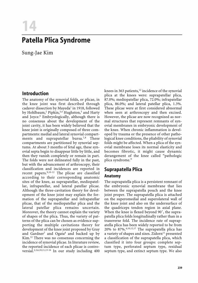
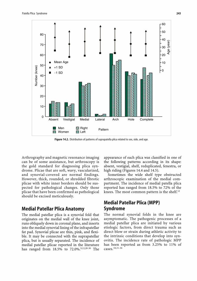
classified each plica as one of the following pat-terns: absent, vestigial, medial, lateral, arch,hole, or complete septum type, of which thearch type is most frequent (Figures 14.1 and14.2).12 Although suprapatella plica is foundfrequently in the knee joint in variable shapesand sizes, the pathologic suprapatella plica israrely reported. There have been some reportsof symptomatic suprapatella plica, or combina-tion of the suprapatella and medial patellaplica.18,19 Dupont20 reported 3 symptomaticsuprapatella plicae in 12,000 arthroscopies. Wereported a case of arch type pathologic sup-rapatella plica that was excised using anarthroscopic technique (Figure 14.3).21 Thepathophysiology of symptomatic plica has not
been clearly defined. Complete or near-com-plete type suprapatella plica has been reportedto cause intermittent painful swelling of theknee because of its one-way valvular mecha-nism.4,22 Some investigators have suggested thatsynovial changes result from a variety of causes.Blunt trauma, localized hemorrhage, and jointlaxity can create symptomatic synovial plica.23,24
Whatever the reason, as a result, the synoviumloses its elasticity, thickens, and becomesinflammatory. This inflammatory process even-tually causes fibrosis of the synovial plicafollowed by serious intra-articular distur-bances. The thickened and inflexible structuraldegeneration of the plica interferes with thepatellofemoral gliding mechanism and may
240 Etiopathogenic Bases and Therapeutic Implications
Patella
Quadriceps
Tendons
Articulasris
Genu
Femoral Condyles
(a)
(b)
Figure 14.1. Illustrations and arthroscopic findings for patterns of suprapatella plica in the right knee. (a) Absent type: No sharp-edged fold ofsynovium between the suprapatella pouch and the knee joint cavity. (b) Vestigial type: A plica with a less than 1 mm protrusion of the synovium.
Ch14.qxd 10/07/05 3:45 PM Page 240
Patella Plica Syndrome 241
(c)
(d)
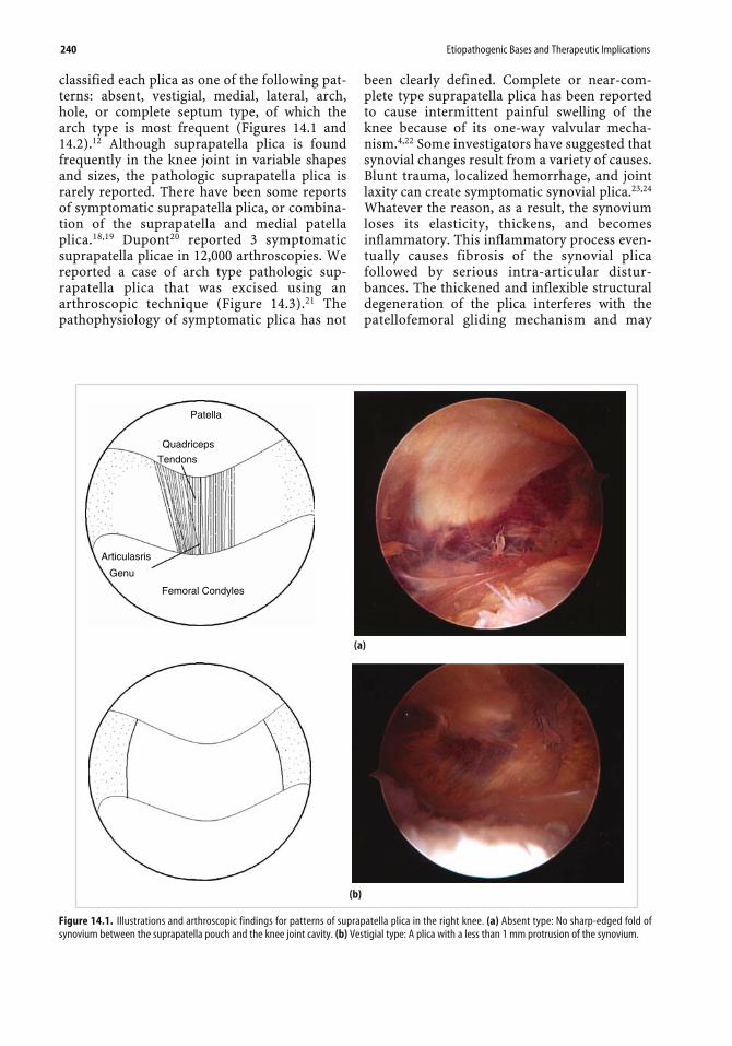
Figure 14.1. (c) Medial type: A plica that lies on th0e medial side of the suprapatella pouch. (d) Lateral type: A plica that lies on the lateral side ofthe suprapatella pouch. (e) Arch type: A plica that is present on the medial, lateral, and anterior aspects of the suprapatella pouch but not over theanterior surface of the femur.
(e)
(continued)
Ch14.qxd 10/07/05 3:45 PM Page 241
Hole
(f)
cause changes in the articular surfaces of thepatella and femoral condyle. This may be themechanism of compression of the femoralcondyle.24,25
Clinical SignificanceThe clinical characteristics of the pathologicsuprapatella plica included chronic intermit-tent pain of the superior aspect of the kneejoint and exercise-related swelling. A palpablebandlike mass on the suprapatella pouch withlocal tenderness and swelling may be present.The pain was aggravated during stair climbingand while sitting for a long time while the kneewas flexed from 45˚ to 90˚. Strover et al.26 andKim21 confirmed that suprapatella plica
impinges between the femoral condyle andquadriceps mechanism when the knee is flexed70˚ to 100˚. Sometimes, a high-pitched snapcan be heard during knee motion. The high-pitched sound characterizes the plica syn-drome and differentiates this snap from soundsassociated with meniscal derangements andloose bodies, which are lower in pitch.17,27 Thesuprapatella plica can provide a good hidingplace for loose bodies, especially in completeseptum type of plica, in which we cannot iden-tify the insertion of the articularis genu.Diagnosis of the suprapatella plica syndrome ismade by recognizing characteristic symptomsand by physical examination. Plain radiogra-phy is of little help in establishing a diagnosis.
242 Etiopathogenic Bases and Therapeutic Implications
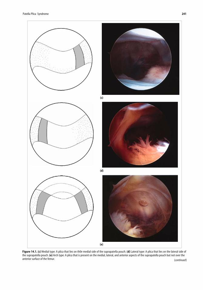
Figure 14.1. (continued ) (f) Hole type: A plica extending completely across the suprapatella pouch but with a central defect. (g) Complete septumtype: A plica dividing the suprapatella pouch into two separate compartments. Each pattern may have lateral cave, or nothing.
Figure 14.2. Distribution of patterns of suprapatella plica related to sex, side, and age.
Arthrography and magnetic resonance imagingcan be of some assistance, but arthroscopy isthe gold standard for diagnosing plica syn-drome. Plicae that are soft, wavy, vascularized,and synovial-covered are normal findings.However, thick, rounded, or shredded fibroticplicae with white inner borders should be sus-pected for pathological changes. Only thoseplicae that have been confirmed as pathologicalshould be excised meticulously.
Medial Patellar Plica AnatomyThe medial patellar plica is a synovial fold thatoriginates on the medial wall of the knee joint,runs obliquely down in coronal plane, and insertsinto the medial synovial lining of the infrapatellarfat pad. Synovial plicae are thin, pink, and flexi-ble. It may be connected with the suprapatellarplica, but is usually separated. The incidence ofmedial patellar plicae reported in the literaturehas ranged from 18.5% to 72.0%.9,12,28-30 The
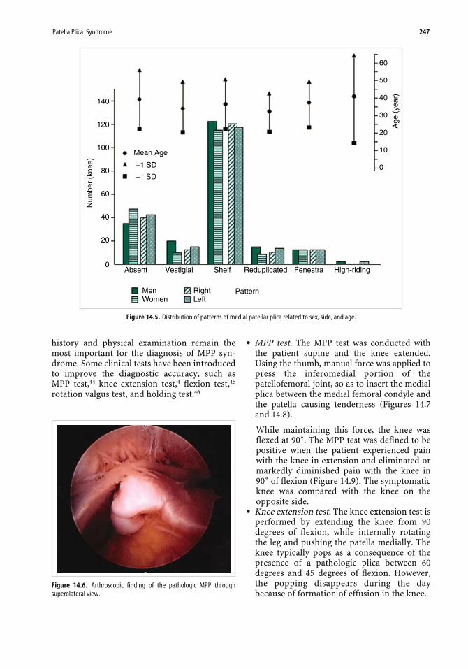
appearance of each plica was classified in one ofthe following patterns according in its shape:absent, vestigial, shelf, reduplicated, fenestra, orhigh riding (Figures 14.4 and 14.5).
Sometimes the wide shelf type obstructedarthroscopic examination of the medial com-partment. The incidence of medial patella plicareported has ranged from 18.5% to 72% of theknees. The most common pattern is the shelf.12
Medial Patellar Plica (MPP)SyndromeThe normal synovial folds in the knee areasymptomatic. The pathogenic processes of amedial patellar plica are initiated by variousetiologic factors, from direct trauma such asdirect blow or strain during athletic activity tothe intrinsic conditions that develop into syn-ovitis. The incidence rate of pathologic MPPhas been reported as from 3.25% to 11% ofcases.18,31-36
Ch14.qxd 10/07/05 3:45 PM Page 243
244 Etiopathogenic Bases and Therapeutic Implications
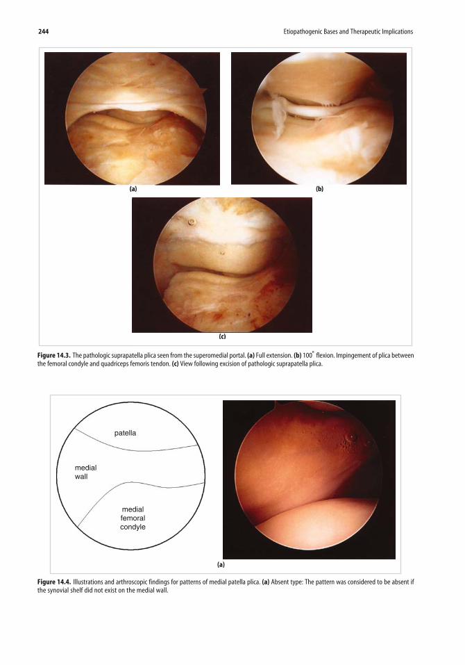
Figure 14.3. The pathologic suprapatella plica seen from the superomedial portal. (a) Full extension. (b) 100̊ flexion. Impingement of plica betweenthe femoral condyle and quadriceps femoris tendon. (c) View following excision of pathologic suprapatella plica.
patella
medialwall
medialfemoralcondyle
(a)
Figure 14.4. Illustrations and arthroscopic findings for patterns of medial patella plica. (a) Absent type: The pattern was considered to be absent ifthe synovial shelf did not exist on the medial wall.
Ch14.qxd 10/07/05 3:45 PM Page 244
Patella Plica Syndrome 245
(b)
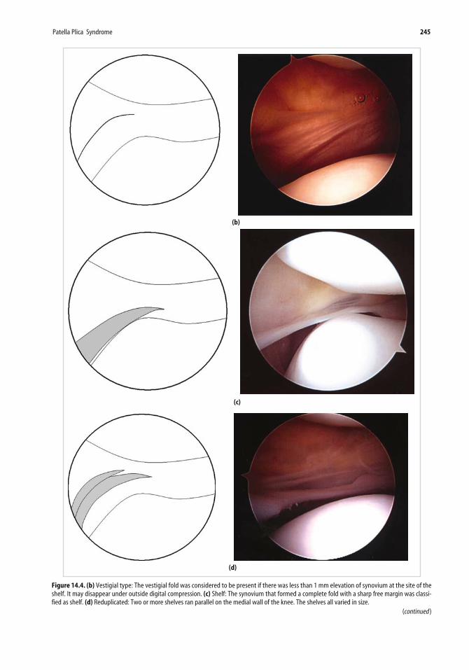
Figure 14.4. (b) Vestigial type: The vestigial fold was considered to be present if there was less than 1 mm elevation of synovium at the site of theshelf. It may disappear under outside digital compression. (c) Shelf: The synovium that formed a complete fold with a sharp free margin was classi-fied as shelf. (d) Reduplicated: Two or more shelves ran parallel on the medial wall of the knee. The shelves all varied in size.
(c)
(d)
(continued)
Ch14.qxd 10/07/05 3:45 PM Page 245
246 Etiopathogenic Bases and Therapeutic Implications
(e)
(f)
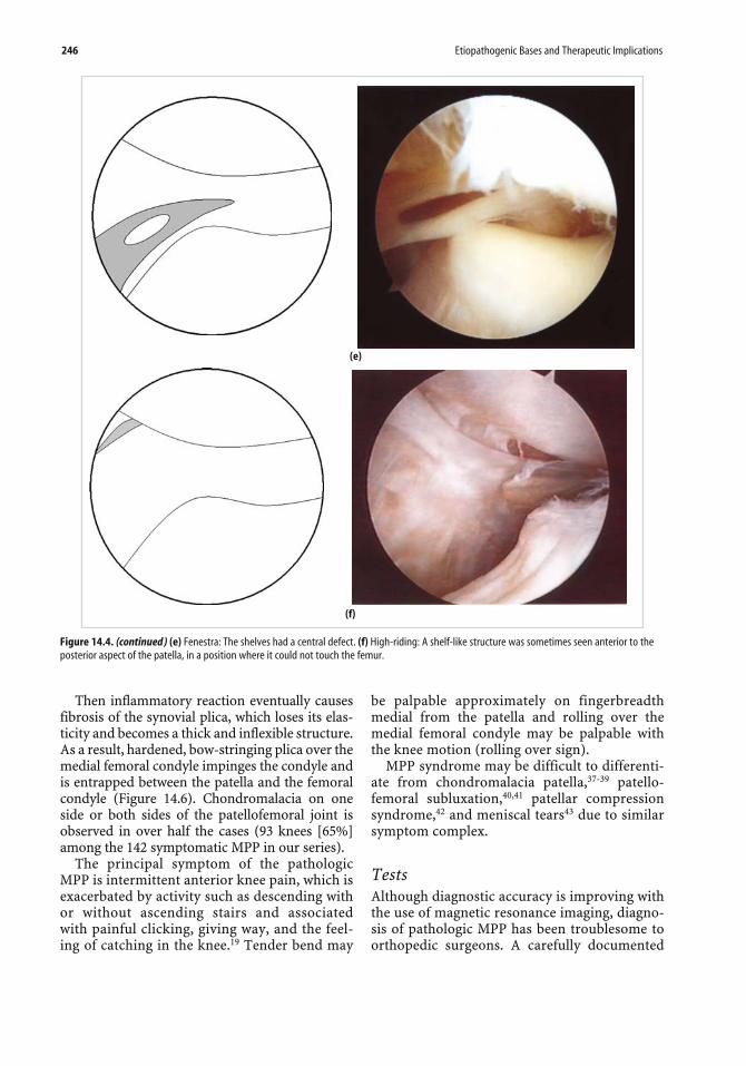
Figure 14.4. (continued ) (e) Fenestra: The shelves had a central defect. (f) High-riding: A shelf-like structure was sometimes seen anterior to theposterior aspect of the patella, in a position where it could not touch the femur.
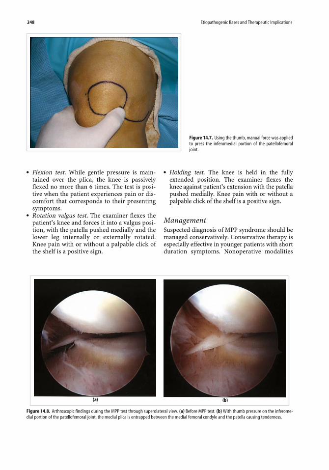
Then inflammatory reaction eventually causesfibrosis of the synovial plica, which loses its elas-ticity and becomes a thick and inflexible structure.As a result, hardened, bow-stringing plica over themedial femoral condyle impinges the condyle andis entrapped between the patella and the femoralcondyle (Figure 14.6). Chondromalacia on oneside or both sides of the patellofemoral joint isobserved in over half the cases (93 knees [65%]among the 142 symptomatic MPP in our series).
The principal symptom of the pathologicMPP is intermittent anterior knee pain, which isexacerbated by activity such as descending withor without ascending stairs and associatedwith painful clicking, giving way, and the feel-ing of catching in the knee.19 Tender bend may
be palpable approximately on fingerbreadthmedial from the patella and rolling over themedial femoral condyle may be palpable withthe knee motion (rolling over sign).
MPP syndrome may be difficult to differenti-ate from chondromalacia patella,37-39 patello-femoral subluxation,40,41 patellar compressionsyndrome,42 and meniscal tears43 due to similarsymptom complex.
TestsAlthough diagnostic accuracy is improving withthe use of magnetic resonance imaging, diagno-sis of pathologic MPP has been troublesome toorthopedic surgeons. A carefully documented
Ch14.qxd 10/07/05 3:46 PM Page 246
history and physical examination remain themost important for the diagnosis of MPP syn-drome. Some clinical tests have been introducedto improve the diagnostic accuracy, such asMPP test,44 knee extension test,4 flexion test,45
rotation valgus test, and holding test.46
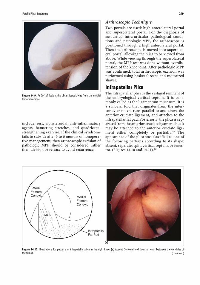
● MPP test. The MPP test was conducted withthe patient supine and the knee extended.Using the thumb, manual force was applied topress the inferomedial portion of thepatellofemoral joint, so as to insert the medialplica between the medial femoral condyle andthe patella causing tenderness (Figures 14.7and 14.8).
While maintaining this force, the knee wasflexed at 90˚. The MPP test was defined to bepositive when the patient experienced painwith the knee in extension and eliminated ormarkedly diminished pain with the knee in90˚ of flexion (Figure 14.9). The symptomaticknee was compared with the knee on theopposite side.
● Knee extension test. The knee extension test isperformed by extending the knee from 90degrees of flexion, while internally rotatingthe leg and pushing the patella medially. Theknee typically pops as a consequence of thepresence of a pathologic plica between 60degrees and 45 degrees of flexion. However,the popping disappears during the daybecause of formation of effusion in the knee.
Figure 14.5. Distribution of patterns of medial patellar plica related to sex, side, and age.
Figure 14.6. Arthroscopic finding of the pathologic MPP throughsuperolateral view.
Ch14.qxd 10/07/05 3:46 PM Page 247
● Flexion test. While gentle pressure is main-tained over the plica, the knee is passivelyflexed no more than 6 times. The test is posi-tive when the patient experiences pain or dis-comfort that corresponds to their presentingsymptoms.
● Rotation valgus test. The examiner flexes thepatient’s knee and forces it into a valgus posi-tion, with the patella pushed medially and thelower leg internally or externally rotated.Knee pain with or without a palpable click ofthe shelf is a positive sign.
● Holding test. The knee is held in the fullyextended position. The examiner flexes theknee against patient’s extension with the patellapushed medially. Knee pain with or without apalpable click of the shelf is a positive sign.
ManagementSuspected diagnosis of MPP syndrome should bemanaged conservatively. Conservative therapy isespecially effective in younger patients with shortduration symptoms. Nonoperative modalities
248 Etiopathogenic Bases and Therapeutic Implications
Figure 14.7. Using the thumb, manual force was appliedto press the inferomedial portion of the patellofemoraljoint.
Figure 14.8. Arthroscopic findings during the MPP test through superolateral view. (a) Before MPP test. (b) With thumb pressure on the inferome-dial portion of the patellofemoral joint, the medial plica is entrapped between the medial femoral condyle and the patella causing tenderness.
Ch14.qxd 10/07/05 3:46 PM Page 248
include rest, nonsteroidal anti-inflammatoryagents, hamstring stretches, and quadriceps-strengthening exercise. If the clinical syndromefails to subside after 3 to 6 months of nonopera-tive management, then arthroscopic excision ofpathologic MPP should be considered ratherthan division or release to avoid recurrence.
Arthroscopic TechniqueTwo portals are used: high anterolateral portaland superolateral portal. For the diagnosis ofassociated intra-articular pathological condi-tions and pathologic MPP, the arthroscope ispositioned through a high anterolateral portal.Then the arthroscope is moved into superolat-eral portal, allowing the plica to be viewed fromabove. While viewing through the superolateralportal, the MPP test was done without overdis-tension of the knee joint. After pathologic MPPwas confirmed, total arthroscopic excision wasperformed using basket forceps and motorizedshaver.
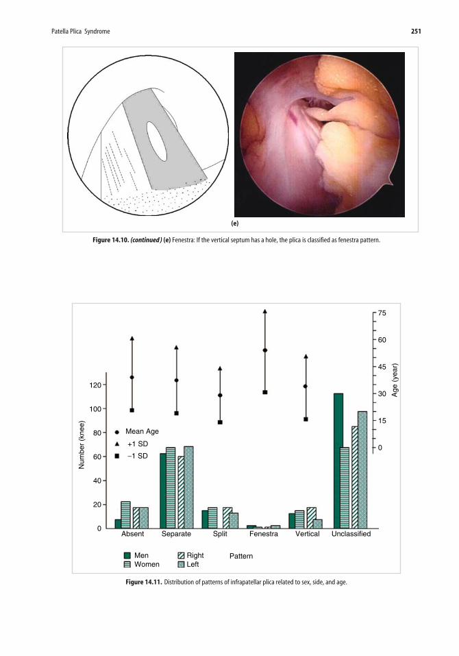
Infrapatellar PlicaThe infrapatellar plica is the vestigial remnant ofthe embryological vertical septum. It is com-monly called as the ligamentum mucosum. It isa synovial fold that originates from the inter-condylar notch, runs parallel to and above theanterior cruciate ligament, and attaches to theinfrapatellar fat pad. Posteriorly, the plica is sep-arated from the anterior cruciate ligament, but itmay be attached to the anterior cruciate liga-ment either completely or partially.47 Theappearance of the plica was classified as one ofthe following patterns according to its shape:absent, separate, split, vertical septum, or fenes-tra. (Figures 14.10 and 14.11).12
Patella Plica Syndrome 249
Figure 14.9. At 90˚ of flexion, the plica slipped away from the medialfemoral condyle.
LateralFemoralCondyle Medial
FemoralCondyle
ACL
InfrapatellaFat Pad
(a)
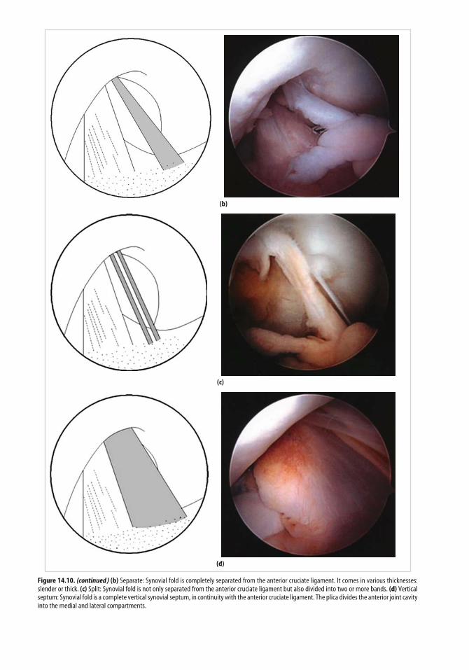
Figure 14.10. Illustrations for patterns of infrapatellar plica in the right knee. (a) Absent: Synovial fold does not exist between the condyles ofthe femur. (continued)
Ch14.qxd 10/07/05 3:46 PM Page 249
(b)
(c)
Figure 14.10. (continued ) (b) Separate: Synovial fold is completely separated from the anterior cruciate ligament. It comes in various thicknesses:slender or thick. (c) Split: Synovial fold is not only separated from the anterior cruciate ligament but also divided into two or more bands. (d) Verticalseptum: Synovial fold is a complete vertical synovial septum, in continuity with the anterior cruciate ligament. The plica divides the anterior joint cavityinto the medial and lateral compartments.
(d)
Ch14.qxd 10/07/05 3:46 PM Page 250
Patella Plica Syndrome 251
Figure 14.10. (continued ) (e) Fenestra: If the vertical septum has a hole, the plica is classified as fenestra pattern.
(e)
120
100
80
60
40
20
0
Mean Age
+1 SD
−1 SD
Absent Separate Split VerticalFenestra Unclassified
MenWomen
Right PatternLeft
0
15
30
45
60
75
Age
(ye
ar)
Num
ber
(kne
e)
Figure 14.11. Distribution of patterns of infrapatellar plica related to sex, side, and age.
Ch14.qxd 10/07/05 3:46 PM Page 251
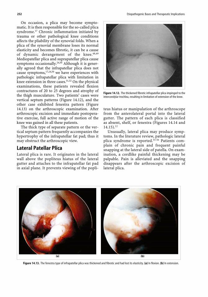
On occasion, a plica may become sympto-matic. It is then responsible for the so-called plicasyndrome.17 Chronic inflammation initiated bytrauma or other pathological knee conditionsaffects the pliability of the synovial folds. When aplica of the synovial membrane loses its normalelasticity and becomes fibrotic, it can be a causeof dynamic derangement of the knee.48,49
Mediopatellar plica and suprapatellar plica causesymptoms occasionally.24,50 Although it is gener-ally agreed that the infrapatellar plica does notcause symptoms,17,24,50 we have experiences withpathologic infrapatellar plica with limitation inknee extension in three cases.51,52 On the physicalexaminations, these patients revealed flexioncontractures of 20 to 25 degrees and atrophy ofthe thigh musculature. Two patients’ cases werevertical septum patterns (Figure 14.12), and theother case exhibited fenestra pattern (Figure14.13) on the arthroscopic examination. Afterarthroscopic excision and immediate postopera-tive exercise, full active range of motion of theknee was gained in all these patients.
The thick type of separate pattern or the ver-tical septum pattern frequently accompanies thehypertrophy of the infrapatellar fat pad; thus itmay obstruct the arthroscopic view.
Lateral Patellar PlicaLateral plica is rare. It originates in the lateralwall above the popliteus hiatus of the lateralgutter and attaches to the infrapatellar fat padin axial plane. It prevents viewing of the popli-
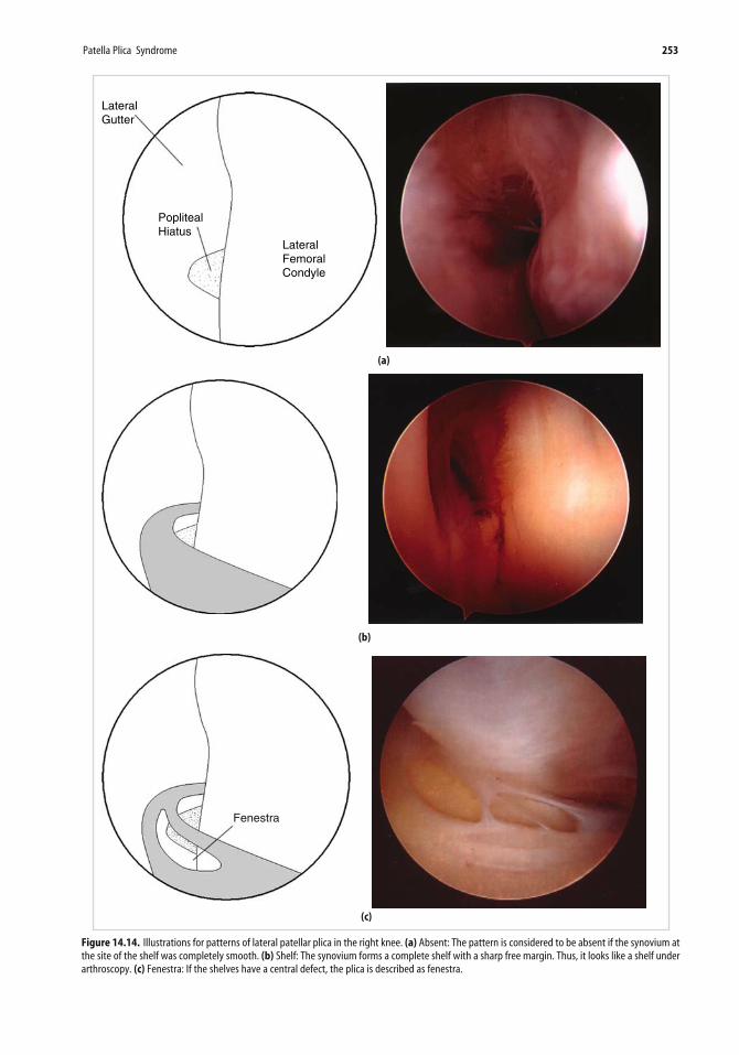
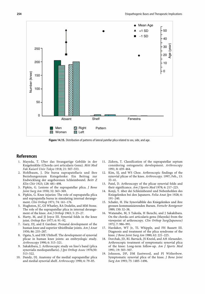
teus hiatus or manipulation of the arthroscopefrom the anterolateral portal into the lateralgutter. The pattern of each plica is classifiedas absent, shelf, or fenestra (Figures 14.14 and14.15).12
Unusually, lateral plica may produce symp-toms. In the literature review, pathologic lateralplica syndrome is reported.53-56 Patients com-plain of chronic pain and frequent painfulsnapping at the lateral side of patella. On exam-ination, a cordlike painful thickening may bepalpable. Pain is alleviated and the snappingdisappears after the arthroscopic excision oflateral plica.
252 Etiopathogenic Bases and Therapeutic Implications
Figure 14.12. The thickened fibrotic infrapatellar plica impinged to theintercondylar trochlea, resulting in limitation of extension of the knee.
Figure 14.13. The fenestra type of infrapatellar plica was thickened and fibrotic and had lost its elasticity. (a) In flexion. (b) In extension.
Ch14.qxd 10/07/05 3:46 PM Page 252
Patella Plica Syndrome 253
LateralFemoralCondyle
PoplitealHiatus
LateralGutter
(a)
(b)
Fenestra
(c)
Figure 14.14. Illustrations for patterns of lateral patellar plica in the right knee. (a) Absent: The pattern is considered to be absent if the synovium atthe site of the shelf was completely smooth. (b) Shelf: The synovium forms a complete shelf with a sharp free margin. Thus, it looks like a shelf underarthroscopy. (c) Fenestra: If the shelves have a central defect, the plica is described as fenestra.
Ch14.qxd 10/07/05 3:46 PM Page 253
254 Etiopathogenic Bases and Therapeutic Implications
250
200
150
100
50
0
Mean Age
+1 SD
−1 SD
50
40
30
20
10
0
Age
(ye
ar)
Absent Shelf Fenestra
MenWomen
RightLeft
Pattern
Num
ber
(kne
e)
Figure 14.15. Distribution of patterns of lateral patellar plica related to sex, side, and age.
References1. Mayeda, T. Uber das Strangartige Gebilde in der
2. Hohlbaum, J. Die bursa suprapatellaris und ihreBeziehungenzum Kniegelenke: Ein Beitrag zurEndwicklung der angeborenen Schleimbeutel. Beitr ZKlin Chir 1923; 128: 481–498.
3. Pipkin, G. Lesions of the suprapatellar plica. J BoneJoint Surg Am 1950; 32: 363–369.
4. Pipkin, G. Knee injuries: The role of suprapatella plicaand suprapatella bursa in simulating internal derange-ment. Clin Orthop 1971; 74: 161–176.
5. Hughston, JC, GS Whatley, RA Dodelin, and MM Stone.The role of the suprapatellar plica in internal derange-ment of the knee. Am J Orthop 1963; 5: 25–27.
6. Harty, M, and JJ Joyce III. Synovial folds in the kneejoint. Orthop Rev 1977; 6: 91–92.
7. Gary, DJ, and E Gardner. Prenatal development of thehuman knee and superior tibiofibular joints. Am J Anat1950; 86: 235–287.
8. Ogata, S, and HK Uhthoff. The development of synovialplicae in human knee joints: an embryologic study.Arthroscopy 1990; 6: 315–321.
9. Sakakibara, J. Arthroscopic study on lino’s band (plicasynovialis mediopatellaris). J Jpn Orthop Assoc 1976;50:513–522.
10. Dandy, DJ. Anatomy of the medial suprapatellar plicaand medial synovial shelf. Arthroscopy 1990; 6: 79–85.
11. Zidorn, T. Classification of the suprapatellar septumconsidering ontogenetic development. Arthroscopy1991; 8: 459–464.
12. Kim, SJ, and WS Choe. Arthroscopic findings of thesynovial plicae of the knee. Arthroscopy. 1997; Feb., 13:33–41.
13. Patel, D. Arthroscopy of the plicae synovial folds andtheir significance. Am J Sports Med 1978; 6: 217–225.
14. Kenji, Y. über dei Schleimbeutel und Nebenhohlen desKniegelenkes bei den Japanern. Folia Anat Jpn 1928; 6:191–240.
15. Schafer, H. Die Synovialhhle des Kniegelenkes und ihregrossen kommunizierenden Bursen. Fortschr Rontgenstr1989; 150: 32–38.
16. Watanabe, M, S Takeda, H Ikeuchi, and J Sakakibara.On the chorda cavi articularis genu (Mayeda) from theviewpoint of arthroscopy. Clin Orthop Surg(Japanese)1972; 7: 986–991.
17. Hardaker, WT Jr, TL Whipple, and FH Bassett III.Diagnosis and treatment of the plica syndrome of theknee. J Bone Joint Surg Am 1980; 62: 221–225.
18. Dorchak, JD, RL Barrack, JS Kneisl, and AH Alexander.Arthroscopic treatment of symptomatic synovial plicaof the knee: Long-term follow-up. Am J Sports Med1991; 19: 503–507.
19. Johnson, DP, DM Eastwood, and PJ Witherbow.Symptomatic synovial plica of the knee. J Bone JointSurg Am 1993; 75: 1485–1496.
Ch14.qxd 10/07/05 3:46 PM Page 254
20. Dupont, JY. Les replies synoviaux du genou: Aspectsanatomiques, physiopathologiques et cliniques. JTraumatol Sport 1990; 7: 25–38.
21. Kim, SJ, SJ Shin, and TY Koo. Arch type pathologicsuprapatella plica. Arthroscopy 2001; 17: 536–538.
22. Ross, KR, and MMS Glassgow. The suprapatella plica. JBone Joint Surg Br 1984; 66: 280.
23. Patel, D. Plica as a cause of anterior knee pain. OrthopClin North Am 1986; 17: 273–277.
24. Tindel, NL, and B Nisonson. The plica syndrome.Orthop Clin North Am 1992; 23: 613–618.
25. Amatuzzi, M, A Fazzi, and MH Varella. Pathologic syn-ovial plica of the knee. Results of conservative treat-ment. Am J Sports Med 1990; 18: 466–469.
26. Strover, AE, E Rouholamin, N Guirguis, and H Behdad.An arthroscopic technique of demonstrating the patho-mechanics of the suprapatella plica. Arthroscopy 1991;7: 308–310.
27. Bae, DK, GU Nam, SD Sun, and YH Kim. The clinicalsignificance of the complete type of suprapatella mem-brane. Arthroscopy 1998; 14: 830–835.
28. Lino, S. Normal arthroscopic findings in the knee jointin adult cadavers. J Jpn Orthop Assoc 1939; 14: 467–523.
29. Aoki, T. The “ledge” lesion in the knee. Proceedings 12thCongress of the International Society of OrthopaedicSurgery and Traumatology. Excerpta MedicaInternational Congress Series, No. 291, Amsterdam,Excerpta Medica, 1973, p. 462.
30. Jackson, RW, DJ Marshall, and Y Fujisawa. The patho-logic medical shelf. Orthop Clin North Am 1982; Apr.,13(2): 307–312.
31. Munzinger, U, J Ruckstuhl, H Scherrer, and NGschwend. Internal derangement of the knee joint dueto pathologic synovial folds: The mediopatellar plicasyndrome. Clin Orthop. 1981; Mar.–Apr., 155: 59–64.
32. Nottage, WM, NF Sprague III, BJ Auerbach, and HShahriaree. The medial patellar plica syndrome. Am JSports Med 1983; July–Aug., 11(4): 211–214.
33. Broom, MJ, and JP Fulkerson. The plica syndrome: anew perspective. Orthop Clin North Am 1986; Apr.,17(2): 279–281.
34. Brabants, K, S Geens, and L Blondeel. Plica synovialismediopatellaris. Acta Orthop Belg 1988; 54(4): 474–476.
35. Richmond, JC, and JB McGinty. Segmental arthroscopicresection of the hypertrophic mediopatellar plica. ClinOrthop 1983; Sep., 178: 185–189.
36. Glasgow, M, DJ McClelland, J Campbell, and RWJackson. The synovial plica and its pathological signifi-cance in the knee. J Bone Joint Surg Br 1981; 63: 630.
37. Aleman, O. Chondromalacia post traumatic patellae.Acta Chir Scandinavica 1928; 63: 149–189.
38. Dehaven, KE, WA Dolan, and PJ Mayer. Chondromalaciapatellae in athletes: Clinical presentation and conservativemanagement. Am J Sports Med 1979; Jan.–Feb., 7(1): 5–11.
39. Ficat, RP, J Philippe, and DS Hungerford. Chondro-malacia patellae: A system of classification. Clin Orthop1979; Oct., 144: 55–62.
40. Broom, HJ, and JP Holkerson. The plica syndrome: Anew perspective. Orthop Clin North America 1986; 17:297–281.
41. Sherman, RMP, and RW Jackson. The pathologicmedial plica: Criteria for diagnosis and prognosis. JBone and Joint Surg 1989; 71-B(2): 351.
42. Larson, RL, HE Cabaud, DB Slocun, SL Hanes, TKeenan, and T Hutchison. The patellar compressionsyndrome: surgical treatment by lateral retinacularrelease. Clin. Orthop 1978; 134: 158–167.
43. Dandy, DJ, and RW Jackson. The diagnosis of problemsafter meniscectomy. J Bone and Joint Surg 1975; 57-B(3): 349–352.
44. Kim, SJ, JH Jeong, YM Cheon, SW Ryu. MPP test in thediagnosis of medial patellar plica syndrome.Arthroscopy 2004; Dec., 20(10): 1101–1103.
45. Flanagan, JP, S Trakru, M Meyer, AB Mullaji, and FKrappel. Arthroscopic excision of symptomatic medialplica. Acta Orthop Scand 1994; 65: 408–411
46. Koshino T, and R Okamoto. Resection of painful shelf(Plic synovialis mediopatellaris) under arthroscopy.Arthroscopy 1985; 1: 136–141.
47. Kim, S-J, B-H Min, and H-K Kim. Arthroscopic anatomyof the infrapatellar plica. Arthroscopy 1996;12: 561–564.
48. Patel, D. Synovial lesions: Plicae. In McGinty, JB, ed.,Operative Arthroscopy. New York: Raven, 1996,pp. 447–458.
49. Mital, M, and J Hayden. Pain in the knee in children:The medial plical shelf syndrome. Orthop Clin NorthAm 1979; 10: 713–722.
50. Subotnick, SI, and P Sisney. The plica syndrome: Acause of knee pain in the athlete. J Am Pediatric MedAssoc 1986; 76: 292–293.
51. Kim, S-J, and W-S Choe. Pathologic infrapatellar plica:A report of two cases and literature review. Arthroscopy1996; 12: 236–239.
52. Kim, S-J, J-Y Kim, and J-W Lee. Pathologic infrapatellarplica. Arthroscopy 2002; 18(5): E25.
53. Bough, BW, and BF Regan. Medial and Lateral synovialplicae of the knee: Pathological significance, diagnosisand treatment by arthroscopic surgery. Irish Med J1985; 78: 279–282.
54. Fujisawa, Y, N Matsumoto, S Shiomi et al. Problemscaused by the medial and lateral synovial folds of thepatella (in Japanese). Kansetsukyo 1976; 1: 40–44.
55. Kurosawa, S, S Koide, T Yaota et al. Disorders of theknee caused by synovial plicae: So-called plica syn-drome. Clin Orthop Surg 1979; 11: 231–237.
56. Kurosaka, M, S Yoshiya, M Yamada, and K Hirohata.Lateral synovial plica syndrome. A case report. Am JSports Med 1992; 20(1): 92–94.