United States Department of Agriculture Forest Service Forest Products Laboratory Research Paper FPL–RP–529 Research on Biodeterioration of Wood, 1987–1992 I. Decay Mechanisms and Biocontrol Terry L. Highley Carol A. Clausen Suki C. Croan Frederick Green III Barbara L. Illman Jessie A. Micales
Transcript
United StatesDepartment ofAgriculture
Forest Service
ForestProductsLaboratory
ResearchPaperFPL–RP–529
Research onBiodeterioration ofWood, 1987–1992I. Decay Mechanisms and Biocontrol
Terry L. HighleyCarol A. ClausenSuki C. CroanFrederick Green IIIBarbara L. IllmanJessie A. Micales
22
Abstract
Growing concerns about the environment present an urgentneed for new approaches to preserving wood. Some com-monly used preservatives have been banned or restricted inseveral countries. Scientists at the Forest Products Labora-tory, USDA Forest Service, are exploring new approachesfor detecting and preventing wood decay. Progress in thisongoing research program is presented in two researchpapers. This report, Part I, describes research on decaymechanisms and biological control (biocontrol). Researchon diagnosis of decay and in-place treatment is presented inPart II (FPL–RP–530).
This research paper first describes current knowledge abouthow white- and brown-rot fungi decay wood and thendelineates research in two problem areas: (1) control of wooddecay through targeting biosynthetic and degradativepathways, and (2) biological control (biocontrol) of wooddecay through nondecay micro-organisms. Finally, direc-tions for further research are described in the areas ofbiology of wood deterioration and protection of woodwithout chemical pesticides.
March 1994
Highley, Terry L.; Clausen, Carol A.; Croan, Suki C. [and others]. 1994.Research on biodeterioration of wood, 1987–1992. I. Decay mechanismsand biocontrol. Res. Pap. FPL–RP–529. Madison, WI: U.S. Department ofAgriculture, Forest Service, Forest Products Laboratory. 20 p.
A limited number of free copies of this publication are available to thepublic from the Forest Products Laboratory, One Gifford Pinchot Drive,Madison, WI 53705–2398. Laboratory publications are sent to more than1,000 libraries in the United States and elsewhere.
The Forest Products Laboratory is maintained in cooperation with theUniversity of Wisconsin.
The United States Department of Agriculture (USDA) prohibits discrimina-tion in its programs on the basis of race, color, national origin, sex, religion,age, disability, political beliefs, and marital or familial status. (Not allprohibited bases apply to all programs.) Persons with disabilities whorequire alternative means for communication of program information(braille, large print, audiotape, etc.) should contact the USDA Office ofCommunication at (202) 720–5881 (voice) or (202) 720–7808 (TDD).
To file a complaint, write the Secretary of Agriculture, U.S. Department ofAgriculture, Washington, DC 20250, or call (202) 720–7327 (voice) or(202) 720–1127 (TDD). USDA is an equal employment opportunityemployer.
The use of trade or firm names in this publication is for reader informationand does not imply endorsement by the U.S. Department of Agriculture ofany product or service.
Research on Biodeteriorationof Wood, 1987–1992I. Decay Mechanisms and Biocontrol
In addition to very low mammalian toxicity, the idealbiocidal treatment must meet other demands: stability, aspectrum of activity graduated according to application,very low environmental hazard, and cost-effectiveness. Thetrend is toward developing narrower spectrum wood-preserving chemicals with enhanced activity and lower end-use concentrations. This emphasis will eventually lead tocarefully tailored or “designer” preservatives. The move tosuch preservatives with low toxicity could be facilitated bythe identification of the unique features of degradingorganisms or their biochemical pathways, which wouldreveal the key mechanisms of the decay process. Thisrequires thorough knowledge about the fundamental prin-ciples of wood decay—including the mechanisms andmethods of deterioration as well as the conditions requiredfor fungal growth. Understanding of the biochemical agentsin fungal attack that are responsible for degradation willprovide tools for “sabotaging” their ability to decay wood.By focusing research on the physiological differencesbetween wood-attacking fungi and nontarget organisms,fungal activity can be inhibited and stopped.
Background
The most important and potent wood-decay organisms arewhite- and brown-rot fungi. Brown-rot decay is the mostcommon and most destructive type of decay in structures(Fig. 1). For decay to occur, the moisture content must beabove fiber saturation (average 30 percent) and the tempera-ture between about 2°C and 38°C. Oxygen must also beavailable because decay fungi are aerobic organisms. Finally,a suitable substrate, such as wood, must be present to nourishthe fungus. Interference with any of these requirements stopsthe decay process. For example, storing logs under waterinterferes with the requirement for oxygen, and thus preventsdecay.
The optimal nutritional needs of white- and brown-rot fungivary, but the basic nutritional requirements are satisfied bythe structural carbohydrates and certain extraneous materials
Terry L. Highley , Research Plant PathologistCarol A. Clausen , MicrobiologistSuki C. Croan , MicrobiologistFrederick Green III , MicrobiologistBarbara L. Illman , Research Plant PathologistJessie A. Micales , Research Plant PathologistForest Products Laboratory, Madison, Wisconsin
Introduction
Failure of wood in service through biodeterioration is aprime cause of user dissatisfaction. More knowledge isneeded about the organisms that attack wood so that we canpredict with confidence the performance of wood products inparticular environments. Although many types of organismsdeteriorate wood, the greatest damage is microbiological—from fungi and, to a relatively minor extent, bacteria. Decayis by far the most serious kind of microbiological deteriora-tion because it can cause structural failure, sometimes veryrapidly. Because damage to wood from decay, thoughwidespread, is seldom spectacular, the tremendous economicand resource loss to this cause is often overlooked. Thisoversight is exemplified by the failure to recognize theimportance of biodeterioration of wood products and thefailure to recognize that extending the service life of wood isa sure way to extend the available timber supply. Extendedservice life also lowers the repair and maintenance cost ofwood construction.
There is an urgent need for new approaches for preventingwood deterioration. The major current wood preservatives—creosote, pentachlorophenol, and inorganic arsenicals—arebroad-spectrum biocides that can pose a serious threat to theenvironment, especially during the treating step. Some ofthese chemicals have already been banned or restricted insome countries. At present, no harmless substitutes for toxicchemicals are available. Consumers are concerned about theenvironment and may be using untreated wood in applica-tions where it may be subject to biodeterioration. Forexample, consumers question the safety of wood treated withchromated copper arsenate (CCA) because of the manyprecautions listed for such wood in consumer informationsheets. Some people think that the only solution is to banCCA-treated wood and to rely on industry to developnontoxic substitutes. Therefore, environmentally acceptablemethods are needed to meet society’s need for environmen-tally safe technologies to increase the durability of woodagainst biodeterioration.
2
efficient mechanism for nitrogen metabolism and reuse (Leviand others 1968). Field evidence (Larsen and others 1978)and results of in vitro culture studies support the theory thatwood-decay Basidiomycetes can conserve and function withthe small amount of nitrogen available in wood by autolysisand reuse of the nitrogen from their own mycelia or by thelysis of other fungi in wood during decay, together with anextremely economical use of nitrogen in metabolism.However, other sources of nitrogen, such as bacterialfixation, may sometimes be required to form sporophores.Studies of growth and nutrition of 42 white- and brown-rotfungi in synthetic media revealed that none of the fungirequired nitrogen in organic form, but growth was usuallygreater with organic nitrogen than with ammonium salts(Jennison 1952). Nitrate did not support the growth of 41 ofthese fungi.
Thiamine, the only vitamin essential for growth of mostwood-decay fungi, is present in wood (Jennison 1952). Thediscovery that most decay fungi require thiamine createdinterest in the possibility of protecting wood foraboveground service by alkaline treatments that presumablydestroy thiamine (Baechler 1959). In laboratory testsdesigned to simulate aboveground exposure (low decay),alkaline-treated wood was resistant to brown-rot fungi butnot white-rot (Highley 1970). Additional study showed thatthe increased resistance of alkaline-treated wood to brown-rot was caused by not only thiamine destruction but also theincreased pH of the wood (Highley 1973).
Various components of the wood cell wall are attackeddifferently by white- and brown-rot fungi. White-rot fungiutilize cellulose and the hemicelluloses at approximately thesame rate relative to the original amounts present, whereaslignin is usually utilized at a somewhat faster rate on arelative basis. These fungi cause the wood to become paler incolor and may eventually reduce it to a fibrous, whitishmass.
Brown-rot fungi utilize the hemicelluloses and cellulose ofthe cell wall, leaving the lignin essentially undigested.However, the fungi do modify lignin, as indicated bydemethylation and the accumulation of oxidized polymericlignin-degradation products. The fungi cause the wood todarken, shrink, and break into brick-shaped pieces thatcrumble easily into a brown powder (Fig. 1). Chemicalanalysis of brown-rotted white pine (Pinus monticola) andhard maple (Acer rubrum) showed that the hemicelluloseglucomannan is removed considerably faster than celluloseor xylan (Highley 1987a). Xylan is usually depleted fasterthan cellulose. Since hemicelluloses form an encrustingenvelope around the cellulose microfibrils, further degrada-tion and removal of depolymerized cellulose may depend onprior removal of the hemicelluloses. Thus, hemicelluloseutilization may be a critical initial step in establishment ofbrown-rot fungi in wood. Consequently, prevention ofhemicellulose utilization should inhibit the decay process.
Figure 1—Stages of brown-rot decay: (Top) earlystage—discoloration in surface and end grain (left),(Bottom) late stage—cracked and collapsed wood.(M 124 928)
in wood. Degradation of lignin does not occur in the absenceof wood carbohydrates, and lignin is probably of limitedimportance as a carbon source (Kirk and Fenn 1982).Successful colonization of wood depends largely on theability of fungi to spread rapidly by using nonstructuralcarbohydrates; the hyphae of both white- and brown-rotfungi are widespread in wood before any significant weightlosses are detectable (Hulme and Shields 1970).
Hyphae penetrate from cell to cell through natural openingsin the wood (pits) or by boring holes in the cell wall. Earlydecay by brown- and white-rot fungi is characterized by thepresence of at least one hyphal strand in each cell, suggestingthat extracellular enzymes may not be able to diffuse outsidethe wood cells in which they are secreted (Wilcox 1970).Thus, any physiochemical feature of the wood or its constitu-ents that limits the ability of an organism to develop in theindividual cells of wood will profoundly influence thewood’s resistance to decay.
Nitrogen plays the most important role of the nonstructuralnutrients in wood, and its part in wood decay has beenextensively studied by Cowling and coworkers (Levi andothers 1968; Merrill and Cowling 1966). The sparsity ofnitrogen in wood (about 0.03 to 0.10 percent (Merrill andCowling 1966)) indicates that wood-decay fungi have an
3
White-rot fungi successively depolymerize cell wall sub-stances only to the extent that the products are used formetabolism. The fungi produce a gradual decrease in averagedegree of polymerization (DP) of holocellulose during allstages of decay and only slightly change the solubilityproperties of wood (Cowling 1961; Highley and Illman1990). Brown-rot fungi, in contrast to white-rot, rapidlydepolymerize holocellulose, and the degradation products areproduced faster than they are utilized (Cowling 1961). Therapid depolymerization of the wood carbohydrates isreflected by the substantial increase in alkali solubilityproducts.
Brown-rot fungi cause a more rapid drop in strength proper-ties than do white-rot fungi, which reflects holocellulosedepolymerization. Early decay of both brown- and white-rotted wood is evidenced only by a loss in toughness. Woodthat has lost only 1 percent of its weight because of decaywill frequently exhibit a 50 percent loss in strength measuredas toughness (Richards 1954). Reduction in toughnesscaused by brown rot could be explained by the sharpreduction in DP of cellulose during decay, but the basis fortoughness loss caused by white rot lies elsewhere becausethese fungi reduce cellulose DP gradually.
While decaying wood, brown-rot fungi create a low pH,primarily by producing oxalic acid (Cowling 1961). White-rot fungi also produce oxalic acid but they metabolize it, andso the pH is not as low.
This report summarizes progress made by theBiodeterioration of Wood Unit at the Forest ProductsLaboratory from 1987 to 1992 in understanding wood-degrading organisms and their unique physiological featuresthat can be used to control decay. The research involves twoproblem directions: (1) study of biosynthetic and degradativepathways that can be targeted to develop better methods forcontrolling decay and (2) enhancement of the ability ofnondecay micro-organisms to prevent fungal attack, com-monly known as biological control.
Decay Control ThroughBiosynthetic andDegradative PathwaysThe objective of this research has been to provide basicinformation on the biochemical agents in fungal attackresponsible for degradation that can be used to develop new,environmentally safe wood preservation methods. Biochemi-cal and physiological studies of wood decay should revealnew information on decay mechanisms that will allow us tointerfere with essential biochemical processes and develophighly active and sharply targeted preservatives withenhanced preservative performance. To achieve our objec-tive, we have conducted research in three major areas:(1) wood/fungus interaction, (2) enzymes in digestion andgrowth, and (3) nature of the brown-rot cellulose depolymer-izing agent.
Figure 2—Early stage of brown-rot degradation ofhemlock. Hyphal sheath (HS) of hyphae (H) growingin lumina (L) is attached to cell wall. Primary (S
1),
secondary (S2), and tertiary (S
3) wall layers and
middle lamella (ML) appear intact. Aldehyde–O2O
4
fixation, TEM, 8,200x. (M89 0167)
Wood/Fungus Interaction
The Biodeterioration of Wood Unit initiated a number ofstudies in the early 1980s to explore the interaction of decayfungi with wood. Electron microscopy and cytochemicalstaining techniques were used to identify the sites of attackand morphological changes that occur during degradation ofcellulose and wood by brown- and white-rot fungi (Palmerand others 1983a,b, 1984; Murmanis and others 1983;Highley and others 1984, 1985; Highley and Murmanis1986, 1987). This work recognized the essential role in wooddecay of a gel-like matrix (sheath) that covers fungal hyphae(Illman and Highley 1989) (Fig. 2). The hyphal sheath iscarbohydrate in nature, surrounds the hyphae, and oftenattaches to the cell wall of wood. This matrix is thought toperform several diverse functions, all critical to the wooddecay process. It certainly must be involved in attachmentand survival mechanisms as well as in protection of thehyphae. It could also serve as a medium to condition thesubstrate and predispose it to enzymatic attack. An isolate ofPostia placenta that is unable to depolymerize cellulose or todecay wood test blocks produced a sheath that was morpho-logically different from those of normal test isolates (Greenand others 1991a). This isolate also failed to form largequantities of fungal glucan in liquid culture (Micales andHighley 1990; Micales and others 1990), which is thought tobe the primary chemical component of the hyphal sheath.These studies support the importance of the hyphal sheath inthe decay process.
The importance of the hyphal sheath was confirmed andexpanded in further work using high resolution scanningelectron microscopy (SEM) and creative SEM preparativeprotocols to offer a new model to account for the rapid
4
penetration of wood-degrading agents during brown-rotdecay (Green and others 1989b, 1991; Larsen and Green1992) (Fig. 3). For the first time, different SEM preparativemethods showed the existence of a mycofibril element of thehyphal sheath that penetrated wood cell wall layers. Theseextracellular structures have a normal diameter of 10 to50 µm and are up to 25 µm in length. Mycofibrils appear tobe linear structural extensions of the hyphal cell wall. Theprecise function of mycofibrils remains to be elucidated.However, we feel that penetration by the extracellularstructures of the sheath into the wood cell wall facilitates thedecay process in an orderly progression, and we propose thisas an alternative hypothesis to simple diffusion of wood-decay agents. The sheath, therefore, appears to consist of anextensive network of microfibrils embedded in a matrix ofglucan; it may also contain trilaminar extracellular membra-nous structures (EMS).
Sterols are a principal component in the membranes of mostfungi. Therefore, sterol-biosynthesis inhibitors, compoundsthat interfere specifically in fungal sterol-metabolism, shoulddestroy membrane integrity and stop decay. Micales andHighley (1991) determined the effect of sterol-synthesisinhibitors, detergents, surfactants, and other membranedisrupters on the growth of the brown-rot fungus Postiaplacenta. The sensitivity of P. placenta isolates to theergosterol-biosynthesis inhibitor fenpropimorph wassomewhat variable; ED
50 values ranged from 0.20 to
0.79 µg/ml. ME20, a strain of P. placenta that does not causesignificant weight loss in wood and that produces a hyphalsheath apparently deficient in glucan, was especiallysensitive to fenpropimorph (ED
50 = 0.08 µg/ml). Similar
variation among isolates was displayed in response to
monensin, an antibiotic that disrupts membrane function byinserting itself into fungal membranes. The ED
50 values
ranged from 1.1 to 1.7 µg/ml; in this instance, however,ME20 was the least sensitive isolate.
Certain strains of wood-decay fungi appear to secretebyproducts that can affect fungal growth (Micales 1992a).These compounds are small (molecular weight < 10,000), arenonenzymatic, and are associated with autolysis. StrainME20, which does not depolymerize cellulose or decaywood test blocks, has an aberrant, appressed colony mor-phology. When exposed to ME20 culture-filtrates, normaltest strains of P. placenta take on the abnormal morphologyof ME20. Colony morphology may be related to hyphalsheath structure. These staling products may disrupt normalhyphal sheath formation and interfere with decay develop-ment. The exact chemical nature of these staling productsand their effect on hyphal sheath development still needs tobe defined.
More work identifying the factors involved in matrixformation certainly seems worthwhile. The inhibition ofmatrix formation could be an effective means of preventingthe damage caused by wood-decay fungi.
Digestive and Growth Enzymes
In the previous section, we discussed some new concepts onhow fungi may move through wood. In this section, wediscuss what is involved in digestion and growth of decayfungi. From the standpoint of control, the regulation of keyenzymes in digestion and growth is of interest. Importantdigestive enzymes are those that decompose cellulose,
Figure 3—Schematic representation of hypha and hyphal sheathstructures as observed on inert substrates.
5
Because previous work by the Biodeterioration of WoodUnit (Highley 1987a) indicated that hemicellulose degrada-tion by brown-rot fungi is an important step in the brown-rotdecay process, we have focused on the purification of theseenzymes (Highley and Wolter 1982). Xylanase was sepa-rated and characterized for the first time from what wasthought to be a large, multienzyme complex (Green andothers 1989). This enzyme was then used as an antigen toproduce monoclonal antibodies (Clausen and others 1993a).
Extracellular wood-decay enzymes of the brown-rot fungusPostia placenta were localized using colloidal-gold-labeledmonoclonal antibodies to the ß–1, 4–xylanase (32 to 36 kDa)fraction of P. placenta (Green and others 1991b). Enzymeswere localized on the hyphal surface and on the clumpedmycofibrillar elements of the hyphal sheath followingfixation with glutaraldehyde. If fixation was omitted,labeling was diffuse and not localized on individual orclumped mycofibrils. We conclude that extracellular decayenzymes are weakly bound (noncovalently) to, but notidentical with, the linear mycofibrillar elements of the hyphalsheath. We conjecture that decay enzymes or agents,especially low molecular weight hemicellulases, mayassociate and migrate in concert with the formation ofmycofibrillar sheath elements as an integral step in theirpresentation to woody substrates during incipient decay.Confirmation of this hypothesis will require isolation andcharacterization of mycofibrils and immunocytochemicallocalization in the hyphal sheath on wood.
Growth Enzymes
Almost nothing is known about nitrogen metabolism inwood-decay fungi, but nitrogen plays the most important roleof the nonstructural nutrients in wood. The nitrogen contentof wood is very low, with an average C/N ratio of about350:500/L; nitrogen is present primarily as protein. Thus,wood-decay fungi must have an efficient mechanism fornitrogen metabolism and reuse. Most likely, these fungireuse the nitrogen in their own mycelium through autolysis.At least in the early stages of colonization, assimilation ofnitrogen might depend on secretion of extracellular pro-teases, which may be a potential target for control.
Polyamines are nitrogen compounds found in fungi that areessential for growth and development. The biosynthesis ofpolyamines in most organisms can proceed through one oftwo metabolic pathways. However, only one pathway hasbeen reported to be the route of synthesis in fungi. Thisfeature makes the pathway an ideal target for specificregulation of polyamine production in wood-decay fungi.The result would be the specific control of decay by affectingonly fungal growth and development. We have shown that“suicide” inhibitors such as diflouromethylornithine(DFMO) inhibit decay fungi (Illman 1990). Further studiesare underway using these inhibitors in the study ofpolyamine transport systems and determining reaction stepsin metabolic pathways. The inhibitors are enzyme-specific
hemicellulose, and lignin. For growth, key enzymes are thoseinvolved in nitrogen metabolism, such as glutaminesynthetase and polyamine enzymes, and in cell-wall forma-tion, such as chitin synthetase.
Digestive Enzymes
For digestion of wood cell wall constituents, enzymes mustbe released externally to the fungus. Thus, one method ofpreventing fungal attack of wood would be to inactivate theextracellular degrading enzymes in situ or prevent theirrelease from fungal hyphae. However, we found thatextracellular enzymes are quite stable; frequently, higherconcentrations of chemical are required to inactivate theenzyme than to prevent growth (Micales and others 1987;Highley and Micales 1990). A more promising approach,therefore, would be to find a way to interfere with processesinvolved in enzyme secretion.
The Tween surfactants have been shown to affect enzymesecretion (Micales 1991). The addition of Tween (20, 40, 60,and 80) changed the cultural morphology of Postia placenta,perhaps by altering the composition of the hyphal sheath.The activity of ß–D–glucosidase increased in culture filtratesthat had been supplemented with the four different Tweens.The modes of action of the various Tweens is not known, butthe increase in enzyme activity may be due to the ability ofTween to solubilize or disperse the hyphal sheath, thusreleasing sheath-bound enzymes into the culture filtrate.Certain detergents, including SDS and Triton X–100, werealso shown to remove the hyphal sheath. These treatmentswere actually incorporated into enzyme isolation proceduresto increase enzyme recovery (Clausen and others 1993b).The disruption of the hyphal sheath may greatly interferewith the decay process. This area needs further study toevaluate the potential of surfactants as control agents.
The effect of tunicamycin, an antibiotic that inhibits secre-tion by preventing the glycosylation of glycoproteins, wasdetermined on growth and enzyme production ofP. placenta. Low levels of tunicamycin prevented the releaseof degradative enzymes. Xylanase formed in the presence oflower concentrations of tunicamycin was smaller and lessstable to conditions of temperature and pH extremes andprotease activity (Micales and Highley 1988). Calmodulinhas also been suggested to be involved in secretion ofcellulases by some fungi, but the role of this material insecretion of enzymes by decay fungi is unknown. Therefore,the effect of anticalmodulin compounds on secretion ofcarbohydrate-degrading enzymes by white- and brown-rotfungi was determined (Highley 1989b). Severalanticalmodulin compounds inhibited production of cellu-lases, hemicellulases, and glycosidases of white-rot but notbrown-rot fungi. This indicates different secretion mecha-nisms by these types of fungi. We do not propose to usethese materials as wood preservatives, but they may serve asa model system in the continuing search for more environ-mentally compatible fungicides.
6
and irreversible, acting at the catalytic site of the enzyme,which results in “suicide.” These inhibitors are idealcandidates for studies to target the “Achilles’ heel” of thedecay fungi. This approach is also being studied to controldiseases in agricultural crops.
One means by which wood-decay fungi reclaim the nitrogenin wood is through the secretion of proteinases, enzymes thatbreak down nitrogenous substances. Proteinase productionwas associated with the autolytic phase of growth in isolatesof Postia placenta, suggesting that the fungus is recoveringthe nitrogen it has used previously for mycelial growth(Micales 1992b). These proteinases had acidic pH optima(Micales 1992b), indicative of a class of enzymes calledaspartic proteinases. There are chemical inhibitors thatinterfere with this type of enzyme. Future research willexamine the effect of these inhibitors on fungal growth andwood decay ability.
Another approach that might be utilized for specific inhibi-tion of fungal growth and development is the use of chemi-cals that prevent synthesis of chitin, an essential componentof fungal cell walls. Vertebrate animals do not possesschitinous tissue. This basic biological difference has stimu-lated interest in the development of pesticides that specifi-cally inhibit chitin synthesis and have little or no effect onnontarget organisms. There has been some success ininhibiting wood-decay fungi by inhibiting chitin synthetase,but the response to inhibition varies among species, probablybecause the chitin synthesizing enzyme is not equallyaccessible in all species (Johnson and others 1992). Addi-tional basic studies are needed to overcome this difficulty.
Brown-Rot Cellulose Depolymerization
Brown-rot fungi are unique among cellulose destroyersbecause they are the only known microbes that can degradethe cellulose in wood without first removing the lignin.These fungi leave a brown residue (hence the name) ofpartially demethylated lignin. Furthermore, brown-rot fungidegrade cellulose in an unusual manner that differs from thatof other cellulolytic organisms. Shortly after colonizingwood, the fungi cause a rapid and extensive depolymeriza-tion of cellulose to the “limit” or crystalline degree ofpolymerization (DP) at low weight loss. How this rapiddepolymerization of cellulose occurs is a perplexing bio-chemical question that has plagued researchers for yearsbecause even the smallest enzymes appear too large topenetrate the pores of wood. Also, cellulases do not mimicthe action of brown-rot fungi in generating cellulosecrystallites. Several hypotheses have been proposed toexplain how brown-rot fungi decompose cellulose (Fig. 4).
Although we have not been able to determine the biochemis-try and physiology of the degradative system employed bybrown-rot fungi to depolymerize cellulose, our research has
A. HemicelluloseAmorphous cellulose
Low molecular weightNonenzymatic
Nonenzymatic
B. Crystalline cellulose
Fibrillar hyphal sheath/EMS
Enzymatic
Fenton(H2O2)
Oxalicacid
Smallmolecules
Glycopeptides(H2O2)
Siderophores(Fe++)
Figure 4—Proposed mechanism of brown-rot decay.
made significant contributions to the understanding of thedegradative process (Highley and Illman 1991). To gaininsight into the mechanisms involved in decomposition ofcellulose by these fungi we conducted research in thefollowing areas: (1) ultrastructural changes in brown-rottedcellulose, (2) characterization of brown-rotted cellulose and(3) nonenzymatic breakdown of cellulose. These studieswere facilitated by use of a solid substrate system. Isolatedcellulose was degraded by placing it in contact with a woodor filter-paper inoculum over a soil or agar medium (Highley1977b; Highley and others 1988).
Ultrastructural Changes
Highley and others (1983) used the solid substrate system tostudy degradation of purified cellulose by 11 brown-rot fungiusing scanning and transmission electron microscopy (SEMand TEM, respectively). Erosion troughs or depressions inthe fiber surface were not visible at points of hyphal contact(Fig. 5). However, the cellulose within the fibers wasextensively degraded, regardless of hyphal invasion.
This type of cellulose degradation is different from thatobserved in studies of most other cellulolyticmicro-organisms, in which a rather localized attack oncellulose occurs near cells of the invading organism. White-rot fungi in contact with cellulose fibers erode the fibersurface (Highley and others 1984). A localized attack oncellulose fibers also occurs with mold fungi such asTrichoderma spp. and Aspergillus spp. These fungi penetrate
7
Characterization of Cellulose
To gain insight into the nature of the cellulase systeminvolved in the depolymerization of cellulose by brown-rotfungi, Highley and others (1989) chemically and physicallycharacterized brown-rotted cellulose. All eight brown-rotfungi studied extensively depolymerized cellulose exposedover soil or agar and increased alkali solubility beforesignificant weight loss occurred. Molecular weight determi-nations of cellulose degraded by the brown-rot fungus Postiaplacenta gave a symmetrical DP distribution, with a fairlytight distribution around DP 200. Thus, random cleavage ofall the cellulose must have occurred in the amorphousregions, not just at the fiber surfaces. The number averagemolecular weight was 188 and the weight average 335. Thisis close enough to a 1:2 ratio to be consistent with randomcleavage. X-ray diffraction analysis indicated a preferentialattack by the fungus on smaller crystallites and amorphousregions of the cellulose. Uronic acids were not detected inacid hydrolysates of the brown-rotted cellulose, indicatingthat oxidation was not at C–6.
Kirk and others (1991) further chemically characterizedcellulose decayed by P. placenta and compared the degradedcellulose with cellulose depolymerized by (1) acid hydroly-sis, (2) ferrous salts (Fe2+)–hydrogen peroxide (Fenton’sreagent), and (3) periodate–bromium oxidation. The Fentonsystem, but not the other systems, mimicked the brown-rotsystem in all measured characteristics. Gas-chromatographicmass spectroscopic analysis of the acid hydrolysates ofbrown-rotted cellulose identified four major acids: glyceric,erythronic, gluconic, and arabonic acids. No uronic acid wasproduced. The presence of glyceric and erythronic acidsindicates oxidative cleavage of the vicinal diol carbon–carbon bond within glucosyl residues. Reaction of cellulosewith Fenton’s reagent generated the same sugar acids but notin the same amounts.
Nonenzymatic Breakdown of Cellulose
Results from ultrastructural and chemical characterizationstudies indicate that rapid oxidation occurs during the earlystages of brown-rot decay, that pore size in wood is too smallfor known enzymes to access wood cellulose, and thatFenton’s reagent mimics brown-rot degradation of cellulose.A model based on Fenton chemistry predicts production ofhydroxyl radical (HO) from hydrogen peroxide and ferroussalts by the Fenton Haber–Weiss reaction: Fe2+ + H
2O
2 Fe3+
+ OH- + HO. The hydroxyl radical would cleave long-chaincellulose into smaller fragments. Thus, one proposal forbrown-rot decompositon of cellulose is that either(1) transition metal chelates convert to potent oxidizingspecies or (2) a free metal may generate an activated oxygenspecies, such as hydroxyl radical, and oxidatively cleavescellulose (Highley and Illman 1991). To test this hypothesis,we have studied hydrogen peroxide and oxygen free radicalproduction by brown-rot fungi and assayed for the presenceof metals.
the fiber to the lumen (Basu and Ghose 1962; Berg andHofsten 1976), and growth is abundant in the lumen andwithin the cell wall. Berg and Hofsten (1976) showed thatTrichoderma viride moves through cellulose by means ofsurface-bound enzymes.
Isolated cellulases also produce a slow, localized erosion ofcellulose near the point of contact (Eriksson 1981; Porter andothers 1960). Thus, morphological changes produced bybrown-rot fungi grown on cellulose are quite different fromthose produced by most other cellulolytic organisms andisolated cellulolytic enzymes. Cellulolytic enzymes would beexpected to produce a localized surface attachment oncellulose because cellulases isolated thus far are too large topenetrate the microstructure of cellulose and because theaffinity of cellulases to cellulose restricts their diffusion.Flournoy and others (1991) found no pores >3.8 nm in soundor decayed wood, which would exclude known cellulasesfrom brown-rot fungi.
Direct contact between the living fungus and cellulose seemsnecessary for effective degradation of native-type cellulosesby brown-rot fungi. Hyphae or hyphal sheath material isalways observed in contact with degraded fibers (Highleyand others 1983). Often, sheath material completely encirclescellulose fibers (Fig. 5). Similarly, in degradation of woodby brown-rot fungi, hyphal sheath structures are observed(1) on the surface of hyphae, (2) extending from hyphae ontothe wood surface and covering the S
3 layer, and (3) penetrat-
ing into the wood cell wall layers (Green and others 1989b).Hyphal sheaths have been proposed to facilitate cellulolyticaction by aiding transport of degradative agents to substrateand uptake of degradative products of cell wall dissolution(Green and others 1989b). Thus, without the hyphal sheath,the fungus may not be able to position the degradativeenzymes near the substrate.
Figure 5—Hyphal sheath (HS) of Postia placentaenwrapping several hyphae and much of cotton fiberwith granular material in its lumen, TEM.
8
source may also influence detection by determining the stageof hyphal growth in a given test. Variable growth rates havebeen observed in adjacent mycelial plugs taken from agarstock plates (Illman, unpublished data).
The role of hydrogen peroxide as a diffusible depolymeriza-tion agent has been predicted (Cowling and Brown 1969;Koenigs 1974a,b) and discussed (Illman 1991; Illman andHighley 1989; Kirk 1983). Its role as the diffusible agentmoving from tracheid lumen to an S
2 site of depolymeriza-
tion has not been demonstrated. If produced, hydrogenperoxide may be generated in close proximity to its site ofaction, in or near the hyphal sheath. This view is supportedby observations that brown-rot degradation of cellulosefibers and wood (Highley and others 1983, 1985) occurswhen the sheath is in contact with these substrates. Theproposed model of the sheath as facilitator of degradationalso supports a model of time-dependent generation ofhydrogen peroxide that is quenched by a number of site-specific oxidative reactions.
The possibility that fungal autolysis is the source of hydro-gen peroxide or other oxidizing agents responsible forpositive results has not been ruled out. The most successfultests for hydrogen peroxide were under conditions ofcarbohydrate and/or nitrogen starvation, which could lead toautolysis (Morton 1951), mimicking fungal action in wood(Merrill and Cowling 1966).
Production of Oxygen Free Radicals —The presump-tive role of hydrogen peroxide is the generation of thehydroxyl radical in a reaction with a metal or a metal chelate.The superoxide radical is a candidate precursor for genera-tion of hydrogen peroxide. Thus, assays for a depolymeriza-tion agent have been based on detection systems for oxygenfree radicals.
Several laboratories have used chemical assays to surveyfungal cultures and rotting wood for the production ofextracellular oxygen free radicals. Filtrates of 21-day-oldcultures of brown-rot fungi were shown to bleach thespectrum of p-nitrosodium methylaniline, suggesting thepresence of the hydroxyl radical (Highley 1982). The assayis not a specific test for oxygen free radicals. Organicradicals and easily auto-oxidizable compounds can producethe same results (Bors and others 1979).
Several attempts have been made to determine radicalproduction by employing oxygen radical inhibitors in fungalsystems. Several different chelators and radical andperoxide-quenching agents had little effect on weight loss ofcellulose or wood blocks by P. placenta, G. trabeum, orL. olivascens (Highley 1982; Highley and Murmanis 1985)in soil-block cultures. In this experiment, the toxicchloramphenicols had no effect on wood decay. Weight losswas <2 percent with thiourea (1.0 M) and 0.5 percent withfluorouracil (0.001 M) compared to 65 percent in the control(Highley 1982).
Production of Hydrogen Peroxide —The first reports ofextracellular hydrogen peroxide production by brown-rotfungi (Koenigs 1972, 1974a,b) were not easily reproducible(Highley 1981, 1982). Many researchers reported variableresults in detecting hydrogen peroxide in cultures of brown-rot fungi (Highley 1981, 1982, 1987a; Koenigs 1972), incontrast to easily detectable levels of hydrogen peroxideproduced by white-rot wood-decay fungi (Highley 1987b).Detection systems were based on color changes in mediafrom oxidation of chromophores ranging from bovinehaemoglobin or sheep’s blood medium to catalase–aminotriazole, 3,3'–diaminobenzidine tetrahydrochloride(DAB), o–dianisidine, and titanium tetrachloride.
When solid culture medium was amended with a moresensitive detection system of 2,2'–azinobis (3–ethyl-benthiazole–6–sulphonic acid) (ABTS) and horseradishperoxidase, hydrogen peroxide was detected in 2 of 13brown-rot fungi and 5 of 7 white-rot fungi tested. The ABTSsystem was used to study the effect of culture conditions onproduction of hydrogen peroxide (Highley 1987a). Byaltering the nitrogen supply in glucose-supported agarmedium, 6 of 13 brown-rot fungi produced hydrogenperoxide, as determined by medium color. In agar mediumwith low nitrogen (0.02 percent NH
4NO
3) supported by
glucose, mannose, xylose, cellobiose, or malt extract,hydrogen peroxide was detected in 11 of 13 brown-rot fungi.All seven white-rot fungi in the study produced hydrogenperoxide (Highley 1987b).
Because agar reportedly acts as a quencher of hydrogenperoxide (Thompson and others 1986), a liquid cultureprotocol was designed to test the time-dependent effects ofnitrogen and carbohydrate limitation on production ofhydrogen peroxide with the ABTS–peroxidase system(Illman and others 1989b). Production of hydrogen peroxideas measured by color change in the culture medium wasinduced by nitrogen and/or carbohydrate starvation in 11 of13 brown-rot fungi. The rate of color change varied witheach species, ranging from 4 to 40 days.
In addition to questions about the generation of hydrogenperoxide, the variable results call to question (1) the specific-ity and sensitivity of detection methods, (2) variability ofspecies and strains of a given species, (3) culture conditionsfor assays, and (4) lack of hydrogen peroxide accumulation.
Detection systems for hydrogen peroxide have yieldedpositive results in chemically defined solid, liquid, andwood-based culture media of many brown-rot fungi underspecific culture conditions. Such results have not beenobtained consistently. Explanations may be found in thedevelopmental stage of the fungus. Hydrogen peroxide maybe produced transiently; that is, in small concentrations for ashort period at a specific hyphal age. If hydrogen peroxide israpidly utilized or quenched by chemicals in the culturemedium, it may not be detected consistently. Inoculum
9
Figure 6—ESR spectra of paramagnetic manganesein wood, with and without the brown-rot fungusPostia placenta. Microwave frequency at 9.003 GHz,microwave power of 10 mW, and modulationamplitude of 10 G.
A more direct detection of hydroxyl radical in brown-rotfungal systems was made by using electron spin resonancespectrometry (ESR) and a spin trapping technique (Illmanand others 1988a, 1989b). The hydroxyl radical was detectedin fungal-inoculated wood using ESR and the radical spintrap, 5,5–dimethylpyrroline–N–oxide (DMPO). The DMPOwas used to derivatize the hydroxyl radical to a more stableadduct radical that gives a specific, four-line ESR signal. TheESR signal of the hydroxyl radical–DMPO adduct wasobserved in wood slivers of Douglas-fir and white fir wheninoculated with P. placenta (Illman and others 1988a,1989b). A hydroxyl radical–DMPO signal was also detectedin 10-day-old nitrogen-limited liquid cultures of P. placentasupported by 0.1 percent cellobiose or 0.1 percent glucose(Illman and others 1988a, 1989b). The chemical reaction orreactions generating the hydroxyl radical have not beendetermined.
The presumptive role of the hydroxyl radical as an agent tooxidize cellulose required formation of its site of action. Thehydroxyl radical is the most reactive of oxygen species, isnonspecific, and does not diffuse, as has been shown forhydrogen peroxide and the superoxide radical (Halliwell andGutteridge 1986). Reactions involving the hydroxyl radicalmay be facilitated by the fungal sheath, as proposed in themodel by Green and others (1989b).
Role of Metals —Fenton’s reagent has been shown tomimic brown-rot degradation of cellulose (Highley andothers 1989), implicating a role for a metal-catalysedFenton–Haber-Weiss reaction in brown-rot wood decay. Wehave assayed for metals in an attempt to determine their rolein brown-rot oxidative processes.
Electron spin resonance spectrometry has been used to detectand follow changes in the oxidative states of paramagneticmetals during brown-rot decay (Illman and others 1988b,1989a). Changes in low-spin iron could not be detected atroom temperature, and changes in high-spin iron were nottested at low temperature (Illman and others 1988b). Acomprehensive study of iron oxidation states using ESR hasyet to be made.
Changes in manganese were observed with ESR afterinoculation of susceptible species of wood with the brown-rot fungus P. placenta (Illman and others 1988a, 1989b)(Fig. 6). These changes were manifested as increases in thesize of the sextet spectra that are specific for manganese(Mn2+). The ESR spectra for Mn2+ were taken over a 4-weekperiod from fungal-inoculated white fir, Douglas-fir,sweetgum, and redwood. The increases in Mn2+ signalscorrelated with wood susceptibility to brown-rot decay. Littleor no increase was found in wood species resistant to brown-rot decay (Illman and others 1989b). The chemical basis ofthe Mn2+ change was not determined.
We found that oxalic acid changed the Mn2+ ESR signal inwood (Illman and Englebert, unpublished data). Oxalic acid
is a low molecular weight fungal metabolite that is primarilyresponsible for the low pH (<3) of wood during brown-rotfungal decay (Takao 1965; Green and others 1992b). Thelow pH is conducive to the Fenton–Haber-Weiss reaction,which occurs more rapidly at lower pH levels (Halliwell andGutteridge 1986). Schmidt and others (1981) demonstratedthat oxalic acid can reduce Fe3+ to Fe2+ under certainconditions. These authors proposed that oxalic acid generatesthe Fe2+ for the Fenton system. In the tests conducted byIllman and Englebert, after treatment of white fir slivers withoxalic acid, the ESR signal for Mn2+ increased with increas-ing oxalic acid concentration although the Mn2+ signal didnot increase as much as in P. placenta decayed wood. Theincreased manganese signal may be a result of lowered pHwith acid solubilization of the metal. Oxalic acid fromseveral species of wood-decay fungi can mobilize calcium
10
from glass and concrete. Alternatively, the increase in theMn2+ ESR signal may be due to chelation of the metal.Oxalate is a known chelator of several elements, includingmanganese, calcium, and potassium. The chemical basis ofoxalic acid effects is being investigated.
Oxalic Acid Metabolism
As demonstrated in the previous sections, the generation ofoxalic acid may be a key step in brown-rot decay. Manydifferent roles, both direct and indirect, have been assignedto oxalic acid during the depolymerization of cellulose.Oxalic acid can depolymerize cellulose directly (Shimadaand others 1991, 1992), but most researchers assign it a moreindirect role (Green and others 1992b). In addition to itspossible role in the generation of hydrogen peroxide(Koenigs 1974a,b; Espejo and Agosin 1991) and its effect onmanganese (Illman and others 1989a), oxalic acid may alsobe involved with the solubilization and hydrolysis ofhemicellulose, thus making the cellulose fibers moreaccessible to cellulases (Bech–Anderson 1987). Green andothers (1991a, 1992, 1993) expanded upon this hypothesisand suggested that the acidic conditions in wood, caused byoxalic acid production, are responsible for early acidhydrolysis and depolymerization of hemicellulose andamorphous cellulose, thus increasing wood porosity.Enzymes and other degrading agents in the hyphal sheathwould then have access to the remaining cellulose and causeits final removal.
Despite its importance, little work has been done on thephysiology of oxalic acid in wood-decay fungi. The path-ways involved with oxalic acid synthesis are just beginningto be explored. Akamatsu and others (1992) isolated theenzyme oxaloacetase (E.C. 3.7.1.1) from the hyphae ofFomitopsis palustris. This enzyme catalyzes the hydrolysisof oxaloacetate to form oxalic acid and acetate. Otherbasidiomycetes are known to produce oxalic acid via theglyoxylate bypass of the tricarboxylic acid cycle (Lapeyrieand others 1987; Maxwell and Bateman 1968a,b,c). Shimadaand others (1992) proposed that both pathways generateoxalic acid to decrease the large pool of organic acids thataccumulates from the degradation of wood carbohydrates.Under in vitro conditions, oxalic acid production by Postiaplacenta was stimulated by conditions of low nitrogen andcertain carbon sources, including glucose and acetic anduronic acids. Strain ME20, a monokaryon that does notdepolymerize cellulose or cause weight loss in wood testblocks, produced little or no oxalic acid under similarconditions. The release of acetic and uronic acids fromhemicellulose early in the decay process would greatlystimulate the production of oxalic acid by the fungus, thusrapidly lowering the pH of the wood in a self-potentiatingsystem. As decay progresses, oxalic acid would continue tobe induced by the release of glucose from cellulose. StrainME20 appears to be defective in these regulatory mecha-nisms and cannot decay wood effectively.
Proposed Mechanism of Brown-RotCellulose Decomposition
Cellulose degradation by brown-rot fungi appears to involvetwo sequential (or concurrent) mechanisms, oxidation andhydrolysis. These appear to be tightly coupled reactions.Oxidation is most likely nonenzymatic, with hydrolysiscatalyzed by a complex of enzymes. Low molecular weight,diffusible element or elements from brown-rot fungi have orare expected to have the capacity to generate oxygen radicalsdirectly or to initiate a chain reaction resulting in theirproduction. The capacity of brown-rot fungi to produceextracellular oxidative metabolites is expressed when theorganisms are maintained under specific culture conditions,especially nitrogen and carbohydrate limitation.
Evidence points to a small (<6,000 daltons) diffusible agentor system as the depolymerization factor with the capacity toset in motion a chemical reaction culminating in the oxida-tion of cellulose. The oxidative mechanism may result in thegeneration of the hydroxyl or other oxygen radical (or ametalo–oxygen species) in close proximity to its site ofaction on cellulose. If hydrogen peroxide is the substrate forhydroxyl radical production, it is most likely produced insmall amounts at a distinct stage of hyphal growth. It isquickly scavenged and does not accumulate. Given thesecriteria, an oxidative agent or agents have or are expected tohave controlled transport from site of origin. The proposedmodel of the fungal sheath provides a vehicle for facilitatingtransport of degradative agents to the site of decomposition.Association of the cellulolytic enzyme complex with ahyphal sheath component could also facilitate the hydrolysisof wood cellulose by appropriately juxtaposing the cellu-lolytic complex with cellulose.
Biocontrol Through NondecayMicro-Organisms
Background
Another approach for environmentally safe wood protectionis to exploit relationships that already exist in nature. Otherorganisms growing in wood, including fungi and bacteria, donot cause decay but do inhibit the growth of wood-decayfungi. These friendly micro-organisms may actually attackand consume wood-decay fungi for their own nutrition, orthay may produce antibiotics that inhibit or kill wood-decayfungi (Fig. 7). This type of activity is termed biologicalcontrol or biocontrol.
The potential of biocontrol to protect wood products againstdeterioration by micro-organisms has long been recognized,as indicated in a 1940 memorandum by Mae S. Chidester,Forest Products Laboratory (FPL). Chidester suggested that abetter understanding of the combined effect of two or morewood-inhabiting fungi on wood would be beneficial inpreventing or controlling the defects brought about by these
11
analysis by Lindgren, such biocontrol remains a virgin fieldthat requires considerable exploratory work, followed byintensive studies.
Verrall (1966) made preliminary incursions into the field ofmicro-organism associations when he commonly observedthe ubiquitous mold Trichoderma viride as the first colonizeron rain-wetted wood in the Southeastern states and proposedthat competition from molds might possibly be an additionalfactor that restricts the number of decay agents on exteriorwoodwork. The effect of two isolates of T. viride on41 isolates of Gloeophyllum saepiarium, G. trabeum, andDaedalea berkeleyi were studied on malt agar and in soil-block tests (Verrall 1966). All three basidiomycetes on maltagar either grew compatibly with T. viride or actuallyovergrew Trichoderma. Trichoderma did not prevent decayof pine by the basidiomycetes. In a number of cases, decaywas greater in the presence of Trichoderma than in pureculture. These findings are important because the threebasidiomycetes are the prevalent decay agents of pine lumberon the exterior of buildings and of other pine productsexposed off the ground but subject to rain wetting.
Following these rudimentary observations, biocontrol ofwood-attacking fungi in wood products did not gain furtherattention by Forest Service scientists until the 1980s.Renewed interest was prompted by increasing environmentalconcerns, which stimulated scientists at FPL to search forless hazardous wood preservatives. Obstacles that hinder theuse of biological fungicides in wood and trees and expandingopportunities for such use are discussed in review articles byRicard and Highley (1988, 1989).
Polyoxin D, one of a group of peptidyl pyrimidine antibioticsproduced by Streptomyces cacaoi var. asoensis was studiedby Johnson (1980, 1982, 1983, 1986) for control of wood-staining mold and wood-decay fungi. This antibiotic is ahighly specific inhibitor of the chitin-synthetase enzyme.Because chitin synthesis occurs only in lower life forms, itsinhibition could be a target-specific approach to pest controlwith minimal effect on nontarget organisms. Wood decay byseveral white- and brown-rot fungi was reduced by 10 ppmpolyoxin mixture and prevented at 100 to 700 ppm, anappreciably lower threshold than that of pentachlorophenol.However, polyoxin was not as effective against mold andstain fungi, and it is not a good candidate for protection ofwood against these fungi (Johnson 1986). Althoughpolyoxins are not themselves suitable wood preservatives,these studies have tentatively validated the concept of woodpreservation via inhibition of fungal chitin synthesis.
The objectives of the research in biocontrol were (1) toidentify potential antagonists and treat wood blocks forexposure to wood-attacking fungi, (2) to investigatemechanisms of antagonism, (3) to improve the ability ofantagonists to colonize wood, and (4) to evaluate deliverysystems.
Figure 7—Early stage of mycoparasitism: (a) small hyphaldiameter of Trichoderma polysporum compared tobasidiomycete (Gloeophyllum trabeum), 2,500x;(b) hyphal coiling by Trichoderma polysporum onGloeophyllum trabeum, 7,000x; (c) hooks produced byTrichoderma harzianum on Antrodia carbonica, 2,300x.(MC 87 9013)
organisms. She further suggested the possible use ofTrichoderma lignorum as a control for wood-decay fungisuch as Lenzites sepiaria (= Gloeophyllum sepiarium).Evidently this proposal was not followed up with experimen-tal work, or at least not published, because there are noreferences to it in the literature.
The first publishable work by Forest Service scientists onbiocontrol of wood-attacking fungi appears to be that ofLindgren and Harvey (1952) at FPL. These researchers usedfluoride pretreatment of Southern Pine pulpwood to enhanceprofuse growth of Trichoderma. They observed reduceddecay in the southern pine pulpwood treated withTrichoderma. However, as recognized in a 1958 problem
12
that wood material removed from poles treated with aTrichoderma (T-75, T-76)-based biocontrol product resistedattack by active fungi for 7 years after the first treatment.This suggests that with further research the use of biocontrolusing these Trichoderma species might be valuable as aprophylactic treatment to protect poles from internal decay.Most important, the decay prevention observed in thisexperiment was achieved using material colonized by theTrichoderma under field conditions. This indicates that atleast some modes of antagonism attributed to this fungusduring laboratory studies must still be active in the nutrient-limiting environment within wood poles.
Wood products, such as poles, are commonly precolonizedby a resident microflora, and any control agent must be ableto either displace or coexist with a variety of mold organ-isms. With this in mind, Bruce and Highley (1989) examinedthe direct influence of prior colonization of wood by typicalmold residents from creosoted poles on the biocontrol abilityof Trichoderma (T-75, T-76) against Lentinus lepideus(= Neolentinus lepideus). The results indicated thatTrichoderma can protect wood from decay by L. lepideuseven when the blocks are colonized with mold organismsprior to treatment with Trichoderma.
We examined the ability of a commercial preparation ofTrichoderma (BINAB T) to colonize and survive in DouglasFir and southern pine timbers exposed above ground invarious climatic areas (Highley 1994). Under field condi-tions, Trichoderma readily colonized southern pine (mostlysapwood) squared timbers and L–joints, but the percentageof recovery in the squared timbers dropped considerably inthe third year of exposure. Trichoderma was isolated up to1.8 m from the point of pellet insertion in the southern pinetimbers. Colonization of Douglas Fir heartwood byTrichoderma in squared timbers and piling exposed aboveground was much poorer than that in southern pine. DouglasFir timbers exposed in ground contact in Madison Wisconsinwere better colonized, but recovery was very poor beyond2.54 cm from pellet insertion.
Although Trichoderma was able to develop and spreadthrough the southern pine timbers, it did not stop decay. Thetimbers eventually were decayed by Gloeophyllum trabeum.In previous laboratory tests (Highley and Ricard 1988), wefound that Trichoderma did not control G. trabeum decay.Control of G. trabeum is important because this fungus is aprevalent cause of decay of pine on the exterior of buildingsand decay of other pine products off the ground but subjectto rain wetting.
A biocontrol treatment would have a better chance forsuccess as a groundline treatment for poles than as a treat-ment for aboveground wood because of the continualmoisture. It is difficult to envisage active control of decay byantagonists in aboveground structures, such as windowframes and waterfront curbing, when the moisture content
Control by Antagonistic Fungi
Highley and Ricard (1988) studied the antagonistic ability ofGliocladium virens and various Trichoderma spp. againstimportant white- and brown-rot fungi. Gliocladium virensand the Trichoderma spp. overgrew the decay fungi culturedon the malt-agar medium and in most cases killed them. Insoil-block tests, pretreatment of southern pine blocks withG. virens prevented brown-rot decay but was ineffectiveagainst the white-rot fungi. Similarly, Trichoderma spp.generally prevented or reduced decay by the brown-rot fungi,except for G. trabeum, but also were generally ineffectiveagainst the white-rot fungi. Various concentrations ofammonium nitrate and glucose in a basal medium did notaffect antagonism of Trichoderma spp. in wood blocks.Gliocladium virens did not confer residual fungistasis towood blocks. In soil-block tests, G. virens arrested thegrowth of Antrodia carbonica but not other decay fungi.This work suggests that Gliocladium and Trichoderma havepotential as natural agents for biocontrol of wood decay.
Several studies were conducted by FPL scientists to assessthe potential and application of commercial Trichodermapreparations for control of wood-decay fungi. These prepara-tions are mainly available as a wettable powder and aspellets; both contain propagules of Trichoderma spp.ATCC 10475 (T-75) and 20476 (T-76) (American TypeCulture Collection). Both preparations are produced inToreboda, Sweden, by Bio Innovation AB (BINAB). Whileexploring the mode of action of T-75 and T-76, Murmanisand others (1988a,b) found neither water-soluble antibioticsnor exoenzymes in wood blocks that were overgrown by theTrichoderma strains and protected against major brown-rotbasidiomycetes. However, the researchers observed abundantchlamydospore formation. Another study (Murmanis andothers 1988), also using SEM microphotographs, showedthat T-75 and T-76 spores became attached readily to hyphaeof wood-decay basidiomycetes. These spores germinated onthe surface of the basidiomycete hyphae and broke throughits wall.
In another study on mode of action by Trichoderma (Bruceand Highley 1990), the interactive effects of Trichodermastrains, including T-75 and T-76, against the range of wood-decay fungi were examined, with particular attention to theproduction of soluble metabolites by the antagonists. Theresults clearly showed that the modes of antagonism ofTrichoderma spp. are most complex, and, unlike the mode ofaction of most chemicals, they may be subject to changeunder different environmental conditions. This point isimportant and must be carefully considered in evaluatingcontrol systems, particularly during screening tests forpotential control agents.
To be a successful biocontrol agent against wood-decayfungi, the antagonistic effect must last for many years. Theresults of studies by Bruce and others (1989, 1991) showed
13
fluctuates substantially. Any biocontrol agent would have tomaintain itself during the times when the timber remaineddry, only becoming active when wetting occurred. On theother hand, if the biocontrol agent left a fungistatic residue,resistance to decay might be improved. Trichoderma hasbeen reported to produce fungistatic substances (Bruce andHighley 1991). However, this evidently did not occur in thefield exposure in our tests because wood removed fromTrichoderma-infected areas of pine timbers and sterilizedwas not resistant to decay. Bruce and others (1991) foundsimilar results with sterilized wood removed fromTrichoderma-treated poles after 7 years. However, theauthors found that unsterilized wood was resistant toN. lepedius but not to Trametes versicolor. Likewise,Highley (1993) found that unsterilized Trichoderma-treatedwood removed from pine timbers reduced decay byN. lepideus, but not T. versicolor or Postia placenta.
All screening of potential biocontrol agents of wood-decayfungi has previously been used against basidiomycetemycelium. Similarly, experiments designed to evaluate themechanisms involved in antagonism (soluble metabolitesand volatile antibiotics) between biocontrol agents and targetfungi have been carried out on mycelial inoculum. Basid-iospores are a primary source of infection that lead to decayof wood exposed above ground and are the pathogenic formof the target fungus in initial contact with a biocontrolantagonist. Thus, Srinivesan and others (1993) examined theantagonistic responses of Trichoderma spp. against wood-decay basidiospores. They also studied the influence ofnutrient composition of growth media on the antagonisticresponses by Trichoderma isolates. The results clearlyshowed that soluble metabolites from Trichoderma do play arole in inhibition of basidiospores. However, the metabolitesexhibited more specific inhibition of basidiospores thaninhibition of mycelial inoculum of basidiomycetes. It is alsoevident that the volatiles produced by the Trichodermaisolates have little or no inhibitory effect on basidiosporegermination. Target specificity seems to play an importantrole in the antagonism of Trichoderma spp.; Trichodermaisolates specifically inhibited germination of brown-rot butnot white-rot basidiospores. The results of this study alsoemphasize the importance of growth media in determiningthe outcome of antagonistic responses. More filtrates fromTrichoderma spp. grown on minimal media were inhibitoryto Trametes versicolor than were filtrates from Trichodermagrown on malt extract.
Trichoderma species examined for antagonistic effect againstdecay fungi have been rather selective in their antagonism inthat they have been unable to control decay by the brown-rotfungus Gloeophyllum trabeum and most white-rot fungi. In asearch for antagonistic fungi with broader antagonismtoward wood-decay fungi, Highley (1989a, 1991) evaluatedthe antagonistic abilities of Scytalidium lignicola againstseveral white- and brown-rot fungi. Pretreatment of Douglas-fir and southern pine blocks with S. lignicola prevented
decay by all fungi. Blocks that were heated or treated withpropylene oxide to kill the antagonist were not resistant todecay. Thus, S. lignicola does not confer a residualfungistatic effect to wood. Scytalidium lignicola was able toeradicate all the decay fungi in wood except for Postiaplacenta and Gloeophyllum trabeum. Wood blocks treatedwith filter-sterilized filtrates of S. lignicola were not decayresistant, and filtrates were not inhibitory to growth of thedecay fungi in agar medium. The antagonistic effect,therefore, apparently does not involve toxins.
The interspecific interactions and antagonism among severalScytalidium isolates with various brown- and white-rot fungiwere studied by Cease and others (1989). Scytalidiuminitially colonized the surface of the blocks and graduallyovergrew the basidiomycetes. In individual wood blocksfrom 11 Scytalidium–basidiomycetes paired treatmentcombinations, the basidiomycete was not inhibited through-out the entire wood block. These wood blocks demonstratedinterspecific interactions and antagonism between thedifferent fungi. The white-rot fungi responded to isolates ofScytalidium by occluding xylem cells with masses of hyphae,forming pseudosclerotial plates in the zone of initial interac-tion. Scytalidium appeared to gain access into portions ofwood colonized by the basidiomycetes only after substantialdecay had resulted by the wood-decay fungus.
An interesting observation was made by Croan and Highley(1991a), who found that the blue-stain fungus Ceratocystiscoerulescens could be controlled by metabolic productsreleased by several wood-decay basidiomycetes. In this case,it might be possible to protect wood against discoloring fungiby treatment with fungitoxic metabolic products.
Control by Antagonistic Bacteria
In several studies at FPL, scientists examined the efficacy ofbacteria on biocontrol agents against wood-decay fungi andsapwood-inhabiting fungi. The type of medium used toevaluate bacterial/fungal interaction had considerable effecton interactions on agar and wood (Benko and Highley1991a,b). In an initial decay test, a bacterial preparationprevented decay by white- and brown-rot fungi when testedby an agar-block procedure (Benko and Highley 1990a).However, in further tests with the soil-block method, thebacterial preparation was ineffective (Benko and Highley1990b). The failure of the autoclaved bacterial solution toprotect wood from decay showed that actively growingbacteria are needed for successful biocontrol. This isdiscouraging from the standpoint of long-term protectionagainst decay because fungistatic material probably wouldnot remain in the wood after death of the bacteria.
Bacterial preparations were found to be very effective inpreventing stain and mold discoloration in laboratory testswith various species of wood (Benko and Highley 1990a,1991b). The greatest potential for use of bacteria may be for
14
temporary protection against mold and stain in green logs orlumber. One bacteria, Streptomyces rimosus, was selected forfurther study.
Initial studies with S. rimosus concerned development ofculture parameters to optimize production of fungitoxicmetabolites (Croan and Highley 1991). The bacteria pro-duced very potent antifungal metabolites that effectivelyprevented mold and stain attack on green pine log sections ina simulated field trial (Croan and Highley 1991b, 1992a,b)(Fig. 8). Streptomyces rimosus has been reported to producethe antifungal antibiotic rimocidin, but the authors have notyet identified the antifungal metabolites responsible for moldand stain prevention. The material is very safe, with an LD
50
of 1,400 mg/kg intraperitoneally in mice compared to 146 to175 mg/kg orally for penta. Boron as a co-biocide withmetabolites from S. rimosus acted synergistically to protectwood first from mold and then from stain attack; the protec-tion was tenfold better than that obtained with concentratedmetabolites (Croan and Highley 1992b).
For longer term protection, it might be advantageous to useliving cells of S. rimosus applied to wood. The authors foundthat application of living S. rimosus cells to green wood is avery effective biotic agent against wood-attacking fungi(Croan and Highley 1992a). The bacterial cells completelyprotected wood blocks and green pine log sections fromwood-attacking organisms and did not alter the physical stateof the wood.
The S. rimosus isolate is a new isolate that differs from itsparent in that it does not sporulate. Therefore, this newisolate was named S. rimosus-nonsporulating isolate, SC-36.
A patent for biological treatment of wood with this isolatehas been applied for.
Research NeedsSince 1987, considerable progress has been made in under-standing and preventing biodeterioration of wood processes.However, wood biodeterioration continues to pose problemsthat only additional research can solve. The followingsections describe general areas in which future researchcould prove valuable.
Biology of Wood Deterioration
• Identification of the elusive agent or agents produced bybrown-rot fungi that facilitate degradation of cellulosein woodWe need a better understanding of the genetics andpathways involved in the production, regulation, andcatabolism of possible nonenzymatic degradative agentssuch as oxalic acid, oxidative agents, or metal chelates,and the role of manganese oxidation. Interfering with theseproducts or pathways could prevent decay.
• Further dissociation and characterization of extracellu-lar degradative enzymes produced by brown-rot fungiStudying the role of specific extracellular enzymes frombrown-rot fungi such as Postia placenta (that is, xylanase,mannanase, and cellulase) has been hampered because ithas not been possible to separate these enzymes from oneanother. Furthermore, precise separation and characteriza-tion of enzymes must precede production of polyclonal ormonoclonal antibodies to individual enzymes. The ability
Figure 8—Control of wood-attacking fungi on green pine log sections by metabolites fromStreptomyces rimosus in simulated field trial. (M 91 0095)
15
we need to fully understand the synergistic relationshipbetween bacteria, both aerobic and anaerobic, and wood-decay fungi.
• Isolation, identification, and physiology of organismsassociated with failed preservative-treated woodKnowledge of these organisms will promote our under-standing of preservative failure and the development ofmore effective preservatives. These organisms may also beuseful in bioremediation.
• Screening of natural compounds from antagonisticmicro-organisms and decay-resistant trees forfungitoxicity against wood-attacking fungiHighly fungitoxic natural compounds with low mamma-lian toxicity may be effective as wood preservatives.
• Study of degradation of wood in compostDuring composting, wood is decayed very rapidly. Decayoccurs in a two-stage process. Temperatures in a compostpile initially increase to about 55°C and microbial activityis restricted to thermophyllic bacteria. The temperaturethen decreases, allowing the growth of mesophyllicbacteria and fungi. Studying the succession of theseorganisms and the mechanism of their interaction todegrade wood so efficiently would provide betterunderstanding of the conditions required for optimal decayand indicate possible ways to interfere with the decayprocess.
Protection Without Chemical Pesticides
• Systematic approach to screening of biocontrol agentsOnly a limited number of biocontrol agents have beenstudied for wood-attacking fungi. A more systematicapproach to screening of biocontrol agents will inevitablyidentify other isolates with improved control qualities.
• Study of in situ modes of antagonismKnowledge of the modes of antagonism will make itpossible to enhance the action of biocontrol agents throughthe use of techniques such as mutation, protoplast fusion,and genetic engineering. Such techniques may enhanceantagonistic traits of the organism or improve its growthand survival capabilities in wood.
• Alteration of physical conditions within wood to make itless favorable to decay fungiOne way to alter wood physically would be to selectivelyenhance resident antagonistic organisms. This will requiredefining the ecological progression of decay.
• Development of integrated biological and chemicalcontrol systemsIntegrated systems will provide an opportunity forreplacing more toxic chemicals by a combination of abiocontrol agent and a less toxic chemical.
to characterize purified enzymes would greatly enhanceefforts to understand and interfere with enzymatic diges-tion of wood cell wall constituents.
• Immunoelectrophoretic analysis and quantitation ofextracellular enzymes of wood-decay fungiImmunoelectrophoresis is a powerful tool for identifyingantigens in complex protein mixtures. Modifications of theoriginal technique have resulted in many applications forqualitative and quantitative analyses of antigen/antibodyreactions. Immunoelectrophoretic analysis will provideinformation critical to the understanding of the role ofextracellular enzymes in wood cell wall digestion.
• Electron microscopic analysis of wood/fungus interac-tion using immunological techniquesElectron microscopes and ancillary equipment are avail-able to provide critical examination of the wood/fungusinteraction. Scanning microscopes permit examination ofthe interaction of decay fungi in the natural state. Usingimmunocytochemical probes to localize either fungalenzymes or extracellular metabolites, it is possible todemonstrate in situ the occurrence and spatial distributionof major wood-degrading agents.
• Study of nitrogen metabolismOf the nonstructural nutrients in wood, nitrogen plays themost important role in growth of wood-decay fungi.Almost nothing is known of the utilization or assimilationof nitrogen. Would inhibition of proteinases that areinvolved with autolysis and the recycling of fungalnitrogen be a target for inhibition of decay? What is therole of polyamines in fungal growth and development?Polyamines may also be a target for specific inhibition offungal decay. What pathways do wood-decay fungi use toassimilate nitrogen? Enzymes involved in nitrogenassimilation could be an effective point of control.
• Study of role of bacteria in biodeterioration of woodWood decay involves higher forms of micro-organisms,such as basidiomycetes and ascomycetes. However,bacteria and actinomycetes are probably the most commonwood-inhabiting micro-organisms. These micro-organismscan be classified in several groups depending on their rolein wood decay: (a) antagonistic inhabiters of wood,(b) organisms that act synergistically with fungal woodinhabiters, (c) organisms that attack wood structure, and(d) organisms that affect wood permeability. Bacteria arethe most ubiquitous organisms and are capable of coloniz-ing wood under both aerobic and anaerobic conditions.Although strict anaerobes were thought to invade woodonly in aqueous environments, wood contains zones of lowoxygen levels that constitute anaerobic microsites (Rogersand Baecker 1988). Most studies involving bacteriaassociated with wood biodeterioration have been con-ducted aerobically, and even studies on obligate anaerobeshave not traditionally been conducted under strictanaerobiasis. To understand all aspects of biodeterioration,
16
• Development of appropriate formulations and deliverysystems for biocontrol agentsCurrent treatment systems for wood preservatives are notsuitable for living control agents. Likewise, formulationsof biocontrol agents used commercially in agriculturalsystems probably will not be suitable for use in wood,which may require extraneous nutrients as part of thetreatment process.
• Genetic modification of nondurable trees to producedecay-resistant woodKnowledge of the characteristics that make wood resistantto microbial attack may allow modification of genes toproduce the desired characteristic for durability. A keyelement is sufficient understanding of the biology andbiochemistry of the wood/microbe system to allow foridentifying proteins important to the characteristic ofinterest.
• Evaluation of natural decay resistance of heartwood ofU.S. commercial species in aboveground exposureReports of early failure of durable species, such as cedar,used in such products like shingles and millwork areincreasing. White cedar shingles, once expected to last50 years in the Midwest, are now failing in 8 to 12 years.Evidently, the heartwood from second- or third-growthtrees is less decay-resistant than is heartwood from old-growth trees. Heartwood from new-growth trees needs tobe evaluated for decay resistance to obtain new estimatesof service-life in structural products.
References
Akamatsu, Y.; Takahashi, M.; Shimada, M. 1992. Cell-free extraction of oxaloacetase from the brown-rot fungusTyromyces palustris. Mokuzai Gakkaishi. 38: 1–6.
Basu, S.N.; Ghose, R. 1962. A microscopical study on thedegradation of jute fiber by microorganisms. Textile Re-search Journal. 32: 677–694.
Bech–Anderson, J. 1987. Production, function, and neutral-ization of oxalic acid produced by the dry-rot fungus andother brown-rot fungi. Doc. IRG/WP/330. The InternationalResearch Group on Wood Preservation.
Benko, R.; Highley, T.L. 1990a. Biological control of woodattacking fungi using bacteria. In: Llewellyn, G.C.; O’Rear,C.E., eds. Biodeterioration Research III. New York: PlenumPress.
Benko, R.; Highley, T.L. 1990b. Evaluation of bacteria forbiological control of wood. Doc. IRG/WP/1426. TheInternational Research Group on Wood Preservation.
Benko, R.; Highley, T.L. 1991a. Selection of media forscreening interaction of wood-attacking and antagonisticbacteria. I. Interaction on agar media. Material undOrganismen. 25(3): 161–171.
Benko, R.; Highley, T.L. 1991b. Selection of media forscreening interaction of wood-attacking fungi and antagonis-tic bacteria. II. Interaction on wood. Material undOrganismen. 25(3): 173–180.
Berg, B.; Hofsten, A. 1976. The ultrastructure of the fungusTrichoderma viride and investigation of its growth oncellulose. Journal of Applied Bacteriology. 41: 395–399.
Bors, W.; Michel, D.; Saran, M. 1979. On the nature ofbiochemically generated hydroxyl radicals. Studies using thebleaching of p-nitrosodimethylaniline as a direct assaymethod. European Journal of Biochemistry. 95: 621–627.
Bruce, A.; Highley, T.L. 1989. The effect ofprecolonization of blocks with pole resident moulds onsubsequent biological control of Lentinus lepideus byTrichoderma spp. Doc. IRG/WP/1387. The InternationalResearch Group on Wood Preservation.
Bruce, A.; Highley, T.L. 1990. Control of growth of wooddecay basidiomycetes by soluble metabolites fromTrichoderma spp. and other potentially antagonistic fungi.Forest Products Journal. 41(2): 63–67.
Bruce, A.; Highley, T.L. 1991. Decay resistance of woodremoved from poles biologically treated with Trichoderma.Holzforschung. 45(4): 307–311.
Bruce, A.; King, B.; Highley, T.L. 1989. Decay resistanceof wood removed from poles biologically treated withTrichoderma. Doc. IRG/WP/1386. The InternationalResearch Group on Wood Preservation.
Cease, K.; Blanchette, R.; Highley, T.L. 1989. Interactionsbetween Scytalidium species and brown- or white-rotbasidiomycetes in birch wood decayed in the laboratory.Wood Science and Technology. 23: 151–161.
Chidester, M.S. 1940. Unpublished Forest ProductsLaboratory memorandum. Madison, WI.
Clausen, C.A.; Green, F. III; Highley, T.L. 1993a.Characterization of monoclonal antibodies to wood-derivedß–1,4–xylanase of Postia placenta and their application todetection of incipient decay. Wood Science and Technology.27: 219–228.
Clausen, C.A.; Green, F. III; Highley, T.L. 1993b.Extracellular polysaccharide degrading enzymes of Postiaplacenta isolated from wood as artificial media.Biodeterioration Research 4.
17
Cowling, E.B. 1961. Comparative biochemistry of the decayof sweetgum by white- and brown-rot fungi. Tech. Bull.1258. Washington, DC: U.S. Department of Agriculture.75 p.
Cowling, E.B.; Brown, W. 1969. Structural features ofcellulosic materials in relation to enzymatic hydrolysis. In:Hajny, G.J. and Reese, E.T., eds. Cellulases and TheirApplications, Advance Chemical Series. 95: 152–187.
Croan, S.C.; Highley, T.L. 1991a. Biological control of theblue stain fungus Ceratocystis coerulescens with fungalantagonists. Material und Organismen. 25(4): 255–266.
Croan, S.; Highley, T.L. 1991b. Antifungal activity inculture filtrates of Streptomyces rimosus. Doc. IRG/WP/1495. The International Research Group on Wood Preserva-tion.
Croan, S.C.; Highley, T.L. 1992a. Biological control ofsapwood-inhabiting fungi by living bacterial cells of Strepto-myces rimosus as a bioprotectant. Doc. IRG/WP/1564. TheInternational Research Group on Wood Preservation.
Croan, S.C.; Highley, T.L. 1992b. Synergistic effect ofboron on Streptomyces rimosus metabolites in preventingconidial germination of sapstain and mold fungi. Doc. IRG/WP/1565.
Espejo, E.; Agosin, E. 1991. Production and degradation ofoxalic acid by brown-rot fungi. Applied and EnvironmentalMicrobiology. 57: 1980–1986.
Flournoy, D.S.; Kirk, T.K.; Highley, T.L. 1991. Changesin pore size and pore volume in wood decayed by the brown-rot fungus Postia (=Poria) placenta. Holzforschung. 45(5):383–388.
Green, F. III; Larsen, M.J.; Murmanis, L.L.; Highley,T.L. 1989b. Proposed model for the penetration and decay ofwood by the hyphal sheath of the brown-rot fungus, Postiaplacenta. Doc. IRG/WP/1391. The International ResearchGroup on Wood Preservation.
Green, F. III; Larsen, M.J.; Winandy, J.E.; Highley, T.L.1991a. Role of oxalic acid in incipient brown-rot decay.Material und Organismen. 26(3): 191–213.
Green, F. III; Clausen, C.A.; Larsen, M.J.; Highley, T.L.1991b. Ultrastructural characterization of the hyphal sheathof Postia placenta by selective removal and imunogoldlabeling. In: Proceedings, 8th International biodeteriorationand biodegradation symposium: 8: 530–532.
Green, F. III; Clausen, C.; Larsen, M.; Highley, T.L.1992a. Immunoscanning electron microscopic localization ofextracellular polysaccharidases within the fibrillar sheath ofthe brown-rot fungus Postia placenta. Canadian Journal ofBotany. 38: 898–904.
Green, F. III; Larsen, M.J.; Hackney, J.M.; Clausen,C.A.; Highley, T.L. 1992b. Acid-mediated depolymeriza-tion of cellulose during incipient brown-rot decay by Postiaplacenta. In: Biotechnology in Pulp and Paper Industry,Kuwahar, M. and Shimila, M., eds. Japan: UNI Publishers:267–272.
Highley, T.L. 1970. Decay resistance of four woods treatedto destroy thiamine. Phytopathology. 60: 1660–1661.
Highley, T.L. 1973. Source of increased decay resistance insodium hydroxide- and ammonia-treated wood. Phytopathol-ogy. 63: 57–61.
Highley, T.L. 1977a. Degradation of cellulose by culturefiltrates of Poria placenta. Material und Organismen. 12(3):161–174.
Highley, T.L. 1977b. Requirements for cellulose degrada-tion by a brown-rot fungus. Material und Organismen. 12(1):25–36.
Highley, T.L. 1981. Catalase-aminotriazole assay—aninvalid method for measurement of hydrogen peroxideproduction by wood-decay fungi. Applied and Environmen-tal Microbiology. 42(2): 925–927.
Highley, T.L. 1982. Is hydrogen peroxide involved incellulose degradation by brown-rot fungi? Material undOrganismen. 17(3): 205–214.
Highley, T.L. 1983. Measurement of cellulase activity inbrown- and white-rot fungi on dyed cellulose. Material undOrganismen. 18(3): 161–170.
Highley, T.L. 1987a. Changes in chemical components ofhardwood and softwood by brown-rot fungi. Material undOrganismen. 21(1): 39–45.
Highley, T.L. 1987b. Effect of carbohydrate and nitrogen onhydrogen peroxide formation by wood decay fungi in solidmedium. FEMS Microbiology Letters. 48: 373–377.
Highley, T.L. 1989a. Antagonism of Scytalidium lignicolaagainst wood decay fungi. Doc. IRG/WP/1392. The Interna-tional Research Group on Wood Preservation.
18
Highley, T.L. 1989b. Influence of calmodulin antagonists onproduction of carbohydrate-degrading enzymnes of brown-and white-rot fungi. Material und Organismen. 24(4): 241–250.
Highley, T.L. 1991. Laboratory studies on antagonism ofScytalidium lignicola to wood decay fungus. Material undOrganismen. 25(3): 181–192.
Highley, T.L. 1994. Colonization and control of decay byTrichoderma in Douglas-fir and southern pine exposedabove ground. Biodeterioration Research 4 (in press).
Highley; T.L.; Illman, B.L. 1990. Changes in cell wallcomponents of white pine and maple by white-rot fungi.Llewellyn, G.C.; O’Rear, C.E., eds. BiodeteriorationResearch III. New York: Plenum Press.
Highley, T.L.; Micales, J.A. 1990. Effect of aromaticmonomers on production of carbohydrate-degrading en-zymes by white-rot and brown-rot fungus. FEMS Microbiol-ogy Letters. 66: 1–22.
Highley, T.L.; Murmanis, L.L. 1986. Determination ofhydrogen peroxide production in Coriolus versicolor andPoria placenta during wood degradation. Material undOrganismen. 20(4): 241–252.
Highley, T.L.; Murmanis, L.L. 1987. Micromorphology ofdegradation in western hemlock and sweetgum by the white-rot fungus Coriolus versicolor. Holzforschung. 41: 67–71.
Highley, T.L.; Ricard, J. 1988. Antagonism ofTrichoderma spp. and Gliocladium virens against wooddecay fungi. Material und Organismen. 23(9): 157–159.
Highley, T.L.; Wolter, K.E. 1982. Properties of a carbohy-drate-degrading enzyme complex from the brown-rot fungusPoria placenta. Material und Organismen. 17(2): 127–134.
Highley, T.L.; Murmanis, L.L.; Palmer, J.G. 1983.Electron microscopy of cellulose decomposition by brown-rot fungi. Holzforschung. 37(6): 271–278.
Highley, T.L.; Murmanis, L.L.; Palmer, J.G. 1984.Ultrastructural aspects of cellulose decomposition by white-rot fungi. Holzforschung. 38(2): 73–78.
Highley, T.L.; Murmanis, L.L.; Palmer, J.G. 1985.Micromorphology of degradation in western hemlock andsweetgum by the brown-rot fungus Poria placenta.Holzforschung. 39(2): 73–78.
Highley, T.L.; Ibach, R.; Kirk, T.K. 1988. Properties ofcellulose degraded by the brown-rot fungus, Postia placenta.Doc. IRG/WP/1350. The International Group on WoodPreservation.
Highley, T.L.; Kirk, T.K.; Ibach, R. 1989. Effect ofbrown-rot fungi on cellulose. Biodeterioration Research 2:511–525.
Hulme, M.A.; Shields, J.K. 1970. Biological control ofdecay fungi in wood by competition for nonstructuralcarbohydrates. Nature. 227(5255): 300–301.
Illman, B.L. 1990. Polyamine synthesis in the brown-rotfungus Postia placenta. Biodeterioration Research. III. NewYork: Plenum Press.
Illman, Barbara L. 1991. Oxidative degradation of wood bybrown-rot fungi. In: Pell, Eva J.; Steffen, Kenneth L., eds.Proceedings, 6th annual Penn State symposium in plantphysiology; 1991 May 23–25; Philadelphia, PA. AmericanSociety of Plant Physiologists. 6: 97–106.
Illman, B.; Highley, T.L. 1989. Decomposition of wood bybrown-rot fungi. In: Llewellyn, G.C.; O’Rear, C.E., eds.Biodeterioration Research II—General biodeterioration,degradation, mycotoxins, biotoxins, and wood decay.Proceedings, 2d meeting Pan American BiodeteriorationSociety; 1989 July 28–31; Washington, DC. New York:Plenum Press: 465–484.
Illman, B.L.; Meinholtz, D.C.; Highley, T.L. 1988a.Generation of hydroxyl radical by the brown-rot fungus,Postia placenta. Document No. IRG/WP/1360. InternationalResearch Group on Wood Preservation.
Illman, B.L.; Meinholtz, D.C.; Highley, T.L . 1988b. Anelectron spin resonance study of manganese changes in wooddecayed by the brown-rot fungus, Postia placenta. Docu-ment No. IRG/WP/1359. The International Research Groupon Wood Preservation.
Illman, B.L.; Meinholtz, D.C.; Highley, T.L. 1989a.Manganese as a probe of fungal decomposition of wood. In:Llewellyn, G.C.; O’Rear, C.E., eds. BiodeteriorationResearch II—General biodeterioration, degradation, myco-toxins, biotoxins, and wood decay. Proceedings, 2d meetingPan American Biodeterioration Society; 1989 July 28–31;Washington, DC. New York: Plenum Press: 485–496.
Illman, B.L.; Meinholtz, D.C.; Highley, T.L. 1989b.Oxygen free radical detection in wood colonized by thebrown-rot fungus, Postia placenta. In: Llewellyn, G.C.;O’Rear, C.E., eds. Biodeterioration Research II—Generalbiodeterioration, degradation, mycotoxins, biotoxins, andwood decay. Proceedings, 2d meeting Pan AmericanBiodeterioration Society; 1989 July 28–31; Washington, DC.New York: Plenum Press: 497–509.
19
Jennison, M.W. 1952. Physiology of the wood-rottingfungi. Report 8 for the Office of Naval Research, Microbiol-ogy Branch, Syracuse University, Syracuse, NY. 151 p.
Johnson, B.R. 1980. Responses of wood decay fungi toPolyoxin D, an inhibitor of chitin synthesis. Material undOrganismen. 15: 924.
Johnson, B.R. 1982. Effects of polyoxin inhibitors of fungalchitin synthesis on the decay of wood. InternationalBiodeterioration Bulletin. 18(2): 37–42.
Johnson, B.R. 1986. Sensitivity of some wood stain andmold fungi to an inhibitor of chitin synthesis. Forest Prod-ucts Journal. 36(3): 54–56.
Johnson, B.R.; Chen, G.C. 1983. Occurrence and inhibitionof chitin in cell walls of wood-decay fungi. Holzforschung.37: 225–229.
Johnson, B.R.; Croan, Suki C.; Illman, Barbara L. 1992.Chitin synthetase activity in cellular fractions of wood-decayfungi. In: Doc. IRG/WP/1524. International Research Groupon Wood Preservation; 1992 May 10-15; Harrogate, UK.Stockholm, Sweden: International Research GroupSecretariat: 6 p.
Kirk, T.K. 1983. Degradation and conversion of lignocellu-loses. In: Smith, J.E.; Berry, D.R.; Kristiansen, B., eds.Fungal Technology 4. The Filamentous Fungi. London, UK:Edward Arnold: 266–295.
Kirk, T.K.; Fenn, P. 1982. Formation and action of theligninolytic system in basidiomycetes. In: Franland, Hedger,and Swift, eds. Decomposer basidiomycetes. Proceedings ofBritish Mycological Society symposium; CambridgeUniversity Press: 4: 67–90.
Kirk, T.K.; Ibach, R.E.; Mozuch, M.D.; Conner, A.H.;Highley, T.L. 1991. Characteristics of cotton cellulosedepolymerized by a brown-rot fungus, by acid, or bychemical oxidants. Holzforschung. 45: 239–244.
Koenigs, J.W. 1972. Production of extracellular hydrogenperoxide and peroxidase by wood-rotting fungi. Phytopathol-ogy. 62: 100–110.
Koenigs J.W. 1974a. Hydrogen peroxide and iron: aproposed system for decomposition of wood by brown-rotbasidiomycetes. Wood Fiber. 6(1): 66–79.
Koenigs, J.W. 1974b. Production of hydrogen peroxide bywood-rotting fungi in wood and its correlation with weightloss, depolymerization, and pH changes. Archives ofMicrobiology. 99: 129–145.
Lapeyrie, F.; Chilvers, G.A.; Bhem, C.A. 1987. Oxalicacid synthesis by the mycorrhizal fungus Paxillus involutus(Batsch. ex Fr.) Fr. New Phytologist. 106: 139–146.
Larsen, M.; Green, F. 1992. Ultrastructural aspects oflinear cell wall extensions of Postia placenta. CanadianJournal of Botany. 38: 905–911.
fixation associated with wood decayed by some fungi inWestern Montana. Canadian Journal of Forest Research. 8:341–345.
Levi, M.P.; Merrill, W.; Cowling, E.B. 1968. Role ofnitrogen in wood deterioration, VI. Mycelial fractions andmodel nitrogen compounds as substrates for growth ofPolyporus versicolor and other wood-decay and wood-inhabiting fungi. Phytopathology. 58(5): 626–634.
Lindgren, R.; Harvey, G.M. 1952. Decay control andincreased permeability in southern pine sprayed withflouride. Proceedings, Forest Products Resesarch Society: 5:250–256.
Merrill, W.; Cowling, E.B. 1966. Rate of nitrogen in wooddeterioration: Amounts and distribution of nitrogen in treestems. Canadian Journal of Botany. 44: 1555–1580.
Micales, J.A. 1991. Increased recovery of ß–D–glucosidasefrom Postia placenta in presence of Tween surfactants.Material und Organismen. 26(1): 63–78.
Micales, J.A. 1992a. Suppression of aerial hyphae by stalingproducts of Postia placenta. International Biodeteriorationand Biodegradation. 3: 285–302.
Micales, J.A. 1992b. Proteinases of the brown-rot fungusPostia placenta. Mycologia. 84: 815–822.
Micales, J.A.; Highley, T.L. 1988. The effect oftunicamycin on production and secretion of extracellularcarboyhydrate-degrading enzymes by Postia placenta. Doc.IRG/WP/1342.
Micales, J.A.; Highley, T.L. 1990. Factors associated withthe decay capacity of the brown-rot fungus Postia placenta.Biodeterioration Research. 3: 285–302.
Micales, J.A.; Highley, T.L. 1991. Effect of membranedisrupters on growth of the brown-rot fungus Postia pla-centa. In: Proceedings, 8th International biodeterioration andbiodegradation symposium: 8: 532–533.
Micales, J.A.; Green, F. III; Clausen, C.A.; Highley, T.L.1987. Physical properties of ß–1, 4–Xylanase produced byPostia (=Poria) placenta: Implications for the control ofbrown rot. Doc. IRG/WP/1318.
Micales, J.A.; Richter, A.L.; Highley, T.L. 1989. Extracel-lular glucan production by Postia (= Poria) placenta.Material und Organismen. 24(4): 259–269.
20
Morton, A.G. 1951. Formation of extracellular nitrogencompounds by fungi. Nature. 168: 333–335.
Murmanis, L.L.; Highley, T.L.; Palmer, J.G. 1983.Electron microscopy of hemlock degradation by the white-rot fungus Ganoderma applanatum. Holzforschung. 38(1):11–18.
Murmanis, L.; Clausen, C.; Highley, T.L. 1988a. Parasit-ism of T. polysporium ATCC 20475 and T. harzianumATCC 20476. Trichoderma Newsletter. 4: 6.
Murmanis, L.L.; Highley, T.L.; Ricard, J. 1988b. Hyphalinteraction of Trichoderma spp. with wood decay fungi.Material und Organismen. 23(4): 271–279.
Palmer, J.G.; Murmanis, L.; Highley, T.L. 1983a.Visualization of hyphal sheath in wood-decayHymenomycetes, I. Brown-rotters. Mycologia. 75:995–1004.
Palmer, J.G.; Murmanis, L.; Highley, T.L. 1983b.Visualization of hyphal sheath in wood-decayHymenomycetes, II. White-rotters. Mycologia. 75:1005–1010.
Porter, B.R.; Carra, J.H.; Tripp, V.W.; Rollins, M.L.1960. Effect of cellulase on cotton fiber microstructure.Part I. Degradation by cellulase in fungal growth filtrates.Part II. Degradation during growth of cellulolytic microor-ganisms. Textile Research Journal. 30: 249–267.
Ricard, J.; Highley, T.L. 1988. Biocontrol of decay orpathogenic fungi in wood and trees. Trichoderma Newsletter.4: 9–15.
Ricard, J.L.; Highley, T.L. 1989. Biocontrol of pathogenicfungi in wood and trees, with particular emphasis on the useof Trichoderma. Biocontrol News and Information. 9(3):133–142.
Richards, D.B. 1954. Physical changes in decaying wood.Journal of Forestry. 52: 260–265.
Rogers, G.M.; Baecker, A.A.W. 1988. A new method forthe study of microbiological decay of wood in a strictlyanaerobic environment. Doc. IRG/WP/2319. The Interna-tional Research Group on Wood Preservation.
Schmidt, C.J.; Whitten, B.K.; Nicholas, D.D. 1981. Aproposed role for oxalic acid in non-enzymatic wood decayby brown-rot fungi. Proceedings, American Wood-Preservers’ Association. 77: 157–164.
Shimada, M.; Akamatsu, Y.; Ohta, A.; Takahashi, M.1991. Biochemical relationships between biodegradation ofcellulose and formation of oxalic acid in brown-rot wooddecay. Doc. IRG/WP/1472. The International ResearchGroup on Wood Preservation.
Shimada, M.; Akamatsu, Y.; Ma, D.B.; Takahashi, M.1992. New biochemical aspects of oxalic acid decompositionby wood-decay fungi. In: Kuwahara, M.; Shimada, M., eds.Biotechnology in Pulp and Paper Industry. Tokyo, Japan:Uni Publishers Co.: 273–278.
Takao, S. 1965. Organic acid production by basidiomycetes.Applied Microbiology. 13: 732–737.
Thompson, H.L.; Stevenson, J.; Brock, J.H. 1986. Theeffect of iron and agar on production of hydrogen peroxideby stimulated and activated mouse peritoneal macrophages.FIBS No. 3615.200(2): 283–286.
Verrall, A.F. 1966. Building decay associated with rainseepage. Tech. Bull. 1356. Washington, DC: USDA ForestService. 58 p.
Wilcox, W.W. 1970. Anatomical changes in wood cell wallsattacked by fungi and bacteria. The Botanical Review. 36:1–28.

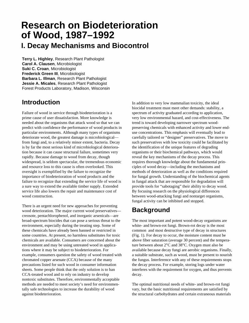
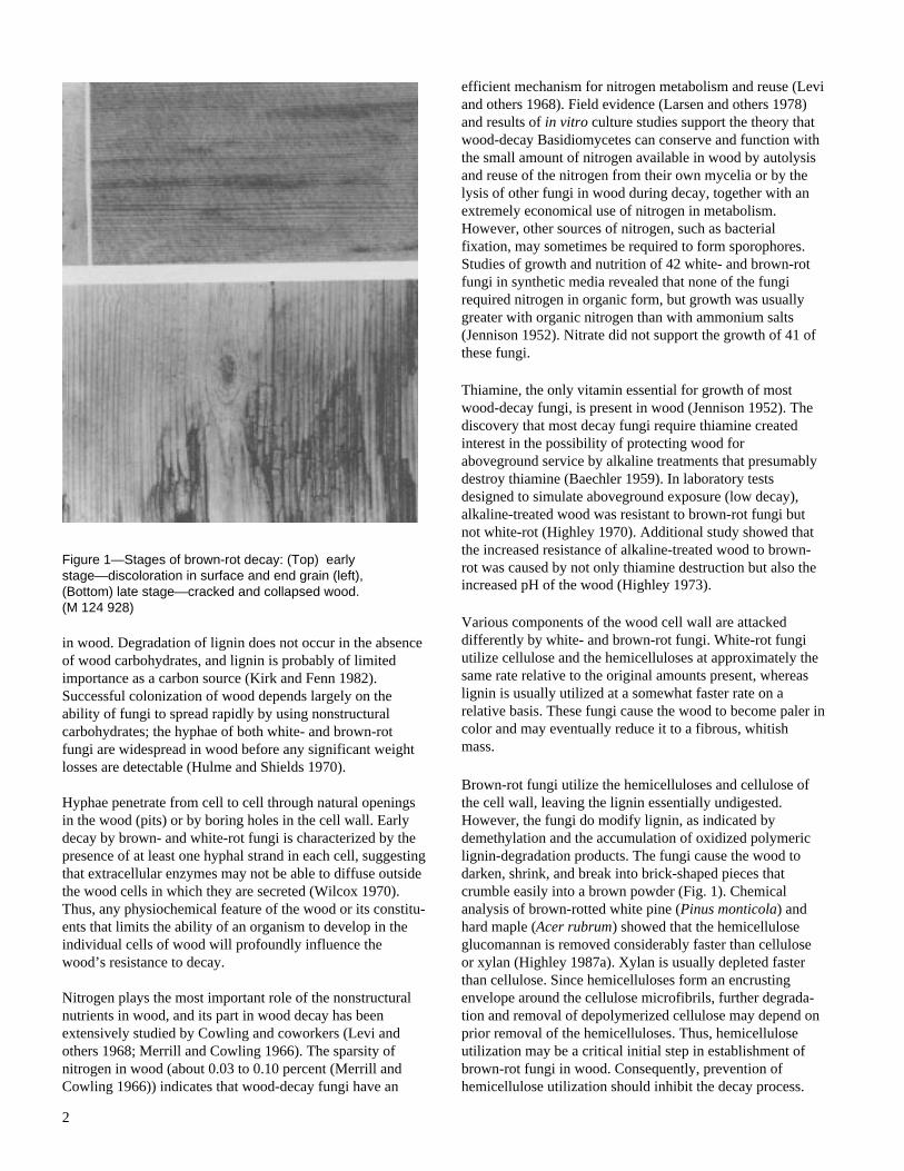
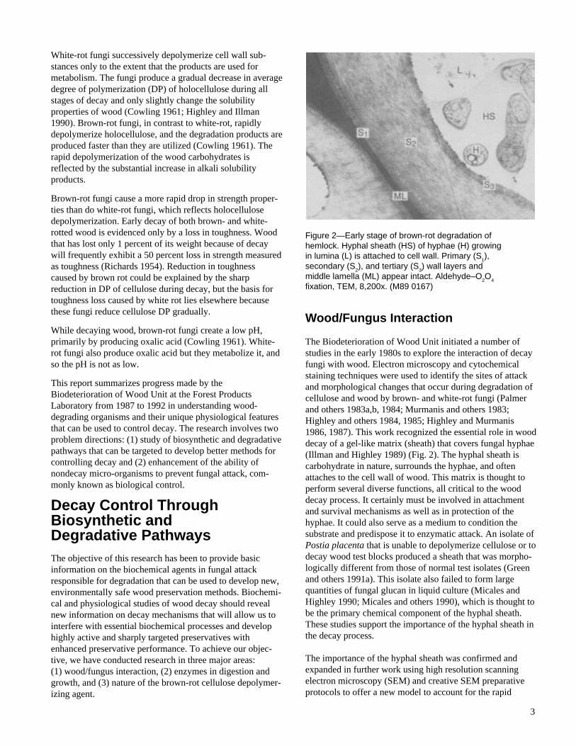
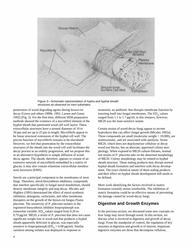
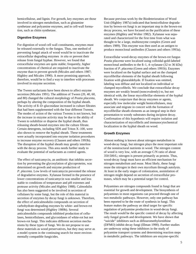
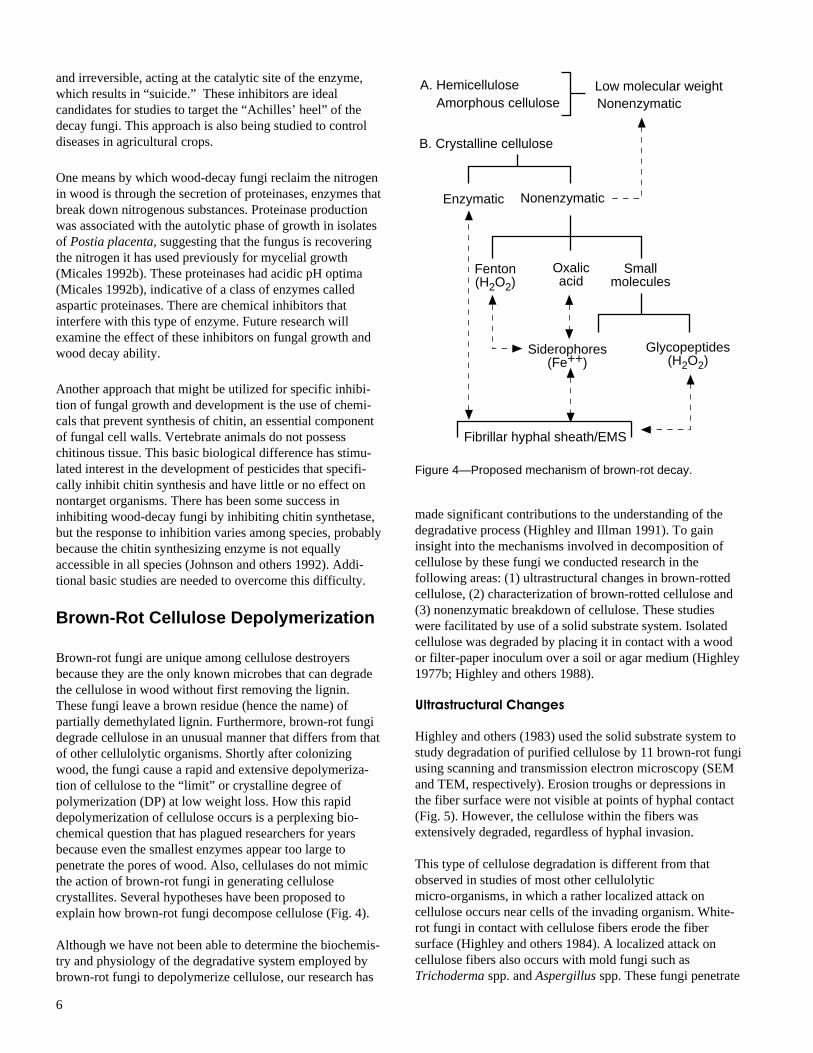
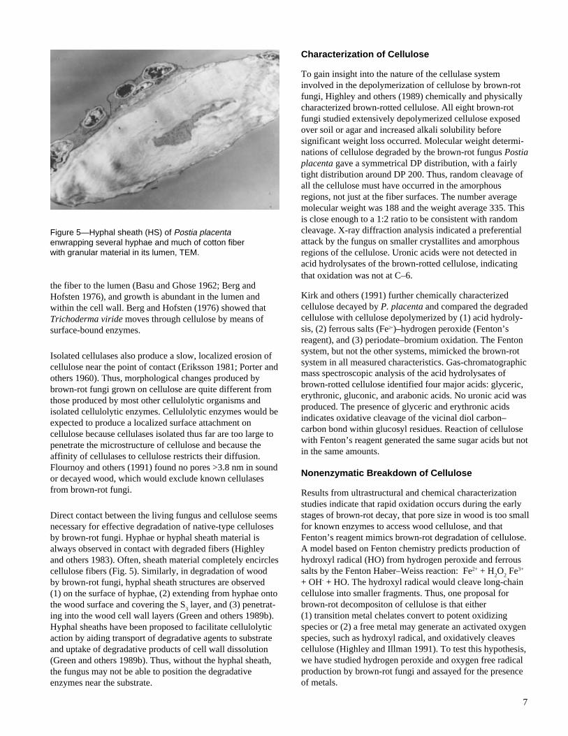
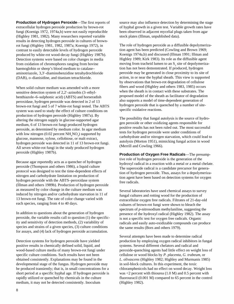
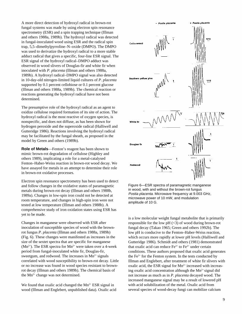
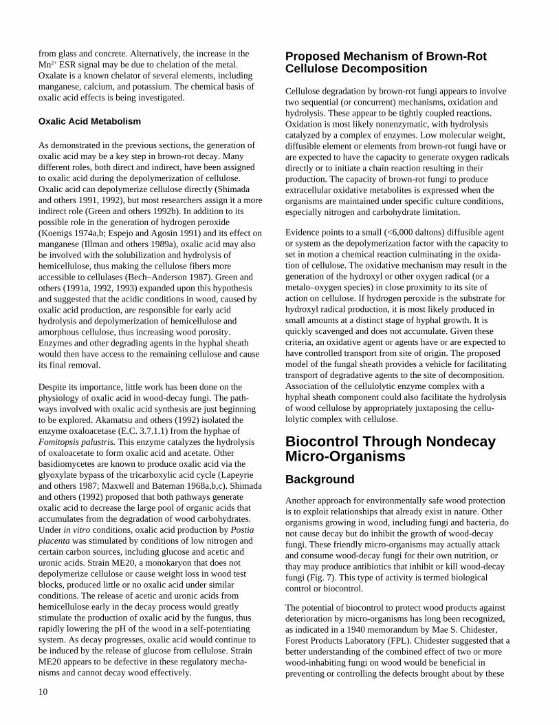
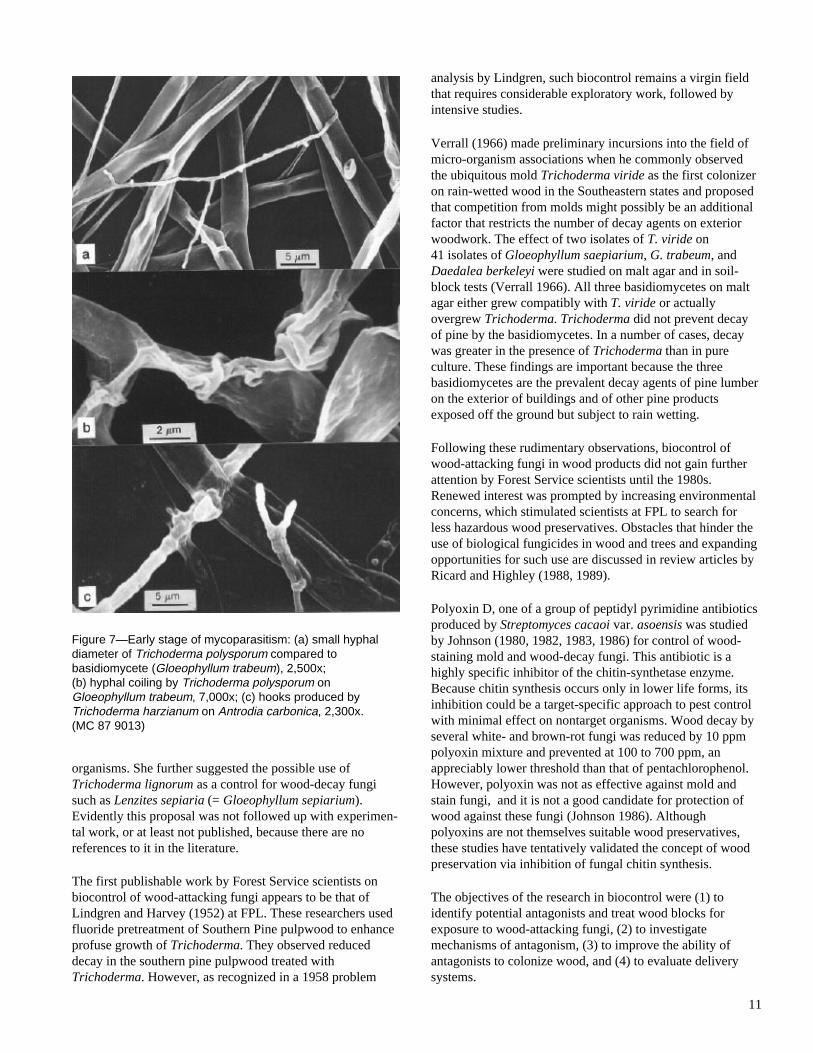
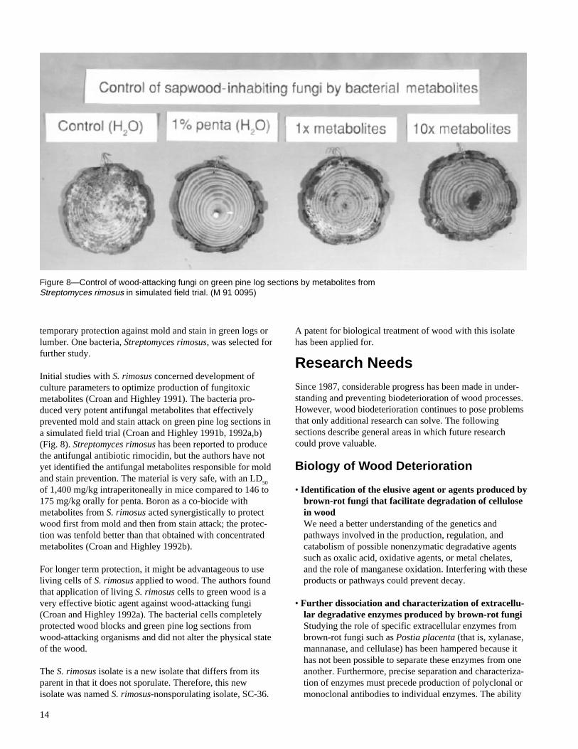


![Funktionsanalysen: En indføring [1987]](https://static.documents.pub/doc/80x56/6352ccbd59ba51b6cb021908/funktionsanalysen-en-indforing-1987.jpg)


