Single-File Diffusion of Protein Drugsthrough Cylindrical NanochannelsSeung Yun Yang,†,� Jeong-A Yang,‡,� Eung-Sam Kim,§ Gumhye Jeon,† Eun Ju Oh,‡ Kwan Yong Choi,§
Sei Kwang Hahn,‡,* and Jin Kon Kim†,*†National Creative Research Center for Block Copolymer Self-Assembly, Departments of Environmental Science & Engineering and Chemical Engineering, ‡Department ofMaterials Science & Engineering, and §Department of Life Science and School of Inter-disciplinary Bioscience and Bioengineering, Pohang University of Science andTechnology, Kyungbuk 790-784, Korea. �These authors contributed equally to this work.
Controlled and long-term proteindrug delivery has been consideredas one of the most promising bio-
medical applications of nanotechnology.1 A
wide range of materials and devices have
been developed for the delivery of protein
drugs in a controlled manner within a thera-
peutic range.2,3 However, conventional
technologies were not successful due to
the protein denaturation during and after
the formulation. Although various protein
conjugates with polyethyleneglycol (PEG)
have been successfully commercialized as
once a week injection formulations, there is
no controlled release depot system in the
market for protein drugs lasting for longer
than a week.
A critical challenge in long-term con-
trolled delivery of protein drugs is to main-
tain the integrity of highly sensitive tertiary
structure and the therapeutic efficacy of
protein drugs in physiological condition for
long periods.4�6 Once protein drugs are de-
natured, they not only are therapeutically
inactive but also cause unpredictable side
effects such as inflammation, toxicity, and
immune responses.2,3 For instance, despite
the successful commercialization of a con-
trolled release system of human growth
hormone (hGH) using poly(lactic-co-glycolic
acid) (PLGA) microparticles under a trade-
name of Nutropin Depot, it was withdrawn
from the market due to the protein denatur-
ation by hydrophobic interaction and/or
harsh acidic microenvironments caused by
the degradation of PLGA inside the body.7
The best long-term controlled delivery
system of protein drugs without denatur-
ation might be possible by exploiting the
passive diffusion through a membrane
without physical and chemical stresses.
This can be achieved when pore sizes in a
membrane are controlled to satisfy thesingle-file diffusion (SFD)8,9 condition of pro-tein drugs. SFD was previously observedfor the diffusion of CF4 gas through thepores in zeolites.8 When pore sizes are care-fully adjusted so that two or more diffusingmolecules are not allowed to pass throughthe pores simultaneously, protein drugs arereleased by SFD through the membranewithout initial burst by the Fickian diffu-sion. The release rate of a protein drug be-comes constant with time irrespective of itsconcentration in the reservoirs. This phe-nomenon is analogous to the constantdropping rate of sands through an hour-glass with time, although the mechanism isfairly different. Ferrari and co-workers10�13
prepared a nanoporous silicon membraneby multiple steps including micropatternfabrication by photolithography, the depo-sition of the sacrificial SiO2 layer, back etch-ing, and removal of the SiO2 layer. They con-firmed controlled in vitro release of insulinand IgG antibody11 when the sizes of thenanochannels in the membrane were care-fully controlled. Later, they showed the con-stant release of labeled bovine serum albu-min (BSA) during in vitro and in vivo tests.12
See the accompanying Perspective byJackson and Hillmyer on p 3548.
mer (PS-b-PMMA). The pore size in the membrane was
carefully tuned from 15 to 6 nm by the control of Au
deposition time. We observed a long-term and constant
in vitro release of both BSA and hGH without denatur-
ation by SFD up to 2 months. We also demonstrated the
feasibility of in vivo applications to long-term con-
trolled delivery of hGH. The novel protein drug deliv-
ery device system using cylindrical block copolymer
nanochannels was discussed for the application to the
treatment of various chronic diseases requiring painful
frequently injection.
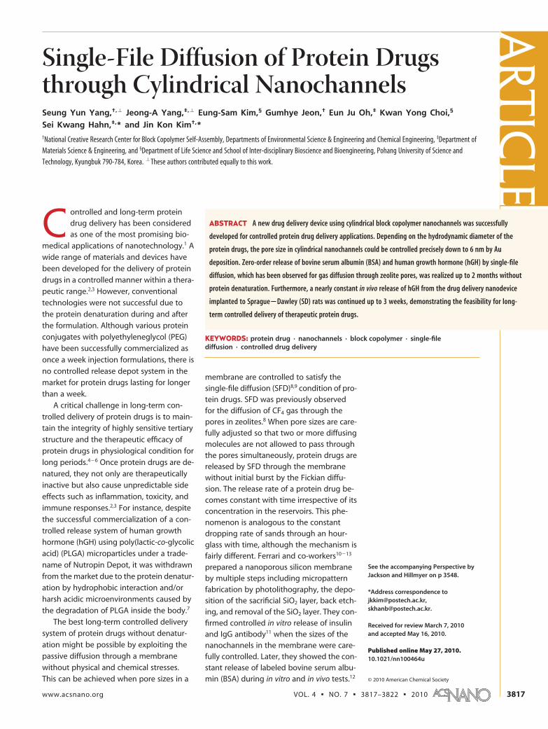
RESULTS AND DISCUSSIONFigure 1a shows schematics for the fabrication of na-
noporous membranes with cylindrical nanochannels.
The upper layer with a thickness of 80 nm was pre-
pared using a thin film of the mixture of PS-b-PMMA
having cylindrical microdomains of PMMA and PMMA
homopolymer on a silicon oxide layer. As previously re-
ported elsewhere,23 the cylindrical microdomains in
the film were oriented vertically to a silicon substrate
with an energetically neutral brush (the left image in
Figure 1a). This film was floated from the substrate in a
buffered HF solution and transferred onto a microfiltra-
tion polysulfone membrane acting as a supporting
layer. Cylindrical nanochannels in the upper layer were
generated by selective removal of the PMMA ho-
mopolymer in the cylindrical PMMA microdomains
with acetic acid. At the same time, the PMMA block mi-
grated onto the PS matrix by swelling in acetic acid
(the middle image in Figure 1a). The resulting mem-
brane had the well-ordered array of vertically aligned
cylindrical nanochannels with a narrow pore size distri-
bution. We could control precisely the pore size by Au
deposition (the right image in Figure 1a). During the Au
deposition, some Au was also deposited on the wall of
the pores as well as on the top of the upper layer of the
membrane. Thus, the pore size decreased with increas-
ing the thickness of the Au deposition layer on the
membrane. Although the pore size also decreased by
the deposition of other metals (for instance, Ti or Al), Au
was chosen because of its good biocompatibility24 and
excellent adhesion with the membrane. Figure 1b�d
shows field-emission scanning electron microscopy (FE-
SEM) images for the top surface of the nanoporous
membrane with different Au deposition thicknesses.
Cylindrical pores with a diameter of ca. 15 nm were uni-
formly formed in the nanoporous membrane, as shown
Figure 1. (a) Schematics for the preparation of block copolymer membrane with cylindrical nanochannels. The mixture ofPS-b-PMMA/PMMA solution in toluene was spin-coated on a modified silicon wafer. The film was floated onto a supportingmembrane by dissolving the silicon oxide layer from the substrate (the left image). Then cylindrical pores were generated bythe removal of PMMA homopolymer and swelling of PMMA block in acetic acid (the middle image). Pore sizes in the nanop-orous membrane were controlled more precisely by Au deposition according to the hydrodynamic diameter of a target pro-tein drug (the right image). (b�d) FE-SEM images of the top surface with a different Au deposition thickness: (b) 0 nm (be-fore Au deposition), (c) 7 nm, and (d) 11 nm. The average pore size decreased from ca. 15 nm to ca. 10 nm and ca. 6 nm.Scale bars in the inset correspond to 30 nm.
ART
ICLE
VOL. 4 ▪ NO. 7 ▪ YANG ET AL. www.acsnano.org3818
in Figure 1b. Pore size decreased with increasing theAu deposition layer. For instance, at a Au depositionlayer with 7 nm thickness, the average diameter of na-nopores became ca. 10 nm (Figure 1c). With further in-crease of Au layer to 11 nm thickness, the diameter ofthe pores at the top of the membrane was reduced to6 nm, as shown in Figure 1d. Narrow size distribution ofpores was maintained, and vertical nanochannels werepreserved after Au deposition (see Figure S1 in the Sup-porting Information).
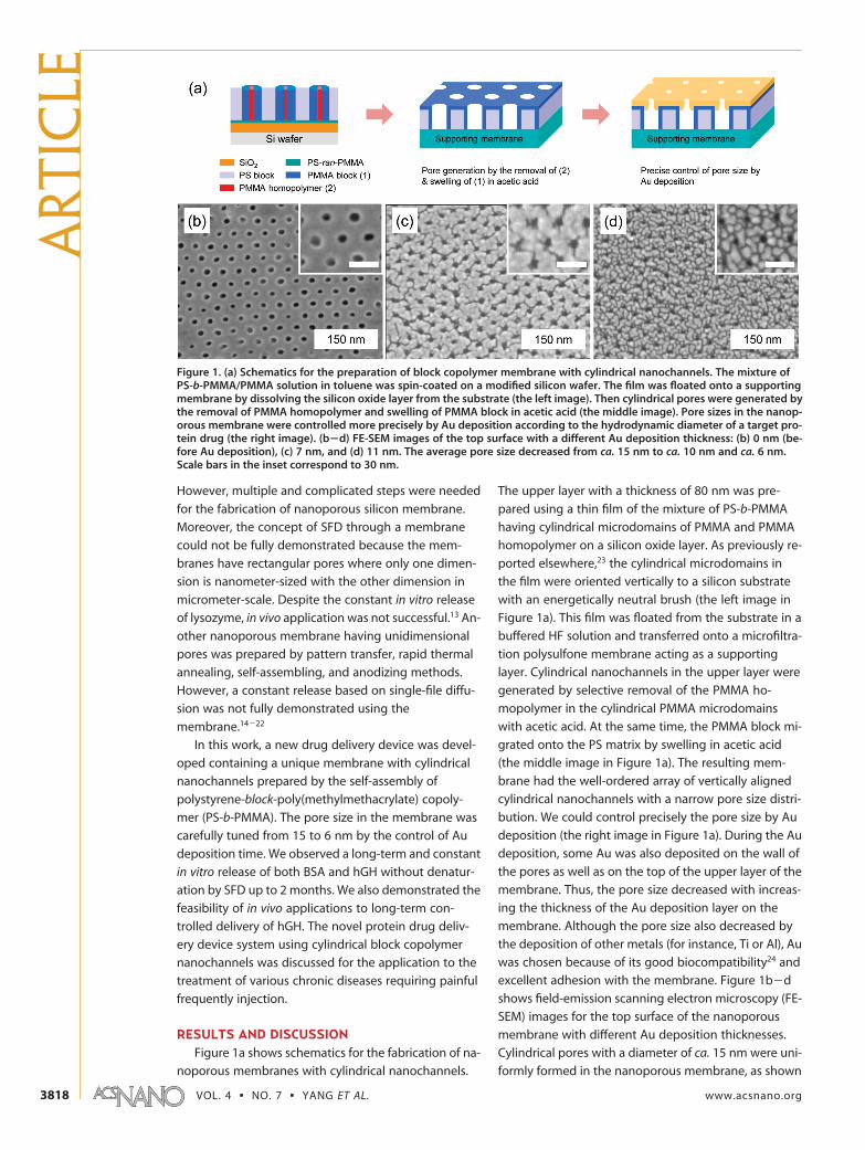
A nanoporous membrane with an average pore di-ameter of 15 nm was used for the delivery of BSA witha molecular weight (MW) of 66 000 Da and a hydrody-namic diameter of ca. 8 nm,12 while another membranewith a pore diameter of 6 nm was employed for the de-livery of hGH with a MW of 22 000 Da and a hydrody-namic diameter of ca. 3.4 nm.25 A good adhesion be-tween the Au layer and nanoporous block copolymerfilm was maintained even after in vitro and in vivo re-lease tests of the protein drugs up to 2 months. Con-trolled release of protein drugs based on the single-filediffusion was assessed using a drug delivery device de-scribed in Figure S2 of the Supporting Information.Two kinds of proteins (BSA and hGH) were selected asmodel protein drugs for in vitro release tests. Figure 2ashows the in vitro release profile of BSA through two dif-ferent membranes in the drug delivery device. Openand closed circles represent the released BSA amountfrom the supporting polysulfone membrane having anominal pore size of 200 nm and the nanoporous blockcopolymer (BCP) membrane with a pore size of ca. 15nm, respectively. The constant release profile of BSA,which is the case of SFD, was obtained for the BCPmembrane, whereas a typical Fickian diffusion releaseprofile was observed for the supporting polysulfonemembrane. The results are quite reasonable becausethe SFD of a protein drug is made possible when the di-ameter of the cylindrical pores in the membrane is lessthan twice the hydrodynamic diameter of the proteindrug. In this situation, two protein drug molecules can-not pass simultaneously through the pores. The pro-tein release rate could be easily controlled by chang-ing the thickness of the BCP membrane (Figure S3 inthe Supporting Information). For the case of hGH witha hydrodynamic diameter of 3.4 nm, however, a BCPmembrane with a pore size of ca. 15 nm showed a non-linear release, that is, a typical Fickian diffusion (opencircles in Figure 2b). Constant release of hGH was ob-served up to 2 months for a Au-deposited BCP mem-brane with a pore size of ca. 6 nm, approximately 1.7times larger than the hydrodynamic diameter of hGH(closed circles in Figure 2b). Constant release of hGHwas observed until the end of the in vitro test (�90days). From the results, Au deposition was thought tobe an effective means to control the pore size of cylin-drical nanochannels. In addition, the Au deposition ap-peared to reduce protein fouling to the polymeric
membrane, especially when erythropoietin (EPO) with
a high affinity to polymer surface was used (Figure S4 in
the Supporting Information).
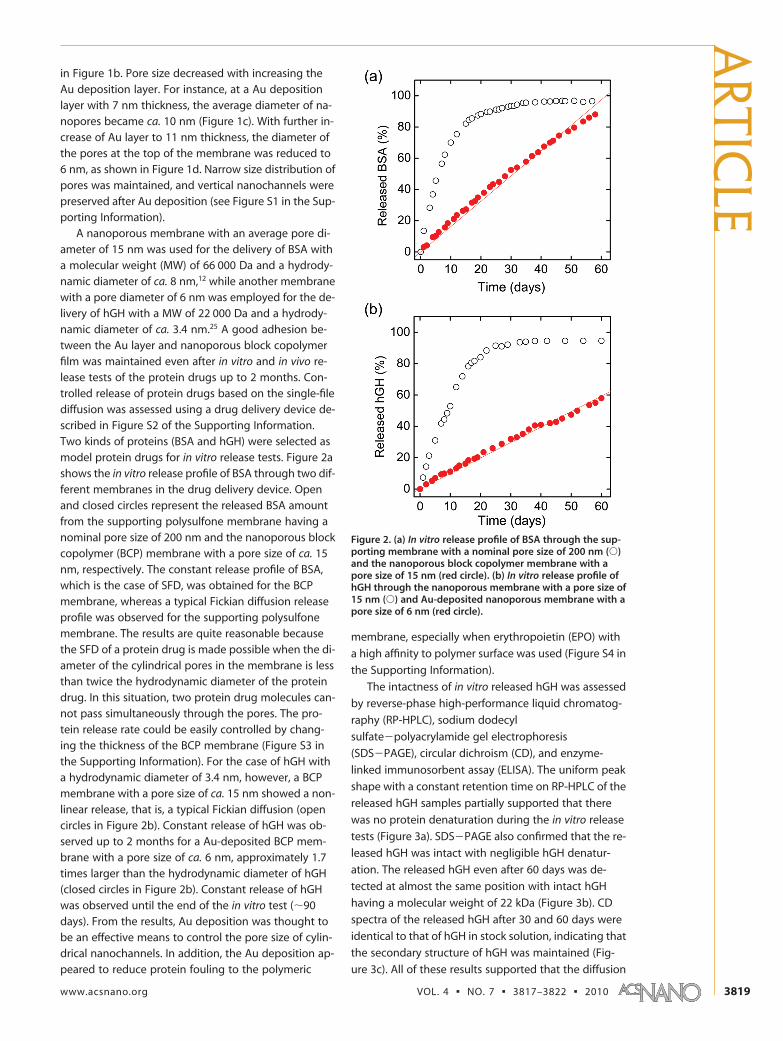
The intactness of in vitro released hGH was assessed
by reverse-phase high-performance liquid chromatog-
raphy (RP-HPLC), sodium dodecyl
sulfate�polyacrylamide gel electrophoresis
(SDS�PAGE), circular dichroism (CD), and enzyme-
linked immunosorbent assay (ELISA). The uniform peak
shape with a constant retention time on RP-HPLC of the
released hGH samples partially supported that there
was no protein denaturation during the in vitro release
tests (Figure 3a). SDS�PAGE also confirmed that the re-
leased hGH was intact with negligible hGH denatur-
ation. The released hGH even after 60 days was de-
tected at almost the same position with intact hGH
having a molecular weight of 22 kDa (Figure 3b). CD
spectra of the released hGH after 30 and 60 days were
identical to that of hGH in stock solution, indicating that
the secondary structure of hGH was maintained (Fig-
ure 3c). All of these results supported that the diffusion
Figure 2. (a) In vitro release profile of BSA through the sup-porting membrane with a nominal pore size of 200 nm (Œ)and the nanoporous block copolymer membrane with apore size of 15 nm (red circle). (b) In vitro release profile ofhGH through the nanoporous membrane with a pore size of15 nm (Œ) and Au-deposited nanoporous membrane with apore size of 6 nm (red circle).
through the BCP nanochannels did not cause physical
and chemical denaturation of hGH. Finally, the biologi-
cal activity of the released hGH was analyzed by ELISA.
To compare the bioactivity of the released hGH, the ra-
tio of in vitro released hGH concentrations determined
by Lowry assay and ELISA was plotted with increasing
time (Figure 3d). ELISA data reflect the amount of bio-
logically active hGH, whereas Lowry assay data corre-
spond to the total amount of hGH regardless of the de-
naturation. Considering the error range of ELISA
measurement, the ratios were very close to unity, sup-
porting the intactness of in vitro released hGH without
denaturation. It was also confirmed by ELISA that the in
vitro released hGH even after 2 months retained the
same biological activity as that of intact hGH.
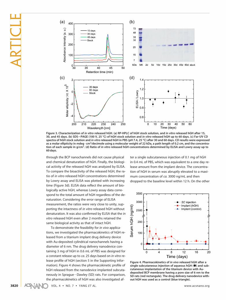
To demonstrate the feasibility for in vivo applica-
tions, we investigated the pharmacokinetics of hGH re-
leased from a titanium implant drug delivery device
with Au-deposited cylindrical nanochannels having a
diameter of 6 nm. The drug delivery nanodevice con-
taining 3 mg of hGH in 0.6 mL of PBS was designed for
a constant release up to ca. 25 days based on in vitro re-
lease profile of hGH (section 5 in the Supporting Infor-
mation). Figure 4 shows the pharmacokinetic profile of
hGH released from the nanodevice implanted subcuta-
neously in Sprague�Dawley (SD) rats. For comparison,
the pharmacokinetics of hGH was also investigated af-
ter a single subcutaneous injection of 0.1 mg of hGH
in 0.4 mL of PBS, which was equivalent to a one day re-
lease amount from the implant device. The concentra-
tion of hGH in serum was abruptly elevated to a maxi-
mum concentration of ca. 3000 ng/mL and then
dropped to the baseline level within 12 h. On the other
Figure 3. Characterization of in vitro released hGH. (a) RP-HPLC of hGH stock solution, and in vitro released hGH after 15,30, and 45 days. (b) SDS�PAGE (100 V, 25 °C) of hGH stock solution and in vitro released hGH up to 60 days. (c) Far-UV CDspectra of hGH stock solution and in vitro released hGH in PBS (pH 7.4, 25 °C) after 30 and 60 days. CD results were expressedas a molar ellipticity in mdeg · cm2/decimole using a molecular weight of 22 kDa, a path length of 0.2 cm, and the concentra-tion of each sample in g/cm3. (d) Ratio of in vitro released hGH concentrations determined by ELISA and Lowry assay up to60 days.
Figure 4. Pharmacokinetics of in vivo released hGH after asingle subcutaneous injection of aqueous hGH (�) and sub-cutaneous implantation of the titanium device with Au-deposited BCP membrane having a pore size of 6 nm to theSD rats (red rectangule). The drug delivery nanodevice with-out hGH was used as a control (blue triangle).
ART
ICLE
VOL. 4 ▪ NO. 7 ▪ YANG ET AL. www.acsnano.org3820
hand, the serum concentration of hGH released fromthe implant device was maintained at a concentrationaround 50 ng/mL. Depending on the therapeutic range,the release rate (or amount) of hGH can be tuned bychanging membrane size in an implant device for fur-ther clinical applications. Considering all of these re-sults, the novel protein drug delivery device with cylin-drical nanochannels would be successfully applied tothe treatment of various chronic diseases with greatlyimproved patient compliance.
In summary, long-term controlled release of proteindrugs by the SFD was successfully demonstrated up to
2 months using the drug delivery device with cylindri-cal BCP nanochannels. According to the hydrodynamicdiameter of a target protein drug, the pore size was pre-cisely controlled down to 6 nm by Au deposition. Therelease rate of protein drugs could also be controlled bychanging the length of BCP nanochannels and thethickness of the Au deposition layer. Due to facile andcost-effective fabrication processes, the drug deliverydevice with cylindrical BCP nanochannels would be suc-cessfully exploited for long-term constant delivery ofprotein drugs without denaturation for the treatmentof various chronic diseases.
EXPERIMENTAL DETAILSFabrication of Nanoporous BCP Membrane. PS-b-PMMA with an av-
erage molecular weight (MW) of 77 000 g/mol and volume frac-tion of PMMA block of 0.25 was prepared by anionic polymeriza-tion. It showed hexagonally-packed cylindrical microdomainwith a lattice domain spacing of 35 nm measured by synchro-tron small angle X-ray scattering (SAXS). A 2% (w/v) solution intoluene of the mixture of PS-b-PMMA and 10 wt % of homopoly-mer PMMA (30k, Sigma) relative to the PMMA block was spin-coated on a silicon oxide sacrificial layer (�200 nm) depositedwafer with a neutral brush layer and annealed at 170 °C undervacuum for 2 days.23,26 Then, the film was floated onto the sur-face of 5 wt % HF solution and then transferred to a microporousmembrane support (HT Tuffryn, Pall Life Science). Nanoporousblock copolymer membrane was prepared by immersing theblock copolymer film floated on the supporting membrane intoacetic acid for 1 h, which results in the complete removal of thePMMA homopolymer located at the center of the cylindricalPMMA microdomain. Finally, the nanoporous membrane wasdried under vacuum in an oven for 6 h at room temperature. Auwas deposited at a rate of 0.1�0.2 Å/s on the top of the nanop-orous block copolymer membrane under 2 � 10�6 Torr by usinga thermal evaporator. This slow deposition rate of Au could en-hance the adhesion between the Au layer and the block copoly-mer film.27 The thickness of the Au layer was measured with aquartz crystal piezometry balance. The surface and cross-sectional morphology of the nanoporous membrane was ob-served by field-emission scanning electron microscopy (FE-SEM,Hitachi S-4600).
In Vitro Release of Protein Drugs. BSA and hGH were dissolved inphosphate buffered saline (PBS, pH 7.4) at a concentration of 5mg/mL. Then, 6 mL of the solutions was put into a test tube withnanoporous membranes and sealed with a cap (Figure S2 in Sup-porting Information). The delivery device was put into a bath(60 mL) with the fresh PBS. Since only BSA and hGH were dif-fused out through the membranes, the total solution volume of6 mL inside the test tubes was maintained during the entire re-lease tests. Before the release test, BCP membrane was soaked in30% ethanol solution for the better wettability with proteindrug solutions. The amount of released BSA and hGH at prede-termined times was determined by Bradford protein assay andLowry assay, respectively. One milliliter of the bath solution wassampled, and the 1 mL of the fresh PBS was added to the bath ateach measurement. The intactness of released hGH was checkedby RP-HPLC, SDS�PAGE, CD, and ELISA.28 RP-HPLC was carriedout using Vydac_218MS51_C4 column at a detection wave-length of 283 nm. The eluent was 0.1 vol % trifluoroacetic acidsolution in deionized water/acetonitrile mixture, and the flowrate was 1 mL/min. For SDS�PAGE analysis, the released hGHwas mixed with loading buffers containing dithiothreitol andboiled at 90 °C for 2 min. The released protein samples of 24 �Lwere loaded onto 15% SDS�PAGE gel (8 cm � 8 cm with 1.0 mmthickness). After gel electrophoresis, the gels were stained withCoomassie Brilliant Blue solution. CD spectra for hGH in PBS (pH7.4) were obtained with a UV spectrophotometer (JASCO J-715)
at 25 °C over the range of 200�250 nm under a nitrogen atmo-sphere. A quartz cuvette with a path length of 2 mm was used.Raw data were acquired at 0.2 mm intervals with a response timeof 1 s. Each spectrum was subtracted by the spectrum of PBS,and the residual ellipticity was calculated as an average of threescans. The bioactivity of released hGH was analyzed with ELISAkits (Diagnostic Systems Laboratory Inc.).
In Vivo Release of hGH. Two groups of 3 SD rats (Japan SLC,Hamamatsu, Japan) with a mean body weight of 200 g wereused for in vivo release test of hGH. For comparison, 0.4 mL ofhGH solution at a concentration of 0.25 mg/mL was injected sub-cutaneously into an SD rat. A titanium device with the Au-deposited BCP membrane was used for in vivo release test ofhGH (section 5 in the Supporting Information). The drug load-ing volume was fixed at 0.6 mL. This device was implanted intothe subcutaneous tissue of the SD rat. For pharmacokineticanalysis, 0.2 mL of blood samples was collected from lateral tailveins. All blood samples were immediately centrifuged, and theplasma was separated and stored at �20 °C. The serum concen-trations of hGH were measured with ELISA kits.
Acknowledgment. We thank Shinpoong Pharmaceutical Co.for the kind contribution to in vivo release tests. This work wassupported by the National Creative Research Initiative Program,the Ministry of Education, Science and Technology (NEST), andKorea Institute for Advancement of Technology (KIAT) throughthe Human Resource Training Project for Regional Innovation.This research was also supported by the Converging ResearchCenter Program through the National Research Foundation ofKorea (NRF) funded by the Ministry of Education, Science andTechnology (2009-0081871). Synchrotron SAXS was performedat PLS beam line 4C2 supported by POSCO and NRF.
Supporting Information Available: Details on in vitro/in vivo re-lease tests of protein drugs and several supporting images. Thismaterial is available free of charge via the Internet at http://pub-s.acs.org.
REFERENCES AND NOTES1. Putney, S. D.; Burke, P. A. Improving Protein Therapeutics
with Sustained-Release Formulations. Nat. Biotechnol.1998, 16, 153–157.
2. Fu, K.; Klibanov, A. M.; Langer, R. Protein Stability inControlled-Release Systems. Nat. Biotechnol. 2000, 18, 24–25.
3. van de Weert, M.; Hennink, W. E.; Jiskoot, W. ProteinInstability in Poly(lactic-co-glycolic acid) Microparticles.Pharm. Res. 2000, 17, 1159–1167.
4. Frokjaer, S.; Otzen, D. E. Protein Drug Stability: AFormulation Challenge. Nat. Rev. Drug Discovery 2005, 4,298–306.
5. Nishiyama, N. Nanomedicine: Nanocarriers Shape up forLong Life. Nat. Nanotechnol. 2007, 2, 203–204.
6. Zhang, L.; Chan, J. M.; Gu, F. X.; Rhee, J.-W.; Wang, A. Z.;Radovic-Moreno, A. F.; Alexis, F.; Langer, R.; Farokhzad,
O. C. Self-Assembled Lipid�Polymer Hybrid Nanoparticles:A Robust Drug Delivery Platform. ACS Nano 2008, 2,1696–1702.
7. Wu, F.; Jin, T. Polymer-Based Sustained-Release DosageForms for Protein Drugs, Challenges, and RecentAdvances. AAPS PharmSciTech. 2008, 9, 1218–1229.
8. Kukla, V.; Kornatowski, J.; Demuth, D.; Girnus, I.; Pfeifer, H.;Rees, L. V. C.; Schunk, S.; Unger, K. K.; Karger, J. NMRStudies of Single-File Diffusion in Unidimensional ChannelZeolites. Science 1996, 272, 702–704.
9. Wei, Q.-H.; Bechinger, C.; Leiderer, P. Single-File Diffusionof Colloids in One-Dimensional Channels. Science 2000,287, 625–627.
10. Chu, W.-H.; Chin, R.; Huen, T.; Ferrari, M. Silicon MembraneNanofilters from Sacrificial Oxide Removal. J.Microelectromech. Syst. 1999, 8, 34–42.
11. Desai, T. A.; Hansford, D. J.; Ferrari, M. MicromachinedInterfaces: New Approaches in Cell Immunoisolation andBiomolecular Separation. Biomol. Eng. 2000, 17, 23–36.
12. Martin, F.; Walczak, R.; Boiarski, A.; Cohen, M.; West, T.;Cosentino, C.; Ferrari, M. Tailoring Width ofMicrofabricated Nanochannels to Solute Size Can Be UsedTo Control Diffusion Kinetics. J. Controlled Release 2005,102, 123–133.
13. Walczak, R. J.; Boiarski, A.; Cohen, M.; West, T.; Melnik, K.;Shapiro, J.; Sharma, S.; Ferrari, M. Long-TermBiocompatibility of NanoGATE Drug Delivery Implant.Nanobiotechnology 2005, 1, 35–42.
14. Black, C. T.; Guarini, K. W.; Breyta, G.; Colburn, M. C.; Ruiz,R.; Sandstrom, R. L.; Sikorski, E. M.; Zhang, Y. Highly PorousSilicon Membrane Fabrication Using Polymer Self-Assembly. J. Vac. Sci. Technol., B 2006, 24, 3188–3191.
15. Striemer, C. C.; Gaborski, T. R.; McGrath, J. L.; Fauchet, P. M.Charge- and Size-Based Separation of MacromoleculesUsing Ultrathin Silicon Membranes. Nature 2007, 445,749–753.
16. Liu, G.; Ding, J.; Hashimoto, T.; Kimishima, K.; Winnik, F. M.;Nigam, S. Thin Films with Densely, Regularly PackedNanochannels: Preparation, Characterization, andApplications. Chem. Mater. 1999, 11, 2233–2240.
17. Thurn-Albrecht, T.; Steiner, R.; DeRouchey, J.; Stafford,C. M.; Huang, E.; Bal, M.; Tuominen, M.; Hawker, C. J.;Russell, T. P. Nanoscopic Templates from Oriented BlockCopolymer Films. Adv. Mater. 2000, 12, 787–791.
18. Kim, J. K.; Lee, J. I.; Lee, D. H. Self-Assembled BlockCopolymers: Bulk to Thin Film. Macromol. Res. 2008, 16,267–292.
19. Phillip, W. A.; Rzayev, J.; Hillmyer, M. A.; Cussler, E. L. Gasand Water Liquid Transport through Nanoporous BlockCopolymer Membranes. J. Membr. Sci. 2006, 286, 144–152.
20. Peinemann, K. V.; Abetz, V.; Simon, P. F. W. AsymmetricSuperstructure Formed in a Block Copolymer via PhaseSeparation. Nat. Mater. 2007, 6, 992–996.
21. Lo, K.-H.; Chen, M.-C.; Ho, R.-M.; Sung, H.-W. Pore-FillingNanoporous Templates from Degradable BlockCopolymers for Nanoscale Drug Delivery. ACS Nano 2009,3, 2660–2666.
22. Peng, L.; Mendelsohn, A. D.; LaTempa, T. J.; Yoriya, S.;Grimes, C. A.; Desai, T. A. Long-Term Small Molecule andProtein Elution from TiO2 Nanotubes. Nano Lett. 2009, 9,1932–1936.
23. Yang, S. Y.; Ryu, I.; Kim, H. Y.; Kim, J. K.; Jang, S. K.; Russell,T. P. Nanoporous Membranes with Ultrahigh Selectivityand Flux for the Filtration of Viruses. Adv. Mater. 2006, 18,709–712.
24. Santini, J. J. T.; Cima, M. J.; Langer, R. A Controlled-ReleaseMicrochip. Nature 1999, 397, 335–338.
25. Maa, Y.-F.; Hsu, C. C. Investigation on Fouling Mechanismsfor Recombinant Human Growth Hormone SterileFiltration. J. Pharm. Sci. 1998, 87, 808–812.
27. Cho, J. H.; Jang, Y.; Lee, W. H.; Ihm, K.; Han, J.-H.; Chung, S.;
Cho, K. Effects of Metal Penetration into OrganicSemiconductors on the Electrical Properties of OrganicThin Film Transistors. Appl. Phys. Lett. 2006, 89,132101–132103.
28. Hahn, S. K.; Kim, S. J.; Kim, M. J.; Kim, D. H. Characterizationand In Vivo Study of Sustained-Release Formulation ofHuman Growth Hormone Using Sodium Hyaluronate.Pharm. Res. 2004, 21, 1374–1381.