125 Research Article Introduction Posttranslational modification of proteins is a rapid and efficient mechanism to modulate protein activity in response to a given stimulus. The s mall u biquitin-like mo difier, SUMO, has emerged as a complex and intriguing posttranslational modifier with a large variety of targets and a wide-range of effects on its substrates. Four SUMO isoforms (SUMO1-4) have been characterized in mammalian cells (Bohren et al., 2004; Su and Li, 2002), although SUMO4 expression is restricted to the kidney and other specific tissues suggesting a limited biological role for SUMO4. In contrast to SUMO4, SUMO1-3 are all widely expressed in mammalian cells (Xu and Au, 2005), though it is likely that these three proteins have at least partially distinct biological roles because SUMO1 shares only 50% identity with the closely related SUMO2 and SUMO3 (Saitoh and Hinchey, 2000). Consistent with the sequence divergence, distinct preferences for substrates (Guo et al., 2005; Manza et al., 2004; Rosas-Acosta et al., 2005b) and for specific de-sumoylating proteases (Melchior et al., 2003) have been observed for each SUMO type. Despite the above differences, all SUMOs are covalently attached to proteins via a series of enzymatic reactions and biochemical steps involving the same SUMO-specific enzymes (Johnson, 2004). The activating enzyme (E1), a heterodimeric protein composed of two subunits, SAE1 and SAE2 (Azuma et al., 2001), transfers SUMO to Ubc9, the only E2-conjugating enzyme (Desterro et al., 1997). The Ubc9-SUMO complex can directly interact with specific substrates, and this interaction leads to the formation of an isopeptide bond between SUMO and a lysine residue on its target (Okuma et al., 1999; Tatham et al., 2003). SUMO ligases (E3 enzymes) such as RanBP2 (Pichler et al., 2002), the PIAS protein family (Kotaja et al., 2002), and the polycomb protein Pc2 (Kagey et al., 2003) are not required for sumoylation in vitro, but when added to a sumoylation reaction they substantially increase the rate of this process and appear to play an essential role in vivo. Finally, there are specific SUMO proteases (SENP protein family) that are involved in both SUMO processing and de-sumoylation (Mossessova and Lima, 2000). The continuous interplay of the enzymes described above makes SUMO modification an active, reversible and dynamic process in mammalian cells. SUMO modification usually exerts significant effects on its targets: it can increase protein stability (Desterro et al., 1998), affect protein-protein interactions (Seeler and Dejean, 2001), alter subcellular localization (Morita et al., 2005; Wilson and Rangasamy, 2001), and impact nuclear trafficking (Pichler and Melchior, 2002). To date, transcription factors (TFs) represent the largest group of SUMO substrates identified, and the most common effect associated with TF sumoylation is modulation of their transcriptional activity (Gill, 2003; Verger et al., 2003). In general, sumoylation results in the negative regulation of the activity of most TFs, including widely expressed factors such SUMO modification regulates the activity of numerous transcription factors that have a direct role in cell-cycle progression, apoptosis, cellular proliferation, and development, but its role in differentiation processes is less clear. Keratinocyte differentiation requires the coordinated activation of a series of transcription factors, and as several crucial keratinocyte transcription factors are known to be SUMO substrates, we investigated the role of sumoylation in keratinocyte differentiation. In a human keratinocyte cell line model (HaCaT cells), Ca 2+ -induced differentiation led to the transient and coordinated transcriptional activation of the genes encoding crucial sumoylation system components, including SAE1, SAE2, Ubc9, SENP1, Miz-1 (PIASx), SUMO2 and SUMO3. The increased gene expression resulted in higher levels of the respective proteins and changes in the pattern of sumoylated substrate proteins during the differentiation process. Similarly to the HaCaT results, stratified human foreskin keratinocytes showed an upregulation of Ubc9 in the suprabasal layers. Abrogation of sumoylation by Gam1 expression severely disrupted normal HaCaT differentiation, consistent with an important role for sumoylation in the proper progression of this biological process. Supplementary material available online at http://jcs.biologists.org/cgi/content/full/120/1/125/DC1 Key words: Keratinocyte, Differentiation, SUMO, HaCaT, Ubc9 Summary Sumoylation dynamics during keratinocyte differentiation Adeline F. Deyrieux 1 , Germán Rosas-Acosta 1 , Michelle A. Ozbun 2 and Van G. Wilson 1, * 1 Department of Molecular and Microbial Pathogenesis, College of Medicine, Texas A&M Health Science Center, College Station, TX 77843-1114, USA 2 Department of Molecular Genetics and Microbiology, and of Obstetrics and Gynecology, University of New Mexico School of Medicine, 915 Camino de Salud NE, Cancer Research Facility (CRF) 303, Albuquerque, NM 87131, USA *Author for correspondence (e-mail: [email protected]) Accepted 25 October 2006 Journal of Cell Science 120, 125-136 Published by The Company of Biologists 2007 doi:10.1242/jcs.03317 Journal of Cell Science JCS ePress online publication date 12 December 2006
Transcript
125Research Article
IntroductionPosttranslational modification of proteins is a rapid andefficient mechanism to modulate protein activity in response toa given stimulus. The small ubiquitin-like modifier, SUMO,has emerged as a complex and intriguing posttranslationalmodifier with a large variety of targets and a wide-range ofeffects on its substrates. Four SUMO isoforms (SUMO1-4)have been characterized in mammalian cells (Bohren et al.,2004; Su and Li, 2002), although SUMO4 expression isrestricted to the kidney and other specific tissues suggesting alimited biological role for SUMO4. In contrast to SUMO4,SUMO1-3 are all widely expressed in mammalian cells (Xuand Au, 2005), though it is likely that these three proteins haveat least partially distinct biological roles because SUMO1shares only 50% identity with the closely related SUMO2 andSUMO3 (Saitoh and Hinchey, 2000). Consistent with thesequence divergence, distinct preferences for substrates (Guoet al., 2005; Manza et al., 2004; Rosas-Acosta et al., 2005b)and for specific de-sumoylating proteases (Melchior et al.,2003) have been observed for each SUMO type. Despite theabove differences, all SUMOs are covalently attached toproteins via a series of enzymatic reactions and biochemicalsteps involving the same SUMO-specific enzymes (Johnson,2004). The activating enzyme (E1), a heterodimeric proteincomposed of two subunits, SAE1 and SAE2 (Azuma et al.,2001), transfers SUMO to Ubc9, the only E2-conjugating
enzyme (Desterro et al., 1997). The Ubc9-SUMO complex candirectly interact with specific substrates, and this interactionleads to the formation of an isopeptide bond between SUMOand a lysine residue on its target (Okuma et al., 1999; Tathamet al., 2003). SUMO ligases (E3 enzymes) such as RanBP2(Pichler et al., 2002), the PIAS protein family (Kotaja et al.,2002), and the polycomb protein Pc2 (Kagey et al., 2003) arenot required for sumoylation in vitro, but when added to asumoylation reaction they substantially increase the rate of thisprocess and appear to play an essential role in vivo. Finally,there are specific SUMO proteases (SENP protein family) thatare involved in both SUMO processing and de-sumoylation(Mossessova and Lima, 2000). The continuous interplay of theenzymes described above makes SUMO modification anactive, reversible and dynamic process in mammalian cells.
SUMO modification usually exerts significant effects on itstargets: it can increase protein stability (Desterro et al., 1998),affect protein-protein interactions (Seeler and Dejean, 2001),alter subcellular localization (Morita et al., 2005; Wilson andRangasamy, 2001), and impact nuclear trafficking (Pichler andMelchior, 2002). To date, transcription factors (TFs) representthe largest group of SUMO substrates identified, and the mostcommon effect associated with TF sumoylation is modulationof their transcriptional activity (Gill, 2003; Verger et al., 2003).In general, sumoylation results in the negative regulation of theactivity of most TFs, including widely expressed factors such
SUMO modification regulates the activity of numeroustranscription factors that have a direct role in cell-cycleprogression, apoptosis, cellular proliferation, anddevelopment, but its role in differentiation processes is lessclear. Keratinocyte differentiation requires the coordinatedactivation of a series of transcription factors, and as severalcrucial keratinocyte transcription factors are known to beSUMO substrates, we investigated the role of sumoylationin keratinocyte differentiation. In a human keratinocytecell line model (HaCaT cells), Ca2+-induced differentiationled to the transient and coordinated transcriptionalactivation of the genes encoding crucial sumoylation systemcomponents, including SAE1, SAE2, Ubc9, SENP1, Miz-1(PIASx�), SUMO2 and SUMO3. The increased geneexpression resulted in higher levels of the respective
proteins and changes in the pattern of sumoylatedsubstrate proteins during the differentiation process.Similarly to the HaCaT results, stratified human foreskinkeratinocytes showed an upregulation of Ubc9 in thesuprabasal layers. Abrogation of sumoylation by Gam1expression severely disrupted normal HaCaTdifferentiation, consistent with an important role forsumoylation in the proper progression of this biologicalprocess.
Supplementary material available online athttp://jcs.biologists.org/cgi/content/full/120/1/125/DC1
Sumoylation dynamics during keratinocytedifferentiationAdeline F. Deyrieux1, Germán Rosas-Acosta1, Michelle A. Ozbun2 and Van G. Wilson1,*1Department of Molecular and Microbial Pathogenesis, College of Medicine, Texas A&M Health Science Center, College Station, TX 77843-1114,USA 2Department of Molecular Genetics and Microbiology, and of Obstetrics and Gynecology, University of New Mexico School of Medicine,915 Camino de Salud NE, Cancer Research Facility (CRF) 303, Albuquerque, NM 87131, USA*Author for correspondence (e-mail: [email protected])
Accepted 25 October 2006Journal of Cell Science 120, 125-136 Published by The Company of Biologists 2007doi:10.1242/jcs.03317
Jour
nal o
f Cel
l Sci
ence JCS ePress online publication date 12 December 2006
126
as AP-1 (Bossis et al., 2005), Sp1/Sp3 (Ross et al., 2002;Spengler and Brattain, 2006), and C/EBP (Kim et al., 2002),though activation has been reported for some TFs, includingTCF4 (Ihara et al., 2005) and IKaros (Arco et al., 2005). Recentstudies have established multiple mechanisms for negativeregulation of TFs by sumoylation, including recruitment oftranscriptional co-repressors such as HDACs (Shiio andEisenman, 2003; Yang and Sharrocks, 2004), sequestration inthe cytoplasm (Morita et al., 2005; Salinas et al., 2004) orubiquitylation and subsequent degradation (Ghioni et al.,2005).
Many of the TFs known to be SUMO targets, including pRB(Ledl et al., 2005), the p53/63/73 family (Ghioni et al., 2005;Melchior and Hengst, 2002), AP-2 (Eloranta and Hurst, 2002),and Sp1/Sp3 (Ross et al., 2002; Spengler and Brattain, 2006)are involved in regulating gene expression during cell-cycleprogression and/or differentiation (Herwig and Strauss, 1997;Li and Kellems, 2003; Santini et al., 2001), suggesting thatsumoylation could coordinate complex transcriptionalprograms in the cell. Several recent studies in metazoansystems support such a role for sumoylation in aspects ofdevelopment and differentiation. Sumoylation is required forcorrect vulvar development in C. elegans (Leight et al., 2005;Poulin et al., 2005), is implicated in male germ cell maturation(Vigodner et al., 2006) and promotes differentiation ofpostsynaptic dendrites (Shalizi et al., 2006). Sumoylation isalso critical for maintaining nuclear structure andchromosomal segregation during blastocyst development, withthe absence of Ubc9 leading to an embryonic lethal phenotype(Nacerddine et al., 2005).
Skin is a complex and renewable organ for which little isknown about the expression and function of the sumoylationsystem, although the presence of the SUMO1 protein in amouse keratinocyte cell line has been reported (Zhong et al.,2000). The epidermis comprises multiple layers ofdifferentiated keratinocytes that are continually regeneratedfrom the replicative basal layer. In skin, keratinocytesdifferentiate vertically because of a Ca2+ gradient establishedthroughout the different epithelial layers, increasing from thebasal to the outermost layer of the epithelium (Menon et al.,1985; Vicanova et al., 1998). Ca2+ signaling drivesproliferating keratinocytes out of the cell cycle and into acommitted path of terminal differentiation, but the intermediatesignaling transduction pathways that lead to this process arestill poorly understood (Bikle et al., 2001; Lansdown, 2002; Tuet al., 2004). A network of keratin markers biochemicallydefines the keratinocyte stage within the epithelium structure(Eichner et al., 1986; Smith, 2003; Sun et al., 1985).Expression of keratin 5 (K5) and keratin 14 (K14)characterizes the basal proliferative phenotype restricted to thestem cell layer where the Ca2+ concentration is low (Schweizerand Winter, 1983). Early in differentiation, K5 and K14 arerepressed and differentiation markers such as K1, K10 andinvolucrin begin to be expressed. Finally, keratinocytescomplete terminal differentiation by producing the outercornified layer of the skin characterized by markers such asfilaggrin and involucrin (Candi et al., 2005; Eichner et al.,1986; Fuchs and Green, 1980). This pattern of markers permitsdiscrimination between proliferating, differentiating andterminally differentiated keratinocytes. The well-coordinatedexpression of specific sets of TFs induces and represses these
keratinocyte genes as the cells migrate through the epitheliumlayers, and a number of these crucial TFs are already knownto be SUMO targets in other tissues (Girdwood et al., 2004;Verger et al., 2003), suggesting that sumoylation might play animportant role in regulating epidermal differentiation.
Cultured keratinocytes, such as HaCaT cells, provide well-established differentiation models that can recapitulate manyaspects of stratified epithelium (Boukamp et al., 1988; Schoopet al., 1999). HaCaTs are spontaneously immortalized and donot express exogenous transforming genes that could interferewith the natural process of sumoylation and differentiation.Moreover, HaCaT cells have the capacity to revert back andforth between the differentiated and the basal phenotype, andtherefore are widely used to model keratinocyte differentiationin culture (Schoop et al., 1999). Here, we examine thesumoylation system in HaCaT cells during Ca2+-induceddifferentiation. We show that the sumoylation system wastransiently upregulated by Ca2+ signaling in HaCaT cells atboth the RNA and protein level, whereas abrogation ofsumoylation led to abnormal differentiation. These resultssuggest cross talk between the sumoylation system and thekeratinocyte differentiation process that contributes to thenormal program of morphological and biochemical changesduring differentiation.
ResultsHaCaT cells express the SUMO machineryThe SUMO system has been extensively studied, and theenzymes required for sumoylation are widely expressed inmany tissues (Chen et al., 1998; Johnson, 2004). However, thesumoylation system remains largely unexplored in humankeratinocytes, an important model for differentiation. To studythe functional importance of this modifier in the context ofkeratinocyte differentiation, we first developed cultureconditions to maintain HaCaT cells stably in either a basal ordifferentiated phenotype. HaCaT cells cultured in 0.03 mMCa2+ were spindle-shaped and loosely packed (Fig. 1A). Inaddition, there was little or no expression of two differentiationmarkers, keratin 1 (K1) and involucrin (Inv), at the RNA (Fig.1B) or protein (Fig. 1C) level, consistent with these cellsremaining in a basal like state. By contrast, HaCaT cellsmaintained in 2.8 mM Ca2+ became more cuboidal, developedcell-cell tight junctions, and expressed both differentiationmarkers, K1 and Inv, at the RNA and protein level (Fig. 1A-C). Under both high and low [Ca2+] conditions, the culturescould be maintained for many passages without any subsequentchanges in morphology or marker expression pattern.Therefore, these growth conditions allow HaCaT cells to bestably maintained in vitro in two alternative states resemblingpre- and post-differentiated skin cells.
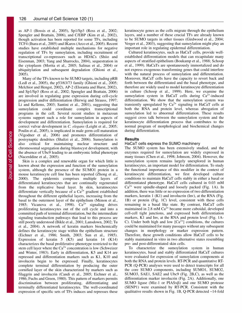
To characterize the sumoylation system in humankeratinocytes, basal and stably differentiated HaCaT cultureswere evaluated for expression of sumoylation components atboth the RNA and protein levels. RT-PCR and quantitative RT-PCR (Q-PCR) analyses were used to detect transcripts for allthe core SUMO components, including SUMO1, SUMO2,SUMO3, SAE1, SAE2 and Ubc9 (Fig. 2B,C), as well as thedifferentiation marker involucrin (Fig. 2A). Additionally, oneSUMO ligase (Miz-1 or PIASx�) and one SUMO protease(SENP1) were examined by RT-PCR. Consistent with thequalitative results shown in Fig. 1B, Q-PCR detected ~14-fold
Journal of Cell Science 120 (1)
Jour
nal o
f Cel
l Sci
ence
127Sumoylation and keratinocyte differentiation
higher levels of involucrin mRNA in the high [Ca2+]-maintained keratinocytes compared with the low [Ca2+]culture, confirming the differentiation state of these cells (Fig.2A). By RT-PCR, transcripts for all of the SUMO componentgenes were detected in both the basal and differentiated HaCaTcells, indicating that the relevant genes for an activesumoylation system were actively transcribed in both cellpopulations (Fig. 2B). Although the HaCaT cells weremaintained in two distinct phenotypic states, the transcriptlevels for the sumoylation components tested were similarunder both culture conditions (Fig. 2C). Immunoblot analysesconfirmed the expression of Ubc9 and SAE1 proteins, and thelevels of each of these proteins were only slightly higher in thedifferentiated cells compared with the undifferentiated HaCaTcells (Fig. 2D). Next, we tested whether the sumoylationsystem was active in HaCaT cells as evidenced by the presenceof sumoylated proteins. Immunoblotting of total cell extractsusing a polyclonal anti-SUMO antibody showed thatsumoylation occurred in both the basal and differentiated cellpopulations (Fig. 2E). Although the differences in expressionlevels of the sumoylation components were small, thesumoylation patterns in differentiated versus basal cells wereslightly different: bands at ~40, 60 and 95 kDa were intensifiedin high [Ca2+] cells compared with basal cells (Fig. 2E,arrows). Since the basal and differentiated cell cultures wereboth equally proliferative and were generated from a singlestock of HaCaT cells, we believe that these changes insumoylation pattern reflect differentiation-related events ratherthan intrinsic differences in genetic background or growthcapacity of the two populations of cells.
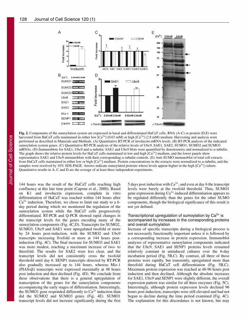
SUMO and Ubc9 are expressed in stratified humanforeskin keratinocytesTo corroborate the HaCaT observations, the expression ofSUMO and Ubc9 was evaluated in primary human foreskinkeratinocytes (HFKs) that were stratified in organotypiccultures. Immunohistochemistry analyses were performed withan anti-SUMO polyclonal antiserum and a purified polyclonalantibody against Ubc9, and antibody specificity was confirmedby blocking experiments using purified SUMO1 or Ubc9 (Fig.3). SUMO was detected in the nucleus and cytoplasm ofkeratinocytes found in all layers of the epithelium (Fig. 3C).However, as this antiserum crossreacts with SUMO2/3 (ourunpublished observations), which SUMO types were presentand whether or not there were changes in type expression indifferent layers is unclear. By contrast, Ubc9 was barelydetectable in the basal layer of the epithelium where the cellsremained relatively unstained (Fig. 3A, arrowheads). Stronganti-Ubc9 staining was detected in the intermediatedifferentiated layers just above the basal layer, and Ubc9 waspresent in both the nucleus and cytoplasm of the keratinocytesin this region (Fig. 3A, black arrows). Although Ubc9 couldbe detected in nuclei throughout the upper layers (Fig. 3A anddata not shown), its overall expression faded as keratinocytesmoved closer to the outer layer of the epithelium. It appearsfrom these results that Ubc9 expression is low in basal cells,transiently increases with the initiation of differentiation in thesuprabasal layers, and declines as terminal differentiationprogresses. As Ubc9 is the only E2 enzyme for sumoylation,we speculate that the pool of sumoylated protein might bedramatically altered as well during this process. Interestingly,Ubc9 expression is maximal in the layers just below wherekeratin 1 expression commences (compare Fig. 3A with 3E).Overall, these analyses demonstrate that sumoylationcomponents are present in stratified human skin epithelium andthat Ubc9 levels change during differentiation. The similarobservations in HaCaT cultures suggest that monolayer HaCaTcells are a good representative model of the sumoylationsystem in normal human keratinocytes.
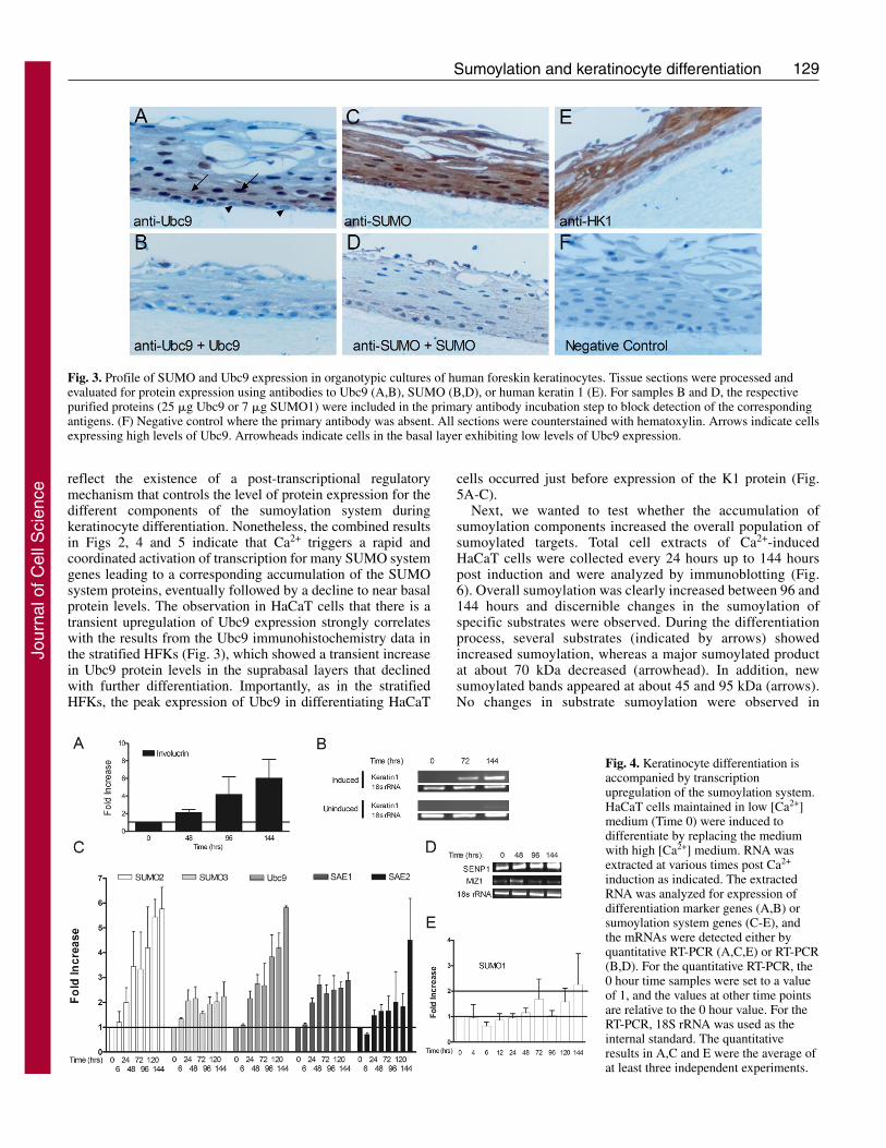
Ca2+-induced differentiation in HaCaT cells upregulatesthe sumoylation systemAlthough only minor differences in expression of thesumoylation components were detected between the twosteady-state populations of HaCaT cells, theimmunohistochemistry results suggested that more substantialchanges in sumoylation might be taking place during theprocess of differentiation. To investigate this possibility weexamined the sumoylation system in HaCaT cells undergoingactive Ca2+-induced differentiation. Addition of Ca2+ to thebasal HaCaT cultures triggered a rapid response resulting inrepression of the basal keratin markers, morphological changes(data not shown), and mRNA expression of differentiationmarkers such as K1 and Inv within 3-4 days (Fig. 4A,B). TheInv transcript detected by Q-PCR progressively increased from48 to 144 hours following Ca2+ induction (Fig. 4A). Likewise,the K1 transcript, which was completely repressed inundifferentiated (0 hours) HaCaT cells, was detected by RT-PCR at 72 hours post Ca2+ induction, reaching a maximumexpression by 120-144 hours (Fig. 4B). No K1 expression wasdetected in uninduced cells for up to 120 hours (Fig. 4B). Note,however, that the faint expression of K1 in uninduced cells by
Fig. 1. HaCaT cell cultures exhibit phenotypic differences inresponse to Ca2+ concentration. (A) Phase-contrast microscopy ofHaCaT cells maintained in low [Ca2+] medium (0.03 mM Ca2+) or inhigh [Ca2+] medium (2.8 mM Ca2+). Upon Ca2+ addition to themedium, basal HaCaT cells take 4-7 days to assume a completedifferentiated state. Inversely, upon Ca2+ depletion, differentiatedHaCaT cells take about 3 weeks to revert to a basal phenotype.(B) RT-PCR analyses of total mRNA harvested from basal (0.03 mMCa2+) and differentiated HaCaT (2.8 mM Ca2+) cultures.(C) Immunoblot analyses showing expression of keratin 1,involucrin, and �-tubulin in total cell extracts harvested from basal(0.03 mM Ca2+) and differentiated (2.8 mM Ca2+) HaCaT cultures.
Jour
nal o
f Cel
l Sci
ence
128
144 hours was the result of the HaCaT cells reaching highconfluency at this late time point (Capone et al., 2000). Basedon K1 and involucrin expression, complete in vitrodifferentiation of HaCaT was reached within 144 hours afterCa2+ induction. Therefore, we chose to limit our study to a 6-day period during which we monitored the regulation of thesumoylation system while the HaCaT cells progressivelydifferentiated. RT-PCR and Q-PCR showed rapid changes inthe transcript levels for the genes encoding many of thesumoylation components (Fig. 4C,D). Transcripts for SUMO2,SUMO3, Ubc9 and SAE1 were upregulated twofold or moreby 24 hours post-induction, with the SUMO2 and Ubc9transcripts increasing fivefold or more at 144 hours post-induction (Fig. 4C). The final increase for SUMO3 and SAE1was more modest, reaching a maximum increase of two- tothreefold. The results for SAE2 were less clear, and thetranscript levels did not consistently cross the twofoldthreshold until day 6. SENP1 transcripts detected by RT-PCRalso gradually increased up to 144 hours, whereas Miz-1(PIASx�) transcripts were expressed maximally at 48 hourspost induction and then declined (Fig. 4D). We conclude fromthese observations that there is a general upregulation oftranscription of the genes for the sumoylation componentsaccompanying the early stages of differentiation. Interestingly,the SUMO1 gene responded differently to Ca2+ induction thandid the SUMO2 and SUMO3 genes (Fig. 4E). SUMO1transcript levels did not increase significantly during the first
5 days post induction with Ca2+, and even at day 6 the transcriptlevels were barely at the twofold threshold. Thus, SUMO1gene expression during Ca2+-induced differentiation appears tobe regulated differently than the genes for the other SUMOcomponents, though the biological significance of this result isnot yet defined.
Transcriptional upregulation of sumoylation by Ca2+ isaccompanied by increases in the corresponding proteinsand overall sumoylationIncrease of specific transcripts during a biological process isnot necessarily functionally important unless it is followed bya corresponding increase in protein expression. Immunoblotanalyses of representative sumoylation components indicatedthat the Ubc9, SAE1 and SENP1 proteins levels remainedrelatively constant in uninduced cultures over the 6-dayincubation period (Fig. 5B,C). By contrast, all three of theseproteins were rapidly, but transiently, upregulated more thanthreefold during HaCaT cell differentiation (Fig. 5B,C).Maximum protein expression was reached at 48-96 hours postinduction and then declined. Although the absolute increasesfor SAE1, Ubc9 and SENP1 were slightly different, the overallexpression pattern was similar for all three enzymes (Fig. 5C).Interestingly, although protein expression levels declined 96hours post induction, transcripts were still elevated and had notbegun to decline during the time period examined (Fig. 4C).The explanation for this discordance is not known, but may
Journal of Cell Science 120 (1)
Fig. 2. Components of the sumoylation system are expressed in basal and differentiated HaCaT cells. RNA (A-C) or protein (D,E) wereharvested from HaCaT cells maintained in either low [Ca2+] (0.03 mM) or high [Ca2+] (2.8 mM) medium. Harvesting and analysis wereperformed as described in Materials and Methods. (A) Quantitative RT-PCR of involucrin mRNA levels. (B) RT-PCR analysis of the indicatedsumoylation system genes. (C) Quantitative RT-PCR analysis of the relative levels of Ubc9, SAE1, SAE2, SUMO1, SUMO2 and SUMO3mRNAs. (D) Immunoblots for SAE1, Ubc9 and �-tubulin. SAE1 and Ubc9 blots were quantified by densitometry and normalized to �-tubulin.The graph shows the relative protein levels for HaCaT cells maintained in low and high [Ca2+] medium, and the lower panels showrepresentative SAE1 and Ubc9 immunoblots with their corresponding �-tubulin controls. (E) Anti-SUMO immunoblot of total cell extractsfrom HaCaT cells maintained in either low or high [Ca2+] medium. Protein concentrations in the extracts were normalized to �-tubulin, and thesamples were resolved by 10% SDS-PAGE. Arrows indicate sumoylated proteins whose levels appear higher in the high [Ca2+] culture.Quantitative results in A, C and D are the average of at least three independent experiments.
Jour
nal o
f Cel
l Sci
ence
129Sumoylation and keratinocyte differentiation
reflect the existence of a post-transcriptional regulatorymechanism that controls the level of protein expression for thedifferent components of the sumoylation system duringkeratinocyte differentiation. Nonetheless, the combined resultsin Figs 2, 4 and 5 indicate that Ca2+ triggers a rapid andcoordinated activation of transcription for many SUMO systemgenes leading to a corresponding accumulation of the SUMOsystem proteins, eventually followed by a decline to near basalprotein levels. The observation in HaCaT cells that there is atransient upregulation of Ubc9 expression strongly correlateswith the results from the Ubc9 immunohistochemistry data inthe stratified HFKs (Fig. 3), which showed a transient increasein Ubc9 protein levels in the suprabasal layers that declinedwith further differentiation. Importantly, as in the stratifiedHFKs, the peak expression of Ubc9 in differentiating HaCaT
cells occurred just before expression of the K1 protein (Fig.5A-C).
Next, we wanted to test whether the accumulation ofsumoylation components increased the overall population ofsumoylated targets. Total cell extracts of Ca2+-inducedHaCaT cells were collected every 24 hours up to 144 hourspost induction and were analyzed by immunoblotting (Fig.6). Overall sumoylation was clearly increased between 96 and144 hours and discernible changes in the sumoylation ofspecific substrates were observed. During the differentiationprocess, several substrates (indicated by arrows) showedincreased sumoylation, whereas a major sumoylated productat about 70 kDa decreased (arrowhead). In addition, newsumoylated bands appeared at about 45 and 95 kDa (arrows).No changes in substrate sumoylation were observed in
Fig. 3. Profile of SUMO and Ubc9 expression in organotypic cultures of human foreskin keratinocytes. Tissue sections were processed andevaluated for protein expression using antibodies to Ubc9 (A,B), SUMO (B,D), or human keratin 1 (E). For samples B and D, the respectivepurified proteins (25 �g Ubc9 or 7 �g SUMO1) were included in the primary antibody incubation step to block detection of the correspondingantigens. (F) Negative control where the primary antibody was absent. All sections were counterstained with hematoxylin. Arrows indicate cellsexpressing high levels of Ubc9. Arrowheads indicate cells in the basal layer exhibiting low levels of Ubc9 expression.
Fig. 4. Keratinocyte differentiation isaccompanied by transcriptionupregulation of the sumoylation system.HaCaT cells maintained in low [Ca2+]medium (Time 0) were induced todifferentiate by replacing the mediumwith high [Ca2+] medium. RNA wasextracted at various times post Ca2+
induction as indicated. The extractedRNA was analyzed for expression ofdifferentiation marker genes (A,B) orsumoylation system genes (C-E), andthe mRNAs were detected either byquantitative RT-PCR (A,C,E) or RT-PCR(B,D). For the quantitative RT-PCR, the0 hour time samples were set to a valueof 1, and the values at other time pointsare relative to the 0 hour value. For theRT-PCR, 18S rRNA was used as theinternal standard. The quantitativeresults in A,C and E were the average ofat least three independent experiments.
Jour
nal o
f Cel
l Sci
ence
130
parallel cultures maintained in low Ca2+ medium (data notshown). Enhanced sumoylation during HaCaT differentiationwas even more pronounced when the samples were evaluatedby 2D gel electrophoresis (unpublished observations). Theseresults indicate that the increased expression of thesumoylation components leads to increased sumoylationactivity, and that Ca2+-induced differentiation ofkeratinocytes is accompanied by dynamic changes in thepattern of sumoylated proteins.
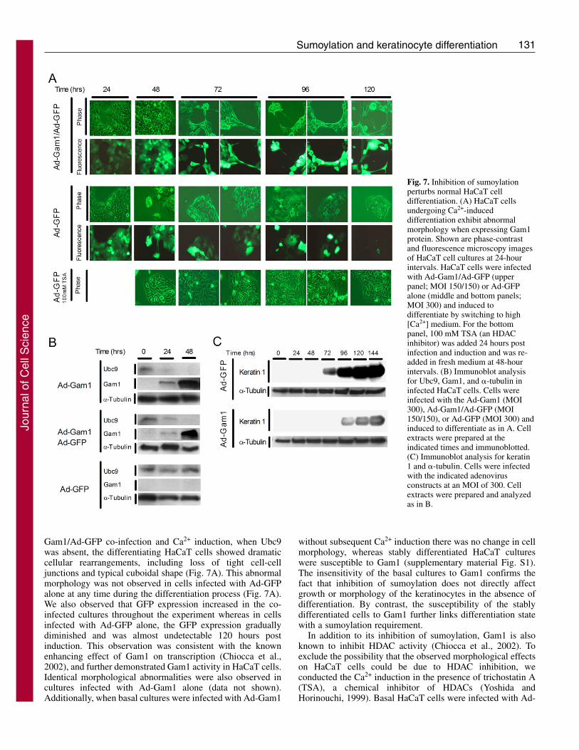
Inhibition of the sumoylation system perturbs the HaCaTcell differentiation processTo investigate the functional importance of sumoylation in theprocess of keratinocyte differentiation, we infected basalHaCaT cells with an adenovirus expressing Myc-tagged Gam1(Ad-Gam1) and then exposed the cells to high [Ca2+] mediumto trigger differentiation. Gam1 is known to inhibitsumoylation by inducing degradation of SAE1/2 and Ubc9(Boggio et al., 2004). By 24 hours post-infection, Gam1 wasdetected in HaCaT cells infected with Ad-Gam1, but not incells infected with Ad-GFP (Fig. 7B). In the Ad-GFP infectedcells, the Ubc9 initially present increased 3.2-fold by 48 hourspost Ca2+ induction, consistent with the response seen in theabsence of viral infection (see Fig. 5B). By contrast, cellsexpressing Gam1 showed diminished Ubc9 levels by 24 hoursand no detectable Ubc9 by 48 hours when Gam1 levels hadreached maximal expression. A similar effect was seen in co-infections with Ad-Gam1 and Ad-GFP, indicating that Ad-GFPhad no stabilizing effect on Ubc9. In contrast to these infectionstudies where the transgene could be delivered effectively intonearly 100% of the cells, alternative attempts to inhibitsumoylation via transfection approaches were unsuccessfulowing to the poor transfection efficiency of HaCaT cells in low[Ca2+] medium (data not shown).
When examined microscopically, HaCaT cells expressingGam1 exhibited a disruption of the normal morphologicalchanges associated with differentiation. By 72 hours after Ad-
Journal of Cell Science 120 (1)
Fig. 5. Keratinocyte differentiation is accompanied by a transientupregulation of several sumoylation system proteins. Parallel HaCaTcell cultures were maintained either in low [Ca2+] medium(Uninduced) or switched from low [Ca2+] to high [Ca2+] medium(Induced). At the indicated times post Ca2+ induction, proteinextracts were prepared from the paired cultures. The extracts wereassayed for expression of K1 (A), Ubc9 (B), SAE1 (B), and SENP1(B) by immunoblotting with specific antibodies. The induced anduninduced cell extracts were also evaluated for �-tubulin expression,and the immunoblots for �-tubulin are duplicated in each panel of Bfor comparative purposes. (C) The immunoblots in B were quantifiedby densitometry, and the values for Ubc9, SAE1, and SENP1 werenormalized against �-tubulin. The 0 hour time values for both theinduced and uninduced samples were set to 1, and the values for thelater time points are relative to the 0 hour time. The quantitativeresults in C are the average of at least three independent experiments.
Fig. 6. The pattern of SUMO-conjugated substrates changes duringHaCaT cell differentiation. Total cell extracts were prepared at theindicated times post Ca2+ induction and analyzed by immunoblottingusing antibodies to SUMO1 (upper panel), �-tubulin (middle panel)or keratin 1 (lower panel). Protein concentrations in the samples wereequalized based on the �-tubulin levels. The positions of molecularsize markers are shown on the right. Arrows and arrowheads indicatebands whose quantity increased or decreased, respectively, during thetime period examined.
Jour
nal o
f Cel
l Sci
ence
131Sumoylation and keratinocyte differentiation
Gam1/Ad-GFP co-infection and Ca2+ induction, when Ubc9was absent, the differentiating HaCaT cells showed dramaticcellular rearrangements, including loss of tight cell-celljunctions and typical cuboidal shape (Fig. 7A). This abnormalmorphology was not observed in cells infected with Ad-GFPalone at any time during the differentiation process (Fig. 7A).We also observed that GFP expression increased in the co-infected cultures throughout the experiment whereas in cellsinfected with Ad-GFP alone, the GFP expression graduallydiminished and was almost undetectable 120 hours postinduction. This observation was consistent with the knownenhancing effect of Gam1 on transcription (Chiocca et al.,2002), and further demonstrated Gam1 activity in HaCaT cells.Identical morphological abnormalities were also observed incultures infected with Ad-Gam1 alone (data not shown).Additionally, when basal cultures were infected with Ad-Gam1
without subsequent Ca2+ induction there was no change in cellmorphology, whereas stably differentiated HaCaT cultureswere susceptible to Gam1 (supplementary material Fig. S1).The insensitivity of the basal cultures to Gam1 confirms thefact that inhibition of sumoylation does not directly affectgrowth or morphology of the keratinocytes in the absence ofdifferentiation. By contrast, the susceptibility of the stablydifferentiated cells to Gam1 further links differentiation statewith a sumoylation requirement.
In addition to its inhibition of sumoylation, Gam1 is alsoknown to inhibit HDAC activity (Chiocca et al., 2002). Toexclude the possibility that the observed morphological effectson HaCaT cells could be due to HDAC inhibition, weconducted the Ca2+ induction in the presence of trichostatin A(TSA), a chemical inhibitor of HDACs (Yoshida andHorinouchi, 1999). Basal HaCaT cells were infected with Ad-
Fig. 7. Inhibition of sumoylationperturbs normal HaCaT celldifferentiation. (A) HaCaT cellsundergoing Ca2+-induceddifferentiation exhibit abnormalmorphology when expressing Gam1protein. Shown are phase-contrastand fluorescence microscopy imagesof HaCaT cell cultures at 24-hourintervals. HaCaT cells were infectedwith Ad-Gam1/Ad-GFP (upperpanel; MOI 150/150) or Ad-GFPalone (middle and bottom panels;MOI 300) and induced todifferentiate by switching to high[Ca2+] medium. For the bottompanel, 100 mM TSA (an HDACinhibitor) was added 24 hours postinfection and induction and was re-added in fresh medium at 48-hourintervals. (B) Immunoblot analysisfor Ubc9, Gam1, and �-tubulin ininfected HaCaT cells. Cells wereinfected with the Ad-Gam1 (MOI300), Ad-Gam1/Ad-GFP (MOI150/150), or Ad-GFP (MOI 300) andinduced to differentiate as in A. Cellextracts were prepared at theindicated times and immunoblotted.(C) Immunoblot analysis for keratin1 and �-tubulin. Cells were infectedwith the indicated adenovirusconstructs at an MOI of 300. Cellextracts were prepared and analyzedas in B.
Jour
nal o
f Cel
l Sci
ence
132
GFP, induced with Ca2+, and then treated with 100 mM TSAat 24 hours after induction and infection. GFP expression wasprolonged in the TSA-treated cultures compared with theuntreated cells, consistent with TSA-mediated enhancement oftranscription through HDAC inhibition (data not shown).However, no abnormal morphology was observed duringdifferentiation in TSA, indicating that HDAC inhibition doesnot contribute to the Gam1 effect on differentiatingkeratinocytes (Fig. 7A).
In addition to keratinocyte morphology, the effect of Gam1expression on the K1 differentiation marker was also evaluated.Gam1 caused a delay in and reduction of K1 expression inCa2+-induced cells (Fig. 7C). Although we cannot exclude thefact that an unknown, off-target effect of Gam1 might alsocontribute to both the defects in morphology and markerexpression, the simplest explanation is that these defects arerelated to the dramatic inhibition of sumoylation. Altogether,the results presented here suggest that modification withSUMO might be a key event for the proper timing of the stepsleading to keratinocyte differentiation, and that sumoylationmay be an important regulator for this biological process.
DiscussionRecent studies on myoblast differentiation (Riquelme et al.,2006), embryonic development (Yamaguchi et al., 2005),genital tissue maturation (Vigodner et al., 2006) and synapticformation (Shalizi et al., 2006) highlight sumoylation not onlyas a modifier of individual proteins but also as a globalregulator of cellular development. In the present study, weinvestigated the role of sumoylation in another complexprocess, keratinocyte differentiation. Using an immortalizedhuman keratinocyte line (HaCaT cells), we demonstrated thatthere was a coordinated transcriptional upregulation of manysumoylation system genes upon exposure to a high [Ca2+]environment, a well-established signal for induction ofkeratinocyte differentiation (Eckert et al., 1997b). In particular,gene expression for Ubc9, the sole conjugating enzyme for the
sumoylation process, was strongly activated. Thistranscriptional upregulation of Ubc9 and several othercomponents of the sumoylation pathway led to increasedexpression of their respective proteins and to changes in theoverall pattern of sumoylated substrates. Furthermore, in theabsence of normal sumoylation, expression of a differentiationmarker, K1 protein, was delayed, and normal keratinocytemorphogenesis was severely perturbed. In agreement with theHaCaT studies, both Ubc9 and SUMO proteins were expressedin organotypic cultures of normal human foreskinkeratinocytes, consistent with an active sumoylation process inthis stratified tissue. We also noted that Ubc9 immunostainingwas most prominent in the intermediate layers of theepithelium just above the basal layer, suggesting increasedaccumulation of Ubc9 in early differentiation similar to thatseen in Ca2+-induced HaCaT cells. These combined resultsstrongly suggest that sumoylation helps to orchestrate propersteps of keratinocyte differentiation and is therefore a keyregulator in this biological event.
One of the salient observations of our studies was thecoordinated transcriptional activation of multiple sumoylationgenes following exposure of basal HaCaT cells to high [Ca2+]culture conditions. Mechanistically, such coordination could beachieved through common promoter elements responding toCa2+-induced TFs. High [Ca2+], both in vitro and in vivo, isknown to trigger keratinocyte differentiation through multiplesignaling pathways that activate a set of effector TFs (Fuchs,1990; Sharpe et al., 1989), which includes NFAT, Sp1, C/EBPand AP1 (Eckert et al., 1997a; Santini et al., 2001). Althoughthe promoters for the sumoylation component genes have notbeen experimentally characterized, bioinformatics analysis(www.genomatrix.de) of the putative promoter regions for thesumoylation genes tested in this study revealed the presence ofbinding sites for various combinations of the above TFs. Therewere single NFAT- and AP1-binding sites and three Sp1-binding sites on the SUMO2 promoter; five Sp1-binding siteson the Ubc9 promoter; three C/EBP-binding sites on the SAE1
Journal of Cell Science 120 (1)
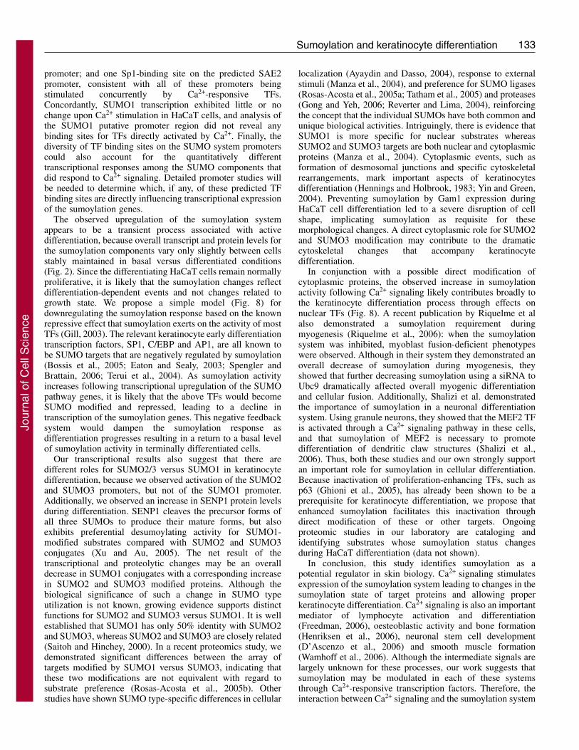
Fig. 8. Model for differentiation-dependent changes in the sumoylation system. The model proposes a negative-feedback mechanism to explainthe transient increase in sumoylation observed during HaCaT cell differentiation. Initially, a pool of TFs, including Sp1, C/EBP and AP1, whichare known to be directly stimulated by Ca2+-induced differentiation, cause upregulation of the sumoylation system. As sumoylation activityincreases, these TFs are in turn modified by SUMO conjugation, which decreases their transcriptional activity and leads to a decline inexpression of the sumoylation pathway genes. A second feature of the model is that increased sumoylation is an active contributor to thedifferentiation process, through SUMO conjugation to downstream effectors of the differentiation signals. Additional details are provided in thetext.
Jour
nal o
f Cel
l Sci
ence
133Sumoylation and keratinocyte differentiation
promoter; and one Sp1-binding site on the predicted SAE2promoter, consistent with all of these promoters beingstimulated concurrently by Ca2+-responsive TFs.Concordantly, SUMO1 transcription exhibited little or nochange upon Ca2+ stimulation in HaCaT cells, and analysis ofthe SUMO1 putative promoter region did not reveal anybinding sites for TFs directly activated by Ca2+. Finally, thediversity of TF binding sites on the SUMO system promoterscould also account for the quantitatively differenttranscriptional responses among the SUMO components thatdid respond to Ca2+ signaling. Detailed promoter studies willbe needed to determine which, if any, of these predicted TFbinding sites are directly influencing transcriptional expressionof the sumoylation genes.
The observed upregulation of the sumoylation systemappears to be a transient process associated with activedifferentiation, because overall transcript and protein levels forthe sumoylation components vary only slightly between cellsstably maintained in basal versus differentiated conditions(Fig. 2). Since the differentiating HaCaT cells remain normallyproliferative, it is likely that the sumoylation changes reflectdifferentiation-dependent events and not changes related togrowth state. We propose a simple model (Fig. 8) fordownregulating the sumoylation response based on the knownrepressive effect that sumoylation exerts on the activity of mostTFs (Gill, 2003). The relevant keratinocyte early differentiationtranscription factors, SP1, C/EBP and AP1, are all known tobe SUMO targets that are negatively regulated by sumoylation(Bossis et al., 2005; Eaton and Sealy, 2003; Spengler andBrattain, 2006; Terui et al., 2004). As sumoylation activityincreases following transcriptional upregulation of the SUMOpathway genes, it is likely that the above TFs would becomeSUMO modified and repressed, leading to a decline intranscription of the sumoylation genes. This negative feedbacksystem would dampen the sumoylation response asdifferentiation progresses resulting in a return to a basal levelof sumoylation activity in terminally differentiated cells.
Our transcriptional results also suggest that there aredifferent roles for SUMO2/3 versus SUMO1 in keratinocytedifferentiation, because we observed activation of the SUMO2and SUMO3 promoters, but not of the SUMO1 promoter.Additionally, we observed an increase in SENP1 protein levelsduring differentiation. SENP1 cleaves the precursor forms ofall three SUMOs to produce their mature forms, but alsoexhibits preferential desumoylating activity for SUMO1-modified substrates compared with SUMO2 and SUMO3conjugates (Xu and Au, 2005). The net result of thetranscriptional and proteolytic changes may be an overalldecrease in SUMO1 conjugates with a corresponding increasein SUMO2 and SUMO3 modified proteins. Although thebiological significance of such a change in SUMO typeutilization is not known, growing evidence supports distinctfunctions for SUMO2 and SUMO3 versus SUMO1. It is wellestablished that SUMO1 has only 50% identity with SUMO2and SUMO3, whereas SUMO2 and SUMO3 are closely related(Saitoh and Hinchey, 2000). In a recent proteomics study, wedemonstrated significant differences between the array oftargets modified by SUMO1 versus SUMO3, indicating thatthese two modifications are not equivalent with regard tosubstrate preference (Rosas-Acosta et al., 2005b). Otherstudies have shown SUMO type-specific differences in cellular
localization (Ayaydin and Dasso, 2004), response to externalstimuli (Manza et al., 2004), and preference for SUMO ligases(Rosas-Acosta et al., 2005a; Tatham et al., 2005) and proteases(Gong and Yeh, 2006; Reverter and Lima, 2004), reinforcingthe concept that the individual SUMOs have both common andunique biological activities. Intriguingly, there is evidence thatSUMO1 is more specific for nuclear substrates whereasSUMO2 and SUMO3 targets are both nuclear and cytoplasmicproteins (Manza et al., 2004). Cytoplasmic events, such asformation of desmosomal junctions and specific cytoskeletalrearrangements, mark important aspects of keratinocytesdifferentiation (Hennings and Holbrook, 1983; Yin and Green,2004). Preventing sumoylation by Gam1 expression duringHaCaT cell differentiation led to a severe disruption of cellshape, implicating sumoylation as requisite for thesemorphological changes. A direct cytoplasmic role for SUMO2and SUMO3 modification may contribute to the dramaticcytoskeletal changes that accompany keratinocytedifferentiation.
In conjunction with a possible direct modification ofcytoplasmic proteins, the observed increase in sumoylationactivity following Ca2+ signaling likely contributes broadly tothe keratinocyte differentiation process through effects onnuclear TFs (Fig. 8). A recent publication by Riquelme et alalso demonstrated a sumoylation requirement duringmyogenesis (Riquelme et al., 2006): when the sumoylationsystem was inhibited, myoblast fusion-deficient phenotypeswere observed. Although in their system they demonstrated anoverall decrease of sumoylation during myogenesis, theyshowed that further decreasing sumoylation using a siRNA toUbc9 dramatically affected overall myogenic differentiationand cellular fusion. Additionally, Shalizi et al. demonstratedthe importance of sumoylation in a neuronal differentiationsystem. Using granule neurons, they showed that the MEF2 TFis activated through a Ca2+ signaling pathway in these cells,and that sumoylation of MEF2 is necessary to promotedifferentiation of dendritic claw structures (Shalizi et al.,2006). Thus, both these studies and our own strongly supportan important role for sumoylation in cellular differentiation.Because inactivation of proliferation-enhancing TFs, such asp63 (Ghioni et al., 2005), has already been shown to be aprerequisite for keratinocyte differentiation, we propose thatenhanced sumoylation facilitates this inactivation throughdirect modification of these or other targets. Ongoingproteomic studies in our laboratory are cataloging andidentifying substrates whose sumoylation status changesduring HaCaT differentiation (data not shown).
In conclusion, this study identifies sumoylation as apotential regulator in skin biology. Ca2+ signaling stimulatesexpression of the sumoylation system leading to changes in thesumoylation state of target proteins and allowing properkeratinocyte differentiation. Ca2+ signaling is also an importantmediator of lymphocyte activation and differentiation(Freedman, 2006), oesteoblastic activity and bone formation(Henriksen et al., 2006), neuronal stem cell development(D’Ascenzo et al., 2006) and smooth muscle formation(Wamhoff et al., 2006). Although the intermediate signals arelargely unknown for these processes, our work suggests thatsumoylation may be modulated in each of these systemsthrough Ca2+-responsive transcription factors. Therefore, theinteraction between Ca2+ signaling and the sumoylation system
Jour
nal o
f Cel
l Sci
ence
134
may also be playing a crucial role for proper development ofthese and other tissues. Lastly, many skin diseases, such aspsoriasis, skin cancer (Eckert et al., 2004) or infectionsresulting from microbial pathogens (Alfandari et al., 1999),hijack the normal process of keratinocyte differentiation andlead to abnormal skin formation. If dysregulation ofsumoylation is contributing to an aberrant differentiationprocess, then modulating sumoylation might have therapeuticbenefits for the treatment of these diseases.
Materials and MethodsCell cultureHaCaT cells (kindly provided by Gary Bokoch, The Scripps Research Institute, LaJolla, CA) were cultured in Ca2+-free DMEM (HyClone), with Ca2+-depleted 10%FBS (Gemini Bioproducts), 4 mM L-glutamine (HyClone), and supplemented withcalcium chloride to a final concentration of 0.03 mM (low [Ca2+] medium) or 2.8mM final concentration (high [Ca2+] medium). FBS was Ca2+ depleted by incubationwith Chelex 100 resin (Bio-Rad Laboratories) for 1 hour at 4°C according to themanufacturer’s protocol. The Chelex was subsequently removed using a 50 mlMillipore 0.22 �m filter unit system (Millipore Corp.). To obtain cells exhibiting abasal keratinocyte phenotype, HaCaT cells were cultured in low [Ca2+] medium forat least 3 weeks and were maintained in the same culture conditions thereafter. Low[Ca2+] HaCaT cultures were never allowed to exceed 85% confluency. For stablydifferentiated HaCaT cells, the cultures were maintained in high [Ca2+] medium forat least 3 weeks before analysis. HaCaT cultures in either medium remained fullyproliferative with similar doubling times (unpublished observations). Humanembryonic kidney 293A (HEK293A) cells (Invitrogen Corp.) were maintained inDMEM supplemented with 10% FBS (Gemini Bioproducts). For the trichostatin A(TSA) experiment, HaCaT cells were treated with 100 mM TSA (Tocris Bioscience)24 hours after infection with an adenoviral vector expressing GFP (Ad-GFP) andCa2+ induction. HaCaT cells were observed up to 144 hours post Ca2+ induction.
RT-PCR and quantitative RT-PCRAll RNAs were extracted using the RNAqueous kit (Ambion). RNA concentrationwas measured using a spectrophotometer and 5 ng/�l aliquots were stored at –80°Cuntil use. All primers were designed to overlap exon-exon junctions, therefore onlyamplifying the cDNAs targets and not the genomic sequences (primer sequencesare provided in the supplementary material, Tables S1 and S2). The one-step RT-PCR mixture contained 50 ng RNA in a final reaction volume of 25 �l. The mixturealso included 2.5 �l of 10� Taq DNA Polymerase buffer (Promega), 50 U MMLVreverse transcriptase, 1 U Platinum Taq (Invitrogen), 4 mM dNTPs, 20 U RNAseOUT (Invitrogen), 6 mM MgCl2 and 0.4 �M of each primer. The one-step RT-PCRwas performed for 20 minutes at 42°C for the reverse transcription step, followedby 90 seconds at 94°C, and 40 cycles of amplification (94°C for 30 seconds; 60°Cfor 30 seconds; 72°C for 60 seconds). Amplifications were performed in a PTC-200 Peltier Thermal Cycler machine (MJ Research). Amplified products wereanalyzed on 2.5% agarose gels and visualized with an Innotech Alphaimager system(Alpha Innotech). For the quantitative RT-PCR (Q-PCR), one step RT reactions wereperformed in a single well using 50 ng of harvested RNA in a 50 �l final volume.In addition to the RT components described above, reactions for Q-PCR contained0.2 �M of LUX primers (designed with the Invitrogen custom primer software), 20U RNAse OUT, 2 �l of Super Script III enzyme solution (which includes MMLV-RT, Platinum Taq, and dNTPs), and 1 �l Rox dye, all from Invitrogen. The LUX�-actin primer set (Invitrogen) was used to detect the internal control gene. ThePCR reaction conditions for Q-PCR were the same as the RT-PCR described above.The Q-PCR plates were read with an ABI 7500 real-time PCR instrument (AppliedBiosystems), and detection of the FAM or JOE label was recorded during the 72°Cstep. Results were graphed as the fold increase of the relative quantitative RQ valueswhere RQ=2(–��Ct). �Ct was calculated as the average Ct for �-actin minus theaverage Ct for the gene of interest, with each sample being run in duplicate. Usingthe �Ct values from each time point, the ��Ct for each mRNA examined wascalculated as the �Ct of the time 0 sample minus the �Ct of time x (in hours) afterinduction. The data shown are the average from at least three independent RNApreparations collected in separate experiments.
Affinity-purified polyclonal antibodies against Ubc9Rabbit polyclonal serum 12741 was produced in-house using affinity-purified Ubc9as the immunogen. A four immunization regime was followed, and 2 weeks afterthe final boost the rabbit was exsanguinated. To affinity purify anti-Ubc9 antibodies,750 �g affinity-purified GST-Ubc9 were diluted in a final volume of 500 �l using1� PBS, and the resulting dilution was dispensed on an 82-mm-diameter, 0.45-�m-pore-size Protran Nitrocellulose filter (Schleicher & Schuell). The membrane wasdried for 30 minutes at room temperature, then re-wetted and blocked by incubationat room temperature for 30 minutes in 15 ml of 1� PBS supplemented with 1%BSA. The blocked membrane was incubated for 2 hours at room temperature with
10 ml of a solution containing 2.5 ml of rabbit polyclonal serum 12741 and 7.5 mlof 1� PBS supplemented with 10 mg/ml BSA and 0.05% Tween 20. The membranewas subsequently washed four times each with 15 ml of 1� PBS supplemented with0.05% Tween 20, and four additional times each with 15 ml of 1� PBS alone. Thebound antibodies were eluted by incubation with 2 ml of elution buffer (100 mMglycine, pH 2.5, 0.02% NaN3) for 5 minutes at room temperature. The elutedantibodies were neutralized with 200 �l of 1.0 M Tris-HCl (pH 8.0), aliquoted, andstored at –70°C. The purity of the affinity-purified antibodies was determined bygel electrophoresis and immunoblotting, and its reactivity against Ubc9 wasdetermined by immunoblotting using purified Ubc9 and unfractionated mammaliancell extracts.
ImmunohistochemistryHuman foreskin keratinocytes were grown in organotypic raft cultures to formstratified epithelium as previously described (Lambert et al., 2005). All chemicalsused for immunohistochemistry were from Biocare Medical (Concord, CA). Slideswere deparaffinized in xylene and rehydrated according to the manufacturer’sprotocols. The following antibodies and dilutions were used: anti-SUMO serum12783 (Rosas-Acosta et al., 2005a), 1:750; affinity purified polyclonal antibodiesagainst Ubc9, 1:20; and polyclonal anti-human keratin 1 (anti-HK1, Covance,Berkeley, CA), 1:5000. Specificity control blocking experiments were performedby adding the corresponding purified proteins (25 �g of Ubc9 or 7 �g of SUMO1)to the cognate antibody dilution. The SUMO and Ubc9 proteins used in theexperiments were purified as previously described (Rosas-Acosta et al., 2005a).After immunostaining, the slides were counterstained with hematoxylin.
Immunoblots and densitometryTotal cell extracts were prepared by adding a 1:1 (v/v) mixture of RIPA buffer (50mM Tris-HCl, pH 8, 150 mM NaCl, 5 mM EDTA, 15 mM MgCl2, 1% NP40, 0.1%SDS, 1 mM DTT, 1:200 protease inhibitor cocktail and 10 mM N-ethylmaleimide)and 4� sample buffer (100 mM Tris-HCl, pH 6.8, 20% glycerol, 8% SDS, 0.02%bromophenol blue, 4% �-mercaptoethanol) directly to the cells. The cells wereshaken gently for 5-10 seconds, and the resulting lysate was collected by pipetting.Samples were heated at 95°C for 5 minutes and sonicated for 30 seconds using aMisonix sonicator 3000 (Misonix). Samples were resolved on 10% or 12.5%polyacrylamide gels and then transferred onto 0.45 �m Immobilon-P membranes(Millipore). The membranes were blocked for at least 15 minutes with 3% non-fatmilk in TTBS (150 mM NaCl, 50 mM Tris-HCl, pH 7.4, 0.005% Tween 20), andincubated for 1 hour or overnight with the primary antibodies listed below at theindicated dilution: rabbit anti �-tubulin (Santa Cruz Biotechnology), 1:15,000; anti-Myc monoclonal antibody (Santa Cruz Biotechnology), 1:500; anti-RanGAPmonoclonal antibody (Zymed/Invitrogen Corp.), 1:2500; rabbit serum 12783 againstSUMO (Rosas-Acosta et al., 2005b), 1:1000; affinity-purified polyclonal antibodiesagainst Ubc9, 1:500; anti-human K1 rabbit serum (Covance), 1:1000; anti-SAE1sheep serum (Axxora), 1:2000; anti-SENP1 rabbit serum (Imgenex), 1:2000; andanti-involucrin rabbit serum (LabVision), 1:1000. After reaction with the primaryantibodies, the membranes were incubated with Horseradish Peroxidase-conjugatedantibodies (Santa Cruz Biotechnology) at 1:10,000 for 1 hour. The membranes weresubsequently rinsed in TTBS, treated with the Western LightningChemiluminescence reagent (PerkinElmer Life and Analytical Sciences), and thenvisualized with X-ray film. Quantitative differences were determined bydensitometry using an Innotech Alphaimager (Alpha Innotech) and were normalizedto the �-tubulin signal. Quantitative results are the average of at least three separateexperiments.
Virus production and infectionMyc-tagged Gam1 adenoviral DNA (Ad-Gam1) was kindly provided by MattCotten (GPC-biotech, Munich, Germany). The Ad-Gam1 DNA was transfected intoHEK 293A cells using Lipofectamine 2000 (Invitrogen) according to themanufacturer’s recommendation. Cells were lysed by freeze-thaw and thesupernatant was collected. The initial viral stock was subsequently amplified withtwo rounds of infection on HEK 293A cells and the final freeze-thaw supernatantcollected as the high-titer stock. Adenovirus expressing GFP (Ad-GFP) was kindlyprovided by G. Davis (Texas A&M Health Science Center, College Station, TX)and amplification was performed as above. Titer was assessed by the limiting-dilution method (Qbiogene Adenovirus Manual, version 1.4) using HEK 293A cellsplated at 1�106 cells/well on six-well plates. For adenovirus experiments, HaCaTcells maintained in low [Ca2+] medium were plated at 5�106 in T75 flasks 20 hoursbefore infection. Each culture was infected at an MOI of 300 in 3 ml of Ca2+-freemedium supplemented with 8 �g/ml polybrene (Fisher Scientific). Three hours afterinfection the medium was removed, cells were trypsinized, and released cells wereresuspended in 10 ml of high [Ca2+] medium to induce differentiation. The resultingcells suspension was split among the wells of a six-well plate at a ratio such thatthe cells in each well would achieve approximately 80% confluency by the time ofcollection. Cells were maintained in high [Ca2+] medium and harvested at varioustimes post plating by direct lysis in the wells using the 1� RIPA:4� sample buffermixture described above. Proteins were analyzed by immunoblotting as describedin the previous section. Cells were visualized by phase contrast and fluorescence
Journal of Cell Science 120 (1)
Jour
nal o
f Cel
l Sci
ence
135Sumoylation and keratinocyte differentiation
microscopy at a magnification of 200� using an Olympus IX70 microscope. Imageswere captured digitally using a Qcolor3 camera (Olympus).
We thank Gary Bokoch for the providing us with the HaCaT cellline, Matthew Cotten for providing us with the Gam1 adenoviralDNA, George Davis for the Ad-GFP vector and Wayne Sampson forassistance with the immunohistochemistry. This research wassupported by a grant from the National Institutes of Health(CA089298).
ReferencesAlfandari, J., Magal, S. S., Jackman, A., Schlegel, R., Gonen, P. and Sherman, L.
(1999). HPV16 E6 oncoprotein inhibits apoptosis induced during serum-calciumdifferentiation of foreskin human keratinocytes. Virology 257, 383-396.
Arco, P. G. D., Koipally, J. and Georgopoulos, K. (2005). Ikaros SUMOylation:Switching out of repression. Mol. Cell. Biol. 25, 2688-2697.
Ayaydin, F. and Dasso, M. (2004). Distinct in vivo dynamics of vertebrate SUMOparalogues. Mol. Biol. Cell 15, 5208-5218.
Azuma, Y., Tan, S. H., Cavenagh, M. M., Ainsztein, A. M., Saitoh, H. and Dasso, M.(2001). Expression and regulation of the mammalian SUMO-1 E1 enzyme. FASEB J.15, 1825-1827.
Bikle, D. D., Ng, D., Tu, C. L., Oda, Y. and Xie, Z. (2001). Calcium- and vitamin D-regulated keratinocyte differentiation. Mol. Cell. Endocrinol. 177, 161-171.
Boggio, R., Colombo, R., Hay, R. T., Draetta, G. F. and Chiocca, S. (2004). Amechanism for inhibiting the SUMO pathway. Mol. Cell 16, 549-561.
Bohren, K. M., Nadkarni, V., Song, J. H., Gabbay, K. H. and Owerbach, D. (2004).A M55V polymorphism in a novel SUMO gene (SUMO-4) differentially activates heatshock transcription factors and is associated with susceptibility to type I diabetesmellitus. J. Biol. Chem. 279, 27233-27238.
Bossis, G., Malnou, C. E., Farras, R., Andermarcher, E., Hipskind, R., Rodriguez,M., Schmidt, D., Muller, S., Jariel-Encontre, I. and Piechaczyk, M. (2005). Down-regulation of c-Fos/c-Jun AP-1 Dimer activity by sumoylation. Mol. Cell. Biol. 25,6964-6979.
Boukamp, P., Petrussevska, R. T., Breitkreutz, D., Hornung, J., Markham, A. andFusenig, N. E. (1988). Normal keratinization in a spontaneously immortalizedaneuploid human keratinocyte cell line. J. Cell Biol. 106, 761-771.
Candi, E., Schmidt, R. and Melino, G. (2005). The cornified envelope: a model of celldeath in the skin. Nat. Rev. Mol. Cell Biol. 6, 328-340.
Capone, A., Visco, V., Belleudi, F., Marchese, C., Carinali, G., Bellocci, M., Picardo,M., Frati, L. and Torrisi, M. R. (2000). Up-modulation of the expression of functionalkeratinocyte growth factor receptor induced by high cell density in the humankeratinocyte HaCaT cell line. Cell Growth Differ. 11, 607-614.
Chen, A., Mannen, H. and Li, S. S. (1998). Characterization of mouse ubiquitin-likeSMT3A and SMT3B cDNAs and gene/pseudogenes. Biochem. Mol. Biol. Int. 46, 1161-1174.
Chiocca, S., Kurtev, V., Colombo, R., Boggio, R., Sciurpi, M. T., Brosch, G., Seiser,C., Draetta, G. F. and Cotten, M. (2002). Histone deacetylase 1 inactivation by anadenovirus early gene product. Curr. Biol. 12, 594-598.
D’Ascenzo, M., Piacentini, R., Casalbore, P., Budoni, M., Pallini, R., Azzena, G. B.and Grassi, C. (2006). Role of L-type Ca2+ channels in neural stem/progenitor celldifferentiation. Eur. J. Neurosci. 23, 935-944.
Desterro, J. M., Thomson, J. and Hay, R. T. (1997). Ubch9 conjugates SUMO but notubiquitin. FEBS Lett. 417, 297-300.
Desterro, J. M., Rodriguez, M. S. and Hay, R. T. (1998). SUMO-1 modification ofIkappaBalpha inhibits NF-kappaB activation. Mol. Cell 2, 233-239.
Eaton, E. M. and Sealy, L. (2003). Modification of CCAAT/enhancer-binding protein-beta by the small ubiquitin-like modifier (SUMO) family members, SUMO-2 andSUMO-3. J. Biol. Chem. 278, 33416-33421.
Eckert, R. L., Crish, J. F., Banks, E. B. and Welter, J. F. (1997a). The epidermis: geneson–genes off. J. Invest. Dermatol. 109, 501-509.
Eckert, R. L., Crish, J. F. and Robinson, N. A. (1997b). The epidermal keratinocyte asa model for the study of gene regulation and cell differentiation. Physiol. Rev. 77, 397-424.
Eckert, R. L., Crish, J. F., Efimova, T. and Balasubramanian, S. (2004). Antioxidantsregulate normal human keratinocyte differentiation. Biochem. Pharmacol. 68, 1125-1131.
Eichner, R., Sun, T. T. and Aebi, U. (1986). The role of keratin subfamilies and keratinpairs in the formation of human epidermal intermediate filaments. J. Cell Biol. 102,1767-1777.
Eloranta, J. J. and Hurst, H. C. (2002). Transcription factor AP-2 interacts with theSUMO-conjugating enzyme UBC9 and is sumolated in vivo. J. Biol. Chem. 277,30798-30804.
Freedman, B. D. (2006). Mechanisms of calcium signaling and function in lymphocytes.Crit. Rev. Immunol. 26, 97-111.
Fuchs, E. (1990). Epidermal differentiation: the bare essentials. J. Cell Biol. 111, 2807-2814.
Fuchs, E. and Green, H. (1980). Changes in keratin gene expression during terminaldifferentiation of the keratinocyte. Cell 19, 1033-1042.
Ghioni, P., D’Alessandra, Y., Mansueto, G., Jaffray, E., Hay, R. T., La Mantia, G.and Guerrini, L. (2005). The protein stability and transcriptional activity of p63 alphaare regulated by SUMO-1 conjugation. Cell Cycle 4, 183-190.
Gill, G. (2003). Post-translational modification by the small ubiquitin-related modifierSUMO has big effects on transcription factor activity. Curr. Opin. Genet. Dev. 13, 108-113.
Girdwood, D. W. H., Tatham, M. H. and Hay, R. T. (2004). SUMO and transcriptionalregulation. Semin. Cell Dev. Biol. 15, 201-210.
Gong, L. and Yeh, E. T. (2006). Characterization of a family of nucleolar SUMO-specific proteases with preference for SUMO-2 or SUMO-3. J. Biol. Chem. 281,15869-15877.
Guo, D. H., Han, J. Y., Adam, B. L., Colburn, N. H., Wang, M. H., Dong, Z., Eizirik,D. L., She, J. X. and Wang, C. Y. (2005). Proteomic analysis of SUM04 substratesin HEK293 cells under serum starvation-induced stress. Biochem. Biophys. Res.Commun. 337, 1308-1318.
Hennings, H. and Holbrook, K. A. (1983). Calcium regulation of cell-cell contact anddifferentiation of epidermal cells in culture. An ultrastructural study. Exp. Cell Res.143, 127-142.
Henriksen, Z., Hiken, J. F., Steinberg, T. H. and Jorgensen, N. R. (2006). Thepredominant mechanism of intercellular calcium wave propagation changes duringlong-term culture of human osteoblast-like cells. Cell Calcium 39, 435-444.
Herwig, S. and Strauss, M. (1997). The retinoblastoma protein: a master regulator ofcell cycle, differentiation and apoptosis. Eur. J. Biochem. 246, 581-601.
Ihara, M., Yamamoto, H. and Kikuchi, A. (2005). SUMO-1 modification of PIASy, anE3 ligase, is necessary for PIASy-dependent activation of Tcf-4. Mol. Cell. Biol. 25,3506-3518.
Johnson, E. S. (2004). Protein modification by SUMO. Annu. Rev. Biochem. 73, 355-382.
Kagey, M. H., Melhuish, T. A. and Wotton, D. (2003). The polycomb protein Pc2 is aSUMO E3. Cell 113, 127-137.
Kim, J., Cantwell, C. A., Johnson, P. F., Pfarr, C. M. and Williams, S. C. (2002).Transcriptional activity of CCAAT/enhancer-binding proteins is controlled by aconserved inhibitory domain that is a target for sumoylation. J. Biol. Chem. 277, 38037-38044.
Kotaja, N., Karvonen, U., Janne, O. A. and Palvimo, J. J. (2002). PIAS proteinsmodulate transcription factors by functioning as SUMO-1 ligases. Mol. Cell. Biol. 22,5222-5234.
Lambert, P. F., Ozbun, M. A., Collins, A., Holmgren, S., Lee, D. and Nakahara, T.(2005). Using an immortalized cell line to study the HPV life cycle in organotypic“raft” cultures. In Human Papillomaviruses: Methods and Protocols. Vol. 119 (ed. C.Davy and J. Doorbar), pp. 141-155. Totowa, NJ: Humana Press.
Lansdown, A. B. (2002). Calcium: a potential central regulator in wound healing in theskin. Wound Repair Regen. 10, 271-285.
Ledl, A., Schmidt, D. and Muller, S. (2005). Viral oncoproteins E1A and E7 and cellularLxCxE proteins repress SUMO modification of the retinoblastoma tumor suppressor.Oncogene 24, 3810-3818.
Leight, E. R., Glossip, D. and Kornfeld, K. (2005). Sumoylation of LIN-1 promotestranscriptional repression and inhibition of vulval cell fates. Development 132, 1047-1056.
Li, M. and Kellems, R. E. (2003). Sp1 and Sp3 Are important regulators of AP-2gammagene transcription. Biol. Reprod. 69, 1220-1230.
Manza, L. L., Codreanu, S. G., Stamer, S. L., Smith, D. L., Wells, K. S., Roberts, R.L. and Liebler, D. C. (2004). Global shifts in protein sumoylation in response toelectrophile and oxidative stress. Chem. Res. Toxicol. 17, 1706-1715.
Melchior, F. and Hengst, L. (2002). SUMO-1 and p53. Cell Cycle 1, 245-249.Melchior, F., Schergaut, M. and Pichler, A. (2003). SUMO: ligases, isopeptidases and
nuclear pores. Trends Biochem. Sci. 28, 612-618.Menon, G. K., Grayson, S. and Elias, P. M. (1985). Ionic calcium reservoirs in
Morita, Y., Kanei-Ishii, C., Nomura, T. and Ishii, S. (2005). TRAF7 sequesters c-Mybto the cytoplasm by stimulating its sumoylation. Mol. Biol. Cell 16, 5433-5444.
Mossessova, E. and Lima, C. D. (2000). Ulp1-SUMO crystal structure and geneticanalysis reveal conserved interactions and a regulatory element essential for cell growthin yeast. Mol. Cell 5, 865-876.
Nacerddine, K., Lehembre, F., Bhaumik, M., Artus, J., Cohen-Tannoudji,M., Babinet, C., Pandolfi, P. P. and Dejean, A. (2005). The SUMO pathway isessential for nuclear integrity and chromosome segregation in mice. Dev. Cell 9, 769-779.
Okuma, T., Honda, R., Ichikawa, G., Tsumagari, N. and Yasuda, H. (1999). In vitroSUMO-1 modification requires two enzymatic steps, E1 and E2. Biochem. Biophys.Res. Commun. 254, 693-698.
Pichler, A. and Melchior, F. (2002). Ubiquitin-related modifier SUMO1 andnucleocytoplasmic transport. Traffic 3, 381-387.
Pichler, A., Gast, A., Seeler, J. S., Dejean, A. and Melchior, F. (2002). The nucleoporinRanBP2 has SUMO1 E3 ligase activity. Cell 108, 109-120.
Poulin, G., Dong, Y., Fraser, A. G., Hopper, N. A. and Ahringer, J. (2005). Chromatinregulation and sumoylation in the inhibition of Ras-induced vulval development inCaenorhabditis elegans. EMBO J. 24, 2613-2623.
Reverter, D. and Lima, C. D. (2004). A basis for SUMO protease specificity providedby analysis of human Senp2 and a Senp2-SUMO complex. Structure 12, 1519-1531.
Riquelme, C., Barthel, K. K., Qin, X. F. and Liu, X. (2006). Ubc9 expression is essentialfor myotube formation in C2C12. Exp. Cell Res. 12, 2132-2141.
Rosas-Acosta, G., Langereis, M. A., Deyrieux, A. and Wilson, V. G. (2005a). Proteinsof the PIAS family enhance the sumoylation of the papillomavirus E1 protein. Virology331, 190-203.
Jour
nal o
f Cel
l Sci
ence
136
Rosas-Acosta, G., Russell, W. K., Deyrieux, A., Russell, D. H. and Wilson, V. G.(2005b). A universal strategy for proteomic studies of SUMO and other ubiquitin-likemodifiers. Mol. Cell. Proteomics 4, 56-72.
Ross, S., Best, J. L., Zon, L. I. and Gill, G. (2002). SUMO-1 modification repressesSp3 transcriptional activation and modulates its subnuclear localization. Mol. Cell 10,831-842.
Saitoh, H. and Hinchey, J. (2000). Functional heterogeneity of small ubiquitin-relatedprotein modifiers SUMO-1 versus SUMO-2/3. J. Biol. Chem. 275, 6252-6258.
Salinas, S., Briancon-Marjollet, A., Bossis, G., Lopez, M. A., Piechaczyk, M., Jariel-Encontre, I., Debant, A. and Hipskind, R. A. (2004). SUMOylation regulates nucleo-cytoplasmic shuttling of Elk-1. J. Cell Biol. 165, 767-773.
Santini, M. P., Talora, C., Seki, T., Bolgan, L. and Dotto, G. P. (2001). Cross talkamong calcineurin, Sp1/Sp3, and NFAT in control of p21(WAF1/CIP1) expression inkeratinocyte differentiation. Proc. Natl. Acad. Sci. USA 98, 9575-9580.
Schoop, V. M., Mirancea, N. and Fusenig, N. E. (1999). Epidermal organization anddifferentiation of HaCaT keratinocytes in organotypic coculture with human dermalfibroblasts. J. Invest. Dermatol. 112, 343-353.
Schweizer, J. and Winter, H. (1983). Keratin biosynthesis in normal mouse epithelia andin squamous cell carcinomas. mRNA-dependent alterations of the primary structure ofdistinct keratin subunits in tumors. J. Biol. Chem. 258, 13268-13272.
Seeler, J. S. and Dejean, A. (2001). SUMO: of branched proteins and nuclear bodies.Oncogene 20, 7243-7249.
Shalizi, A., Gaudilliere, B., Yuan, Z. Q., Stegmuller, J., Shirogane, T., Ge, Q. Y., Tan,Y., Schulman, B., Harper, J. W. and Bonni, A. (2006). A calcium-regulated MEF2sumoylation switch controls postsynaptic differentiation. Science 311, 1012-1017.
Sharpe, G. R., Gillespie, J. I. and Greenwell, J. R. (1989). An increase in intracellularfree calcium is an early event during differentiation of cultured human keratinocytes.FEBS Lett. 254, 25-28.
Shiio, Y. and Eisenman, R. N. (2003). Histone sumoylation is associated withtranscriptional repression. Proc. Natl. Acad. Sci. USA 100, 13225-13230.
Smith, F. (2003). The molecular genetics of keratin disorders. Am. J. Clin. Dermatol. 4,347-364.
Spengler, M. L. and Brattain, M. G. (2006). Sumoylation inhibits cleavage of Sp1 N-terminal negative regulatory domain and inhibits Sp1-dependent transcription. J. Biol.Chem. 281, 5567-5574.
Su, H. L. and Li, S. S. L. (2002). Molecular features of human ubiquitin-like SUMOgenes and their encoded proteins. Gene 296, 65-73.
Sun, T. T., Tseng, S. C., Huang, A. J., Cooper, D., Schermer, A., Lynch, M. H., Weiss,R. and Eichner, R. (1985). Monoclonal antibody studies of mammalian epithelialkeratins: a review. Ann. N. Y. Acad. Sci. 455, 307-329.
Tatham, M. H., Chen, Y. and Hay, R. T. (2003). Role of two residues proximal to theactive site of Ubc9 in substrate recognition by the Ubc9 center dot SUMO-1 thiolestercomplex. Biochemistry 42, 3168-3179.
Tatham, M. H., Kim, S., Jaffray, E., Song, J., Chen, Y. and Hay, R. T. (2005). Uniquebinding interactions among Ubc9, SUMO and RanBP2 reveal a mechanism for SUMOparalog selection. Nat. Struct. Mol. Biol. 12, 67-74.
Terui, Y., Saad, N., Jia, S., McKeon, F. and Yuan, J. Y. (2004). Dual role of sumoylationin the nuclear localization and transcriptional activation of NFAT1. J. Biol. Chem. 279,28257-28265.
Tu, C. L., Oda, Y., Komuves, L. and Bikle, D. D. (2004). The role of the calcium-sensingreceptor in epidermal differentiation. Cell Calcium 35, 265-273.
Verger, A., Perdomo, J. and Crossley, M. (2003). Modification with SUMO – A role intranscriptional regulation. EMBO Rep. 4, 137-142.
Vicanova, J., Boelsma, E., Mommaas, A. M., Kempenaar, J. A., Forslind, B., Pallon,J., Egelrud, T., Koerten, H. K. and Ponec, M. (1998). Normalization of epidermalcalcium distribution profile in reconstructed human epidermis is related toimprovement of terminal differentiation and stratum corneum barrier formation. J.Invest. Dermatol. 111, 97-106.
Vigodner, M., Ishikawa, T., Schlegel, P. N. and Morris, P. L. (2006). SUMO-1, humanmale germ cell development, and the androgen receptor in the testis of men with normaland abnormal spermatogenesis. Am. J. Physiol. Endocrinol. Metab. 290, E1022-E1033.
Wamhoff, B. R., Bowles, D. K. and Owens, G. K. (2006). Excitation-transcriptioncoupling in arterial smooth muscle. Circ. Res. 98, 868-878.
Wilson, V. G. and Rangasamy, D. (2001). Intracellular targeting of proteins bysumoylation. Exp. Cell Res. 271, 57-65.
Xu, Z. and Au, S. W. N. (2005). Mapping residues of SUMO precursors essential indifferential maturation by SUMO-specific protease, SENP1. Biochem. J. 386, 325-330.
Yamaguchi, T., Sharma, P., Athanasiou, M., Kumar, A., Yamada, S. and Kuehn, M.R. (2005). Mutation of SENP1/SuPr-2 reveals an essential role for desumoylation inmouse development. Mol. Cell. Biol. 25, 5171-5182.
Yang, S. H. and Sharrocks, A. D. (2004). SUMO promotes HDAC-mediatedtranscriptional repression. Mol. Cell 13, 611-617.
Yin, T. and Green, K. J. (2004). Regulation of desmosome assembly and adhesion.Semin. Cell Dev. Biol. 15, 665-677.
Yoshida, M. and Horinouchi, S. (1999). Trichostatin and leptomycin. Inhibition ofhistone deacetylation and signal-dependent nuclear export. Ann. N. Y. Acad. Sci. 886,23-36.
Zhong, S., Muller, S., Ronchetti, S., Freemont, P. S., Dejean, A. and Pandolfi, P. P.(2000). Role of SUMO-1-modified PML in nuclear body formation. Blood 95, 2748-2753.