Review Article Visual Perception in Preterm Children: What Are We Currently Measuring? Els L. Ortibus MD * , Paul P. De Cock MD, PhD, Lieven G. Lagae MD, PhD Department of Pediatric Neurology, University Hospitals Leuven, Leuven, Belgium article information Article history: Received 25 October 2010 Accepted 28 February 2011 abstract Over the past two decades, cerebral visual impairment has been recognized as a principal deficit in preterm children, and in particular those with cerebral palsy. We review the current knowledge of visual processing deficits in these children, and provide an overview of the tools for assessing cerebral visual impairment. Commercially available instruments are usually directed at evaluating visuospatial skills rather than detecting object recognition difficulties. Particularly in children aged 3 years or younger and in children with multiple handicaps, cerebral visual impairment is difficult to diagnose. This difficulty may be attributable to limitations specific to the instrument, such as a test that is inappropriate for age, or to child-specific limitations such as motor impairment or speech delay. We therefore include an overview of relevant neuroimaging findings reported in these children, focusing on the most recent imaging modalities. Novel techniques such as diffusion tensor imaging may provide sensitive markers of cerebral visual impairment in situations where clinical diagnosis is difficult, and such approaches may allow for early intervention. Ó 2011 Elsevier Inc. All rights reserved. Introduction Since the 1970s, the survival of preterm born children born has steadily improved [1]. In particular, an increase in survival of 25-73% was reported for infants born before 26 weeks of post- menstrual age [2]. Although this positive evolution raised concerns that the burden of disability in this population would demonstrate a similar increase, a decrease in significant disabilities such as cerebral palsy and severe visual and hearing problems was repor- ted during the last decade [3]. However, a spectrum of more subtle cognitive deficits has become apparent with age [4]. Deficits in the domains of executive functioning, attention, language, behavior, and learning skills were reported, particularly in extremely preterm children, and these difficulties tend to persist into school age and adolescence [4,5]. This finding is reflected in the current definition of cerebral palsy, which includes accompanying disturbances on the sensory and perceptual levels, leading to limited activity and participation [6]. Within this spectrum of “new” cognitive deficits, cerebral visual impairment has emerged as a major problem, and its occurrence is not restricted to extremely preterm children or children with brain abnormalities [7]. Although the problem is becoming better understood, the exact definition of cerebral visual impairment is still under debate, and diagnostic approaches vary widely [8,9]. Cerebral visual impairment not only occurs in association with other deficits, but by itself exerts a major impact on other devel- opmental domains, and this renders the clinical picture of the individual child with cerebral visual impairment very complex. Probably as a consequence of these factors, solid research efforts to address the development of rehabilitative strategies for cerebral visual impairment are lacking. This review aims to provide an overview of the visual processing deficits occurring in preterm children, of the diagnostic tools in current use to assess cerebral visual impairment, and of neuro- imaging findings reported in association with cerebral visual impairment. Development of visual function in healthy, term children A great deal of research has been directed at understanding the maturation of sensory function in preterm babies, and these efforts initially relied on behavioral measures. Daum et al. [10] reported in 1980 that fixation and ocular pursuit are present from post- menstrual age 34 weeks. In their research, they used the “l’oeil de boeuf,” a cardboard disc with concentric black and white circles presented to the baby at a distance of 20-30 cm. In 1982, Morante et al. [11] described the development of a preference for patterns and gratings after postmenstrual age 30 weeks. Subsequently, Tsuneishi et al. succeeded in eliciting visual evoked responses in preterm infants as of postmenstrual age 24 weeks [12]. With increasing age, the waveform of visual evoked responses changed, * Communications should be addressed to: Dr. Ortibus; Department of Pediatric Neurology; University Hospitals Leuven; Herestraat 49; B-3000 Leuven, Belgium. E-mail address: [email protected]. Contents lists available at ScienceDirect Pediatric Neurology journal homepage: www.elsevier.com/locate/pnu 0887-8994/$ - see front matter Ó 2011 Elsevier Inc. All rights reserved. doi:10.1016/j.pediatrneurol.2011.02.008 Pediatric Neurology 45 (2011) 1e10
Transcript
lable at ScienceDirect
Pediatric Neurology 45 (2011) 1e10
Contents lists avai
Pediatric Neurology
journal homepage: www.elsevier .com/locate/pnu
Review Article
Visual Perception in Preterm Children: What Are We Currently Measuring?
Els L. Ortibus MD *, Paul P. De Cock MD, PhD, Lieven G. Lagae MD, PhDDepartment of Pediatric Neurology, University Hospitals Leuven, Leuven, Belgium
article information
Article history:Received 25 October 2010Accepted 28 February 2011
a
Opp
* Communications should be addressed to: Dr. OrtiNeurology; University Hospitals Leuven; Herestraat 4
0887-8994/$ - see front matter � 2011 Elsevier Inc. Adoi:10.1016/j.pediatrneurol.2011.02.008
bstract
ver the past two decades, cerebral visual impairment has been recognized as a principal deficit inreterm children, and in particular those with cerebral palsy. We review the current knowledge of visualrocessing deficits in these children, and provide an overview of the tools for assessing cerebral visual
impairment. Commercially available instruments are usually directed at evaluating visuospatial skillsrather than detecting object recognition difficulties. Particularly in children aged 3 years or younger andin children with multiple handicaps, cerebral visual impairment is difficult to diagnose. This difficultymay be attributable to limitations specific to the instrument, such as a test that is inappropriate for age,or to child-specific limitations such as motor impairment or speech delay. We therefore include anoverview of relevant neuroimaging findings reported in these children, focusing on the most recentimaging modalities. Novel techniques such as diffusion tensor imaging may provide sensitive markers ofcerebral visual impairment in situations where clinical diagnosis is difficult, and such approaches mayallow for early intervention.
� 2011 Elsevier Inc. All rights reserved.
Introduction
Since the 1970s, the survival of preterm born children born hassteadily improved [1]. In particular, an increase in survival of25-73% was reported for infants born before 26 weeks of post-menstrual age [2]. Although this positive evolution raised concernsthat the burden of disability in this population would demonstratea similar increase, a decrease in significant disabilities such ascerebral palsy and severe visual and hearing problems was repor-ted during the last decade [3]. However, a spectrum of more subtlecognitive deficits has become apparent with age [4]. Deficits in thedomains of executive functioning, attention, language, behavior,and learning skills were reported, particularly in extremely pretermchildren, and these difficulties tend to persist into school age andadolescence [4,5]. This finding is reflected in the current definitionof cerebral palsy, which includes accompanying disturbances onthe sensory and perceptual levels, leading to limited activity andparticipation [6].
Within this spectrum of “new” cognitive deficits, cerebral visualimpairment has emerged as a major problem, and its occurrence isnot restricted to extremely preterm children or children with brainabnormalities [7]. Although the problem is becoming betterunderstood, the exact definition of cerebral visual impairment is
bus; Department of Pediatric9; B-3000 Leuven, Belgium.
ll rights reserved.
still under debate, and diagnostic approaches vary widely [8,9].Cerebral visual impairment not only occurs in association withother deficits, but by itself exerts a major impact on other devel-opmental domains, and this renders the clinical picture of theindividual child with cerebral visual impairment very complex.Probably as a consequence of these factors, solid research efforts toaddress the development of rehabilitative strategies for cerebralvisual impairment are lacking.
This review aims to provide an overview of the visual processingdeficits occurring in preterm children, of the diagnostic tools incurrent use to assess cerebral visual impairment, and of neuro-imaging findings reported in association with cerebral visualimpairment.
Development of visual function in healthy, term children
A great deal of research has been directed at understanding thematuration of sensory function in preterm babies, and these effortsinitially relied on behavioral measures. Daum et al. [10] reported in1980 that fixation and ocular pursuit are present from post-menstrual age 34 weeks. In their research, they used the “l’oeil deboeuf,” a cardboard disc with concentric black and white circlespresented to the baby at a distance of 20-30 cm. In 1982, Moranteet al. [11] described the development of a preference for patternsand gratings after postmenstrual age 30 weeks. Subsequently,Tsuneishi et al. succeeded in eliciting visual evoked responses inpreterm infants as of postmenstrual age 24 weeks [12]. Withincreasing age, the waveform of visual evoked responses changed,
E.L. Ortibus et al. / Pediatric Neurology 45 (2011) 1e102
and a positive deflection (N1) was discovered at around age 34weeks [13].
Enhanced extrauterine visual experience was revealed toaccelerate the maturation of these visual evoked responses signif-icantly, but not that of the electroretinogram [14]. Ricci et al. [15]also showed that extrauterine visual experience positively influ-enced the maturational aspects of visual function relating to ocularstability and tracking, but not those relating to attention at distanceand the perception of color contrast. Therefore, subcorticallymediated functions were assumed to mature more rapidly withextrauterine visual exposure than aspects of visual function thatrequired cortical input, because the maturation of those aspectsdepended more on postmenstrual age. However, in children withbrain lesions such as periventricular white matter injury, periven-tricular hemorrhage, and occipital lesions, the maturation of visualevoked responses demonstrated a manifest delay, the extent ofwhich was related to the degree of neurologic abnormality [16].
Behavioral and electrophysiologic methods enabled researchersto demonstrate that visual acuity in preterm children changed froma 160-minute to a 40-minute arc between postmenstrual ages 30and 40 weeks. Van hof-van Duin [17] indicated that visual acuitydramatically increases before age 1 year (from fourfold to tenfold),reaching an adult level of acuity (1-minute arc) at age 3-5 years.Visual acuity may be defined as the visual capacity to discriminatefine detail, and according to the stimulus, three categories arediscerned: detection acuity (the detection of a small object againsta plain background), resolution acuity (the discrimination of indi-vidual elements in repetitive patterns), and recognition acuity (thediscrimination of fine detail of an optotype). In 1989, recognitionacuity had already been demonstrated to involve not only the eyeand the occipital brain areas, but also the temporal and parietalcortex, along with the eye and the occipital brain areas.
Along with the maturation of lower-order visual functions,various cortical visual functions reach maturity, each at specificages. For example, orientation selectivity is considered mature atage 8 weeks. From birth, human infants preferentially attend toface-like patterns. A newborn first recognizes its mother’s face onthe basis of information from both the outer contour of the headand hairline, and the internal configuration of eyes, nose, andmouth [18]. After age 6 weeks, recognition is based solely on theinternal configuration of the face. Very early on, infants also detectbiologic motion, can discriminate that motion from other forms ofmotion, and show preferential attention for upright humanmovement, a preference crucial in the ability to recognize peopleand make social contacts [19]. Visual attention is a process thatmaturesmore slowly, and that continues until school age [20]. Aftera phase of alertness between birth and age 8-10 weeks, the ori-enting system becomes fully functional during the first 6months. Inthis period, the duration of looking decreases, reflecting animproved ability to disengage attention. Afterward, infants developan ability to manifest sustained attention, enabling them to exploreobjects in the environment. Finally, at around age 18-24 months,the frontal cortex undergoes further development, enablingtoddlers to begin looking at complex visual displays such as tele-vision [21].
Before age 16 months, form-processing and motion-processingabilities have also begun to develop, but these are not completeuntil age 4 years [22].
Lower-order visual function in preterm children
Overall, preterm children are at risk for developing visualdisorders, irrespective of a history of retinopathy of prematurity.
Visual acuity is significantly reduced in preterm children withneurologic problems, such as those that develop after asphyxia and
intraventricular hemorrhage [23]. Studies reported not onlya reduction in resolution, but also in recognition acuity. Acuity wasdemonstrated to be normal in infants with prolonged flares orperiventricular leukomalacia grades 1 and 2, but it was clearlyaffected in infants with periventricular leukomalacia grades 3 and 4[7,24]. However, in preterm children attending mainstream school,decreased visual acuity was reported to occur two to three timesmore frequently than in term-born peers [25,26]. These problemswith visual acuity can be ascribed principally to refractive errors.High myopia, in particular, confers a risk for developing anisome-tropia and secondary visual disorders such as amblyopia andstrabismus [27]. However, such early reductions of visual acuity arereportedly subject to “catch-up” by age 5 years [28].
The prevalence of strabismus in preterm children varies from 3%in infants without retinopathy of prematurity, to 57% in 5-year-oldchildren born at a postmenstrual age of less than 28 weeks. Thepresence of brain lesions partly accounts for this wide range inprevalence [25,27]. Considerable variation exists in the type ofstrabismus, but the proportion of children with divergence-typestrabismus is known to be higher in preterm than in term chil-dren. This knowledge is important for purposes of detection,because the presence of strabismus affects the development ofbinocular vision, including stereo acuity [28].
In addition to deficits in acuity and eye alignment, pretermchildren also run a higher risk for reduced contrast sensitivity andvisual field loss [26].
Visual perception/higher-level visual processing in preterminfants
A structural framework for visual perception
Cerebral visual perception is an important and early developingaspect of brain function. Visual processing is complex, and in itsfirst phase involves a relay of sensory signals from the retina to thevisual cortex (striate cortex or area V1). This relay is responsible forthe initial processing of visual information. Lesions in the striatecortex primarily lead to reduced visual acuity, contrast sensitivity,and color detection, and a loss of visual field. From the striatecortex onward, information is processed by the extrastriate areas.In a simplified view, these extrastriate regions are organized astwo streams, i.e., ventral and dorsal. The ventral stream runs fromthe striate cortex to the middle and inferotemporal regions, andplays a role in the recognition of colors, objects, shapes, faces, androute finding [20,29]. These representations are stored for futurereference, and help build visual memory. The dorsal stream alsostarts in area V1 but orients its fibers to the posterior parietalcortex, and plays a role in processing complex visual information.It is also responsible for online unconscious visual processing,which allows for the visual guidance of movement. Goodale andMilner [29] introduced the terms “What” (vision for perception)for the ventral stream, and “How” (vision for action) for the dorsalstream.
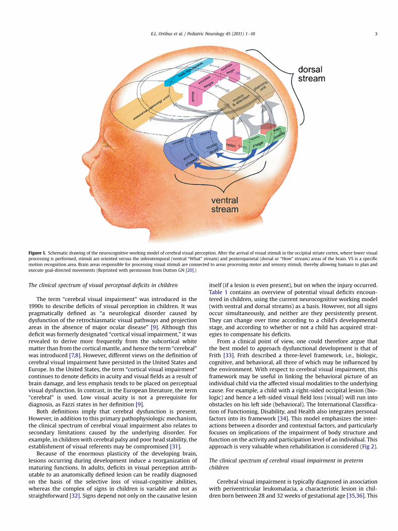
From the posterior part of the brain, visual information is led toV5, a specific projection area where motion is detected. This area isresponsible for the recognition of radial flow and translationalmotion, which enables us to detect the approach to an object or todistinguish figures from their background, among other abilities.Biologic motion is processed in both the temporal lobe and parietalareas, where posterior portions of the superior temporal sulcusrepresent the intersection within this distributed network [19].Finally, visual attention is regulated by both the posterior parietaland frontal areas [20,30]. Figure 1 illustrates the different brainareas involved in visual processing.
Figure 1. Schematic drawing of the neurocognitive working model of cerebral visual perception. After the arrival of visual stimuli in the occipital striate cortex, where lower visualprocessing is performed, stimuli are oriented versus the inferotemporal (ventral “What” stream) and posteroparietal (dorsal or “How” stream) areas of the brain. V5 is a specificmotion recognition area. Brain areas responsible for processing visual stimuli are connected to areas processing motor and sensory stimuli, thereby allowing humans to plan andexecute goal-directed movements (Reprinted with permission from Dutton GN [20].)
The clinical spectrum of visual perceptual deficits in children
The term “cerebral visual impairment” was introduced in the1990s to describe deficits of visual perception in children. It waspragmatically defined as “a neurological disorder caused bydysfunction of the retrochiasmatic visual pathways and projectionareas in the absence of major ocular disease” [9]. Although thisdeficit was formerly designated “cortical visual impairment,” it wasrevealed to derive more frequently from the subcortical whitematter than from the cortical mantle, and hence the term “cerebral”was introduced [7,8]. However, different views on the definition ofcerebral visual impairment have persisted in the United States andEurope. In the United States, the term “cortical visual impairment”continues to denote deficits in acuity and visual fields as a result ofbrain damage, and less emphasis tends to be placed on perceptualvisual dysfunction. In contrast, in the European literature, the term“cerebral” is used. Low visual acuity is not a prerequisite fordiagnosis, as Fazzi states in her definition [9].
Both definitions imply that cerebral dysfunction is present.However, in addition to this primary pathophysiologic mechanism,the clinical spectrum of cerebral visual impairment also relates tosecondary limitations caused by the underlying disorder. Forexample, in childrenwith cerebral palsy and poor head stability, theestablishment of visual referents may be compromised [31].
Because of the enormous plasticity of the developing brain,lesions occurring during development induce a reorganization ofmaturing functions. In adults, deficits in visual perception attrib-utable to an anatomically defined lesion can be readily diagnosedon the basis of the selective loss of visual-cognitive abilities,whereas the complex of signs in children is variable and not asstraightforward [32]. Signs depend not only on the causative lesion
itself (if a lesion is even present), but on when the injury occurred.Table 1 contains an overview of potential visual deficits encoun-tered in children, using the current neurocognitive working model(with ventral and dorsal streams) as a basis. However, not all signsoccur simultaneously, and neither are they persistently present.They can change over time according to a child’s developmentalstage, and according to whether or not a child has acquired strat-egies to compensate his deficits.
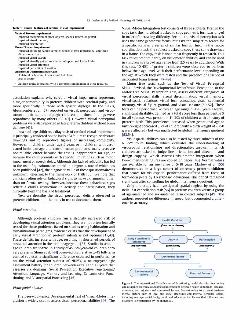
From a clinical point of view, one could therefore argue thatthe best model to approach dysfunctional development is that ofFrith [33]. Frith described a three-level framework, i.e., biologic,cognitive, and behavioral, all three of which may be influenced bythe environment. With respect to cerebral visual impairment, thisframework may be useful in linking the behavioral picture of anindividual child via the affected visual modalities to the underlyingcause. For example, a child with a right-sided occipital lesion (bio-logic) and hence a left-sided visual field loss (visual) will run intoobstacles on his left side (behavioral). The International Classifica-tion of Functioning, Disability, and Health also integrates personalfactors into its framework [34]. This model emphasizes the inter-actions between a disorder and contextual factors, and particularlyfocuses on implications of the impairment of body structure andfunction on the activity and participation level of an individual. Thisapproach is very valuable when rehabilitation is considered (Fig 2).
The clinical spectrum of cerebral visual impairment in pretermchildren
Cerebral visual impairment is typically diagnosed in associationwith periventricular leukomalacia, a characteristic lesion in chil-dren born between 28 and 32 weeks of gestational age [35,36]. This
Figure 2. The International Classification of Functioning model classifies functioningand disability, viewed as outcomes of interactions between health conditions (diseases,disorders, and injuries) and contextual factors. Context refers to external environ-mental factors, such as legal and social structures and internal personal factors,including sex, age, social background, and education, i.e., factors that influence howdisability is experienced by the individual.
Table 1. Clinical features of cerebral visual impairment*
Ventral Stream ImpairmentImpaired recognition of faces, objects, shapes, letters, or gestaltImpaired visual memoryImpaired orientation
Dorsal Stream ImpairmentImpaired ability to handle complex scenes in two-dimensional and three-dimensional spaceImpaired visual searchImpaired visually guided movement of upper and lower limbsImpaired visual attentionImpaired perception of motion
Additional Ophthalmologic CuesUnilateral or bilateral lower visual field lossLoss of acuity
* Children typically present with a complex combination of these features.
E.L. Ortibus et al. / Pediatric Neurology 45 (2011) 1e104
association explains why cerebral visual impairment representsa major comorbidity in preterm children with cerebral palsy, andmore specifically in those with spastic diplegia. In the 1960s,Abercrombie et al. [37] reported on visual, perceptual, and visuo-motor impairments in diplegic children, and those findings werereproduced by many others [38-40]. However, visual perceptualproblems were also reported in late preterm childrenwithout braindamage [41].
In school-age children, a diagnosis of cerebral visual impairmentis principally rendered on the basis of a failure to recognize abstractdrawings and to reproduce figures of increasing complexity.However, in children under age 5 years or in children with asso-ciated brain damage and central motor problems, many tests arenot reliable, either because the test is inappropriate for age, orbecause the child presents with specific limitations such as motorimpairment or speech delay. Although this lack of reliability has ledto the use of questionnaires to aid in diagnosis, one of which hasbeen published [42], the diagnostic value of these questionnaires isunknown. Referring to the framework of Frith [33], we note thatclinicians often rely on behavioral signs to make a diagnosis, ratherthan on formal testing. However, because these behavioral signsreflect a child’s restrictions in activity and participation, theycurrently form the basis of treatment.
Next we describe the visual perceptual deficits observed inpreterm children, and the tools in use to document them.
Visual attention
Although preterm children run a strongly increased risk ofdeveloping visual attention problems, they are not often formallytested for these problems. Based on studies using habituation anddishabituation paradigms, evidence exists that the development ofearly visual attention in preterm infants is not optimal [15,43].These deficits increase with age, resulting in shortened periods ofsustained attention in the toddler age group [23]. Studies in school-age children are sparse. In a study of 45 7-9-year-old children bornvery preterm, Shum et al. [44] observed that relative to 49 full-termcontrol subjects, a significant difference occurred in performanceon the visual attention subtest of NEPSY, a neuropsychologicassessment battery for children between ages 3 and 12 years thatassesses six domains: Social Perception, Executive Functioning/Attention, Language, Memory and Learning, Sensorimotor Func-tioning, and Visuospatial Processing [45].
Visuospatial abilities
The Beery-Buktenica Developmental Test of Visual-Motor Inte-gration is widely used to assess visual perceptual abilities [46]. The
Visual-Motor Integration test consists of three subtests. First, in thecopy task, the individual is asked to copy geometric forms, arrangedin order of increasing difficulty. Second, the visual perception taskuses the same geometric forms, but asks the subject to search fora specific form in a series of similar forms. Third, in the motorcoordination task, the subject is asked to copy these same drawingsin a frame. The copy task is used most frequently in research. Thistask relies predominantly on visuomotor abilities, and can be usedin children in a broad age range from 2.5 years to adulthood. Withthis test, 10-45% of preterm children were observed to performbelow their age level, with their performance level depending onthe age at which they were tested and the presence or absence ofassociated brain lesions [47-49].
Motor free tests, such as the Test of Visual PerceptualSkillsdRevised, the Developmental Test of Visual Perception, or theMotor Free Visual Perception Test, assess different categories ofvisual perceptual skills: visual discrimination, visual memory,visual-spatial relations, visual form-constancy, visual sequentialmemory, visual figure ground, and visual closure [50-52]. Thesetests can be performed within an age range of 4-18 years. Overall,significant disability, defined as a total score less than percentile 5for all subtests, was present in 11-20% of children with a history ofpreterm birth. This prevalence increased when gestational age orbirth weight decreased (17% of childrenwith a birth weight of<750g were affected), but was unaffected by global intelligence quotient[53,54].
Visuospatial abilities can also be tested by three subtests of theNEPSY: route finding, which evaluates the understanding ofvisuospatial relationships and directionality; arrows, in whichchildren are asked to judge line orientation and direction; anddesign copying, which assesses visuomotor integration whentwo-dimensional figures are copied on paper [45]. Normal valuesare available for an age range of 5-16 years. Marlow et al. [55]demonstrated in a large cohort of extremely preterm childrenthat scores for visuospatial performance differed from those ofterm-born peers by 1.6 standard deviations. This deficit remainedsignificant after controlling for global intelligence quotient.
Only one study has investigated spatial neglect by using theBells Test cancellation task [56] in preterm children versus a groupof age-matched and sex-matched term control subjects [49]. Theauthors reported no difference in speed, but documented a differ-ence in accuracy.
In the Hooper Visual Organisation Test, the patient is asked toidentify 30 objects, represented as puzzle pieces [57]. The test reliesmainly on concept formation, on visual analysis and synthesis, andon labeling familiar objects, and can be used from age 6 yearsonward. In a group of 216 extremely low birth weight childrenolder than age 6 years, Jakobson et al. [58] observed no differencesin performance on this test, independently of whether brain lesionswere documented by ultrasound in the neonatal period.
The L94 Visual Perceptual Battery is another object recognitiontest, comprising eight visual perceptual tasks for which normativedata are available for children aged between 2.75 and 6.5 years. Infive computer tasks, the child is asked to identify everyday objects,and thus is tested for semantic and perceptional categorization (foran overview and examples of individual subtasks, see Stierset al.[35] and Fig 3). In a study by van den Hout et al. [59],impairment indicated by the L94 was particularly evident inpreterm children with periventricular leukomalacia, but also inchildren with transient periventricular echo densities. Later on,impaired L94 results were observed to be increased in childrenborn between 30 and 37 weeks of postmenstrual age with cerebralpalsy, and the severity of impairment increased when periven-tricular leukomalacia was present [36].
Finally, Fazzi et al. [9] studied 22 preterm born children withbrain damage, using a neuropsychologic battery that evaluatesnot only form, object, and spatial recognition, but also visualmemory. They identified object recognition difficulties in 70% ofsubjects. Visual memory was also significantly deficient, a findingconfirmed by others [60]. The authors also investigated faceperception, with four out of 20 preterm children performing poorly.
Perception of motion
In addition to form recognition and visuospatial problems,preterm children exhibit impaired sensitivity to motion coherence,which is thought to depend on the functional integrity of V5. [61].Jakobson et al. [62] demonstrated significant associations betweenmotion-defined form-processing deficits and problems with visualsearch, stereopsis, visuoconstructive and graphomotor skills, motordevelopment, and performance intelligence quotient. Moreover,the presence of retinopathy of prematurity andmild periventricularleukomalacia exerted a negative influence on motion sensitivity.These results suggest that the assessment of sensitivity to motion-defined forms may allow for the early identification of pretermchildren at greatest risk for visual problems associated with dorsalstream impairment.
Their findings were reinforced by Birtles et al. [63] and Guzzettaet al. [64]. These groups found that the performance of pretermchildren on global motion perception was diminished, irrespectiveof whether brain damage was present. In the experimental condi-tions inwhich the perceptual task could be accomplished by relyingon form information, the sensitivities of preterm children withoutbrain lesions were similar to those of age-matched healthy chil-dren, whereas preterm children with brain lesions performed lesswell.
In addition to deficits in the recognition of global motion andform coherence, the recognition of biologic motion is also impairedin preterm children [65,66]. Biologic motion is crucial for a varietyof daily-life activities such as adaptive social behavior and safe self-locomotion. Klin et al. [67] found that in children with autismspectrum disorder, the perception of biologic motion was impairedas of age 2 years. In a comparative study of 23 5-9-year-old childrenwho were born at less than 32 weeks of gestation and whoexhibited no major disabilities, Taylor et al. [66] demonstrated
a clear reduction in sensitivity to biologic motion relative to normalcontrol subjects. At older ages, these children exhibited bettersensitivity, but the results remained significantly worse than intheir term peers. When deficits in biologic motion perception wereevident, they occurred either as an isolated problem or in combi-nation with deficits in global form perception, global motionperception, or both.
Correlation of visual perception with cranial imaging findings
Structural magnetic resonance imaging
Because cerebral visual impairment was first recognized inchildren with cerebral palsy or acquired brain damage, imagingstudies initially focused on this group of children. Cerebral visualimpairment was first thought to derive only from lesions in thecorticalmantle, i.e., the occipital lobe [68]. However, subcortical andwhite matter lesions were also observed to underlie cerebral visualimpairment [7,59]. A few years later, the excellent overview ofDutton and Jacobson [69] described the wide spectrum of condi-tions inwhich cerebral visual impairment has been diagnosed. Thisspectrum included brain malformations, periventricular leucoma-lacia, and occipital damage attributable to infections or hypoxic-ischemic encephalopathy, but also closed head injuries andhydrocephalus. As a consequence, structural magnetic resonanceimaging was thought to correlate in a nonspecific way with signs ofcerebral visual impairment.
However, in preterm children, bilateral periventricular leuko-malacia and unilateral lesions are associated with visual perceptualimpairment. Van den Hout et al. [59,70] demonstrated a strongcorrelation between visual perceptual impairment (measured withthe L94 visual perceptual battery) and the presence of corticaldamage or peritrigonal white matter damage (even if unilateral)and the size of the lateral ventricles. A better visual prognosis wasevident when the splenium of the corpus callosum was preserved.Laterality clearly played a role, because right temporal lobe damagein the patients of Van den Hout et al. [59,70] was associated withproblems of recognition, whereas this finding was less clear for lefttemporal damage, an area responsible for orientation and naviga-tion. Further, Ortibus et al. [36] demonstrated that the presence ofperiventricular damage correlated with the extent of visualperceptual signs.
Other authors studied the impact of central gray matter damageand reported thalamic and cerebellar atrophy in nearly half oftheir studied children with visual perceptual problems [71,72].Moreover, thewhite matter volume of the cerebellum could predicttest scores on the copy and visual perception subtasks of theBeery-Buktenica Developmental Test of Visual-Motor Integration,whereas the volume of the thalamus predicted only the scores ofthe copy subtask. When these structures were found to be intact,this finding was associated with normal visual function.
The absence of structural lesions, however, does not guaranteeintact circuitry. This finding became clear when cerebral visualimpairment was described in children without structural braindamage, e.g., in children with velocardiofacial or Williamssyndrome [73,74].
Diffusion tensor imaging and tracking
The understanding that structural magnetic resonance imagingcannot provide sufficiently detailed information on the prognosisof cerebral visual impairment underscores the need for improvedimaging techniques. Diffusion tensor imaging is a new, noninvasivemagnetic resonance modality that can demonstrate the orientationand integrity of white matter fibers in vivo by measuring fractional
Figure 3. Subtests from the L94 Visual Perceptual Battery. (Reprinted with permission from Ortibus et al. [36].)
E.L. Ortibus et al. / Pediatric Neurology 45 (2011) 1e106
anisotropy [75]. Verhoeven et al. [76] elegantly illustrated thematuration of white matter from infancy to adolescence using thismethod. In preterm children, however, the integrity of the globalwhite matter seems generally deficient [77,78]. Behrman et al. [79]reported more specifically on a correlation of optic tract integrityand visual maturity in preterm neonates by testing visual fixation.In a cohort of 36 premature neonates at gestational ages from 29-41weeks, optic radiation fractional anisotropy correlated significantlywith scores from the visual fixation tracking assessment,
independent of gestational age. Using a more comprehensive visualassessment battery, Bassi et al. [80] demonstrated that fractionalanisotropy of the optic tract correlated with visual assessmentscores in preterm neonates, and this relationship remained signif-icant when gestational age at birth, postmenstrual age at time ofscanning, and the presence of lesions on conventional magneticresonance imaging were taken into account. Moreover, this corre-lation was specific to the optic radiations, and thus did not reflecta general problem of the white matter.
In a group of low birth weight children assessed at age 15 yearswith low scores on the Beery-Buktenica Developmental Test ofVisual-Motor Integration, Skranes et al. [81] documented lowfractional anisotropy values in the external capsule, the posteriorpart of the internal capsule, and the inferior longitudinal fasciculus.
Voxel-based morphometry
Voxel-based morphometry is a recently developed method toexamine the regional and subregional microstructural brainchanges associated with prematurity. Using this method in pretermadolescents, Nosarti et al. [82] and Kessler et al. [83] documentedsignificantly lower white matter volumes in the cingulum andcorticospinal tract, but also in the superior and inferior longitudinalfascicules. The cingulum is (among other areas) involved in spatialattention. The superior and inferior longitudinal fascicules arethought to be associated with dorsal and ventral stream function,respectively.
Functional magnetic resonance imaging
Functional magnetic resonance imaging allows for a noninva-sive investigation of neural activity by interpreting dynamicchanges in blood oxygen level-dependent signals [84]. Studies ofyoung, healthy children reported considerable variability, becauseboth positive and negative blood oxygen level-dependent signalresponses could be identified within the same study population.Whether this heterogeneity is attributable to technical issueswith the stimulus paradigms, to analytical procedures, or togenuine physiologic differences in the developing brain remainsunclear.
In adults, functional magnetic resonance imaging can be used tomap multiple visual areas. Stiers et al. [85], for example, askedadults to passively view simple stimulus sequences, consisting ofstatic object photographs alternating with videos of movementthrough natural indoor and outdoor scenes, and with a controlfixation task. They demonstrated that even the processing of shortsequence stimuli involved multiple visual areas, in both the ventraland dorsal areas of the brain.
Narberhaus et al. [86] performed an assessment of visualperceptual learning processing (by means of encoding, recognition,and same/different discrimination) in a cohort of very preterm-bornadults compared with control subjects. Despite good task perfor-mance, Narberhaus et al. [86] found that different neural networkswereactivated.Duringencoding, the test subjects exhibited increasedblood oxygen level-dependent signal responses relative to controlsubjects in the left caudate nucleus, the right cuneus, and the leftsuperior parietal lobule, and decreased signal responses in the rightinferior frontal gyrus. During recognition, they exhibited increasedsignal responses relative to control subjects in the right cerebellumand bilaterally in the anterior cingulate gyrus.
Active functional magnetic resonance imaging experiments inpreschool children are difficult to perform because of limitedcooperation in this age group. Passive functional magnetic reso-nance imaging, on the other hand, is a feasible examination forinfants, including those with neurologic disabilities [87]. Usingstroboscopic light stimulation, Born et al. [88] compared corticalactivation in 12 preterm children with cerebral visual impair-ment, 15 healthy preterm children, and 10 term-born normalcontrol subjects, all at term equivalent age. The overall signal wassignificantly decreased in infants with cerebral visual impair-ment. We expect this form of passive functional magneticresonance imaging to be a useful tool for assessment in other agegroups.
Plasticity of the visual system: clinical variety explained?
Evidence exists that the motor system exhibits significantplasticity. Hopefully, this would also be the case for the visualsystem [89]. Experiments with functional magnetic resonanceimaging demonstrated that Braille reading leads to activation of thevisual cortex in adults who have been blind from an early age. Incontrast, in healthy control subjects, only the sensorimotor cortexwas activated during this task [90]. Therefore, during adulthood,remarkable residual plasticity remains when vision is lost eithertemporarily or permanently. The important questions regardingchildren, however, involve whether alternative brain areas arecapable of substituting the function of the visual cortex or the visualassociation areas, whether these areas are slower to mature whenthey are damaged, and whether interpretation is more time-consuming because of the use of alternative pathways. Answeringthese questions will be important when envisaging early diagnosesof cerebral visual problems and early intervention.
According to evidence from studies in healthy animals, theenrichment of an environment that produces increased visualexperience also gives rise to an increase in visual acuity [91].Studies of children with congenital cataracts and blindness alsoindicate that early visual experience builds the infrastructure forlater learning, and this finding involves both the dorsal and ventralstreams [92]. In a cohort of children with congenital cataracts, theglobal motion coherence threshold was significantly higher inchildren with bilateral rather than unilateral disease, whereas inthe unilaterally affected group, the thresholds were only slightlyhigher than in normal control subjects [93].
Bova et al. [94] reported on the spontaneous recovery of highervisual function in the case report of a child who underwent gas-troenterologic surgery at age 2 years 6 months, and who acquiredbilateral occipital damage resulting in complete and bilateral visualloss. The authors described how the child was able to localize lightin the lower visual field, imitate hand movements, and recognizemoving cars. Over a time frame of 4 years, nearly every visualfunction had recovered. At age 6 years and 8 months, however, thechild still failed tests evaluating memory for location, complexvisuospatial design, and recognition of overlapping figures, gestalt,and unusual perspectives. The authors concluded that in compar-ison to adults, visual recovery in children seems more extensive,but that it is incomplete and occurs randomly over long periods oftime [95].
The proposedmechanisms for the recovery observed in childrenare based on the assumption that functional tissue remains withina lesion, or that a reservoir of neuronal cells and synapses isavailable [92]. This assumption, however, is extremely difficult todocument clinically in individuals with early brain damage,because such evaluations rely on behavioral responses, and activecooperation in such situations is not possible [96]. However, thesemechanisms were studied in part using diffusion tensor imagingtractography. Seghier et al. [97] followed an infant who hadmanifested a perinatal stroke and unilateral visual field loss. Theyassessed visual system recovery by combining diffusion tensorimaging and event-related functional magnetic resonance imagingat ages 12 and 20 months. At age 20 months, event-related func-tional magnetic resonance imaging indicated significant activationin the visual cortex of the injured left hemisphere that had not beenevident at age 12 months. These observations were reinforced bythe finding of structural modifications on diffusion tensor imaging.
Finally, an interesting concept to explain the enormous clinicalvariability is based on the developmental origins theory, in whichgene-environment interaction is considered central. Cell differen-tiation and migration are influenced by both genetic programs andthe environment. Alterations in these processes, e.g., by a mutation
E.L. Ortibus et al. / Pediatric Neurology 45 (2011) 1e108
or an insult, may lead to misconnected circuits and disturbeddevelopment. Particularly among children inwhom a lesion cannotbe documented, this hypothesis is intriguing [98].
Conclusions
With age, during both intrauterine and postnatal life andthroughout the acquisition of skills, the developing brainundergoes significant changes in functional organization. Notsurprisingly, therefore, preterm birth exerts long-lasting effects onbrain development, including the domain of visual function.Importantly, cerebral visual impairment can occur in the absence ofany identifiable brain lesion. On the other hand, secondary limita-tions, e.g., in children with cerebral palsy, may contribute to theemergence of visual perceptual problems.
The term “cerebral visual impairment” is used to denote visualperceptual deficits that can appear over time. Diagnoses of cerebralvisual impairment have relied mostly on the formal testing ofvisuospatial abilities, e.g., because the available object recognitionbatteries are not appropriate for children under age 6 years.Assessments of motion and form coherence have thus far beenperformed only in the research setting. Furthermore, in childrenwith multiple handicaps, standardized assessments are notappropriate, and in these cases, clinicians are forced to rely onobservational information. Therefore, the development ofa comprehensive test battery for young children and for those withmultiple handicaps should be regarded as a priority.
As the model of Frith [33] states, behavioral signs are ultimatelythe result of a disturbance at the biologic level. Therefore, analternative approach to study whether visual perception is intactwould involve correlating clinical signs with measures of impair-ment at the neuroanatomic level. Given the evidence that theventral and dorsal streams may be impaired in the absence ofstructural lesions to the brain, recent imaging techniques (e.g.,diffusion tensor imaging) are emerging as promising tools to studythe functional integrity of white matter in children. A problem inneed of further clarification involves the contributions of cerebellarand central grey matter maldevelopment to the visual perceptualand visuomotor deficits in preterm children. A combination ofintact circuits is probably required for visual perceptual function todevelop normally [99]. Functional magnetic resonance imagingmay be the appropriate technique to bring insights to this complexproblem.
We hope that studying the link between early damage andclinical signs will enable clinicians to predict visual problems at anearly stage in preterm children, and to begin interventions soonafter discharge from the neonatal unit, exactly when brain plas-ticity is at its highest potential.
Until then, rehabilitation programs will rely on trial and error.Indeed, some stimulation programs to encourage visual recoverywere reported in the literature, but the results are difficult tointerpret, in particular because every patient with cerebral visualimpairment is different [100]. In any case, stimulation programsshould benefit from the growing understanding of mechanismsinvolved in the plasticity of the brain and of the effects of enrichedenvironments. Ultimately, however, any individual rehabilitationprogram should be designed as a patient-tailored treatment.
References
[1] Saigal S, Doyle LW. An overview of mortality and sequelae of preterm birthfrom infancy to adulthood. Lancet 2008;19:261e9.
[2] Doyle LW, Victorian Infant Collaborative Study Group. Evaluation of neonatalintensive care for extremely low birth weight infants in Victoria over twodecades: 1. Effectiveness. Pediatrics 2004;113:505e9.
[3] Aylward GP. Cognitive and neuropsychological outcomes: More than IQscores. Ment Retard Dev Disabil Res Rev 2002;8:234e40.
[4] Johnson S, Fawke J, Hennessy E, et al. Neurodevelopmental disability through11 years of age in children born before 26 weeks of gestation. Pediatrics2009;124:249e57.
[5] Marret S, Ancel PY, Marpeau L, et al. Neonatal and 5-year outcomes afterbirth at 30e34 weeks of gestation. Obstet Gynecol 2007;110:72e80.
[6] Rosenbaum P, Paneth N, Leviton A, et al. A report: The definition andclassification of cerebral palsy: April 2006. Dev Med Child Neurol2007;109(Suppl.):8e14.
[7] Cioni G, Fazzi B, Ipata AE, Canapicchi R, van Hof-van Duin J. Correlationbetween cerebral visual impairment and magnetic resonance imaging inchildren with neonatal encephalopathy. Dev Med Child Neurol1996;38:120e32.
[8] Good WV. Cortical visual impairment: New directions. Optom Vis Sci2009;86:663e5.
[9] Fazzi E, Bova S, Giovenzana A, Signorini S, Uggetti C, Bianchi P. Cognitivevisual dysfunctions in preterm children with periventricular leukomalacia.Dev Med Child Neurol 2009;51:974e81.
[10] Daum C, Kurtzberg D, Ruff H, Vaughan HJ. Preterm development of visualand auditory orienting in very low birthweight infants. Pediatr Res1980;14:432.
[11] Morante A, Dubowitz LM, Leven M, Dubowitz V. The development of visualfunction in normal and neurologically abnormal preterm and fullterminfants. Dev Med Child Neurol 1982;24:771e84.
[12] Tsuneishi S, Casaer P, Fock JM, Hirano S. Establishment of normal values forflash visual evoked potentials (VEPs) in preterm infants: A longitudinal studywith special reference to two components of the N1 wave. Electro-encephalogr Clin Neurophysiol 1995;96:291e9.
[13] Pike AA, Marlow N. The role of cortical evoked responses in predictingneuromotor outcome in very preterm infants. Early Hum Dev 2000;57:123e35.
[14] Tsuneishi S, Casaer P. Effects of preterm extrauterine visual experience onthe development of the human visual system: A flash VEP study. Dev MedChild Neurol 2000;42:663e8.
[15] Ricci D, Cesarini L, Romeo DM, et al. Visual function at 35 and 40 weeks’postmenstrual age in low-risk preterm infants. Pediatrics 2008;122:1193e8.
[16] Scherjon S, Briët J, Oosting H, Kok J. The discrepancy between maturation ofvisual-evoked potentials and cognitive outcome at five years in very preterminfants with and without hemodynamic signs of fetal brain-sparing. Pedi-atrics 2000;105:385e91.
[17] van Hof-van Duin J. The development and study of visual acuity. Dev MedChild Neurol 1989;31:547e52.
[18] Bushnell J, Sai F, Mullin J. Neonatal recognition of the mother’s face. Br J DevPsychol 1989;7:3e15.
[19] Yoon JM, Johnson SC. Biological motion displays elicit social behavior in 12-month-olds. Child Dev 2009;80:1069e71.
[20] Dutton GN. Cognitive vision, its disorders and differential diagnosis in adultsand children: Knowing where and what things are. Eye 2003;17:289e304.
[21] van de Weijer-Bergsma E, Wijnroks L, Jongmans M. Attention developmentin infants and preschool children born preterm: A review. J Infant Behav Dev2008;31:333e51.
[22] Braddick O, Atkinson J, Wattam-Bell J. Normal and anomalous developmentof visual motion processing, motion coherence and “dorsal-stream” vulner-ability. Neuropsychologia 2003;43:1769e84.
[23] Harvey EM, Dobson V, Luna B, Scher MS. Grating acuity and visual-fielddevelopment in children with intraventricular hemorrhage. Dev Med ChildNeurol 1997;39:305e12.
[24] Guzzetta A, Cioni G, Cowan F, Mercuri E. Visual disorders in children withbrain lesions: 1. Maturation of visual function in infants with neonatal brainlesions: Correlation with neuroimaging. Eur J Paediatr Neurol2001;5:107e14.
[25] Cooke RW, Foulder-Hughes L, Newsham D, Clarke D. Ophthalmic impair-ment at 7 years of age in children born very preterm. Arch Dis Child FetalNeonatal Ed 2004;89:F249e53.
[26] Powls A, Botting N, Cooke RW, Stephenson G, Marlow N. Visual impairmentin very low birthweight children. Arch Dis Child Fetal Neonatal Ed1997;76:F82e7.
[28] O’Connor AR, Birch EE, Spencer R. Factors affecting development of motorskills in extremely low birth weight children. Strabismus 2009;17:20e3.
[29] Goodale MA, Milner AD. Separate visual pathways for perception and action.Trends Neurosci 1992;15:20e5.
[30] Blake R, Shiffrar M. Perception of human motion. Annu Rev Psychol2007;58:47e73.
[31] Dan B, Bouillot E, Bengoetxea A, Noël P, Kahn A, Cheron G. Head stabilityduring whole body movements in spastic diplegia. Brain Dev 2000;22:99e101.
[32] De Renzi E. Disorders of visual recognition. Semin Neurol 2000;20:479e85.[33] Frith U. What framework should we use for understanding developmental
disorders? Dev Neuropsychol 2001;20:555e63.[34] World Health Organization. International classification of functioning,
disability and health: ICF. Geneva: World Health Organization; 2001.
[35] Stiers P, van den Hout M, Haers M, et al. The variety of visual perceptualimpairments in pre-school children with perinatal brain damage. Brain Dev2001;23:33e48.
[36] Ortibus E, Lagae L, Casteels I, Demaerel P, Stiers P. Assessment of cerebralvisual impairment with the L94 visual perceptual battery: Clinical value andcorrelation with MRI findings. Dev Med Child Neurol 2009;51:209e17.
[37] Abercrombie MLJ, Gardiner PA, Hansen E, et al. Visual perceptual andvisuomotor impairment in physically handicapped children. Percept MotSkills 1964;18:561e625.
[38] Ito JU, Saijo H, Araki A, et al. Assessment of visuoperceptual disturbance inchildren with spastic diplegia using measurements of the lateral ventricleson cerebral MRI. Dev Med Child Neurol 1996;38:496e502.
[39] Fazzi E, Orcesi S, Caffi L, et al. Neurodevelopmental outcome at 5e7 years inpreterm infants with periventricular leukomalacia. Neuropediatrics1994;25:134e9.
[40] Jacobson L, Ek U, Fernell E, Flodmark O, Broberger U. Visual impairment inpreterm children with periventricular leukomalaciadVisual, cognitive andneuropaediatric characteristics related to cerebral imaging. Dev Med ChildNeurol 1996;38:724e35.
[41] Foreman N, Fielder A, Minshell C, Hurrion E, Sergienko E. Visual search,perception, and visual-motor skill in “healthy” children born at 27e32weeks’ gestation. J Exp Child Psychol 1997;64:27e41.
[42] McCulloch DL, Mackie RT, Dutton GN, et al. A visual skills inventory forchildren with neurological impairments. Dev Med Child Neurol 2007;49:757e63.
[43] Kavsek M, Bornstein MH. Visual habituation and dishabituation in preterminfants: A review and meta-analysis. Res Dev Disabil 2010;31:951e75.
[44] Shum D, Neulinger K, O’Callaghan M, Mohay H. Attentional problems inchildren born very preterm or with extremely low birth weight at 7e9 years.Arch Clin Neuropsychol 2008;23:103e12.
[45] Korkman M, Kirk U, Kemp SL. NEPSY II. 2nd ed. San Antonio, TX: PsychCorp/Pearson Clinical Assessment; 2007.
[46] Beery K. The Beery-Buktenica Developmental Test of Visual-Motor Integra-tion: VMI with supplemental developmental tests of visual perception andmotor coordination: Administration, scoring and teaching manual. Parsip-pany, NJ: Modern Curriculum Press; 1997.
[47] Evensen KA, Lindqvist S, Indredavik MS, Skranes J, Brubakk AM, Vik T. Dovisual impairments affect risk of motor problems in preterm and term lowbirth weight adolescents? Eur J Paediatr Neurol 2009;13:47e56.
[48] Pietz J, Peter J, Graf R, et al. Physical growth and neurodevelopmentaloutcome of nonhandicapped low-risk children born preterm. Early Hum Dev2004;79:131e43.
[49] Torrioli MG, Frisone MF, Bonvini L, et al. Perceptual-motor, visual andcognitive ability in very low birthweight preschool children withoutneonatal ultrasound abnormalities. Brain Dev 2000;22:163e8.
[50] Gardner MF. TVPS-R: Test of visual-perceptual skills (non-motor)dRevised.San Francisco: Psychological and Educational Publications, Inc.; 1996.
[51] Hammill DD, Pearson NA, Voress JK. Developmental test of visual perception,2nd ed. Austin: Tx: Pro-Ed;1993.
[53] Anderson PJ, Doyle LW. Cognitive and educational deficits in children bornextremely preterm. Semin Perinatol 2008;32:51e8.
[54] Caravale B, Tozzi C, Albino G, Vicari S. Cognitive development in low riskpreterm infants at 3e4 years of life. Arch Dis Child Fetal Neonatal Ed2005;90:F474e9.
[55] Marlow N, Hennessy EM, Bracewell MA, Wolke D, EPICure Study Group.Motor and executive function at 6 years of age after extremely preterm birth.Pediatrics 2007;120:793e804.
[56] Gauthier L, Dehaut F, Joanette Y. The Bells test: A quantitative and qualitativetest for visual neglect. Int J Clin Neuropsychol 1989;11:49e54.
[57] Hooper HE. The Hooper visual organization test. Los Angeles: WesternPsychological Services; 1983.
[58] Jakobson LS, Frisk V, Knight RM, Downie AL, Whyte H. The relationshipbetween periventricular brain injury and deficits in visual processing amongextremely-low-birthweight (<1000 g) children. J Pediatr Psychol2001;26:503e12.
[59] van den Hout BM, Stiers P, Haers M, et al. Relation between visual perceptualimpairment and neonatal ultrasound diagnosis of haemorrhagic-ischaemicbrain lesions in 5-year-old children. Dev Med Child Neurol 2000;42:376e86.
[60] Baron IS, Erickson K, Ahronovich MD, Coulehan K, Baker R, Litman FR.Visuospatial and verbal fluency relative deficits in “complicated” late-preterm preschool children. Early Hum Dev 2009;85:751e4.
[61] MacKay TL, Jakobson LS, Ellemberg D, Lewis TL, Maurer D, Casiro O. Deficitsin the processing of local and global motion in very low birthweight children.Neuropsychologia 2005;43:1738e48.
[63] Birtles DB, Braddick OJ, Wattam-Bell J, Wilkinson AR, Atkinson J. Orientationand motion-specific visual cortex responses in infants born preterm. Neu-roreport 2007;18:1975e9.
[64] Guzzetta A, Tinelli F, Del Viva MM, et al. Motion perception in pretermchildren: Role of prematurity and brain damage. Neuroreport 2009;20:1339e43.
[65] Pavlova M, Sokolov A, Birbaumer N, Krägeloh-Mann I. Biological motionprocessing in adolescents with early periventricular brain damage. Neuro-psychologia 2006;44:586e93.
[66] Taylor NM, Jakobson LS, Maurer D, Lewis TL. Differential vulnerability ofglobal motion, global form, and biological motion processing in full-term andpreterm children. Neuropsychologia 2009;47:2766e78.
[67] Klin A, Lin DJ, Gorrindo P, Ramsay G, Jones W. Two-year-olds with autismorient to non-social contingencies rather than biological motion. Nature2009;459:257e61.
[68] Lambert SR, Hoyt CS, Jan JE, Barkovich J, Flodmark O. Visual recovery fromhypoxic cortical blindness during childhood: Computed tomographic andmagnetic resonance imaging predictors. Arch Ophthalmol 1987;105:1371e7.
[70] van den Hout BM, de Vries LS, Meiners LC, et al. Visual perceptual impair-ment in children at 5 years of age with perinatal haemorrhagic or ischaemicbrain damage in relation to cerebral magnetic resonance imaging. Brain Dev2004;26:251e61.
[71] Ricci D, Anker S, Cowan F, et al. Thalamic atrophy in infants with PVL andcerebral visual impairment. Early Hum Dev 2006;82:591e5.
[72] Martinussen M, Flanders DW, Fischl B, et al. Segmental brain volumes andcognitive and perceptual correlates in 15-year-old adolescents with lowbirth weight. J Pediatr 2009;155:848e53.
[73] Stiers P, Swillen A, De Smedt B, et al. Atypical neuropsychological profile ina boy with 22q11.2 deletion syndrome. Child Neuropsychol2005;11:87e108.
[74] Atkinson J, King J, Braddick O, Nokes L, Anker S, Braddick F. A specific deficitof dorsal stream function in Williams’ syndrome. Neuroreport1997;8:1919e22.
[76] Verhoeven JS, Sage CA, Leemans A, et al. Construction of a stereotaxic DTIatlas with full diffusion tensor information for studying white mattermaturation from childhood to adolescence using tractography-basedsegmentations. Hum Brain Map 2010;31:470e86.
[77] Anjari M, Srinivasan L, Allsop JM, et al. Diffusion tensor imaging with tract-based spatial statistics reveals local white matter abnormalities in preterminfants. Neuroimage 2007;35:1021e7.
[78] Counsell SJ, Shen Y, Boardman JP, et al. Axial and radial diffusivity in preterminfants who have diffuse white matter changes on magnetic resonanceimaging at term-equivalent age. Pediatrics 2006;117:376e86.
[79] Behrman JI, Glass HC, Miller SP, et al. Quantitative fiber tracking analysis ofthe optic radiation correlated with visual performance in prematurenewborns. AJNR 2009;30:120e4.
[80] Bassi L, Ricci D, Volzone A, et al. Probabilistic diffusion tractography of theoptic radiations and visual function in preterm infants at term equivalentage. Brain 2008;131:573e82.
[81] Skranes J, Evensen KI, Løhaugen GC, et al. Abnormal cerebral MRI findingsand neuroimpairments in very low birth weight (VLBW) adolescents. Eur JPaediatr Neurol 2008;12:273e83.
[82] Nosarti C, Giouroukou E, Healy E, et al. Grey and white matter distribution invery preterm adolescents mediates neurodevelopmental outcome. Brain2008;131:205e17.
[83] Kesler S, Reiss A, Vohr B, et al. Brain volume reductions within multiplecognitive systems in male preterm children at age twelve. J Pediatr2008;152:513e20.
[84] Kwong KK, Belliveau JW, Chesler DA, et al. Dynamic magnetic resonanceimaging of human brain activity during primary sensory stimulation. ProcNatl Acad Sci USA 1992;89:5675e9.
[85] Stiers P, Peeters R, Lagae L, Van Hecke P, Sunaert S. Mapping multiple visualareas in the human brain with a short fMRI sequence. Neuroimage2006;29:74e89.
[86] Narberhaus A, Lawrence E, Allin MP, et al. Neural substrates of visual pairedassociates in young adults with a history of very preterm birth: Alterations infronto-parieto-occipital networks and caudate nucleus. Neuroimage2009;47:1884e93.
[87] Seghier ML, Hüppi PS. The role of functional magnetic resonance imaging inthe study of brain development, injury, and recovery in the newborn. SeminPerinatol 2010;34:79e86.
[88] Born AP, Miranda MJ, Rostrup E, et al. Functional magnetic resonanceimaging of the normal and abnormal visual system in early life. Neuro-pediatrics 2000;31:24e32.
[89] Staudt M, Gerloff C, Grodd W, Holthausen H, Niemann G, Krägeloh-Mann I.Reorganization in congenital hemiparesis acquired at different gestationalages. Ann Neurol 2004;56:854e63.
[90] Maurer D, Lewis TL, Mondloch CJ. Missing sights: Consequences for visualcognitive development. Trends Cogn Sci 2005;9:144e51.
[91] Sale A, Putignano E, Cancedda L, et al. Enriched environment and accelera-tion of visual system development. Neuropharmacology 2004;47:649e60.
[92] Lewis TL, Maurer D. Effects of early deprivation on visual development.Optom Vis Sci 2009;86:640e6.
[93] Ellemberg D, Lewis TL, Maurer D, Brar S, Brent HP. Better perception of globalmotion after monocular than after binocular deprivation. Vision Res2002;42:169e79.
E.L. Ortibus et al. / Pediatric Neurology 45 (2011) 1e1010
[94] Bova SM, Giovenzana A, Signorini S, et al. Recovery of visual functions afterearly acquired occipital damage. Dev Med Child Neurol 2008;50:311e5.
[95] Hoyt CS. Visual function in the brain-damaged child. Eye 2003;17:369e84.[96] Guzzetta A, D’Acunto G, Rose S, Tinelli F, Boyd R, Cioni G. Plasticity of the visual
system after early brain damage. Dev Med Child Neurol 2010;52:891e900.[97] Seghier ML, Lazeyras F, Zimine S, Saudan-Frei S, Safran AB, Hüppi PS. Visual
recovery after perinatal stroke evidenced by functional and diffusion MRI:Case report. BMC Neurol 2005;5:17.
[98] Ben-Ari Y. Neuro-archaeology: Pre-symptomatic architecture and signatureof neurological disorders. Trends Neurosci 2008;31:626e36.
[99] Limperopoulos C, Bassan H, Gauvreau K, et al. Does cerebellar injury inpremature infants contribute to the high prevalence of long-term cognitive,learning, and behavioral disability in survivors? Pediatrics 2007;120:584e93.
[100] Malkowicz DE, Myers G, Leisman G. Rehabilitation of cortical visualimpairment in children. Int J Neurosci 2006;116:1015e33.