
8/8/2019 100205HM-SB CRA Poster Final 3
http://slidepdf.com/reader/full/100205hm-sb-cra-poster-final-3 1/1
DESCRIPTION AND VALIDATION OF A SIMPLE CLINICALTEST TO SORT OUT CHRONIC LOW BACK PAIN.
A PILOT STUDYSimon Bergeron¹, O. Maria1, M-J.Morneau², H.A. Ménard1
Division of Rheumatology, McGill University¹, Montréal, andClinique Action Sport Physio, Boucherville, Qc, Canada2.
1.1.A new simple clinical observation has herein been established: lumbar para-spinal muscles normallyA new simple clinical observation has herein been established: lumbar para-spinal muscles normally
relax during early lumbar extension.relax during early lumbar extension.
2.2.A new simple clinical test has been described and validated with excellent intra- and inter-observer A new simple clinical test has been described and validated with excellent intra- and inter-observer
reproducibility and correlation with surface EMG.reproducibility and correlation with surface EMG.
3.3.The test clearly distinguishes normal = asymptomatic AS = mechanical LBP/OA from early-inflamedThe test clearly distinguishes normal = asymptomatic AS = mechanical LBP/OA from early-inflamed
and late-fused AS patients and can be used to confirm the facet origin of an inflammatory pain pattern.and late-fused AS patients and can be used to confirm the facet origin of an inflammatory pain pattern.
4.4.The test can be restored to normal with appropriate early treatmentThe test can be restored to normal with appropriate early treatment..
Rationale and Objective. Chronic Low back pain (LBP) pain is a difficult clinical problem because its precise
diagnosis is often delayed. We posit that it is possible, using a simple clinical test, to distinguish the origin of LBP as
posterior (synovitis), anterior (disk-vertebra) or referred. Methods. 1. We describe a clinical test with its intra- and
inter-observer reproducibility. 2. We used it in 28 normal individuals (A), 6 patients with symptomatic Ankylosing
Spondylitis (AS) (B), 10 asymptomatic AS (C) and 21 mechanical LBP (D). 3. We validated it using surface
electromyography (EMG) of lumbar para-vertebral muscles. Statistical significance was set at p<0.05. Results.
During a standardized lumbar extension of the first 0-50% range of motion (ROM), palpation of the lumbar para-
vertebral muscles detects either relaxation (normal) or no relaxation or a contraction (abnormal). Intra-observer
reproducibility was 100%. Inter-observer agreement was 95.5% (42/44). Muscles relax in group A, C and D. They do
not in group B. EMG performed by a blinded observer confirms the clinical evaluation in all cases: downward tracings
in group A, C and D and, flat or upward tracings in group B. Comparing the mean group difference in μvolts/sec
between the minimum and maximum stable readings obtained in each patient, we find striking differences between the
active AS group B vs. A, C or D (p<0.001). There are no differences between the normal group A vs. C or D (p=0.06
or 0.9) and, C vs. D (p=0,6). Conclusions. The clinical test we designed for MSK primary care givers is a reliable,easy to learn and perform, reproducible qualitative appreciation of the state of contraction of the lumbar para-vertebral
muscles during extension of the lumbar spine. In normal individuals, those muscles relax during early extension. If
they don’t, the test is abnormal and a reason must be found. The facet joints and those muscles sharing common
innervations, we propose that the earliest objective manifestation of AS in the lumbar spine is their reflex contraction in
early extension. Preliminary data suggest that this can normalize with early treatment as a measure of disease activity.
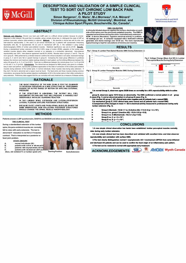
1.1.In normal Group A, observers agree 26/28 times on normality for a 93% reproducibility while in activeIn normal Group A, observers agree 26/28 times on normality for a 93% reproducibility while in active
ASAS
group B, observers agree 10/10 times on abnormality. The EMG confirmed a normal pattern in allgroup B, observers agree 10/10 times on abnormality. The EMG confirmed a normal pattern in all groupgroup
A cases (Fig. 1) and an abnormal pattern in all group B cases (Fig. 2).A cases (Fig. 1) and an abnormal pattern in all group B cases (Fig. 2).2.2.In inactive AS group C, 6/6 clinical tests were negative and all patients had a normal EMG.In inactive AS group C, 6/6 clinical tests were negative and all patients had a normal EMG.
3.3.In mechanical group D, 21/21 clinical tests were normal and all patients had a normal EMG.In mechanical group D, 21/21 clinical tests were normal and all patients had a normal EMG.
4.4.Comparison of the changes in mean +/- SE in electrical activity measured inComparison of the changes in mean +/- SE in electrical activity measured in μμvoltvolt /second during early /second during early
lumbar extension (Fig. 3).lumbar extension (Fig. 3).
▶ Group A (Normal): -18.0± 1.7 vs. B (Active AS): +7.5 ±1.8 (p < 2 x 10Group A (Normal): -18.0± 1.7 vs. B (Active AS): +7.5 ±1.8 (p < 2 x 10 -7-7););
▶ Group A vs. group C (Inactive AS): -15,9 ± 2.0 (p = 0.6);Group A vs. group C (Inactive AS): -15,9 ± 2.0 (p = 0.6);
▶ Group A vs. D (Mechanical): -18.2 ± 1,4 (p = 0.9);Group A vs. D (Mechanical): -18.2 ± 1,4 (p = 0.9);
▶ Group B vs. C: p < 2 x 10Group B vs. C: p < 2 x 10 -3-3;;
▶ Group B vs. D: p < 1 x 10Group B vs. D: p < 1 x 10 -8-8;;
▶ Group C vs. D: p = 0.6Group C vs. D: p = 0.6
ABSTRACT
EMG VALIDATIONA clinically blinded observer applies the surface electrodes on each
side of the spine over the previously palpated muscles. The EMG isrepeated several times during the same controlled early extensionROM. A device (Neurotrac) registers electrical activity in μvolt/sec. at16 readings per sec. A computer equipped with a dedicated softwaregenerates and stores tracings over 25-30 sec. The more reproducibletypical tracing is kept for calculations of minima and maxima.
Fig 3. Voltage Change (Mean And SE) In Lumbar Para-spinal Muscles During Early Extension
T T T
T* **
* = Significant difference (P < 0,05)1. THE BASIC PRINCIPLE OF THE MSK EXAM IS FOR THE EXAMINER
TO INCREASE PRESSURE IN A MSK STRUCTURE BY PERFORMINGPASSIVE OR ACTIVE RANGE OF MOTION OR APPLYING EXTERNALPRESSURE.
2. IF THE STRUCTURE IS ABNORMAL THE PATIENT WILL FEELDISCOMFORT OR PAIN AND THAT WILL PROVOKE A VARIABLE BUT
OBSERVABLE ANTALGIC GUARDING REACTION.
3. IN THE LUMBAR SPINE, EXTENSION AND LATERAL/POSTERIOR-LATERAL FLEXIONS EXPLORE POSTERIOR STRUCTURES.
4. BECAUSE FACET JOINTS AND PARA-SPINAL MUSCLES SHARE THESAME INNERVATION, ROM SOLLICITING POSTERIOR STRUCTURESSHOULD CHANGE THE SPINAL MUSCLE KINOPHYSIOLOGY.
Patients answer a LBP questionnaire, BASFAI and BASDAI and allow access to their medical files.
THE CLINICAL TEST
During a standardized extension of the lumbar
spine, the para-vertebral muscles are normally
felt to relax with early extension. The test is
abnormal if relaxation is not felt or if muscles
contract. That is interpreted as a posterior or
facet joint problem.
STUDY GROUPS
A. normal individuals (28);B. patients with active or advanced AS (10);C. patients with asymptomatic AS (6);D. patients with mechanical LBP (scol iosis,discs, old vertebral Fx or lumbar sprain (21). Starting Position Early Extension
RATIONALE
RESULTS
+
Fig 1. Group A Lumbar Para-Spinal Muscles EMG During Extension (↕)
-
Fig 2. Group B Lumbar Paraspinal Muscles EMG During Extension (↕)
Seconds
M i c
r o v o
l t
M i c
r o v o
l t
Seconds
METHODS
CONCLUSIONS
ACKNOWLEDGEMENTS Wyeth Canada


