
PRESENTED BY
DR .HESHAM ADEL EL-NOUBYUNDER SUPERVISION OF
PROF. DR. LATIFA ABD EL GAWAD
Antibacterial effects of laser
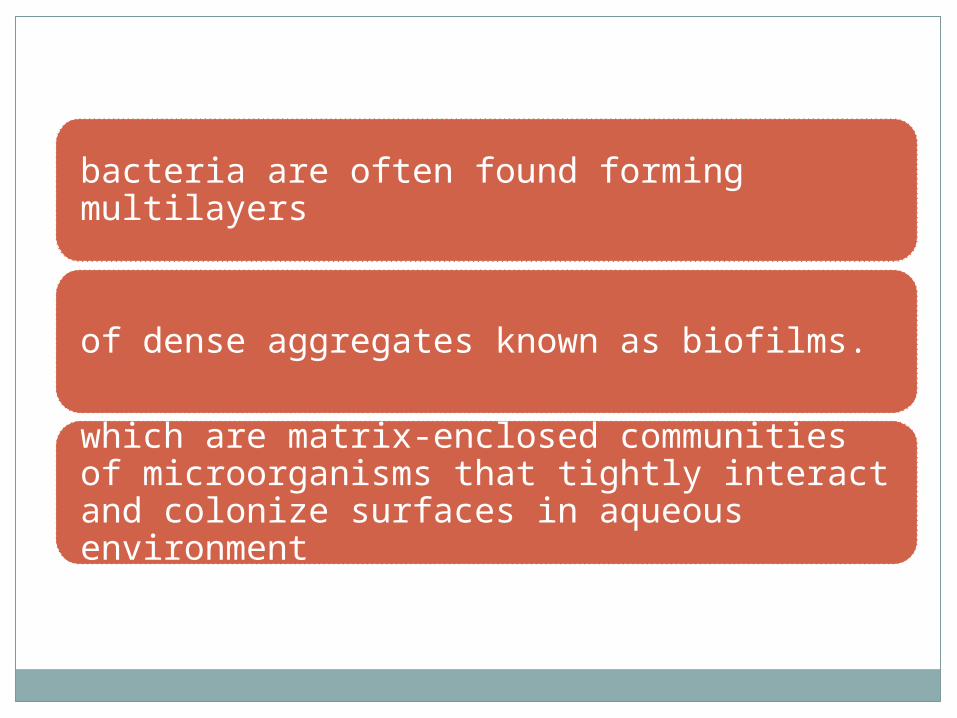
bacteria are often found forming multilayers
of dense aggregates known as biofilms.
which are matrix-enclosed communities of microorganisms that tightly interact and colonize surfaces in aqueous environment
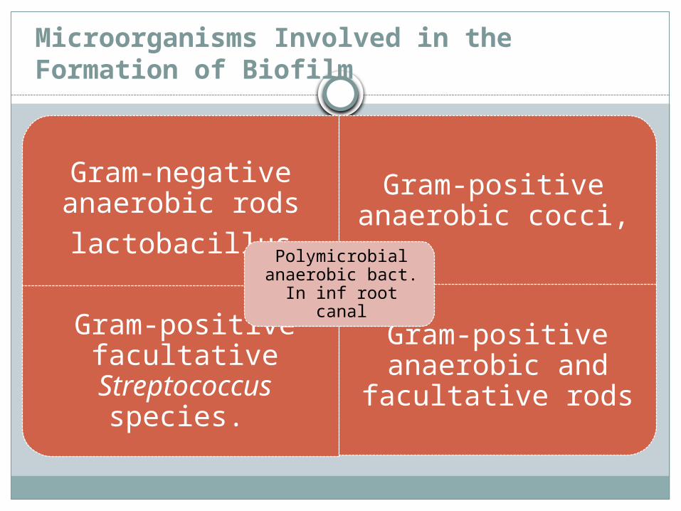
Microorganisms Involved in the Formation of Biofilm
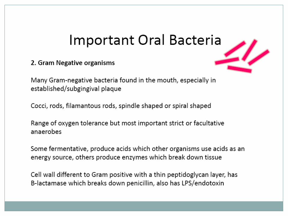
Gram-negative anaerobic rodslactobacillus
Gram-positive anaerobic cocci,
Gram-positive facultative
Streptococcus species.
Gram-positive anaerobic and
facultative rods
Polymicrobial anaerobic bact. In
inf root canal
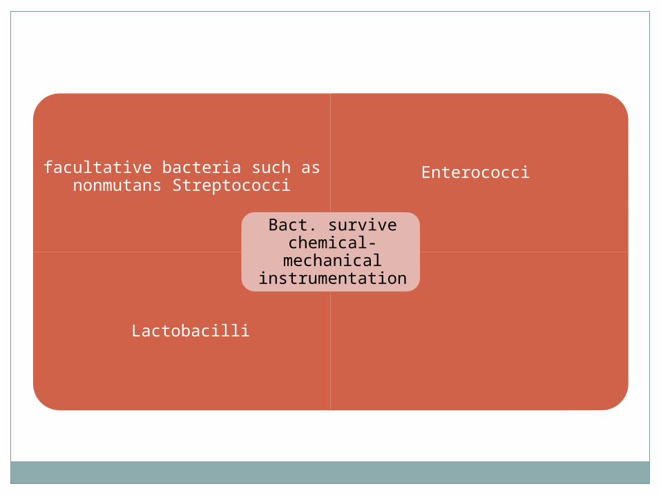
facultative bacteria such as nonmutans Streptococci
Enterococci
Lactobacilli
Bact. survive chemical-
mechanical instrumentation
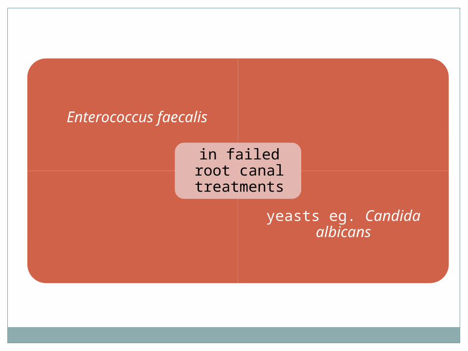
Enterococcus faecalis
yeasts eg. Candida albicans
in failed root canal
treatments
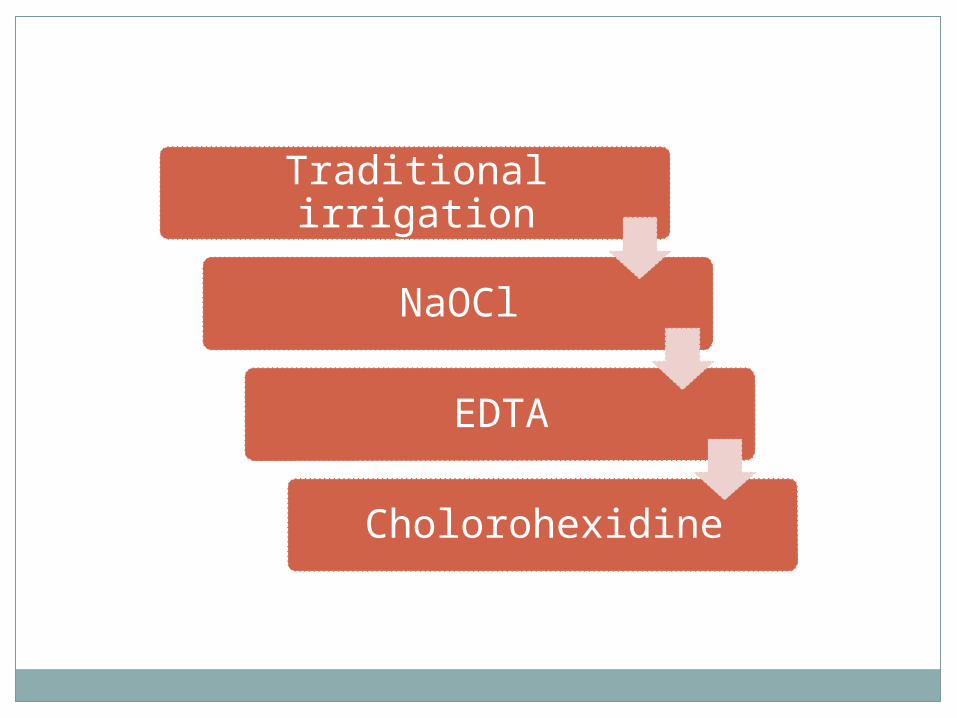
Traditional irrigation
NaOCl
EDTA
Cholorohexidine
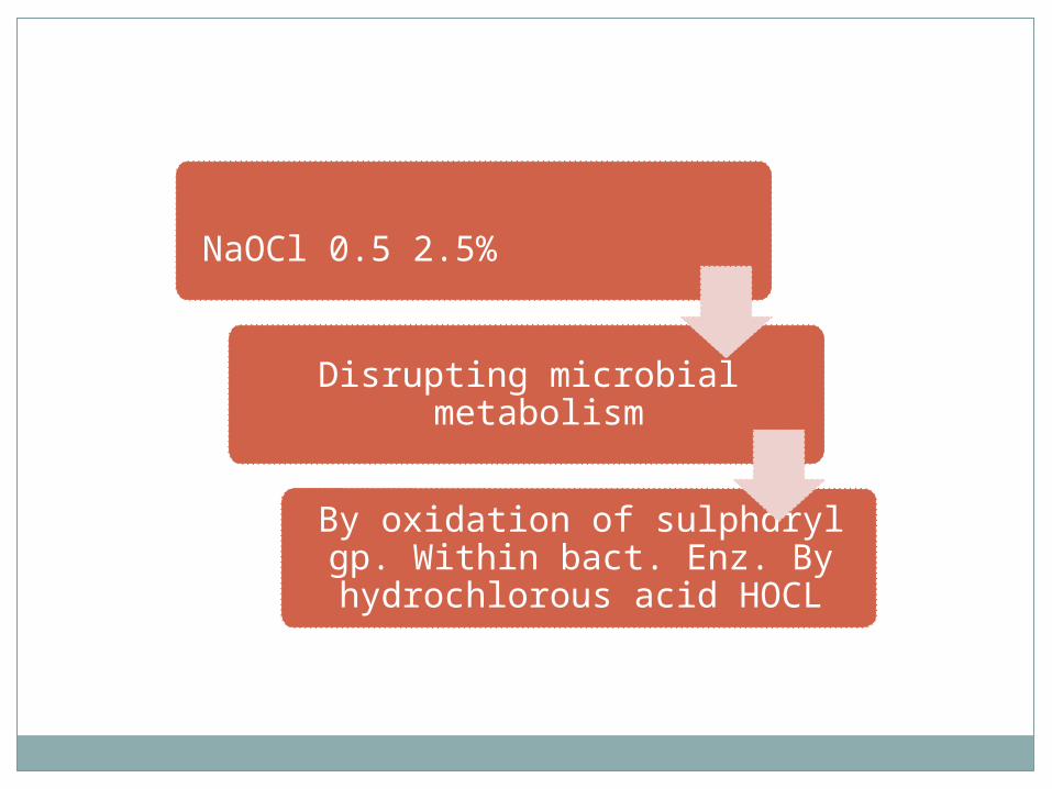
NaOCl 0.5 2.5%
Disrupting microbial metabolism
By oxidation of sulphdryl gp. Within
bact. Enz. By hydrochlorous acid
HOCL
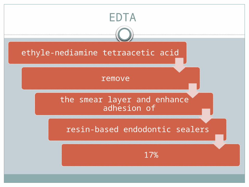
EDTA
ethyle-nediamine tetraacetic acid
remove
the smear layer and enhance adhesion of
resin-based endodontic sealers
17%
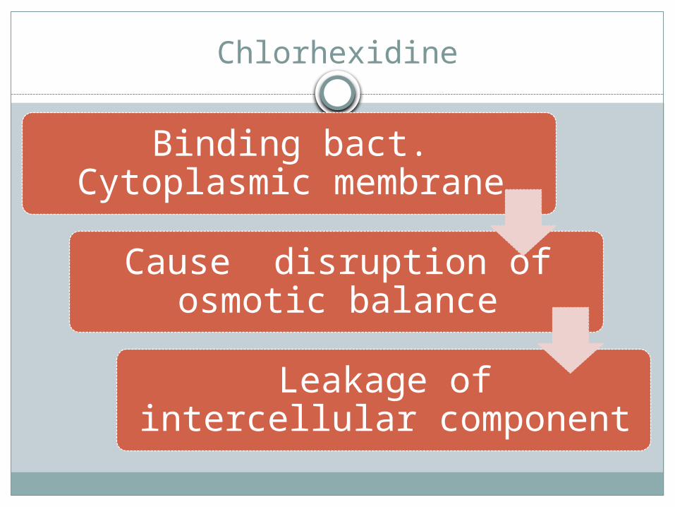
Chlorhexidine
Binding bact. Cytoplasmic membrane
Cause disruption of osmotic balance
Leakage of intercellular component
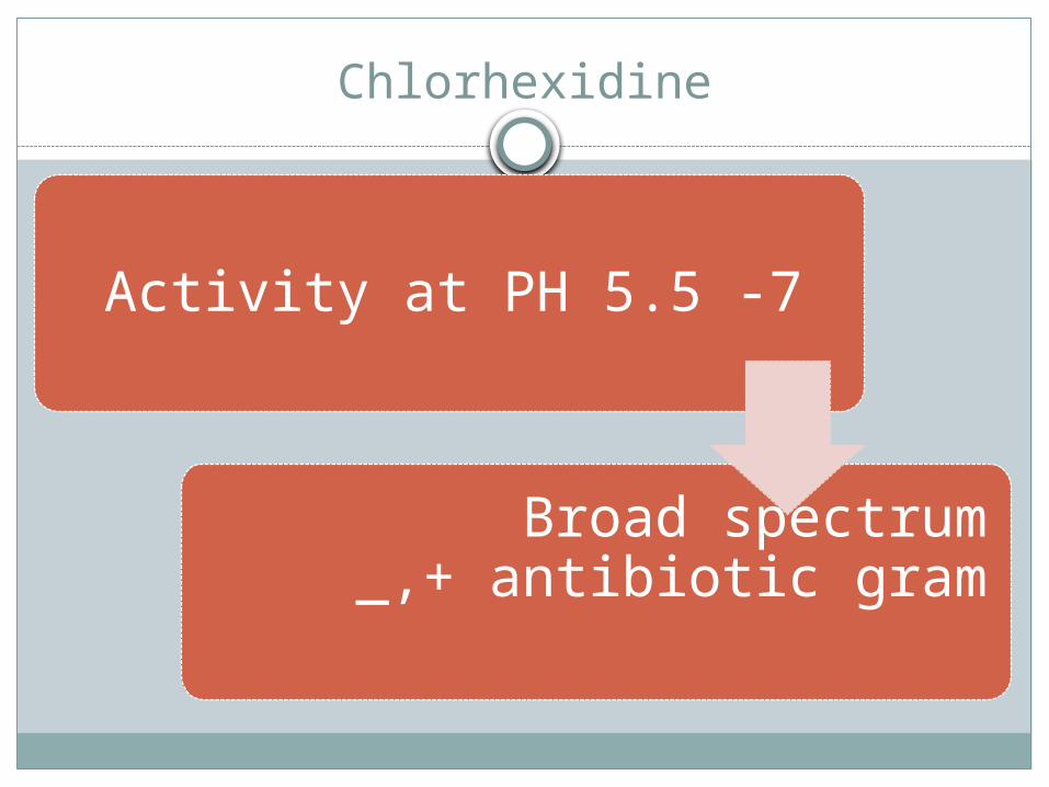
Chlorhexidine
Activity at PH 5.5 -7
Broad spectrum antibiotic gram
_,+
HIGH-POWER NEARIR LASER BACTERIAL KILLING IS
THE MOST IMPORTANT PARAMETER IS THE MAXIMUM TEMPERATURE. LASER IRRADIATION OF BACTERIA AT LOW TEMPERATURES DOES NOT RESULT IN KILLING .
Laser action
A laser is a photo-thermal device.
1. It acts directly on cellular structures.
2. Destroying cell walls.3 .Altering DNA .
4. Modifying metabolic processes .
5.Ungluing the polysaccharide structure of the biofilm.
Nd:YAG and Diode lasers have an antibacterial but not sterilizing capability, substantiating that laser irradiation is a possible supplement for disinfection but not an alternative
ND:YAG Bactericidal effect
To evaluate the bactericidal effects of Nd:YAG laser 3-W laser beam for 10 sec on biofilm of Enterococcus faecalis.
Based on the results of the present study, the effect of Nd:YAG laser beam on E . faecalisbiofilm is less than that of sodium hypochlorite solution. A combination of laser and sodium hypochlorite results in complete elimination of E. faecalis biofilm.
Bactericidal activity of pulsed Nd-YAG laser radiation
At 1994 in vitro for oral bacteria using several pulse energies and exposure duration
120-mJ laser pulses proved more efficient with 99.9% kills .
90% in 80-mJ pulses kills after exposure to
1800 pulses
Antimicrobial effects of 2.94 μm Er:YAG laser
After application of 75 laserpulses1.E. coli was reduced by the Er:YAG laser
radiation after exposure to 105 laser pulses to 5.5% of the initial count
2.In the Staph. aureus group to 15.1%.3.The number of bacteria in case of A.
actinomytemcomitans was reduced to 8.3%,
4.In E. corrodens to 3.0% 5.In case of Peptostreptococcus micros to
22%
Besides the selective removal of plaque and calculus, the 2.94 μm Er:YAG laser radiation causes reduction in bacteria on root surfaces.
AT 1.5 W, THE BEST RESULTS WERE OBTAINED BY THE ER:YAG LASER ACHIEVING A MEAN BACTERIAL ELIMINATION OF 99.64%,FOLLOWED BY THE ND:YAG LASER (99.16%)
THE HO:YAG LASER(99.05%. )
The Bactericidal Effect of Nd:YAG, Ho:YAG, and Er:YAG Laser Irradiation in the Root Canal: An in Vitro Comparison 1999.
Diode effects on Streptococcus sanguis in biofilm J. Antimicrob. Chemother
BiofUms were grown on hydroxyapatite, irradiated with up to 12.2 J of light from a gallium aluminium arsenide laser in the presence of aluminium disulphonated phthalocyanine
(AlPcS2) and survivors enumerated.
No significant decrease in the viable count was found when either the AlPcS2 or the laser light was used alone.
No viable streptococci were detectable following irradiation with 12.2 J of laser light
Co2 bactericidal effect
The energy density required to kill greater than 99.5% of the bacteria is less than 200 J/cm2
The effects of super pulsed CO2 laser irradiation on periodontopathic bacteria
The effects of super pulsed CO2 laser irradiation on 1 . periodontopathic bacteria .
2 .lipopolysaccharide (LPS). The irradiation at low energy densities of 7.5
and 12.5 J/cm2 killed more than 99.9 and 99.999% of Porphyromonas gingivalis .
More than 99% of Actinobacillus actinomycetemcomitans was sterilized by the irradiation at 7.5 J/cm2.
Super pulsed CO2 on LPS
LPS biological activity was significantly decreased by laser irradiation at energy densities of more than 7.5 J/cm2 , and the components of LPS analyzed by SDS-PAGE was diminished non-specifically.
CO2
The results indicate that CO2 laser irradiation at low power is capable of bactericidal effect on periodontopathic bacteria and decreasing LPS activity.
CO2 laser (1.064nm, 1.5W, 100mJ, 15Hzsec) showed a higher antibacterial efficacy
against E. faecalis as compared to the Nd:YAG laser
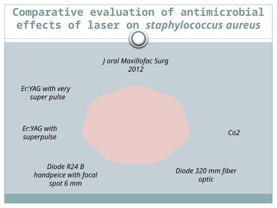
Comparative evaluation of antimicrobial effects of laser on staphylococcus aureus
J oral Maxillofac Surg 2012
Co2
Diode 320 mm fiber optic
Diode R24 B handpeice with focal spot 6 mm
Er:YAG with superpulse
Er:YAG with very super pulse
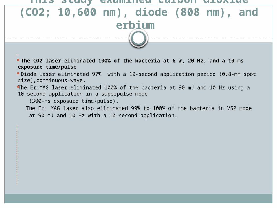
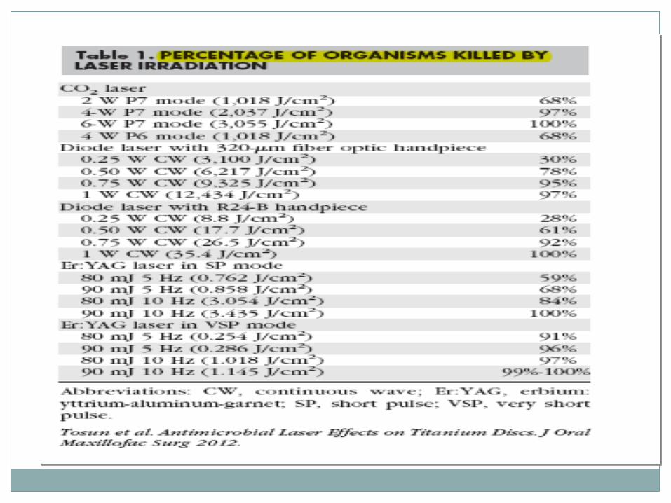
This study examined carbon dioxide (CO2; 10,600 nm), diode (808 nm), and
erbium
The CO2 laser eliminated 100% of the bacteria at 6 W, 20 Hz, and a 10-ms exposure time/pulse
Diode laser eliminated 97% with a 10-second application period (0.8-mm spot size),continuous-wave.
The Er:YAG laser eliminated 100% of the bacteria at 90 mJ and 10 Hz using a 10-second application in a superpulse mode
(300-ms exposure time/pulse). The Er: YAG laser also eliminated 99% to 100% of the bacteria in VSP mode at 90 mJ and 10 Hz with a 10-second application.
Application of photodynamic therapy in dentistry
Antimicrobial PDT (aPDT) non thermal light induced inactivation of cells,microorganism .
Can be considered as an adjunctive to conventional mechanical therapy.
Antimicrobial PDT not only kills the bacteria, but may also lead to the detoxification of endotoxins such as lipopolysaccharide. These lipopolysaccharides treated by PDT do not stimulate the production of pro-inflammatory cytokines by mononuclear cells.
light source
helium – neon lasers (633 nm) visible red
gallium – aluminum – arsenide diode lasers (630-690, 830 or 906 nm) IR
argon laser (488-514 nm)Visible blue
PS
Methylene blue and toluidine blue O .
very effective photosensitizing agents for the inactivation of both gram-positive and gram-negative
PDT
P. i
nter
me
dia,
P.gingivalis
Ent
er
ococcus
f
aecali
s
Fusobacteri
um
PDT could reduce the bacterial count
of
PDT
It has been demonstrated that bacteria associated with periodontal disease can be killed through
photosensitization with toulidine blue O
By irradiating with helium – neon soft laser. Data from an in vitro study indicated that PDT
could kill bacteria organized in a biofilm.In an animal study, bleeding on probing, and
porphyromonas gingivalis levels.
PDT
The optimal concentration of toluidine blue O to kill P. gingivalis was 12.5 μg/ml with helium-neon laser irradiations.
This was caused by the disruption of outer membrane proteins of these bacteria
PDT
Methylene blue at the wavelength of 632.8 nm & helium-neon laser 665 & 830 nm (diode laser) has a high bactericidal effect.
Methylene blue served as the photosensitizer and was used as a mouth rinse
PDT
PDT on endodontic pathogens in planktonic phase as well as on Enterococcus faecalis biofilms in experimentally infected root canals of extracted teeth. Strains of microorganisms were sensitized with methylene blue (25 μg/ml) for five minutes followed by exposure to red light of 665 nm with an energy florescence of 30 J/cm2.
Methylene blue fully eliminated all bacterial species with the exception of E. faecalis (53% killed). The same concentration of methylene blue in combination with red light (222 J/cm2) was able to eliminate 97% of E. faecalis biofilm bacteria in root canals using an optical fiber with multiple cylindrical diffusers that uniformly distributed light at 360
PDT
Recently, non laser light source, such as light – emitting diodes (LED), has been used as new light activators in PDT.
LED devices are more 1 compact
2 portable 3 cost effective compared to traditional lasers.
Effect of Low-Level Laser Therapy onTypical Oral Microbial Biofilms
LLLT had an inhibitory effect on typical oral microbialbiofilms, and this capacity can be altered according to the interactions between different species Streptococcus mutans, Candida albicans or an
association of both species. Single and dual-species biofilms - SSB and DSB – were exposed to laser doses of 5, 10 or 20 J/cm2 from a near infrared InGaAsP diode laser prototype 780 ± 3 nm, 0.04 W
LLLT
Figure 1 illustrates the effects of LLLT on S mutans The irradiation promoted a decrease in the number of microorganisms, though without significant difference among the laser doses or the types of biofilm.
LLLT
Figure 1. SEM micrograph showing the morphology and structure of SSB of S. mutans (Original magnification ×5000). A= Control,
B-D= Biofilm after irradiation with laser dose of 5, 10 and 20 J/cm2.
Antibacterial effect of nanoparticles on Staphylococcu
Previous studies have reportedsignificant antibacterial effect of1. Chitosan nanoparticles (CS-Np).2. Zinc oxide nanoparticles (ZnO-Np) against
planktonic Enterococcus faecalis.the CS-Np and ZnO-Np reduce and disrupt the biofilm structure.
3. Silver nanoparticles (AgNPs).
Thank You
أسنان. طب ماجستير النوبي عادل هشام - دN.I.L.E 2015 2015ليزر


