
OU Neurology
APHASIA
David Lee Gordon, M.D., FAAN, FANA, FAHA
Professor and Chair
Department of Neurology
The University of Oklahoma Health Sciences Center
OU Neurology
APHASIALEARNING OBJECTIVES
Define aphasia & list the 4 components of a language exam
Differentiate the 7 aphasia types based on exam findings
Localize a lesion based on aphasia type and associated signs
OU Neurology
APHASIA: DEFINITION
Language abnormality due to brain dysfunction
Involves both spoken & written language
Although “dysphasia” is technically a more correct term for a partial language deficit, most neurologists only use the term “aphasia” and categorize aphasias as mild, moderate, or severe

OU Neurology
APHASIA: FACTS
Due to focal dysfunction in “dominant” hemisphere (by definition, dominant hemisphere controls language)
➢Handedness suggests dominant hemisphere
➢Most people are LEFT brain dominant
▪ R-handed 99% / L-handed 75%
➢ Some people have “mixed dominance”
Clinical features vary based on lesion location
Dysnomia (difficulty naming) is the only common feature among all 7 types of aphasias
In recovery, 1° language returns first

OU Neurology
THE ANATOMY OF NORMAL LANGUAGE: RECEPTION Wernicke area in posterior aspect of the superior
temporal gyrus “receives” language and distributes information to temporal and parietal lobes for comprehension
Wernicke area (W) is near auditory cortex
Receptive (= posterior = sensory) aphasias are often associated with visual field & sensory deficits
FRONTALPARIETAL
TEMPORAL
BW
Ear

OU Neurology
THE ANATOMY OF NORMAL LANGUAGE: EXPRESSION Spontaneous language is created in the frontal lobe
and is “expressed” via the Broca area in the posterior aspect of the inferior frontal gyrus
FRONTALPARIETAL
TEMPORAL
BW
Mouth
Broca area (B) is near facial motor cortex
Expressive (= anterior = motor) aphasias are often associated with motor deficits

OU Neurology
THE ANATOMY OF NORMAL LANGUAGE: REPETITION To “repeat,” language enters Wernicke area,
travels along arcuate fasciculus in perisylvian area, and exits via Broca area (“the repetition loop”)
“Repetition loop”: Wernicke arcuate fasciculus Broca
FRONTALPARIETAL
TEMPORAL
BW
Mouth
Ear
arcuatefasciculus
OU Neurology
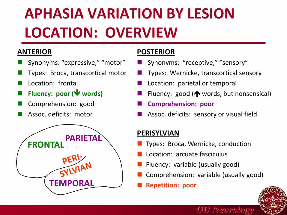
APHASIA VARIATION BY LESION LOCATION: OVERVIEW
ANTERIOR
Synonyms: “expressive,” “motor”
Types: Broca, transcortical motor
Location: frontal
Fluency: poor ( words)
Comprehension: good
Assoc. deficits: motor
POSTERIOR
Synonyms: “receptive,” ”sensory”
Types: Wernicke, transcortical sensory
Location: parietal or temporal
Fluency: good ( words, but nonsensical)
Comprehension: poor
Assoc. deficits: sensory or visual field
PERISYLVIAN
Types: Broca, Wernicke, conduction
Location: arcuate fasciculus
Fluency: variable (usually good)
Comprehension: variable (usually good)
Repetition: poor
FRONTALPARIETAL
TEMPORAL
OU Neurology
APHASIA: CLINICAL FEATURES
Dysnomia / anomia
Nonfluent speech
Fluent speech
Auditory comprehension impairment
Repetition impairment
Jargon aphasia
Reading & writing difficulty
OU Neurology
APHASIA CLINICAL FEATURES:DYSNOMIA / ANOMIA Difficulty naming or finding words
➢ Impaired retrieval of target words
➢Occurs in all aphasias
➢Non-localizing: occurs with lesions anywhere in dominant hemisphere
➢ Isolated dysnomia may occur as result of incomplete resolution of any aphasia
Hesitations
Circumlocution
➢ “Talking around” difficult-to-retrieve words
➢Definition or description instead of target word
OU Neurology
APHASIA CLINICAL FEATURES:NONFLUENT SPEECH Rate, quantity, ease of speech production
➢Verbal output decreased (< 50 words/min)
➢Phrase length short (1-4 words)
➢Production effortful
➢Articulation often poor
➢Prosody (melodic contour) disturbed
➢Telegraphic speech: Preferential use of nouns & verbs without small connecting words
Frontal language areas damaged
OU Neurology
APHASIA CLINICAL FEATURES:FLUENT SPEECH Rate, quantity, ease of speech production normal
➢Verbal output normal or increased
➢Phrase length normal (> 5 words)
➢Production easy
➢Articulation usually normal
➢Prosody (melodic contour) normal
➢May be nonsensical
Frontal language centers intact

OU Neurology
APHASIA CLINICAL FEATURES:AUDITORY COMPREHENSION IMPAIRMENT
Ranges from complete lack of understanding to subtle failure to extract full meaning of complex sentences
Informal conversation may be misleading
➢Clues from gestures, tones, setting
➢Automatic (previously stored) words & phrases
Formal testing without nonverbal clues necessary
Temporoparietal language areas damaged
OU Neurology
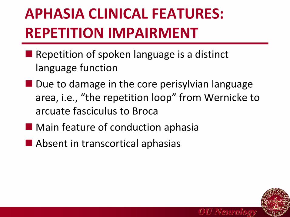
APHASIA CLINICAL FEATURES:REPETITION IMPAIRMENT Repetition of spoken language is a distinct
language function
Due to damage in the core perisylvian language area, i.e., “the repetition loop” from Wernicke to arcuate fasciculus to Broca
Main feature of conduction aphasia
Absent in transcortical aphasias

OU Neurology
APHASIA CLINICAL FEATURES:JARGON APHASIA (NONSENSICAL SPEECH)
Paraphasic errors
➢ Substitution of incorrect words for intended words; 2 types
➢Verbal (semantic) paraphasia▪ Real word similar in meaning to intended word
▪ Lesion often frontal, associated with expressive aphasia
➢ Literal (phonemic) paraphasia▪ Real or made-up word similar in sound to intended word
▪ Lesion often temporoparietal, associated with receptive aphasia
Neologisms (“new words”)
➢Made-up words unrelated to intended words
➢ Lesion temporoparietal, associated with receptive aphasia

OU Neurology
APHASIA CLINICAL FEATURES:READING & WRITING DIFFICULTY Difficulty reading = alexia
Difficulty writing = agraphia
Usually, alexia & agraphia parallel oral deficits
Both may occur in isolation due to damage to perisylvian area plus other areas
➢Alexia: occipital & inferior parietal
➢Agraphia: frontal & inferior parietal
OU Neurology
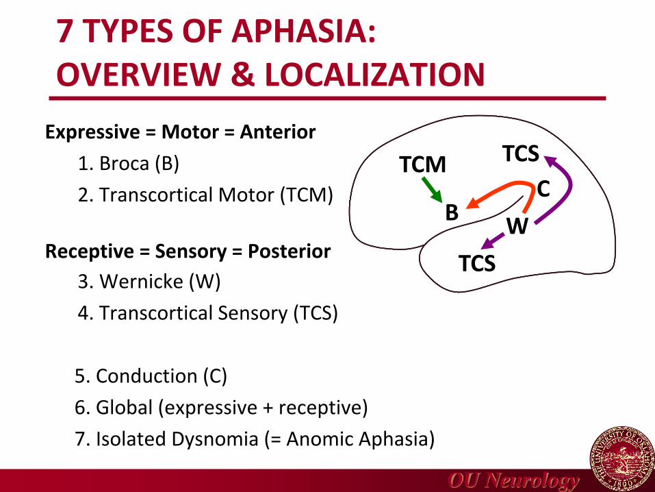
7 TYPES OF APHASIA:OVERVIEW & LOCALIZATION
Expressive = Motor = Anterior
1. Broca (B)
2. Transcortical Motor (TCM)
Receptive = Sensory = Posterior
3. Wernicke (W)
4. Transcortical Sensory (TCS)
5. Conduction (C)
6. Global (expressive + receptive)
7. Isolated Dysnomia (= Anomic Aphasia)
TCM TCS
TCS
BW
C

OU Neurology
LANGUAGE EXAM COMPONENTSTO DETERMINE APHASIA TYPE, LOCALIZE LESION
These 4 items combined take less than 1 minute to perform
Naming
Fluency
Commands
Repetition
Perform all 4 items if patient has dysnomia on “essential exam.”
Avoid giving nonverbal clues when testing aphasia pts, but
use nonverbal clues when communicating with aphasia pts
OU Neurology
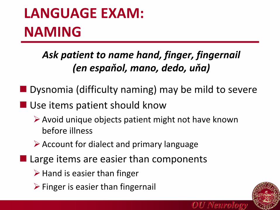
LANGUAGE EXAM: NAMING
Ask patient to name hand, finger, fingernail(en espaňol, mano, dedo, uňa)
Dysnomia (difficulty naming) may be mild to severe
Use items patient should know
➢Avoid unique objects patient might not have known before illness
➢Account for dialect and primary language
Large items are easier than components
➢Hand is easier than finger
➢ Finger is easier than fingernail
OU Neurology
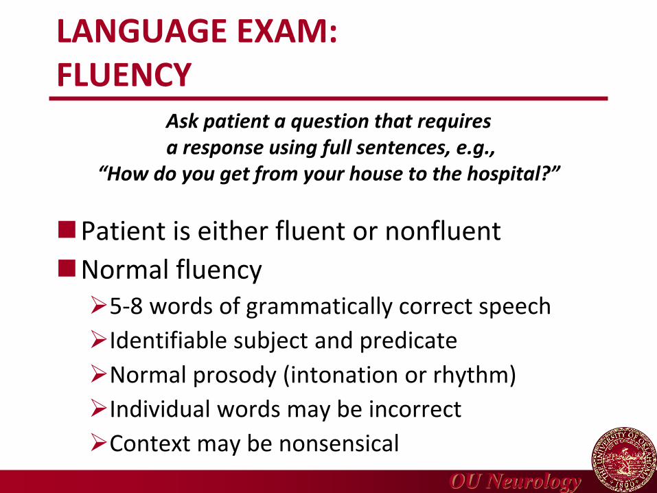
LANGUAGE EXAM:FLUENCY
Ask patient a question that requiresa response using full sentences, e.g.,
“How do you get from your house to the hospital?”
Patient is either fluent or nonfluent
Normal fluency➢5-8 words of grammatically correct speech
➢Identifiable subject and predicate
➢Normal prosody (intonation or rhythm)
➢Individual words may be incorrect
➢Context may be nonsensical

OU Neurology
LANGUAGE EXAM: COMMANDS
Ask patient a 3-step command across the midline, e.g.,“With your left hand, touch your right shoulder, then
point to the ceiling, then close your eyes.”
Deficit may be mild to severe
Number of steps➢1-step easier than 2-step easier than 3-step
Relationship to midline➢ “Midline” easier than “to midline” easier than
“crossing midline”
Easiest command: 1-step, midline
Most difficult command: 3-step, crossing midline
OU Neurology
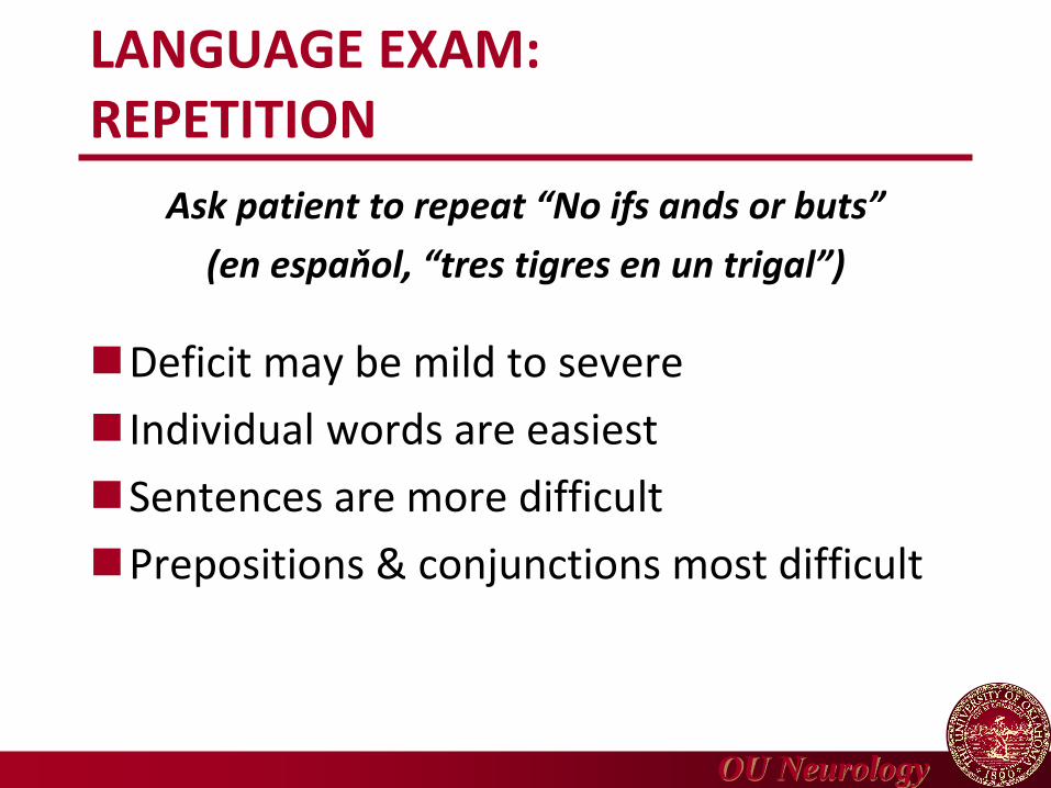
LANGUAGE EXAM: REPETITION
Ask patient to repeat “No ifs ands or buts”
(en espaňol, “tres tigres en un trigal”)
Deficit may be mild to severe
Individual words are easiest
Sentences are more difficult
Prepositions & conjunctions most difficult

OU Neurology
7 TYPES OF APHASIA:OVERVIEW & LOCALIZATION
Expressive = Motor = Anterior
1. Broca (B)
2. Transcortical Motor (TCM)
Receptive = Sensory = Posterior
3. Wernicke (W)
4. Transcortical Sensory (TCS)
5. Conduction (C)
6. Global (expressive + receptive)
7. Isolated Dysnomia (= Anomic Aphasia)
TCM TCS
TCS
BW
C

OU Neurology
EXPRESSIVE APHASIASDIAGNOSIS BASED ON LANGUAGE EXAM
TCM B C W TCS
NAMING — — — — —
FLUENCY — — + + +
COMMANDS + + + — —
REPETITION + — — — +
EXPRESSIVE
Both expressive aphasias are nonfluent with normal comprehension.Broca cannot repeat—transcortical motor can.
+ normal— abnormal
TCM TCS
TCS
BW
C

OU Neurology
RECEPTIVE APHASIASDIAGNOSIS BASED ON LANGUAGE EXAM
TCM B C W TCS
NAMING — — — — —
FLUENCY — — + + +
COMMANDS + + + — —
REPETITION + — — — +
RECEPTIVE
Both receptive aphasias are fluent with abnormal comprehension.Wernicke cannot repeat—transcortical sensory can.
+ normal— abnormal
TCM TCS
TCS
BW
C

OU Neurology
CONDUCTION APHASIA PLUSDIAGNOSIS BASED ON LANGUAGE EXAM
TCM B C W TCS
NAMING — — — — —
FLUENCY — — + + +
COMMANDS + + + — —
REPETITION + — — — +
ARCUATEFASCICULUS
Aphasias on “repetition loop” cannot repeat—conduction, Broca, Wernicke.
+ normal— abnormal
TCM TCS
TCS
BW
C
OU Neurology
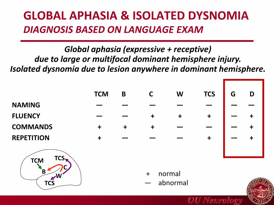
GLOBAL APHASIA & ISOLATED DYSNOMIADIAGNOSIS BASED ON LANGUAGE EXAM
TCM B C W TCS
NAMING — — — — —
FLUENCY — — + + +
COMMANDS + + + — —
REPETITION + — — — +
G D
— —
— +
— +
— +
Global aphasia (expressive + receptive)due to large or multifocal dominant hemisphere injury.
Isolated dysnomia due to lesion anywhere in dominant hemisphere.
+ normal— abnormal
TCM TCS
TCS
BW
C
OU Neurology
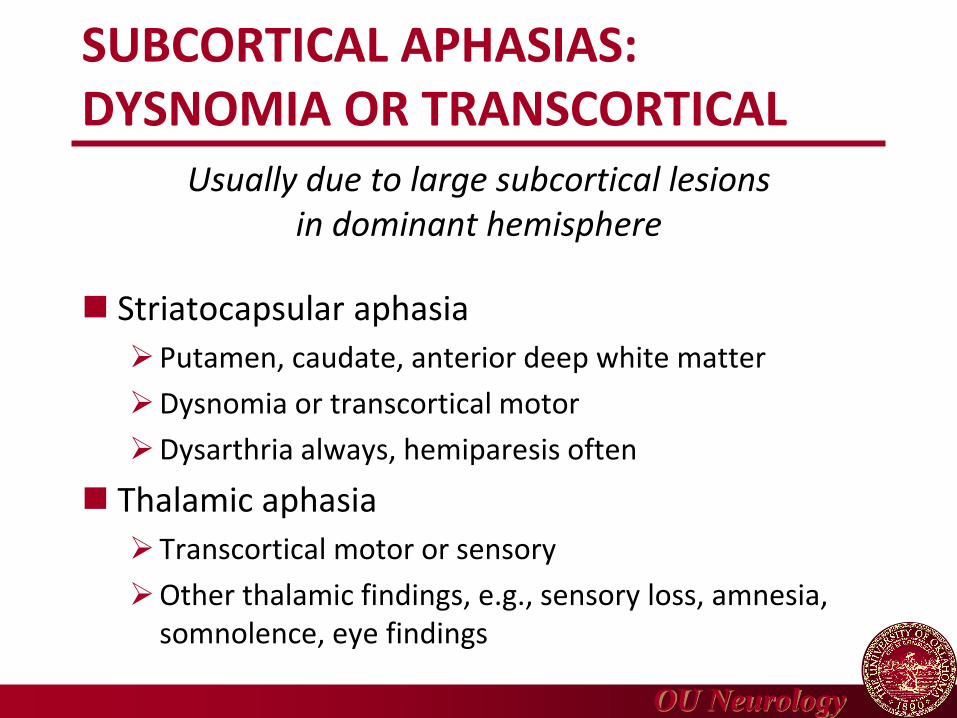
SUBCORTICAL APHASIAS:DYSNOMIA OR TRANSCORTICAL
Usually due to large subcortical lesionsin dominant hemisphere
Striatocapsular aphasia
➢Putamen, caudate, anterior deep white matter
➢Dysnomia or transcortical motor
➢Dysarthria always, hemiparesis often
Thalamic aphasia
➢Transcortical motor or sensory
➢Other thalamic findings, e.g., sensory loss, amnesia, somnolence, eye findings
OU Neurology
ETIOLOGIES OF APHASIAS
Stroke – ischemia or hemorrhage➢Perisylvian language zone supplied by MCA
➢Classic syndromes usually due to ischemic stroke
➢ Large subcortical hemorrhages can cause aphasia
Mass lesions (tumor, abscess)
Primary progressive aphasia➢ Focal degenerative disease with slow progression
➢ Form of frontotemporal dementia
Diffuse lesions➢Traumatic brain injury or Alzheimer Disease
➢Common causes of aphasia—but not in isolation
TIA, migraine, or seizure (transient aphasia)
OU Neurology
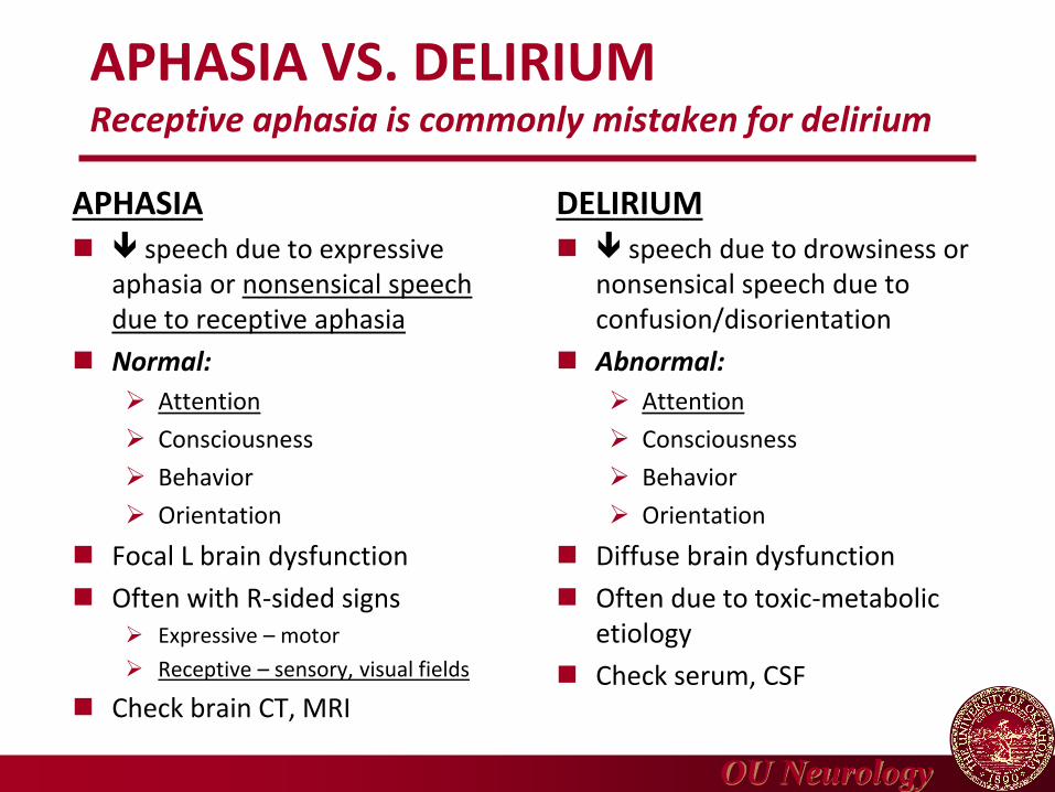
APHASIA VS. DELIRIUMReceptive aphasia is commonly mistaken for delirium
APHASIA speech due to expressive
aphasia or nonsensical speech due to receptive aphasia
Normal:
➢ Attention
➢ Consciousness
➢ Behavior
➢ Orientation
Focal L brain dysfunction
Often with R-sided signs➢ Expressive – motor
➢ Receptive – sensory, visual fields
Check brain CT, MRI
DELIRIUM speech due to drowsiness or
nonsensical speech due to confusion/disorientation
Abnormal:
➢ Attention
➢ Consciousness
➢ Behavior
➢ Orientation
Diffuse brain dysfunction
Often due to toxic-metabolic etiology
Check serum, CSF

OU Neurology
APHASIALEARNING OBJECTIVES
Define aphasia & list the 4 components of a language exam
Differentiate the 7 aphasia types based on exam findings
Localize a lesion based on aphasia type and associated signs
OU Neurology
THE END


