
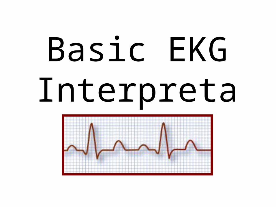
Basic EKG Interpretati
on
What is an EKG?
Also called an ECG, it is an electrocardiogram
Recording of the heart's electrical activityA series of "waves" that represent the
electrical impulsesCan be used to monitor patients for
extended periods of timeCan be used to test for heart conditions
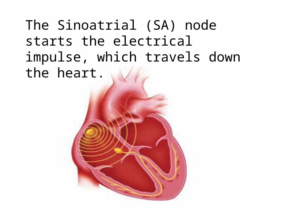
The Sinoatrial (SA) node starts the electrical impulse, which travels down the heart.
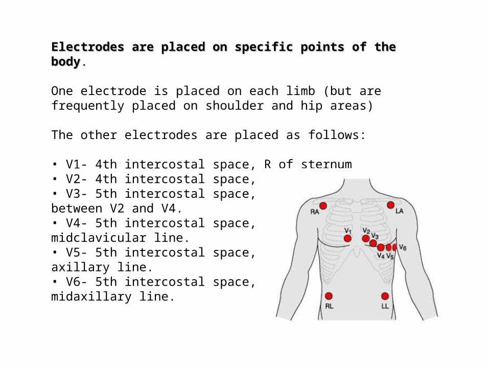
Electrodes are placed on specific points of the bodyElectrodes are placed on specific points of the body..
One electrode is placed on each limb (but are frequently placed on shoulder and hip areas)
The other electrodes are placed as follows:
• V1- 4th intercostal space, R of sternum• V2- 4th intercostal space, L of sternum• V3- 5th intercostal space, halfwaybetween V2 and V4.• V4- 5th intercostal space, Leftmidclavicular line.• V5- 5th intercostal space, L anterioraxillary line.• V6- 5th intercostal space, Lmidaxillary line.
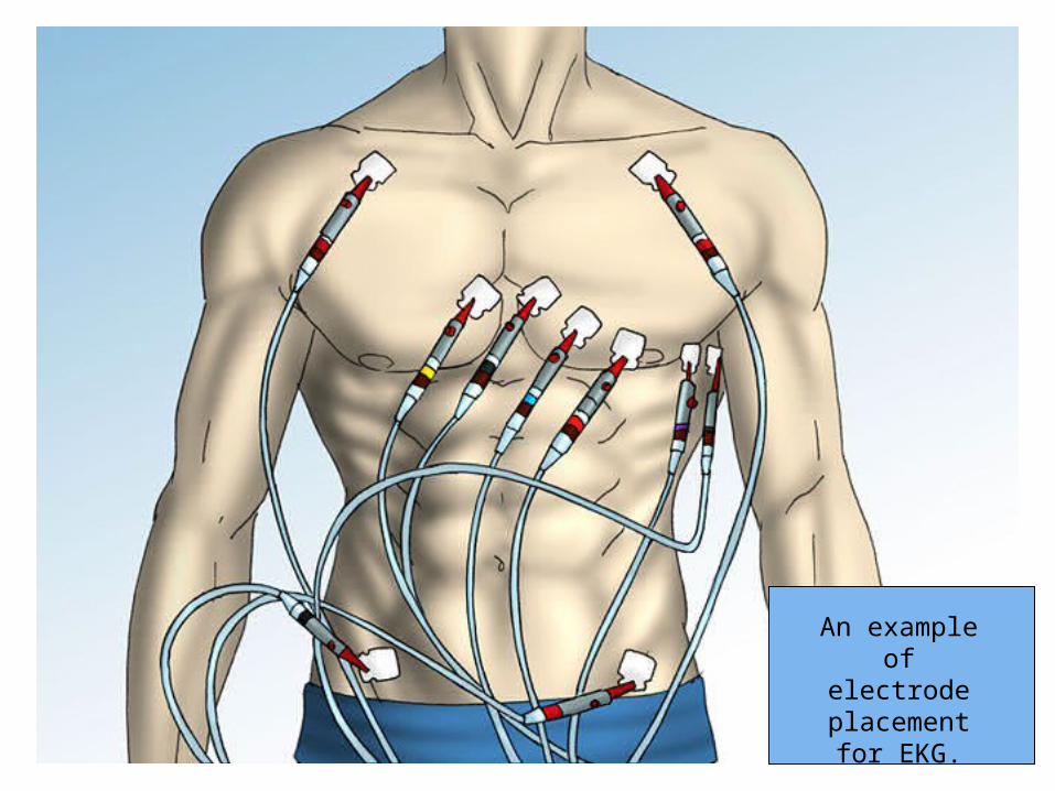
An example of electrode
placement for EKG.
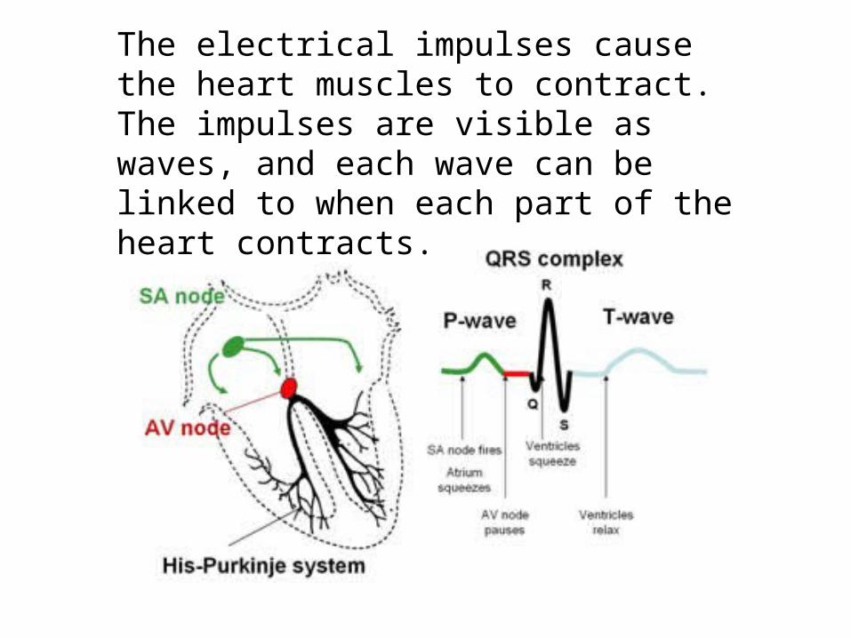
The electrical impulses cause the heart muscles to contract. The impulses are visible as waves, and each wave can be linked to when each part of the heart contracts.
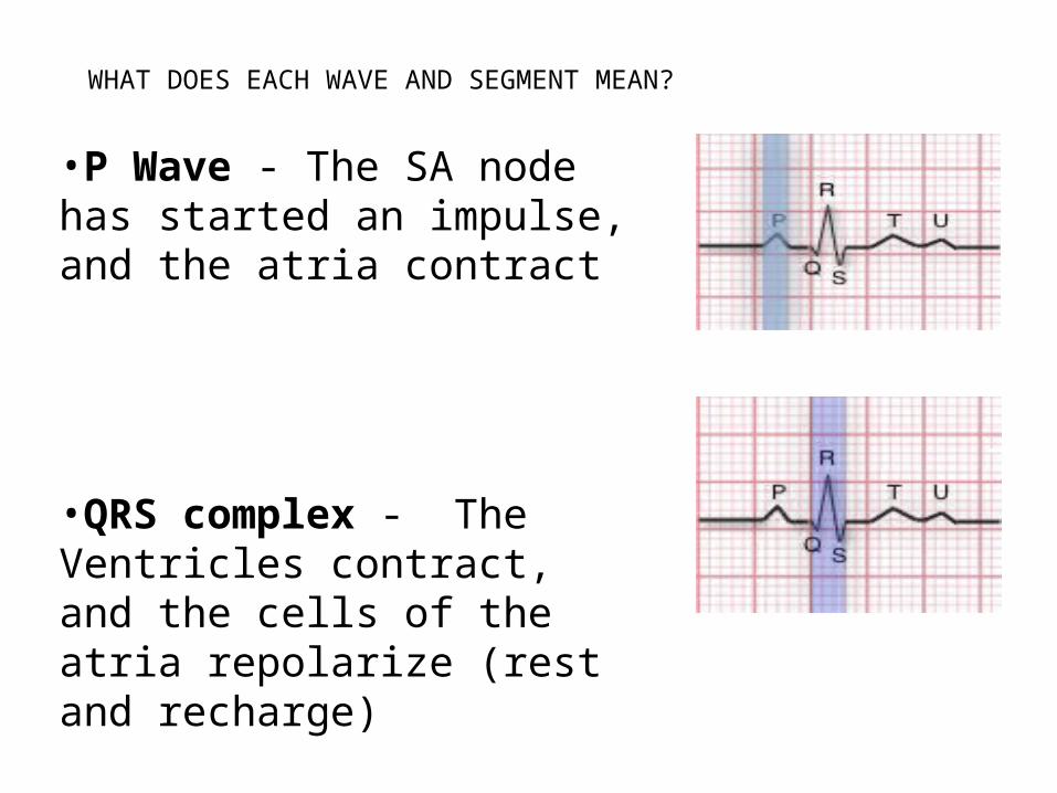
WHAT DOES EACH WAVE AND SEGMENT MEAN?
•P Wave - The SA node has started an impulse, and the atria contract
•QRS complex - The Ventricles contract, and the cells of the atria repolarize (rest and recharge)
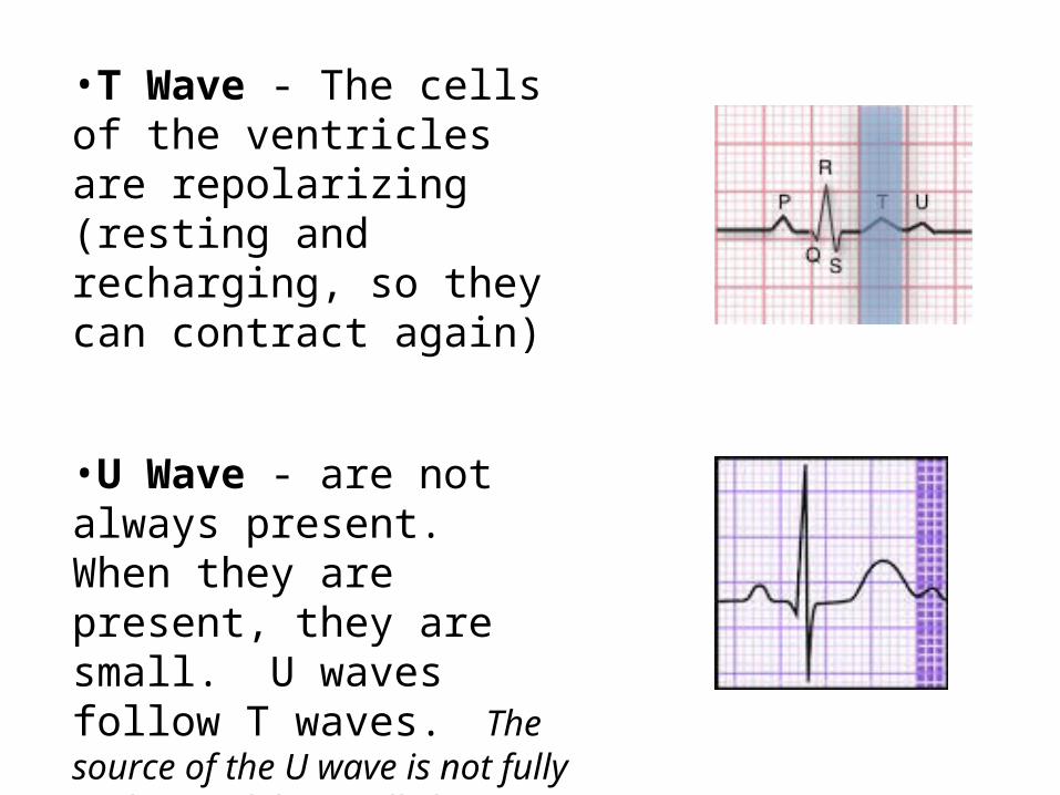
•T Wave - The cells of the ventricles are repolarizing (resting and recharging, so they can contract again)
•U Wave - are not always present. When they are present, they are small. U waves follow T waves. The source of the U wave is not fully understood, but really large U waves, or inverted (upside down) U waves can indicate problems.
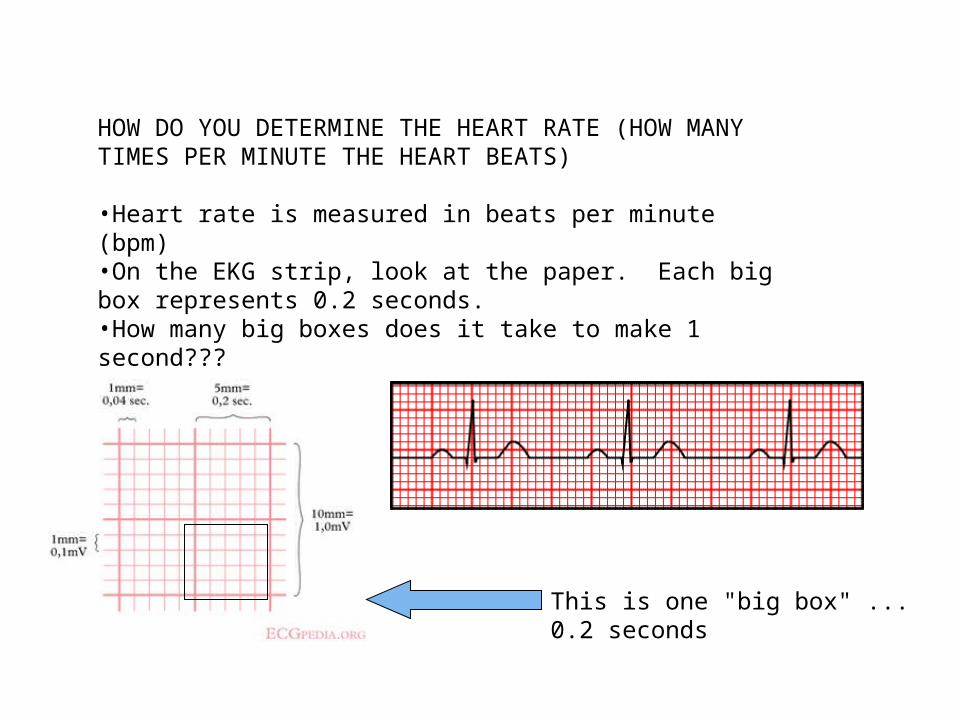
HOW DO YOU DETERMINE THE HEART RATE (HOW MANY TIMES PER MINUTE THE HEART BEATS)
•Heart rate is measured in beats per minute (bpm)•On the EKG strip, look at the paper. Each big box represents 0.2 seconds. •How many big boxes does it take to make 1 second???
This is one "big box" ... 0.2 seconds
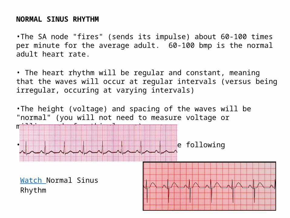
NORMAL SINUS RHYTHM
•The SA node "fires" (sends its impulse) about 60-100 times per minute for the average adult. 60-100 bmp is the normal adult heart rate.
• The heart rhythm will be regular and constant, meaning that the waves will occur at regular intervals (versus being irregular, occuring at varying intervals)
•The height (voltage) and spacing of the waves will be "normal" (you will not need to measure voltage or milliseconds for this lesson).
•Sinus Rhythm can look like any of the following
Watch Normal Sinus Rhythm
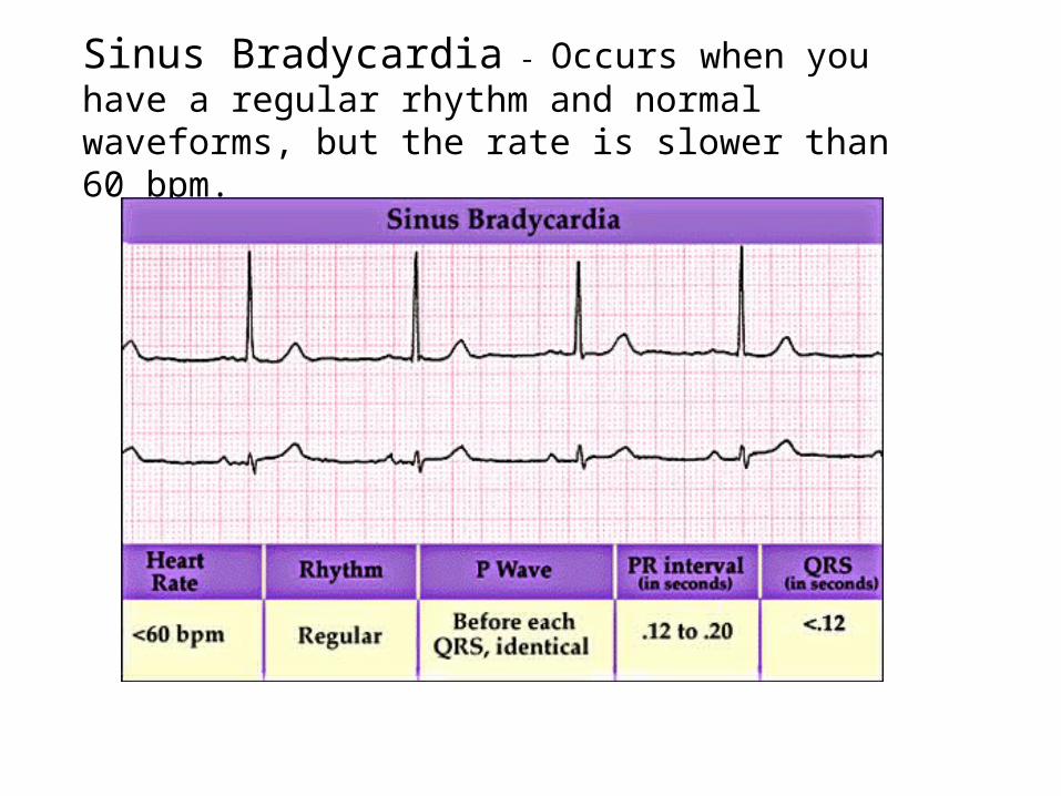
Sinus Bradycardia - Occurs when you have a regular rhythm and normal waveforms, but the rate is slower than 60 bpm.
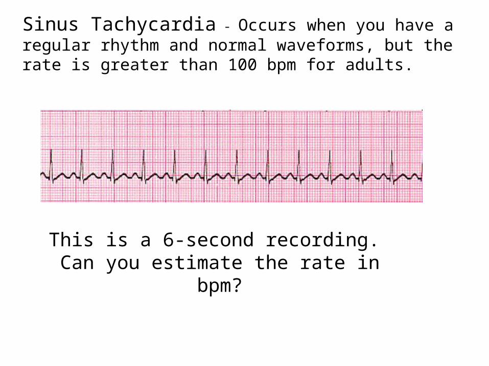
Sinus Tachycardia - Occurs when you have a regular rhythm and normal waveforms, but the rate is greater than 100 bpm for adults.
This is a 6-second recording. Can you estimate the rate in bpm?
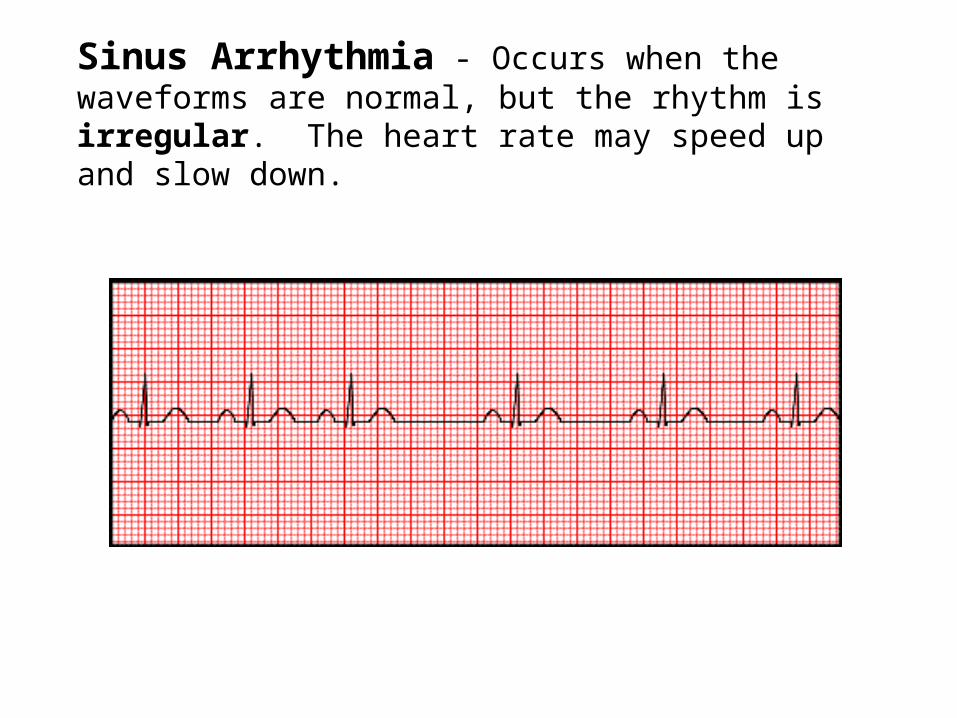
Sinus Arrhythmia - Occurs when the waveforms are normal, but the rhythm is irregular. The heart rate may speed up and slow down.
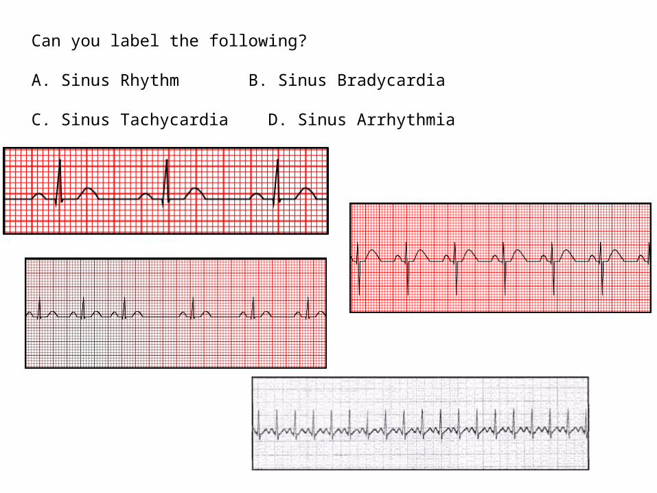
Can you label the following?
A. Sinus Rhythm B. Sinus Bradycardia
C. Sinus Tachycardia D. Sinus Arrhythmia
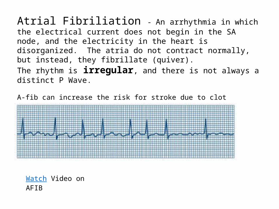
Atrial Fibriliation - An arrhythmia in which the electrical current does not begin in the SA node, and the electricity in the heart is disorganized. The atria do not contract normally, but instead, they fibrillate (quiver).
The rhythm is irregular, and there is not always a distinct P Wave.
A-fib can increase the risk for stroke due to clot formation.Many patients with A-fib will be on anticlotting medications.
Watch Video on AFIB
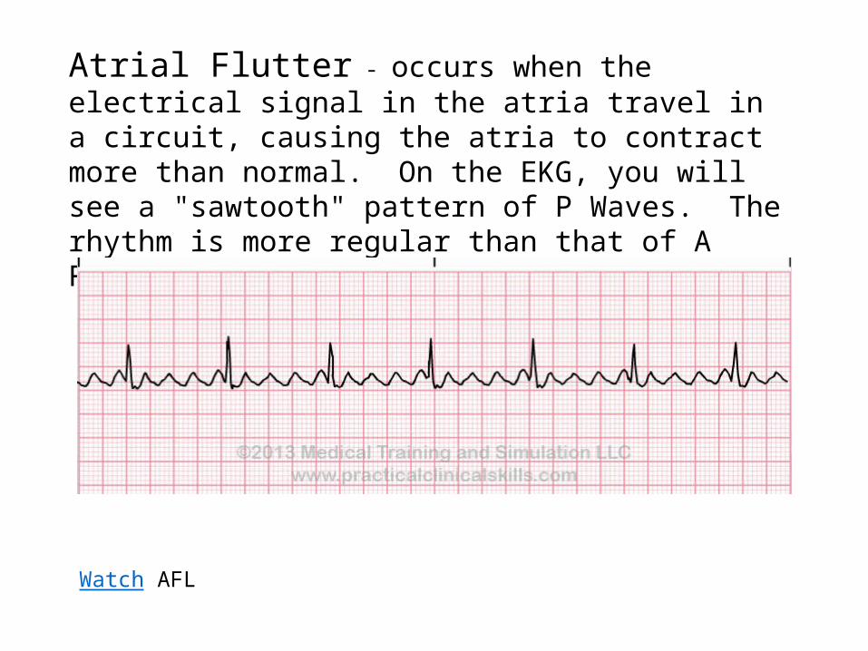
Atrial Flutter - occurs when the electrical signal in the atria travel in a circuit, causing the atria to contract more than normal. On the EKG, you will see a "sawtooth" pattern of P Waves. The rhythm is more regular than that of A Fib.
Watch AFL
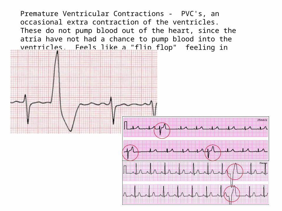
Premature Ventricular Contractions - PVC's, an occasional extra contraction of the ventricles. These do not pump blood out of the heart, since the atria have not had a chance to pump blood into the ventricles. Feels like a "flip flop" feeling in the chest.
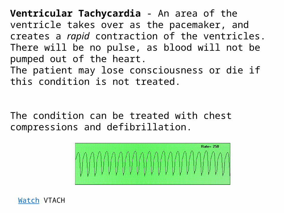
Ventricular Tachycardia - An area of the ventricle takes over as the pacemaker, and creates a rapid contraction of the ventricles. There will be no pulse, as blood will not be pumped out of the heart.The patient may lose consciousness or die if this condition is not treated.
The condition can be treated with chest compressions and defibrillation.
Watch VTACH
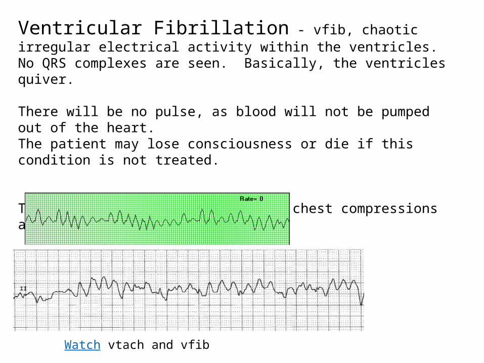
Ventricular Fibrillation - vfib, chaotic irregular electrical activity within the ventricles. No QRS complexes are seen. Basically, the ventricles quiver.
There will be no pulse, as blood will not be pumped out of the heart.The patient may lose consciousness or die if this condition is not treated.
The condition can be treated with chest compressions and defibrillation
Watch vtach and vfib
Watch agonal rhythm to asystole
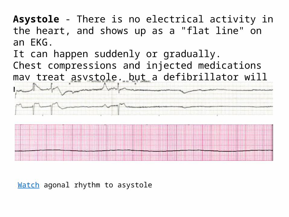
Asystole - There is no electrical activity in the heart, and shows up as a "flat line" on an EKG.It can happen suddenly or gradually.Chest compressions and injected medications may treat asystole, but a defibrillator will not work!
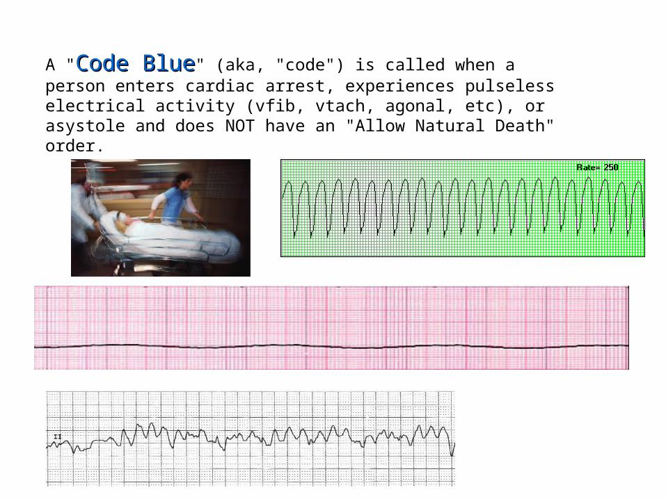
A "Code BlueCode Blue" (aka, "code") is called when a person enters cardiac arrest, experiences pulseless electrical activity (vfib, vtach, agonal, etc), or asystole and does NOT have an "Allow Natural Death" order.
These are a FEW of the dysrrhythmias that can occur, and a very basic way to interpret EKG's.
Questions? Comments?
References:
www.nih.govwww.medicinenet.comwww.bostonscientific.comwww.quia.com


