
DISTRIBUTION AND ELIMINATION OF DRUGS
By:-Dr. Roohana Hasan
JR-1Moderator:-
Dr. Dilshad Ali Rizvi

DRUG DISTRIBUTION:

3
AFTER ABSORPTION OF A DRUG IT MAY:-
Reversibly attached with its site of action.
Bound to plasma Proteins.
Accumulate in various storage sites.
Enter into tissues.
4
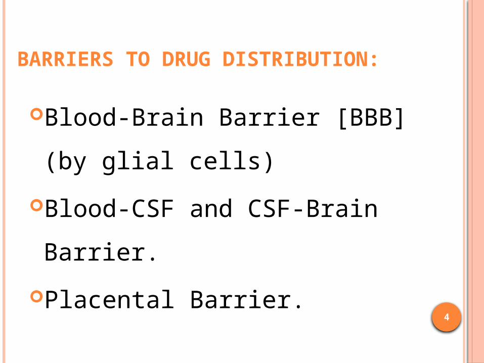
BARRIERS TO DRUG DISTRIBUTION:
Blood-Brain Barrier [BBB] (by glial cells)
Blood-CSF and CSF-Brain Barrier.
Placental Barrier.

5
BLOOD BRAIN BARRIER BBB Protects brain tissue from toxic substances.
Only lipid soluble non ionised drug can pass through BBB.
Inflammatory conditions like cerebral meningitis alter permeability of BBB
Drugs like Penicillin, Chloramphenicol exhibit increased
permeability.

6
BLOOD CSF AND CSF BRAIN BARRIER CSF secreted by the epithelial cells and lined by
occluding zonulae. But CSF brain barrier is composed epithelial cells lining
the ventricles , not connected by occluding zonulae.
CSF-Brain barrier permeable to drug molecules
If drug given by intrathecal route it reach the brain in sufficient concentrations. eg: Penicillin in Brain Abscess.
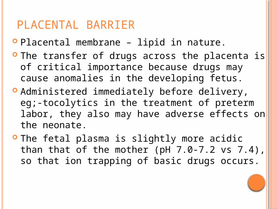
PLACENTAL BARRIER Placental membrane – lipid in nature. The transfer of drugs across the placenta is of
critical importance because drugs may cause anomalies in the developing fetus.
Administered immediately before delivery, eg;-tocolytics in the treatment of preterm labor, they also may have adverse effects on the neonate.
The fetal plasma is slightly more acidic than that of the mother (pH 7.0-7.2 vs 7.4), so that ion trapping of basic drugs occurs.

8
SPECIAL COMPARTMENTS FOR DRUG DISTRIBUTION: Cellular Reservoir. Fat as Reservoir. Transcellular Reservoir. Bones & Connective Tissue as Reservoir. Plasma protein binding as Drug Reservoir.

CELLULAR RESERVOIR If the tissue has higher affinity for the drug.
Binding to tissue proteins or nucleoproteins. Eg:- digoxin and emetine- skeletal muscles,
heart,liver,kidney( bound to muscle proteins) Iodine in thyroid, chloroquine in liver (tissue
proteins) Cadmium, lead, mercury in kidney (muscle
protein)

Fat as Reservoir.- Highly lipid soluble drugs.eg. Thiopentone & DDT
Sluggish reservoir due to decreased blood flow- may cause toxicity.
Transcellular Reservoir. eg. Chloramphenicol in aqueous humour, CSF (amino sugars), pericardial and peritoneal sacs serve as drug reservoirs.
Bones & Connective Tissue as Reservoir. Many drugs like tetracyclines,cisplatin,lead, fluorides form complexes with bone salts and get deposited in nails,bones and teeth.
eg. Griseofulvin in Keratin precursor cells, selectively accumulated in skin and nails.
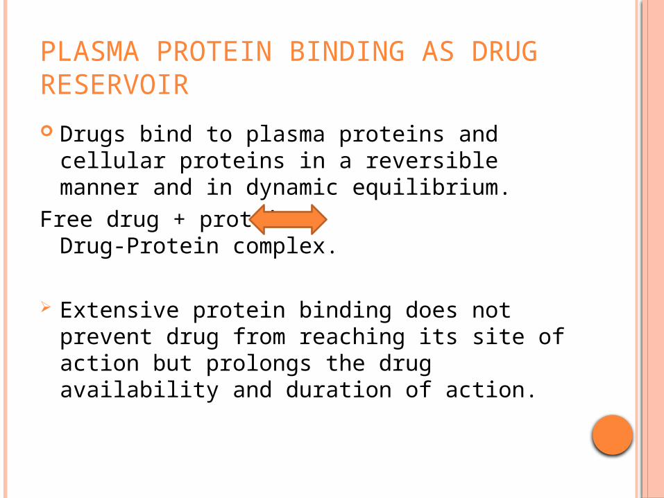
PLASMA PROTEIN BINDING AS DRUG RESERVOIR Drugs bind to plasma proteins and cellular
proteins in a reversible manner and in dynamic equilibrium.
Free drug + protein Drug-Protein complex.
Extensive protein binding does not prevent drug from reaching its site of action but prolongs the drug availability and duration of action.

12
IMPORTANT PROTEINS THAT CONTRIBUTE TO DRUG BINDING:
1) Plasma Albumin. [acidic drugs]
2) α1-Acid Glycoproteins (α1-AGP). [basic drug]
3) Tissue Proteins & Nucleoproteins. [drugs with high aVd]
4) Miscellaneous Binding Proteins. [thyroxin to α globulin, antigens to gamma globulins]

13
CLINICAL IMPORTANCE OF PLASMA PROTEIN BINDING:
Highly plasma protein bound drugs
Restricts to vascular compartment and have lower Vd.
Highly protein bound drugs are Difficult to remove by Dialysis
In diseases causing hypoalbuminemia therapeutic dose can lead to higher conc. of drug.
Plasma α1-AGP = acute phase reactant protein. Increases in MI, Crohn’s disease etc. Binding of basic drug increases. eg. Propanolol

14
Displacement interactions increase free drug concentration (of
the displaced drug) causing adverse effects.
Displacement is significant when:-
Displaced drug is more than 95% protein bound
Displaced drug with extensive protein bounding but lower aVd

Volume of distribution: It has physiological meaning, & related to the
Body Water.
15
4

Digoxin is widely distributed in the body including muscles and adipose tissue, leaving a small fraction to be distributed in the plasma.
Volume of distribution does not represent a real volume but must be regarded as the size of the body or fluids that would be required if the drug was distributed eqaully in all portions of the body.

17
APPARENT VOLUME OF DISTRIBUTION (AVD):
aVd = Total amount of drug in body (mg/kg)_ Conc. Of the Drug in the Plasma (mg/L)
It is the total space which should apparently be available in the body to contain the known amount of the drug.

a. If a drug does not capillary walls and is given by IV route , aVd = plasma water ie; 3L.
b. Drugs highly bound to plasma proteins have low aVd.
c. The lesser the plasma protein binding ,greater is the aVd.
d. aVd is > actual body volume
- widely distributed in the body - difficult to remove by dialysis.

aVd < 5L – vascular compartment aVd~ 15L – extracellular fluid aVd > 20L – distribution through out the body.
Pathological states can alter aVd of many drugs by altering distribution of body water and protein binding.
19

20
REDISTRIBUTION OF DRUGS: Typical mode of drug distribution observed with highly Lipid-soluble drugs.
eg: Anaesthetic effect of Thiopentone is rapid but effect get terminated due to redistribution in muscle and fat.
21
DRUG ELIMINATION:

22
ROUTES OF ELIMINATION:Major Routes Minor Routes
Renal Milk
Biliary Skin
Fecal Hair
Alveolar Sweat & Saliva

CLEARANCE Volume of plasma that is cleared of drug per unit
time. Unit= volume/time. Clearance is the propotionality factor used to
determine the rate of elimination. Rate of elimination = CL * concentration CLtotal = CLrenal + CLhepatic + CLlungs

24
RENAL EXCRETION:
Most important organ for Elimination.
Free drugs (eg. Furosemide, gentamicin)
Drug Metabolites.
25
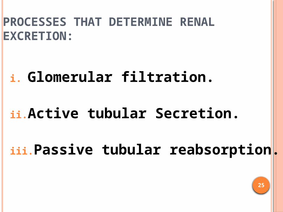
PROCESSES THAT DETERMINE RENAL EXCRETION:
i. Glomerular filtration.
ii. Active tubular Secretion.
iii. Passive tubular reabsorption.

26
FACTORS OF GLOMERULAR FILTRATION:
i. Molecular size.
ii. Plasma protein
binding
iii. Renal Blood Flow.

27
TUBULAR SECRETION: Energy, requiring carrier mediated active transport. Two independent carrier systems
For acidic drug (eg. Penicillin, salicylic acid) For basic drugs (eg. Morphine)
Clinical Importance:Weakly acidic drug (salicylic acid, lactic acid) interfere with secretion of Uric Acid
Increase plasma Uric acid Level
Precipitates GOUT

Probenecid (a weak acid) competitively inhibits the tubular secretion of penicillins and amoxycillin,
increase plasma half-life and effectiveness of penicillians in the treatment of infective diseases.
29
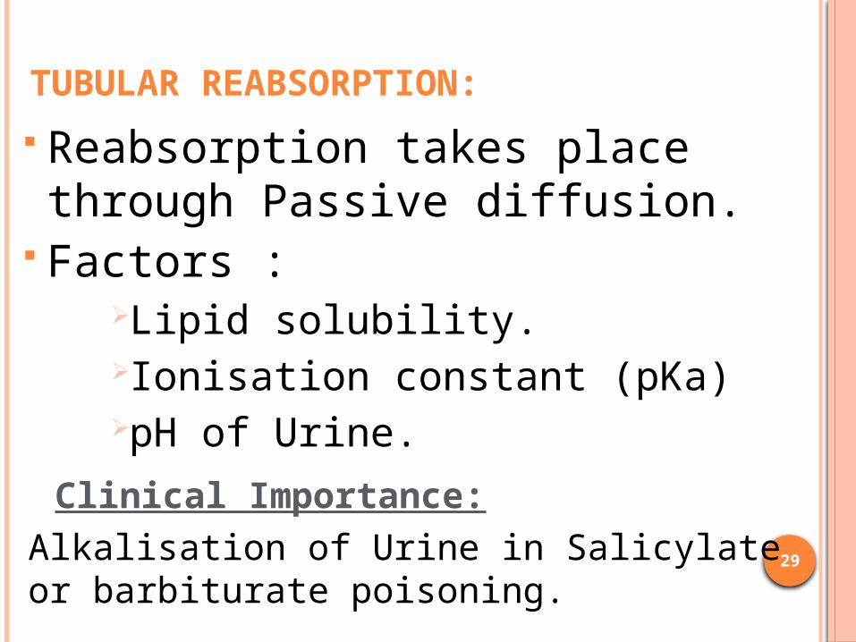
TUBULAR REABSORPTION: Reabsorption takes place through Passive diffusion.
Factors :Lipid solubility.Ionisation constant (pKa)pH of Urine.
Clinical Importance:Alkalisation of Urine in Salicylate or barbiturate poisoning.
30
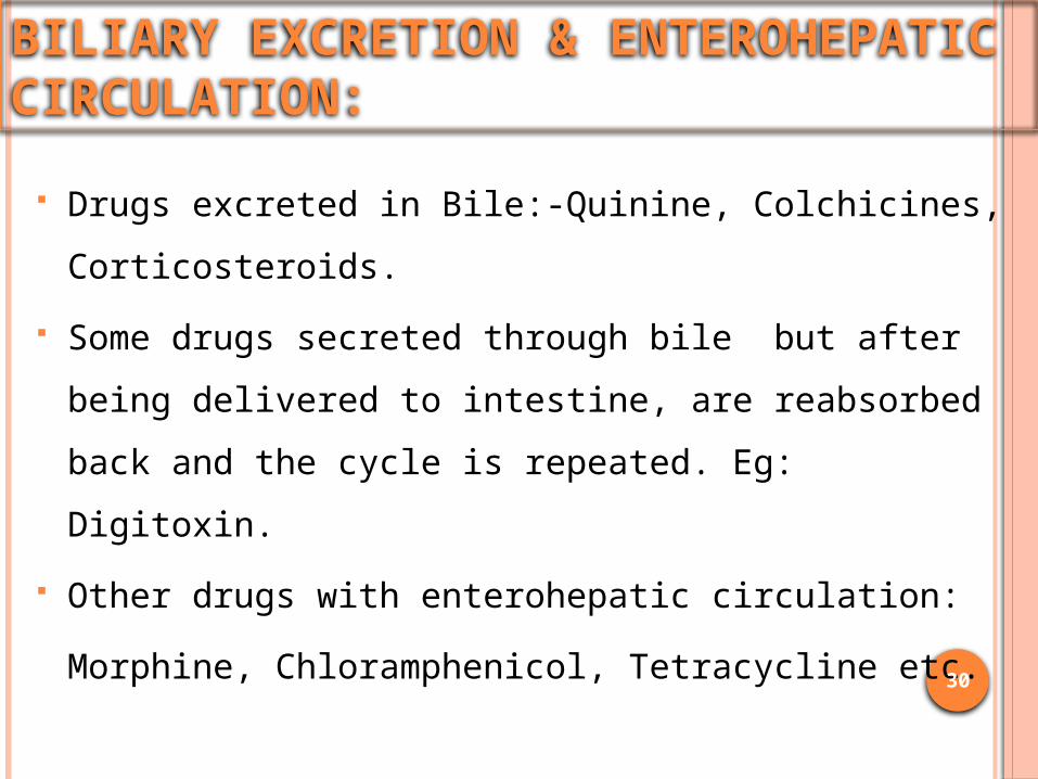
BILIARY EXCRETION & ENTEROHEPATIC CIRCULATION:
Drugs excreted in Bile:-Quinine, Colchicines, Corticosteroids.
Some drugs secreted through bile but after being delivered to intestine, are reabsorbed back and the cycle is repeated. Eg: Digitoxin.
Other drugs with enterohepatic circulation:Morphine, Chloramphenicol, Tetracycline etc.

31
Clinical Importance of Biliary excretion and Enterohepatic circulation:
In morphine poisoning Gastric lavage is done to prevent Enterohepatic Circulation.
Enterohepatic circulation prolongs the drug action.

32
FECAL ELIMINATION:
Orally ingested drug not absorbed in Guteg. MgSO4, Neomycin, Certain
purgatives
Drugs excreted in bile & not absorbed from intestinal tract.
eg. Erythromycin, Corticosteroids.

33
ALVEOLAR EXCRETION: Gases & Volatile liquids
eg: General Anaesthetics, Ether, Alcohol Depends on partial pressure in the blood. Eucalyptus oil and garlic oil eliminated through
expectoration.

34
ELIMINATION THROUGH BREAST MILK: May cause unwanted effect in Nursing infant. Drugs transferred to breast milk according to pH
partition principle. Basic drugs not ionised at plasma alkaline pH, get
accumulated in Milk.Eg: Chloramphenicol, Tetracycline, Morphine etc.

Certain acidic drugs may also be secreted in the milk and can causeSulfonamides- kericterus and allergyPenicillin- allergy Dapsone- heamolytic anemia Phenobarbiton- drowsiness Phenytoin- methaemoglobineamia Infants are sensitive to drug induced hemolysis-
chloramphenicol, quinine, quinidine, dapsone etc. should not be given to breastfeeding mother.

36
EXCRETION THROUGH SKIN, HAIR, SWEAT & SALIVA: Griseofulvin is secreted through
keratin precursor cells. Arsenic, Mercury salts & Iodides
Hair Follicles. Iodine, KI, Li & Phenytoin Saliva. Amines & Urea derivatives Sweat.

37
KINETICS OF DRUG ELIMINATION: First order kinetics. Zero order kinetics. Mixed order kinetics.

38
FIRST ORDER KINETICS: Majority of the drugs follow this type of elimination.
A constant fraction of the drug is eliminated at a constant interval of time.
eg: Plasma concentration declining at a rate of 50% per two hours: 100 µg/ ml 50 µg/ml 25 µg/ ml
12.5 µg/ml and so on.
39
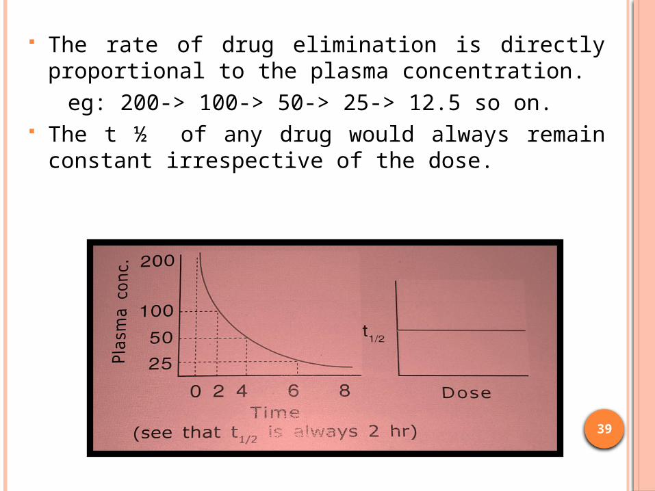
The rate of drug elimination is directly proportional to the plasma concentration.
eg: 200-> 100-> 50-> 25-> 12.5 so on. The t ½ of any drug would always remain
constant irrespective of the dose.

40
Plasma concentration is plotted against time , the resultant “ plasma fall-out curve” curvilinear,
Log of plasma concentration are plotted against time , the resultant curve linear.
41
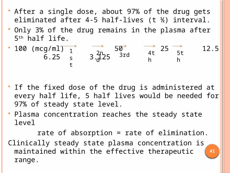
After a single dose, about 97% of the drug gets eliminated after 4-5 half-lives (t ½) interval.
Only 3% of the drug remains in the plasma after 5th half life.
100 (mcg/ml) 50 25 12.5 6.25 3.125
If the fixed dose of the drug is administered at every half life, 5 half lives would be needed for 97% of steady state level.
Plasma concentration reaches the steady state level rate of absorption = rate of elimination.Clinically steady state plasma concentration is
maintained within the effective therapeutic range.
1st
2nd
3rd 4th
5th

42
If the dose of the drug is doubled, its duration of action is prolonged for one more half-life.
The “log plasma concentration fall-out curve” of a drug having high aVd, exhibits 2 slopes.
An initial rapid declining phase due to distribution (called as α phase)
Later linearly declining phase due to elimination (called as β phase)

43FIG : Α AND Β-PHASE OF DRUG CLEARANCE

44
ZERO ORDER KINETICS: A constant or a fixed quantity of drug is eliminated
per unit time. Ethyl alcohol exhibit zero order at virtually all plasma concentrations. For eg: if plasma concentration falls at a rate of 25
µg per hour then 50 25 nil

45
The rate of elimination is independent of the concentration of the drug in the plasma. So increasing the dose does not result in a proportionate rise in the extent of elimination.
100 75 50 25 Nil The t ½ of a drug following zero order is never
constant.

46
If such a fall in plasma concentration is plotted against time, the resultant “plasma fall-out curve” is steeply linear, but if logarithm of plasma concentration are plotted against time , then the curve becomes curvilinear.

47
MIXED ORDER KINETICS/ SATURATION KINETICS / MICHAELIS-MENTEN KINETICS: Dose-dependent kinetics where smaller doses are
eliminated by first order kinetics but as the plasma concentration reaches higher values ,the rate of drug elimination becomes zero order.
Phenytoin, warfarin, digoxin, dicumarol.

48
After a single dose administration, if the plasma concentrations are plotted against time, the resultant plasma fall out curve remains linear in the beginning (zero order) and then become predominantly exponential ( curvilinear i.e. first order).
Fig : Plasma concentration fall-out curve in mixed order kinetics.

49
CLINICAL IMPORTANCE: Drugs having very short half-life are given by constant
i.v. infusion to maintain steady state concentration. Drugs having t1/2 = 30 mins to 2 hrs , it becomes
incovenient to administer it every half life. In such cases, provides the drug is having high safety margin and obeying 1st order kinetics , dose can be so increased that the drug can be administered every 6-8 hours.
The drugs having t1/2 = 4-12 hours, administered at every half life interval.

50
The drugs having medium half life usually given at 12 hours interval.
Drugs having 24 hours half life, half of the therapeutic dose is given at every half of half life.
For drugs having longer t ½, with high Vd & slow rate of clearance also are cumulative in nature. To reach steady state Loading dose given Maintenance dose.
Loading dose= Desired plasma conc. (mg/L) * aVd (L/kg).

THANK YOU..!!


