

1
Insertional mutagenesis in PLNR9C
transgenic mouse
Insertional mutagenesis in PLNR9C transgenic mouse: a case report and a
review of the literature on cardiac-specific transgenic expression
Alexander Kraev
University of Toronto; 27 King's College Circle, Toronto, Ontario, Canada M5S 1A1; Email
address: [email protected], Phone: 905-847-8891
Abstract A mouse line with heterozygous transgenic expression of phospholamban carrying a
substitution of cysteine for arginine 9 (PLNR9C) under the control of α-myosin heavy chain
(αMHC) promoter features dilated cardiomyopathy, heart failure and premature death. In this
line the transgenic array of 13 PLNR9C expression cassettes, arranged in a head-to-tail tandem
orientation, has integrated into the homologous genomic site, the bi-directional promoter of
the αMHC (Myh6) gene and the gene for the regulatory non-coding RNA Myheart (Mhrt), both
of which are involved in the execution of the α/β MHC switch during cardiac development and
pathology. PLNR9C overexpression is evident at the age of 1 month but declines dramatically
along with a less pronounced concomitant decline of the resident PLN expression, until the
animals die. Expression of the non-coding RNA Mhrt in PLNR9C mice also exhibits a profound
deregulation, despite the presence of the second, intact allele. Hence the mouse strain does not
faithfully model a human PLNR9C heterozygote, wherein both the mutant and the wildtype PLN
alleles have, in all likelihood, the same temporal expression profile. The intricate regulatory
circuit of the α/β MHC switch, involving the non-coding RNA Mhrt, was described in detail only
recently, and since publications about αMHC-driven transgenes rarely contain the definition of
the transgene integration site or temporal expression profile, it is suggested that some of the
pathological phenomena attributed to expression of αMHC-driven transgenes may have an
alternative explanation.
.CC-BY-NC-ND 4.0 International licensepeer-reviewed) is the author/funder. It is made available under aThe copyright holder for this preprint (which was not. http://dx.doi.org/10.1101/075671doi: bioRxiv preprint first posted online Sep. 16, 2016;
2
Insertional mutagenesis in PLNR9C
transgenic mouse
Introduction
Complex genetic landscape of inherited cardiomyopathy includes many genes, among
which are the genes encoding contractile proteins, such as the subunits of the myosin complex,
as well as the gene encoding a small phosphoprotein phospholamban (McNally et al. 2015).
Myosin is a hexameric protein complex, often called a molecular motor (Gupta 2007), consisting
of two heavy chains (MHC) and four light chains (MLC). The two MHCs, designated αMHC and
βMHC, are encoded by two closely spaced genes, called MYH6 and MYH7 in man, that are
transcribed in the same direction (Morkin 2000). The products of individual genes combine to
make three myosin isoforms V1, V2 and V3, by association of αα, αβ and ββ chains, respectively.
Expression of the two MHC gene transcripts is known to occur at a high level in all of the
cardiomyocytes by 8 d p.c. in the mouse embryo (Lyons et al. 1990), when an S-shaped tubular
heart has formed and contractions start (Sissman 1970). Myosin isoform switch happens more
than once during development of a healthy mouse heart, and any gene put under control of the
αMHC promoter should follow its expression profile in transgenic animals (Subramaniam et al.
1991). Due to its spatially restricted, cardiac-specific pattern of expression (Gulick et al. 1991),
since 1991 the αMHC promoter has become the promoter of choice for transgenic expression of
proteins, suspected to be involved in heart dysfunction. In hindsight, its use looked problematic
from the beginning (Subramaniam et al. 1991), since only spatial, but not temporal or regulatory
characteristics of the promoter were taken into consideration. However, neither the knowledge
that the α/β MHC isoform switch is affected by the thyroid hormone (Gustafson et al. 1986;
Izumo et al. 1986; Subramaniam et al. 1991) as well as observed in experimental cardiac
hypertrophy (Morkin and Kimata 1974), nor the knowledge that the regulatory effects of
hormones and experimental cardiac hypertrophy are exerted differently in ventricles and atria
(Morkin 1993), had prevented the extensive use of αMHC-promoter driven transgenes in
cardiovascular research (Molkentin and Robbins 2009).
Phospholamban (PLN), a 52-aminoacid transmembrane phosphoprotein, abundant in the
endoplasmic reticulum of cardiac and skeletal muscle, regulates activity of sarcoendoplasmic
reticulum Ca2+ ATPAses (SERCA). Since its discovery by Kirchberber and Katz in 1975
(Kirchberber et al. 1975) it received much attention as a possible pharmacological target in heart
.CC-BY-NC-ND 4.0 International licensepeer-reviewed) is the author/funder. It is made available under aThe copyright holder for this preprint (which was not. http://dx.doi.org/10.1101/075671doi: bioRxiv preprint first posted online Sep. 16, 2016;

3
Insertional mutagenesis in PLNR9C
transgenic mouse
failure, since it is expressed in ventricles, atria (Koss et al. 1995) and sino-atrial node of the
pacemaker (Maltsev et al. 2006). Transgenic expression of PLN mutants in mice has become one
of the mainstream models of heart failure in the last two decades (Koss and Kranias 1996;
Molkentin and Robbins 2009), reinforced by the discovery of actual, albeit rare, patients that
carry PLN mutations (Fish et al. 2016; Haghighi et al. 2003; Liu et al. 2015; Schmitt et al. 2003).
Recent whole genome association studies mapped the quantitative trait locus on chromosome
6q22 that includes the PLN gene, as a strong candidate for resting heart rate (Eijgelsheim et al.
2010).
Researchers in the field of PLN functional studies were among the first to embrace the
transgenic technology, and a pioneering study succeeded in creating two transgenic lines,
having 2 and 3 copies of the αMHC promoter–driven PLN transgene per genome, and featuring
a two-fold excess of the transgenic PLN in the ventricles (Kadambi et al. 1996). These mice
demonstrated no gross pathology and had normal life span.
In contrast, a mouse strain with transgenic expression of a heterozygous substitution of
cysteine for arginine 9 in phospholamban (PLNR9C) (Schmitt et al. 2003) under the control of
αMHC promoter, reported in 2003, featured dilated cardiomyopathy, heart failure and life
expectancy of only 21±6 weeks. The ensuing line of physiological/genetic (Schmitt et al. 2009),
transcriptomic (Burke et al. 2016) and proteomic (Gramolini et al. 2008) studies of this strain was
mostly focused on Ca2+ dysregulation and did not address the possible impact of other
mechanisms, unrelated to dysregulation of SERCA2a. Here, it is reported that the PLNR9C
transgenic line has a peculiar genomic structure, wherein the tandem array of 13 expression
cassettes has integrated into the bi-directional promoter that drives the transcription of the
resident αMHC gene (Myh6) and the gene for long non-coding RNA Mhrt (Han and Chang
2015; Han et al. 2014), involved in developmental and pathological α/β MHC switch.
Materials and methods
Transgenic mice and genotyping
.CC-BY-NC-ND 4.0 International licensepeer-reviewed) is the author/funder. It is made available under aThe copyright holder for this preprint (which was not. http://dx.doi.org/10.1101/075671doi: bioRxiv preprint first posted online Sep. 16, 2016;
4
Insertional mutagenesis in PLNR9C
transgenic mouse
The transgenic mouse line, carrying rabbit phospholamban with a substitution of
cysteine for arginine 9 under the control of the mouse αMHC promoter (TgPLNR9C) was
described previously (Schmitt et al. 2003). The mice were propagated by backcross to FVB/NCrl
(strain code 207, Charles River Canada), thus obtaining only transgenic hemizygotes, and
transgenes were identified by PCR (Zvaritch et al. 2000).
RNA and DNA isolation
Mice were dissected under total anesthesia with diethyl ether and subsequently killed by
cervical dislocation. Hearts were excised, gently squeezed while submerged in cold phosphate-
buffered saline to reduce blood contamination, dissected into atria and ventricles and flash
frozen in liquid nitrogen. Total RNA was extracted with Trizol reagent (Thermo Fisher Scientific #
15596-026) essentially as described by the manufacturer, subsequently treated with RNAse-free
DNAse I (Thermo Fisher Scientific # AM2222) and purified by phenol extraction and ethanol
precipitation. RNA from mutant and wild type litter mates (three of each genotype) were always
processed in the same batch to minimize variations of quality. RNA amount and quality was
estimated using Nanodrop 1000 spectrophotometer. 120 ng portions of total RNA from the
mice of each genotype were pooled for cDNA synthesis. Mouse genomic DNA was isolated from
either tail or ear clips as described (Miller et al. 1988) or recovered from crude preparations of
heart RNA.
Quantitative reverse transcription-assisted polymerase chain reaction
Total RNA was reverse transcribed into complementary DNA (cDNA) using Transcriptor®
enzyme, Roche Diagnostics, Laval, QC), primed by 10 pmol of T7-oligo-dT24VN primer (Thermo
Fisher Scientific #AM5712) by incubation for 15 min at 42 °C followed by 1 hour at 50 °C. For the
detection of long non-coding RNA the cDNA synthesis was primed with specific primers and M-
MLV Reverse Transcriptase, RNase H Minus (Promega #M3682). A 4-µl portion of each
completed cDNA reaction was diluted 1:25 with reagent-grade water and 5 µl aliquots of diluted
cDNA were added to 20-µl qRT-PCR reactions, along with 5 µl of a 1 µM primer pair and 10 µl of
.CC-BY-NC-ND 4.0 International licensepeer-reviewed) is the author/funder. It is made available under aThe copyright holder for this preprint (which was not. http://dx.doi.org/10.1101/075671doi: bioRxiv preprint first posted online Sep. 16, 2016;

5
Insertional mutagenesis in PLNR9C
transgenic mouse
SYBR® Select reagent master mix (Applied Biosystems #4472919). A “no primer” control
reaction was analyzed in parallel with properly made cDNAs. The identity of all fragments used
in real-time PCR was verified by sequencing.
Reactions were run in triplicate on the Mini-Opticon qRT-PCR instrument (Bio-Rad
Laboratories Canada Ltd.). The hypoxanthine phosphoribosyl transferase gene was used as a
reference. The qRT-PCR Ct values were exported as Excel files and analyzed using online
software provided by Qiagen Inc [http://pcrdataanalysis.sabiosciences.com], which included two-
tailed Student’s t-test, and finally converted into a graph using GraphPad Prism v.6. Data were
considered significant with p-value less than 0.05.
Determination of the ratio of native to transgenic PLN transcripts
PLN- and TgPLNR9C primer pairs were used to amplify the respective PCR fragments from
cDNA by conventional PCR, which were purified and quantitated. After two million-fold dilution
of each fragment in a glycogen solution (20 µg/ml), synthetic molar ratios of 5:1, 2:1, 1:1, 1:2 and
1:5 were made and each mixture was tested with the same two non-overlapping primer pairs in
a quantitative PCR reaction. A calibration curve, plotting the function of constructed ratio to
experimentally observed ratio was thus obtained which was used to correct the experimentally
observed ratio in a tissue. Thus, the ratio of 2-∆∆Ct of PLN relative to TgPLNR9C can be adjusted for
primer efficiency according to the formula y=4.4136x-0.0855, where x is the experimentally
observed ratio, and y is the corrected ratio. By design, this formula applies only to the two
specific primer batches that were actually used, and the procedure needs to be repeated for any
new primer batches.
DNA amplification and sequence analysis
Genomic location of the transgenic insert was determined by inverse PCR (Does et al.
1991). Genomic DNA (0.5 µg) was cut with Nco I (one of the sites was present at the start codon
of the TgPLNR9C gene but not elsewhere within its transcript), diluted to 700 µl and circularized
with T4 DNA ligase overnight at room temperature. The DNA was phenol extracted and ethanol
precipitated and dissolved in reagent grade water at a concentration of 25 ng/µl. Primers P1 and
P2, specific for the rabbit TgPLNR9C gene, directed outward, were used to amplify the circularized
.CC-BY-NC-ND 4.0 International licensepeer-reviewed) is the author/funder. It is made available under aThe copyright holder for this preprint (which was not. http://dx.doi.org/10.1101/075671doi: bioRxiv preprint first posted online Sep. 16, 2016;

6
Insertional mutagenesis in PLNR9C
transgenic mouse
fragment. The location of the junction sequence, deduced from sequencing of the fragment, was
verified by regular PCR using appropriate flanking primers P1-P4 and P3-P5 (Fig. 3a and 4).
Rapid amplification of cDNA 3’-ends was done with T7-oligo-dT24V primed cDNA, using PCR
with anchor-specific primer and gene-specific primer P1. Due to high concentration of the PLN
transcript it was readily amplified by a single round PCR. If purified genomic DNA was not
available, genomic DNA amplification could be successfully carried out from the crude heart
RNA preparations (not treated with DNAse). Amplifications were carried out either with
Platinum® Taq polymerase (Thermofisher) or with LongAmp® Taq polymerase (New England
Biolabs) on a Bio-Rad C1000 Touch™ thermal cycler. All PCR products were analyzed by agarose
gel electrophoresis, purified on Qiaquick MinElute columns (Qiagen) and sequenced at the
Toronto Centre for Applied Genomics facility. Sequence assembly was done with Sequencher
4.10 (Gene Codes, Ann Arbor, MI). Reference Mus musculus C57BL/6J genomic and cDNA
sequences were retrieved from Ensembl [http://www.ensembl.org]. Oligonucleotide primers
were synthetized by ACGT (Toronto, ON, Canada) or Eurofins MWG Operon LLC (Huntsville, AL,
USA). Primer sequences are listed in Table 1.
Results
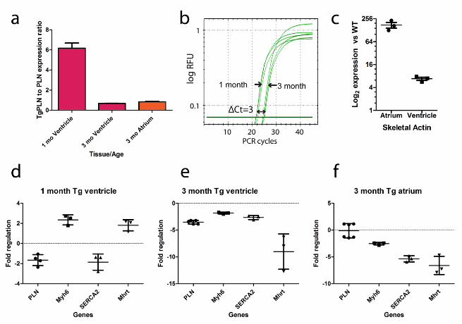
Developmental suppression of PLNR9C expression in transgenic hearts
Overexpression of wildtype PLN, if driven by the αMHC promoter, is expected to disturb
the natural atrio-ventricular PLN gradient (Koss et al. 1995), wherein 3 to 5 fold more PLN is
found in normal murine ventricles than in atria; besides, the expression of αMHC begins
somewhat earlier in atria than in ventricles (Lyons et al. 1990). In a wildtype mouse the activity of
the αMHC promoter should peak between 1 and 2 months of age (Han et al. 2014; Ng et al.
1991), while βMHC expression drops to a very low level after birth (Ng et al. 1991). Therefore, it
was first sought to compare gene expression of the transgenic and native PLN at 1 month. Using
real-time PCR, the ratio of TgPLNR9C to native PLN transcripts at 1 month was found to be
slightly above 6-fold (Fig.1a), while native PLN in transgenic hearts was found downregulated
1.8-fold relative to the wildtype (Fig.1d). If the level of TgPLNR9C overexpression at 1 month is
maintained in translated PLN protein, a ratio required to trap the native PLN in a non-productive
.CC-BY-NC-ND 4.0 International licensepeer-reviewed) is the author/funder. It is made available under aThe copyright holder for this preprint (which was not. http://dx.doi.org/10.1101/075671doi: bioRxiv preprint first posted online Sep. 16, 2016;
7
Insertional mutagenesis in PLNR9C
transgenic mouse
pentameric complex, where it cannot be phosphorylated by protein kinase A (Ha et al. 2011;
Wittmann et al. 2015) is substantially exceeded.
TgPLNR9C animals start to die (Gramolini et al. 2008; Schmitt et al. 2009), at about 3
months of age. Gene expression experiments done with the same starting amount of RNA,
allowing a direct comparison of Ct values, show that absolute expression of TgPLNR9C goes down
~8 fold in the timeline from 1 month to 3 months (Fig. 1b). The ratio of TgPLNR9C to native PLN
transcript in the 3 month ventricle is only 0.69 (Fig.1a). Evidently, at 3 months there is no
overexpression of TgPLNR9C in the ventricle anymore, similarly to previous reports (Babu et al.
2006; James et al. 2000; Kadambi et al. 1996; Mende et al. 1998; Nakayama et al. 2003; Sanbe et
al. 1999; Zvaritch et al. 2000), even though the native PLN is downregulated 3.7 times relative to
the wildtype (Fig.1e). Presumably, somewhere at an intermediate age between 1 and 3 months
the transgenic PLN expression transiently passes the 1:1 ratio with the native PLN, to model a
situation thought to exist continuously in a human patient heterozygous for the PLN mutation.
In contrast, the TgPLNR9C heart initially receives a large excess of mutant PLN around the age of
1 month, but the transgene expression then gradually subsides on the background of
developing cardiomyopathy, until the animal dies. The documented use of a similar construct to
create transgenic mice may result in dramatic suppression (Mende et al. 1998), as well as in no
suppression (Santini et al. 2007) of the transgene. A peculiar feature of the 3-month TgPLNR9C
heart is a strong upregulation of skeletal actin, especially in the atrium (over 160-fold, Fig.1c),
incidentally, also observed in heterozygous mice with αMHC gene ablation (Jones et al. 1996),
suggests that overexpression of TgPLNR9C may not be the only factor of the morbid phenotype.
Determination of the chromosomal structure and location of the transgene
In search for the reasons of peculiar changes in the gene expression of the TgPLNR9C
mouse heart, in particular of the dramatic downregulation of the TgPLNR9C, the genomic
sequence of the TgPLNR9C chimeric gene and also the sequence of its transcript were
determined from appropriate PCR products but no potentially detrimental structural alterations
were found. By using primers P1 and P2 (Fig.3a), specific to the TgPLNR9C gene and facing
outward in a long-range PCR, it was confirmed that each unit contains the expected 5.5 kb
.CC-BY-NC-ND 4.0 International licensepeer-reviewed) is the author/funder. It is made available under aThe copyright holder for this preprint (which was not. http://dx.doi.org/10.1101/075671doi: bioRxiv preprint first posted online Sep. 16, 2016;

8
Insertional mutagenesis in PLNR9C
transgenic mouse
fragment containing the αMHC promoter [Genbank Accession U71441] and exists exclusively in
a head-to-tail orientation in a tandem array, since a single product (Fig.2a, leftmost panel) with
an unambiguous sequence reading across the unit junction was obtained [Genbank Accession
KU665646]. Using real-time PCR on genomic DNA with αMHC (Myh6) gene as reference, it was
also found in one randomly chosen animal, using Myh6 gene as a reference, that the transgenic
array contains 13 copies of the expression cassette (not shown).
Chromosomal location was determined with appropriate primers in the conventional and
the inverse PCR format (Fig.2a). Determination of the side of the transgenic array, facing the
Mhrt gene, presented no technical problems and it was found to be identical in 7 transgenic
animals (3,3,1) from three different litters (genomic coordinate 14:54968214 of Mouse Genome
assembly GRCm38.p4 (GCA_000001635.6)). The transgene portion contains a filled-in Not I site
of a polylinker sequence (Fig.2b ad Fig.3c), presumably used to excise the construct from the
vector (Schmitt et al. 2003), indicating that it is the original end of the construct (Pawlik et al.
1995). Determination of the flanking sequence at the other end of the transgene presented
certain technical problems. Similarly, 7 transgenic animals from three different litters were
analyzed. Initially, a flanking primer (P5) was designed to be close to the projected junctional
sequence from the results of the inverse PCR and paired with a primer specific for the Myh7 3’-
UTR in the transgene (P3). The first use of this primer pair recovered products only in mice Tg1
(one litter) and Tg5, Tg6 (another litter), which were of slightly different size (Fig.2c). Absence of
product in other animals could be due to a deletion affecting one of the primer sites or due to a
PCR failure. However, by using other primers and conditions junctional sequences were
eventually obtained from the remaining animals. The common junctional sequence contains a
double polylinker, identical to the one in KU665646 and 26 bp of the sequence derived from the
human growth hormone gene. Thus, it corresponds to a severely truncated original cassette (Fig.
4). The truncated transgenic unit, however, is not directly juxtaposed to mouse genomic DNA,
corresponding to the aMHC promoter, but is separated from it by a 19-bp “filler” sequence
(Mayerhofer et al. 1991), the origin of which could not be established. Comparison of junctional
sequences revealed that some of them contain short internal deletions (Fig.2c) relative to a
common sequence, as originally found in animals Tg1 and Tg5. Finally, long range PCR with the
.CC-BY-NC-ND 4.0 International licensepeer-reviewed) is the author/funder. It is made available under aThe copyright holder for this preprint (which was not. http://dx.doi.org/10.1101/075671doi: bioRxiv preprint first posted online Sep. 16, 2016;

9
Insertional mutagenesis in PLNR9C
transgenic mouse
primer P3 paired with a primer placed on Myh6 exon 3 outside of the portion included in the
transgene (P6) produced a fragment of expected size from all litters (Fig. 2a) that is consistent
with the location of the junctional sequence facing the Mhrt gene. An attempt to sequence the
long range PCR product across the junction encountered a sequence heterogeneity (not shown)
around the location of the polylinker and supported the conclusion that the small deletions,
observed in short PCR products, are likely PCR artefacts, induced by a local secondary structure.
Thus, no extensive rearrangement or large genomic deletions accompanying the transgenic
insertion were found, unlike those reported for other transgenes (1, 2).
Taken together, these experiments showed that the transgenic array is integrated into
the bi-directional promoter (Haddad et al. 2003; Han et al. 2014) driving the resident Myh6 gene
and the resident long non-coding RNA Mhrt in such a way that the TgPLNR9C transcription is
driven by the transgenic copies of the promoter in the opposite direction to that of the resident
Myh6 gene (Fig.3b). The insertion of the transgene results in 83 bp deletion of the target site in
the mice analyzed. The resident Myh6/Mhrt promoter appears to be cut approximately in half by
the ~85 kb transgenic array insertion, while the other Myh6 allele is intact, judging from the
ongoing transcription of Myh6, verified by real-time PCR (Fig.1d-1f). From the small number of
animals available for analysis no definite conclusion about the transgene stability could be
made.
Deregulation of non-coding RNA Mhrt expression in 3 month old transgenic hearts
reflects insertional mutagenesis of its promoter
Myh6 promoter substantially overlaps with the promoter of the long non-coding RNA
Mhrt (Han et al. 2014), thus it was of interest to determine if the transgenic insertion had indeed
any influence on its expression. Using a pair of primers, specific for the 3’-end of Mhrt in a
quantitative PCR format, about 2-fold upregulation was observed in transgenic ventricle at 1
month and over five-fold downregulation both in transgenic ventricles and atria at 3 months
(Fig.1d-1f), wherein the level of Mhrt expression drops close to a reliable limit of detection. A
confirmatory end-to-end amplification and sequencing of Mhrt was done from both transgenic
and wildtype ventricle cDNA, using one of the 3’-end specific primers utilized for cDNA synthesis
.CC-BY-NC-ND 4.0 International licensepeer-reviewed) is the author/funder. It is made available under aThe copyright holder for this preprint (which was not. http://dx.doi.org/10.1101/075671doi: bioRxiv preprint first posted online Sep. 16, 2016;

10
Insertional mutagenesis in PLNR9C
transgenic mouse
and quantitative PCR and a nested pair of primers, specific for the first exon of Mhrt (Table 1).
The deregulation of Mhrt expression occurs despite the presence of the intact allele in the
hemizygote, as if the damaged allele exerts a dominant negative effect over the intact allele.
.CC-BY-NC-ND 4.0 International licensepeer-reviewed) is the author/funder. It is made available under aThe copyright holder for this preprint (which was not. http://dx.doi.org/10.1101/075671doi: bioRxiv preprint first posted online Sep. 16, 2016;
11
Insertional mutagenesis in PLNR9C
transgenic mouse
Discussion
The peculiar genomic structure of the TgPLNR9C mouse line, containing damaged
Myh6/Mhrt allele and presumably impaired α/β MHC switch is a probable contributing cause of
the morbid phenotype of this line, along with the excessive amount of mutant PLN that the
mouse heart receives during the 1st month after birth. Indeed, only a weak downregulation of
αMHC (this study) and upregulation of βMHC (Schmitt et al. 2009) is observed, and the non-
coding RNA Mhrt is paradoxically downregulated on the background of the developing
cardiomyopathy (Han et al. 2014). Even in the absence of data about gene expression in
humans, it can be concluded that this line does not faithfully model a heterozygous human
patient carrying a cysteine for arginine 9 mutation in PLN, as the latter not only expresses the
mutant and the wildtype PLN from identical promoters, but also does not carry a Myh6/Mhrt
allele damaged by insertional mutagenesis. While it may be argued that the transgene
localization, as presented here, is but an elaborate PCR artefact, however, it is supported by the
observed deregulation of Mhrt that is a predicted consequence of the damage to its promoter.
It can also be argued that the transgene structure as revealed in this study may not
necessarily have existed in the founder transgenic mouse, but may have appeared as a result of
a secondary transgene rearrangement (or several ones) at some point in 12 years of
conventional breeding. It has become customary to cite Aigner and colleagues (Aigner et al.
1999) who did a thorough study to prove that transgenes, once obtained, are stable over many
generations. However, their conclusions from a study of just one gene appear to be an
unjustified generalization and may not apply to all conceivable transgenes. Specifically, there are
two important differences between their case and that of TgPLNR9C. First, TgPLNR9C animals have
severely reduced viability; hence a spontaneous transgenic array expansion (Barlow et al. 1995),
contraction (Alexander et al. 2004) or even change of location (Kearns et al. 2001) may be
selected for if it leads to a weaker morbid phenotype. Second, the transgenic array is inserted in
an inverted orientation into a homologous sequence, which makes it prone to instability.
Furthermore, the current genotyping protocol would not reveal most of the interunit
rearrangements, while only a complete transgene excision will be genotyped as wildtype and
discarded. However, in the absence of extensive structural data in a large pedigree no definite
.CC-BY-NC-ND 4.0 International licensepeer-reviewed) is the author/funder. It is made available under aThe copyright holder for this preprint (which was not. http://dx.doi.org/10.1101/075671doi: bioRxiv preprint first posted online Sep. 16, 2016;
12
Insertional mutagenesis in PLNR9C
transgenic mouse
conclusion can be made. The issue of the PLNR9C transgene stability may be an interesting topic
for a future dedicated study.
In the past, the problem of transgene silencing was well recognized (Garrick et al. 1998;
Martin and Whitelaw 1996; Robertson et al. 1995; Sanbe et al. 2003; Sutherland et al. 2000), so
obtaining a clinically relevant phenotype in a TgPLNR9C mouse line could have been a reason for
its initial selection. However, even though the case of the TgPLNR9C mouse line may be rare or
even unique, it brings to light two issues of general importance for transgenic research that, in
this author’s opinion, supersede the importance of mechanistic insights derived from the morbid
phenotype of a mouse line allegedly modelling a rare cause of cardiomyopathy. One is the
lingering lack of attention to the transgene integration site, an apparent relic of the “pre-
genomic era”, quite surprising in view of the current availability of the refined mouse genomic
sequence (Church et al. 2009). The other is the far-reaching implications of the recently defined
regulatory elements of the α/β MHC switch (Han et al. 2014; Hang et al. 2010) to the
interpretation of data collected over 25 years of use of αMHC-driven transgenes.
Determination of transgene integration site is inherent to the gene trap methodology
(Friedrich and Soriano 1991), which in essence is an ingenuous exploitation of a drawback of
unguided transgenesis (Wigler et al. 1977). However, it is disconcerting to find that the majority
of cardiovascular researchers working with mice obtained by unguided transgenesis are
apparently not concerned with the issue of transgene integration site. Even though it clearly
does not cover all relevant studies, a search in PubMed database for titles containing ”cardiac-
specific overexpression” found 81 hits from 1997 to 2015 (as of April 2016), wherein 72 studies
used the αMHC promoter (Subramaniam et al. 1991). Further search with “cardiomyocyte-
specific overexpression” found additional 17 references starting from 2012, of which 8 used
αMHC promoter and 8 used binary/inducible systems (Sanbe et al. 2003; Sohal et al. 2001; Yu et
al. 1996) . However, only one study of the 98 used both targeting to a specific locus and
inducible expression with a binary system (Schuster et al. 2016) and none actually determined
the transgene integration site, regardless of whether the mouse line was novel or re-used, while
the mouse phenotype as a model of a human disease was always addressed in detail (see
Supplementary References).
.CC-BY-NC-ND 4.0 International licensepeer-reviewed) is the author/funder. It is made available under aThe copyright holder for this preprint (which was not. http://dx.doi.org/10.1101/075671doi: bioRxiv preprint first posted online Sep. 16, 2016;

13
Insertional mutagenesis in PLNR9C
transgenic mouse
Some of the pitfalls of the process of transgene creation have been known for quite a
long time (Bronson and Smithies 1994; Robertson et al. 1995) and were difficult to avoid in the
absence of the complete genomic sequence. In 1992 Meisler stated that about 5% of transgenic
insertions damaged a gene (Meisler 1992). Detrimental consequences of an unguided insertion
occasionally lead to discoveries of new genes (Krakowsky et al. 1993; Maguire et al. 2014).
However, after nearly 40 years the appreciation of chances of transgene causing complex
disruption of a genomic region (West et al. 2016) has grown considerably, since now we are well
aware of the ubiquitous presence of non-coding RNA genes, intricately entangled with protein-
coding genes (Mattick and Makunin 2006), as well as of long-range enhancers (Smallwood and
Ren 2013).
Furthermore, the presence of a transgene should also pass the test of non-interference
with embryonic development. Here, the αMHC promoter stands apart from many other
promoters utilized in transgenic research in that, unbeknown to its users for decades, it contains
a truncated regulatory circuit, involving a long non-coding RNA, Myheart (Haddad et al. 2003;
Han et al. 2014). Revisiting the pioneering study of the αMHC promoter as a driver in transgenic
mice (Subramaniam et al. 1991), one finds an interesting fact: of the 17 lines generated, 9 were
from the 5.5 kb construct (the αMHC promoter), 7 from the 1.3 kb construct (reportedly
inactive), and one from the 3 kb construct. Curiously, the αMHC gene-distal border of the 3 kb
construct (Sph I site at nucleotide 2656 of Genbank Accession U71441) is very close to the site of
the 85-kb insertion (at nucleotide 2732 of U71441), observed in the TgPLNR9C mouse line. Thus,
in the latter, one Myh6 allele is effectively driven by the equivalent of the 3 kb construct
(Subramaniam et al. 1991), with the adjacent transgenic sequence containing an inverted repeat
of it. It is probable that any mice displaying a morbid phenotype in the context of the study of a
protein having no known function in mouse (chloramphenicol acetyl transferase), would have
been discarded. To the best of this author’s knowledge, the 3 kb αMHC promoter construct was
never used again in stably transformed transgenic mice.
25 years of successful outcomes of transgenic expression of functionally relevant proteins
under the αMHC promoter feature a wide spectrum of examples: from just one breeder line, a
minimal requirement for publication, with one or a few copies of transgene per genome and an
.CC-BY-NC-ND 4.0 International licensepeer-reviewed) is the author/funder. It is made available under aThe copyright holder for this preprint (which was not. http://dx.doi.org/10.1101/075671doi: bioRxiv preprint first posted online Sep. 16, 2016;
14
Insertional mutagenesis in PLNR9C
transgenic mouse
unimpressive level of overexpression, up to generation of multiple lines with a wide range of the
number of copies per genome and/or of transgene expression rate, sometimes far exceeding
that of the resident gene (examples are found in the Supplementary References).
Even though the parameters of transgenic strains, such as transgene integration site,
copy number or expression rate may span a wide range of values (where determined), the
variety should not necessarily prompt a conclusion that the parameters are independent of each
other or of the transgene function, and are determined only by chance. If chance were the main
factor, the use of the original αMHC promoter was remarkably successful in view of the possible
insertional mutagenesis, as only a few disconcerting events have been documented. For
example, it was found that green fluorescent protein gene expression driven by the αMHC
promoter could cause dilated cardiomyopathy (Huang et al. 2000) and that the αMHC promoter
driven Cre recombinase (Sohal et al. 2001) should be carefully controlled (Lexow et al. 2013) to
avoid cardiac fibrosis (Koitabashi et al. 2009). Theoretically, once such facts have come to light,
every author should be defending his/her position that any αMHC-driven transgenic phenotype
is not an artifact, i.e. by presenting appropriate controls (Huang et al. 2000). Instead, a mere
possibility of an artifact is confidently dismissed. It took 15 years to reveal that the αMHC-driven
Cre recombinase transgene (Sohal et al. 2001, prone to induction of cardiac fibrosis, has, in fact,
produced a dramatic genomic rearrangement {Harkins, 2016 #254).
10 years after the αMHC promoter introduction, Krenz and Robbins, commenting on the
results of transgenic studies of the Bcl-2 gene in an article entitled “Gates of Fate” (Krenz and
Robbins 2001) admitted that one “cannot differentiate between the possibilities of either direct
causality, or whether we are simply observing a secondary, compensatory effect due to
transgenically mediated overexpression”. Incidentally, the same applies to the TgPLNR9C mouse
line. While the allusion to fate points to the role of the Bcl-2 protein in cell death, from today’s
perspective the αMHC promoter itself may have more to do with the Gates of Fate than
currently appreciated, as it is a site of remarkable phenomena that take place during
embryogenesis and after birth, by virtue of containing a binding site for a nucleosome
remodeling complex with DNA-helicase Brg1 (Khavari et al. 1993; Randazzo et al. 1994) and an
overlap with the promoter for regulatory long non-coding RNA Mhrt (Han et al. 2014).
.CC-BY-NC-ND 4.0 International licensepeer-reviewed) is the author/funder. It is made available under aThe copyright holder for this preprint (which was not. http://dx.doi.org/10.1101/075671doi: bioRxiv preprint first posted online Sep. 16, 2016;
15
Insertional mutagenesis in PLNR9C
transgenic mouse
As mentioned earlier, in wildtype mouse the αMHC transcript achieves maximal
postnatal level between 1 and 2 months after birth (Ng et al. 1991) and then declines, however,
one does not often find studies of the temporal αMHC-driven transgene expression profile
(Mende et al. 1998; Santini et al. 2007; Sheridan et al. 2000). The relatively few studies that did it,
have uncovered that transgene expression falls into two principal scenarios: one where it closely
follows the expression profile of the αMHC promoter in the wildtype mouse (Santini et al. 2007),
and another where its rate first increases and then drops to a level close or even below that of
the wildtype at the same age ((Mende et al. 1998) and this study). In some cases, the transgene
suppression is not evident, but appears after the mechanical stress is applied to the heart
(Sheridan et al. 2000). Many studies, where the overexpression level was measured only once at
a relatively advanced age, report a value not exceeding 2-fold excess over the resident protein
level (Babu et al. 2006; James et al. 2000; Kadambi et al. 1996; Nakayama et al. 2003; Sanbe et al.
1999; Zvaritch et al. 2000).
The transcriptional suppression can be explained by the fact that Brg1 is normally
expressed in embryonic but not in adult cardiomyocytes (Hang et al. 2010). Pro-hypertrophic
stimuli can induce its expression, resulting in an attempt to execute a pathological α>β MHC
isoform switch (Papait and Condorelli 2010). Any αMHC transgenic array has multiple binding
sites for Brg1, thus it may enforce a quantitative (partial) failure of α>β switch under stress
conditions and eventually cardiac failure on a macroscopic level, e.g. due to uncoordinated
remodeling of ventricles and atria. The cardiac failure would be promptly attributed to the action
of the transgene (such as TgPLNR9C), while actually it could be a combination of at least two
factors and in some cases could have nothing to do with the transgenically expressed protein at
all. As noted in (Huang et al. 2000), the transgenes that behave according to this scenario are
actually presented without appropriate controls (which may amount to much additional work),
thus observations based on such studies may do little beside skewing the knowledge space
related to cardiac pathology.
The native αMHC promoter is one of the targets of Brg1-containing nucleosome-
remodeling SWI/SNF complex (Whitehouse et al. 1999) throughout the development and in the
postnatal life (Hang et al. 2010), and it is conceivable that its supernumerary copies in a
.CC-BY-NC-ND 4.0 International licensepeer-reviewed) is the author/funder. It is made available under aThe copyright holder for this preprint (which was not. http://dx.doi.org/10.1101/075671doi: bioRxiv preprint first posted online Sep. 16, 2016;
16
Insertional mutagenesis in PLNR9C
transgenic mouse
transgenic array may interfere with the availability of Brg1. Hence, there appears to be an
entirely novel aspect of the αMHC promoter in a transgenic environment, albeit one of a more
speculative nature. Mammalian SWI/SNF complexes utilize one of the two highly similar ATP-
dependent DNA helicases Brm (Brahma, also known as SMARCA2) or Brg1 (Brahma-related gene
1, also known as SMARCA4) as the catalytic subunit (de la Serna et al. 2006; Ho and Crabtree
2010). Upon association with promoters by sequence-specific transcription factors, SWI/SNF
complexes displace nucleosomes away from transcriptional start sites to allow RNA Polymerase
II access and initiation of transcription. Cardiogenic transcription factors TBX5, GATA4, and
Nkx2–5 interact with SWI/SNF to program non-cardiac mesoderm into cardiomyocytes (Bruneau
2010). SWI/SNF complexes participate in many diverse developmental processes (reviewed in
(Smith-Roe and Bultman 2013)).
Brg1 is expressed at much higher levels than Brm in the early embryo (LeGouy et al.
1998), and the embryonic stem cell SWI/SNF complex (esBAF) incorporates Brg1 as the catalytic
subunit (Ho et al. 2009). However, Brm participates in a subset of developmental processes that
are sensitive to Brg1 dosage; when the combined gene dosage is diminished from four copies in
wild-type (Brg1+/+;Brm+/+) embryos to one copy in Brg1+/-;Brm-/- double mutant embryos, it
drops below a threshold required for implantation to occur (Smith-Roe and Bultman 2013).
Conceivably, fluctuations in Brg1 availability may alter many developmental processes.
Long non-coding RNA Mhrt was known to be involved in the execution of the α/β
myosin switch for over a decade (Haddad et al. 2003), but only recently a detailed study has
defined its regulatory mode in chromatin remodeling (Han et al. 2014). According to it, Mhrt
competes with Brg1 in regulation of transcription of both αMHC gene and its own gene from the
bidirectional promoter (Han et al. 2014). Multiple copies of the αMHC promoter in a transgene
are equipped with intact Mhrt promoter, but not full-length Mhrt gene itself, so the truncated
copies may interfere with the execution of the α/β regulatory switch. Hence, the Mhrt/Brg1
circuit may actually contribute to the fate of αMHC transgenes, i.e. determine at multiple points
in time which of them are embryonic lethal; in other words, determine which of the genes may
ultimately be considered essential for heart development and pathology from the results of their
transgenic expression. For example, from day 8 pc the starting position of the α/β myosin heavy
.CC-BY-NC-ND 4.0 International licensepeer-reviewed) is the author/funder. It is made available under aThe copyright holder for this preprint (which was not. http://dx.doi.org/10.1101/075671doi: bioRxiv preprint first posted online Sep. 16, 2016;

17
Insertional mutagenesis in PLNR9C
transgenic mouse
chain switch is set to favor the expression of βMHC over αMHC, while after birth the toggle
quickly reverses. Brg1 and Mhrt play active (and opposing) roles in each case (Hang et al. 2010).
After birth the surviving transgene-carrying embryos are subject to a different kind of selection,
when expression of the transgene, driven by the αMHC promoter, may reach a level far greater
(Ng et al. 1991), than that of the respective resident gene (a true overexpression), with
downstream effects that may have little to do with its original function. Admittedly, similar
arguments have already spurred the development of more sophisticated transgenic systems in
the early 21st century (discussed in (Sanbe et al. 2003; Tasic et al. 2011)).
In conclusion, the complex chromatin-remodeling machinery associated with the αMHC
promoter implies that in its use not only the transgene integration site and the number of
copies in the unguided transgenesis are likely not due to chance alone, moreover, the effects in
adult animals may only demonstrate how they cope with a protein, fluctuating excess of which is
not under investigator’s control. It could be reminded that the αMHC promoter was originally
selected not only for its spatially restricted activity, but also for the specific goals of the Robbins
laboratory that required high level transgenic expression (Robbins 1997). Subsequently,
however, it was used to express proteins with an inherently low and/or tightly controlled level of
expression. Regrettably, there is no information about genes that failed to be expressed by
unguided transgenesis with this promoter.
Currently, it is widely accepted (a recent review, (Nandi and Mishra 2015)) that fetal
reprogramming is one of the common features of the failing heart. Re-expression of one or
more of the genes normally active in the fetus is often a part of αMHC-driven transgenic
phenotype, however, in view of the arguments presented above, the similarity to cardiac disease
or to data from non-transgenic models should be taken with a grain of salt. Now, with the new
data about the functional elements of the αMHC promoter, the insights obtained from some of
such transgenes may be in need of reconsideration. In particular, the existing αMHC transgenes
need to be further characterized with experiments outlined here, i.e. with regard to transgene
insertion point, temporal transgene expression profile and expression of Mhrt. Furthermore, any
gross discrepancies between the phenotype of human patient and its mouse model, such as
lethality in one case and its absence in the other, should not be taken lightly and dismissed as a
.CC-BY-NC-ND 4.0 International licensepeer-reviewed) is the author/funder. It is made available under aThe copyright holder for this preprint (which was not. http://dx.doi.org/10.1101/075671doi: bioRxiv preprint first posted online Sep. 16, 2016;

18
Insertional mutagenesis in PLNR9C
transgenic mouse
species difference. A successful transgene may have passed through the Gates of Fate, but if the
end result represents a faithful model of the disease under study may still be a matter of debate.
Fortunately, today an impressive palette of alternative genome modification strategies is
available to verify these data (Giraldo and Montoliu 2001; Sanbe et al. 2003; Van Keuren et al.
2009; Woltjen et al. 2011; Yu et al. 1996). In addition, while mouse may serve well as an interim
model, genome targeting of another animal may be considered as an alternative, using the
CRISPR/Cas9 technology (Seruggia and Montoliu 2014).
Conclusions and perspectives
Methods of transgene creation have come a long way since the first pioneering
experiments of the 1970s, and so did the techniques of determining chromosomal localization
and profiling of gene expression. A fundamentally new, no longer protein-centric, picture of the
mammalian genome is slowly emerging, to which transgenic studies have made a crucial
contribution. It would be unfair to pass judgement on the reasoning behind the strategy to
create the PLNR9C transgene that was devised over 15 years ago, in particular, to remark that the
goal of modeling a human heterozygote could have been better served with a conventional
knock-in. Nevertheless, it is plausible that the entire field of PLN functional research may have
taken a different route, were the details of the genomic location of rather numerous PLN
transgenes addressed early in their respective projects. Today not only the techniques of
transgene characterization (review, (Stefano et al. 2016)) have become substantially more
accessible and affordable, but also the results of genomic mapping can be compared to a
refined genome sequence and its functional annotation. They may immediately alert the
researcher of any possible adverse genomic effects before physiological or behavioral studies
may begin. This recommendation is almost 20 years old (Robbins 1997), and there is even more
incentive to follow it today.
Making the determination of the transgene parameters such as the genomic location
and copy number mandatory for publication would bring unguided transgenes on par with the
high standards used in the current generations of genetically modified mice, produced by large
scale projects (Ryder et al. 2013). It would also be extremely helpful to routinely obtain at least a
.CC-BY-NC-ND 4.0 International licensepeer-reviewed) is the author/funder. It is made available under aThe copyright holder for this preprint (which was not. http://dx.doi.org/10.1101/075671doi: bioRxiv preprint first posted online Sep. 16, 2016;

19
Insertional mutagenesis in PLNR9C
transgenic mouse
crude temporal profile of transgene expression. The presence of an aberrant temporal profile
would then alert researcher to the possibility of artefactual phenomena on the physiological
level.
The notion that the popular αMHC promoter in its original form spells trouble for studies
of experimentally induced cardiac pathology is less evident, since the importance of long non-
coding RNAs only recently began to gain recognition in cardiovascular research (De Windt and
Thum 2015). The fact that one of these RNAs is a component of the αMHC promoter construct
and hence is included, possibly in multiple copies, in many existing transgenes, means that re-
evaluation of the transgenes may be necessary, especially in those cases where the transgene
features a cardiac pathology. Advanced approaches, such as creating a transgene with the native
promoter on a large genomic fragment (Van Keuren et al. 2009), targeting transgene to a
specific location (Tasic et al. 2011) and reversible transgene insertion (Woltjen et al. 2011) are
available to verify the transgenic phenotype. In cardiovascular research, there is clearly a need
for development of alternative tissue-specific promoters that could be used instead of the
αMHC promoter in transgene creation. The concept of targeting a transgene to a pre-defined
location, a safe “landing pad” (Soriano 1999; Zhu et al. 2014), can be expanded to locations
where an insertion has minimal off-target effects, utilizing the data from genomic sequence and
large-scale gene-trap projects (West et al. 2016; West et al. 2015).
From a more general perspective, the case of the TgPLNR9C, as well as of the αMHC
promoter, discussed here, are examples of a downside of shared resources. Availability of
diverse shared resources, such as genetically modified mice or gene expression cassettes, is an
undisputable advantage of mouse research as compared to research in other laboratory animals.
However, hidden faults of the shared genetics resources are similar to the bugs of widely used
computer software, in that they may insidiously affect a large number of users. Therefore, the
importance of data provenance in biomedical research, transgenic or otherwise, should not be
underrated.
Acknowledgements
.CC-BY-NC-ND 4.0 International licensepeer-reviewed) is the author/funder. It is made available under aThe copyright holder for this preprint (which was not. http://dx.doi.org/10.1101/075671doi: bioRxiv preprint first posted online Sep. 16, 2016;
20
Insertional mutagenesis in PLNR9C
transgenic mouse
The Author is grateful to Dr. Elena Zvaritch for her generous help with mouse surgery
and for enlightening discussions. This work did not receive a dedicated funding, but it used
resources from another project in which the Author participated under the direction of Dr. David
H. MacLennan, whose de facto financial support as well as valuable comments are gratefully
acknowledged. University of Toronto is acknowledged for online library access, used in the
manuscript preparation, however, the views expressed in this article are those of the Author, and
do not necessarily reflect endorsement by University of Toronto, or by any of its constituent
departments and units.
Ethical Approval
All applicable international, national, and/or institutional guidelines for the care and use
of animals were followed. All procedures performed in studies involving animals were in
accordance with the ethical standards of the University of Toronto.
Conflict of Interest
The author declares that he has no conflict of interest.
.CC-BY-NC-ND 4.0 International licensepeer-reviewed) is the author/funder. It is made available under aThe copyright holder for this preprint (which was not. http://dx.doi.org/10.1101/075671doi: bioRxiv preprint first posted online Sep. 16, 2016;

21
Insertional mutagenesis in PLNR9C
transgenic mouse
Figure Captions
Fig. 1 Genotype-specific and developmental changes in gene expression in the hearts of
transgenic mice expressing mutant PLN. a: Expression ratios of transgenic PLN to resident PLN in
transgenic hearts only b: Quantitative PCR curves, showing dramatic downregulation of
transgenic PLN in postnatal development of transgenic ventricle c: Upregulation of skeletal actin
in 3-month old transgenic heart relative to wildtype heart d-f: Expression levels of selected
genes in transgenic heart relative to wildtype heart. P<0.05 for all inter-genotype comparisons
Fig. 2 Results of PCR experiments used to determine the structure and the chromosomal
location of the transgenic array. a: From left to right, PCR amplification with outwardly directed
primers across two adjacent repetitive units; inverse PCR amplification of genomic DNA treated
with Nco I and ligase; long range PCR amplification defining the distance of the transgenic array
from Myh6 exon 3 b: PCR amplification defining the distance of the transgenic array from the
Mhrt exon 1. c: Artefactual deletion of junctional sequences in short PCR products spanning the
transgenic/genomic junction. Numbers beside the gel panels denote the marker fragment
length in kilobase pairs, Tg1 to Tg7 denote transgenic animals, and P1 to P6 denote PCR
primers, listed in Table 1. Litters are numbered in chronological order, starting from #1, but are
not adjacent generations
Fig. 3 A portion of mouse genome on chromosome 14, showing the origin of αMHC
promoter fragment and the transgenic array integration point (grey arrow), as deduced from
PCR and sequencing experiments on genomic DNA. a: Relative locations and direction of
transcription of the genes encoding α-myosin heavy chain (Myh6), β-myosin heavy chain (Myh7)
and long non-coding RNA Myheart (Mhrt). b: Deduced structure of the transgenic array. One
complete expression cassette is shown as a long arrow. Fewer cassettes are shown for clarity.
Short arrows denote the location of oligonucleotide primers P1 to P6, listed in Table 1. c:
Junctional sequence of the transgenic array and the mouse genomic DNA, facing the Mhrt gene,
from a PCR fragment generated with primers P1 and P4 (Fig.3 and Table 1)
Fig. 4 Junctional sequence of the transgenic array and the mouse genomic DNA, facing
the Myh6 gene, from a PCR fragment generated with primers P3 and P5 (Fig. 3 and Table 1)
.CC-BY-NC-ND 4.0 International licensepeer-reviewed) is the author/funder. It is made available under aThe copyright holder for this preprint (which was not. http://dx.doi.org/10.1101/075671doi: bioRxiv preprint first posted online Sep. 16, 2016;
22
Insertional mutagenesis in PLNR9C
transgenic mouse
References
Aigner B, Fleischmann M, Muller M, Brem G (1999) Stable long-term germ-line transmission of
transgene integration sites in mice Transgenic Res 8:1-8
Alexander GM, Erwin KL, Byers N, Deitch JS, Augelli BJ, Blankenhorn EP, Heiman-Patterson TD
(2004) Effect of transgene copy number on survival in the G93A SOD1 transgenic mouse
model of ALS Brain Res Mol Brain Res 130:7-15 doi:10.1016/j.molbrainres.2004.07.002
Babu GJ et al. (2006) Targeted overexpression of sarcolipin in the mouse heart decreases
sarcoplasmic reticulum calcium transport and cardiac contractility J Biol Chem 281:3972-
3979 doi:10.1074/jbc.M508998200
Barlow C, Meister B, Lendahl U, Vennstrom B (1995) Altered expression after expansion of a v-
erbA transgene in transgenic mice Transgenic Res 4:378-387
Bronson SK, Smithies O (1994) Altering mice by homologous recombination using embryonic
stem cells J Biol Chem 269:27155-27158
Bruneau BG (2010) Chromatin remodeling in heart development Curr Opin Genet Dev 20:505-
511 doi:10.1016/j.gde.2010.06.008
Burke MA et al. (2016) Molecular profiling of dilated cardiomyopathy that progresses to heart
failure JCI Insight 1 doi:10.1172/jci.insight.86898
Church DM et al. (2009) Lineage-specific biology revealed by a finished genome assembly of the
mouse PLoS Biol 7:e1000112 doi:10.1371/journal.pbio.1000112
de la Serna IL, Ohkawa Y, Imbalzano AN (2006) Chromatin remodelling in mammalian
differentiation: lessons from ATP-dependent remodellers Nat Rev Genet 7:461-473
doi:10.1038/nrg1882
De Windt LJ, Thum T (2015) State-of-the-art on non-coding RNA bioinformatics, diagnostics and
therapeutics in cardiovascular diseases: Preface to SI Non-coding RNAs in cardiovascular
disease J Mol Cell Cardiol 89:1-2 doi:10.1016/j.yjmcc.2015.11.021
Does MP, Dekker BM, de Groot MJ, Offringa R (1991) A quick method to estimate the T-DNA
copy number in transgenic plants at an early stage after transformation, using inverse
PCR Plant Mol Biol 17:151-153
Eijgelsheim M et al. (2010) Genome-wide association analysis identifies multiple loci related to
resting heart rate Hum Mol Genet 19:3885-3894 doi:10.1093/hmg/ddq303
Fish M et al. (2016) Mutation analysis of the phospholamban gene in 315 South Africans with
dilated, hypertrophic, peripartum and arrhythmogenic right ventricular cardiomyopathies
Sci Rep 6:22235 doi:10.1038/srep22235
Friedrich G, Soriano P (1991) Promoter traps in embryonic stem cells: a genetic screen to identify
and mutate developmental genes in mice Genes Dev 5:1513-1523
Garrick D, Fiering S, Martin DI, Whitelaw E (1998) Repeat-induced gene silencing in mammals
Nat Genet 18:56-59 doi:10.1038/ng0198-56
Giraldo P, Montoliu L (2001) Size matters: use of YACs, BACs and PACs in transgenic animals
Transgenic Res 10:83-103
Gramolini AO et al. (2008) Comparative proteomics profiling of a phospholamban mutant
mouse model of dilated cardiomyopathy reveals progressive intracellular stress
responses Mol Cell Proteomics 7:519-533 doi:10.1074/mcp.M700245-MCP200
.CC-BY-NC-ND 4.0 International licensepeer-reviewed) is the author/funder. It is made available under aThe copyright holder for this preprint (which was not. http://dx.doi.org/10.1101/075671doi: bioRxiv preprint first posted online Sep. 16, 2016;
23
Insertional mutagenesis in PLNR9C
transgenic mouse
Gulick J, Subramaniam A, Neumann J, Robbins J (1991) Isolation and characterization of the
mouse cardiac myosin heavy chain genes J Biol Chem 266:9180-9185
Gupta MP (2007) Factors controlling cardiac myosin-isoform shift during hypertrophy and heart
failure J Mol Cell Cardiol 43:388-403 doi:10.1016/j.yjmcc.2007.07.045
Gustafson TA, Markham BE, Morkin E (1986) Effects of thyroid hormone on alpha-actin and
myosin heavy chain gene expression in cardiac and skeletal muscles of the rat:
measurement of mRNA content using synthetic oligonucleotide probes Circ Res 59:194-
201
Ha KN, Masterson LR, Hou Z, Verardi R, Walsh N, Veglia G, Robia SL (2011) Lethal Arg9Cys
phospholamban mutation hinders Ca2+-ATPase regulation and phosphorylation by
protein kinase A Proc Natl Acad Sci U S A 108:2735-2740 doi:10.1073/pnas.1013987108
Haddad F, Bodell PW, Qin AX, Giger JM, Baldwin KM (2003) Role of antisense RNA in
coordinating cardiac myosin heavy chain gene switching J Biol Chem 278:37132-37138
doi:10.1074/jbc.M305911200
Haghighi K et al. (2003) Human phospholamban null results in lethal dilated cardiomyopathy
revealing a critical difference between mouse and human J Clin Invest 111:869-876
doi:10.1172/JCI17892
Han P, Chang CP (2015) Long non-coding RNA and chromatin remodeling RNA Biol 12:1094-
1098 doi:10.1080/15476286.2015.1063770
Han P et al. (2014) A long noncoding RNA protects the heart from pathological hypertrophy
Nature 514:102-106 doi:10.1038/nature13596
Hang CT et al. (2010) Chromatin regulation by Brg1 underlies heart muscle development and
disease Nature 466:62-67 doi:10.1038/nature09130
Ho L, Crabtree GR (2010) Chromatin remodelling during development Nature 463:474-484
doi:10.1038/nature08911
Ho L, Jothi R, Ronan JL, Cui K, Zhao K, Crabtree GR (2009) An embryonic stem cell chromatin
remodeling complex, esBAF, is an essential component of the core pluripotency
transcriptional network Proc Natl Acad Sci U S A 106:5187-5191
doi:10.1073/pnas.0812888106
Huang WY, Aramburu J, Douglas PS, Izumo S (2000) Transgenic expression of green
fluorescence protein can cause dilated cardiomyopathy Nat Med 6:482-483
doi:10.1038/74914
Izumo S, Nadal-Ginard B, Mahdavi V (1986) All members of the MHC multigene family respond
to thyroid hormone in a highly tissue-specific manner Science 231:597-600
James J, Zhang Y, Osinska H, Sanbe A, Klevitsky R, Hewett TE, Robbins J (2000) Transgenic
modeling of a cardiac troponin I mutation linked to familial hypertrophic
cardiomyopathy Circ Res 87:805-811
Jones WK et al. (1996) Ablation of the murine alpha myosin heavy chain gene leads to dosage
effects and functional deficits in the heart J Clin Invest 98:1906-1917
doi:10.1172/JCI118992
Kadambi VJ, Ponniah S, Harrer JM, Hoit BD, Dorn GW, 2nd, Walsh RA, Kranias EG (1996) Cardiac-
specific overexpression of phospholamban alters calcium kinetics and resultant
cardiomyocyte mechanics in transgenic mice J Clin Invest 97:533-539
doi:10.1172/JCI118446
.CC-BY-NC-ND 4.0 International licensepeer-reviewed) is the author/funder. It is made available under aThe copyright holder for this preprint (which was not. http://dx.doi.org/10.1101/075671doi: bioRxiv preprint first posted online Sep. 16, 2016;

24
Insertional mutagenesis in PLNR9C
transgenic mouse
Kearns M, Morris C, Whitelaw E (2001) Spontaneous germline amplification and translocation of
a transgene array Mutat Res 486:125-136
Khavari PA, Peterson CL, Tamkun JW, Mendel DB, Crabtree GR (1993) BRG1 contains a conserved
domain of the SWI2/SNF2 family necessary for normal mitotic growth and transcription
Nature 366:170-174 doi:10.1038/366170a0
Kirchberber MA, Tada M, Katz AM (1975) Phospholamban: a regulatory protein of the cardiac
sarcoplasmic reticulum Recent Adv Stud Cardiac Struct Metab 5:103-115
Koitabashi N et al. (2009) Avoidance of transient cardiomyopathy in cardiomyocyte-targeted
tamoxifen-induced MerCreMer gene deletion models Circ Res 105:12-15
doi:10.1161/CIRCRESAHA.109.198416
Koss KL, Kranias EG (1996) Phospholamban: a prominent regulator of myocardial contractility
Circ Res 79:1059-1063
Koss KL, Ponniah S, Jones WK, Grupp IL, Kranias EG (1995) Differential phospholamban gene
expression in murine cardiac compartments. Molecular and physiological analyses Circ
Res 77:342-353
Krakowsky JM, Boissy RE, Neumann JC, Lingrel JB (1993) A DNA insertional mutation results in
microphthalmia in transgenic mice Transgenic Res 2:14-20
Krenz M, Robbins J (2001) Gates of fate J Mol Cell Cardiol 33:2079-2082
doi:10.1006/jmcc.2001.1486
LeGouy E, Thompson EM, Muchardt C, Renard JP (1998) Differential preimplantation regulation
of two mouse homologues of the yeast SWI2 protein Dev Dyn 212:38-48
doi:10.1002/(SICI)1097-0177(199805)212:1<38::AID-AJA4>3.0.CO;2-3
Lexow J, Poggioli T, Sarathchandra P, Santini MP, Rosenthal N (2013) Cardiac fibrosis in mice
expressing an inducible myocardial-specific Cre driver Dis Model Mech 6:1470-1476
doi:10.1242/dmm.010470
Liu GS et al. (2015) A novel human R25C-phospholamban mutation is associated with super-
inhibition of calcium cycling and ventricular arrhythmia Cardiovasc Res 107:164-174
doi:10.1093/cvr/cvv127
Lyons GE, Schiaffino S, Sassoon D, Barton P, Buckingham M (1990) Developmental regulation of
myosin gene expression in mouse cardiac muscle J Cell Biol 111:2427-2436
Maguire S et al. (2014) Targeting of Slc25a21 is associated with orofacial defects and otitis
media due to disrupted expression of a neighbouring gene PLoS One 9:e91807
doi:10.1371/journal.pone.0091807
Maltsev VA, Vinogradova TM, Lakatta EG (2006) The emergence of a general theory of the
initiation and strength of the heartbeat J Pharmacol Sci 100:338-369
Martin DI, Whitelaw E (1996) The vagaries of variegating transgenes Bioessays 18:919-923
doi:10.1002/bies.950181111
Mattick JS, Makunin IV (2006) Non-coding RNA Hum Mol Genet 15 Spec No 1:R17-29
doi:10.1093/hmg/ddl046
Mayerhofer R et al. (1991) T-DNA integration: a mode of illegitimate recombination in plants
EMBO J 10:697-704
McNally EM, Barefield DY, Puckelwartz MJ (2015) The genetic landscape of cardiomyopathy and
its role in heart failure Cell Metab 21:174-182 doi:10.1016/j.cmet.2015.01.013
.CC-BY-NC-ND 4.0 International licensepeer-reviewed) is the author/funder. It is made available under aThe copyright holder for this preprint (which was not. http://dx.doi.org/10.1101/075671doi: bioRxiv preprint first posted online Sep. 16, 2016;

25
Insertional mutagenesis in PLNR9C
transgenic mouse
Meisler MH (1992) Insertional mutation of 'classical' and novel genes in transgenic mice Trends
Genet 8:341-344
Mende U, Kagen A, Cohen A, Aramburu J, Schoen FJ, Neer EJ (1998) Transient cardiac expression
of constitutively active Galphaq leads to hypertrophy and dilated cardiomyopathy by
calcineurin-dependent and independent pathways Proc Natl Acad Sci U S A 95:13893-
13898
Miller SA, Dykes DD, Polesky HF (1988) A simple salting out procedure for extracting DNA from
human nucleated cells Nucleic Acids Res 16:1215
Molkentin JD, Robbins J (2009) With great power comes great responsibility: using mouse
genetics to study cardiac hypertrophy and failure J Mol Cell Cardiol 46:130-136
doi:10.1016/j.yjmcc.2008.09.002
Morkin E (1993) Regulation of myosin heavy chain genes in the heart Circulation 87:1451-1460
Morkin E (2000) Control of cardiac myosin heavy chain gene expression Microsc Res Tech
50:522-531 doi:10.1002/1097-0029(20000915)50:6<522::AID-JEMT9>3.0.CO;2-U
Morkin E, Kimata S (1974) Replacement of myosin during development of cardiac hypertrophy
Circ Res 35 Suppl 3:50-57
Nakayama H et al. (2003) Cardiac-specific overexpression of a high Ca2+ affinity mutant of
SERCA2a attenuates in vivo pressure overload cardiac hypertrophy FASEB J 17:61-63
doi:10.1096/fj.02-0474fje
Nandi SS, Mishra PK (2015) Harnessing fetal and adult genetic reprograming for therapy of heart
disease J Nat Sci 1
Ng WA, Grupp IL, Subramaniam A, Robbins J (1991) Cardiac myosin heavy chain mRNA
expression and myocardial function in the mouse heart Circ Res 68:1742-1750
Papait R, Condorelli G (2010) Epigenetics in heart failure Ann N Y Acad Sci 1188:159-164
doi:10.1111/j.1749-6632.2009.05096.x
Pawlik KM, Sun CW, Higgins NP, Townes TM (1995) End joining of genomic DNA and transgene
DNA in fertilized mouse eggs Gene 165:173-181
Randazzo FM, Khavari P, Crabtree G, Tamkun J, Rossant J (1994) brg1: a putative murine
homologue of the Drosophila brahma gene, a homeotic gene regulator Dev Biol
161:229-242 doi:10.1006/dbio.1994.1023
Robbins J (1997) Altering Cardiac Function via Transgenesis A Nuts and Bolts Approach Trends
Cardiovasc Med 7:185-191 doi:10.1016/S1050-1738(97)00048-0
Robertson G, Garrick D, Wu W, Kearns M, Martin D, Whitelaw E (1995) Position-dependent
variegation of globin transgene expression in mice Proc Natl Acad Sci U S A 92:5371-
5375
Ryder E et al. (2013) Molecular characterization of mutant mouse strains generated from the
EUCOMM/KOMP-CSD ES cell resource Mamm Genome 24:286-294 doi:10.1007/s00335-
013-9467-x
Sanbe A et al. (1999) Abnormal cardiac structure and function in mice expressing
nonphosphorylatable cardiac regulatory myosin light chain 2 J Biol Chem 274:21085-
21094
Sanbe A, Gulick J, Hanks MC, Liang Q, Osinska H, Robbins J (2003) Reengineering inducible
cardiac-specific transgenesis with an attenuated myosin heavy chain promoter Circ Res
92:609-616 doi:10.1161/01.RES.0000065442.64694.9F
.CC-BY-NC-ND 4.0 International licensepeer-reviewed) is the author/funder. It is made available under aThe copyright holder for this preprint (which was not. http://dx.doi.org/10.1101/075671doi: bioRxiv preprint first posted online Sep. 16, 2016;

26
Insertional mutagenesis in PLNR9C
transgenic mouse
Santini MP et al. (2007) Enhancing repair of the mammalian heart Circ Res 100:1732-1740
doi:10.1161/CIRCRESAHA.107.148791
Schmitt JP et al. (2009) Alterations of phospholamban function can exhibit cardiotoxic effects
independent of excessive sarcoplasmic reticulum Ca2+-ATPase inhibition Circulation
119:436-444 doi:10.1161/CIRCULATIONAHA.108.783506
Schmitt JP et al. (2003) Dilated cardiomyopathy and heart failure caused by a mutation in
phospholamban Science 299:1410-1413 doi:10.1126/science.1081578
Schuster I, Mahmoodzadeh S, Dworatzek E, Jaisser F, Messaoudi S, Morano I, Regitz-Zagrosek V
(2016) Cardiomyocyte-specific overexpression of oestrogen receptor beta improves
survival and cardiac function after myocardial infarction in female and male mice Clin Sci
(Lond) 130:365-376 doi:10.1042/CS20150609
Seruggia D, Montoliu L (2014) The new CRISPR-Cas system: RNA-guided genome engineering to
efficiently produce any desired genetic alteration in animals Transgenic Res 23:707-716
doi:10.1007/s11248-014-9823-y
Sheridan DJ, Autelitano DJ, Wang B, Percy E, Woodcock EA, Du XJ (2000) Beta(2)-adrenergic
receptor overexpression driven by alpha-MHC promoter is downregulated in
hypertrophied and failing myocardium Cardiovasc Res 47:133-141
Sissman NJ (1970) Developmental landmarks in cardiac morphogenesis: comparative
chronology Am J Cardiol 25:141-148
Smallwood A, Ren B (2013) Genome organization and long-range regulation of gene expression
by enhancers Curr Opin Cell Biol 25:387-394 doi:10.1016/j.ceb.2013.02.005
Smith-Roe SL, Bultman SJ (2013) Combined gene dosage requirement for SWI/SNF catalytic
subunits during early mammalian development Mamm Genome 24:21-29
doi:10.1007/s00335-012-9433-z
Sohal DS et al. (2001) Temporally regulated and tissue-specific gene manipulations in the adult
and embryonic heart using a tamoxifen-inducible Cre protein Circ Res 89:20-25
Soriano P (1999) Generalized lacZ expression with the ROSA26 Cre reporter strain Nat Genet
21:70-71 doi:10.1038/5007
Stefano B, Patrizia B, Matteo C, Massimo G (2016) Inverse PCR and Quantitative PCR as
Alternative Methods to Southern Blotting Analysis to Assess Transgene Copy Number
and Characterize the Integration Site in Transgenic Woody Plants Biochem Genet
doi:10.1007/s10528-016-9719-z
Subramaniam A, Jones WK, Gulick J, Wert S, Neumann J, Robbins J (1991) Tissue-specific
regulation of the alpha-myosin heavy chain gene promoter in transgenic mice J Biol
Chem 266:24613-24620
Sutherland HG, Kearns M, Morgan HD, Headley AP, Morris C, Martin DI, Whitelaw E (2000)
Reactivation of heritably silenced gene expression in mice Mamm Genome 11:347-355
Tasic B, Hippenmeyer S, Wang C, Gamboa M, Zong H, Chen-Tsai Y, Luo L (2011) Site-specific
integrase-mediated transgenesis in mice via pronuclear injection Proc Natl Acad Sci U S
A 108:7902-7907 doi:10.1073/pnas.1019507108
Van Keuren ML, Gavrilina GB, Filipiak WE, Zeidler MG, Saunders TL (2009) Generating transgenic
mice from bacterial artificial chromosomes: transgenesis efficiency, integration and
expression outcomes Transgenic Res 18:769-785 doi:10.1007/s11248-009-9271-2
.CC-BY-NC-ND 4.0 International licensepeer-reviewed) is the author/funder. It is made available under aThe copyright holder for this preprint (which was not. http://dx.doi.org/10.1101/075671doi: bioRxiv preprint first posted online Sep. 16, 2016;

27
Insertional mutagenesis in PLNR9C
transgenic mouse
West DB et al. (2016) Transcriptome Analysis of Targeted Mouse Mutations Reveals the
Topography of Local Changes in Gene Expression PLoS Genet 12:e1005691
doi:10.1371/journal.pgen.1005691
West DB et al. (2015) A lacZ reporter gene expression atlas for 313 adult KOMP mutant mouse
lines Genome Res 25:598-607 doi:10.1101/gr.184184.114
Whitehouse I, Flaus A, Cairns BR, White MF, Workman JL, Owen-Hughes T (1999) Nucleosome
mobilization catalysed by the yeast SWI/SNF complex Nature 400:784-787
doi:10.1038/23506
Wigler M, Silverstein S, Lee LS, Pellicer A, Cheng Y, Axel R (1977) Transfer of purified herpes virus
thymidine kinase gene to cultured mouse cells Cell 11:223-232
Wittmann T, Lohse MJ, Schmitt JP (2015) Phospholamban pentamers attenuate PKA-dependent
phosphorylation of monomers J Mol Cell Cardiol 80:90-97
doi:10.1016/j.yjmcc.2014.12.020
Woltjen K, Hamalainen R, Kibschull M, Mileikovsky M, Nagy A (2011) Transgene-free production
of pluripotent stem cells using piggyBac transposons Methods Mol Biol 767:87-103
doi:10.1007/978-1-61779-201-4_7
Yu Z, Redfern CS, Fishman GI (1996) Conditional transgene expression in the heart Circ Res
79:691-697
Zhu F et al. (2014) DICE, an efficient system for iterative genomic editing in human pluripotent
stem cells Nucleic Acids Res 42:e34 doi:10.1093/nar/gkt1290
Zvaritch E et al. (2000) The transgenic expression of highly inhibitory monomeric forms of
phospholamban in mouse heart impairs cardiac contractility J Biol Chem 275:14985-
14991
.CC-BY-NC-ND 4.0 International licensepeer-reviewed) is the author/funder. It is made available under aThe copyright holder for this preprint (which was not. http://dx.doi.org/10.1101/075671doi: bioRxiv preprint first posted online Sep. 16, 2016;

28
Insertional mutagenesis in PLNR9C
transgenic mouse
Table 1 Synthetic oligonucleotides used in the study
name sequence specificity
hGH-UR GATGCAACTTAATTTTATTAGGACAAGGCT human growth hormone 3'-UTR
anchorT7 TAATACGACTCACTATAGGG anchored cDNA primer
Myh6-F CAGCCTCTGCTACTCCTCTTCCTGCCTGTTC mouse myh6, exon 2 (genotyping primer)
rabPLN-F1 (P1) TGATATGTCTCCTGCTGATCTGCATCATCGTC rabbit phospholamban
rabPLN-R2 (P2) GACGTGCTTGTTGAGGCATTTCAATGGTTGAGG rabbit phospholamban (genotyping primer)
Myh7UR (P3) GCTTCCACCTAAAGGGCTGTTG mouse myosin light chain, beta, 3'-UTR
Mhrt1-1F (P4) GAAAGAGTATGTGAGAGCCAGGGGAGAC First exon of Mhrt gene
Myh6IR (P5) CTTTTCAGAGGTCGTAGATGTTGGGTC mouse intergenic region between Myh7 and Myh6
Myh6-R(P6) CGAAGTCAGCCATCTGGGCGTCCGTCA mouse myosin light chain, alpha, exon 3
mPLN-UF GCATAATGTAGCTGCCAATTTCCTC mouse phospholamban 3'-UTR
mPLN-UR CCCAGACTGGAGCTATAAAGTGA mouse phospholamban 3'-UTR
mMhrt-F CAGAGAGCATTTGGGGATGGTA mouse Mhrt, last exon
mMhrt-R CTCTCTGCTTCATTGCCTCTGT mouse Mhrt, last exon
mMhrt1EF GCCCTACAGTCTGATGAACATTCT mouse Mhrt, first exon
mMhrt1EF2 GAGTATGTGGACACAGATGGAC mouse Mhrt, first exon (nested)
mSerca2-F CCAGACCTGGACATCATGAACAA mouse SERCA2
mSerca2-R CTGGTAGAAGGAGACTCTTGGA mouse SERCA2
ACTA-1F GCTCCATCCTGGCCTCGCTGTCCA mouse actin common primer
ACTA-1R GGCCACCCTGCAACCACAGCACGAT mouse α-actin, skeletal muscle isoform 3’-UTR
.CC-BY-NC-ND 4.0 International licensepeer-reviewed) is the author/funder. It is made available under aThe copyright holder for this preprint (which was not. http://dx.doi.org/10.1101/075671doi: bioRxiv preprint first posted online Sep. 16, 2016;
.CC-BY-NC-ND 4.0 International licensepeer-reviewed) is the author/funder. It is made available under aThe copyright holder for this preprint (which was not. http://dx.doi.org/10.1101/075671doi: bioRxiv preprint first posted online Sep. 16, 2016;
.CC-BY-NC-ND 4.0 International licensepeer-reviewed) is the author/funder. It is made available under aThe copyright holder for this preprint (which was not. http://dx.doi.org/10.1101/075671doi: bioRxiv preprint first posted online Sep. 16, 2016;

.CC-BY-NC-ND 4.0 International licensepeer-reviewed) is the author/funder. It is made available under aThe copyright holder for this preprint (which was not. http://dx.doi.org/10.1101/075671doi: bioRxiv preprint first posted online Sep. 16, 2016;
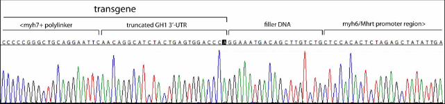
.CC-BY-NC-ND 4.0 International licensepeer-reviewed) is the author/funder. It is made available under aThe copyright holder for this preprint (which was not. http://dx.doi.org/10.1101/075671doi: bioRxiv preprint first posted online Sep. 16, 2016;


![Mutant SOD1 Transgenic Rodents as Models of FALS NDI G93A [b].pdfVariables in the Clinical Phenotype of the Drexel University School of Medicine G93A SOD1 Transgenic Mouse Colony1](https://static.documents.pub/doc/80x56/5f59b9b96e1b173fdc4ad324/mutant-sod1-transgenic-rodents-as-models-of-ndi-g93a-bpdf-variables-in-the-clinical.jpg)
