

8/3/2019 Lecture 8 VIII Nerve
http://slidepdf.com/reader/full/lecture-8-viii-nerve 1/26
The VIII Cranial Nerve
(also known the Vestibulocochlear Nerve)-Made up of the Auditory branch from the
cochlea and the Vestibular branch from
the SSCs and Otolith organs

8/3/2019 Lecture 8 VIII Nerve
http://slidepdf.com/reader/full/lecture-8-viii-nerve 2/26
STRUCTURE
8/3/2019 Lecture 8 VIII Nerve
http://slidepdf.com/reader/full/lecture-8-viii-nerve 3/26
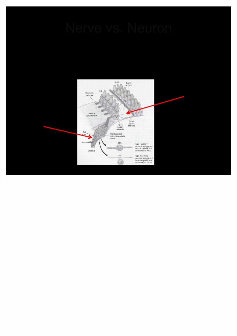
Nerve vs. Neuron
A neuron is one single cell
A nerve is a collection of neurons
Neuron
Nerve

8/3/2019 Lecture 8 VIII Nerve
http://slidepdf.com/reader/full/lecture-8-viii-nerve 4/26
VIII nerve
Links peripheral (outer, middle and inner ear)and central auditory system (brain and spinalcord)
Tonotopic (by frequency) arrangement of fibers from hair cells in the cochlea
Two types of afferent (ascending) fibers ± Type I: 85-95 % - connected to IHC- many fibers
connect to one IHC (many-to-one)
± Type II: 5-15 % - connected to OHC ± one fiber connects to many OHC (one-to-many)
Between 40,000-50,000 neurons
8/3/2019 Lecture 8 VIII Nerve
http://slidepdf.com/reader/full/lecture-8-viii-nerve 5/26
Types of Neurons Type I Radial Afferent
neurons
± Fewer IHC¶s than OHC¶s (3
rows vs. 1 row)
Type I neurons, however, are themost common in the auditory
system
± Many Type I afferent neurons
attach to 1 IHC (10-20 on each
IHC)
Type II Spiral Afferent
neurons
± Connect one-to-many to OHC¶s
from Donald Sinex USU
Spiral Ganglion
8/3/2019 Lecture 8 VIII Nerve
http://slidepdf.com/reader/full/lecture-8-viii-nerve 6/26
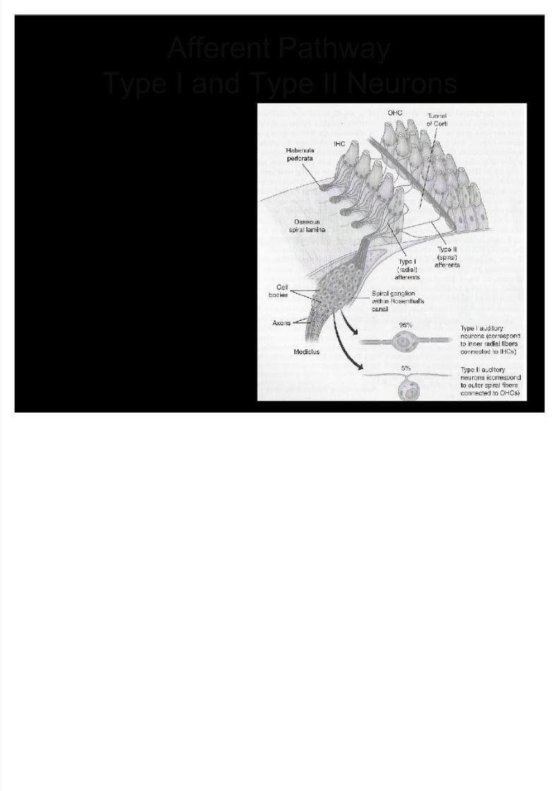
Afferent Pathway
Type I and Type II Neurons Type I Radial±many neuronsconnect to one IHC
± 85-95% of afferentneurons are radial
Type II Spiral ±one fiber connectsto many OHCs ± 5-15% of afferents
are spiral
8/3/2019 Lecture 8 VIII Nerve
http://slidepdf.com/reader/full/lecture-8-viii-nerve 7/26
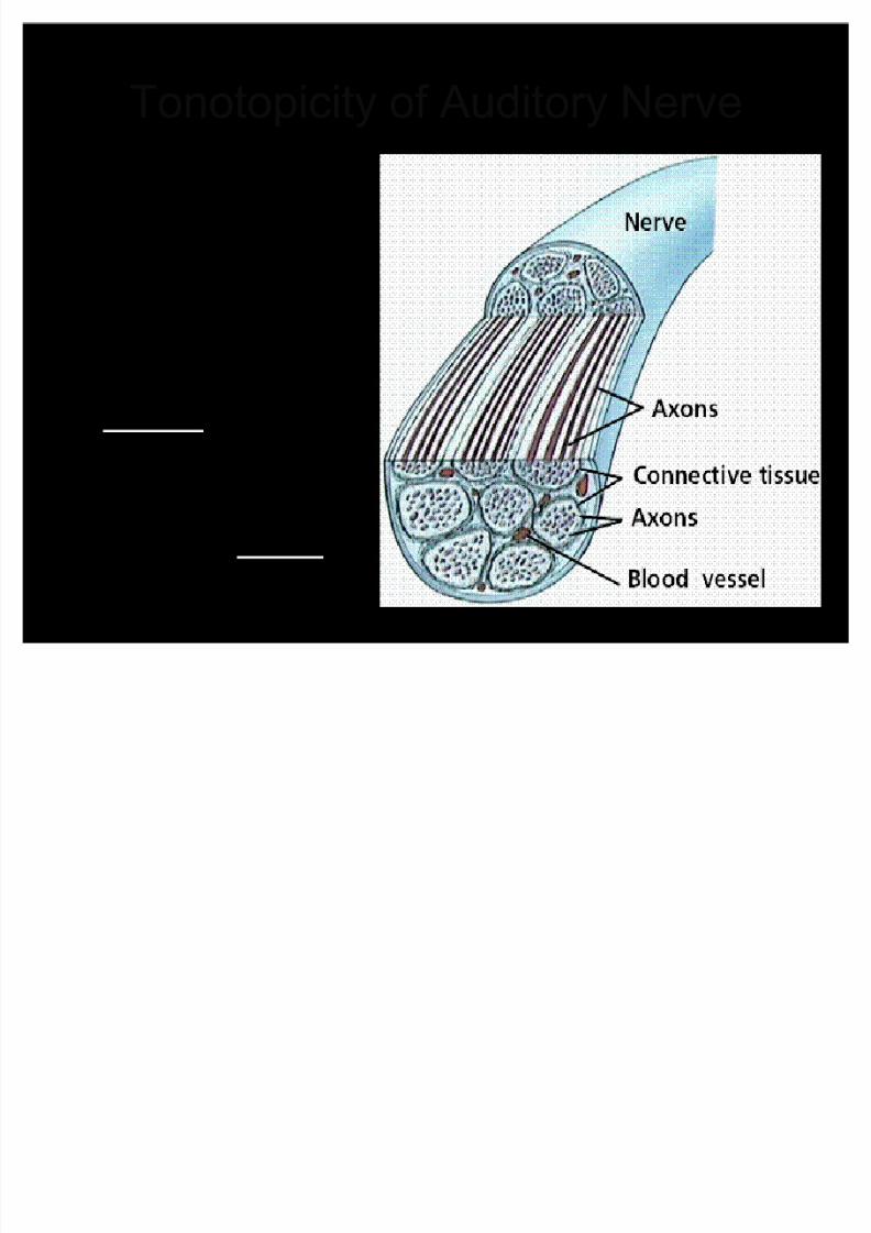
Tonotopicity of Auditory Nerve
Auditory nerve ismade up of manyneurons comingfrom the hair cells
Neurons comingfrom the base of the cochlea (HF)surround theoutside of the
auditory nerve Neurons from the
apex (LF) traveldown the center of the auditory nerve

8/3/2019 Lecture 8 VIII Nerve
http://slidepdf.com/reader/full/lecture-8-viii-nerve 8/26
A nerve is a bundle of individual
neurons
myelin
telodendria
8/3/2019 Lecture 8 VIII Nerve
http://slidepdf.com/reader/full/lecture-8-viii-nerve 9/26
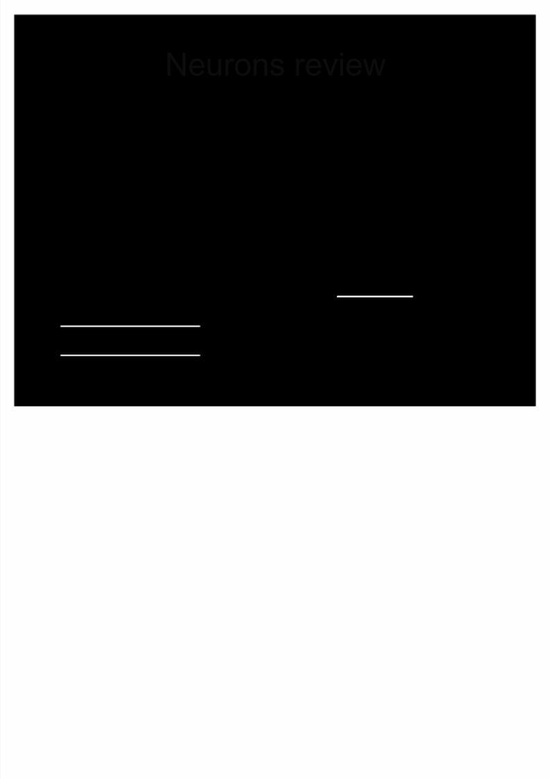
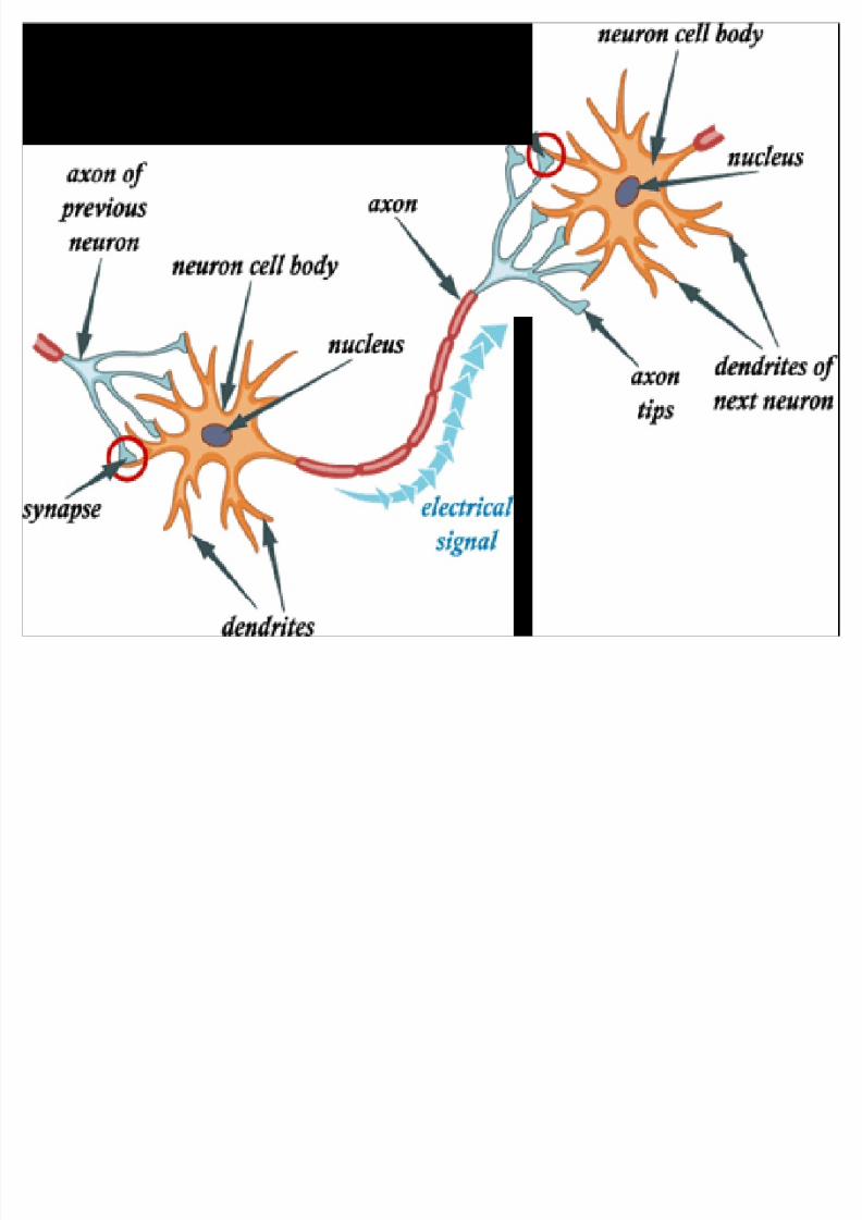
Neurons review
Neurons - building blocks of CNS
± Communicate information from neuron to neuron
(interneuron), organ, gland, muscle fiber (motor
neuron), or sensory receptor (sensory neuron)
Components of a neuron: dendrites, cell body,
axon, myelin, nodes of ranvier, telodendria
The dendrite is separated from the transmitting
axon by a narrow gap called a synapse Afferent neuron ± Ascending to the brain
Efferent neuron ± Descending from the brain

8/3/2019 Lecture 8 VIII Nerve
http://slidepdf.com/reader/full/lecture-8-viii-nerve 10/26
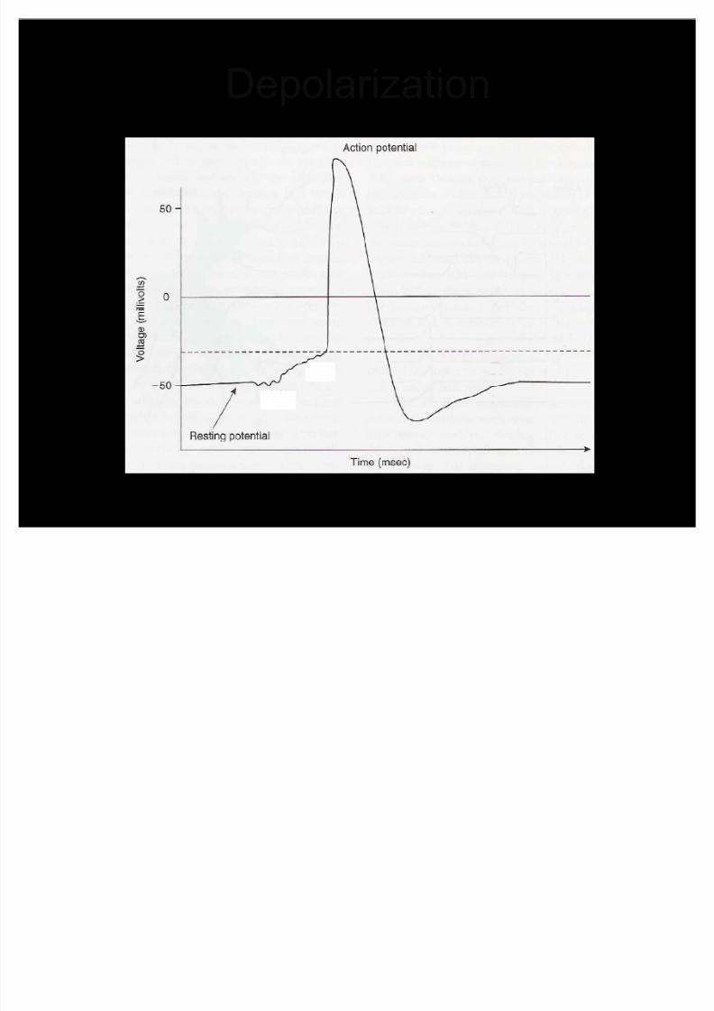
Neurons Review
Information is received from another cellthrough a synapse at the dendrites
Depolarization of the cell occurs
± If enough of a polarity change, impulse istransmitted along the axon to the telodendria
± Neurotransmitters are released into thesynapse and picked up by another neuron,muscle fiber, sensory organ, etc.
Input can be excitatory (passed on) or inhibitory (stopping)
8/3/2019 Lecture 8 VIII Nerve
http://slidepdf.com/reader/full/lecture-8-viii-nerve 11/26

8/3/2019 Lecture 8 VIII Nerve
http://slidepdf.com/reader/full/lecture-8-viii-nerve 12/26
Synapse
8/3/2019 Lecture 8 VIII Nerve
http://slidepdf.com/reader/full/lecture-8-viii-nerve 13/26
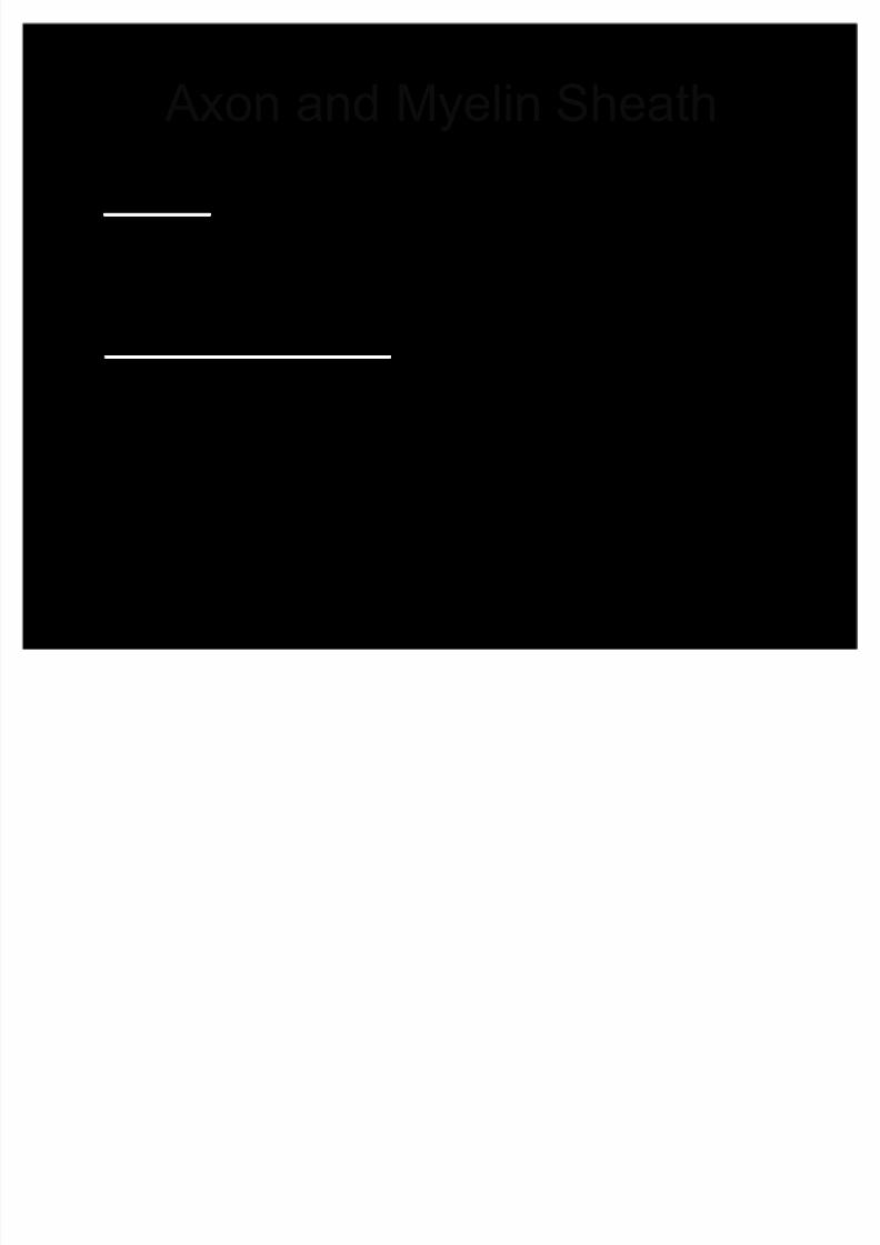
Axon and Myelin Sheath
Myelin ± a fatty substance that insulates
the axon and prevents the diffusion of ions
through the wall of the axon
Nodes of Ranvier ± electrical current
jumps from node to node rather than
diffusing along the entire length of the
membrane

8/3/2019 Lecture 8 VIII Nerve
http://slidepdf.com/reader/full/lecture-8-viii-nerve 14/26
Myelin, Nodes, Axon and
Bundle
A nerve is a bundle of axons
Nodes of Ranvier
8/3/2019 Lecture 8 VIII Nerve
http://slidepdf.com/reader/full/lecture-8-viii-nerve 15/26
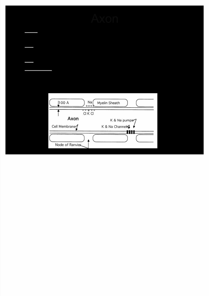
Axon Na+ positively charged sodium (pumped into the cell at
rest) K+ positively charged potassium (constantly being
pushed out of the cell at rest)
Cl- chlorine with extra electron, negatively charged
Cell Wall ± some ions flow through, others cannot fit
± Always want to reach equilibrium ± Resting potential -50mV (inside the cell)
3 K+ out for every 2 Na+ ions in = overall negative charge
8/3/2019 Lecture 8 VIII Nerve
http://slidepdf.com/reader/full/lecture-8-viii-nerve 16/26
Depolarization

8/3/2019 Lecture 8 VIII Nerve
http://slidepdf.com/reader/full/lecture-8-viii-nerve 17/26
A nerve impulse travels along the axon by a combination of
electrical and chemical conduction

8/3/2019 Lecture 8 VIII Nerve
http://slidepdf.com/reader/full/lecture-8-viii-nerve 18/26
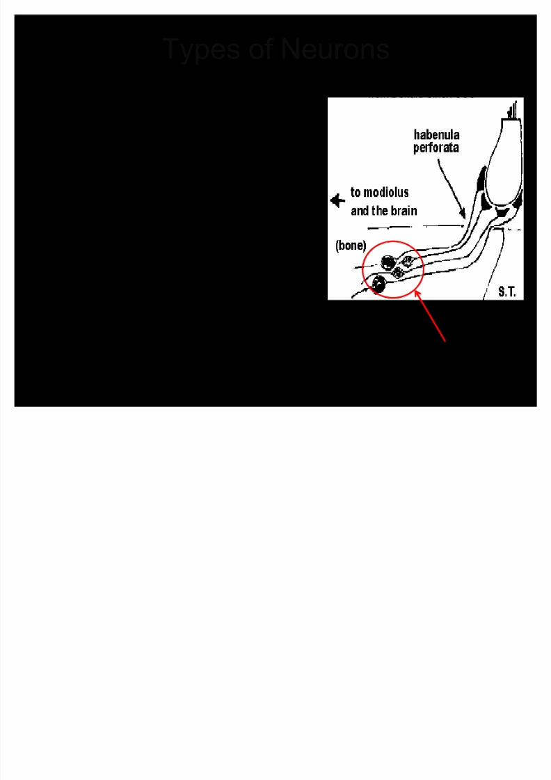
Nuclei
Soma - one cell body
Spiral Ganglion - a cluster of cell bodies
(collection of cell bodies)
Groups of neural cell bodies
in the nervous system are
called Spiral Ganglia
Soma
Spiral Ganglion
8/3/2019 Lecture 8 VIII Nerve
http://slidepdf.com/reader/full/lecture-8-viii-nerve 19/26
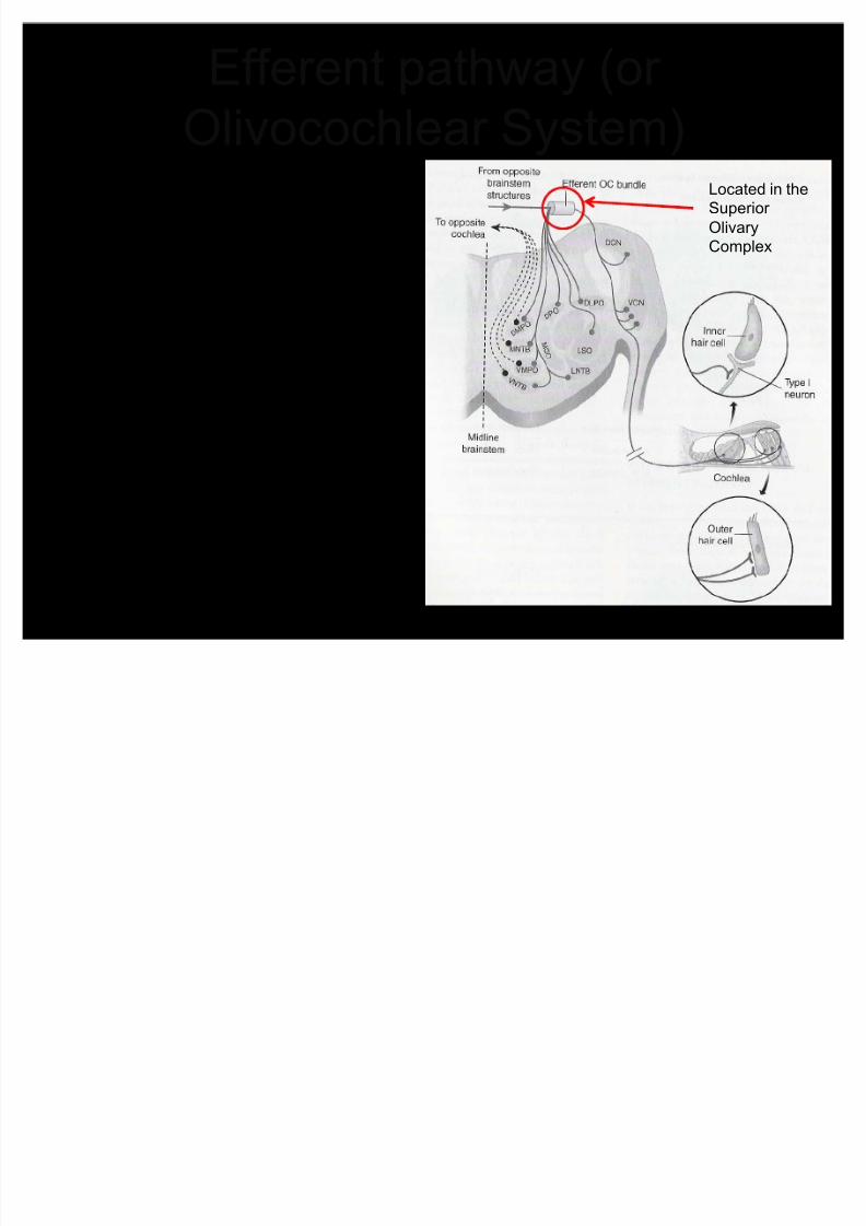
Efferent pathway (or
Olivocochlear S
ystem) Descendingpathway that carriesinformation from thebrainstem to thecochlea
Uncrossed pathwayfrom brainstem toipsilateral (same
side) ear Crossed pathway to
contralateral(opposite side) ear
Located in the
Superior
Olivary
Complex

8/3/2019 Lecture 8 VIII Nerve
http://slidepdf.com/reader/full/lecture-8-viii-nerve 20/26
FUNCTION
8/3/2019 Lecture 8 VIII Nerve
http://slidepdf.com/reader/full/lecture-8-viii-nerve 21/26
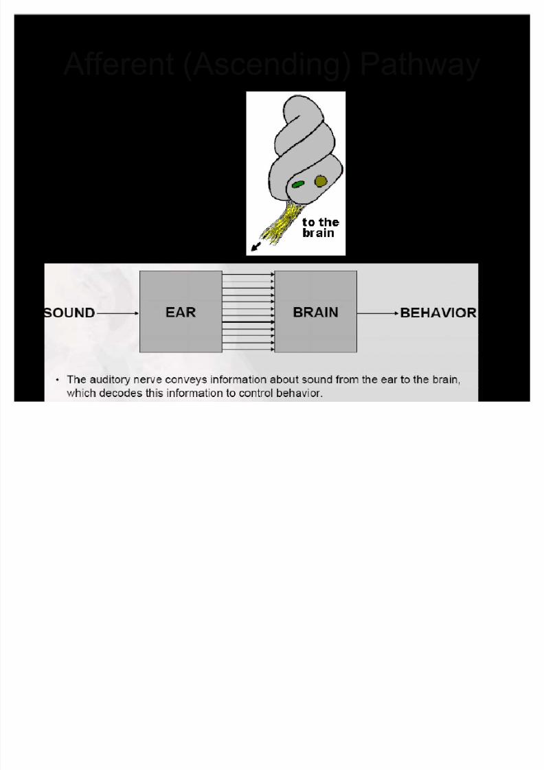
Afferent (Ascending) Pathway
The pathway that carries
infor mation from the
cochlea to the brain

8/3/2019 Lecture 8 VIII Nerve
http://slidepdf.com/reader/full/lecture-8-viii-nerve 22/26
Efferent (Descending) Pathway
Has inhibitory (opposite of excitatory) function
± Can stop neurotransmitters from HC¶s beingtransmitted to nerves
Originate in the superior olive and endprimarily on the HC¶s to modulate function
Not studied as much ± difficult to isolatedescending pathway in alive mammals
H
ypotheses of Function: ± 1) Helps with selective attention ± allows brain toinhibit/suppress/turn off certain backgroundnoises while attending to signal
± 2) Protects the ear from damage caused by
intense sounds

8/3/2019 Lecture 8 VIII Nerve
http://slidepdf.com/reader/full/lecture-8-viii-nerve 23/26
Tuning Curves
Frequency tuning curves of six differentfibers in the auditory nerve.
Each graph plots, across all frequencies
to which the fiber responds, i.e., the
minimum sound level required to increase
the fiber's firing rate above its
spontaneous firing level
The lowest point in the plot is the
weakest sound intensity to which the
neuron will respond
The frequency at this point is called the
neuronµs characteristic frequency Tips of afferent tuning curves
become narrower with increasing
characteristic frequency
8/3/2019 Lecture 8 VIII Nerve
http://slidepdf.com/reader/full/lecture-8-viii-nerve 24/26
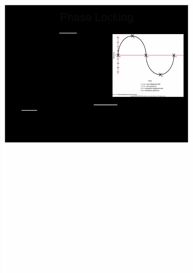
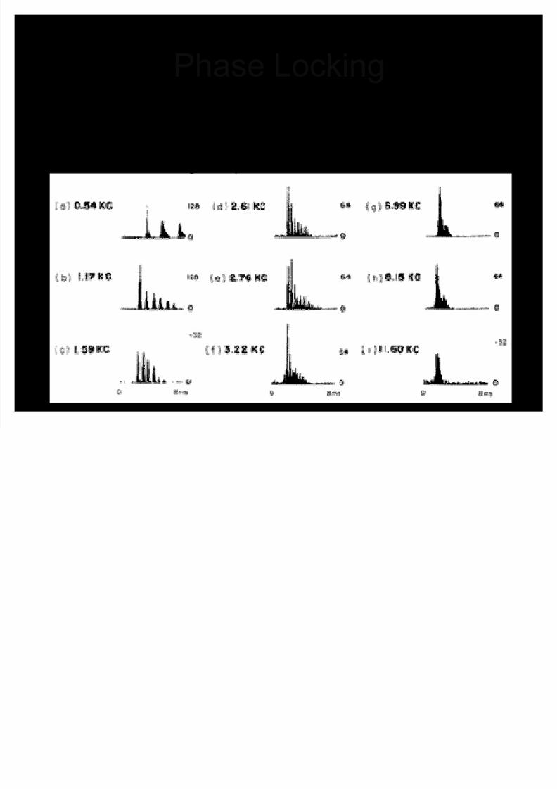
Phase Locking Remember, the phase of a waveform
depends on the point in the cyclebetween max displacement in onedirection and max displacement inanother
Neurons as a group tend to fire at
specific points on the waveform(i.e. at a specific phase/degree) ± Only true of low frequency sounds
Why not high frequency? ± Neurons can¶t keep up
± Once neurons fire, there is a refractory period where they need to rest beforefiring again
± With high frequency sounds, the nextcycle occurs before the refractory periodis over and cannot fire precisely at the
same point in the cycle

8/3/2019 Lecture 8 VIII Nerve
http://slidepdf.com/reader/full/lecture-8-viii-nerve 25/26
Phase Locking
Temporal response patterns of a low-frequency axon in the auditory nerve.
The stimulus waveform is indicated beneath the histograms, which show the
phase-locked responses to a 50-ms tone pulse of 260 Hz.
Note that the spikes are all timed to the same phase of the sinusoidalstimulus.
8/3/2019 Lecture 8 VIII Nerve
http://slidepdf.com/reader/full/lecture-8-viii-nerve 26/26
Phase Locking Poststimulus-time (PST) histograms of auditory nervefibers
At low frequencies, neurons have enough time to rest andfire again to the next cycle
Not able to at high frequencies


