
Low-dose exposure to bisphenol A and replacementbisphenol S induces precocious hypothalamicneurogenesis in embryonic zebrafishCassandra D. Kincha,b,c, Kingsley Ibhazehiebob,c, Joo-Hyun Jeongb,c, Hamid R. Habibia, and Deborah M. Kurraschb,c,1
Departments of aBiological Sciences and bMedical Genetics and cAlberta Children’s Hospital Research Institute, University of Calgary, Calgary, AB, CanadaT2N 4N1
Edited* by Joan V. Ruderman, Harvard Medical School, Boston, MA, and approved November 26, 2014 (received for review September 16, 2014)
Bisphenol A (BPA), a ubiquitous endocrine disruptor that is presentin many household products, has been linked to obesity, cancer,and, most relevant here, childhood neurological disorders such asanxiety and hyperactivity. However, how BPA exposure translatesinto these neurodevelopmental disorders remains poorly under-stood. Here, we used zebrafish to link BPA mechanistically todisease etiology. Strikingly, treatment of embryonic zebrafishwith very low-dose BPA (0.0068 μM, 1,000-fold lower than theaccepted human daily exposure) and bisphenol S (BPS), a commonanalog used in BPA-free products, resulted in 180% and 240%increases, respectively, in neuronal birth (neurogenesis) withinthe hypothalamus, a highly conserved brain region involved inhyperactivity. Furthermore, restricted BPA/BPS exposure specifi-cally during the neurogenic window caused later hyperactivebehaviors in zebrafish larvae. Unexpectedly, we show that BPA-mediated precocious neurogenesis and the concomitant behav-ioral phenotype were not dependent on predicted estrogen recep-tors but relied on androgen receptor-mediated up-regulation ofaromatase. Although human epidemiological results are stillemerging, an association between high maternal urinary BPA dur-ing gestation and hyperactivity and other behavioral disturbances inthe child has been suggested. Our studies here provide mechanisticsupport that the neurogenic period indeed may be a window ofvulnerability and uncovers previously unexplored avenues of researchinto how endocrine disruptors might perturb early brain develop-ment. Furthermore, our results show that BPA-free products are notnecessarily safer and support the removal of all bisphenols fromconsumer merchandise.
endocrine disruption | androgen receptor | aromatase | hyperactivity
In humans and rodent models, gestational exposure to bisphe-nol A (BPA) has been associated with increased risk of de-
veloping social (e.g., aggression), psychiatric (e.g., depression),and behavioral (e.g., hyperactivity) challenges later in life (1–7).BPA is a compound used in the production of diverse consumerproducts, ranging from baby bottles to thermal paper used forcredit card receipts (8–10). Even though adults can experienceadverse health following continued exposure to BPA, the fetalbrain is especially vulnerable because of an immature xenobiotic-metabolizing system and blood–brain barrier (11, 12). Exactlyhow BPA exposure in utero translates into neurodevelopmentaldisorders later in life is only beginning to be explored (13–15).Despite a wide body of research illustrating adverse effects of
BPA, controversy exists around the true effects of low-dose ex-posure, as is most often the case in humans. In accordance withstandardized toxicological testing procedures, government agen-cies in the United States (the US Environmental ProtectionAgency, USEPA), Canada (Health Canada), and Europe (theEuropean Food Safety Authority, EFSA) have established toler-able daily intake levels, ranging from 25–50 μg BPA·kg bodyweight−1·d−1 (16–18). Given these restrictions and societal pres-sure, manufacturers seeking BPA alternatives have turned toprimarily bisphenol S (BPS) to produce “BPA-free” products (19).
Indeed, a strong negative correlation between BPA and BPSlevels exists, whereby thermal paper that contained high quan-tities of BPA (milligrams per gram) demonstrated low quanti-ties of BPS (nanograms per gram), and vice versa, suggestingthat BPS is the primary replacement for BPA in thermal receipts(20). A recent examination of urine samples in the United Statesand Asia confirmed previous work showing that 93% of people haddetectable levels of BPA but surprisingly showed that 81% haddetectable levels of BPS, illustrating the wide-spread use of thispoorly known bisphenol analog in consumer products (21).The physiological effects of BPA on adults are well docu-
mented, but the mode of BPA action on the developing brain hasyet to be defined clearly, especially in the hypothalamus, whichplays a known role in neuroendocrine disorders that are on therise, including obesity and precocious puberty, as well as anxietyand hyperactivity (1, 2, 4, 10, 22). BPA is commonly thought toexert its effects by acting as a weak estrogen receptor (ER) ag-onist (8), although antagonism at androgen receptors (ARs) andthyroid receptors (ThRs) has been shown also (23). Proliferatingcells in the developing hypothalamus express ERs and ARs (24,25), and a role for sex steroids (e.g., estrogen, testosterone) inregulating neurogenesis is emerging (26–29). Although resultsare mixed (30, 31), neurobehavioral studies in humans suggestthat the prenatal period could be a window of BPA vulnerability(1–4, 7). Given that BPA is a known endocrine disruptor andthat steroid hormones increasingly are being shown to play a rolein cell differentiation, our objective here was to determine whetherBPA-mediated behavioral phenotypes were the consequence
Significance
Here we demonstrate that bisphenol A (BPA) exposure duringa time point analogous to the second trimester in humans hasreal and measurable effects on brain development and behav-ior. Furthermore, our study is the first, to our knowledge, toshow that bisphenol S, a replacement used in BPA-free prod-ucts, equally affects neurodevelopment. These findings sug-gest that BPA-free products are not necessarily safe andsupport a societal push to remove all structurally similar bisphe-nol analogues and other compounds with endocrine-disruptiveactivity from consumer goods. Our data here, combined withover a dozen physiological and behavioral human studies thatbegin to point to the prenatal period as a BPA window of vul-nerability, suggest that pregnant mothers limit exposure to plas-tics and receipts.
Author contributions: C.D.K., H.R.H., and D.M.K. designed research; C.D.K., K.I., and J.-H.J.performed research; C.D.K. analyzed data; C.D.K. and D.M.K. wrote the paper; and H.R.H.provided intellectual input on experimental design and data analysis.
The authors declare no conflict of interest.
*This Direct Submission article had a prearranged editor.
Freely available online through the PNAS open access option.1To whom correspondence should be addressed. Email: [email protected].
This article contains supporting information online at www.pnas.org/lookup/suppl/doi:10.1073/pnas.1417731112/-/DCSupplemental.
www.pnas.org/cgi/doi/10.1073/pnas.1417731112 PNAS | February 3, 2015 | vol. 112 | no. 5 | 1475–1480
ENVIRONMEN
TAL
SCIENCE
S
of altered neurogenesis, a developmental process that occursduring the second trimester of gestation. Further, we alsostudied whether BPS, a common replacement for BPA, likewisecauses precocious neurogenesis and concomitant hyperactivebehaviors.
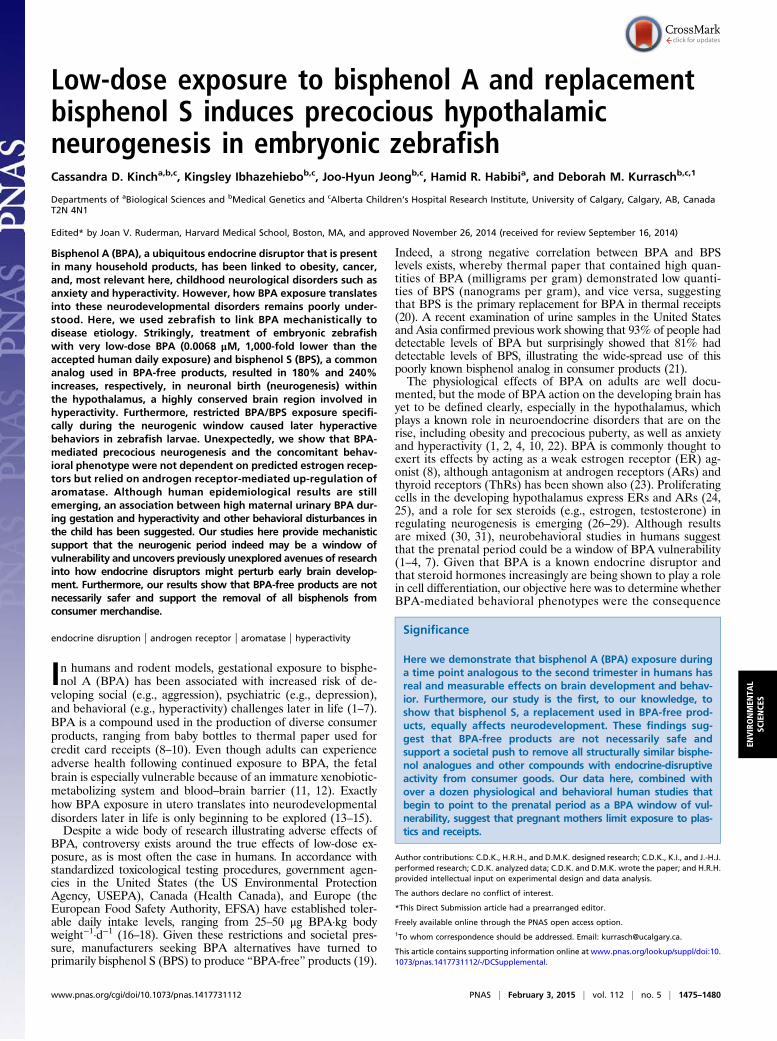
ResultsBPA Exposure During Hypothalamic Neurogenesis Induces Hyperactivity.First, we sought to recapitulate the human data using the neuro-developmentally similar zebrafish as a model and asked whetheracute BPA exposure during a time point analogous to gestationwould cause behavioral changes in larvae. Specifically, zebrafishembryos were exposed to BPA just before (10–16 h post fertiliza-tion; hpf), at the onset of (16–24 hpf), and at the peak of (24–36hpf) hypothalamic neurogenesis. BPA was washed out after eachrestricted time point, and zebrafish were assayed at the larval stage,5 d post fertilization (dpf), for changes in locomotor activity (Fig.1A; see SI Materials and Methods for further dosing information).Low-dose BPA exposure at 0.1 μM just before neurogenesis (10–16hpf) had no effect on locomotion but resulted in 2.8- and 2.9-foldincreases in locomotor activity when exposure occurred during theearly (16–24 hpf) and late (24–36 hpf) neurogenic periods, re-spectively (Fig. 1 B and C). Precedents in zebrafish demonstratethat this type of locomotor activity (i.e., hyperactivity burst) can bean indicator of anxiety-like behavior (32, 33). We also found thatchronic BPA exposure (across 0–5 dpf) resulted in an invertedU-shaped hyperactivity dose–response curve (Fig. S1A), consistentwith previous findings (32). Given that physiological responses toendocrine disruptors are known to be biphasic (10), with elevatedresponses at nanomolar concentrations, we next asked whethera lower dose of BPA also would cause hyperactive behavior. Ad-ditionally, because BPA is thought to act via ERs, we sought to
determine whether ER inhibition alleviated BPA effects onhyperactivity. Indeed, the same BPA dose found in a local waterbody (Oldman River, Alberta, Canada) (34) (0.0068 μM, consid-ered a very low dose) (Fig. 1D) and a more moderate BPA dose(1 μM) (Fig. S1B) caused hyperactivity that, unexpectedly, was notblocked by the broad ER ligand ICI 182,780 (hereafter, “ICI”)(Figs. 1D and 2C). This failure to antagonize BPA effects indicatedthat BPA might not act via classic nuclear (i.e., ERα, ERβ) ormembrane-bound (i.e., mER, GPR30) ERs. This same ICI dosewas sufficient to antagonize ER activity, because it blocked vtg1expression in gonad and liver tissues (Fig. S2).These data were puzzling, because BPA generally is consid-
ered to act via ERs. We therefore looked to other components ofthe estrogen synthesis pathway and asked whether aromatase B(AroB), the key enzyme for local estradiol synthesis, which isexpressed in hypothalamic progenitor cells, mediates the effectsof BPA. First, we relied on transient knockdown of AroB withtargeted morpholinos (MOs) (Fig. S3) and observed a completeblock of the BPA-mediated increase in locomotor activity at bothvery low (0.0068 μM) (Fig. 1E) and moderate (1 μM) (Fig. S1C)BPA doses. Second, we used the selective aromatase inhibitorfadrozole (FAD) (1 μM) to determine whether AroB catalyticactivity was required for BPA-mediated behavioral changes. In-deed, coexposure to very-low-dose BPA (0.0068 μM) + FAD(1 μM) (Fig. 1F) and moderate-dose BPA (1 μM) + FAD (1 μM)(Fig. S1D) during the hypothalamic neurogenic window (24–48hpf) lowered BPA-induced hyperactivity nearly to control levels,suggesting that AroB enzymatic activity indeed is required.
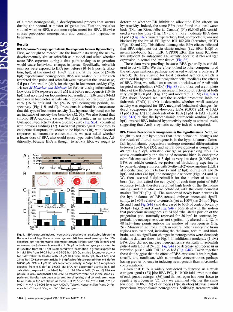
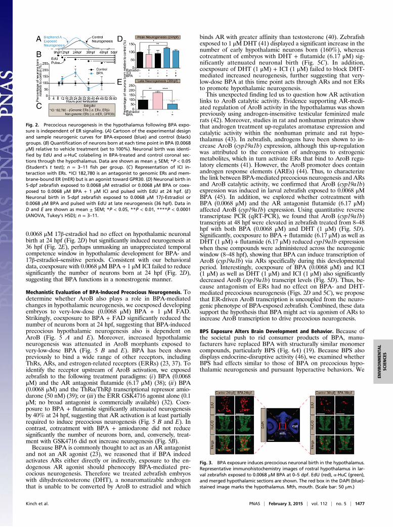
BPA Causes Precocious Neurogenesis in the Hypothalamus. Next, wesought to test our hypothesis that these behavioral changes arethe result of altered neurogenesis in the hypothalamus. Zebra-fish hypothalamic progenitors undergo neuronal differentiationbetween 18–36 hpf (35), and neural development is complete by48 hpf. By 5 dpf, zebrafish emerge as prey-seeking larva. Toassess quantitatively the timing of neuronal birth in embryoniczebrafish exposed from 0–5 dpf to very-low-dose (0.0068 μM)BPA or vehicle control, we performed birthdating experimentsby pulse-labeling embryos with 5-ethynyl-2′-deoxyuridine (EdU)at specific time points before (9 and 12 hpf), during (24 and 36hpf), and after (48 hpf) the neurogenic window (Figs. 2A and 3).We then assessed 5-dpf zebrafish for the number of neuronsborn (i.e., that exited the cell cycle) at each time point of EdUexposure (which therefore retained high levels of the thymidineanalog) and that also were colabeled with the early neuronalmarker HuC/D (Fig. 3). The number of newly born neurons inthe hypothalamus of BPA-treated embryos increased signifi-cantly, to 180% relative to controls (set at 100%), at 24 hpf (Figs.2B and 3 and Fig. S4A) and decreased to 60% of control levels by36 hpf (Figs. 2 and 3 and Fig. S4B), consistent with the notionthat precocious neurogenesis at 24 hpf exhausted a portion of theprogenitor pool normally reserved for 36 hpf. In contrast, hy-pothalamic neurogenesis was not significantly altered at 9, 12, or48 hpf—time points outside the window of neurogenesis (Fig.2B). Moreover, neuronal birth in several other embryonic brainregions was examined, including the thalamus, tectum, and hind-brain, and no significant changes in neurogenesis were detected;thalamic data are shown in Fig. 4. In addition, a moderate (1 μM)BPA dose did not increase neurogenesis statistically in zebrafishpulsed with EdU at 24 hpf (Fig. S4A) or decrease neurogenesis inzebrafish pulsed with EdU at 36 hpf (Fig. S4B). Taken togetherthese data suggest that the effect of BPA exposure is brain region-specific and nonlinear, with nanomolar concentrations perhapshaving greater potency in inducing neurogenesis than micromolarconcentrations.Given that BPA is widely considered to function as a weak
estrogen agonist (23) [the BPA EC50 is 10,000-fold lower than thatof endogenous estrogen (36)] and that estrogen has been shown toregulate neurogenesis (26, 28), we examined whether the samelow dose (0.0068 μM) of estrogen (17β-estradiol) likewise causedprecocious hypothalamic neurogenesis. Strikingly, treatment with
Fig. 1. BPA exposure induces hyperactive behaviors in larval zebrafish duringthe window of hypothalamic neurogenesis. (A) Treatment paradigm for BPAexposure. (B) Representative locomotor activity scribes with fish (green) andmovement (red) shown. Locomotion in 5-dpf controls and groups exposed to0.1 μM BPA from 10–16 hpf is compared with locomotion in groups exposed to0.1 μM BPA from 16–24 hpf and 24–36 hpf. (C) Quantified locomotor activityfor 5-dpf zebrafish treated with 0.1 μM BPA from 10–16 hpf, 16–24 hpf, and24–36 hpf. (D) Locomotor activity in 5-dpf zebrafish coexposed from 0–5 dpf to0.0068 μM BPA + 1 μM ICI. (E) Locomotor activity in 5-dpf AroB morphantsexposed from 0–5 dpf to 0.0068 μM BPA. (F) Locomotor activity in 5-dpfzebrafish coexposed from 24–48 hpf to 1 μM BPA + FAD. (D and E) BPA ex-posure in AroB morphants and BPA+ICI treatment were run in the same ex-periment. Results have been separated for simplicity, and controls are showntwice. Data in C–F are shown as mean ± SEM; *P < 0.05, **P < 0.01, ***P <0.001, ****P < 0.0001 [one-way ANOVA, Tukey’s Honestly Significant Differ-ence test (Tukey’s HSD)]; n = 5–10 fish per group.
1476 | www.pnas.org/cgi/doi/10.1073/pnas.1417731112 Kinch et al.
0.0068 μM 17β-estradiol had no effect on hypothalamic neuronalbirth at 24 hpf (Fig. 2D) but significantly induced neurogenesis at36 hpf (Fig. 2E), perhaps unmasking an unappreciated temporalcompetence window in hypothalamic development for BPA- and17β-estradiol–sensitive periods. Consistent with our behavioraldata, coexposure with 0.0068 μMBPA + 1 μM ICI failed to reducesignificantly the number of neurons born at 24 hpf (Fig. 2D),suggesting that BPA functions in a nonestrogenic manner.
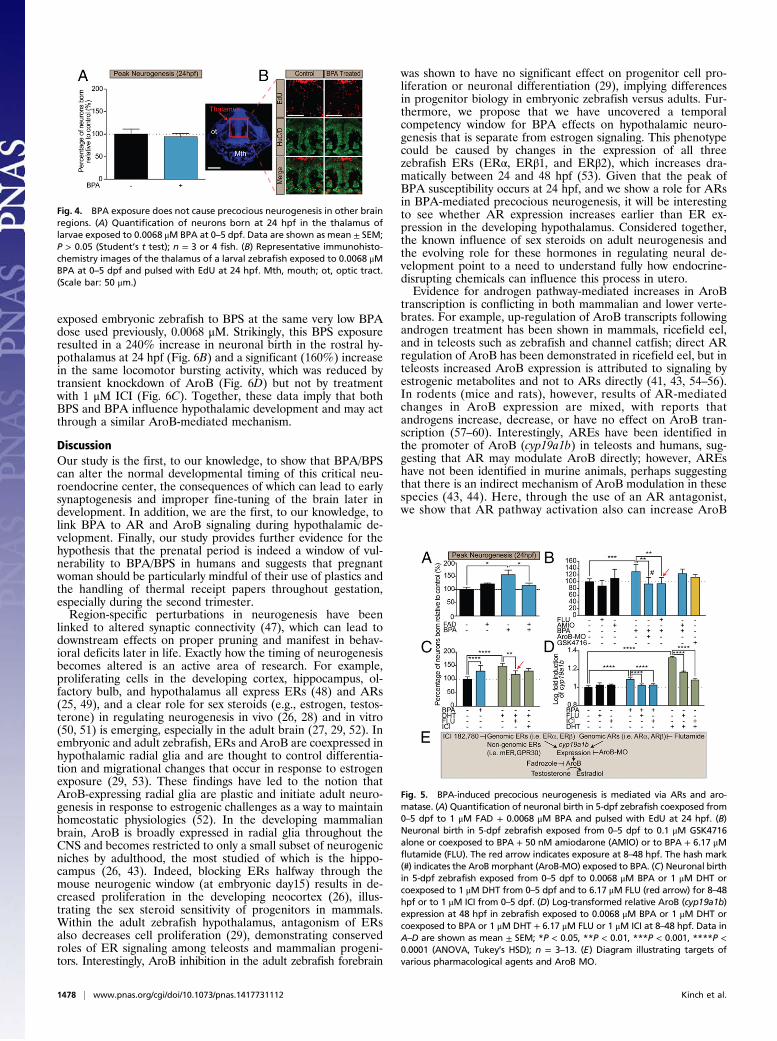
Mechanistic Evaluation of BPA-Induced Precocious Neurogenesis. Todetermine whether AroB also plays a role in BPA-mediatedchanges in hypothalamic neurogenesis, we coexposed developingembryos to very-low-dose (0.0068 μM) BPA + 1 μM FAD.Strikingly, coexposure to BPA + FAD significantly reduced thenumber of neurons born at 24 hpf, suggesting that BPA-inducedprecocious hypothalamic neurogenesis also is dependent onAroB (Fig. 5 A and E). Moreover, increased hypothalamicneurogenesis was attenuated in AroB morphants exposed tovery-low-dose BPA (Fig. 5 B and E). BPA has been shownpreviously to bind a wide range of other receptors, includingThRs, ARs, and estrogen-related receptors (ERRs) (23, 37). Toidentify the receptor upstream of AroB activation, we exposedzebrafish to the following treatment paradigms: (i) BPA (0.0068μM) and the AR antagonist flutamide (6.17 μM) (38); (ii) BPA(0.0068 μM) and the ThRα/ThRβ transcriptional repressor amio-darone (50 nM) (39); or (iii) the ERR GSK4716 agonist alone (0.1μM; no broad antagonist is commercially available) (32). Coex-posure to BPA + flutamide significantly attenuated neurogenesisby 40% at 24 hpf, suggesting that AR activation is at least partiallyrequired to induce precocious neurogenesis (Fig. 5 B and E). Incontrast, cotreatment with BPA + amiodarone did not reducesignificantly the number of neurons born, and, conversely, treat-ment with GSK4716 did not increase neurogenesis (Fig. 5B).Because BPA is commonly thought to act as an AR antagonist
and not an AR agonist (23), we reasoned that if BPA indeedactivates ARs either directly or indirectly, exposure to the en-dogenous AR agonist should phenocopy BPA-mediated pre-cocious neurogenesis. Therefore we treated zebrafish embryoswith dihydrotestosterone (DHT), a nonaromatizable androgenthat is unable to be converted by AroB to estradiol and which
binds AR with greater affinity than testosterone (40). Zebrafishexposed to 1 μMDHT (41) displayed a significant increase in thenumber of early hypothalamic neurons born (160%), whereascotreatment of embryos with DHT + flutamide (6.17 μM) sig-nificantly attenuated neuronal birth (Fig. 5C). In addition,coexposure of DHT (1 μM) + ICI (1 μM) failed to block DHT-mediated increased neurogenesis, further suggesting that very-low-dose BPA at this time point acts through ARs and not ERsto promote hypothalamic neurogenesis.This unexpected finding led us to question how AR activation
links to AroB catalytic activity. Evidence supporting AR-medi-ated regulation of AroB activity in the hypothalamus was shownpreviously using androgen-insensitive testicular feminized malerats (42). Moreover, studies in rat and nonhuman primates showthat androgen treatment up-regulates aromatase expression andcatalytic activity within the nonhuman primate and rat hypo-thalamus (43). In zebrafish, androgens have been shown to in-crease AroB (cyp19a1b) expression, although this up-regulationwas attributed to the conversion of androgens to estrogenicmetabolites, which in turn activate ERs that bind to AroB regu-latory elements (41). However, the AroB promoter does containandrogen response elements (AREs) (44). Thus, to characterizethe link between BPA-mediated precocious neurogenesis and ARsand AroB catalytic activity, we confirmed that AroB (cyp19a1b)expression was induced in larval zebrafish exposed to 0.0068 μMBPA (45). In addition, we explored whether cotreatment withBPA (0.0068 μM) and the AR antagonist flutamide (6.17 μM)affected AroB (cyp19a1b) expression. Using quantitative reversetranscriptase PCR (qRT-PCR), we found that AroB (cyp19a1b)transcripts at 48 hpf were elevated in zebrafish treated from 8–48hpf with both BPA (0.0068 μM) and DHT (1 μM) (Fig. 5D).Significantly, coexposure to BPA + flutamide (6.17 μM) as well asDHT (1 μM) + flutamide (6.17 μM) reduced cyp19a1b expressionwhen these compounds were administered across the neurogenicwindow (8–48 hpf), showing that BPA can induce transcription ofAroB (cyp19a1b) via ARs specifically during this developmentalperiod. Interestingly, coexposure of BPA (0.0068 μM) and ICI(1 μM) as well as DHT (1 μM) and ICI (1 μM) also significantlydecreased AroB (cyp19a1b) transcript levels (Fig. 5D). Thus, be-cause antagonism of ERs had no effect on BPA- and DHT-mediated precocious neurogenesis (Figs. 2D and 5C), we proposethat ER-driven AroB transcription is uncoupled from the neuro-genic phenotype of BPA-exposed zebrafish. Combined, these datasupport the hypothesis that BPA might act via agonism of ARs toincrease AroB transcription to drive precocious neurogenesis.
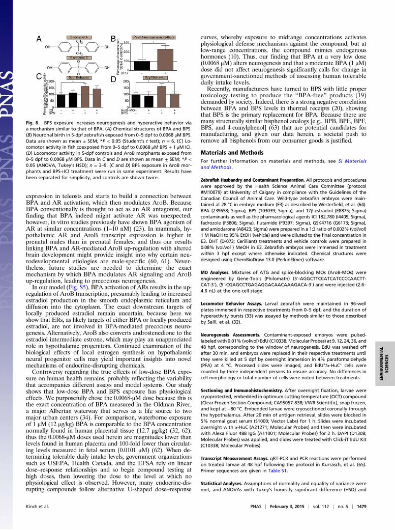
BPS Exposure Alters Brain Development and Behavior. Because ofthe societal push to rid consumer products of BPA, manu-facturers have replaced BPA with structurally similar monomercompounds, particularly BPS (Fig. 6A) (19). Because BPS alsodisplays endocrine-disruptive activity (46), we examined whetherBPS had effects similar to those of BPA on precocious hypo-thalamic neurogenesis and pursuant hyperactive behaviors. We
Fig. 2. Precocious neurogenesis in the hypothalamus following BPA expo-sure is independent of ER signaling. (A) Cartoon of the experimental designand sample neurogenic curves for BPA-exposed (blue) and control (black)groups. (B) Quantification of neurons born at each time point in BPA (0.0068μM) relative to vehicle treatment (set to 100%). Neuronal birth was identi-fied by EdU and α-HuC colabeling in BPA-treated and control coronal sec-tions through the hypothalamus. Data are shown as mean ± SEM; *P < 0.05(Student’s t test); n = 3–11 fish per group. (C) Representation of ICI in-teraction with ERs. *ICI 182,780 is an antagonist to genomic ERs and mem-brane-bound ER (mER) but is an agonist toward GPR30. (D) Neuronal birth in5-dpf zebrafish exposed to 0.0068 μM estradiol or 0.0068 μM BPA or coex-posed to 0.0068 μM BPA + 1 μM ICI and pulsed with EdU at 24 hpf. (E)Neuronal birth in 5-dpf zebrafish exposed to 0.0068 μM 17β-Estradiol or0.0068 μM BPA and pulsed with EdU at late neurogenesis (36 hpf). Data inD and E are shown as mean ± SEM; *P < 0.05, **P < 0.01, ****P < 0.0001(ANOVA, Tukey’s HSD); n = 3–11.
Fig. 3. BPA exposure induces precocious neuronal birth in the hypothalamus.Representative immunohistochemistry images of rostral hypothalamus in lar-val zebrafish exposed to 0.0068 μM BPA at 0–5 dpf. EdU (red), α-HuC (green),and merged hypothalamic sections are shown. The red box in the DAPI (blue)-stained image marks the hypothalamus. Mth, mouth. (Scale bar: 50 μm.)
Kinch et al. PNAS | February 3, 2015 | vol. 112 | no. 5 | 1477
ENVIRONMEN
TAL
SCIENCE
S
exposed embryonic zebrafish to BPS at the same very low BPAdose used previously, 0.0068 μM. Strikingly, this BPS exposureresulted in a 240% increase in neuronal birth in the rostral hy-pothalamus at 24 hpf (Fig. 6B) and a significant (160%) increasein the same locomotor bursting activity, which was reduced bytransient knockdown of AroB (Fig. 6D) but not by treatmentwith 1 μM ICI (Fig. 6C). Together, these data imply that bothBPS and BPA influence hypothalamic development and may actthrough a similar AroB-mediated mechanism.
DiscussionOur study is the first, to our knowledge, to show that BPA/BPScan alter the normal developmental timing of this critical neu-roendocrine center, the consequences of which can lead to earlysynaptogenesis and improper fine-tuning of the brain later indevelopment. In addition, we are the first, to our knowledge, tolink BPA to AR and AroB signaling during hypothalamic de-velopment. Finally, our study provides further evidence for thehypothesis that the prenatal period is indeed a window of vul-nerability to BPA/BPS in humans and suggests that pregnantwoman should be particularly mindful of their use of plastics andthe handling of thermal receipt papers throughout gestation,especially during the second trimester.Region-specific perturbations in neurogenesis have been
linked to altered synaptic connectivity (47), which can lead todownstream effects on proper pruning and manifest in behav-ioral deficits later in life. Exactly how the timing of neurogenesisbecomes altered is an active area of research. For example,proliferating cells in the developing cortex, hippocampus, ol-factory bulb, and hypothalamus all express ERs (48) and ARs(25, 49), and a clear role for sex steroids (e.g., estrogen, testos-terone) in regulating neurogenesis in vivo (26, 28) and in vitro(50, 51) is emerging, especially in the adult brain (27, 29, 52). Inembryonic and adult zebrafish, ERs and AroB are coexpressed inhypothalamic radial glia and are thought to control differentia-tion and migrational changes that occur in response to estrogenexposure (29, 53). These findings have led to the notion thatAroB-expressing radial glia are plastic and initiate adult neuro-genesis in response to estrogenic challenges as a way to maintainhomeostatic physiologies (52). In the developing mammalianbrain, AroB is broadly expressed in radial glia throughout theCNS and becomes restricted to only a small subset of neurogenicniches by adulthood, the most studied of which is the hippo-campus (26, 43). Indeed, blocking ERs halfway through themouse neurogenic window (at embryonic day15) results in de-creased proliferation in the developing neocortex (26), illus-trating the sex steroid sensitivity of progenitors in mammals.Within the adult zebrafish hypothalamus, antagonism of ERsalso decreases cell proliferation (29), demonstrating conservedroles of ER signaling among teleosts and mammalian progeni-tors. Interestingly, AroB inhibition in the adult zebrafish forebrain
was shown to have no significant effect on progenitor cell pro-liferation or neuronal differentiation (29), implying differencesin progenitor biology in embryonic zebrafish versus adults. Fur-thermore, we propose that we have uncovered a temporalcompetency window for BPA effects on hypothalamic neuro-genesis that is separate from estrogen signaling. This phenotypecould be caused by changes in the expression of all threezebrafish ERs (ERα, ERβ1, and ERβ2), which increases dra-matically between 24 and 48 hpf (53). Given that the peak ofBPA susceptibility occurs at 24 hpf, and we show a role for ARsin BPA-mediated precocious neurogenesis, it will be interestingto see whether AR expression increases earlier than ER ex-pression in the developing hypothalamus. Considered together,the known influence of sex steroids on adult neurogenesis andthe evolving role for these hormones in regulating neural de-velopment point to a need to understand fully how endocrine-disrupting chemicals can influence this process in utero.Evidence for androgen pathway-mediated increases in AroB
transcription is conflicting in both mammalian and lower verte-brates. For example, up-regulation of AroB transcripts followingandrogen treatment has been shown in mammals, ricefield eel,and in teleosts such as zebrafish and channel catfish; direct ARregulation of AroB has been demonstrated in ricefield eel, but inteleosts increased AroB expression is attributed to signaling byestrogenic metabolites and not to ARs directly (41, 43, 54–56).In rodents (mice and rats), however, results of AR-mediatedchanges in AroB expression are mixed, with reports thatandrogens increase, decrease, or have no effect on AroB tran-scription (57–60). Interestingly, AREs have been identified inthe promoter of AroB (cyp19a1b) in teleosts and humans, sug-gesting that AR may modulate AroB directly; however, AREshave not been identified in murine animals, perhaps suggestingthat there is an indirect mechanism of AroB modulation in thesespecies (43, 44). Here, through the use of an AR antagonist,we show that AR pathway activation also can increase AroB
Fig. 4. BPA exposure does not cause precocious neurogenesis in other brainregions. (A) Quantification of neurons born at 24 hpf in the thalamus oflarvae exposed to 0.0068 μM BPA at 0–5 dpf. Data are shown as mean ± SEM;P > 0.05 (Student’s t test); n = 3 or 4 fish. (B) Representative immunohisto-chemistry images of the thalamus of a larval zebrafish exposed to 0.0068 μMBPA at 0–5 dpf and pulsed with EdU at 24 hpf. Mth, mouth; ot, optic tract.(Scale bar: 50 μm.)
Fig. 5. BPA-induced precocious neurogenesis is mediated via ARs and aro-matase. (A) Quantification of neuronal birth in 5-dpf zebrafish coexposed from0–5 dpf to 1 μM FAD + 0.0068 μM BPA and pulsed with EdU at 24 hpf. (B)Neuronal birth in 5-dpf zebrafish exposed from 0–5 dpf to 0.1 μM GSK4716alone or coexposed to BPA + 50 nM amiodarone (AMIO) or to BPA + 6.17 μMflutamide (FLU). The red arrow indicates exposure at 8–48 hpf. The hash mark(#) indicates the AroB morphant (AroB-MO) exposed to BPA. (C) Neuronal birthin 5-dpf zebrafish exposed from 0–5 dpf to 0.0068 μM BPA or 1 μM DHT orcoexposed to 1 μM DHT from 0–5 dpf and to 6.17 μM FLU (red arrow) for 8–48hpf or to 1 μM ICI from 0–5 dpf. (D) Log-transformed relative AroB (cyp19a1b)expression at 48 hpf in zebrafish exposed to 0.0068 μM BPA or 1 μM DHT orcoexposed to BPA or 1 μM DHT + 6.17 μM FLU or 1 μM ICI at 8–48 hpf. Data inA–D are shown as mean ± SEM; *P < 0.05, **P < 0.01, ***P < 0.001, ****P <0.0001 (ANOVA, Tukey’s HSD); n = 3–13. (E) Diagram illustrating targets ofvarious pharmacological agents and AroB MO.
1478 | www.pnas.org/cgi/doi/10.1073/pnas.1417731112 Kinch et al.
expression in teleosts and starts to build a connection betweenBPA and AR activation, which then modulates AroB. BecauseBPA conventionally is thought to act as an AR antagonist, ourfinding that BPA indeed might activate AR was unexpected;however, in vitro studies previously have shown BPA agonism ofAR at similar concentrations (1–10 nM) (23). In mammals, hy-pothalamic AR and AroB transcript expression is higher inprenatal males than in prenatal females, and thus our resultslinking BPA and AR-mediated AroB up-regulation with alteredbrain development might provide insight into why certain neu-rodevelopmental etiologies are male-specific (60, 61). Never-theless, future studies are needed to determine the exactmechanism by which BPA modulates AR signaling and AroBup-regulation, leading to precocious neurogenesis.In our model (Fig. S5), BPA activation of ARs results in the up-
regulation of AroB transcription, presumably leading to increasedestradiol production in the smooth endoplasmic reticulum anddiffusion into the cytoplasm. The exact downstream targets oflocally produced estradiol remain uncertain, because here weshow that ERs, as likely targets of either BPA or locally producedestradiol, are not involved in BPA-mediated precocious neuro-genesis. Alternatively, AroB also converts androstenedione to theestradiol intermediate estrone, which may play an unappreciatedrole in hypothalamic progenitors. Continued examination of thebiological effects of local estrogen synthesis on hypothalamicneural progenitor cells may yield important insights into novelmechanisms of endocrine-disrupting chemicals.Controversy regarding the true effects of low-dose BPA expo-
sure on human health remains, probably reflecting the variabilitythat accompanies different assays and model systems. Our studyshows that low-dose BPA and BPS exposure has physiologicaleffects. We purposefully chose the 0.0068-μM dose because this isthe exact concentration of BPA measured in the Oldman River,a major Albertan waterway that serves as a life source to twomajor urban centers (34). For comparison, waterborne exposureof 1 μM (12 μg/kg) BPA is comparable to the BPA concentrationnormally found in human placental tissue (12.7 μg/kg) (32, 62);thus the 0.0068-μM doses used herein are magnitudes lower thanlevels found in human placenta and 100-fold lower than circulat-ing levels measured in fetal serum (0.0101 μM) (62). When de-termining tolerable daily intake levels, government organizationssuch as USEPA, Health Canada, and the EFSA rely on lineardose–response relationships and so begin compound testing athigh doses, then lowering the dose to the level at which nophysiological effect is observed. However, many endocrine-dis-rupting compounds follow alternative U-shaped dose–response
curves, whereby exposure to midrange concentrations activatesphysiological defense mechanisms against the compound, but atlow-range concentrations, the compound mimics endogenoushormones (10). Thus, our finding that BPA at a very low dose(0.0068 μM) alters neurogenesis and that a moderate BPA (1 μM)dose did not affect neurogenesis significantly calls for change ingovernment-sanctioned methods of assessing human tolerabledaily intake levels.Recently, manufacturers have turned to BPS with little proper
toxicology testing to produce the “BPA-free” products (19)demanded by society. Indeed, there is a strong negative correlationbetween BPA and BPS levels in thermal receipts (20), showingthat BPS is the primary replacement for BPA. Because there aremany structurally similar bisphenol analogs [e.g., BPB, BPE, BPF,BPS, and 4-cumylphenol] (63) that are potential candidates formanufacturing, and given our data herein, a societal push toremove all bisphenols from our consumer goods is justified.
Materials and MethodsFor further information on materials and methods, see SI Materialsand Methods.
Zebrafish Husbandry and Contaminant Preparation. All protocols and procedureswere approved by the Health Science Animal Care Committee (protocol#M10079) at University of Calgary in compliance with the Guidelines of theCanadian Council of Animal Care. Wild-type zebrafish embryos were main-tained at 28 °C in embryo medium (E3) as described by Westerfield, et al. (64).BPA (239658; Sigma), BPS (103039; Sigma), and 17β-estradiol (E8875; Sigma)contaminants as well as the pharmacological agents ICI 182,780 (I4409; Sigma),fadrozole (F3806; Sigma), flutamide (F9397; Sigma), GSK4716 (G6173; Sigma),and amiodarone (A8423; Sigma) were prepared in a 1:3 ratio of 0.002% (vol/vol)1 M NaOH to 95% EtOH (vehicle) and were diluted to the final concentration inE3. DHT (D-073; Cerilliant) treatments and vehicle controls were prepared in0.08% (vol/vol ) MeOH in E3. Zebrafish embryos were immersed in treatmentwithin 3 hpf except where otherwise indicated. Chemical structures weredesigned using ChemBioDraw 13.0 (PerkinElmer) software.
MO Analyses. Mixtures of ATG and splice-blocking MOs (AroB-MOs) wereengineered by Gene-Tools (Philomath) (5′-AGGCTTCCATCATCCCCAACTT-CAT-3′), (5′-CGAGCCTGAGAGGACAACAAAGACA-3′) and were injected (2.6–4.6 nL) at the one-cell stage.
Locomotor Behavior Assays. Larval zebrafish were maintained in 96-wellplates immersed in respective treatments from 0–5 dpf, and the duration ofhyperactivity bursts (33) was assayed by methods similar to those describedby Saili, et al. (32).
Neurogenesis Assessments. Contaminant-exposed embryos were pulsed-labeledwith 0.01% (vol/vol) EdU (C10338;Molecular Probes) at 9, 12, 24, 36, and48 hpf, corresponding to the window of neurogenesis. EdU was washed offafter 30 min, and embryos were replaced in their respective treatments untilthey were killed at 5 dpf by overnight immersion in 4% paraformaldehyde(PFA) at 4 °C. Processed slides were imaged, and EdU+/α-HuC+ cells werecounted by three independent persons to ensure accuracy. No differences incell morphology or total number of cells were noted between treatments.
Sectioning and Immunohistochemistry. After overnight fixation, larvae werecryoprotected, embedded in optimum cutting temperature (OCT) compound(Clear Frozen Section Compound; CA95057-838; VWR Scientific), snap frozen,and kept at −80 °C. Embedded larvae were cryosectioned coronally throughthe hypothalamus. After 20 min of antigen retrieval, slides were blocked in5% normal goat serum (S1000; Vector Labs) for 1 h. Slides were incubatedovernight with α-HuC (A21271; Molecular Probes) and then were incubatedwith Alexa Fluor 488 IgG (A11001; Molecular Probes) for 2 h. DAPI (D1308;Molecular Probes) was applied, and slides were treated with Click-iT EdU Kit(C10338; Molecular Probes).
Transcript Measurement Assays. qRT-PCR and PCR reactions were performedon treated larvae at 48 hpf following the protocol in Kurrasch, et al. (65).Primer sequences are given in Table S1.
Statistical Analyses. Assumptions of normality and equality of variance weremet, and ANOVAs with Tukey’s honestly significant difference (HSD) and
Fig. 6. BPS exposure increases neurogenesis and hyperactive behavior viaa mechanism similar to that of BPA. (A) Chemical structures of BPA and BPS.(B) Neuronal birth in 5-dpf zebrafish exposed from 0–5 dpf to 0.0068 μM BPS.Data are shown as mean ± SEM; *P < 0.05 (Student’s t test); n = 6. (C) Lo-comotor activity in fish coexposed from 0–5 dpf to 0.0068 μM BPS + 1 μM ICI.(D) Locomotor activity in 5-dpf controls and AroB morphants exposed from0–5 dpf to 0.0068 μM BPS. Data in C and D are shown as mean ± SEM; *P <0.05 (ANOVA, Tukey’s HSD); n = 3–9. (C and D) BPS exposure in AroB mor-phants and BPS+ICI treatment were run in same experiment. Results havebeen separated for simplicity, and controls are shown twice.
Kinch et al. PNAS | February 3, 2015 | vol. 112 | no. 5 | 1479
ENVIRONMEN
TAL
SCIENCE
S
Student’s t test were performed where indicated using Prism 6 (GraphPadSoftware). qRT-PCR data were log transformed before analyses.
ACKNOWLEDGMENTS. We thank Gaurav Kaushik and Natalia Klenin fortechnical assistance on this project. The research described in these
studies was supported by National Sciences and Engineering ResearchCouncil of Canada (NSERC) Grant DG386445 (to D.M.K.), NSERC DiscoveryGrants Program–Individual Grant 156910 (to H.R.H.), and NSERC Post-graduate Scholarships–Doctoral (2-year) Program Grant 459881-2014(to C.D.K.).
1. Braun JM, et al. (2009) Prenatal bisphenol A exposure and early childhood behavior.Environ Health Perspect 117(12):1945–1952.
2. Braun JM, et al. (2011) Impact of early-life bisphenol A exposure on behavior andexecutive function in children. Pediatrics 128(5):873–882.
3. Perera F, et al. (2012) Prenatal bisphenol a exposure and child behavior in an inner-city cohort. Environ Health Perspect 120(8):1190–1194.
4. Harley KG, et al. (2013) Prenatal and early childhood bisphenol A concentrations andbehavior in school-aged children. Environ Res 126:43–50.
5. Anderson OS, et al. (2013) Perinatal bisphenol A exposure promotes hyperactivity,lean body composition, and hormonal responses across the murine life course. FASEBJ 27(4):1784–1792.
6. Tian YH, Baek JH, Lee SY, Jang CG (2010) Prenatal and postnatal exposure to bisphenola induces anxiolytic behaviors and cognitive deficits in mice. Synapse 64(6):432–439.
7. Evans SF, et al. (2014) Prenatal bisphenol A exposure and maternally reported be-havior in boys and girls. Neurotoxicology 45C:91–99.
8. Krishnan AV, Stathis P, Permuth SF, Tokes L, Feldman D (1993) Bisphenol-A: An es-trogenic substance is released from polycarbonate flasks during autoclaving. Endo-crinology 132(6):2279–2286.
9. Liao C, Kannan K (2011) Widespread occurrence of bisphenol A in paper and paperproducts: Implications for human exposure. Environ Sci Technol 45(21):9372–9379.
10. Vandenberg LN, Maffini MV, Sonnenschein C, Rubin BS, Soto AM (2009) Bisphenol-Aand the great divide: A review of controversies in the field of endocrine disruption.Endocr Rev 30(1):75–95.
11. Nishikawa M, et al. (2010) Placental transfer of conjugated bisphenol A and sub-sequent reactivation in the rat fetus. Environ Health Perspect 118(9):1196–1203.
12. Adinolfi M (1985) The development of the human blood-CSF-brain barrier. Dev MedChild Neurol 27(4):532–537.
13. Kim K, et al. (2009) Potencies of bisphenol A on the neuronal differentiation andhippocampal neurogenesis. J Toxicol Environ Health A 72(21-22):1343–1351.
14. Komada M, et al. (2012) Maternal bisphenol A oral dosing relates to the acceleration ofneurogenesis in the developing neocortex of mouse fetuses. Toxicology 295(1-3):31–38.
15. Itoh K, Yaoi T, Fushiki S (2012) Bisphenol A, an endocrine-disrupting chemical, andbrain development. Neuropathology 32(4):447–457.
16. US Environmental Protection Agency (2008) Child-specific exposure factors hand-book, EPA/600/R-06/096F, September 2008 (National Center for Environmental As-sessment, Office of Research and Development, Washington, DC). Available at www.epa.gov/ncea. Accessed Dec. 13, 2013.
17. Health Canada (2009) Survey of bisphenol A in canned drink products. Available atwww.hc-sc.gc.ca/fn-an/securit/packagemball/bpa/bpa_survey-enquete-can-eng.php.Accessed December 13, 2013.
18. European Food Safety Authority (2007) EFSA re-evaluates safety of bisphenol A and setstolerable daily intake. Available at www.efsa.europa.eu/en/press/news/afc070129.htm. Ac-cessed December 13, 2013.
19. Grignard E, Lapenna S, Bremer S (2012) Weak estrogenic transcriptional activities ofBisphenol A and Bisphenol S. Toxicol In Vitro 26(5):727–731.
20. Liao C, Liu F, Kannan K (2012) Bisphenol s, a new bisphenol analogue, in paperproducts and currency bills and its association with bisphenol a residues. Environ SciTechnol 46(12):6515–6522.
21. Liao C, et al. (2012) Bisphenol S in urine from the United States and seven Asiancountries: Occurrence and human exposures. Environ Sci Technol 46(12):6860–6866.
22. Bourguignon JP, et al. (2010) Neuroendocrine disruption of pubertal timing and in-teractions between homeostasis of reproduction and energy balance. Mol Cell En-docrinol 324(1-2):110–120.
23. Wetherill YB, et al. (2007) In vitro molecular mechanisms of bisphenol A action. Re-prod Toxicol 24(2):178–198.
24. MacLusky NJ, Lieberburg I, McEwen BS (1979) The development of estrogen receptorsystems in the rat brain: Perinatal development. Brain Res 178(1):129–142.
25. Gorelick DA, Watson W, Halpern ME (2008) Androgen receptor gene expression inthe developing and adult zebrafish brain. Dev Dyn 237(10):2987–2995.
26. Martínez-Cerdeño V, Noctor SC, Kriegstein AR (2006) Estradiol stimulates progenitorcell division in the ventricular and subventricular zones of the embryonic neocortex.Eur J Neurosci 24(12):3475–3488.
27. Fowler CD, Liu Y, Wang Z (2008) Estrogen and adult neurogenesis in the amygdalaand hypothalamus. Brain Res Brain Res Rev 57(2):342–351.
28. Vosges M, et al. (2012) 17α-Ethinylestradiol and nonylphenol affect the developmentof forebrain GnRH neurons through an estrogen receptors-dependent pathway. Re-prod Toxicol 33(2):198–204.
29. Diotel N, et al. (2013) Effects of estradiol in adult neurogenesis and brain repair inzebrafish. Horm Behav 63(2):193–207.
30. Yolton K, et al. (2011) Prenatal exposure to bisphenol A and phthalates and infantneurobehavior. Neurotoxicol Teratol 33(5):558–566.
31. Miodovnik A, et al. (2011) Endocrine disruptors and childhood social impairment.Neurotoxicology 32(2):261–267.
32. Saili KS, et al. (2012) Neurodevelopmental low-dose bisphenol A exposure leads to earlylife-stage hyperactivity and learning deficits in adult zebrafish. Toxicology 291(1-3):83–92.
33. Kalueff AV, et al.; Zebrafish Neuroscience Research Consortium (2013) Towards a compre-hensive catalog of zebrafish behavior 1.0 and beyond. Zebrafish 10(1):70–86.
34. Sosiak A, Hebben T (2005) A Preliminary Survey of Pharmaceuticals and EndocrineDisrupting Compounds in Treated Municipal Wastewaters and Receiving Rivers ofAlberta. Technical Report AE T/773 (Alberta Environment, Edmonton, AB, Canada).
35. Nesan D, Vijayan MM (2013) Role of glucocorticoid in developmental programming:Evidence from zebrafish. Gen Comp Endocrinol 181:35–44.
36. Matthews JB, Twomey K, Zacharewski TR (2001) In vitro and in vivo interactions ofbisphenol A and its metabolite, bisphenol A glucuronide, with estrogen receptorsalpha and beta. Chem Res Toxicol 14(2):149–157.
37. Teng C, et al. (2013) Bisphenol A affects androgen receptor function via multiplemechanisms. Chem Biol Interact 203(3):556–564.
38. Schiller V, et al. (2013) Transcriptome alterations in zebrafish embryos after exposureto environmental estrogens and anti-androgens can reveal endocrine disruption.Reprod Toxicol 42:210–223.
39. Liu YW, Chan WK (2002) Thyroid hormones are important for embryonic to larvaltransitory phase in zebrafish. Differentiation 70(1):36–45.
40. Grino PB, Griffin JE, Wilson JD (1990) Testosterone at high concentrations interacts with thehuman androgen receptor similarly to dihydrotestosterone. Endocrinology 126(2):1165–1172.
41. Mouriec K, et al. (2009) Androgens upregulate cyp19a1b (aromatase B) gene ex-pression in the brain of zebrafish (Danio rerio) through estrogen receptors. Biol Re-prod 80(5):889–896.
42. Roselli CE, Salisbury RL, Resko JA (1987) Genetic evidence for androgen-dependent andindependent control of aromatase activity in the rat brain. Endocrinology 121(6):2205–2210.
43. Roselli CE, Liu M, Hurn PD (2009) Brain aromatization: Classic roles and new per-spectives. Semin Reprod Med 27(3):207–217.
44. Tong SK, Chung BC (2003) Analysis of zebrafish cyp19 promoters. J Steroid BiochemMol Biol 86(3-5):381–386.
45. Chung E, Genco MC, Megrelis L, Ruderman JV (2011) Effects of bisphenol A and tri-clocarban on brain-specific expression of aromatase in early zebrafish embryos. ProcNatl Acad Sci USA 108(43):17732–17737.
46. Ji K, Hong S, Kho Y, Choi K (2013) Effects of bisphenol s exposure on endocrinefunctions and reproduction of zebrafish. Environ Sci Technol 47(15):8793–8800.
47. Itoh K, Yaoi T, Fushiki S (2012) Bisphenol A, an endocrine-disrupting chemical, andbrain development. Neuropathology 32(4):447–457.
48. MacLusky NJ, Chaptal C, McEwen BS (1979) The development of estrogen receptor systemsin the rat brain and pituitary: Postnatal development. Brain Res 178(1):143–160.
49. Young WJ, Chang C (1998) Ontogeny and autoregulation of androgen receptormRNA expression in the nervous system. Endocrine 9(1):79–88.
50. Toran-Allerand CD (1976) Sex steroids and the development of the newborn mousehypothalamus and preoptic area in vitro: Implications for sexual differentiation. BrainRes 106(2):407–412.
51. Murashov AK, Pak ES, Hendricks WA, Tatko LM (2004) 17beta-Estradiol enhancesneuronal differentiation of mouse embryonic stem cells. FEBS Lett 569(1-3):165–168.
52. Diotel N, et al. (2010) Aromatase in the brain of teleost fish: Expression, regulationand putative functions. Front Neuroendocrinol 31(2):172–192.
53. Mouriec K, et al. (2009) Early regulation of brain aromatase (cyp19a1b) by estrogenreceptors during zebrafish development. Dev Dyn 238(10):2641–2651.
54. Zhang Y, et al. (2012) Androgen rather than estrogen up-regulates brain-type cytochromeP450 aromatase (cyp19a1b) gene via tissue-specific promoters in the hermaphrodite teleostricefield eel Monopterus albus. Mol Cell Endocrinol 350(1):125–135.
55. Lassiter CS, Linney E (2007) Embryonic expression and steroid regulation of brainaromatase cyp19a1b in zebrafish (Danio rerio). Zebrafish 4(1):49–57.
56. Kazeto Y, Trant JM (2005) Molecular biology of channel catfish brain cytochrome P450aromatase (CYP19A2): Cloning, preovulatory induction of gene expression, hormonal generegulation and analysis of promoter region. J Mol Endocrinol 35(3):571–583.
57. Karolczak M, Küppers E, Beyer C (1998) Developmental expression and regulation ofaromatase- and 5alpha-reductase type I mRNA in the male and female mouse hy-pothalamus. J Neuroendocrinol 10(4):267–274.
58. Abe-Dohmae S, Tanaka R, Harada N (1994) Cell type- and region-specific expression ofaromatase mRNA in cultured brain cells. Brain Res Mol Brain Res 24(1-4):153–158.
59. Lephart ED, Simpson ER, Ojeda SR (1992) Effects of Cyclic AMP and Andre-gens on invitro Brain Aromatase Enzyme Activity During Prenatal Development in the Rat.J Neuroendocrinol 4(1):29–36.
60. Negri-Cesi P, Colciago A, Motta M, Martini L, Celotti F (2001) Aromatase expressionand activity in male and female cultured rat hypothalamic neurons: Effect of an-drogens. Mol Cell Endocrinol 178(1-2):1–10.
61. Beyer C, Hutchison JB (1997) Androgens stimulate the morphological maturation ofembryonic hypothalamic aromatase-immunoreactive neurons in the mouse. Brain ResDev Brain Res 98(1):74–81.
62. Schönfelder G, et al. (2002) Parent bisphenol A accumulation in the human maternal-fetal-placental unit. Environ Health Perspect 110(11):A703–A707.
63. Rosenmai AK, et al. (2014) Are structural analogues to bisphenol a safe alternatives?Toxicol Sci 139(1):35–47.
64. Westerfield M (2000) The Zebrafish Book. A Guide for the Laboratory Use of Zebrafish(Danio rerio) (Univ of Oregon Press, Eugene, OR), 4th Ed.
65. Kurrasch DM, Nevin LM, Wong JS, Baier H, Ingraham HA (2009) Neuroendocrinetranscriptional programs adapt dynamically to the supply and demand for neuro-peptides as revealed in NSF mutant zebrafish. Neural Dev 4(22):1–16.
1480 | www.pnas.org/cgi/doi/10.1073/pnas.1417731112 Kinch et al.


