

Manisha Mehta, M.D

On Feb 26 , 2008 , a 63 year old white male was referred for
• Red and painful right eye with decreased vision • Anterior uveitis not responding to topical steroids• With increasing posterior synechial formation
Past history:
•HLA-B27 positive recurrent anterior uveitis Affecting the right eye 2-3 times/ year Responding to steroids and cycloplegicsfor the past 10 years
•Macular edema OD

Medical history:
•Psoriatic arthritis and skin lesions treated with narrow beam UV radiation
•Chronic lymphoid leukemia for one year on treatment with Rituxan and Fludarabine
•Family history:Father: CancerGrandfather: DiabetesAunt: Arthritis
•Medications:IVIG (for treatment of peripheral neuropathy 2 years before CLL)Aspirin

Review of systems:
Fatigue
Poor appetite
Severe / recurrent nose bleeds
Skin rashes
Stiff joints
Painful or swollen joints
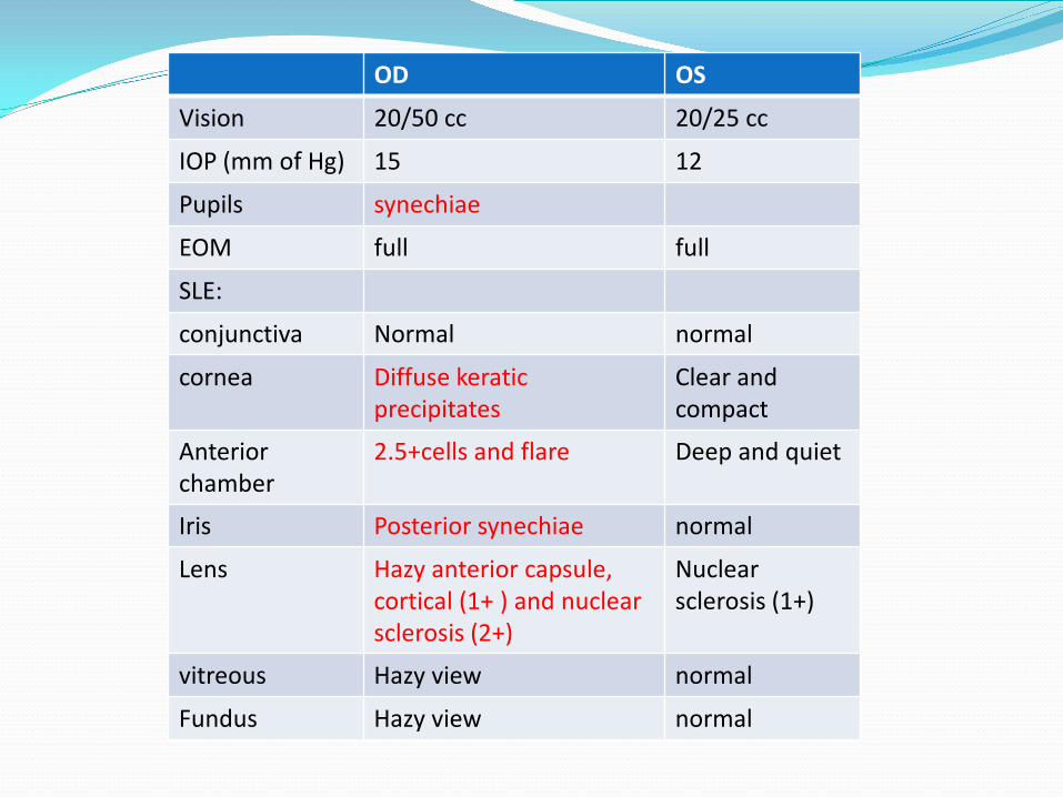
OD OS
Vision 20/50 cc 20/25 cc
IOP (mm of Hg) 15 12
Pupils synechiae
EOM full full
SLE:
conjunctiva Normal normal
cornea Diffuse keraticprecipitates
Clear and compact
Anterior chamber
2.5+cells and flare Deep and quiet
Iris Posterior synechiae normal
Lens Hazy anterior capsule, cortical (1+ ) and nuclear sclerosis (2+)
Nuclear sclerosis (1+)
vitreous Hazy view normal
Fundus Hazy view normal

Management:
Investigations:OCT:
Foveal thickness OD: 274 OS: 229
Medications:Posterior synechiae broken with a ‘ dynamite cocktail ‘(adrenalin, atropine and cocaine)Transeptal Kenalog (40 mg ) and IV Solumedrol ( 1 gm)Plan to start Humira (anti TNF alpha) after discussion with Oncologists
Enconopred Plus (Prednisolone Acetate 1%) q 1 hour ODXibrom (Bromfenac Sodium) bid ODHomatropine tid-qid OD
Time line Event Management
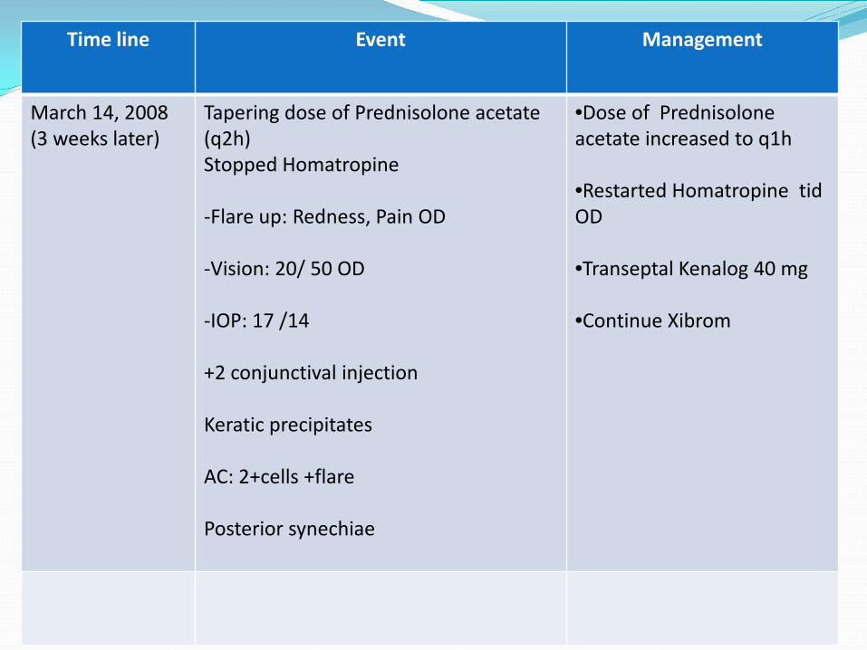
March 14, 2008(3 weeks later)
Tapering dose of Prednisolone acetate (q2h)Stopped Homatropine
-Flare up: Redness, Pain OD
-Vision: 20/ 50 OD
-IOP: 17 /14
+2 conjunctival injection
Keratic precipitates
AC: 2+cells +flare
Posterior synechiae
•Dose of Prednisoloneacetate increased to q1h
•Restarted Homatropine tidOD
•Transeptal Kenalog 40 mg
•Continue Xibrom

Time line Event Management
March 26, 2008(3 weeks later )
Pain and Redness OD: 1 dayHeadachesDecreased vision OD
-Vision: LP OD20/20 OS
-IOP: 60 OD22 OS
-Cornea: stromal edema 1+anterior synechiae
-AC: 2+ cell and flare
-Iris: Iris bombe
-Fundus: OD: RPE mottling at maculaAttenuated vessels
Emergent treatment:
50 cc of 25% Mannitol
500 mg Diamox
YAG iridotomy and AC paracentesis-22 mg Hg OD
Cytoxan infusion: dose 1 gm q 2 weekly
Cosopt (Timolol maleate/dorzolam)
Pred forte q2h OD
Homatropine q4 h OD
Xibrom bid OD

Time line Event Management
April 01, 2008(1 week later)
No pain or rednessBetter vision
Vision: cc 20/ 50
-IOP: 11 /11
-Pupils : round and reactive
-AC: rare cell, well formed chamber
-Iris: Posterior synechiae at 11 o’clock
-Fundus: OD: Vitreous strandsPale ODRPE mottling at maculaAttenuated vessels
WBC: 6.2 k/ul
Pred forte tapered to 6 times/day
Cosopt continued
Xibrom continued

Time line Event Management
May 07, 2008(5 weeks later)
Trouble with nocturnal vision with oncoming lights while driving
Vision: cc 20/40 OD
IOP: 14/14
Pupils : round and reactive
AC: deep and quiet
Lens: nuclear sclerosis and posterior subcapsular cataract OD
WBC: 4.3 k/ul
Cytoxan withheld
Pred forte bid
Xibrom continued

Time line Event Management
May 15, 2008(1 week later)
Fatigue Vision: 20/40 ccAC: PI open, posterior synechiae
IOP: 14
WBC: 5.6 k/ul
Cytoxan resumed at lower dose 750 mg
Pred forte qd
Cosopt bid
May 30, 2008(2 weeks later)
Fatigue and tiredness
Vision: 20/30AC: 1+ flareWBC: 2.9 k/ul
Cytoxan withheld
Solumedrol infusion +
June 05, 2008(1 week later)
No new redness or pain
Vision: cc 20/30IOP 16/14
WBC: 5.0 k/ul
Cytoxan (750 mg, q 2 weekly)SolumedrolPred forte qdXibromCosopt

Time line Event Management
July 7, 2008(1 month later)
CE/IOL OD Cytoxan reduced to 500 mg (every 3 weekly)
Solumedrol 1 gm
Xibrom
Cosopt
Aug 07, 2008(1 month later)
Increasing fatigue
Advised to decreaseCytoxan by Oncologist
Cytoxan changed to Methotrexate 15 mg/week
Xibrom
Cosopt
Pred forte qd OD
November 25, 2008(3.5 months later)
Vision: cc 20/20
IOP: 8/9
AC: deep and quiet
Methotrexate 15 mg/week
Cosopt
Xibrom

Time line Event Management
January, 2009 Restarted chemotherapy for CLL with Rituxan, Fludarabine and Cytoxan
Stopped MTXXibrom continuedCosopt continued
Feb 03, 2009 Vision cc 20/20
OCT:
Foveal thickness OD 248 OS 215IOP: 8/10OD quiet
Xibrom
Cosopt
April 28, 2009 Follow upVision: cc 20/20IOP: 12/10OD quiet
Xibrom reduced to qd
Cosopt
July 28, 2009 Follow up Vision cc 20/20IOP 12/11OD quiet
Xibrom qd
Cosopt bid

HLA-B27 syndromes
•HLA molecules are genetically encoded by the major histocompatibility complex (MHC) found on chromosome 6
•Role in immunity and in self-recognition in all nucleated cells and tissues
•Mechanisms of HLA-B27 associated inflammatory response:
-Molecular mimicry
-Arthritogenic peptide
-Innate etiology unrelated to HLA
-Marker closely linked to unidentified true immune gene responsible for inflammatory response.

HLA-B27 associated Acute Anterior Uveitis:
•Male predominance
•Age: 20- 40 yrs
•Associated with seronegative arthritic syndromes
-Ankylosing spondylitis
-Reactive arthritis
-Psoriatic arthritis
-Inflammatory bowel disease
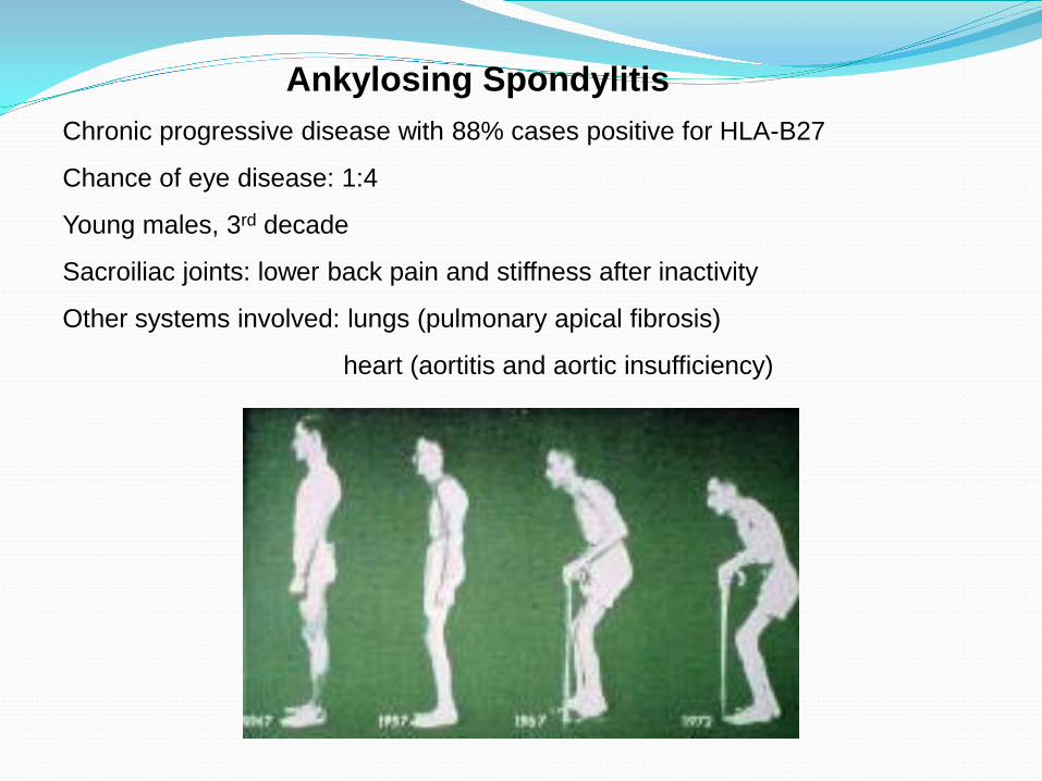
Ankylosing SpondylitisChronic progressive disease with 88% cases positive for HLA-B27
Chance of eye disease: 1:4
Young males, 3rd decade
Sacroiliac joints: lower back pain and stiffness after inactivity
Other systems involved: lungs (pulmonary apical fibrosis)
heart (aortitis and aortic insufficiency)

Reactive Arthritis (Reiter syndrome)
•18-40 years
• Acute nonpurulent arthritis secondary to an infection elsewhere
•Enteric (diarrhoea) or urogenital infections (dysuria) in HLA-B27 positive individuals
•60-85% individuals with reactive arthritis are HLA-B27 positive
•Organisms:
Shigella flexneri, Salmonella species, Yersinia enterocolitica, Campylobacter jejuni, Chlamydia trachomatis, Chlamydia pneumoniae, Clostridium difficile, Ureaplasma urealyticum

Reactive Arthritis•Non specific urethritis,
•Conjunctivitis (mucopurulent and papillary)
•Arthritis (knees, ankles, feet, wrists)
Minor diagnostic criteria: plantar fasciitis, Achilles tendonitis, nail bed pitting, palate ulcers and tongue ulcers
Major diagnostic criteria: keratoderma blennorrhagicum, circinate balanitis

Inflammatory bowel disease
•Ulcerative colitis (5-12%) and Crohn disease (2.4%) are associated with AAU
•50-60% cases with spondylitis in association with inflammatory bowel disease are positive for HLA-B27
•Small bony erosions and joint space narrowing
•Ankylosing spondylitis

Psoriatic Arthritis
•HLA-B27 is associated with the pustular form of psoriasis
•60-70% of cases with spondylitis associated psoriasis are HLA-B27 positive
•3-4 th decade
•Mild intermittent arthritis (sausage shaped digits) except arthritis mutilans
•Psoriatic skin lesions: look like eczema and seborrheic dermatitis

HLA-B27 associated Acute anterior uveitis
• Non granulomatous unilateral disease (pain, redness, photophobia)
• Corneal: fine KP, fibrin on endothelium, corneal edema, band keratopathy
•AC: fibrinous exudate in AC, cells and flare, iris bombe, hypopyon
•Rare posterior segment involvement
•Cystoid macular edema, disc edema, pars plana exudates, choroiditis

HLA-B27 associated Acute anterior uveitis
•Tendency to recur
•Complications: cataract, glaucoma, hypotony, CME, synechiae formation
•Poorer prognosis than HLA-B27 negative AAU

Treatment
•Steroids: Topical, periocular, intravitreal and oral
•Cycloplegics
•Immunosuppressive therapy: - refractory cases- steroid induced adverse effects, steroid dependant cases- vision threatening inflammation
-Azathioprine, Cyclophosphamide, Chlorambucil, Methotrexate, Cyclosporin
•Immunomodulation therapy: Infliximab (antiTNF-alpha), Etanercept (anti TNF alpha and beta)
•Sulfasalazine (in reactive arthritis)
• HLA-B27 derived peptide (B27PD) oral tolerance therapy
•Rheumatology consult

Thank you…..


