
Universidade do MinhoEscola de Psicologia
outubro de 2014
Sara Figueiredo Cruz
Neuropsychophysiological correlates of interactive behavior: evidence from infant development and implications for therapeutic relationship
Sara
Fig
ueire
do C
ruz
N
eu
rop
sych
op
hys
iolo
gic
al c
orr
ela
tes
of
inte
ract
ive
be
ha
vio
r: e
vid
en
ce
fro
m in
fan
t d
eve
lop
me
nt
an
d im
plic
ati
on
s fo
r th
era
pe
uti
c re
lati
on
ship
UM
inho
|201
4
This dissertation was financed by the Portuguese Foundation for Science and Technology (grant reference – SFRH/BD/68263/2010) supported by the Ministry of Science, Technology, and Higher Education, in the scope of – Advanced Training, reimbursed by the European Social Fund and byMSTHE funds.
Tese de Doutoramento em Psicologia Especialidade em Psicologia Clínica
Trabalho efetuado sob a orientação daDoutora Eugénia Maria Ribeiro Pereira e daDoutora Adriana da Conceição Soares Sampaio
Universidade do MinhoEscola de Psicologia
outubro de 2014
Sara Figueiredo Cruz
Neuropsychophysiological correlates of interactive behavior: evidence from infant development and implications for therapeutic relationship

iii
STATEMENT OF INTEGRITY
I hereby declare having conducted my thesis with integrity. I confirm that I have not used plagiarism or
any form of falsification of results in the process of the thesis elaboration.
I further declare that I have fully acknowledged the Code of Ethical Conduct of the University of Minho.
University of Minho, October 31st, 2014
Full name: Sara Figueiredo Cruz
Signature: _________________________________________________________________

iv
v
Acknowledgements
Foremost I am very grateful to my supervisors who accompanied me in the preparation of this
dissertation. To Dr. Eugénia Ribeiro for the guidance, kindness and knowledge transmitted along this
path and for encouraging me to pursue my research interests. To Dr. Adriana Sampaio for the wisdom,
autonomy and friendship that allowed me to grow as a researcher. Your teachings and mentoring are
extremely valuable and I will always take them with me. Likewise, I would like to thank School of
Psychology, particularly to Dr. Óscar Gonçalves and Dr. Carla Martins, for contributing to my scientific
growth and for all the opportunities given to me along these years.
My appreciation to Fundação para Ciência e Tecnologia (FCT) for the PhD grant
(SFRH/BD/68263/2010) which was essential for the fulfillment of the studies that constitute this
dissertation, and to Hospital Pedro Hispano, Matosinhos, for the generosity and kindness of all
professionals that helped me throughout the infant’s recruitment and data collection moments.
To all the participants, especially to the families and infants that accepted to collaborate with this
work, I am indebted with your generosity.
To Luis Jorge for the collaboration in the developmental studies data collection. I am thankful for
the friendship and for facilitating this journey.
I would like to thank Dr. Alberto Crego for the collaboration in this dissertation and for the
knowledge conveyed along this work. To Patricia Oliveira-Silva for the friendship, collaboration and help
throughout this process and to Dr. Fernando Ferreira-Santos for his knowledge and contributions to this
work. To Dr. Maria de Góis Eanes, I am grateful for the help and guidance in the implementation of the
developmental studies.
To Ana Osório, thank you for all the support and enlighten in all questions and doubts that
emerged along this path. But foremost I am thankful for your friendship and encouragement in fulfilling
my goals, particularly this dissertation.

vi
To all the Neuropsychophysiology Lab members, especially to Ana Ganho, Ana Raquel, Catarina,
Liliana, Luciana, Rosana and Sónia, I am thankful for your friendship, laughter and support along these
years. Thank you to Dr. Eugénia team, particularly to Dulce for the contributions to the therapeutic
relationship studies and to Nuno for the confidences and shared perspectives about the PhD process.
Thank you to all of my friends, although they are too many to enumerate they know who they are:
for the company, laughter and all the support. The shared experiences, emotions and endless
conversations transformed this journey into an easier process.
Last but not the least, a special thanks and appreciation to my family for all the encouragement. I
am forever grateful to my parents for the love, support and for always believing in this project. To my
sister, for always supporting me and believing I was able to finish this dissertation. I am for certain that
without your help, support and advice this dissertation would not be possible.
vii
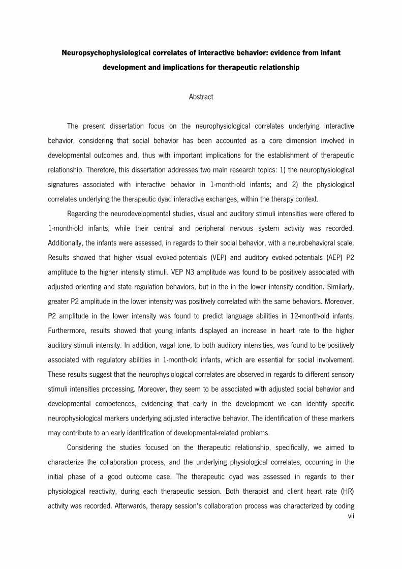
Neuropsychophysiological correlates of interactive behavior: evidence from infant
development and implications for therapeutic relationship
Abstract
The present dissertation focus on the neurophysiological correlates underlying interactive
behavior, considering that social behavior has been accounted as a core dimension involved in
developmental outcomes and, thus with important implications for the establishment of therapeutic
relationship. Therefore, this dissertation addresses two main research topics: 1) the neurophysiological
signatures associated with interactive behavior in 1-month-old infants; and 2) the physiological
correlates underlying the therapeutic dyad interactive exchanges, within the therapy context.
Regarding the neurodevelopmental studies, visual and auditory stimuli intensities were offered to
1-month-old infants, while their central and peripheral nervous system activity was recorded.
Additionally, the infants were assessed, in regards to their social behavior, with a neurobehavioral scale.
Results showed that higher visual evoked-potentials (VEP) and auditory evoked-potentials (AEP) P2
amplitude to the higher intensity stimuli. VEP N3 amplitude was found to be positively associated with
adjusted orienting and state regulation behaviors, but in the in the lower intensity condition. Similarly,
greater P2 amplitude in the lower intensity was positively correlated with the same behaviors. Moreover,
P2 amplitude in the lower intensity was found to predict language abilities in 12-month-old infants.
Furthermore, results showed that young infants displayed an increase in heart rate to the higher
auditory stimuli intensity. In addition, vagal tone, to both auditory intensities, was found to be positively
associated with regulatory abilities in 1-month-old infants, which are essential for social involvement.
These results suggest that the neurophysiological correlates are observed in regards to different sensory
stimuli intensities processing. Moreover, they seem to be associated with adjusted social behavior and
developmental competences, evidencing that early in the development we can identify specific
neurophysiological markers underlying adjusted interactive behavior. The identification of these markers
may contribute to an early identification of developmental-related problems.
Considering the studies focused on the therapeutic relationship, specifically, we aimed to
characterize the collaboration process, and the underlying physiological correlates, occurring in the
initial phase of a good outcome case. The therapeutic dyad was assessed in regards to their
physiological reactivity, during each therapeutic session. Both therapist and client heart rate (HR)
activity was recorded. Afterwards, therapy session’s collaboration process was characterized by coding
viii
the different therapeutic exchanges according to the Therapeutic Collaboration Coding System (TCCS).
In order to verify if HR pattern between the dyad was associated with the collaboration process,
physiological concordance and discordance was calculated for each therapeutic episodes identified.
Results showed that, collaboration process in the first session is mainly characterized by
collaborative exchanges, where therapist’s interventions that are coded as supporting problem and
client’s response as safety. Therapist’s challenging interventions occurring in this session were
commonly invalidated, producing non-collaborative episodes. As the initial phase of the therapy moves
on, novelty interventions introduced by the therapist tend to be more frequently validated. By the forth
session, the episodes now occurring are more often challenging, from the therapist’s side, followed by
safety or tolerable risk responses, from the client’s side (producing collaborative episodes). In general,
collaborative episodes, specifically supporting problem-safety, were accompanied by physiological
concordance, as therapist and client’s HR is similar. In non-collaborative episodes, commonly
invalidated challenges, the client tends to increase his HR, which is not accompanied by the therapist’s
HR and, therefore, physiological discordance is observed. Nevertheless, in collaborative episodes that
are characterized as validated challenging interventions, both therapist and client’s HR increased, which
may be associated with the client’s perceiving and accepting new perspectives. This acceptance is
followed by physiological signature (i.e. increase HR), which seems to be associated with cognitive and
emotional processes. These results suggest that specific collaboration exchanges are happen in the
initial phase of a good outcome case and, furthermore, such exchanges seems to be accompanied by
different HR activity patterns that are associated with collaborative and non-collaborative episodes.
Overall, this dissertation presents evidence that specific neurophysiological correlates are
associated with interactive behavior involved in infancy development that may offer a framework to
understand the dynamics of the physiological processes regarding interactive behaviors in the
therapeutic relationship.
ix
Correlatos neuropsicofisiológicos do comportamento interativo: evidência do
desenvolvimento infantil e implicações para a relação terapêutica
Resumo
A presente dissertação foca-se no estudo dos correlatos neurofisiológicos subjacentes ao
comportamento interativo, uma vez que o comportamento social tem sido largamente considerado
como um aspecto fundamental no desenvolvimento infantil e, da mesma forma, está associado ao
desenvolvimento do processo terapêutico, com importantes implicações para a relação entre terapeuta
e cliente. Assim, a presentação dissertação aborda dois tópicos de investigação: 1) os correlatos
neurofisiológicos associados a comportamentos sociais em crianças com 30 dias de vida; e 2) os
correlatos fisiológicos subjacentes à interação da díade terapêutica, no contexto da terapia.
Considerando os estudos apresentados sobre o neurodesenvolvimento infantil, duas intensidade
de estímulos visuais e auditivos foram oferecidos aos bebés, enquanto a atividade dos sistemas
nervoso central e periférico era registada. Adicionalmente, estes bebés foram avaliados considerando o
seu comportamento social, através de uma escala neurocomportamental. Os resultados indicam que
maior amplitude do componente de onda P2, na maior intensidade, é observada tanto em relação aos
potenciais evocados visuais (PEVs), como nos potenciais evocados auditivos (PEAs). A amplitude do
componente N3 nos PEVs parece estar positivamente associada com comportamentos de orientação e
de regulação dos estados, na intensidade mais baixa. De igual modo, a amplitude do componente P2
nos PEAs, na intensidade mais baixa, parece estar associada aos mesmos comportamentos interativos
e, mais ainda, a predizer o desenvolvimento da linguagem destas crianças a 1 ano de idade. Além
disso, os resultados indicam que os bebés apresentam um aumento da frequência cardíaca perante o
estímulos auditivos de a maior intensidade. Adicionalmente, a resposta vagal, em ambas as
intensidades, parece estar relacionada com comportamento de regulação dos estados nos bebés,
essencial para o envolvimento social. Estes resultados parecem indicar que correlatos neurofisiológicos
estão subjacentes ao processamento sensorial e associados ao comportamento social e a
competências desenvolvimentais. Assim, evidenciamos que desde cedo, podemos identificar
marcadores neurofisiológicos específicos que estão subjacentes ao comportamento interativo. A
identificação destes marcadores pode contribuir para a identificação precoce de problemas associados
ao desenvolvimento.
x
Relativamente aos estudos centrados na relação terapêutica, o principal objectivo era descrever
a processo de colaboração terapêutica e os correlatos fisiológicos subjacentes a este processo, durante
fase inicial de um processo terapêutico de caso de sucesso. A atividade cardíaca do terapeuta e cliente
foi registada simultaneamente ao longo das sessões terapêuticas. De seguida, cada sessão foi
codificada em relação ao processo de colaboração de acordo com o Sistema de Codificação da
Colaboração Terapêutica (SCCT). Seguidamente, e de forma a perceber se a atividade cardíaca está
associada ao processo colaborativo, concordância e discordância fisiológica foi calculada para cada
episódio terapêutico de colaboração codificado.
Os resultados mostram que na primeira sessão terapêutica o processo de colaboração é
essencialmente caracterizado por interações colaborativas, em que as intervenções do terapeuta são
maioritariamente suporte no problema e as respostas do cliente segurança. As intervenções de desafio,
por parte do terapeuta, são na maior parte invalidadas, resultando em episódios não-colaborativos.
Assim que a fase inicial da terapia avança, as intervenções suporte na novidade por parte do terapeuta
tendem a ser mais frequentemente validadas pelo cliente. Já na quarta sessão, os episódios mais
recorrentes são intervenções de desafio seguidas por respostas de segurança ou risco tolerável
(episódios colaborativos). Em relação à atividade fisiológica, de uma forma geral, os episódios
colaborativos suporte no problema-segurança são acompanhados por concordância fisiológica, uma
vez que a frequência cardíaca do terapeuta e cliente é semelhante. Em episódios não-colaborativos,
maioritariamente desafios invalidados, o cliente apresenta um aumento da frequência cardíaca, o que
não é observado no terapeuta, e, portanto, há discordância fisiológica. Em intervenções de desafio que
são validadas produzindo episódios colaborativos, a frequência cardíaca da díade aumenta
simultaneamente, o que parece estar indicar que o cliente percebe e aceita novas perspectivas
introduzidas pelo terapeuta. A aceitação de novidade pelo cliente parece ser acompanhada um padrão
fisiológico (aumento de frequência cardíaca) que parece estar associado a processos cognitivos e
emocionais. Estes resultados sugerem episódios específicos de colaboração terapêutica ocorrem na
fase inicial da terapia de um caso de sucesso e, adicionalmente, estes episódios parecem ser
acompanhados por diferentes padrões de atividade cardíaca associada a episódios colaborativos e não-
colaborativos.
De uma forma geral, esta dissertação apresenta evidência para a existência de correlatos
neurofisiológicos específicos que estão associados ao desenvolvimento do comportamento interativo
em crianças e que podem permitir uma melhor compreensão dos correlatos fisiológicos envolvidos nas
interações terapêuticas.
xi
INDEX
Introduction .................................................................................................................. 1
CHAPTER 1 Neuropsychophysiological correlates of interactive behavior: evidence from
infant’s development ..................................................................................................... 7
Neural and psychophysiological correlates of infant’s socio-cognitive development .... 9
A VEP study in sleeping and awaked one-month-old infants and its relation with social
behavior .................................................................................................................. 49
Auditory neural correlates of infant's development: a longitudinal study ................... 69
Vagal regulation to auditory stimuli is associated with neurobehavioral regulatory
abilities in one-month-old infants .............................................................................. 93
Conclusion ............................................................................................................ 111
CHAPTER 2 Neuropsychophysiological correlates of interactive behavior: implications for
therapeutic relationship ............................................................................................ 121
Physiological correlates of therapeutic relationship ............................................... 123
Therapeutic collaboration and the underlying physiological profile in the first session of
psychotherapy ....................................................................................................... 137
Therapeutic Collaboration and the underlying physiological profile: concordance and
discordance in the early phase of a CBT good outcome case .................................. 167
Conclusion ............................................................................................................ 193
General Conclusion ................................................................................................... 199

xii
xiii
LIST OF FIGURES CHAPTER 1 Neuropsychophysiological correlates of interactive behavior: evidence from
infant’s development
A VEP study in sleeping and awaked one-month-old infants and its relation with social
behavior
Figure 1 - VEP response in sleeping and awaked infants to the 2 visual stimuli intensities. . .. 57
Auditory neural correlates of infant's development: a longitudinal study
Figure 1 - Infant's AEPs response regarding the two auditory stimuli intensities. .................. 78
Figure 2 Graphic representation of the correlations between the P2 response and the behavior
clusters.. .................................................................................................................... 80
Vagal regulation to auditory stimuli is associated with neurobehavioral regulatory
abilities in one-month-old infants
Figure 1 - Vagal regulation and its association with state regulation behavior. .................... 102
CHAPTER 2 Neuropsychophysiological correlates of interactive behavior: implications for
therapeutic relationship
Physiological correlates of therapeutic relationship
Figure 1 Client's developmental continuum according to the TZPD (Ribeiro et al., 2013). .. 127
Therapeutic collaboration and the underlying physiological profile in the first
session of psychotherapy
Figure 1 Therapist's interventions across the therapeutic session as indexes. .................... 153
Figure 2 - Client's responses across the therapeutic session presented as indexes............. 154
Figure 3 Indexes (%) of the therapeutic exchanges across the therapeutic session.. ........... 155
Figure 4 Grid of the trajectory of the therapeutic exchanges. ............................................. 156
Figure 5 Therapist's and client's physiological pattern across the session and considering the
different TCCS episodes. ................................................................................................... 158
Therapeutic Collaboration and the underlying physiological profile: concordance
and discordance in the early phase of a CBT good outcome case
Figure 1 Therapist's interventions, client's responses and therapeutic collaborative exchanges
that occurred most frequently in the 4 therapeutic sessions.. ............................................. 178
Figure 2 SSGs method for the representation of the collaborative episodes occurring in the first
session ............................................................................................................................. 179
xiv
Figure 3 Heart rate (beats per minute) concordance/discordance between therapist and client
regarding SP-S, C-S, C-IR and C-TR exchanges, in the first session. .................................... 180
Figure 4 SSGs method for the representation of collaborative exchanges occurring in the
second session ................................................................................................................. 181
Figure 5 Heart rate concordance/discordance between therapist and client in SP-S, C-S, C-IR
and SI-S exchanges, in the second session. ....................................................................... 182
Figure 6 SSGs method for the representation of collaborative exchanges occurring in the third
session. ............................................................................................................................ 182
Figure 7 Heart rate concordance/discordance analysis between therapist and client in SP-S, C-
S, C-IR and C-TR exchanges, in the third session. ............................................................... 183
Figure 8 SSGs method for the representation of the collaborative exchanges occurring in the
forth session ..................................................................................................................... 184
Figure 9 Heart rate concordance/discordance analysis between therapist and client in SP-S, C-
S, C-IR and SI-S exchanges, in the forth session ................................................................. 184
xv
LIST OF TABLES CHAPTER 1 Neuropsychophysiological correlates of interactive behavior: evidence from
infant’s development
A VEP study in sleeping and awaked one-month-old infants and its relation with social
behavior
Table 1 Infant’s health characteristics at the time of recruitment and collection .................... 53
Table 2 Mean (SEs) values for N2, P2 and N3 peak amplitudes (µV) and latency (ms) recorded
at O1, O2 and Oz. ............................................................................................................... 58
Table 3 Infants' neurobehavioral profile considering all the NBAS clusters. .......................... 58
Table 4 Regression analyses results considering the VEPs components. .............................. 60
Auditory neural correlates of infant's development: a longitudinal study
Table 1 Infant's health information at the time of recruitment and collection. ....................... 73
Table 2 Mean (SEs) values for P2 and N2 peaks amplitude (µV) and latency (ms) recorded at
Cz. ...................................................................................................................................... 79
Table 3 NBAS behavior clusters considering its mean and standed deviation values. ............ 79
Table 4 Linear regression regarding P2 amplitude and latency values, in Cz, and behavior
clusters. .............................................................................................................................. 80
Table 5 Bayley-III cognitive, language and motor scale descriptive analysis. ........................ 81
Table 6 Regression analyses: association between AEPs and cognitive, language and motor
scales. ................................................................................................................................ 81
Vagal regulation to auditory stimuli is associated with neurobehavioral regulatory
abilities in one-month-old infants
Table 1 Infant's health information at the time of recruitment and collection. ....................... 97
Table 2 Mean (standard deviation) amplitude for baseline, HRV and RSA metrics. .............. 101
Table 3 NBAS behavior clusters considering its mean and standard deviation values. ......... 101
CHAPTER 2 Neuropsychophysiological correlates of interactive behavior: implications for
therapeutic relationship
Therapeutic collaboration and the underlying physiological profile in the first
session of psychotherapy
Table 1 Therapist's interventions. ...................................................................................... 145
Table 2 Client's responses. ................................................................................................ 146
Table 3 Therapeutic exchanges. ........................................................................................ 148

xvi
1
Introduction
From early on, research in the developmental field has been focusing on debating nature vs.
nurture question. It is undeniable that developmental psychologists are cautious when attributing the
emergence of different developmental pathways to purely biological or environmental experiences.
Indeed, professionals on the field postulate that both biological processes and experience factors
modulate the developmental changes occurring at multiple levels (i.e., neural and physiological
maturational processes) (Schwartz et al., 2012; Calkins & Fox, 2002; Calkins, Fox & Marshall, 1996).
From birth, infants unfold through different qualitative developmental changes, which are
translated into age-related specific behavioral, cognitive and socio-emotional characteristics. These
developmental changes are expected to be orderly, cumulative and directional as behavior moves
consistently towards greater organization and complexity (Sroufe, Cooper & Ganie). These behavioral
transitions, across infancy, are translated into adjusted interactive routines with the environment and
caregivers.
At a postnatal age, young infants interaction with the environment occurs mainly through
sensory experiences. Indeed, from early on, newborn infants interact with the environment through
their senses within their social network (Brazelton & Nugent, 1995). Specifically, infants display a set of
behaviors that allows them to experience, regulate and express emotions in secure relationships and
within the community (Zero to Three, 2001). The emergence of such capacities is synonymous of
healthy social and emotional development and mirrors the infant’s mental wellbeing (Sampaio & Lifter,
2014; Góis-Eanes, Gonçalves, Caldeira-da-Silva, & Sampaio, 2012). Moreover, evidence suggests that
these developmental abilities have neurobiological underpinnings that are molding infants’ future
social, emotional and cognitive response since birth (Congdon et al., 2012; Sheese, Voelker, Posner, &
Rothbart, 2009; Fox & Calkins, 2003).
Several studies have suggested that sensory processing abilities in young infants seem to be
associated with brain maturational processes (McGlone et al., 2013; Marcoux, 2011; Lippé, Martinez-
Montes, Arcand, & Lassonde, 2009; Atkinson, 2002)and with physiological reactivity competences
(Porges, 2011; Feldman & Eidelman, 2006; Bazhenova, Plonskaia & Porges, 2001). Furthermore,
these abilities seem to be correlated with adjusted behavioral and developmental outcomes, as well as
with positive engagement social interactions, across infancy (Perry et al., 2013; Nelson & McCleery,
2008; Benasich, Thomas, Choudhury, & Leppanen, 2002; Colombo, 2001; Johnson, 2000; Stifter &
2
Fox, 1990). In fact, studies focusing on visual and auditory processing in infants have showed that
specific visual (VEP) and auditory evoked-potentials (AEP) components (P2 or N3) are associated with
sensory processing in 1-month-old infants, suggesting that the presence of these components are
mirroring brain maturational processes (McGlone et al., 2013; Lippé, Martinez-Montes, Arcand, &
Lassonde, 2009) and, additionally, adjusted social behavior, cognitive and language abilities (Benasich,
Thomas, Choudhury, & Leppanen, 2002; Kushnerenko et al., 2013). Likewise, studies focusing on
physiological activity have showed that regulated cardiac vagal response is underlying positive
engagement behaviors displayed by young infants, as its assessment may be used as an index of
developmental behavior as demonstrated by several studies (Perry et al., 2013).
As physiological signatures are mediating developmental processes and pro-social behavior,
evidence suggests that physiological reactivity is crucial in mediating social relationships (Decety,
Norman, Berntson, & Cacioppo, 2012; Porges, 2009; Decety & Lamm, 2006; Porges, 2003). Indeed,
physiological alterations have been observed in different social interactions (e.g. engagement or
disengagement behavior) and described in multiple psychological disorders (Nahshoni et al., 2004;
Papousek & Schulter, 2001; Cohen et al., 2000). Different investigation has showed that, although
physiological processes have been mainly studied as an intrapersonal aspect, it has also has been
largely accepted as a domain under influence of interpersonal relationships occurring in different social
contexts (Heaphy & Dutton, 2008; Roy, Steptoe & Kirschbaum, 1998). Accepting therapeutic context
as a social environment, where social exchanges and communication is happen in the dyad, we are
assuming that physiological reactivity signatures are subjacent to different therapeutic exchanges
occurring in therapy sessions.
Therefore, the present dissertation is organized around two chapters with different articles. The
first chapter “Neuropsychophysiological correlates of interactive behavior: evidence from infant’s
development” is focused on describing the neuropsychophysiological correlates associated with social
and cognitive abilities in 1-month-old infants. The chapter initiates with a literature review study “Neural
and physiological correlated of infant’s development” describing age-related sensory processing,
cognitive and socio-emotional developmental abilities and its association with brain developmental and
physiological reactivity characteristics across the infancy period. Then, in order to address sensory
processing abilities in young infants, we present the second article “A VEP study in sleeping and
awaked one-month-old infants and its relation with social behavior”, showing specific visual processing
neurophysiological markers underlying social behavior in 1-moth-old infant’s. Because visual and
3
auditory cues are essential for orienting behaviors, the third study “Auditory neural correlates of infant’s
development: a longitudinal study” demonstrates that auditory evoked-potentials in 1-month-old infants
are associated with social behavior at the same age and, additionally, are predicting languages abilities
at 12-months of age. To complement this neurophysiological evidence, the last study of this chapter
“Vagal regulation to auditory stimuli is associated with regulatory abilities in one-month-old infants”
shows that regulated young infant’s regulated cardiac vagal response to auditory stimuli intensities is
associated with state regulation abilities, implied in social behavior.
As physiological reactivity is crucial in mediating social relationships and accepting therapeutic
context as a social environment, we started the second chapter “Neuropsychophysiological correlates
of interactive behavior: implications for therapeutic relationship” with an overview of the physiological
signatures of therapeutic relationship “Physiological correlates of therapeutic relationship”, and
provides general review regarding therapeutic alliance, collaboration process and physiological activity
underlying the therapeutic context. The main objective of this chapter is to understand the collaboration
process and the underlying physiological correlates within the therapy. The second article “Therapeutic
collaboration and the underlying physiological profile in the first session of psychotherapy” builds on
understanding the collaboration process in the initial session of a good outcome case as well as
uncovering heart rate activity underlying therapeutic exchanges coded. The last study “Therapeutic
collaboration and the underlying physiological profile: concordance and discordance in the early phase
of a CBT good outcome case” focuses on the initial phase of a therapeutic process (initial 4 sessions)
and, similarly, describes how the collaboration process behaves in this phase and the dyad
concordance/discordance heart rate response associated with the therapeutic exchanges.
We conclude with a general conclusion regarding the studies composing this dissertation.
4
References
Atkinson, J. (2002). The developing visual brain. (Vol. 32). New York, United States: Oxford University
Press.
Bazhenova, O. V., Plonskaia, O., & Porges, S. W. (2001). Vagal reactivity and affective adjustment in
infants during interaction challenges. Child Dev, 72(5), 1314-1326.
Benasich, A. A., Thomas, J. J., Choudhury, N., & Leppanen, P. H. (2002). The importance of rapid
auditory processing abilities to early language development: evidence from converging
methodologies. Dev Psychobiol, 40(3), 278-292.
Brazelton, T. B., & Nugent, J. K. (1995). Neonatal Behavioral Assessment Scale. (3rd Edition ed. Vol.
137). London: MacKeith Press.
Calkins, S. D., & Fox, N. A. (2002). Self-regulatory processes in early personality development: a
multilevel approach to the study of childhood social withdrawal and aggression. Dev
Psychopathol, 14(3), 477-498.
Calkins, S. D., Fox, N. A., & Marshall, T. R. (1996). Behavioral and physiological antecedents of
inhibited and uninhibited behavior. Child Dev, 67(2), 523-540.
Cohen, H., Benjamin, J., Geva, A. B., Matar, M. A., Kaplan, Z., & Kotler, M. (2000). Autonomic
dysregulation in panic disorder and in post-traumatic stress disorder: application of power
spectrum analysis of heart rate variability at rest and in response to recollection of trauma or
panic attacks. Psychiatry Res, 96(1), 1-13.
Colombo, J. (2001). The development of visual attention in infancy. Annual review of psychology,
52(1), 337-367.
Congdon, E., Service, S., Wessman, J., Seppanen, J. K., Schonauer, S., Miettunen, J., . . . Freimer, N.
B. (2012). Early environment and neurobehavioral development predict adult temperament
clusters. PLoS One, 7(7), e38065. doi: 10.1371/journal.pone.0038065
Decety, J., & Lamm, C. (2006). Human empathy through the lens of social neuroscience.
ScientificWorldJournal, 6, 1146-1163. doi: 10.1100/tsw.2006.221
Decety, J., Norman, G. J., Berntson, G. G., & Cacioppo, J. T. (2012). A neurobehavioral evolutionary
perspective on the mechanisms underlying empathy. Prog Neurobiol, 98(1), 38-48. doi:
10.1016/j.pneurobio.2012.05.001
5
Feldman, R., & Eidelman, A. I. (2006). Neonatal state organization, neuromaturation, mother-infant
interaction, and cognitive development in small-for-gestational-age premature infants.
Pediatrics, 118(3), e869-878. doi: 10.1542/peds.2005-2040
Fox, N. A., & Calkins, S. D. (2003). The development of self-control of emotion: Intrinsic and extrinsic
influences. Motivation and emotion, 27(1), 7-26.
Góis-Eanes, M., Gonçalves, Ó. F., Caldeira-da-Silva, P., & Sampaio, A. (2012). Biological and
physiological markers of tactile sensorial processing in healthy newborns. Infant Mental Health
Journal, 33(5), 535-542. doi: 10.1002/imhj.21328
Heaphy, E. D., & Dutton, J. E. (2008). Positive social interactions and the human body at work: Linking
organizations and physiology. Academy of Management Review, 33(1), 137-162.
Johnson, M. H. (2000). Functional brain development in infants: elements of an interactive
specialization framework. Child Dev, 71(1), 75-81.
Lippé, S., Martinez-Montes, E., Arcand, C., & Lassonde, M. (2009). Electrophysiological study of
auditory development. Neuroscience, 164(3), 1108-1118. doi:
http://dx.doi.org/10.1016/j.neuroscience.2009.07.066
Marcoux, A. M. (2011). Maturation of auditory function related to hearing threshold estimations using
the auditory brainstem response during infancy. Int J Pediatr Otorhinolaryngol, 75(2), 163-170.
doi: 10.1016/j.ijporl.2010.10.027
McGlone, L., Hamilton, R., McCulloch, D. L., Boulton, R., Bradnam, M. S., Weaver, L. T., & Mactier, H.
(2013). Neonatal visual evoked potentials in infants born to mothers prescribed methadone.
Pediatrics, 131(3), e857-863. doi: 10.1542/peds.2012-2113
Nahshoni, E., Aravot, D., Aizenberg, D., Sigler, M., Zalsman, G., Strasberg, B., . . . Weizman, A. (2004).
Heart rate variability in patients with major depression. Psychosomatics, 45(2), 129-134. doi:
10.1176/appi.psy.45.2.129
Nelson, C. A., & McCleery, J. P. (2008). Use of event-related potentials in the study of typical and
atypical development. J Am Acad Child Adolesc Psychiatry, 47(11), 1252-1261. doi:
10.1097/CHI.0b013e318185a6d8
Papousek, I., & Schulter, G. (2001). Associations between EEG asymmetries and electrodermal lability
in low vs. high depressive and anxious normal individuals. Int J Psychophysiol, 41(2), 105-117.
6
Perry, N. B., Nelson, J. A., Swingler, M. M., Leerkes, E. M., Calkins, S. D., Marcovitch, S., & O'Brien,
M. (2013). The relation between maternal emotional support and child physiological regulation
across the preschool years. Dev Psychobiol, 55(4), 382-394.
Porges, S. W. (2003). The Polyvagal Theory: phylogenetic contributions to social behavior. Physiol
Behav, 79(3), 503-513.
Porges, S. W. (2009). The polyvagal theory: new insights into adaptive reactions of the autonomic
nervous system. Cleve Clin J Med, 76 Suppl 2, S86-90. doi: 10.3949/ccjm.76.s2.17
Porges, S. W. (2011). The Polyvagal Theory: Neurophysiological Foundations of Emotions, Attachment,
Communication, and Self-regulation (Norton Series on Interpersonal Neurobiology): WW Norton
& Company.
Roy, M. P., Steptoe, A., & Kirschbaum, C. (1998). Life events and social support as moderators of
individual differences in cardiovascular and cortisol reactivity. Journal of personality and social
psychology, 75(5), 1273.
Sampaio, A., & Lifter, K. (2014). Neurosciences of infant mental health development: Recent findings
and implications for counseling psychology. Journal of counseling psychology, 61(4), 513.
Schwartz, C. E., Kunwar, P. S., Greve, D. N., Kagan, J., Snidman, N. C., & Bloch, R. B. (2012). A
phenotype of early infancy predicts reactivity of the amygdala in male adults. Mol Psychiatry,
17(10), 1042-1050. doi: 10.1038/mp.2011.96
Sheese, B. E., Voelker, P., Posner, M. I., & Rothbart, M. K. (2009). Genetic variation influences on the
early development of reactive emotions and their regulation by attention. Cogn
Neuropsychiatry, 14(4-5), 332-355. doi: 10.1080/13546800902844064
Sroufe, L. A., Cooper, R. G., & Ganie, B. Dehart (1996). Child development: Its nature and course.
Stifter, C. A., & Fox, N. A. (1990). Infant reactivity: Physiological correlates of newborn and 5-month
temperament. Dev Psychol, 26(4), 582.
Zero to Three. (2001). Washington, DC: Zero to Three Infant Mental Health Steering Committee.

7
CHAPTER 1 Neuropsychophysiological correlates of interactive behavior: evidence from infant’s
development

8
9
Neural and psychophysiological correlates of infant’s socio-cognitive development1
Cruz, S.1, Lifter, K.2, & Sampaio, A.1
1 Neuropsychophysiology Laboratory, CIPsi, School of Psychology, University of Minho, Braga, Portugal
2Department of Counseling and Applied Educational Psychology, Bouvé College of Health Sciences,
Northeastern University, Boston, MA, US
Correspondent address: Sara Cruz, Neuropsychophysiology Laboratory, School of Psychology,
University of Minho, Campus de Gualtar, 4710-057, Braga, Portugal.
Email address: [email protected]
Telephone number: +351 253 604 220
1 This study was submitted to: Developmental Psychology
10
Abstract
The current article presents a literature review focusing on the neural and psychophysiological
correlates associated with infant’s development. Infant’s sensory processing, cognitive and socio-
emotional abilities are described in regards to the neuropsychophysiological processes sustaining its
emergence. Study results are presented considering specific age-related characteristics across the
infancy period. Evidence suggests that age-related developmental behaviors seem to be accompanied
by specific neural and physiological signatures that are associated with adjusted developmental
outcomes.
Keywords: Infancy; Development; Neuropsychophysiological markers
11
Neural and psychophysiological correlates of infant’s socio-cognitive development
Recent research in developmental cognitive neuroscience has shown that brain development is
accompanying the different developmental changes as the infant is accomplishing new behavioral
milestones throughout the first years of life (Paterson, Heim, Friedman, Choudhury, & Benasich, 2006;
Johnson, 2000; Casey, Giedd & Thomas, 2000)
A reciprocal system between brain and behavior seems evident as the brain development is
characterized by a continuous specialization and differentiation process, which is related with several
behavioral, cognitive and socio-emotional development (Nelson & Luciana, 2008).
In this review we will inform about the neural and psychophysiological correlates underlying
infant’s social and emotional development. We will start by providing an overview of the brain
developmental processes, elucidating some of the techniques available to map infant developmental-
related changes in anatomy and function of the brain and, finally how they are linked to the maturation
of behavioral, cognitive and socio-emotional abilities.
Brain development during infancy
Brain development starts prior to birth. The cortex formation initiates when in uterus and
develops throughout the first years of life until circa mid-adolescence period / early adulthood (Giedd et
al., 1999).
Prenatal neurogenesis is under genetic control and maturational processes occur in a consistent
and rapid way. Immediately after birth, we observe myelination of the subcortical white matter in
parallel with an increase in the number of synapses and synaptic density, as well as dendritic growth.
Paralleled with these processes, subcortical structures are already defined at birth and functional
changes in brain activity occur (Huttenlocher, 2009). Although sulci and gyri definition are evident at
birth, the inter- and intraregional connectivity is still immature, only achieving full development around
3-years of age, being also the moment when synaptogenesis processes seems to stabilize, decreasing
during childhood until adulthood (O’Hare & Sowell, 2008; Huttenlocher, 2002). Myelination and
synaptogenesis are, therefore, crucial for brain function and, at term, are highly evident. Nevertheless,
the myelination and synaptogenesis pruning phase seem to vary when comparing cortices development
in young infants, as it has been evidenced that synaptic density in the auditory cortex peaks earlier
than in the visual cortex (Huttenlocher, 2009; Casey, Giedd & Thomas, 2000). Similar, at the frontal
12
cortex, synaptic density seems to reach its peak around 15 months of age, suggesting that these
processes may be associated with the emergence of higher cognitive functions (Casey, Giedd &
Thomas, 2000).
Moreover, studies focusing on developmental structural brain changes have showed that
cerebral volumes seems to stabilize around 5-years of age (Webb, Monk & Nelson, 2001). Later it is
observed that around 12 years of age cortical gray matter (Giedd et al., 1999), as cerebral white matter
increases during childhood until later adolescence period (Schneider, Il'yasov, Hennig, & Martin, 2004;
Giedd et al., 1999). Therefore, evidence seems to suggest that indeed, brain development
accompanies different emotional and socio-cognitive abilities, as brain maturational processes seems
to be associated with the emergence of specific age-related behavioral outcomes (Casey, Giedd &
Thomas, 2000).
Methods to assess brain function during infancy: central and peripheral central
nervous measures.
After birth, the central nervous system (CNS) development may be assessed through a variety of
noninvasive neuroimaging techniques such as electroencephalography and electroencephalogram
(EEG)/event-related potential (ERPs), psychophysiology, magnetic resonance imaging (MRI, structural
and functional), positron emission tomography (PET), magneto-encephalography, and functional near-
infrared spectroscopy (NIRS). The use of these techniques allows a better understanding of the neural
characteristics that have made possible to begin to fine map the neurodevelopmental trajectories that
occur during infancy.
In fact, diverse neuroimaging and psychophysiological methodologies have been largely used
with different purposes, either to differentiate structural and sensorial neural responses in normal and
at risk infants (Rosander, Nystrom, Gredeback, & von Hofsten, 2007; Pihko et al., 2004; Kushnerenko
et al., 2002), to detect abnormal patterns of neural activity (Smyser et al., 2010; Tao & Mathur, 2010)
or to identify neural signatures as predictors of future developmental outcomes (Nelson & McCleery,
2008; Lawson & Ruff, 2004; Benasich, Thomas, Choudhury, & Leppanen, 2002).
Neuroimaging techniques (MRI, PET or NIRS) create images from the brain areas that are being
recruited and activated during stimuli presentation. Specifically, such techniques show the brain’s
metabolic changes in specific areas that are recruited for a task performance. The main advantage
related to these techniques is related with their noninvasive approach that allows the identification of
13
structures and functionality of the brain. For instance, near infrared spectroscopy (NIRS) is a non-
invasive neuroimaging technique that monitors the blood volume and oxygenation processes in the
brain. It gives an index of cerebral function assessed through the changes in oxygenated (OxyHb) and
deoxygenated (deoxyHb) hemoglobin concentration measured through NIR transmittance diffuse light
in an appropriate gamma of wavelength. Studies using these techniques have showed that specific
brain structures seem to be associated with different stimuli, such as face processing (Rossion et al.,
2003; Vuilleumier, Armony, Driver, & Dolan, 2001; Johnson, 2000), language performance (Holland et
al., 2007; Dehaene-Lambertz et al., 2006; Belin et al., 1998) or cognitive development (Nagy,
Westerberg & Klingberg, 2004; Peterson et al., 2000; Diamond, 2000).
EEG/ERPs is one of the most widely used techniques (Thierry, 2005) and allows either to record
the infant’s on-going neural state (ECG) or their response to sensory stimuli processing (ERPs),
translating the brain electric signals related to external and internal events and therefore providing a
real time measure of the neural processing. This technique implies that in order to have a reliable
electric response, infants must be repeatedly presented with the same type of sensory stimulus.
Indeed, different studies have revealed that specific ERP component were elicited to particular stimuli,
for instance N170 to emotional face processing (Blau, Maurer, Tottenham, & McCandliss, 2007; Batty
& Taylor, 2003; Eimer & Holmes, 2002), mismatch negativity to language development (Bishop, 2007;
Naatanen, Paavilainen, Rinne, & Alho, 2007; Korpilahti, Krause, Holopainen, & Lang, 2001) or even
N2 component associated to response inhibition in a go/no go task (Jodo & Kayama, 1992).
In the infancy period, physiological processes are commonly studied to index different
developmental processes such as orientation, attention or habituation behaviors (Bradley, 2009;
Bornstein & Suess, 2000), as well as early cognitive and perceptual development, most frequently in
the pre-verbal period (McClelland & Siegler, 2001). Physiological reactivity in infants is measured
through cardiac activity, electrodermal activity or respiratory frequency, although respiratory frequency
is mostly associated with cardiac activity. These measures are under the influence of the autonomic
nervous system (ANS), which is responsible for the control of involuntary or visceral body functions,
and is subdivided into three main systems: sympathetic, parasympathetic and enteric. Sympathetic
nervous system (SNS) and the parasympathetic nervous system’s (PSNS) typically function opposite to
each other and complement each other. SNS is responsible for the increase and stimulation of activity,
preparing the body for action (e.g. increase heart rate), generally involving mechanisms in fight-or-flight
responses. On the contrary, PSNS is responsible for the decrease of activity, activating calm and
14
peaceful conditions (e.g. rest states). In the electrocardiogram (ECG) is recorded the electric sign that
is produced by the heart and represents events occurring in the cardiac cycle, which can be decoded
through different metrics: heart rate, interbeat-interval and heart rate variability. Heart rate (HR) is the
number of beats happening in a given time period, either translating accelerating or decelerating
cardiac activity patterns. The interbeat-interval (IBI), measures the time interval occurring between
individual’s positive peaks, known as R-Wave peaks, which are commonly greater in amplitude than the
other peaks. Shorter distances between these peaks are associated with an acceleration of the heart
rate. Heart rate variability (HRV) translates changes in the normal HR pattern due to the central and
autonomic systems influence and is measured by the variation in the beat-to-beat interval.
Physiological reactivity and social adaption in young infants has been tackled by the influence of
the Vagus nerve (the 10th cranial nerve) functions - the Polyvagal theory (Porges, 2011, 2009, 2007,
2003, 2001, 1995). Indeed, the Vagus has connections to both motor and sensory pathways and can
rapidly inhibit or uninhibited cardiac output and, consequently, rapidly display mobilization or calming
behaviors. Being a primary component of the ANS, the Vagus have different functions: connects the
brainstem and several visceral organs, such as heart, lungs or maxillary muscles; carries multiple
signals to and from the brain, providing and transmitting information about the body constant
information; and controls a range of reflex responses (Porges, 2001).
Moreover, the Vagus has been implied on the development of evolutionary stress response in
mammals and its branches can be characterized into two types: the myelinated and the unmyelinated.
The myelinated branches are responsible for two processes: 1) increasing the metabolic output, by
inhibiting the visceral Vagus and, thus, producing mobilization behaviors (e.g., fight or flight response;
mainly characterized by high vagal tone); and 2) regulating the cardiac output to enable engagement or
disengagement behaviors with the environment, being this a feature unique to mammals and it
characterizes social behavior. The unmyelinated branches are responsible for depressing the metabolic
activity, acting when the stimulation is perceived as life threating and, therefore, producing
immobilization behaviors (Porges, 2003). When vagal tone is high, the Vagus nerve can act as a brake,
restraining the HR and producing calm behaviors; when the vagal tone is considered to be low, then
the Vagus nerve increases the HR (Porges, 2001).
Thus, the polyvagal theory proposes that the vagal pathways may mediate the subject reaction to
the environment, either by reducing cardiac output and promoting calm states, or by increasing cardiac
output and promoting mobilization behaviors. These vagal pathways are considered to be the biological
15
basis that are underlying the emergence of social behavior in young infants, impacting behavioral
characteristics and, similarly, physiological states, in order to attend to social stimuli (Porges &
Furman, 2011). At birth, the Vagus myelinated pathway is not yet full maturated as this process
evolves through the first few months of life (Porges, 2009).
This physiological reactivity can be assessed through the measurement of a component in the
beat-to-beat heart rate pattern known as RSA. As seen previously, RSA quantifies the natural rhythm in
the heart rate pattern that oscillates approximately at the frequency of the spontaneous breathing and
can be used as an index of the vagal regulation. The RSA, as the measure of the dynamic regulation of
the myelinated Vagus, can be use to study physiological reactivity of infants and young children to
people and objects (Porges, 1986). It has been showed that a decrease in the RSA value is associated
with more mobilization behaviors and, contrarily, a RSA increase is associated with more social
engagement behaviors (Bazhenova, Plonskaia & Porges, 2001).
Studies focusing on vagal balance and its relation with social behaviors have showed alterations
in the vagal response to social environment (Field, Dempsey, Hatch, Ting, & Clifton, 1979; Porges,
Arnold & Forbes, 1973; Sameroff, Cashmore & Dykes, 1973). Bazhenova, Plonskaia and Porges
(2001) have evidencing a decrease in RSA, and, therefore, an increase in heart rate to stressful events
and show more disengagement behaviors. On the contrary, and increase in RSA would happen during
tasks that elicited more positive states or feelings.
As the infant grows, these cortical and subcortical maturational processes, addressed by
different CNS and ANS measures, seem to follow the multiple cognitive, emotional and social changes
that emerge across infancy. Therefore, evidence suggests that maturational processes seem to be
accompanying and reflecting functional and behavioral development across infancy. The study of the
association between brain developmental processes and specific milestones has been addressed by
multiple studies. We will then briefly review the main studies underlying critical developmental and
neurodevelopmental milestones during infancy, namely providing an overview of the sensory
processing and its contributions to motor, language and social emotional development.
Sensory Processing in the Infancy
Since birth, the brain is organized in order to respond to the different sensory stimuli, which is
associated with the infants’ future social development (Grossmann & Johnson, 2007; Paterson et al.,
2006; Casey, Giedd & Thomas, 2000).
16
Particularly, in young infants, sensory processing has been largely tackled once sensorial
processing (i.e., olfactory, tactile, visual and auditory stimuli) is the first form of interaction happening
between the infant and the social environment. This is in accordance with neuroimaging evidence
showing that the primary sensory cortices and related subcortical brain regions are the first to mature
(Chugani, 1998). Specifically, increased glucose metabolism in the primary sensorimotor and cingulate
cortex, thalamus, brain stem, cerebellum and hippocampal regions was described in the newborn
(Muller et al., 1998; Chugani, 1998; Chugani & Phelps, 1986). Moreover, these results are consistent
with activity of the resting state networks detected in the infant brain (Fransson et al., 2007), in which
the sensoriomotor networks are already being activated.
Moreover, sensory processing is contributing for a better understanding on the infant’s early
expression of emotional states and their interactive response to social contexts. Specifically, infants
display a behavioral repertoire that enables them to interact with others, in particular with the
caregiver, as well as regulate themselves in order to attend to and to discriminate emotional and social
cues (Gartstein & Rothbart, 2003; Nelson & De Haan, 1996; Brazelton & Nugent, 1995). Infants
respond differently to sensory stimulation and, thus, clarifying how sensory processing is associated
with perceiving, expressing and regulating emotional states is essential to uncover its contributions to
the development of social, emotional and cognitive processes. We will then describe the main findings
that uncover sensory processing abilities throughout infancy.
Olfactory Processing in the Infancy.
The neonate is already able to apprehend and explore the environment, through multiple sensory
cues (e.g., olfactory, visual, auditory, and somatosensory). One of the most salient cues to the neonate
is the perception of their mother’s odor, as olfactory learning occurs during the first hours of postnatal
life (Porter & Winberg, 1999). In fact, there is functional data (NIRS) showing that in the 6h to 192
hours after birth, the neonate shows a differential pattern of changes in blood flow over the left
orbitofrontal region (brain region that belongs to secondary olfactory cortex) when exposed to vanilla
and colostrum smell (Bartocci et al., 2000). A greater activation of the orbitofrontal cortex to maternal
breast milk odor when compared to formula milk odor was also demonstrated (Aoyama et al., 2010).
Tactile Processing in the Infancy.
17
Human tactile stimulation has been reported to be associated with improvement in certain
biological and behavioral conditions, by promoting body and mental development in infants (Góis-
Eanes, Gonçalves, Caldeira-da-Silva, & Sampaio, 2012). Cortical somatosensory evoked potentials
(SEPs) can be measured in newborns from the 7th gestational month. At this time, the somatosensory
pathways can conduct peripheral impulses to the cortex, which is mature enough to produce responses
(for a review, see (Pihko et al., 2004)). SEPs have also been studied across different infant age groups
(George & Taylor, 1991; Laureau, Majnemer, Rosenblatt, & Riley, 1988). Overall, these studies
reported a consistent early cortical response that in newborns is called N1 (equivalent to the N20 in
adults). In addition, fMRI studies showed that the passive stimulation of coetaneous and proprioceptive
receptors in newborns’ hands resulted in a significant bilateral activation of the cortex and thalamus in
newborns (Erberich et al., 2006) (Erberich et al., 2006). An increase in the activation of the
contralateral primary somatosensory cortex was evidenced by other research group, suggesting an
early hemispheric lateralization of the somatosensory system (Arichi et al., 2010).
Although heart rate is the autonomic response most studied in infants, others have focused on
better understanding electrodermal activity in young infants. Great amount of this studies have focused
on assessing a physiological marker of pain and discomfort in full-term newborns, finding that skin
conductance is a useful method to do so and, also, presents a close correlation with behavioral state
(Gladman & Chiswick, 1990; Harpin & Rutter, 1983). Moreover, SC level has revealed to be a
consistent measure of neonatal stress associated with unpleasant tactile stimulation (Eriksson, Storm,
Fremming, & Schollin, 2008; Hellerud & Storm, 2002). Pleasant tactile stimulation was found to be
associated with greater increase in physical wellbeing of newborn infants (Góis-Eanes et al., 2012;
Feldman & Eidelman, 2006)). Hellerud and Storm (2002) recorded plantar SC activity and behavioral
state in full-term newborns and 3-month-old infants to painful stimulation (heel stick in newborns and
immunization processes in 3-months infants). The authors observed an increased in both SC levels and
behavioral state arousal in the newborns to the stimulation. Whereas 3-month-old infants displayed an
increase in the SC level to the stimulation, no behavioral alterations were documented.
Visual Processing in the Infancy.
Visual cortex activity, in response to visual stimulation in neonates and infants, mirrors a mature
vascular system that is translated into an increased metabolic demand (Martin et al., 1999). Indeed, a
NIRS study assessing the hemodynamic response in full-term, healthy and quiet resting within 3 days
18
of life infants has evidenced a visual cortex activation specifically localized in the occipital region (Liao
et al., 2010). Consistent with these functional data, evidence from visual evoked-potentials (VEPs)
studies has suggested that around 1-month-old, infants’ P2 and N3 VEP components to visual
stimulation are the most robust indicators of healthy brain development, as their presence is
associated with visual processing and proper neural maturation of the visual cortex (McGlone et al.,
2013; Kato & Watanabe, 2006; Benavente, Tamargo, Tajada, Yuste, & Olivan, 2005; Kraemer,
Abrahamsson & Sjostrom, 1999). Around 6-months-old, the P2 continues to emerge as the main
component involved in visual processing, translating neural maturation at this age (Benavente et al.,
2005). By 2-years of age, a N2 followed by P2 was found to be consistent components evident in visual
processing paradigms (Shi et al., 2011).
This pattern of maturation is reflected in specific structural neurodevelopmental changes.
Although at birth the neonate’s ability to differentiate visual cues is still immature, visual development
occurs throughout the first developmental year and is characterized by an increase of the synaptic
density in parallel with intense myelination of the visual tracts in the first 3-4 postnatal months (Dubois
et al., 2008; Grill-Spector, Golarai & Gabrieli, 2008; Kriss & Russell-Eggitt, 1992), evidenced also by
microstructural white matter maturational changes in the optic radiations in 1-to-4-months of age
infants (Dubois et al., 2008).
Face Processing.
Although results from functional visual processing studies at birth are controversial, it is known
that newborns display a series of preferential looking behaviors for moving, patterned, high-contrast
and three-dimensional objects (Slater, 1993). Furthermore, newborns show clear preference for face-
like stimuli, dispending more gazing time, particularly to their mothers’ face, than to inanimate or non-
face-like stimuli (Farroni et al., 2005; Johnson, Dziurawiec, Ellis, & Morton, 1991). Behavioral studies
have evidenced that, from birth, infants are able to discriminate emotional faces. Specifically, when
newborns were presented to happy, fearful and neutral faces, they showed clear preference for a
happy face over a fearful face (Farroni et al., 2007). By 3-4-months of age, infants can discriminate
between happy and surprised or angry faces (Barrera & Maurer, 1981) and around 6-month-old they
can distinguish different levels of intensities of happy and angry faces (Farroni, Menon, Rigato, &
Johnson, 2007).
19
A study focused on cortical activation to face stimuli has found that 2-month-old infants show
activation in fusiform face area, as well as in the inferior occipital cortex, to face stimuli, in a way
similar to adults (Tzourio-Mazoyer et al., 2002). Moreover, specific ERP components were found to be
associated with face processing – Nc (negative central component at the frontocentral electrodes)
associated with recognition of new/old facial identity, eye gaze and emotional content of the facial
stimuli and N170 and P400 (parietoccipital middle latency ERPs) with the ability to encode the physical
attributes of the faces (Bentin & Deouell, 2000) (Bentin & Deouell, 2000). Therefore, studies showed
that newborns, within the first hours of life, are already able to discriminate happy from fearful
expressions, fixating more time in happy face and by 4-month-old they present increased N170
amplitude and increased Nc amplitude to happy expressions (Hoehl, Reid, Mooney, & Striano, 2008;
Farroni et al., 2007; Farroni, Csibra, Simion, & Johnson, 2002). In 6-month-old infants, N170
component was identified in face processing, which is also observed in adults, although its peak
latency and distribution differed (de Haan, Pascalis & Johnson, 2002). At the same age, P400 latency
at occipital site was found shorter for faces than objects and, furthermore, Nc larger amplitude at
frontotemporal site was evident for familiar stimuli than unfamiliar (de Haan & Nelson, 1999). Around
7 months of age the preference for happy faces shifts to fearful expressions. Behavioral and EEG/ERP
evidence shows that infants display more attention to fearful expressions by spending more time
looking to such emotional faces than to happy ones, accompanied by an increase in Nc component to
fearful faces (Peltola, Leppanen, Maki, & Hietanen, 2009; Leppanen, Moulson, Vogel-Farley, & Nelson,
2007; Kotsoni, de Haan & Johnson, 2001). At 9-month of age, a more specific processing for human
faces is verified as N290 and P400 are involved in the neural processing of familiar faces (Scott,
Shannon & Nelson, 2006).
Auditory Processing in the Infancy.
While he newborn display behavioral preferences regarding specific visual stimuli, the same is
true for auditory stimuli. Similarly, infant’s heart rate activity to auditory stimulation has also been
studied. Initial studies (Keen, Chase & Graham, 1965) argued that newborn infant’s were presenting
heart rate acceleration to 75dB auditory stimulation and, therefore, were unable to elicit an orienting
response. However, further investigation demonstrated that such experimental paradigms were only
eliciting defensive reflex and a startle response and that the infant’s states were crucial during this test
procedure (which was not being considering) (Graham & Jackson, 1970). A study carried out by
20
Vranekovic, Hock, Isaac, and Cordero (1974) evidenced that sleeping newborn infants present a
diphasic response, beginning by HR acceleration, followed by a deceleration, to auditory stimulation.
Similarly, later, a study demonstrated that 75 dB auditory stimulation intensity is, indeed, eliciting HR
deceleration in awake newborns but is provoking HR acceleration in sleeping infants (Pomerleau &
Malcuit, 1981).
Moreover, infants prefer human voices to other auditory stimuli (Ecklund-Flores & Turkewitz,
1996) and their preference for their mother’s voice over another voice is clear (DeCasper & Fifer,
1980). They are also sensitive to auditory information such as frequency or intensity (Ceponiene et al.,
2002).
Studies have demonstrated newborns’ preference for infant-direct (ID) speech over adult-direct
speech (Cooper & Aslin, 1994; DeCasper & Fifer, 1980). ID speech is a type of maternal speech
commonly known as “motherese” characterized by its exaggerated prosodic features and the high
arousal and positive vocal emotion conveyed in ID is understood to significantly contribute to this
infant’s preference (Corbeil, Trehub & Peretz, 2013). Thus, is believed that maternal speech is
important to the infants’ attention, arousal and regulation processes as it has been proven, through
psychophysiological data, the infant’s ability to regulate his affective state, mainly characterized by the
deceleration of his heart rate when presented to ID speech (Santesso, Schmidt & Trainor, 2007).
Purhonen and colleagues (2004) found that 4-month-old infants still allocate more attentional
resources to their mother’s voice than to unfamiliar voices as ERP evidence revealed a negative shift to
the mother’s voice around 350 milliseconds. Indeed, smaller P350 and increased N450 amplitudes
are proposed to be an ERP correlate associated with mother’s voice processing in (Purhonen,
Kilpelainen-Lees, Valkonen-Korhonen, Karhu, & Lehtonen, 2004). By 7-months of age, infants are able
to discriminate and recognize emotional content as they allocate more attention to angry prosody
words than to happy or neutral words (Grossmann, Striano & Friederici, 2005).
A functional NIRS study with 7-month-old infants revealed a cortical activation in a more posterior
location of the temporal cortex than in adults to human voices (Grossmann, Oberecker, Koch, &
Friederici, 2010). More recently, an fMRI study, focusing on non-speech vocalization processing, in 3 to
7-month-old infants, has found that, from very early, a functional specialization for processing human
voice and negative emotions (Blasi et al., 2011). A significant differential activation in the anterior
region of the temporal cortex (for human voice), and activation in orbitofrontal cortex and insula (sad
vocalizations) was reported.
21
In fact, these different studies evidence suggests that auditory processing is additionally of
extremely importance for the emergence of emotional and social behavior. The ability to hear is one of
the first functions to emerge, starting even in uterus (Huotilainen et al., 2003; Kushnerenko, 2003;
Winkler et al., 2003). Cortical activation related to auditory function is evident since birth, as auditory
processing has been associated with left and right temporal brain activation as well as in the middle
and superior temporal gyri of the temporal lobes (Tervaniemi & Hugdahl, 2003). Different neural
networks within the temporal lobe are thus involved in the perception and representation of different
features of auditory stimuli in 4-month-old infants (Dehaene-Lambertz, 2000). Indeed, cortical auditory
evoked-potentials (AEPs) are frequently used to investigate auditory function (cortical auditory
discrimination ability or auditory threshold detection) in neonates and infants (Beauchemin et al.,
2011; Vestergaard et al., 2009; Ceponiene et al., 2002; Kushnerenko, Ceponiene, Fellman,
Huotilainen, & Winkler, 2001a). Full-term born infants display a discernable P2 wave component to
auditory stimulus, translating auditory neural maturational processes (Lippé, Martinez-Montes, Arcand,
& Lassonde, 2009; Telkemeyer et al., 2009; Wunderlich, Cone-Wesson & Shepherd, 2006b;
Wunderlich, Cone-Wesson & Shepherd, 2006a), and until 3-years of age, the auditory components
involved in auditory processing undergo maturational changes that are mainly characterized by an
increase of peaks amplitude and the decrease of peaks latency (Wunderlich, Cone-Wesson &
Shepherd, 2006b; Wunderlich, Cone-Wesson & Shepherd, 2006a; Kushnerenko et al., 2002).
More commonly during interactive routines is the integration of both visual and auditory cues.
Studies focusing on the combination of these sensory modalities conveyed through face and voice
processing, in emotional information have been of great interest (Campanella & Belin, 2007;
Grossmann, Striano & Friederici, 2005; Meltzoff & Kuhl, 1994). By 5-month of age, infants’ ability to
attend to synchronized stimuli seems evident as auditory-visual integration for young infants begins
early in development through sensory processing as showed by ERP signatures to asynchronous and
synchronous audio-visual speech in infants (Hyde, Jones, Flom, & Porter, 2011). Similarly, an ERP
study revealed that from 5 to 9-months of age, neural networks that are activated in face-voice
congruent stimuli change from Nc component associated with attentional processes (5-month-olds) to
N290 and P400 now associated with perceptual processes (9-month-olds) (Vogel, Monesson & Scott,
2012). By 7-month-old, infants are able to recognize common affect and emotional content across
modalities evidenced through EPR signatures (Grossmann, Striano & Friederici, 2006). Pairing
congruent and incongruent tone of voice with facial expression (angry and happy) the authors showed
22
that the 7-month-old infants display larger negative component to emotionally incongruent pair and, on
the contrary, larger positive component to emotionally congruent pair. Moreover, they evidenced more
negative Nc component amplitude in incongruent voice-face pairs (Grossmann, Striano & Friederici,
2006). The combination of these multiple results suggest that in the first developmental months, the
neural networks associated with multisensory integration, conveyed through voice and face in
emotional information, change and mature along the developmental course.
Motor Development in Infancy
At birth, the motor behavior repertoire consists of an array of neurological reflexes (i.e., grasping,
sucking) and specific combination of limb and muscle movements (flexors activity, extensor activity).
These motor behaviors have been proposed as flexibly, controlled goal-directed actions and adapted to
the environment (Craig & Lee, 1999) (Angulo-Barroso & Tiernan, 2008).
From birth to about three-four months of age, in parallel with advancements of the visual system,
postural trunk control, eye-hand coordination, and arm velocity and muscle forces regulation, infants
develop a phase of pre-reaching movements (Angulo-Barroso & Tiernan, 2008). During this phase,
infants learn which are the optimal patterns in order to reach the object and within the first year they
reveal significant improvements in their manipulative skills. Additional, it is during the first two years of
life that infants move into the upright locomotion adult-like position. These adaptive motor behaviors
are expressed as the sensorimotor brain networks matures, including the corticospinal and cerebellar
systems (Angulo-Barroso & Tiernan, 2008; Martin, 2005; Swinny, van der Want & Gramsbergen, 2005;
Meng, Li & Martin, 2004). This evidence is also in accordance with neuroimaging studies showing that
the primary sensory cortices and related subcortical brain regions are the first to mature (Chugani,
1998). Increased glucose metabolism in the primary sensorimotor cortex, cingulate cortex, thalamus,
brain stem, cerebellum, and hippocampal region was described in the newborn (Chugani, 1998;
Chugani & Phelps, 1986). These results are also consistent with activity of the resting state networks
(e.g. sensorimotor), detected in the infant brain (Fransson et al., 2007), including the including the
primary visual cortex, bilateral sensorimotor areas, bilateral auditory cortex, an anterior prefrontal
network (medial and dorsolateral), precuneus, lateral parietal cortex and cerebellum.
Maturation of these brain networks is associated with adjustments in motor behavior but also in
motivation, attention, anticipation, learning about current and prospective control of movement, motor
planning, motor memory and recognition of motor actions. Abundant evidence supports the view that
23
the infant is able to activate motor areas during action observation, recruiting a complex brain network
– the “mirror neuron system” (Virji-Babul, Rose, Moiseeva, & Makan, 2012; Marshall, Young &
Meltzoff, 2011; Shimada & Hiraki, 2006). Moreover, maturation of these brain regions is modulated by
the experience, in which the infant experiences and learns new motor skills that will have a structural
and functional impact in the development of these neuromotor systems (Angulo-Barroso & Tiernan,
2008).
All these developmental abilities combined have great impact in social development, as it is
through these experiences that infants initiate and are able to apprehend and learn diverse social
competences from early ages. Considering the evidence described above, it seems clear that, prior to
birth and during the first developmental years, cerebral cortical development and specialization,
accompanied by neural maturational processes, are occurring. These cortical and neural
developmental mechanisms seem to be underlying infants’ developmental milestones as well as
preparing them to perceive emotional cues and initiate social interactions. Moreover, these structural
and neural processes are accompanying the infants’ adaptation to different environment challenges
from birth.
Indeed, very early at a postnatal age, maturation processes are occurring and assume an
important role for the initiation of interactive behaviors. Sensory experiences are the infants’ first
interaction with the environment, laying down the foundation for high-level functions appearing along
the developmental course. Typical development is considered to have specific neural signatures that
can be identified early in the development process and, therefore, facilitating atypical developmental-
related problems to be recognized.
From this overview, we described that the neonate is born with the ability to regulate his
sensorial and motor developmental responses in order to act and react in his context (Schmidt &
Segalowitz, 2008). In the infancy period, physiological processes are commonly studied to index
different developmental processes such as orientation, attention or habituation behaviors (Bradley,
2009; Bornstein & Suess, 2000), as well as early cognitive and perceptual development, most
frequently in the pre-verbal period (McClelland & Siegler, 2001). Moreover, the neurophysiological and
biological signatures underlying the emergence of cognitive, and social emotional development has
been of great interest ((Calkins & Fox, 2002; Calkins, Fox & Marshall, 1996) and will be further
addressed.
24
Cognitive and Language Development in Infancy
In parallel with sensorial and motor developmental processes, cognitive development is of
extreme importance for social development as concepts such attention, memory and object
permanence play an important role in social interactions. All these components are intimately related
and unfolding the neural correlates underlying them allows a better understanding of the development
of cognitive abilities throughout the infancy period.
The study of recognition memory is essentially performed by habituation paradigms by
familiarizing infants to a stimulus and then presenting them with a novel stimulus, while ERPs are
recorded (Reynolds & Richards, 2005; Nelson, 1994). ERP studies focusing on the development of
visual recognition memory in infants have identified Nc component, characterized by a negative
deflection that peaks around 400 to 800 milliseconds over fronto-central regions, as a neural signature
related to attentional processes, once it is associated with alert and attentive states to novel stimulus in
6-month-old infants ((Pascalis, de Haan, Nelson, & de Schonen, 1998). However, this is not yet
observed in 4-month-olds (Nelson & Monk, 2001; Nelson, 1994; Nelson & Collins, 1991).
Furthermore, in a facial recognition task, Snyder, Webb and Nelson (2002) found a lateralization
specialization for familiar and unfamiliar faces processing in 6-month-old infants. Indeed, greater Nc
amplitude was observed at midline and right, anterior-temporal sites to familiar stimulus and, on the
contrary, greater Nc amplitude in left, posterior-temporal sites to unfamiliar faces (Snyder, Webb &
Nelson, 2002).
In parallel with attention and memory processes, object permanence is an important milestone
associated with cognitive development. Such competence seems to emerge around 5-months of age
and, considering different studies, seems that consensus over an activation on frontal regions to object
permanence tasks is verified (Baird et al., 2002; Munakata, 1998; Bell & Fox, 1992). A PET study
(Chugani & Phelps, 1986) evidenced an increase in glucose metabolism in the frontal cortex when 8 to
12-month-old infants were presented to an object permanence task. Additionally, a NIRS study has
demonstrated that 5 to- 12-month-olds displayed greater activation in frontal regions, indicating higher
levels of neural activity in this cortex, when object permanence task was accomplished successfully
(Baird et al., 2002). Similarly, 6-month-old infants, also assessed through NIRS, have displayed neural
activation in primary visual and inferior cortex during an object permanence task, suggesting that these
areas support object processing in such young ages (Wilcox, Boas, Bortfeld, Woods, & Wruck, 2005).
This data is supported by an EEG study, demonstrating that 8-month-old infants exhibit frontal and
25
occipital activation, which were associated with performance on an object permanence task (Bell &
Fox, 1992). Taken together, it seems clear the main role that frontal cortex plays in the development of
different cognitive processes. Additionally, the developments of these processes, together with
language development are contributing significantly to immerge infants in the social interactive
routines.
Associated with cognitive development, language competences are, likewise, being decoded by
neural signatures. Prior to birth, infant is already exposed to language stimuli and, after birth, the
infant’s contact with language increases and is based on acoustic-phonetic and phonological
information from others (Friederici, 2006). Different studies have provided evidence suggesting cortical
preparation for language development, from birth. Indeed, a NIRS study evidenced that sleeping
newborns displayed an increase in cerebral blood volume in left temporal region to speech sounds
(Peña et al., 2003). Similarly, left hemispheric activation, specifically in the superior temporal gyrus to
speech sounds, was found in 3-month-old infants in an fMRI study (Dehaene-Lambertz, Dehaene &
Hertz-Pannier, 2002). Moreover, around the same age, through a NIRS experiment, bilateral activation
in tempo-parietal and frontal cortex was recorded to normal speech and speech with flattened
intonational contours (Homae, Watanabe, Nakano, Asakawa, & Taga, 2006). Furthermore, around 3-
months of age, infants seem already able to discriminate speech in normal and reversed order,
evidence sustained by greater functional activation of left temporal areas to speech stimuli and a right
frontal activation when processing normal speech, which is found in adults (Dehaene-Lambertz,
Dehaene & Hertz-Pannier, 2002). These data are consistent with fMRI and magneto-encephalography
studies showing activation on speech production brain regions when infants are processing speech
sounds stimuli (Dehaene-Lambertz et al., 2006; Imada et al., 2006). Imada and colleagues (2006)
found that around 6 to 12-months of age, infants display an increasing synchronized perceptual-motor
brain activity when listening to speech (activation of auditory and motor brain regions simultaneously),
associated with the emergence of the first words. At 12 to 18- months of age, infants recruit left fronto-
temporal regions, implied in lexico-semantic information processing for new and old words
understanding, similar to adults (Travis et al., 2011).
Likewise, language development is corroborated by EEG/ERP data, showing neural maturation
processes throughout the acquisition of these competences. Throughout the first developmental years,
the mismatch negativity (MMN) response has been of great interest for multiple authors (Kushnerenko
et al., 2013; Ceponiene et al., 2002; Cheour, Leppanen & Kraus, 2000). MMN is associated with
26
sound processing, also elicited to speech sounds, and is produced by a physical deviant sound that is
infrequently presented among repetitive sounds (Kushnerenko et al., 2001a). This neural response has
been recorded in healthy infants, either sleeping or awake, produced by a deviant sound at the latency
of 200 to 400 milliseconds (Alho, Sainio, Sajaniemi, Reinikainen, & Naatanen, 1990). Similarly, in 1 to
5-days-old neonates, a MMN response, recorded around 300-500 milliseconds, was elicited in a task to
discriminate duration changes of fricative consonant in bi-syllabic nonsense words (Kushnerenko et al.,
2001b). Later, 5-month-old infants display a MMN response to different words, either when stressing
the first syllable or when stressing the second syllable, showing that at this age they are already able to
discriminate between two types of stress pattern in words (Friederici, Friedrich & Christophe, 2007). At
10 months of age, infants can recognize two syllable words, evidencing greater negativity between 350
and 500 milliseconds, on the left hemisphere, to familiar words (Kooijman, Hagoort & Cutler, 2005).
EPR components are more widely distributed over bilateral anterior and posterior brain regions to
comprehended and unknown words in 13 to 17- month-old infants and more focal and limited
activation of left temporal and parietal regions at 20 months (Mills, Coffey-Corina & Neville, 1993)
(Mills, Coffey‐Corina & Neville, 1997).
Additionally, emerging around 9 to 10-months of age, joint attention is an essential social
cognitive competence and is considered to be an important milestone in language development
(Charman et al., 2000). Joint attention is understood as the ability to attend and follow social cues with
other person in a social interaction and this behavior can be either initiated by the infant (request for
the other’s attention to a specific object or circumstance) or the infant may follow the attention of other
(Moore & Dunham, 2014). Many studies have suggested that frontal lobe plays an important role in
performing successfully in joint attention tasks in infants (Caplan et al., 1993). At 5-months, neural
precursors of mechanisms involved in joint attention development, as activation at left dorsal prefrontal
cortex and increase Nc component (associated with attention allocation), have been documented
(Grossmann & Johnson, 2007; Striano & Reid, 2006). Similarly, at 6-months of age, white matter
integrity of the uncinate fasciculus, right fronto-limbic connection, was predicting joint attention abilities
at 9-months of age (Elison et al., 2013). This evidence seems consistent with a longitudinal EEG study
demonstrating that infants from 14 to 18-months-old displayed left frontal and left and right central
activation, which was associated with joint attention competences (Mundy, Card & Fox, 2000).
All these developmental processes acquaint the infant with the ability to attend, process and act
to internal and external stimuli, integrating this information by regulating himself and, therefore, being
27
able to respond within the social world. Therefore, emotional and social development milestones will be
further addressed in the last section of this article.
Social and Emotional Development in Infancy
As neural maturational processes occur, self-regulatory mechanisms improve, allowing the
infant’s to be less dependent on others for physiological regulation. This new competence (self-
regulatory abilities) allows the appearance of other levels of social interaction, such as the display of
more pro-social engagement behaviors with others (Porges & Furman, 2011; Bazhenova, Plonskaia &
Porges, 2001). Therefore, the vagal activity pathways are underlying social engagement behaviors and
allow the infant to change his repertoire of adaptive behaviors, either by limiting or expanding the
expression of social communication and, consequently, facilitating self-regulatory behaviors to attend to
social environment. In fact, at birth, infants display orienting behaviors to specific objects attending
essential to their physical characteristics. Alert and vigilant states are minimal but around 2 to 4-
months of age infants are more prepared to attend and detect familiar stimuli in the surrounding
environment, as attentional processes seem to emerge (Fogel, 2009). Attentional processes are
commonly combined with visual recognition memory tasks in young infants (de Haan & de Haan,
2007; Richards, 2003; Nelson, 2001).
The first behavioral responses being associated with HR activity is the orienting and defensive
reflexes (Davis, Buchwald & Frankmann, 1955; Zeaman, Deane & Wegner, 1954). From birth,
orienting reflex has been considered to be the first response to a stimulus by the neonate and is
associated with arousal, learning, perceptual and attentional processes (Reynolds & Richards, 2008). It
has been reported that infant’s displaying orienting behaviors tend to decrease their HR, implied in
attentional processes, and is associated with a decrease in sensory thresholds. On the contrary,
defensive reflex, associated with an increase in sensory thresholds, is displayed when a stimulus is
being considered as unpleasant or threatening and, therefore, increased the HR in infants (Lang, Davis
& Öhman, 2000). Overall, it seems that infants display larger and longer HR decelerations when gazing
to visual stimuli (Leppänen et al., 2010).
More commonly, heart rate response has been associated with attentional phases in infants with
14, 20 and 26 weeks of age (Richards & Casey, 1991). Indeed, different patterns of cardiac activity
have been found regarding the different attentional states: automatic interruption, orienting, sustained
attention and attention termination (Frick & Richards, 2001; Richards & Casey, 1991). Automatic
28
interrupt phase is the initial attention phase where a change in the environment is detected and the
infant attends it. A bi-phasic HR response happens, where a deceleration followed by acceleration in
HR is observed; the orienting phase is similar to the orienting reflex, previously explain, and a
deceleration in HR is verified (attending to stimulus); sustained attention is characterized by longer
periods of maintaining attention to the stimulus (longer time than in the orienting phase) and a
decrease level of the HR variability, accompanied by decrease in respiratory frequency, is evidenced; at
last, attention termination is when the attention to the stimulus is finished and the infant changes his
focus, presenting an increase in HR and an increase in HR variability levels (Richards & Casey, 1992).
This attentional patterns followed by HR changes are commonly observed in infants while
performing visual fixation tasks. For instance, a study assessing 14 and 20-weeks-old infants’ HR, HR
variability and RSA in 5-minute baseline and to a visual attentional task found that cardiac variability
predicts the level of cardiac attentional responsivity (Richards, 1985). In fact, the author evidenced
that, in the 20-week-old infants, RSA level in the baseline assessment was correlated with cardiac
deceleration during the visual task; the visual fixation durations were associated with HR variability in
14 and 20-week infants; and that cardiac and respiratory responses, as well as the RSA level, during
the attention task were stronger in the 20-week infants.
Different studies have been conducted focusing HR change in infants attending to television
programs (Richards & Turner, 2001; Richards & Gibson, 1997). In a particular work, studying
attentional behavior in 3-months-old to 2-years of age infants, when viewing “Sesame Street” show,
showed that infants that displayed prolonged attentional periods (2 minutes duration) also presented
typical HR deceleration if the orienting response occurred (Richards & Anderson, 2004). Moreover,
infants from 6 months to 2 years of age, that view the program for long time, this attentional behavior
was accompanied by HR changes that were associated with an increasing resistance to distraction by
other stimuli (Richard & Turner, 2001).
Indeed, a study focused on the HR congruence between level of attention and distractibility, 6-,
9- and 12-month-old infants showed that distraction latencies are congruent with the infants’ attention
level to the stimulus (Lansink & Richards, 1997). The authors showed longer distraction latencies that
were congruent with HR changes during the attentional task (HR deceleration and focused attention)
and shorter distraction latencies for congruent values of inattention (HR acceleration and causal
attention) (Lansink & Richards, 1997).
29
Additionally, physiological correlates have been of particularly interest in the study of
temperament in infants. Such studies focused on the infant’s ability of regulating themselves to stimuli
(Feldman et al., 2009; Rothbart, Ellis & Posner, 2004). In a study conducted by Fox (1989) the author
evidenced that greater RSA in 5 and 14-month-old infants is associated with greater behavioral
reactivity to environmental stimulation when older. Similarly, high vagal tone in 5-month-old infants
seems to be associated with reactivity to frustration and novelty (Calkins & Fox, 2002). Around 6-
months of age, infants classified as easily frustrated were found to be more reactive physiologically and
less able to regulate this physiological reactivity ((Calkins, Dedmon, Gill, Lomax, & Johnson, 2002).
Skin conductance levels are frequently associated with emotional arousal (Dawson, Schell & Filion,
2007). In a study conducted by Ham and Tronick (2009) they found that 5-month-old SC level
increased to both unconditional and conditional clap stimuli, provoking a startle response, when
engaged with another person. However, the authors found that infants’ emotional distress was not
related to either the unconditioned or the conditioned response.
At 12-weeks of age, a relation between temperament and vagal tone was found, evidencing that
young infants that displayed higher baseline cardiac vagal tone showed fewer negative behaviors and
were less disrupted by the experimental procedure; on the contrary, infants with lower cardiac vagal
tone were described as having longer attention spans and being more easily soother (Huffman et al.,
1998).
Likewise, 5-month-old infants who were classified as presenting more reactive behavioral
characteristics also displayed higher vagal tone (Stifter & Fox, 1990). Similarly, in a study conducted by
Bazhenova, Plonskaia and Porges (2001), the authors presented 5-month-old infants to object-
mediated (Toy attention paradigm) and person-mediated (SF paradigm) experimental paradigms. They
found that during the object-mediated situation increases in RSA were only displayed to positive
engagements combinations; however, in the person-mediated situation, physiological reactivity was
more complex as increases and decreases in RSA and heart period, combined with positive
engagement, negative affect, and motor activity were observed. Afterwards, infants were divided into 2
groups according to their RSA pattern response during the person-mediated situation and the authors
found that infants that earlier exhibited a decrease RSA to that interactive situation were the only ones
demonstrating better regulatory behavior (Bazhenova, Plonskaia & Porges, 2001). This is consistent
with data in older infants (12 and 13-month-old) showing that their ability to suppress cardiac vagal
tone during a cognitive challenge situation was associated with social behavior (Stifter & Corey, 2001).
30
Indeed, the authors found that the infants who were able to suppress vagal tone during the cognitive
task were also classified as displaying more social abilities.
Through a clinical perspective, an optimal Polyvagal regulation leads to more adaptive social
behavior (Porges, 2003; Porges, 2001). Without functioning myelinated fibers, more primitive defensive
strategies would be more frequently expressed, like fight or flight actions, tantrums and shutdown
behaviors (Porges, 2007). This way, an appropriate vagal response seems critical for the newborn and
young infants to produce engagement and/or disengagement behaviors with the caregiver and,
consequently, explore social reciprocity as a mechanism of psychophysiology and behavioral
regulation.
As observed, physiological responses in infants have been studied to index emotional states,
specifically by studying physiological reactivity to Still-face (SF) paradigm (Conradt & Ablow, 2010;
Moore & Calkins, 2004; Haley & Stansbury, 2003), which is an interaction task where infant and
mother are seated face-to-face to each other. The task is divided into three parts, each lasting
approximately 90-120 seconds: in the first moment mothers are asked to interact with the infant using
facial expression and voice; in the second one, they are asked to assume a neutral, non-responsive SF
and not to talk to the infant; lastly, in the third moment, mothers are asked to resume the first
interactive behavior.
Greater RSA withdrawal in 6-month-old infants was found to mother’s SF than in infants exposed
to other emotions (Moore, 2009). In fact, behavioral evidence suggest that during SF paradigm, where
the mother is asked to look at the infant with a neutral emotional face, infants reduce their similes
direct to the mother and increase their gazing away from her (Toda & Fogel, 1993). This behavioral
evidence is corroborated by physiological correlates demonstrating that infants, at 3 and 5-months of
age, display increase HR and decrease vagal tone during SF experiment, indicating physiological
regulation to stress (Moore & Calkins, 2004). On the contrary, infants that did not suppress vagal tone
to SF were more probably to show less positive affect and higher reactivity in this situation.
Similarly, greater suppression in vagal tone of observed in 6-month-old when SF paradigm was
not accompanied by maternal touch (Feldman, Singer & Zagoory, 2010). In a study focused on touch
and vagal reaction, Feldman and colleagues (2010) evidenced that touch synchrony between mother
and infant, during a free play task, was accompanied by higher vagal tone, whereas this was not
observed in touch myssynchrony. Moreover, mother and 3-months-old infants heart rates seem to
coordinate in interaction synchrony (Feldman, Magori-Cohen, Galili, Singer, & Louzoun, 2011). Around
31
4, 5-months of age, infants display behaviors of emotional regulation at the third moment of the SF
paradigm, the reunion part, as infants’ HR decreases and RSA increases in consequence of mother-
infant interaction (MacLean et al., 2014; Conradt & Ablow, 2010). Besides, maternal speech is
contributing to the development of infant’s attention, arousal and regulation behaviors. Indeed, studies
focused on emotional development, have showed that infant’s display a heart rate deceleration to ID
speech, independently of the affective content in 9-month olds (Santesso, Schmidt & Trainor, 2007).
Conclusion
In summary, and similar to neural maturation processes, physiological reactivity emerges a
signature of the developmental process. Indeed, and particularly in the studies focusing on HR patterns
throughout the infancy period, it seems clear that from very early infant’s physiological response to
emotional and social stimuli is underlying behavioral characteristics that, depending on the infant’s
environment, can be changed or not. It is known that infants can be classified according to their
physiological reactivity thresholds, being considered less or more reactive to stimuli. Behavioral
characteristics are, therefore, accompanied by physiological responses that can be used, early in a
postnatal age, to identify future behavioral reactivity to emotional and social contexts and, similarly,
developmental-related problems.
32
References
Alho, K., Sainio, K., Sajaniemi, N., Reinikainen, K., & Naatanen, R. (1990). Event-related brain potential
of human newborns to pitch change of an acoustic stimulus. Electroencephalogr Clin
Neurophysiol, 77(2), 151-155.
Angulo-Barroso, R. M., & Tiernan, C. H. (2008). Motor Systems Development. In C. A. Nelson & M.
Luciana (Eds.), Handbook of developmental cognitive neuroscience (2nd ed.). Cambridge, MA:
The MIT Press.
Aoyama, S., Toshima, T., Saito, Y., Konishi, N., Motoshige, K., Ishikawa, N., . . . Kobayashi, M. (2010).
Maternal breast milk odour induces frontal lobe activation in neonates: a NIRS study. Early
Hum Dev, 86(9), 541-545. doi: 10.1016/j.earlhumdev.2010.07.003
Arichi, T., Moraux, A., Melendez, A., Doria, V., Groppo, M., Merchant, N., . . . Edwards, A. D. (2010).
Somatosensory cortical activation identified by functional MRI in preterm and term infants.
Neuroimage, 49(3), 2063-2071. doi: 10.1016/j.neuroimage.2009.10.038
Baird, A. A., Kagan, J., Gaudette, T., Walz, K. A., Hershlag, N., & Boas, D. A. (2002). Frontal lobe
activation during object permanence: Data from near-infrared spectroscopy. Neuroimage,
16(4), 1120-1126.
Bartocci, M., Winberg, J., Ruggiero, C., Bergqvist, L. L., Serra, G., & Lagercrantz, H. (2000). Activation
of olfactory cortex in newborn infants after odor stimulation: a functional near-infrared
spectroscopy study. Pediatr Res, 48(1), 18-23. doi: 10.1203/00006450-200007000-00006
Batty, M., & Taylor, M. J. (2003). Early processing of the six basic facial emotional expressions. Brain
Res Cogn Brain Res, 17(3), 613-620.
Bazhenova, O. V., Plonskaia, O., & Porges, S. W. (2001). Vagal reactivity and affective adjustment in
infants during interaction challenges. Child Dev, 72(5), 1314-1326.
Beauchemin, M., Gonzalez-Frankenberger, B., Tremblay, J., Vannasing, P., Martinez-Montes, E., Belin,
P., . . . Lassonde, M. (2011). Mother and stranger: an electrophysiological study of voice
processing in newborns. Cereb Cortex, 21(8), 1705-1711. doi: 10.1093/cercor/bhq242
Belin, P., Zilbovicius, M., Crozier, S., Thivard, L., Fontaine, A., Masure, M. C., & Samson, Y. (1998).
Lateralization of speech and auditory temporal processing. J Cogn Neurosci, 10(4), 536-540.
Bell, M. A., & Fox, N. A. (1992). The relations between frontal brain electrical activity and cognitive
development during infancy. Child Dev, 63(5), 1142-1163.
33
Benasich, A. A., Thomas, J. J., Choudhury, N., & Leppanen, P. H. (2002). The importance of rapid
auditory processing abilities to early language development: evidence from converging
methodologies. Dev Psychobiol, 40(3), 278-292.
Benavente, I., Tamargo, P., Tajada, N., Yuste, V., & Olivan, M. J. (2005). Flash visually evoked
potentials in the newborn and their maturation during the first six months of life. Doc
Ophthalmol, 110(2-3), 255-263. doi: 10.1007/s10633-005-0818-0
Bentin, S., & Deouell, L. Y. (2000). Structural encoding and identification in face processing: erp
evidence for separate mechanisms. Cogn Neuropsychol, 17(1), 35-55. doi:
10.1080/026432900380472
Bishop, D. V. (2007). Using mismatch negativity to study central auditory processing in developmental
language and literacy impairments: where are we, and where should we be going? Psychol
Bull, 133(4), 651-672. doi: 10.1037/0033-2909.133.4.651
Blasi, A., Mercure, E., Lloyd-Fox, S., Thomson, A., Brammer, M., Sauter, D., . . . Murphy, Declan G. M.
(2011). Early Specialization for Voice and Emotion Processing in the Infant Brain. Current
Biology, 21(14), 1220-1224. doi: http://dx.doi.org/10.1016/j.cub.2011.06.009
Blau, V. C., Maurer, U., Tottenham, N., & McCandliss, B. D. (2007). The face-specific N170
component is modulated by emotional facial expression. Behav Brain Funct, 3, 7. doi:
10.1186/1744-9081-3-7
Bornstein, M. H., & Suess, P. E. (2000). Physiological self-regulation and information processing in
infancy: cardiac vagal tone and habituation. Child Dev, 71(2), 273-287.
Bradley, M. M. (2009). Natural selective attention: orienting and emotion. Psychophysiology, 46(1), 1-
11. doi: 10.1111/j.1469-8986.2008.00702.x
Brazelton, T. B., & Nugent, J. K. (1995). Neonatal behavioral assessment scale: Cambridge University
Press.
Calkins, S. D., Dedmon, S. E., Gill, K. L., Lomax, L. E., & Johnson, L. M. (2002). Frustration in infancy:
Implications for emotion regulation, physiological processes, and temperament. Infancy, 3(2),
175-197.
Calkins, S. D., & Fox, N. A. (2002). Self-regulatory processes in early personality development: a
multilevel approach to the study of childhood social withdrawal and aggression. Dev
Psychopathol, 14(3), 477-498.
34
Calkins, S. D., Fox, N. A., & Marshall, T. R. (1996). Behavioral and physiological antecedents of
inhibited and uninhibited behavior. Child Dev, 67(2), 523-540.
Campanella, S., & Belin, P. (2007). Integrating face and voice in person perception. Trends Cogn Sci,
11(12), 535-543.
Caplan, R., Chugani, H. T., Messa, C., Guthrie, D., Sigman, M., de Traversay, J., & Mundy, P. (1993).
Hemispherectomy for intractable seizures: presurgical cerebral glucose metabolism and post-
surgical non-verbal communication. Dev Med Child Neurol, 35(7), 582-592.
Casey, B. J., Giedd, J. N., & Thomas, K. M. (2000). Structural and functional brain development and
its relation to cognitive development. Biol Psychol, 54(1-3), 241-257.
Ceponiene, R., Kushnerenko, E., Fellman, V., Renlund, M., Suominen, K., & Naatanen, R. (2002).
Event-related potential features indexing central auditory discrimination by newborns. Brain Res
Cogn Brain Res, 13(1), 101-113.
Charman, T., Baron-Cohen, S., Swettenham, J., Baird, G., Cox, A., & Drew, A. (2000). Testing joint
attention, imitation, and play as infancy precursors to language and theory of mind. Cognitive
Development, 15(4), 481-498.
Cheour, M., Leppanen, P. H., & Kraus, N. (2000). Mismatch negativity (MMN) as a tool for
investigating auditory discrimination and sensory memory in infants and children. Clin
Neurophysiol, 111(1), 4-16.
Chugani, H. T. (1998). A critical period of brain development: studies of cerebral glucose utilization
with PET. Prev Med, 27(2), 184-188. doi: 10.1006/pmed.1998.0274
Chugani, H. T., & Phelps, M. E. (1986). Maturational changes in cerebral function in infants
determined by 18FDG positron emission tomography. Science, 231(4740), 840-843.
Conradt, E., & Ablow, J. (2010). Infant physiological response to the still-face paradigm: contributions
of maternal sensitivity and infants' early regulatory behavior. Infant Behav Dev, 33(3), 251-
265. doi: 10.1016/j.infbeh.2010.01.001
Cooper, R. P., & Aslin, R. N. (1994). Developmental differences in infant attention to the spectral
properties of infant-directed speech. Child Dev, 65(6), 1663-1677.
Corbeil, M., Trehub, S. E., & Peretz, I. (2013). Speech vs. singing: infants choose happier sounds.
Front Psychol, 4, 372. doi: 10.3389/fpsyg.2013.00372
Craig, C. M., & Lee, D. N. (1999). Neonatal control of nutritive sucking pressure: evidence for an
intrinsic tau-guide. Exp Brain Res, 124(3), 371-382.
35
Davis, R. C., Buchwald, A. M., & Frankmann, R. W. (1955). Autonomic and muscular responses, and
their relation to simple stimuli. Psychological Monographs: General and Applied, 69(20), 1.
Dawson, M. E., Schell, A. M., & Filion, D. L. (2007). 7 The Electrodermal System. Handbook of
psychophysiology, 159.
de Haan, M., & de Haan, M. (2007). Visual attention and recognition memory in infancy. Infant EEG
and event-related potentials, 101-143.
de Haan, M., & Nelson, C. A. (1999). Brain activity differentiates face and object processing in 6-
month-old infants. Dev Psychol, 35(4), 1113-1121.
de Haan, M., Pascalis, O., & Johnson, M. H. (2002). Specialization of neural mechanisms underlying
face recognition in human infants. J Cogn Neurosci, 14(2), 199-209. doi:
10.1162/089892902317236849
DeCasper, A. J., & Fifer, W. P. (1980). Of human bonding: newborns prefer their mothers' voices.
Science, 208(4448), 1174-1176.
Dehaene-Lambertz, G. (2000). Cerebral specialization for speech and non-speech stimuli in infants. J
Cogn Neurosci, 12(3), 449-460.
Dehaene-Lambertz, G., Dehaene, S., & Hertz-Pannier, L. (2002). Functional neuroimaging of speech
perception in infants. Science, 298(5600), 2013-2015.
Dehaene-Lambertz, G., Hertz-Pannier, L., Dubois, J., Meriaux, S., Roche, A., Sigman, M., & Dehaene,
S. (2006). Functional organization of perisylvian activation during presentation of sentences in
preverbal infants. Proc Natl Acad Sci U S A, 103(38), 14240-14245. doi:
10.1073/pnas.0606302103
Diamond, A. (2000). Close interrelation of motor development and cognitive development and of the
cerebellum and prefrontal cortex. Child Dev, 71(1), 44-56.
Dubois, J., Dehaene-Lambertz, G., Soares, C., Cointepas, Y., Le Bihan, D., & Hertz-Pannier, L. (2008).
Microstructural correlates of infant functional development: example of the visual pathways. J
Neurosci, 28(8), 1943-1948. doi: 10.1523/JNEUROSCI.5145-07.2008
Ecklund-Flores, L., & Turkewitz, G. (1996). Asymmetric headturning to speech and nonspeech in
human newborns. Dev Psychobiol, 29(3), 205-217. doi: 10.1002/(SICI)1098-
2302(199604)29:3<205::AID-DEV2>3.0.CO;2-V
Eimer, M., & Holmes, A. (2002). An ERP study on the time course of emotional face processing.
Neuroreport, 13(4), 427-431.
36
Elison, J. T., Wolff, J. J., Heimer, D. C., Paterson, S. J., Gu, H., Hazlett, H. C., . . . Network, I. (2013).
Frontolimbic neural circuitry at 6 months predicts individual differences in joint attention at 9
months. Dev Sci, 16(2), 186-197. doi: 10.1111/desc.12015
Erberich, S. G., Panigrahy, A., Friedlich, P., Seri, I., Nelson, M. D., & Gilles, F. (2006). Somatosensory
lateralization in the newborn brain. Neuroimage, 29(1), 155-161. doi:
10.1016/j.neuroimage.2005.07.024
Eriksson, M., Storm, H., Fremming, A., & Schollin, J. (2008). Skin conductance compared to a
combined behavioural and physiological pain measure in newborn infants. Acta Paediatr,
97(1), 27-30. doi: 10.1111/j.1651-2227.2007.00586.x
Farroni, T., Csibra, G., Simion, F., & Johnson, M. H. (2002). Eye contact detection in humans from
birth. Proc Natl Acad Sci U S A, 99(14), 9602-9605. doi: 10.1073/pnas.152159999
Farroni, T., Johnson, M. H., Menon, E., Zulian, L., Faraguna, D., & Csibra, G. (2005). Newborns'
preference for face-relevant stimuli: effects of contrast polarity. Proc Natl Acad Sci U S A,
102(47), 17245-17250. doi: 10.1073/pnas.0502205102
Farroni, T., Menon, E., Rigato, S., & Johnson, M. H. (2007). The perception of facial expressions in
newborns. European Journal of Developmental Psychology, 4(1), 2-13.
Feldman, R., & Eidelman, A. I. (2006). Neonatal state organization, neuromaturation, mother-infant
interaction, and cognitive development in small-for-gestational-age premature infants.
Pediatrics, 118(3), e869-878. doi: 10.1542/peds.2005-2040
Feldman, R., Granat, A., Pariente, C., Kanety, H., Kuint, J., & Gilboa-Schechtman, E. (2009). Maternal
depression and anxiety across the postpartum year and infant social engagement, fear
regulation, and stress reactivity. Journal of the American Academy of Child & Adolescent
Psychiatry, 48(9), 919-927.
Feldman, R., Magori-Cohen, R., Galili, G., Singer, M., & Louzoun, Y. (2011). Mother and infant
coordinate heart rhythms through episodes of interaction synchrony. Infant Behav Dev, 34(4),
569-577. doi: 10.1016/j.infbeh.2011.06.008
Feldman, R., Singer, M., & Zagoory, O. (2010). Touch attenuates infants' physiological reactivity to
stress. Dev Sci, 13(2), 271-278. doi: 10.1111/j.1467-7687.2009.00890.x
Field, T. M., Dempsey, J. R., Hatch, J., Ting, G., & Clifton, R. K. (1979). Cardiac and behavioral
responses to repeated tactile and auditory stimulation by preterm and term neonates. Dev
Psychol, 15(4), 406.
37
Fogel, A. (2009). Infancy: Infant, family, and society (Fifth ed.): Sloan Publishing.
Fox, N. A. (1989). Psychophysiological correlates of emotional reactivity during the first year of life. Dev
Psychol, 25(3), 364.
Fransson, P., Skiold, B., Horsch, S., Nordell, A., Blennow, M., Lagercrantz, H., & Aden, U. (2007).
Resting-state networks in the infant brain. Proc Natl Acad Sci U S A, 104(39), 15531-15536.
doi: 10.1073/pnas.0704380104
Frick, J. E., & Richards, J. E. (2001). Individual differences in infants' recognition of briefly presented
visual stimuli. Infancy, 2(3), 331-352.
Friederici, A. D. (2006). The neural basis of language development and its impairment. Neuron, 52(6),
941-952. doi: 10.1016/j.neuron.2006.12.002
Friederici, A. D., Friedrich, M., & Christophe, A. (2007). Brain responses in 4-month-old infants are
already language specific. Current Biology, 17(14), 1208-1211.
Gartstein, M. A., & Rothbart, M. K. (2003). Studying infant temperament via the revised infant behavior
questionnaire. Infant Behavior and Development, 26(1), 64-86.
George, S. R., & Taylor, M. J. (1991). Somatosensory evoked potentials in neonates and infants:
developmental and normative data. Electroencephalogr Clin Neurophysiol, 80(2), 94-102.
Giedd, J. N., Blumenthal, J., Jeffries, N. O., Castellanos, F. X., Liu, H., Zijdenbos, A., . . . Rapoport, J.
L. (1999). Brain development during childhood and adolescence: a longitudinal MRI study. Nat
Neurosci, 2(10), 861-863. doi: 10.1038/13158
Gladman, G., & Chiswick, M. L. (1990). Skin conductance and arousal in the newborn. Arch Dis Child,
65(10 Spec No), 1063-1066.
Góis-Eanes, M., Gonçalves, Ó. F., Caldeira-da-Silva, P., & Sampaio, A. (2012). Biological and
physiological markers of tactile sensorial processing in healthy newborns. Infant Mental Health
Journal, 33(5), 535-542. doi: 10.1002/imhj.21328
Graham, F. K., & Jackson, J. C. (1970). Arousal systems and infant heart rate responses. Advances in
child development and behavior, 5, 59-117.
Grill-Spector, K., Golarai, G., & Gabrieli, J. (2008). Developmental neuroimaging of the human ventral
visual cortex. Trends Cogn Sci, 12(4), 152-162. doi: 10.1016/j.tics.2008.01.009
Grossmann, T., & Johnson, M. H. (2007). The development of the social brain in human infancy. Eur J
Neurosci, 25(4), 909-919. doi: 10.1111/j.1460-9568.2007.05379.x
38
Grossmann, T., Oberecker, R., Koch, S. P., & Friederici, A. D. (2010). The Developmental Origins of
Voice Processing in the Human Brain. Neuron, 65(6), 852-858. doi:
http://dx.doi.org/10.1016/j.neuron.2010.03.001
Grossmann, T., Striano, T., & Friederici, A. D. (2005). Infants' electric brain responses to emotional
prosody. Neuroreport, 16(16), 1825-1828.
Grossmann, T., Striano, T., & Friederici, A. D. (2006). Crossmodal integration of emotional information
from face and voice in the infant brain. Dev Sci, 9(3), 309-315. doi: 10.1111/j.1467-
7687.2006.00494.x
Haley, D. W., & Stansbury, K. (2003). Infant Stress and Parent Responsiveness: Regulation of
Physiology and Behavior During Still‐Face and Reunion. Child Dev, 74(5), 1534-1546.
Ham, J., & Tronick, E. (2009). Relational psychophysiology: Lessons from mother–infant physiology
research on dyadically expanded states of consciousness. Psychotherapy Research, 19(6),
619-632.
Harpin, V. A., & Rutter, N. (1983). Barrier properties of the newborn infant's skin. The journal of
pediatrics, 102(3), 419-425.
Hellerud, B. C., & Storm, H. (2002). Skin conductance and behaviour during sensory stimulation of
preterm and term infants. Early Hum Dev, 70(1-2), 35-46.
Hoehl, S., Reid, V., Mooney, J., & Striano, T. (2008). What are you looking at? Infants' neural
processing of an adult's object-directed eye gaze. Dev Sci, 11(1), 10-16. doi: 10.1111/j.1467-
7687.2007.00643.x
Holland, S. K., Vannest, J., Mecoli, M., Jacola, L. M., Tillema, J. M., Karunanayaka, P. R., . . . Byars, A.
W. (2007). Functional MRI of language lateralization during development in children. Int J
Audiol, 46(9), 533-551. doi: 10.1080/14992020701448994
Homae, F., Watanabe, H., Nakano, T., Asakawa, K., & Taga, G. (2006). The right hemisphere of
sleeping infant perceives sentential prosody. Neurosci Res, 54(4), 276-280. doi:
10.1016/j.neures.2005.12.006
Huffman, L. C., Bryan, Y. E., Carmen, R., Pedersen, F. A., Doussard‐Roosevelt, J. A., & Forges, S. W.
(1998). Infant temperament and cardiac vagal tone: Assessments at twelve weeks of age.
Child Dev, 69(3), 624-635.
39
Huotilainen, M., Kujala, A., Hotakainen, M., Shestakova, A., Kushnerenko, E., Parkkonen, L., . . .
Naatanen, R. (2003). Auditory magnetic responses of healthy newborns. Neuroreport, 14(14),
1871-1875. doi: 10.1097/01.wnr.0000090589.35425.10
Huttenlocher, P. R. (2002). Neural Plasticity. The effects of environment on the development of the
crebral cortex. Cambridge, MA: Harvard University Press.
Huttenlocher, P. R. (2009). Neural plasticity: Harvard University Press.
Hyde, D. C., Jones, B. L., Flom, R., & Porter, C. L. (2011). Neural signatures of face-voice synchrony in
5-month-old human infants. Dev Psychobiol, 53(4), 359-370. doi: 10.1002/dev.20525
Imada, T., Zhang, Y., Cheour, M., Taulu, S., Ahonen, A., & Kuhl, P. K. (2006). Infant speech perception
activates Broca's area: a developmental magnetoencephalography study. Neuroreport, 17(10),
957-962. doi: 10.1097/01.wnr.0000223387.51704.89
Jodo, E., & Kayama, Y. (1992). Relation of a negative ERP component to response inhibition in a
Go/No-go task. Electroencephalogr Clin Neurophysiol, 82(6), 477-482.
Johnson, M. H. (2000). Functional brain development in infants: elements of an interactive
specialization framework. Child Dev, 71(1), 75-81.
Johnson, M. H., Dziurawiec, S., Ellis, H., & Morton, J. (1991). Newborns' preferential tracking of face-
like stimuli and its subsequent decline. Cognition, 40(1-2), 1-19.
Kato, T., & Watanabe, K. (2006). Visual evoked potential in the newborn: does it have predictive value?
Semin Fetal Neonatal Med, 11(6), 459-463. doi: 10.1016/j.siny.2006.08.003
Keen, R. E., Chase, H. H., & Graham, F. K. (1965). Twenty-four hour retention by neonates of an
habituated heart rate response. Psychonomic Science, 2(1-12), 265-266.
Kooijman, V., Hagoort, P., & Cutler, A. (2005). Electrophysiological evidence for prelinguistic infants'
word recognition in continuous speech. Brain Res Cogn Brain Res, 24(1), 109-116. doi:
10.1016/j.cogbrainres.2004.12.009
Korpilahti, P., Krause, C. M., Holopainen, I., & Lang, A. H. (2001). Early and late mismatch negativity
elicited by words and speech-like stimuli in children. Brain Lang, 76(3), 332-339. doi:
10.1006/brln.2000.2426
Kotsoni, E., de Haan, M., & Johnson, M. H. (2001). Categorical perception of facial expressions by 7-
month-old infants. Perception, 30(9), 1115-1125.
Kraemer, M., Abrahamsson, M., & Sjostrom, A. (1999). The neonatal development of the light flash
visual evoked potential. Doc Ophthalmol, 99(1), 21-39.
40
Kriss, A., & Russell-Eggitt, I. (1992). Electrophysiological assessment of visual pathway function in
infants. Eye (Lond), 6 ( Pt 2), 145-153. doi: 10.1038/eye.1992.30
Kushnerenko, E., Ceponiene, R., Balan, P., Fellman, V., Huotilaine, M., & Naatane, R. (2002).
Maturation of the auditory event-related potentials during the first year of life. Neuroreport,
13(1), 47-51.
Kushnerenko, E., Ceponiene, R., Fellman, V., Huotilainen, M., & Winkler, I. (2001a). Event-related
potential correlates of sound duration: similar pattern from birth to adulthood. Neuroreport,
12(17), 3777-3781.
Kushnerenko, E., Cheour, M., Ceponiene, R., Fellman, V., Renlund, M., Soininen, K., . . . Naatanen, R.
(2001b). Central auditory processing of durational changes in complex speech patterns by
newborns: an event-related brain potential study. Dev Neuropsychol, 19(1), 83-97. doi:
10.1207/S15326942DN1901_6
Kushnerenko, E., Tomalski, P., Ballieux, H., Ribeiro, H., Potton, A., Axelsson, E. L., . . . Moore, D. G.
(2013). Brain responses to audiovisual speech mismatch in infants are associated with
individual differences in looking behaviour. Eur J Neurosci, 38(9), 3363-3369. doi:
10.1111/ejn.12317
Kushnerenko, E. V. (2003). Maturation of the cortical auditory event-related brain potentials in infancy.,
University of Helsinki.
Lang, P. J., Davis, M., & Öhman, A. (2000). Fear and anxiety: animal models and human cognitive
psychophysiology. Journal of affective disorders, 61(3), 137-159.
Lansink, J. M., & Richards, J. E. (1997). Heart rate and behavioral measures of attention in six-, nine-,
and twelve-month-old infants during object exploration. Child Dev, 68(4), 610-620.
Laureau, E., Majnemer, A., Rosenblatt, B., & Riley, P. (1988). A longitudinal study of short latency
somatosensory evoked responses in healthy newborns and infants. Electroencephalogr Clin
Neurophysiol, 71(2), 100-108.
Lawson, K. R., & Ruff, H. A. (2004). Early focused attention predicts outcome for children born
prematurely. J Dev Behav Pediatr, 25(6), 399-406.
Leppänen, J., Peltola, M. J., Mäntymaa, M., Koivuluoma, M., Salminen, A., & Puura, K. (2010).
Cardiac and behavioral evidence for emotional influences on attention in 7-month-old infants.
International Journal of Behavioral Development, 34(6), 547-553.
41
Leppanen, J. M., Moulson, M. C., Vogel-Farley, V. K., & Nelson, C. A. (2007). An ERP study of
emotional face processing in the adult and infant brain. Child Dev, 78(1), 232-245. doi:
10.1111/j.1467-8624.2007.00994.x
Liao, S. M., Gregg, N. M., White, B. R., Zeff, B. W., Bjerkaas, K. A., Inder, T. E., & Culver, J. P. (2010).
Neonatal hemodynamic response to visual cortex activity: high-density near-infrared
spectroscopy study. J Biomed Opt, 15(2), 026010. doi: 10.1117/1.3369809
Lippé, S., Martinez-Montes, E., Arcand, C., & Lassonde, M. (2009). Electrophysiological study of
auditory development. Neuroscience, 164(3), 1108-1118. doi:
http://dx.doi.org/10.1016/j.neuroscience.2009.07.066
MacLean, P. C., Rynes, K. N., Aragon, C., Caprihan, A., Phillips, J. P., & Lowe, J. R. (2014). Mother-
infant mutual eye gaze supports emotion regulation in infancy during the Still-Face paradigm.
Infant Behav Dev, 37(4), 512-522. doi: 10.1016/j.infbeh.2014.06.008
Marshall, P. J., Young, T., & Meltzoff, A. N. (2011). Neural correlates of action observation and
execution in 14-month-old infants: an event-related EEG desynchronization study. Dev Sci,
14(3), 474-480. doi: 10.1111/j.1467-7687.2010.00991.x
Martin, E., Joeri, P., Loenneker, T., Ekatodramis, D., Vitacco, D., Hennig, J., & Marcar, V. L. (1999).
Visual processing in infants and children studied using functional MRI. Pediatr Res, 46(2), 135-
140.
Martin, J. H. (2005). The corticospinal system: from development to motor control. Neuroscientist,
11(2), 161-173. doi: 10.1177/1073858404270843
McClelland, J. L., & Siegler, R. (2001). Mechanisms of cognitive development: Behavioral and neural
perspectives: Psychology Press.
McGlone, L., Hamilton, R., McCulloch, D. L., Boulton, R., Bradnam, M. S., Weaver, L. T., & Mactier, H.
(2013). Neonatal visual evoked potentials in infants born to mothers prescribed methadone.
Pediatrics, 131(3), e857-863. doi: 10.1542/peds.2012-2113
Meltzoff, A. N., & Kuhl, P. K. (1994). Faces and speech: Intermodal processing of biologically relevant
signals in infants and adults. The development of intersensory perception: Comparative
perspectives, 335-369.
Meng, Z., Li, Q., & Martin, J. H. (2004). The transition from development to motor control function in
the corticospinal system. J Neurosci, 24(3), 605-614. doi: 10.1523/JNEUROSCI.4313-
03.2004
42
Mills, D. L., Coffey-Corina, S. A., & Neville, H. J. (1993). Language acquisition and cerebral
specialization in 20-month-old infants. J Cogn Neurosci, 5(3), 317-334.
Mills, D. L., Coffey‐Corina, S., & Neville, H. J. (1997). Language comprehension and cerebral
specialization from 13 to 20 months. Dev Neuropsychol, 13(3), 397-445.
Moore, C., & Dunham, P. (2014). Joint attention: Its origins and role in development: Psychology
Press.
Moore, G. A. (2009). Infants’ and mothers’ vagal reactivity in response to anger. Journal of Child
Psychology and Psychiatry, 50(11), 1392-1400.
Moore, G. A., & Calkins, S. D. (2004). Infants' vagal regulation in the still-face paradigm is related to
dyadic coordination of mother-infant interaction. Dev Psychol, 40(6), 1068-1080. doi:
10.1037/0012-1649.40.6.1068
Muller, R. A., Rothermel, R. D., Behen, M. E., Muzik, O., Mangner, T. J., & Chugani, H. T. (1998).
Developmental changes of cortical and cerebellar motor control: a clinical positron emission
tomography study with children and adults. J Child Neurol, 13(11), 550-556.
Munakata, Y. (1998). Infant perseveration and implications for object permanence theories: A PDP
model of the AB task. Dev Sci, 1(2), 161-184.
Mundy, P., Card, J., & Fox, N. (2000). EEG correlates of the development of infant joint attention skills.
Dev Psychobiol, 36(4), 325.
Naatanen, R., Paavilainen, P., Rinne, T., & Alho, K. (2007). The mismatch negativity (MMN) in basic
research of central auditory processing: a review. Clin Neurophysiol, 118(12), 2544-2590. doi:
10.1016/j.clinph.2007.04.026
Nagy, Z., Westerberg, H., & Klingberg, T. (2004). Maturation of white matter is associated with the
development of cognitive functions during childhood. J Cogn Neurosci, 16(7), 1227-1233. doi:
10.1162/0898929041920441
Nelson, C. A. (1994). Neural correlates of recognition memory in the first postnatal year.
Nelson, C. A. (2001). The development and neural bases of face recognition. Infant and child
development, 10(1‐2), 3-18.
Nelson, C. A., & Collins, P. F. (1991). Event-related potential and looking-time analysis of infants'
responses to familiar and novel events: Implications for visual recognition memory. Dev
Psychol, 27(1), 50.
43
Nelson, C. A., & De Haan, M. (1996). Neural correlates of infants' visual responsiveness to facial
expressions of emotion. Dev Psychobiol, 29(7), 577-595. doi: 10.1002/(SICI)1098-
2302(199611)29:7<577::AID-DEV3>3.0.CO;2-R
Nelson, C. A., & Luciana, M. (2008). Handbook of developmental cognitive neuroscience: MIT Press.
Nelson, C. A., & McCleery, J. P. (2008). Use of event-related potentials in the study of typical and
atypical development. J Am Acad Child Adolesc Psychiatry, 47(11), 1252-1261. doi:
10.1097/CHI.0b013e318185a6d8
Nelson, C. A., & Monk, C. S. (2001). The use of event-related potentials in the study of cognitive
development. Handbook of developmental cognitive neuroscience, 125-136.
O’Hare, E. D., & Sowell, E. R. (2008). Imaging developmental changes in gray and white matter in the
human brain. Handbook of developmental cognitive neuroscience, 2, 23-38.
Pascalis, O., de Haan, M., Nelson, C. A., & de Schonen, S. (1998). Long-term recognition memory for
faces assessed by visual paired comparison in 3-and 6-month-old infants. Journal of
Experimental Psychology: Learning, Memory, and Cognition, 24(1), 249.
Paterson, S. J., Heim, S., Friedman, J. T., Choudhury, N., & Benasich, A. A. (2006). Development of
structure and function in the infant brain: implications for cognition, language and social
behaviour. Neurosci Biobehav Rev, 30(8), 1087-1105. doi: 10.1016/j.neubiorev.2006.05.001
Peltola, M. J., Leppanen, J. M., Maki, S., & Hietanen, J. K. (2009). Emergence of enhanced attention
to fearful faces between 5 and 7 months of age. Soc Cogn Affect Neurosci, 4(2), 134-142. doi:
10.1093/scan/nsn046
Peña, M., Maki, A., Kovac ić, D., Dehaene-Lambertz, G., Koizumi, H., Bouquet, F., & Mehler, J. (2003).
Sounds and silence: an optical topography study of language recognition at birth. Proceedings
of the National Academy of Sciences, 100(20), 11702-11705.
Peterson, B. S., Vohr, B., Staib, L. H., Cannistraci, C. J., Dolberg, A., Schneider, K. C., . . . Ment, L. R.
(2000). Regional brain volume abnormalities and long-term cognitive outcome in preterm
infants. JAMA, 284(15), 1939-1947.
Pihko, E., Lauronen, L., Wikstrom, H., Taulu, S., Nurminen, J., Kivitie-Kallio, S., & Okada, Y. (2004).
Somatosensory evoked potentials and magnetic fields elicited by tactile stimulation of the hand
during active and quiet sleep in newborns. Clin Neurophysiol, 115(2), 448-455.
Pomerleau, A., & Malcuit, G. (1981). State effects on concomitant cardiac and behavioral responses to
a rocking stimulus in human newborns. Infant Behavior and Development, 4, 163-174.
44
Porges, S. W. (1986). Respiratory sinus arrhythmia: Physiological basis, quantitative methods, and
clinical implications Cardiorespiratory and cardiosomatic psychophysiology (pp. 101-115):
Springer.
Porges, S. W. (1995). Cardiac vagal tone: a physiological index of stress. Neuroscience & Biobehavioral
Reviews, 19(2), 225-233.
Porges, S. W. (2001). The polyvagal theory: phylogenetic substrates of a social nervous system.
International Journal of Psychophysiology, 42(2), 123-146.
Porges, S. W. (2003). The Polyvagal Theory: phylogenetic contributions to social behavior. Physiol
Behav, 79(3), 503-513.
Porges, S. W. (2007). The polyvagal perspective. Biol Psychol, 74(2), 116-143.
Porges, S. W. (2009). The polyvagal theory: new insights into adaptive reactions of the autonomic
nervous system. Cleve Clin J Med, 76 Suppl 2, S86-90. doi: 10.3949/ccjm.76.s2.17
Porges, S. W. (2011). The Polyvagal Theory: Neurophysiological Foundations of Emotions, Attachment,
Communication, and Self-regulation (Norton Series on Interpersonal Neurobiology): WW Norton
& Company.
Porges, S. W., Arnold, W. R., & Forbes, E. J. (1973). Heart rate variability: An index of attentional
responsivity in human newborns. Dev Psychol, 8(1), 85.
Porges, S. W., & Furman, S. A. (2011). The early development of the autonomic nervous system
provides a neural platform for social behaviour: A polyvagal perspective. Infant and child
development, 20(1), 106-118.
Porter, R. H., & Winberg, J. (1999). Unique salience of maternal breast odors for newborn infants.
Neurosci Biobehav Rev, 23(3), 439-449.
Purhonen, M., Kilpelainen-Lees, R., Valkonen-Korhonen, M., Karhu, J., & Lehtonen, J. (2004). Cerebral
processing of mother's voice compared to unfamiliar voice in 4-month-old infants. Int J
Psychophysiol, 52(3), 257-266. doi: 10.1016/j.ijpsycho.2003.11.003
Reynolds, G., & Richards, J. E. (2008). ADevelopmental Psychophysiological Perspective.
Developmental psychophysiology: Theory, systems, and methods, 173.
Reynolds, G. D., & Richards, J. E. (2005). Familiarization, attention, and recognition memory in
infancy: an event-related potential and cortical source localization study. Dev Psychol, 41(4),
598.
45
Richards, J. E. (1985). Respiratory sinus arrhythmia predicts heart rate and visual responses during
visual attention in 14 and 20 week old infants. Psychophysiology, 22(1), 101-109.
Richards, J. E. (2003). Attention affects the recognition of briefly presented visual stimuli in infants: An
ERP study. Dev Sci, 6(3), 312-328.
Richards, J. E., & Anderson, D. R. (2004). Attentional inertia in children’s extended looking at
television. Advances in child development and behavior, 32, 163-212.
Richards, J. E., & Casey, B. J. (1991). Heart rate variability during attention phases in young infants.
Psychophysiology, 28(1), 43-53.
Richards, J. E., & Casey, B. J. (1992). Development of sustained visual attention in the human infant.
Attention and information processing in infants and adults: Perspectives from human and
animal research, 30-60.
Richards, J. E., & Gibson, T. L. (1997). Extended visual fixation in young infants: Look distributions,
heart rate changes, and attention. Child Dev, 68(6), 1041-1056.
Richards, J. E., & Turner, E. D. (2001). Extended Visual Fixation and Distractibility in Children from Six
to Twenty‐Four Months of Age. Child Dev, 72(4), 963-972.
Rosander, K., Nystrom, P., Gredeback, G., & von Hofsten, C. (2007). Cortical processing of visual
motion in young infants. Vision Res, 47(12), 1614-1623. doi: 10.1016/j.visres.2007.03.004
Rossion, B., Caldara, R., Seghier, M., Schuller, A. M., Lazeyras, F., & Mayer, E. (2003). A network of
occipito-temporal face-sensitive areas besides the right middle fusiform gyrus is necessary for
normal face processing. Brain, 126(Pt 11), 2381-2395. doi: 10.1093/brain/awg241
Rothbart, M. K., Ellis, L. K., & Posner, M. I. (2004). Temperament and self-regulation. Handbook of
self-regulation: Research, theory, and applications, 2, 284-299.
Sameroff, A. J., Cashmore, T. F., & Dykes, A. C. (1973). Heart rate deceleration during visual fixation in
human newborns. Dev Psychol, 8(1), 117.
Santesso, D. L., Schmidt, L. A., & Trainor, L. J. (2007). Frontal brain electrical activity (EEG) and heart
rate in response to affective infant-directed (ID) speech in 9-month-old infants. Brain Cogn,
65(1), 14-21. doi: 10.1016/j.bandc.2007.02.008
Schmidt, L. A., & Segalowitz, S. J. (2008). Developmental psychophysiology: theory, systems, and
methods: Cambridge University Press New York.
46
Schneider, J. F., Il'yasov, K. A., Hennig, J., & Martin, E. (2004). Fast quantitative diffusion-tensor
imaging of cerebral white matter from the neonatal period to adolescence. Neuroradiology,
46(4), 258-266. doi: 10.1007/s00234-003-1154-2
Scott, L. S., Shannon, R. W., & Nelson, C. A. (2006). Neural Correlates of Human and Monkey Face
Processing in 9‐Month‐Old Infants. Infancy, 10(2), 171-186.
Shi, F., Yap, P. T., Wu, G., Jia, H., Gilmore, J. H., Lin, W., & Shen, D. (2011). Infant brain atlases from
neonates to 1- and 2-year-olds. PLoS One, 6(4), e18746. doi: 10.1371/journal.pone.0018746
Shimada, S., & Hiraki, K. (2006). Infant's brain responses to live and televised action. Neuroimage,
32(2), 930-939. doi: 10.1016/j.neuroimage.2006.03.044
Slater, A. M. (1993). Visual perceptual abilities at birth: Implications for face perception Developmental
neurocognition: Speech and face processing in the first year of life (pp. 125-134): Springer.
Smyser, C. D., Inder, T. E., Shimony, J. S., Hill, J. E., Degnan, A. J., Snyder, A. Z., & Neil, J. J. (2010).
Longitudinal analysis of neural network development in preterm infants. Cereb Cortex, 20(12),
2852-2862. doi: 10.1093/cercor/bhq035
Snyder, K., Webb, S. J., & Nelson, C. A. (2002). Theoretical and methodological implications of
variability in infant brain response during a recognition memory paradigm. Infant Behavior and
Development, 25(4), 466-494.
Stifter, C. A., & Corey, J. M. (2001). Vagal regulation and observed social behavior in infancy. Social
Development, 10(2), 189-201.
Stifter, C. A., & Fox, N. A. (1990). Infant reactivity: Physiological correlates of newborn and 5-month
temperament. Dev Psychol, 26(4), 582.
Striano, T., & Reid, V. M. (2006). Social cognition in the first year. Trends Cogn Sci, 10(10), 471-476.
Swinny, J. D., van der Want, J. J., & Gramsbergen, A. (2005). Cerebellar development and plasticity:
perspectives for motor coordination strategies, for motor skills, and for therapy. Neural Plast,
12(2-3), 153-160; discussion 263-172. doi: 10.1155/NP.2005.153
Tao, J. D., & Mathur, A. M. (2010). Using amplitude-integrated EEG in neonatal intensive care. J
Perinatol, 30 Suppl, S73-81. doi: 10.1038/jp.2010.93
Telkemeyer, S., Rossi, S., Koch, S. P., Nierhaus, T., Steinbrink, J., Poeppel, D., . . . Wartenburger, I.
(2009). Sensitivity of newborn auditory cortex to the temporal structure of sounds. J Neurosci,
29(47), 14726-14733. doi: 10.1523/JNEUROSCI.1246-09.2009
47
Tervaniemi, M., & Hugdahl, K. (2003). Lateralization of auditory-cortex functions. Brain Research
Reviews, 43(3), 231-246.
Thierry, G. (2005). The use of event-related potentials in the study of early cognitive development.
Infant and child development, 14(1), 85-94. doi: 10.1002/icd.353
Toda, S., & Fogel, A. (1993). Infant response to the still-face situation at 3 and 6 months. Dev Psychol,
29(3), 532.
Travis, K. E., Leonard, M. K., Brown, T. T., Hagler, D. J., Jr., Curran, M., Dale, A. M., . . . Halgren, E.
(2011). Spatiotemporal neural dynamics of word understanding in 12- to 18-month-old-infants.
Cereb Cortex, 21(8), 1832-1839. doi: 10.1093/cercor/bhq259
Tzourio-Mazoyer, N., De Schonen, S., Crivello, F., Reutter, B., Aujard, Y., & Mazoyer, B. (2002). Neural
correlates of woman face processing by 2-month-old infants. Neuroimage, 15(2), 454-461. doi:
10.1006/nimg.2001.0979
Vestergaard, M. D., Haden, G. P., Shtyrov, Y., Patterson, R. D., Pulvermuller, F., Denham, S. L., . . .
Winkler, I. (2009). Auditory size-deviant detection in adults and newborn infants. Biol Psychol,
82(2), 169-175. doi: 10.1016/j.biopsycho.2009.07.004
Virji-Babul, N., Rose, A., Moiseeva, N., & Makan, N. (2012). Neural correlates of action understanding
in infants: influence of motor experience. Brain Behav, 2(3), 237-242. doi: 10.1002/brb3.50
Vogel, M., Monesson, A., & Scott, L. S. (2012). Building biases in infancy: the influence of race on face
and voice emotion matching. Dev Sci, 15(3), 359-372. doi: 10.1111/j.1467-
7687.2012.01138.x
Vranekovic, G., Hock, E., Isaac, P., & Cordero, L. (1974). Heart rate variability and cardiac response to
an auditory stimulus. Biol Neonate, 24(1), 66-73.
Vuilleumier, P., Armony, J. L., Driver, J., & Dolan, R. J. (2001). Effects of attention and emotion on face
processing in the human brain: an event-related fMRI study. Neuron, 30(3), 829-841.
Webb, S. J., Monk, C. S., & Nelson, C. A. (2001). Mechanisms of postnatal neurobiological
development: implications for human development. Dev Neuropsychol, 19(2), 147-171. doi:
10.1207/S15326942DN1902_2
Wilcox, T., Boas, D. A., Bortfeld, H., Woods, R., & Wruck, E. (2005). Using near-infrared spectroscopy
to assess neural activation during object processing in infants. J Biomed Opt, 10(1), 011010-
0110109.
48
Winkler, I., Kushnerenko, E., Horvath, J., Ceponiene, R., Fellman, V., Huotilainen, M., . . . Sussman, E.
(2003). Newborn infants can organize the auditory world. Proc Natl Acad Sci U S A, 100(20),
11812-11815. doi: 10.1073/pnas.2031891100
Wunderlich, J. L., Cone-Wesson, B. K., & Shepherd, R. (2006a). Maturation of the cortical auditory
evoked potential in infants and young children. Hear Res, 212(1), 185-202.
Wunderlich, J. L., Cone-Wesson, B. K., & Shepherd, R. (2006b). Maturation of the cortical auditory
evoked potential in infants and young children. Hear Res, 212(1-2), 185-202. doi:
10.1016/j.heares.2005.11.010
Zeaman, D., Deane, G., & Wegner, N. (1954). Amplitude and latency characteristics of the conditioned
heart response. The Journal of Psychology, 38(1), 235-250.
49
A VEP study in sleeping and awaked one-month-old infants and its relation with social behavior2
Sara Cruza, MSc, Alberto Cregoa, PhD, Eugénia Ribeirob, PhD, Óscar Gonçalvesa, c,d, PhD, &
Adriana Sampaioa, PhD
a Neuropsychophysiology Laboratory, CIPsi, School of Psychology, University of Minho, Braga, Portugal
b CIPsi, School of Psychology, University of Minho, Braga, Portugal
c Department of Counseling and Applied Educational Psychology, Bouvé College of Health Sciences,
Northeastern University, Boston, MA, US
d Spaulding Center of Neuromodulation, Department of Physical Medicine & Rehabilitation, Spaulding
Rehabilitation Hospital and Massachusetts General Hospital, Harvard Medical School, Boston, MA, USA
Correspondent address: Sara Cruz, Neuropsychophysiology Laboratory, School of Psychology,
University of Minho, Campus de Gualtar, 4710-057, Braga, Portugal.
Email address: [email protected]
Telephone number: +351 253 604 220
2 This study was resubmitted for publication at International Journal of Developmental Neuroscience.
50
Abstract
With the present study we aimed to analyze the relationship between infants’ behavior and their
visual evoked-potential (VEPs) response. Specifically, we want to verify differences regarding the VEP
response in sleeping and awaked infants and if an association between VEP components, in both
groups, with neurobehavioral outcome could be identified. To do so, thirty-two full-term and healthy
infants, approximately 1-month of age, were assessed through a VEP unpatterned flashlight stimuli
paradigm, offered in two different intensities, and were assessed using a neurobehavioral scale.
However, only 18 infants have both assessments and, therefore, these is the total included in both
analysis. Infants displayed a mature neurobehavioral outcome, expected for their age. We observed
that P2 and N3 components were present in both sleeping and awaked infants. Differences between
intensities were found regarding the P2 amplitude, but only in awake infants. Regression analysis
showed that N3 amplitude predicted an adequate social interactive and internal regulatory behavior in
infants who were awaked during the stimuli presentation. Taking into account that social orientation
and regulatory behaviors are fundamental keys for social-like behavior in 1-month-old infants, this study
provides an important approach for assessing physiological biomarkers (VEPs) and its relation with
social behavior, very early in postnatal development. Moreover, we evidence the importance of the
infant’s state when studying differences regarding visual threshold processing and its association with
behavioral outcome.
Keywords: Infancy; State; VEPs; Neurobehavioral assessment; NBAS; Development
51
Introduction
From birth, infants respond differently to the surrounding environment by changing their state,
being able to attend to distinct visual stimuli. This ability is especially noticeable in their preference for
the human face, particularly their mother’s, which is addressed by the infant’s eye gaze and the
imitation of face-like patterns (Johnson, Dziurawiec, Ellis, & Morton, 1991). Once this orientation
behavior towards the environment is displayed very early, several authors have been interested in
characterizing young infants according to their reactivity to external stimuli (Mikkola et al., 2007; Pihko
et al., 2004; Ceponiene et al., 2002). This reactivity can be decoded through different behavioral
characteristics (Rothbart, 2007; Calkins, Fox & Marshall, 1996). For instance, Feldman and Eidelman
(2006) showed that full-term newborns exhibit mature neurobehavioral profiles emphasizing their state
organization, motor maturity and higher orientation scores to both social and nonsocial stimuli, as well
as more settled cognitive development and interactive behavior when assessed later.
Across the infancy period, developmental and behavioral changes are accompanied by brain
alterations as the infant’s response to the environmental stimuli changes in parallel to brain maturation
mechanims (Huttenlocher, 2009). Different studies have used psychophysiological techniques, such as
event-related potentials (ERP), to assess changes in the infant’s brain activity that occur in response to
a stimulus that is repeatedly presented. Particularly, the study of visual evoked potentials (VEPs) has
been widely used to expand our knowledge about the different neurodevelopmental pathways in very
young infants, allowing for further comprehension about the visual maturation and cortical function
mechanisms, as well as visual sensory processing (McGlone et al., 2013).
Evidence from VEP studies have suggested that, at approximately 1 month of age, the presence
of early VEP components, such as P2 or N3, play an important role as indicators of healthy brain
development, as their presence is associated with visual processing and proper neural maturation of
the visual cortex (McGlone et al., 2013; Kato & Watanabe, 2006; Benavente, Tamargo, Tajada, Yuste,
& Olivan, 2005; Kraemer, Abrahamsson & Sjostrom, 1999). Indeed, evidence from several studies has
shown that the P2 and N3 are present in early ages, being characterized as the most robust
components in sensory stimuli processing, translating a mature VEP neural development (McGlone et
al., 2013). However, there seem to be little consensus regarding the VEP characteristics when
assessed in awake or sleeping infants (Mercuri, von Siebenthal, Tutuncuoglu, Guzzetta, & Casaer,
1995; Whyte, Pearce & Taylor, 1987). In fact, some studies have suggested differences regarding VEP
components’ latency and amplitude depending on the infant’s alertness state, particularly reporting
52
that awake infants display greater P2 amplitudes and shorter latencies (Benavente et al., 2005;
Mercuri et al., 1995). In a study conducted by Shepherd, Saunders and McCulloch (1999), with only a
full-term infant, the authors have found differences regarding the N1 and P2 amplitude and peak
latencies depending on the infant’s behavioral state. Indeed, infants’ state and its implications for
development have been addressed, indicating that both sleeping and awake states seem essential for
development and neural maturation mechanisms (Mento & Bisiacchi, 2012; Fifer et al., 2010;
Majnemer, Rosenblatt & Riley, 1990).
More commonly, alterations in VEP morphology are linked to atypical developmenta features l
that are mirrored in neurobehavioral changes, with implications for both cognitive and social domains
(Liu et al., 2010; Kato & Watanabe, 2006; Tsuneishi, Casaer, Fock, & Hirano, 1995). These
physiological differences, when correlated with neurodevelopmental outcomes, may be used as
physiological markers for the early identification of developmental pathways (Liu et al., 2010; Isler et
al., 2007; Majnemer, Rosenblatt & Riley, 1990). Associating visual processing through a VEP
assessment in very young infants may be a useful approach to identify abnormal developmental
characteristics (Stanley, Wright, Pike, Marlow, & Pike, 2009), thereby contributing to a better
understanding about its implications in cognitive and behavior abnormalities (Kirk, Hocking, Riby, &
Cornish, 2013; Sampaio, Sousa, Fernandez, Henriques, & Goncalves, 2008).
Therefore, with the present study, our objective was to identify VEP components in 1-month-old
infants’ response to an unpatterned flashlight visual stimulus offered in two different intensities in
awake and sleeping infants. Additionally, we aimed to analyze if the VEP response can predict adjusted
neurobehavioral outcomes. Taking into account previous studies, our hypothesis was that the VEP
components could be identified in very young infants in the two intensities, with greater activation being
displayed in response to the higher intensity stimulus. Moreover, we hypothesized that this response
differed according to the infants’ state (sleeping vs. awake infants), with this physiological response
predicting mature neurobehavioral profiles with respect to their reactivity to both external
(orienting/interactive characteristics) and internal stimuli (regulation characteristics).
Methods
Participants
This study was reviewed and accepted by the ethical committee from Hospital Pedro Hispano in
Matosinhos, Portugal. Mother/infant dyads were recruited at the Obstetric Department when the infant
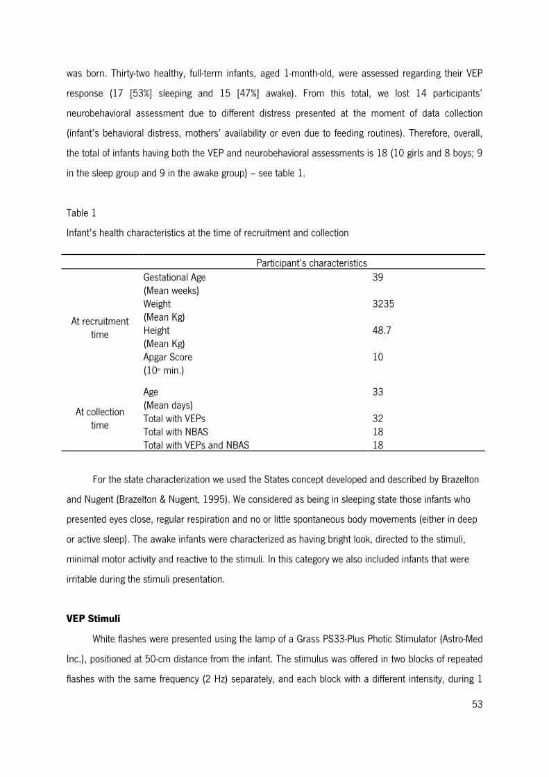
53
was born. Thirty-two healthy, full-term infants, aged 1-month-old, were assessed regarding their VEP
response (17 [53%] sleeping and 15 [47%] awake). From this total, we lost 14 participants’
neurobehavioral assessment due to different distress presented at the moment of data collection
(infant’s behavioral distress, mothers’ availability or even due to feeding routines). Therefore, overall,
the total of infants having both the VEP and neurobehavioral assessments is 18 (10 girls and 8 boys; 9
in the sleep group and 9 in the awake group) – see table 1.
Table 1
Infant’s health characteristics at the time of recruitment and collection
Participant’s characteristics
At recruitment time
Gestational Age (Mean weeks)
39
Weight (Mean Kg)
3235
Height (Mean Kg)
48.7
Apgar Score (10th min.)
10
At collection time
Age (Mean days)
33
Total with VEPs 32 Total with NBAS 18 Total with VEPs and NBAS 18
For the state characterization we used the States concept developed and described by Brazelton
and Nugent (Brazelton & Nugent, 1995). We considered as being in sleeping state those infants who
presented eyes close, regular respiration and no or little spontaneous body movements (either in deep
or active sleep). The awake infants were characterized as having bright look, directed to the stimuli,
minimal motor activity and reactive to the stimuli. In this category we also included infants that were
irritable during the stimuli presentation.
VEP Stimuli
White flashes were presented using the lamp of a Grass PS33-Plus Photic Stimulator (Astro-Med
Inc.), positioned at 50-cm distance from the infant. The stimulus was offered in two blocks of repeated
flashes with the same frequency (2 Hz) separately, and each block with a different intensity, during 1
54
minute. The stimulation intensity was set at 1 (0.09 J - intensity 1) and 2 (0.18 J - intensity 2) in the
flash position (Odom et al., 2010). For each block presentation, the flash position was organized for the
purpose of achieving different combinations and offered in a pseudo-randomized way (1-2; 2-1) so that
we could control the presentation order effect.
VEP Data Recording and Analysis
Electroencephalographic activity was recorded with a Quick- AmpTM system, with a 32-electrode
ActicapTM System inserted in a cap with a frontopolar ground and average referenced. 32 recording
electrodes were placed at Fp1, Fp2, F3, F4, Fz, F7, F8, FC1, FC2, FC5, FC6, T7, C3, Cz, C4, T8, TP9,
CP1, CP2, CP5, CP6, TP10, P3, P4, Pz, P7, P8, PO9, O1, Oz, O2, PO10 in accordance with the
International 10–20 System (Jasper, 1958) and electrode impedances were kept below 10 kΩ for all
participants. EEG signals were continuously amplified, digitized at sample rate of 250 Hz and filtered
on-line with a 0.01-100 Hz (12 dB/octave slope) band pass filter using a Quick-AmpTM system
amplifier and Brain Vision Recorder software (Version 1.20). All EEG data was analyzed with Brain
Vision Analyzer software (Version 2.0.1). The EEG was digitally filtered off-line with a 0.2-20 Hz band
pass filter and 50 Hz notch filter. It was then corrected for ocular artifacts by the semiautomatic
procedure in Independent Component Analysis (ICA) (Jung et al., 2000) and segmented into epochs of
600 ms from 100 ms pre-stimulus to 500 ms post-stimulus. Next, baseline correction was applied and
epochs exceeding ±200 µV at any scalp electrode were rejected. Finally, individual subject averages
ERPs time-locked to the two different stimuli (lower and higher intensity) were computed separately. A
minimum of 30 trials per average was recorded and the procedure repeated to check reproducibility.
Grand mean averages were computed for each stimulus and used to determine the latency
ranges in which the cortical VEP components were measured. The identification of peaks in individual
averages were made with a semiautomatic peak detection procedure and, subsequently reviewed and
manually corrected at O1, Oz and O2 electrodes for each participant. When peak identification was
doubtful, responses from all electrodes were compared, and the response was compared to the grand
mean averages.
The variability in the response was considerable, which is common among infants, but,
according to the typical neonatal VEP waveform morphology reported by McGlone et al. (2013) it was
generally characterized by a positive wave (P2) peaking around 200 ms followed by a broad negative
wave (N3) peaking around 350 ms (see table 2), clearly identified in all participants. Earlier peaks, N1,
55
P1 and N2 were much less frequently evoked and were smaller in comparison with the later peaks;
therefore, these components were not analyzed.
Neurobehavioral Assessment
For the neurobehavioral assessment we used the Neonatal Behavioral Assessment Scale (NBAS)
(Als, Tronick, Lester, & Brazelton, 1977). A trained and reliable examiner on the NBAS carried out the
assessment and the codification process. This scale assesses the newborns’ infant behavioral
repertoire through 28 behavioral items coded on a 9-point scale and the neurological state through 18
reflex items coded on a 4-point scale. It is organized into 7 clusters (Lester, Als & Brazelton, 1982):
habituation, orientation, motor, range of state, regulation of state, autonomic stability and reflexes.
Taking into account the objectives of this study, we considered the following clusters to
characterize the infants’ external and internal behavior: a) orientation, for external behavior
assessment, and b) regulation of state to assess their internal regulation ability (Sprangler, Fremmer-
Bombik & Grossmann, 1996; Lester, Als & Brazelton, 1982; Als et al., 1977). Indeed, both clusters are
assumed to mirror social characteristics in such young infants as social involvement imply alert and
orienting abilities, as well as internal regulatory processes, in order to attend and respond to the
surrounding stimuli (Brazelton & Nugent, 1995). And, once the visual assessment is proposed as an
indicator of cortical function (Atkinson, 2002; Brazelton & Nugent, 1995) when associated to infants’
external and internal responses to stimuli, we assume it as a psychophysiological marker of social
behavior.
For the orientation score, the mean of the items inanimate visual, inanimate auditory, inanimate
visual-auditory, animate visual, animate auditory, animate visual-auditory and alertness was calculated;
likewise, regarding the regulation of state, the score was obtained by calculating the mean of the items
cuddliness, consolability, self-quieting and hand-to-mouth. In both clusters, higher punctuations reflect
mature behavioral performances, as expected for this age.
Procedure
Data collection was carried out in a quiet room with a temperature approximately 20 to 25ºC
with luminosity and sound features controlled. Once the family arrived, the informed consent was
obtained. We started either with the neurobehavioral assessment or with the psychophysiological
recordings, considering the newborns’ state. Regarding the physiological procedure, we began the data
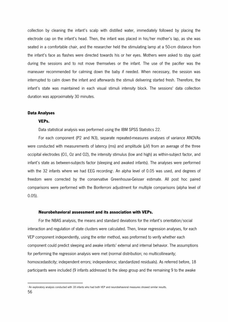
56
collection by cleaning the infant’s scalp with distilled water, immediately followed by placing the
electrode cap on the infant’s head. Then, the infant was placed in his/her mother’s lap, as she was
seated in a comfortable chair, and the researcher held the stimulating lamp at a 50-cm distance from
the infant’s face as flashes were directed towards his or her eyes. Mothers were asked to stay quiet
during the sessions and to not move themselves or the infant. The use of the pacifier was the
maneuver recommended for calming down the baby if needed. When necessary, the session was
interrupted to calm down the infant and afterwards the stimuli delivering started fresh. Therefore, the
infant’s state was maintained in each visual stimuli intensity block. The sessions' data collection
duration was approximately 30 minutes.
Data Analyses
VEPs.
Data statistical analysis was performed using the IBM SPSS Statistics 22.
For each component (P2 and N3), separate repeated-measures analyses of variance ANOVAs
were conducted with measurements of latency (ms) and amplitude (µV) from an average of the three
occipital electrodes (O1, Oz and O2), the intensity stimulus (low and high) as within-subject factor, and
infant’s state as between-subjects factor (sleeping and awaked infants). The analyses were performed
with the 32 infants where we had EEG recording3. An alpha level of 0.05 was used, and degrees of
freedom were corrected by the conservative Greenhouse-Geisser estimate. All post hoc paired
comparisons were performed with the Bonferroni adjustment for multiple comparisons (alpha level of
0.05).
Neurobehavioral assessment and its association with VEPs.
For the NBAS analysis, the means and standard deviations for the infant’s orientation/social
interaction and regulation of state clusters were calculated. Then, linear regression analyses, for each
VEP component independently, using the enter method, was preformed to verify whether each
component could predict sleeping and awake infants’ external and internal behavior. The assumptions
for performing the regression analysis were met (normal distribution; no multicollinearity;
homoscedasticity; independent errors; independence; standardized residuals). As referred before, 18
participants were included (9 infants addressed to the sleep group and the remaining 9 to the awake
3 An exploratory analysis conducted with 18 infants who had both VEP and neurobehavioral measures showed similar results.
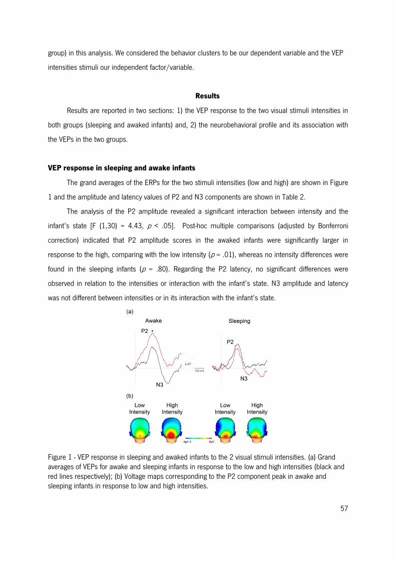
57
group) in this analysis. We considered the behavior clusters to be our dependent variable and the VEP
intensities stimuli our independent factor/variable.
Results
Results are reported in two sections: 1) the VEP response to the two visual stimuli intensities in
both groups (sleeping and awaked infants) and, 2) the neurobehavioral profile and its association with
the VEPs in the two groups.
VEP response in sleeping and awake infants
The grand averages of the ERPs for the two stimuli intensities (low and high) are shown in Figure
1 and the amplitude and latency values of P2 and N3 components are shown in Table 2.
The analysis of the P2 amplitude revealed a significant interaction between intensity and the
infant’s state [F (1,30) = 4.43, p < .05]. Post-hoc multiple comparisons (adjusted by Bonferroni
correction) indicated that P2 amplitude scores in the awaked infants were significantly larger in
response to the high, comparing with the low intensity (p = .01), whereas no intensity differences were
found in the sleeping infants (p = .80). Regarding the P2 latency, no significant differences were
observed in relation to the intensities or interaction with the infant’s state. N3 amplitude and latency
was not different between intensities or in its interaction with the infant’s state.
Figure 1 - VEP response in sleeping and awaked infants to the 2 visual stimuli intensities. (a) Grand averages of VEPs for awake and sleeping infants in response to the low and high intensities (black and red lines respectively); (b) Voltage maps corresponding to the P2 component peak in awake and sleeping infants in response to low and high intensities.
58
Table 2 Mean (SEs) values for N2, P2 and N3 peak amplitudes (µV) and latency (ms) recorded at O1, O2 and Oz.
P2 N3
Latency Amplitude Latency Amplitude
Awake infants
Low intensity
204.36 (8.86)
5.69 (1.43)
340.84 (15.68)
-2.52 (0.64)
High intensity 227.69 (15.89) 8.61 (1.86) 377.24 (15.32) -1.76 (0.66)
Sleeping infants
Low intensity 199.71 (11.20) 6.24 (1.33) 320.31 (15.04) -2.30 (1.01)
High intensity 213.61 (15.30) 5.98 (1.28) 335.16 (18.99) -3.44 (1.17)
Association between VEP and neurobehavioral clusters
All infants displayed organized, coherent and focused behavior, observed in their
neurobehavioral profile (see Table 3).
Table 3 Infants' neurobehavioral profile considering all the NBAS clusters.
NBAS Cluster Mean (SD)
Habituation 6.6 (2.5)
Orientation 7.4 (1)
Motor System 5.7 (1.5)
Range of States 4 (1.7)
Regulation of States 5.4 (2)
Autonomic System 4.4 (1.5)
Reflexes 1.9 (0.3)
Regarding its association with VEPs, we found that the N3 component amplitude in the lower
intensity was correlated with both an adequate external, orienting behavioral performance (R2= 0.38, P
= 0.04) and an adjusted internal regulation (R2= 0.6, P = 0.009) but only in awoken infants. However,
no differences were found regarding the N3 latency for the orienting/social interactive behavior (R2 =

59
0.2, p = 0.1) neither for the regulation of state (R2 = 0.1, p = 0.2). Likewise, in the higher intensity in
awoken infants was not associated with the orienting/social interactive behavior [N3 amplitude (R2 = -
0.14, p = 0.9); N3 latency (R2 = -0.14, p = 0.9)] and the regulation of state [N3 amplitude (R2 = -0.14, p
= 0.8); N3 latency (R2 = -0.08, p = 0.5)]. We did not find a statistically significant association regarding
the P2 component (amplitude and latency) in awoken infants and the behavior clusters in the lower
and higher intensities (table 4). Finally, in sleeping infants, we did not find associations between VEPs
components (N3 and P2 latency and amplitude) regarding both stimuli intensities (for detailed
information see Table 4).
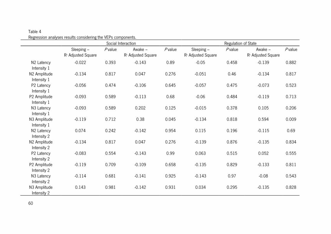
60
Table 4 Regression analyses results considering the VEPs components.
Social Interaction Regulation of State Sleeping –
R2 Adjusted Square P value Awake –
R2 Adjusted Square P value Sleeping –
R2 Adjusted Square P value Awake –
R2 Adjusted Square P value
N2 Latency Intensity 1
-0.022
0.393 -0.143 0.89 -0.05 0.458 -0.139 0.882
N2 Amplitude Intensity 1
-0.134 0.817 0.047 0.276 -0.051 0.46 -0.134 0.817
P2 Latency Intensity 1
-0.056 0.474 -0.106 0.645 -0.057 0.475 -0.073 0.523
P2 Amplitude Intensity 1
-0.093 0.589 -0.113 0.68 -0.06 0.484 -0.119 0.713
N3 Latency Intensity 1
-0.093 0.589 0.202 0.125 -0.015 0.378 0.105 0.206
N3 Amplitude Intensity 1
-0.119 0.712 0.38 0.045 -0.134 0.818 0.594 0.009
N2 Latency Intensity 2
0.074 0.242 -0.142 0.954 0.115 0.196 -0.115 0.69
N2 Amplitude Intensity 2
-0.134 0.817 0.047 0.276 -0.139 0.876 -0.135 0.834
P2 Latency Intensity 2
-0.083 0.554 -0.143 0.99 0.063 0.515 0.052 0.555
P2 Amplitude Intensity 2
-0.119 0.709 -0.109 0.658 -0.135 0.829 -0.133 0.811
N3 Latency Intensity 2
-0.114 0.681 -0.141
0.925 -0.143 0.97 -0.08 0.543
N3 Amplitude Intensity 2
0.143 0.981 -0.142 0.931 0.034 0.295 -0.135 0.828
61
Discussion
With the present study, our main objectives were: a) determining which VEP response
components were present in 1-month-old infants when presenting them 2 visual unpatterned flash
stimuli intensities; b) verifying if there were differences regarding the visual stimuli processing in the
infants who were sleeping and awake during the stimuli presentation; and c) understanding if the VEP
components could predict an adjusted orientation and/or regulation behavior in one-month-old infants.
We found that in both sleeping and awake one-month-old infants, P2 and N3 components were
present during the flashlight stimuli, in both lower and higher intensities. Indeed, the presence of these
components was a consistent and robust finding observed among all infants. These results are
consistent with previous findings (McGlone et al., 2013) suggesting that visual components such as P2
or N3 may reflect a mature brain development in the first weeks of life.
Considering the flash stimuli intensities, our results revealed that the infants displayed greater
P2 amplitude in response to the higher intensity stimulus. This was evident only when the infants were
awoken. Therefore, these results suggest that the infant’s state is crucial for determining different
visual processing thresholds. In fact, as it has been reported before (Shepherd, Saunders & McCulloch,
1999), infants’ state seem to influence the VEP peaks latency and amplitude regarding the flash visual
response, once infants that are awake were reported to display greater amplitudes and shorter peak
latencies (Benavente et al., 2005). The present study did not corroborate previous evidence suggesting
that there are no differences regarding the VEP characterization in sleeping and awake infants (Barnet
et al., 1980; Ellingson, 1970; Ferriss, Davis, Dorsen, & Hackett, 1967). However, it demonstrated that
when studying differences regarding threshold processing, infants’ state is essential to the VEP
response. This can be due possibly to the fact that similar behavior state may reduce intra and inter-
subject variability in very young infants, as suggested by Apkarian and colleagues (Apkarian, Mirmiran
& Tijssen, 1991). Our study suggests that the infant’s neural response to different stimuli may be
depending on their behavior state (Prechtl, 1974).
Regarding the neurobehavioral assessment, we hypothesized that one-month-old infants would
respond to sensorial stimulation in two behavioral ways: a) through an external response to the stimuli,
and b) through the infants’ ability to regulate themselves in order to respond to that stimulation. These
behavioral characteristics are translating social abilities displayed by very young infants (Brazelton &
Nugent, 1995). We observed an association between a mature neurobehavioral outcome and the VEP’s
positive N3 amplitude (once N3 is a negative component higher amplitude means more negativity) in
62
the lower flash intensity. Moreover, once again, these results were only evident in infants that were
awoken during the visual stimulation. Indeed, the brain development at young ages is characterized as
a complex process that occurs very rapidly, namely a fast increase in synaptic density in the visual
cortex in parallel with intense myelination of the visual tracts in the first four postnatal months (Dubois
et al., 2008). The infant’s physiological response to sensorial stimulation is translated into behavioral
characteristics that are associated with spending attentional resources in order to display adequate
orienting/social interactive and regulating behavior outcomes (Atkinson, 2002). In fact, infants show
different sensorial stimulation input necessities and/or difficulties that are present early in the
developmental process (Magnee, de Gelder, van Engeland, & Kemner, 2011). For instance, infants
who display lower stimuli processing thresholds or seem too disorganized to deal with their context will
need different stimuli inputs than those infants who are calmer or require more stimulation to react
(Brazelton & Nugent, 1995). Furthermore, evidenced have been suggested that in the first months of
life, infants present an increase sensitivity to lower sensorial information intensities which is consistent
with our results regarding the association of N3 in the lower intensity with behavior (Kushnerenko,
Tomalski, Ballieux, Ribeiro, et al., 2013).
This way, as suggested by our results, the VEP response to the lower intensity is correlated with
an adjusted social interactive and a mature internal regulation profile once the attentional and
physiological processes displayed seem to be a demanding process at very young ages. Infants
characterized as behaviorally organized are able to regulate themselves to the surrounding stimuli and,
consequently, maintaining focused interactive routines. Therefore, an awake state is a requirement for
visual processing, as attending and following visual stimuli implies behavioral characteristics only
exhibited by awake infants (Atkinson, 2002). Overall, our results seem to suggest that P2 component is
associated with visual stimuli threshold discrimination, and N3 component is more likely to be
associated with complex interactive abilities. Specifically, P2 has been associated with processing the
physical properties of stimuli while N3 with attentional behaviors underlying interactive conditions (Luo,
Feng, He, Wang, & Luo, 2010; McCulloch & de Haan, 2007).
Nevertheless, we should highlight that the study of VEP in one-month-old infants is susceptible
of great variability. It is clear that additional work is required before a complete understanding of these
processes. Thus, further studies contemplating different intensities thresholds in visual processing and
its relation to behavioral characteristics should be done with a larger number of participants, as the
number of infants in each group is limited. Future studies should also assess infants’ physiological
63
characteristics with respect to different sensorial modalities (such as auditory or olfactory processing)
and their relationships with neurobehavioral outcome as well as their implications for brain
development. Additionally, longitudinal analysis of the VEPs during infancy is required.
Conclusion
VEP components displayed during visual processing in one-month-old infants reflect possible
brain maturation and are related with mature regulation and interactive abilities at early ages (Isler et
al., 2007; Feldman & Eidelman, 2006; Huttenlocher, 2002). Although P2 and N3 components can be
identified in sleeping and awaked one-month-old infants, our results suggest that the infants’ state is
crucial for the visual processing of different intensities. Furthermore, our results suggest that VEP’s N3
component amplitude is associated with mature neurobehavioral outcome in one-month-old infants
regarding their interactive and internal regulation behavior. Identifying physiological markers of
behavioral outcome can be crucial for an early detection of developmental-related problems.
Acknowledgements
We would like to thank the families and infants who participated in this study. Also, we would like
to thank Pedro Hispano Hospital, particularly to Dr. J. Lopes dos Santos, for all the kindness
throughout the time that we were there for the data collection process. We thank to Maria de Góis-
Eanes for all the help and knowledge entailed throughout this study. Furthermore, we thank Associação
Viver a Ciência for the Simbiontes prize in 2013. This work was supported by Fundação Bial (grant
number 42/08) and financed by Fundação para a Ciência e Tecnologia (FCT) under a PhD grant
(reference SFRH/BD/68263/2010).
64
References
Als, H., Tronick, E., Lester, B., & Brazelton, T. B. (1977). The Brazelton Neonatal Behavioral
Assessment Scale (BNBAS). Journal of Abnormal Child Psychology, 5(3), 215-229. doi:
10.1007/BF00913693
Apkarian, P., Mirmiran, M., & Tijssen, R. (1991). Effects of behavioural state on visual processing in
neonates. Neuropediatrics, 22(2), 85-91. doi: 10.1055/s-2008-1071422
Atkinson, J. (2002). The developing visual brain. (Vol. 32). New York, United States: Oxford University
Press.
Barnet, A. B., Friedman, S. L., Weiss, I. P., Ohlrich, E. S., Shanks, B., & Lodge, A. (1980). VEP
development in infancy and early childhood. A longitudinal study. Electroencephalogr Clin
Neurophysiol, 49(5-6), 476-489.
Benavente, I., Tamargo, P., Tajada, N., Yuste, V., & Olivan, M. J. (2005). Flash visually evoked
potentials in the newborn and their maturation during the first six months of life. Doc
Ophthalmol, 110(2-3), 255-263. doi: 10.1007/s10633-005-0818-0
Brazelton, T. B., & Nugent, J. K. (1995). Neonatal Behavioral Assessment Scale. (3rd Edition ed. Vol.
137). London: MacKeith Press.
Calkins, S. D., Fox, N. A., & Marshall, T. R. (1996). Behavioral and physiological antecedents of
inhibited and uninhibited behavior. Child Dev, 67(2), 523-540.
Ceponiene, R., Kushnerenko, E., Fellman, V., Renlund, M., Suominen, K., & Naatanen, R. (2002).
Event-related potential features indexing central auditory discrimination by newborns. Brain Res
Cogn Brain Res, 13(1), 101-113.
Dubois, J., Dehaene-Lambertz, G., Soares, C., Cointepas, Y., Le Bihan, D., & Hertz-Pannier, L. (2008).
Microstructural correlates of infant functional development: example of the visual pathways. J
Neurosci, 28(8), 1943-1948. doi: 10.1523/JNEUROSCI.5145-07.2008
Ellingson, R. J. (1970). Variability of visual evoked responses in the human newborn.
Electroencephalogr Clin Neurophysiol, 29(1), 10-19.
Feldman, R., & Eidelman, A. I. (2006). Neonatal state organization, neuromaturation, mother-infant
interaction, and cognitive development in small-for-gestational-age premature infants.
Pediatrics, 118(3), e869-878. doi: 10.1542/peds.2005-2040
65
Ferriss, G. S., Davis, G. D., Dorsen, M. M., & Hackett, E. R. (1967). Changes in latency and form of the
photically induced average evoked response in human infants. Electroencephalogr Clin
Neurophysiol, 22(4), 305-312.
Huttenlocher, P. R. (2002). Neural Plasticity. The effects of environment on the development of the
crebral cortex. Cambridge, MA: Harvard University Press.
Isler, J. R., Grose-Fifer, J., Fifer, W. P., Housman, S., Stark, R. I., & Grieve, P. G. (2007). Frequency
domain analyses of neonatal flash VEP. Pediatr Res, 62(5), 581-585. doi:
10.1203/PDR.0b013e31815586a1
Johnson, M. H., Dziurawiec, S., Ellis, H., & Morton, J. (1991). Newborns' preferential tracking of face-
like stimuli and its subsequent decline. Cognition, 40(1-2), 1-19.
Jung, T. P., Makeig, S., Westerfield, M., Townsend, J., Courchesne, E., & Sejnowski, T. J. (2000).
Removal of eye activity artifacts from visual event-related potentials in normal and clinical
subjects. Clin Neurophysiol, 111(10), 1745-1758.
Kato, T., & Watanabe, K. (2006). Visual evoked potential in the newborn: does it have predictive value?
Semin Fetal Neonatal Med, 11(6), 459-463. doi: 10.1016/j.siny.2006.08.003
Kirk, H. E., Hocking, D. R., Riby, D. M., & Cornish, K. M. (2013). Linking social behaviour and anxiety
to attention to emotional faces in Williams syndrome. Res Dev Disabil. doi:
10.1016/j.ridd.2013.09.042
Kraemer, M., Abrahamsson, M., & Sjostrom, A. (1999). The neonatal development of the light flash
visual evoked potential. Doc Ophthalmol, 99(1), 21-39.
Lester, B. M., Als, H., & Brazelton, T. B. (1982). Regional obstetric anesthesia and newborn behavior:
a reanalysis toward synergistic effects. Child Dev, 53(3), 687-692.
Liu, J., Bann, C., Lester, B., Tronick, E., Das, A., Lagasse, L., . . . Bada, H. (2010). Neonatal
neurobehavior predicts medical and behavioral outcome. Pediatrics, 125(1), e90-98. doi:
10.1542/peds.2009-0204
Magnee, M. J., de Gelder, B., van Engeland, H., & Kemner, C. (2011). Multisensory integration and
attention in autism spectrum disorder: evidence from event-related potentials. PLoS One, 6(8),
e24196. doi: 10.1371/journal.pone.0024196
McGlone, L., Hamilton, R., McCulloch, D. L., Boulton, R., Bradnam, M. S., Weaver, L. T., & Mactier, H.
(2013). Neonatal visual evoked potentials in infants born to mothers prescribed methadone.
Pediatrics, 131(3), e857-863. doi: 10.1542/peds.2012-2113
66
Mercuri, E., von Siebenthal, K., Tutuncuoglu, S., Guzzetta, F., & Casaer, P. (1995). The effect of
behavioural states on visual evoked responses in preterm and full-term newborns.
Neuropediatrics, 26(4), 211-213. doi: 10.1055/s-2007-979756
Mikkola, K., Kushnerenko, E., Partanen, E., Serenius-Sirve, S., Leipala, J., Huotilainen, M., & Fellman,
V. (2007). Auditory event-related potentials and cognitive function of preterm children at five
years of age. Clin Neurophysiol, 118(7), 1494-1502. doi: 10.1016/j.clinph.2007.04.012
Odom, J. V., Bach, M., Brigell, M., Holder, G., McCulloch, D., Tormene, A., & Vaegan. (2010). ISCEV
standard for clinical visual evoked potentials (2009 update). Documenta Ophthalmologica,
120(1), 111-119. doi: 10.1007/s10633-009-9195-4
Pihko, E., Lauronen, L., Wikstrom, H., Taulu, S., Nurminen, J., Kivitie-Kallio, S., & Okada, Y. (2004).
Somatosensory evoked potentials and magnetic fields elicited by tactile stimulation of the hand
during active and quiet sleep in newborns. Clin Neurophysiol, 115(2), 448-455.
Prechtl, H. F. (1974). The behavioural states of the newborn infant (a review). Brain Res, 76(2), 185-
212.
Rothbart, M. K. (2007). Temperament, Development, and Personality. Current Directions in
Psychological Science, 16(4), 207-212.
Sampaio, A., Sousa, N., Fernandez, M., Henriques, M., & Goncalves, O. F. (2008). Memory abilities in
Williams syndrome: dissociation or developmental delay hypothesis? Brain Cogn, 66(3), 290-
297. doi: 10.1016/j.bandc.2007.09.005
Shepherd, A., Saunders, K., & McCulloch, D. (1999). Effect of sleep state on the flash visual evoked
potential. A case study. Doc Ophthalmol, 98(3), 247-256.
Sprangler, G., Fremmer-Bombik, E., & Grossmann, K. (1996). Social and individual determinants of
infant attachment security and disorganization. Infant Mental Health Journal, 17(2), 127-139.
doi: 10.1002/(SICI)1097-0355(199622)17:2<127::AID-IMHJ3>3.0.CO;2-N
Stanley, O., Wright, M., Pike, A., Marlow, N., & Pike, R. (2009). 'Weight of Evidence' analysis of
neonatal sensory evoked potentials. J Neurosci Methods, 185(1), 99-107. doi:
10.1016/j.jneumeth.2009.09.020
Tsuneishi, S., Casaer, P., Fock, J. M., & Hirano, S. (1995). Establishment of normal values for flash
visual evoked potentials (VEPs) in preterm infants: a longitudinal study with special reference to
two components of the N1 wave. Electroencephalogr Clin Neurophysiol, 96(4), 291-299.

67
Whyte, H. E., Pearce, J. M., & Taylor, M. J. (1987). Changes in the VEP in preterm neonates with
arousal states, as assessed by EEG monitoring. Electroencephalogr Clin Neurophysiol, 68(3),
223-225.

68
69
Auditory neural correlates of infant's development: a longitudinal study4
Sara Cruza, MSc, Alberto Cregoa, PhD, Eugénia Ribeirob, PhD, Óscar Gonçalvesa, c,d, PhD, &
Adriana Sampaioa, PhD
a Neuropsychophysiology Laboratory, CIPsi, School of Psychology, University of Minho, Braga, Portugal
b CIPsi, School of Psychology, University of Minho, Braga, Portugal
c Department of Counseling and Applied Educational Psychology, Bouvé College of Health Sciences,
Northeastern University, Boston, MA, US
d Spaulding Center of Neuromodulation, Department of Physical Medicine & Rehabilitation, Spaulding
Rehabilitation Hospital and Massachusetts General Hospital, Harvard Medical School, Boston, MA, USA
Correspondent address: Sara Cruz, Neuropsychophysiology Laboratory, School of Psychology,
University of Minho, Campus de Gualtar, 4710-057, Braga, Portugal.
Email address: [email protected]
Telephone number: +351 253 604 220
4 This study was submitted for publication at Developmental Neuropsychology.
70
Abstract
We aimed to identify in full-term 1-month-old infants, AEPs components to two auditory stimuli
intensities and if they were associated with adjusted interactive and internal regulatory behavior, at the
same age, as well as predicting development outcomes at 12 months of age. Greater P2 amplitude
was observed to the higher intensity stimuli and was associated with orienting and regulatory
behaviors. Increased P2 latency was associated with regulatory abilities. Similarly, P2 amplitude is
predicting language competences at 12 months of age. Our study supports previous findings and
evidence that specific auditory physiological markers underlie adequate behavior throughout the first
developmental year.
Keywords: Auditory Evoked Potentials; Developmental assessment; Infant; Language;
Neurobehavioral assessment
71
Introduction
The human interaction with the environment and its reciprocal relation with sensorial systems
are crucial for brain development. During the gestation time, the fetus central nervous system
formation and its maturation process develops consistently and rapidly. By the end of the gestation
period, the cortex formation is already complete and, after birth, as the brain is still developing, the
main event now occurring is the cortex maturation processes, which is characterized by the increase of
synapses and synaptic density, as well as, dendritic growth (Huttenlocher, 2002). Paralleled with these
structural developments, functional maturation is also happening. One of the earliest functions to
emerge is auditory function (hearing) (Kushnerenko, 2003), indicating that the infant is capable of
sound perception, which has been already developed in uterus. Nevertheless, a continuous
specialization of the auditory processing occurs throughout the infancy period (until circa 3-years-old)
as the maturation of auditory evoked-potentials (AEPs) is considered to be a continuum process where
peaks amplitude increase and the peaks latency decrease through time (Wunderlich, Cone-Wesson &
Shepherd, 2006).
Several authors have been studying cortical auditory discrimination ability and auditory threshold
detection in young infants (Beauchemin et al., 2011; Vestergaard et al., 2009; Ceponiene et al., 2002;
Kushnerenko, Ceponiene, Fellman, Huotilainen, & Winkler, 2001). To achieve this purpose, Event-
related Potentials (ERP) have been widely used once, besides being a non-invasive and non-harmful
technique, it translates brain electric signals that are related to external and internal events providing
real time neural information processing, essential to assess neural changes occurring during the
infancy (Kushnerenko, Van den Bergh & Winkler, 2013).
Previous studies have addressed auditory processing abilities in young infants and the
emergence of auditory components, since birth, that are underlying auditory neural maturation
processes (Lippé, Martinez-Montes, Arcand, & Lassonde, 2009; Telkemeyer et al., 2009; Wunderlich,
Cone-Wesson & Shepherd, 2006). In a study conducted by Lippé and collaborators (2009), the authors
found that the cortical components complex P1-N1-P2-N2, present in adults, is not yet identifiable in 1-
month-old infants. The authors have identified the presence of a first negative component, appearing
around 80ms, and a discernable P2 in these young infants. Similarly, in another study, P1 and N1
components were infrequently observed in newborns but P2 and N2 components were present
consistently, being the P2 the most prominent positive peak around this age (Wunderlich, Cone-Wesson
& Shepherd, 2006). Moreover, electrophysiological studies focusing on auditory threshold
72
discrimination in healthy full-term newborns have evidenced that the ability to perceive different sound
intensities is already present at birth (Marcoux, 2011; Ceponiene et al., 2002).
Furthermore, the relation between auditory perception and discrimination with developmental
characteristics has been addressed (Magnee et al., 2011; Mikkola et al., 2010; Nelson & McCleery,
2008; Mikkola et al., 2007; Feldman & Eidelman, 2006; Fellman et al., 2004; Benasich & Tallal,
2002; Benasich, Thomas, Choudhury, & Leppanen, 2002). Indeed, the study of the association
between these physiological features with behavioral outcomes in infants seems crucial for a better
understanding of the physiological characteristics underlying different cognitive processes emerging
throughout the developmental course (Schwartz et al., 2012; Calkins, Fox & Marshall, 1996).
In fact, a correlation between cortical ERPs waveform morphology recorded at birth and
language performance in pre-school children (Molfese & Molfese, 1997) has been demonstrated, as
auditory processing abilities assessed in newborn infants seems to predict communicative abilities
(Benasich et al., 2002). Studies focused on rapid auditory processing in healthy, full-term infants have
shown that those who present greater difficulty in processing rapidly sounds may display later language
impairment (Benasich et al., 2006; Benasich & Tallal, 2002; Benasich et al., 2002). Likewise, infants
at risk of developing disorders or born preterm present auditory neural correlates that are associated
with poor language competences. For instance, studies comparing newborn infants at risk for dyslexia
and control participants were able to differentiate the at risk group regarding their auditory processing
abilities, particularly their mismatch negative (MMN) response (Guttorm et al., 2005; Naatanen, 2001;
Bradlow et al., 1999). Similarly, infants that were born preterm seem to display a different MMN
pattern response when compared with the control group and, additionally, exhibit worst performances
in development assessment tasks when older (Mikkola et al., 2007; Fellman et al., 2004;
Kushnerenko, Ceponiene, Balan, Fellman, & Naatanen, 2002).
The importance of assessing AEPs has been evident from early infancy, as it assumes a main
role in mirroring maturational processes and in predicting later development outcomes. The
neurophysiologic assessment of infants throughout their first year of development can provide
information about functional changes of the brain (deRegnier, 2008), particularly for auditory
processing, perception and discrimination (Ceponiene et al., 2002) as well as for studying behavioral
and developmental aspects (Kushnerenko et al., 2013a; Kushnerenko et al., 2013b; Benasich et al.,
2006; Benasich & Tallal, 2002; Benasich et al., 2002). Hence, with the present study we aim to
identify AEPs components in 1-month-old infants using two auditory stimuli intensities. We hypothesize
73
that cortical auditory components can be identified in these young infants and we expect to find greater
activation to the higher stimulus intensity. Moreover, we want to correlate the AEP response with the
neurobehavioral profile in order to verify if this physiological response is correlated with infant’s
behavioral reactivity to external (orienting/interactive characteristics) and internal (regulation
characteristics) stimuli. Additionally, with the longitudinal study we are aiming to verify if the
physiological response recorded at one-month-old, in both intensities, is predicting development
outcomes when the infants are assessed at 12 months of age.
Methods
Participants
Mother/infant dyads were recruited at the moment the infant was born and sixty-one families
accepted to participate in this study. Infants were assessed in two different moments: the first moment
when they were 1-month-old and the second when they completed 1-year-old. All infants were
caucasian, born full-term and healthy at the moment of data collection.
From the 61 participants, at 1-month-old, AEP were collected for 30 infants, whereas 28 (46%)
AEPs were lost due to distress presented by the baby at the time of data collection or due to artifacts in
the auditory register. Additionally, neurobehavioral assessment was lost for 14 infants (23%), as its
application was discontinued once they were distressed at that time. Therefore, in the first assessment
moment, the total of participants having both AEPs and neurobehavioral assessments is 19 infants [9
boys and 10 girls (table 1)].
Regarding the second moment evaluation, 39 (64%) 12-month-old infants returned for the
development assessment. Consequently, the total number of participants having both AEPs recording
and the development assessment is 18 infants [8 boys and 10 girls (table 1)].
The present study was reviewed and accepted by the ethical committee of the hospital where the
assessment was performed.
Table 1 Infant's health information at the time of recruitment and collection.
Participant’s characteristics
At recruitment time
Gestational Age 39 (mean weeks) Weight 3278 (mean kilograms) Height 48,9 (centimeters)
74
Apgar Score (10th min.) 10 At collection
time Age 34 (mean days) Total Infants with AEPs assessment 30 Total infants with NBAS assessment 47 Total Infants with AEPs and NBAS assessments 19
Physiological assessment at 1-month-old
Auditory Stimuli.
Presentation® software (Version 0.61.3, www.neurobs.com) was used to build and deliver the
auditory stimuli. Two auditory thresholds intensities were created: intensity 1, a lower intensity (50dB)
and intensity 2, a higher intensity (70dB). The stimulus delivering was organized in a block paradigm
considering the auditory stimulus intensity. Each block was offered during 1 minute, 1 stimulus per
second, with a 20-second interval period between intensities. Two speakers were positioned
approximately at 20 centimeters distant from each infant’s ear for the delivery of the auditory stimuli.
The order of the intensities presentation was counterbalanced across participants, and offered in a
pseudo-randomized way so that we could control the presentation order effect.
EEG recording and ERP analysis.
Electroencephalographic activity was recorded with a Quick- AmpTM system, with a 32-electrode
ActicapTM System inserted in a cap with a frontopolar ground and average referenced. 32 recording
electrodes were placed at Fp1, Fp2, F3, F4, Fz, F7, F8, FC1, FC2, FC5, FC6, T7, C3, Cz, C4, T8, TP9,
CP1, CP2, CP5, CP6, TP10, P3, P4, Pz, P7, P8, PO9, O1, Oz, O2, PO10 in accordance with the
International 10–20 System (Jasper, 1958) and electrode impedances were kept below 10 kΩ for all
participants. Electroencephalogram (EEG) signals were continuously amplified, digitized at sample rate
of 250 Hz and filtered on-line with a 0.01-100 Hz (12 dB/octave slope) band pass filter using a Quick-
AmpTM system amplifier and Brain Vision Recorder software (Version 1.20). All EEG data were
analyzed with Brain Vision Analyzer software (Version 2.0.1). The EEG was digitally filtered off-line with
a 1-15 Hz band pass filter and 50 Hz notch filter. It was then corrected for ocular artefacts by the
semiautomatic procedure in Independent Component Analysis (ICA) (Jung et al., 2000) and segmented
into epochs of 1000 ms from 100 ms pre-stimulus to 900 ms post-stimulus. Next, baseline correction
was applied and epochs exceeding ±150 µV at any scalp electrode were rejected. Finally, individual
subject averages ERPs time-locked to the two different stimuli (lower and higher intensity) were
computed separately.
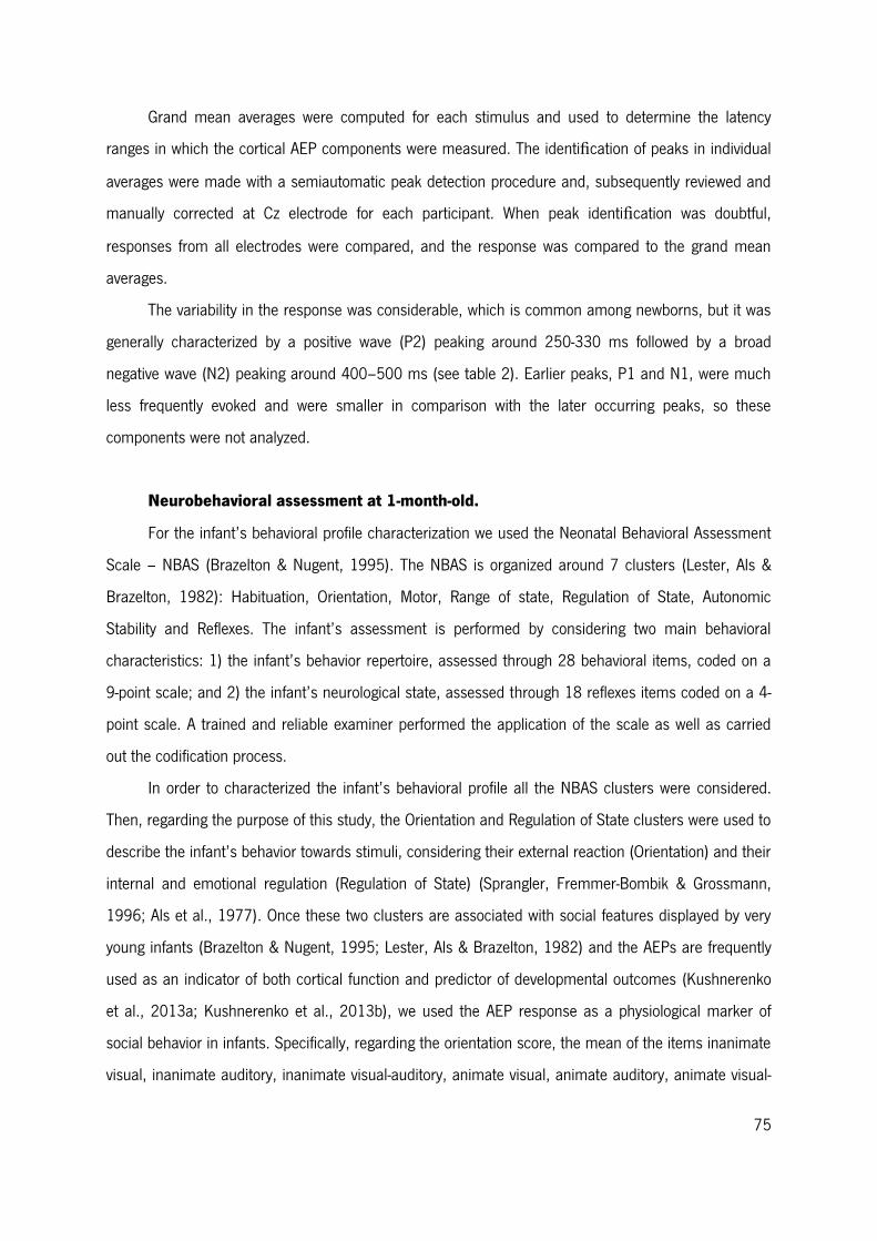
75
Grand mean averages were computed for each stimulus and used to determine the latency
ranges in which the cortical AEP components were measured. The identification of peaks in individual
averages were made with a semiautomatic peak detection procedure and, subsequently reviewed and
manually corrected at Cz electrode for each participant. When peak identification was doubtful,
responses from all electrodes were compared, and the response was compared to the grand mean
averages.
The variability in the response was considerable, which is common among newborns, but it was
generally characterized by a positive wave (P2) peaking around 250-330 ms followed by a broad
negative wave (N2) peaking around 400–500 ms (see table 2). Earlier peaks, P1 and N1, were much
less frequently evoked and were smaller in comparison with the later occurring peaks, so these
components were not analyzed.
Neurobehavioral assessment at 1-month-old.
For the infant’s behavioral profile characterization we used the Neonatal Behavioral Assessment
Scale – NBAS (Brazelton & Nugent, 1995). The NBAS is organized around 7 clusters (Lester, Als &
Brazelton, 1982): Habituation, Orientation, Motor, Range of state, Regulation of State, Autonomic
Stability and Reflexes. The infant’s assessment is performed by considering two main behavioral
characteristics: 1) the infant’s behavior repertoire, assessed through 28 behavioral items, coded on a
9-point scale; and 2) the infant’s neurological state, assessed through 18 reflexes items coded on a 4-
point scale. A trained and reliable examiner performed the application of the scale as well as carried
out the codification process.
In order to characterized the infant’s behavioral profile all the NBAS clusters were considered.
Then, regarding the purpose of this study, the Orientation and Regulation of State clusters were used to
describe the infant’s behavior towards stimuli, considering their external reaction (Orientation) and their
internal and emotional regulation (Regulation of State) (Sprangler, Fremmer-Bombik & Grossmann,
1996; Als et al., 1977). Once these two clusters are associated with social features displayed by very
young infants (Brazelton & Nugent, 1995; Lester, Als & Brazelton, 1982) and the AEPs are frequently
used as an indicator of both cortical function and predictor of developmental outcomes (Kushnerenko
et al., 2013a; Kushnerenko et al., 2013b), we used the AEP response as a physiological marker of
social behavior in infants. Specifically, regarding the orientation score, the mean of the items inanimate
visual, inanimate auditory, inanimate visual-auditory, animate visual, animate auditory, animate visual-
76
auditory and alertness was calculated; similarly, for the regulation of state, the mean score was
obtained by calculating the values obtained in the cuddliness, consolability, self-quieting and hand-to-
mouth items. In both clusters, higher punctuations reflected a typical behavioral performance; only the
infant’s best performance was considered for the coding process.
Developmental assessment at 1-year-old
For the 1-year-old infant’s development assessment the Bayley Scales of Infant and Toddler
Development, 3rd edition (Bayley, 2006) – Bayley-III – was used. A psychologist carried out the
development assessment. The Bayley-III has acceptable levels of reliability (internal consistency > 0.86;
test-retest reliability > 0.67).
The Bayley-III consists of 5 scales: cognitive (91 items), language (97 items), motor (138 items),
social-emotional (35 items) and adaptive behavior (241 items) (Bayley, 2006). For the aims of the
present study, only the first three were included in the analysis. Regarding the language scale,
receptive and expressive communication items are assessed; likewise, the motor scale consists of fine
and gross motor items. The raw scores for cognition, receptive communication, expressive
communication, fine motor and gross motor are obtained. Then, each raw score was transformed into
a scaled score (mean = 10 and SD = 3) and additionally converted into cognitive, language and motor
composite scores (mean = 100 and SD = 15).
Procedure
At the first assessment moment, the study objectives and procedures were explained and the
informed consent was obtained as the mother/infant dyad arrived at the hospital. The data collection
was performed in a room where luminosity and sound features were controlled and the temperature
kept at 20 to 25 Celsius degrees. We either started with the physiological or with the neurobehavioral
assessment considering the infant’s state. For the physiological assessment the infant’s scalp was
cleaned with distilled water and then the electrodes cap was placed in his/her head. Next, the infant
was placed in his/her mother’s lap, who was sat at a comfortable chair and was asked to stay quiet
and not to move her or the infant during the session. The electrolytic gel was then applied into the
electrodes to diminish the impedances. As the auditory stimuli delivered started, the speakers were
hold by the researcher at approximately 20 centimeters distant from each ear. During this time the
infants were awake, either in an alert calm (16.4%) or drowsy (42.6%) state, sleeping (13.1%) or even
77
irritable (11.5%). Through the session, using the pacifier was the maneuver recommended for calming
down the infant, if necessary, and the session was interrupted when the infant was too distressed. The
sessions' collection duration was approximately 30 minutes.
One year later, mothers were once again contacted and those who accepted participating once
again returned to the hospital for the infant’s development assessment. The data collection occurred in
an ample room with minimal distracting objects. As the mother arrived, the study’s second phase
objectives and procedures were explained and the informed consent obtained. As the development
assessment was performed the infant was sat at the mother’s lap. If the infant was drowsy or
distressed at the moment, the assessment session was interrupted and continued later, in the same
session. The assessment duration was approximately 45 to 60 minutes.
Statistical Analysis
Data statistical analysis was performed using the IBM SPSS Statistics 22. For each component
(P2 and N2), separate repeated-measures analyses of variance ANOVAs were conducted with
measurements of latency (ms) and amplitude (µV) from a single electrode (Cz) and the intensity
stimulus (lower and higher) as within-subject factor. The analyses were performed with the 30 infants
in who EEG was recorded. However, an exploratory analysis was performed with the 19 infants who
had both AEPs and neurobehavioral measures and, likewise, with the 18 infants having AEPs and
development assessment at 1-year-old. We did so to verify if AEP results obtained previously with the
30 infants were similar when only including the infants that have both assessments, at 1-month-old
and at 1-year-old, in the statistical analysis. An alpha level of 0.05 was used and, whenever
appropriate, degrees of freedom were corrected by the conservative Greenhouse-Geisser estimate. All
post hoc paired comparisons were performed with the Bonferroni adjustment for multiple comparisons.
Then, the mean and standard deviation for the NBAS clusters were calculated in order to classify the
behavioral profile of our sample.
Further analyses were performed to examine the association between the physiological response
with neurobehavior measures as well as with the developmental assessment. Regarding the
neurobehavioral clusters, we performed a linear regression analysis, where we defined the
orientation/social interactive and the regulation of state NBAS clusters as our dependent factor and the
identified AEPs components latency and amplitude values, for the two intensities, as our Independent
factor. Likewise, to examine the association between the physiological response and the development
78
assessment at 1-year-old, a linear regression analysis was performed. Similarly, we defined the Bayley-
III domains Cognitive, Language and Motor composite scores as our dependent factor and the
identified AEPs components latency and amplitude values, for the two intensities, as our Independent
factor. The assumptions for performing the regression analysis were met.
Results
Electrophysiological results
The grand averages of the ERPs for the two stimuli (lower and high intensities) are described in
Figure 1 and the mean and standard error (SE) values of amplitude and latency of P2 and N2
components are shown in Table 2.
Figure 1 - Infant's AEPs response regarding the two auditory stimuli intensities. (a) Grand averages of ERPs for the infants in response to low and high auditory intensities (red and black lines respectively). Averages are presented for midline central (Cz) electrode. Infants showed significantly larger P2 amplitude for the high intensity than for the low intensity; (b) Voltage maps corresponding to the P2 peak component in infants in response to low and high intensities.
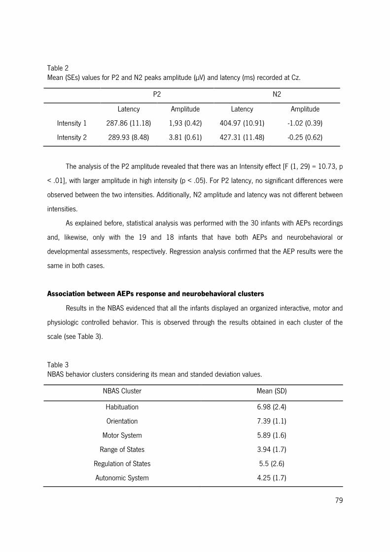
79
Table 2 Mean (SEs) values for P2 and N2 peaks amplitude (µV) and latency (ms) recorded at Cz.
P2 N2
Latency Amplitude Latency Amplitude
Intensity 1 287.86 (11.18) 1,93 (0.42) 404.97 (10.91) -1.02 (0.39)
Intensity 2 289.93 (8.48) 3.81 (0.61) 427.31 (11.48) -0.25 (0.62)
The analysis of the P2 amplitude revealed that there was an Intensity effect [F (1, 29) = 10.73, p
< .01], with larger amplitude in high intensity (p < .05). For P2 latency, no significant differences were
observed between the two intensities. Additionally, N2 amplitude and latency was not different between
intensities.
As explained before, statistical analysis was performed with the 30 infants with AEPs recordings
and, likewise, only with the 19 and 18 infants that have both AEPs and neurobehavioral or
developmental assessments, respectively. Regression analysis confirmed that the AEP results were the
same in both cases.
Association between AEPs response and neurobehavioral clusters
Results in the NBAS evidenced that all the infants displayed an organized interactive, motor and
physiologic controlled behavior. This is observed through the results obtained in each cluster of the
scale (see Table 3).
Table 3 NBAS behavior clusters considering its mean and standed deviation values.
NBAS Cluster Mean (SD)
Habituation 6.98 (2.4)
Orientation 7.39 (1.1)
Motor System 5.89 (1.6)
Range of States 3.94 (1.7)
Regulation of States 5.5 (2.6)
Autonomic System 4.25 (1.7)
80
Reflexes 1.94 (0.2)
Taking into account that differences regarding auditory stimuli intensities were observed only for
the P2 component, we analyzed the association between P2 and neurobehavioral results. The
regression analysis showed that greater P2 amplitude in the lower intensity (intensity 1) was predicting
a mature oriented, social interactive behavior in 1-month-old infant (R2 = 0.18, P < 0.05). Likewise, a
greater P2 amplitude (R2 = 0.18, P < 0.05) and latency (R2 = 0.1, P < 0.05), also in the lower intensity
(intensity 1), were predicting an adjusted internal regulation in such young infants (Figure 2) (see Table
4).
Figure 2 Graphic representation of the correlations between the P2 response and the behavior clusters. a) Association between P2 amplitude in the lower intensity and social interaction NBAS cluster; b) association between P2 amplitude and in the lower intensity and state regulation NBAS cluster; c) association between P2 latency in the lower intensity and state regulation NBAS cluster.
Table 4 Linear regression regarding P2 amplitude and latency values, in Cz, and behavior clusters.
Orientation Cluster Regulation Cluster
R2 Adjusted Square P value R2 Adjusted Square P value
P2 - Intensity 1 Amplitude 0.18 0.04* 0.18 0.04*
P2 - Intensity 2 Amplitude 0.06 0.1 0.01 0.3
P2 - Intensity 1 Latency 0.11 0.08 0.1 0.03*
P2 - Intensity 2 Latency 0.05 0.2 0.04 0.6
81
Association between AEPs response and development assessment
Regarding the Bayley-III assessment, the infants scored above the basal scoring threshold for the
cognitive scale (mean = 115, SD = 11) and within average for the language (mean = 96, SD = 10.6)
and motor (mean = 97, SD = 7.5) scales, considering the composite scores (table 5).
Table 5 Bayley-III cognitive, language and motor scale descriptive analysis.
Bayley-III Scales Median (Range) Mean (SD)
Cognitive composite score 111.5 (100-130) 115 (11)
Language composite score 97 (71-115) 96 (10.6)
Motor composite score 97 (88-115) 97 (7.5)
Associations between AEPs P2 and N2 amplitude and latency values for both auditory stimuli
intensities, assessed at 1-month-old, and cognitive, language and motor composite scores were
preformed.
The regression analysis showed that greater P2 amplitude in the lower intensity (intensity 1) at 1-
month-old was predicting the language outcome in 1-year-old infants (R2 = 0.17, P = 0.04). No
significant results were obtained for the cognitive and motor scales and, likewise, regarding the N2 and
the association between cognitive, language and motor scales (table 6).
Table 6 Regression analyses: association between AEPs and cognitive, language and motor scales.
Cognitive Scale Language Scale Motor Scale
R2 Adjusted
Square
P value R2 Adjusted
Square
P value R2 Adjusted
Square
P value
P2 Latency – Int1 0.07 0.3 -0.06 0.7 -0.03 0.5
P2 Amplitude – Int1 -0.03 0.5 0.17 0.049 0.04 0.2
P2 Latency – Int2 -0.06 0.7 -0.06 0.9 -0.05 0.6
P2 Amplitude – Int2 -0.06 0.9 -0.06 0.7 -0.04 0.5
N2 Latency – Int1 0.06 0.2 -0.01 0.4 -0.04 0.5
N2 Amplitude – Int1 -0.05 0.7 0.02 0.3 -0.02 0.4
82
N2 Latency – Int2 -0.06 0.9 -0.02 0.4 -0.05 0.6
N2 Amplitude – Int2 0.05 0.2 -0.03 0.5 0.008 0.3
Discussion
With the present study we analyzed AEPs in 1-month-old healthy infants to two stimuli intensities
and, additionally, we verified if this response could be associated with an adjusted and mature
interactive and internal regulatory behavior in such young infants. Furthermore, we examined if the
neural auditory response was also predicting development outcomes when assessing the infants at 1-
year-old.
Our results showed that very early in development we are able to identify AEPs that are
associated with auditory stimuli processing. We found that all infants presented P2 and N2
components to both intensities, as they were the most consistent components appearing regarding the
auditory stimulation. This result is consistent with previous findings in the literature, showing that the
P2 and N2 seem to be the most coherent and robust components present in the auditory processing in
young infants (Lippé et al., 2009; Wunderlich, Cone-Wesson & Shepherd, 2006). In accordance, these
results suggest that AEPs play an important role regarding the auditory processing in 1-month-old-
infants, as they appear to be associated with auditory cortical maturation processes and developmental
features (van de Weijer-Bergsma, Wijnroks & Jongmans, 2008; Kushnerenko, 2003; Cheour,
Kushnerenko, Ceponiene, Fellman, & Naatanen, 2002). Indeed, around these ages the auditory cortex
development is still occurring as it is characterized by the emergence, in the first months of life, of AEP
components that are underlying an adequate maturation process (Kushnerenko, Van den Bergh &
Winkler, 2013; Telkemeyer et al., 2009; Lippé et al., 2009). Moreover, we found greater P2 amplitude
displayed to the higher intensity and no differences were observed in the lower intensity. Regarding the
N2, no differences were found between intensities either in its amplitude and latency. Therefore, our
results seem to indicate that the presence of the P2 component is essential for auditory perception. In
fact, the P2 amplitude seems to be related with threshold discrimination (Ceponiene et al., 2002).
Once again, this ability seems to be associated with auditory cortical maturation once it reflects the
infant’s ability to differently processing distinctive sound intensities (Marcoux, 2011).
Moreover, we observed that P2 response (either its latency or amplitude) was predicting the
infants’ external orientation and internal regulatory behavior. Therefore, our results indicate that greater
83
P2 amplitude in the lower intensity is predicting both an adjusted orienting / interactive and internal
regulatory abilities. This may be occurring due to the fact that during the first months of life (until circa
3 month of age) infants present an increase sensitivity to lower auditory sensorial information, that is
decoded through their behavioral features (Kushnerenko et al., 2013b). Thus, we hypothesize that this
neural response to the lower intensity underlies the infant’s ability to perceive lower auditory
information which is associated with coherent and focused interactive and regulatory characteristics,
and enables infants to attend to different social and sensorial cues from the surrounding environment
(Kushnerenko et al., 2013b; Brazelton & Nugent, 1995). Likewise, a larger P2 latency seems to be
predicting an adjusted regulatory behavior. Once the central nervous system is still immature at 1
month of age and the neural response to sensory processing is mainly characterized by employing
more time at a given stimulus, it is possible that these findings suggest that infant’s with better
regulatory abilities attend repeatedly to the stimuli, spending more attentional resources and, therefore,
taking more time in auditory stimuli processing (Kushnerenko, Van den Bergh & Winkler, 2013;
Atkinson, 2002). Over all, our results suggest that 1-month-old infants who are able to attend to lower
auditory stimuli threshold are also successfully able to regulate themselves in order to attend to social
cues and interactive routines. This behavioral outcome seems to be supported by their AEPs response.
Interestingly, our results show that an auditory neural response at 1-month-old is predicting
language outcome at 12 months of age. In fact, we verify that, similarly, greater P2 amplitude in the
lower intensity is predicting adjusted language competences in 1-year-old infants. Our results are
consistent with previous findings showing that auditory processing competences are associated with
language outcomes (Benasich et al., 2006; Benasich & Tallal, 2002; Benasich et al., 2002; Molfese &
Molfese, 1997). It seems clear that auditory stimuli processing is crucial for the development of
communicative abilities as it is through auditory competences that language processes emerges, as
infants are able to distinguish speech from non-speech sounds, as well as processing acoustic-phonetic
and phonological information (Friederici, 2006). Infants circa 1-month-old are able of auditory
frequency discrimination, which, according to our results, seems indicative of language development.
In fact, greater P2 amplitude, in the lower intensity, seems to be indicative of social behaviors in such
young infants, once the ability of detecting different intensities seems to be associated with cortical
maturation and, consequently, with the infant’s competence of regulatory and social behaviors
(Marcoux, 2001).
84
Previous studies have shown the importance of the AEP assessment in infants from early ages
and how the presence and maturation of the auditory neural response have implication for the
development of higher cognitive processes and social behavior (Mikkola et al., 2010; Nelson &
McCleery, 2008; Mikkola et al., 2007; Feldman & Eidelman, 2006; Fellman et al., 2004). In fact, the
infant is already born with a repertoire of social behavioral characteristics that emerge through the
different sensorial systems and is nurtured across the developmental process, reflecting the infant’s
brain maturation. Our data suggests that identifying AEP that are related with the presence (or
absence) of adjusted interactive behavior and regulatory abilities, as well as with language
competences at 1-year-old, and may be an important method to detect physiological markers
underlying development, in very early ages. Indeed, our study seems to support the fact that through
the first developmental year there is an auditory neural response signature – P2 component – that is
crucial for auditory intensities processing. Additionally, this neural response is associated with
interactive and regulatory abilities in young infants and is predicting language competences later, which
may suggest that such competences rely on an auditory neural correlate that seem to be stable and
coherent through the first developmental year. This way, assuming the presence or the absence of P2
and N2 for auditory stimuli processing and the P2 association with the infants’ orienting and internal
regulation characteristics and its prediction of language competences, results from this study may
contribute for a better understanding of mature neurobehavioral performance and it may consequently
be useful to predict future typical and atypical behaviors.
In conclusion, in the present study, we found, in 1-month-old infants, AEPs components that
are associated with auditory threshold processing and its presence may be reflecting possible auditory
cortical maturation. Specifically, our results show that greater P2 amplitude is displayed to higher
auditory intensities. Furthermore, our results indicate that P2 component is associated with mature
interactive and regulatory abilities, in 1-month-old, and, likewise, is predicting language outcomes at 1-
year-old. Assuming the role of auditory stimuli processing in unraveling cognitive and social behavior
development, we hypothesize that this approach may be helpful for an early detection of developmental
problems. This study suggests evidence for the presence of electrophysiological indices of auditory
neural correlates that may be indicative of orienting/interactive and regulatory behavior in 1-month-old
infants and language outcome at 12 months of age.

85
Acknowledgements
We would like to thank the families and infants for their availability to participate in this study.
Also, we would like to thank Pedro Hispano Hospital, particularly to Dr. J. Lopes dos Santos, for all the
kindness throughout the time that we were there for the data collection process. We thank to Maria de
Góis-Eanes for all the help and knowledge entailed throughout this study. Furthermore, we thank
Associação Viver a Ciência for the Simbiontes prize in 2013. This work was supported by Fundação
Bial (grant number 42/08) and the Fundação para a Ciência e Tecnologia (FCT) under a PhD grant
(reference SFRH/BD/68263/2010).
86
References
Als, H., Tronick, E., Lester, B., & Brazelton, T. B. (1977). The Brazelton Neonatal Behavioral
Assessment Scale (BNBAS). Journal of Abnormal Child Psychology, 5(3), 215-229. doi:
10.1007/BF00913693
Apkarian, P., Mirmiran, M., & Tijssen, R. (1991). Effects of behavioural state on visual processing in
neonates. Neuropediatrics, 22(2), 85-91. doi: 10.1055/s-2008-1071422
Atkinson, J. (2002). The developing visual brain. (Vol. 32). New York, United States: Oxford University
Press.
Barnet, A. B., Friedman, S. L., Weiss, I. P., Ohlrich, E. S., Shanks, B., & Lodge, A. (1980). VEP
development in infancy and early childhood. A longitudinal study. Electroencephalogr Clin
Neurophysiol, 49(5-6), 476-489.
Bayley, N. (2006). Bayley Scales of Infant and Toddler Development Technical Manual (3rd ed.). San
Antonio TX: Pro-Ed.
Beauchemin, M., Gonzalez-Frankenberger, B., Tremblay, J., Vannasing, P., Martinez-Montes, E., Belin,
P., . . . Lassonde, M. (2011). Mother and stranger: an electrophysiological study of voice
processing in newborns. Cereb Cortex, 21(8), 1705-1711. doi: 10.1093/cercor/bhq242
Benasich, A. A., Choudhury, N., Friedman, J. T., Realpe-Bonilla, T., Chojnowska, C., & Gou, Z. (2006).
The infant as a prelinguistic model for language learning impairments: predicting from event-
related potentials to behavior. Neuropsychologia, 44(3), 396-411. doi:
10.1016/j.neuropsychologia.2005.06.004
Benasich, A. A., & Tallal, P. (2002). Infant discrimination of rapid auditory cues predicts later language
impairment. Behav Brain Res, 136(1), 31-49.
Benasich, A. A., Thomas, J. J., Choudhury, N., & Leppanen, P. H. (2002). The importance of rapid
auditory processing abilities to early language development: evidence from converging
methodologies. Dev Psychobiol, 40(3), 278-292.
Benavente, I., Tamargo, P., Tajada, N., Yuste, V., & Olivan, M. J. (2005). Flash visually evoked
potentials in the newborn and their maturation during the first six months of life. Doc
Ophthalmol, 110(2-3), 255-263. doi: 10.1007/s10633-005-0818-0
Bradlow, A. R., Kraus, N., Nicol, T. G., McGee, T. J., Cunningham, J., Zecker, S. G., & Carrell, T. D.
(1999). Effects of lengthened formant transition duration on discrimination and neural
87
representation of synthetic CV syllables by normal and learning-disabled children. J Acoust Soc
Am, 106(4 Pt 1), 2086-2096.
Brazelton, T. B., & Nugent, J. K. (1995). Neonatal Behavioral Assessment Scale. (3rd Edition ed. Vol.
137). London: MacKeith Press.
Calkins, S. D., Fox, N. A., & Marshall, T. R. (1996). Behavioral and physiological antecedents of
inhibited and uninhibited behavior. Child Dev, 67(2), 523-540.
Ceponiene, R., Kushnerenko, E., Fellman, V., Renlund, M., Suominen, K., & Naatanen, R. (2002).
Event-related potential features indexing central auditory discrimination by newborns. Brain Res
Cogn Brain Res, 13(1), 101-113.
Cheour, M., Kushnerenko, E., Ceponiene, R., Fellman, V., & Naatanen, R. (2002). Electric brain
responses obtained from newborn infants to changes in duration in complex harmonic tones.
Dev Neuropsychol, 22(2), 471-479. doi: 10.1207/S15326942DN2202_3
deRegnier, R. A. (2008). Neurophysiologic evaluation of brain function in extremely premature newborn
infants. Semin Perinatol, 32(1), 2-10. doi: 10.1053/j.semperi.2007.12.003
Dubois, J., Dehaene-Lambertz, G., Soares, C., Cointepas, Y., Le Bihan, D., & Hertz-Pannier, L. (2008).
Microstructural correlates of infant functional development: example of the visual pathways. J
Neurosci, 28(8), 1943-1948. doi: 10.1523/JNEUROSCI.5145-07.2008
Ellingson, R. J. (1970). Variability of visual evoked responses in the human newborn.
Electroencephalogr Clin Neurophysiol, 29(1), 10-19.
Feldman, R., & Eidelman, A. I. (2006). Neonatal state organization, neuromaturation, mother-infant
interaction, and cognitive development in small-for-gestational-age premature infants.
Pediatrics, 118(3), e869-878. doi: 10.1542/peds.2005-2040
Fellman, V., Kushnerenko, E., Mikkola, K., Ceponiene, R., Leipala, J., & Naatanen, R. (2004). Atypical
auditory event-related potentials in preterm infants during the first year of life: a possible sign
of cognitive dysfunction? Pediatr Res, 56(2), 291-297. doi:
10.1203/01.PDR.0000132750.97066.B9
Ferriss, G. S., Davis, G. D., Dorsen, M. M., & Hackett, E. R. (1967). Changes in latency and form of the
photically induced average evoked response in human infants. Electroencephalogr Clin
Neurophysiol, 22(4), 305-312.
88
Fifer, W. P., Byrd, D. L., Kaku, M., Eigsti, I. M., Isler, J. R., Grose-Fifer, J., . . . Balsam, P. D. (2010).
Newborn infants learn during sleep. Proc Natl Acad Sci U S A, 107(22), 10320-10323. doi:
10.1073/pnas.1005061107
Friederici, A. D. (2006). The neural basis of language development and its impairment. Neuron, 52(6),
941-952. doi: 10.1016/j.neuron.2006.12.002
Guttorm, T. K., Leppanen, P. H., Poikkeus, A. M., Eklund, K. M., Lyytinen, P., & Lyytinen, H. (2005).
Brain event-related potentials (ERPs) measured at birth predict later language development in
children with and without familial risk for dyslexia. Cortex, 41(3), 291-303.
Huttenlocher, P. R. (2002). Neural Plasticity. The effects of environment on the development of the
crebral cortex. Cambridge, MA: Harvard University Press.
Huttenlocher, P. R. (2009). Neural plasticity: Harvard University Press.
Isler, J. R., Grose-Fifer, J., Fifer, W. P., Housman, S., Stark, R. I., & Grieve, P. G. (2007). Frequency
domain analyses of neonatal flash VEP. Pediatr Res, 62(5), 581-585. doi:
10.1203/PDR.0b013e31815586a1
Jasper, H. H. (1958). The ten twenty electrode system of the international federation.
Electroencephalogr Clin Neurophysiol, 10, 371-375.
Johnson, M. H., Dziurawiec, S., Ellis, H., & Morton, J. (1991). Newborns' preferential tracking of face-
like stimuli and its subsequent decline. Cognition, 40(1-2), 1-19.
Jung, T. P., Makeig, S., Westerfield, M., Townsend, J., Courchesne, E., & Sejnowski, T. J. (2000).
Removal of eye activity artifacts from visual event-related potentials in normal and clinical
subjects. Clin Neurophysiol, 111(10), 1745-1758.
Kato, T., & Watanabe, K. (2006). Visual evoked potential in the newborn: does it have predictive value?
Semin Fetal Neonatal Med, 11(6), 459-463. doi: 10.1016/j.siny.2006.08.003
Kirk, H. E., Hocking, D. R., Riby, D. M., & Cornish, K. M. (2013). Linking social behaviour and anxiety
to attention to emotional faces in Williams syndrome. Res Dev Disabil. doi:
10.1016/j.ridd.2013.09.042
Kraemer, M., Abrahamsson, M., & Sjostrom, A. (1999). The neonatal development of the light flash
visual evoked potential. Doc Ophthalmol, 99(1), 21-39.
Kushnerenko, E., Ceponiene, R., Balan, P., Fellman, V., & Naatanen, R. (2002). Maturation of the
auditory change detection response in infants: a longitudinal ERP study. Neuroreport, 13(15),
1843-1848.
89
Kushnerenko, E., Ceponiene, R., Fellman, V., Huotilainen, M., & Winkler, I. (2001). Event-related
potential correlates of sound duration: similar pattern from birth to adulthood. Neuroreport,
12(17), 3777-3781.
Kushnerenko, E., Tomalski, P., Ballieux, H., Potton, A., Birtles, D., Frostick, C., & Moore, D. G.
(2013a). Brain responses and looking behavior during audiovisual speech integration in infants
predict auditory speech comprehension in the second year of life. Front Psychol, 4, 432. doi:
10.3389/fpsyg.2013.00432
Kushnerenko, E., Tomalski, P., Ballieux, H., Ribeiro, H., Potton, A., Axelsson, E. L., . . . Moore, D. G.
(2013b). Brain responses to audiovisual speech mismatch in infants are associated with
individual differences in looking behaviour. Eur J Neurosci, 38(9), 3363-3369. doi:
10.1111/ejn.12317
Kushnerenko, E. V. (2003). Maturation of the cortical auditory event-related brain potentials in infancy.,
University of Helsinki.
Kushnerenko, E. V., Van den Bergh, B. R., & Winkler, I. (2013). Separating acoustic deviance from
novelty during the first year of life: a review of event-related potential evidence. Front Psychol,
4, 595. doi: 10.3389/fpsyg.2013.00595
Lester, B. M., Als, H., & Brazelton, T. B. (1982). Regional obstetric anesthesia and newborn behavior:
a reanalysis toward synergistic effects. Child Dev, 53(3), 687-692.
Lippé, S., Martinez-Montes, E., Arcand, C., & Lassonde, M. (2009). Electrophysiological study of
auditory development. Neuroscience, 164(3), 1108-1118. doi:
http://dx.doi.org/10.1016/j.neuroscience.2009.07.066
Liu, J., Bann, C., Lester, B., Tronick, E., Das, A., Lagasse, L., . . . Bada, H. (2010). Neonatal
neurobehavior predicts medical and behavioral outcome. Pediatrics, 125(1), e90-98. doi:
10.1542/peds.2009-0204
Magnee, M. J., de Gelder, B., van Engeland, H., & Kemner, C. (2011). Multisensory integration and
attention in autism spectrum disorder: evidence from event-related potentials. PLoS One, 6(8),
e24196. doi: 10.1371/journal.pone.0024196
Majnemer, A., Rosenblatt, B., & Riley, P. S. (1990). Prognostic significance of multimodality evoked
response testing in high-risk newborns. Pediatr Neurol, 6(6), 367-374.
90
Marcoux, A. M. (2011). Maturation of auditory function related to hearing threshold estimations using
the auditory brainstem response during infancy. Int J Pediatr Otorhinolaryngol, 75(2), 163-170.
doi: 10.1016/j.ijporl.2010.10.027
McGlone, L., Hamilton, R., McCulloch, D. L., Boulton, R., Bradnam, M. S., Weaver, L. T., & Mactier, H.
(2013). Neonatal visual evoked potentials in infants born to mothers prescribed methadone.
Pediatrics, 131(3), e857-863. doi: 10.1542/peds.2012-2113
Mento, G., & Bisiacchi, P. S. (2012). Neurocognitive development in preterm infants: insights from
different approaches. Neurosci Biobehav Rev, 36(1), 536-555. doi:
10.1016/j.neubiorev.2011.08.008
Mercuri, E., von Siebenthal, K., Tutuncuoglu, S., Guzzetta, F., & Casaer, P. (1995). The effect of
behavioural states on visual evoked responses in preterm and full-term newborns.
Neuropediatrics, 26(4), 211-213. doi: 10.1055/s-2007-979756
Mikkola, K., Kushnerenko, E., Partanen, E., Serenius-Sirve, S., Leipala, J., Huotilainen, M., & Fellman,
V. (2007). Auditory event-related potentials and cognitive function of preterm children at five
years of age. Clin Neurophysiol, 118(7), 1494-1502. doi: 10.1016/j.clinph.2007.04.012
Mikkola, K., Wetzel, N., Leipala, J., Serenius-Sirve, S., Schroger, E., Huotilainen, M., & Fellman, V.
(2010). Behavioral and evoked potential measures of distraction in 5-year-old children born
preterm. Int J Psychophysiol, 77(1), 8-12. doi: 10.1016/j.ijpsycho.2010.03.009
Molfese, D. F., & Molfese, V. J. (1997). Discrimination of language skills at five years of age using
event-related potentials recorded at birth. Dev Neuropsychol, 13(2), 135-156.
Naatanen, R. (2001). The perception of speech sounds by the human brain as reflected by the
mismatch negativity (MMN) and its magnetic equivalent (MMNm). Psychophysiology, 38(1), 1-
21.
Nelson, C. A., & McCleery, J. P. (2008). Use of event-related potentials in the study of typical and
atypical development. J Am Acad Child Adolesc Psychiatry, 47(11), 1252-1261. doi:
10.1097/CHI.0b013e318185a6d8
Odom, J. V., Bach, M., Brigell, M., Holder, G., McCulloch, D., Tormene, A., & Vaegan. (2010). ISCEV
standard for clinical visual evoked potentials (2009 update). Documenta Ophthalmologica,
120(1), 111-119. doi: 10.1007/s10633-009-9195-4
91
Pihko, E., Lauronen, L., Wikstrom, H., Taulu, S., Nurminen, J., Kivitie-Kallio, S., & Okada, Y. (2004).
Somatosensory evoked potentials and magnetic fields elicited by tactile stimulation of the hand
during active and quiet sleep in newborns. Clin Neurophysiol, 115(2), 448-455.
Prechtl, H. F. (1974). The behavioural states of the newborn infant (a review). Brain Res, 76(2), 185-
212.
Rothbart, M. K. (2007). Temperament, Development, and Personality. Current Directions in
Psychological Science, 16(4), 207-212.
Sampaio, A., Sousa, N., Fernandez, M., Henriques, M., & Goncalves, O. F. (2008). Memory abilities in
Williams syndrome: dissociation or developmental delay hypothesis? Brain Cogn, 66(3), 290-
297. doi: 10.1016/j.bandc.2007.09.005
Schwartz, C. E., Kunwar, P. S., Greve, D. N., Kagan, J., Snidman, N. C., & Bloch, R. B. (2012). A
phenotype of early infancy predicts reactivity of the amygdala in male adults. Mol Psychiatry,
17(10), 1042-1050. doi: 10.1038/mp.2011.96
Shepherd, A., Saunders, K., & McCulloch, D. (1999). Effect of sleep state on the flash visual evoked
potential. A case study. Doc Ophthalmol, 98(3), 247-256.
Sprangler, G., Fremmer-Bombik, E., & Grossmann, K. (1996). Social and individual determinants of
infant attachment security and disorganization. Infant Mental Health Journal, 17(2), 127-139.
doi: 10.1002/(SICI)1097-0355(199622)17:2<127::AID-IMHJ3>3.0.CO;2-N
Stanley, O., Wright, M., Pike, A., Marlow, N., & Pike, R. (2009). 'Weight of Evidence' analysis of
neonatal sensory evoked potentials. J Neurosci Methods, 185(1), 99-107. doi:
10.1016/j.jneumeth.2009.09.020
Telkemeyer, S., Rossi, S., Koch, S. P., Nierhaus, T., Steinbrink, J., Poeppel, D., . . . Wartenburger, I.
(2009). Sensitivity of newborn auditory cortex to the temporal structure of sounds. J Neurosci,
29(47), 14726-14733. doi: 10.1523/JNEUROSCI.1246-09.2009
Tsuneishi, S., Casaer, P., Fock, J. M., & Hirano, S. (1995). Establishment of normal values for flash
visual evoked potentials (VEPs) in preterm infants: a longitudinal study with special reference to
two components of the N1 wave. Electroencephalogr Clin Neurophysiol, 96(4), 291-299.
van de Weijer-Bergsma, E., Wijnroks, L., & Jongmans, M. J. (2008). Attention development in infants
and preschool children born preterm: a review. Infant Behav Dev, 31(3), 333-351. doi:
10.1016/j.infbeh.2007.12.003
92
Vestergaard, M. D., Haden, G. P., Shtyrov, Y., Patterson, R. D., Pulvermuller, F., Denham, S. L., . . .
Winkler, I. (2009). Auditory size-deviant detection in adults and newborn infants. Biol Psychol,
82(2), 169-175. doi: 10.1016/j.biopsycho.2009.07.004
Whyte, H. E., Pearce, J. M., & Taylor, M. J. (1987). Changes in the VEP in preterm neonates with
arousal states, as assessed by EEG monitoring. Electroencephalogr Clin Neurophysiol, 68(3),
223-225.
Wunderlich, J. L., Cone-Wesson, B. K., & Shepherd, R. (2006). Maturation of the cortical auditory
evoked potential in infants and young children. Hear Res, 212(1-2), 185-202. doi:
10.1016/j.heares.2005.11.010
93
Vagal regulation to auditory stimuli is associated with neurobehavioral regulatory abilities in one-month-old infants5
Sara Cruza, MSc, Fernando Ferreira-Santosb, c, PhD, Patricia Oliveira-Silvaa, MSc, Eugénia Ribeiroa,
PhD, Óscar Gonçalvesa, d, e, f, PhD, & Adriana Sampaioa, PhD
aCIPsi, School of Psychology, University of Minho, Braga, Portugal
bHuman Neurobehavioral Laboratory, Faculty of Education and Psychology, Catholic University of
Portugal, Porto
cLaboratory of Neuropsychophysiology, Faculty of Psychology and Education Sciences, University
of Porto, Porto
dDepartment of Counseling and Applied Educational Psychology, Bouvé College of Health
Sciences, Northeastern University, Boston, MA, US
eSpaulding Center of Neuromodulation, Department of Physical Medicine & Rehabilitation,
Spaulding fRehabilitation Hospital and Massachusetts General Hospital, Harvard Medical School,
Boston, MA, USA
Correspondent address: Sara Cruz, Neuropsychophysiology Laboratory, School of Psychology,
University of Minho, Campus de Gualtar, 4710-057, Braga, Portugal.
Email address: [email protected]
Telephone number: +351 253 604 220
5 This study was submitted at: Infancy
94
Abstract
Vagal regulation is the vagus nerve influence over the heart rate response and has been largely
associated with social engagement processes. Infants display a behavioral repertoire that enables them
to interact with other through sensorial processing, enabling them to regulate themselves in order to
attend to and to discriminate emotional and social cues. Therefore, vagal regulation to sensory
processing and its association with behavioral outcomes remains to clarify during infancy. Therefore,
with the present study, our main aim was to identify cardiac vagal reactivity to two auditory stimuli (a
lower and a higher intensity) in 1-month-old infants. Furthermore, we wanted to verify if the cardiac
vagal response was associated with social interactive and state regulation abilities. Cardiac and
respiratory physiological responses were successfully recorded in 28 infants. Results shows that higher
heart rate variability (HRV) and respiratory sinus arrhythmia (RSA) amplitudes were displayed in the
lower intensity condition. On the contrary, lower HRV and RSA amplitudes were observed to the higher
auditory intensity. Moreover, higher RSA amplitude was positively correlated with adjusted state
regulation, suggesting implications of vagal response to self-regulatory abilities in order to attend to the
stimuli. Results are discussed in light of implications for social engagement from an early age.
Furthermore, the present study suggests evidence of the presence of physiological markers, in
particular vagal regulation abilities that may be associated with adjusted social behavioral outcomes.
Keywords: Sensory processing; Infancy; Social orientation; State regulation; Neurodevelopment;
Vagal regulation
95
Introduction
Infant’s physiological reactivity to multiple sensory stimuli has been largely studied. Indeed,
since birth, infants are already able to interact within their context, through social orienting and
regulatory behaviors, that enables them to attend to the environment and to the caregiver. These
interactive abilities seem to have a physiological basis, as young infants are capable of regulate
themselves in order to attend and respond to the environment (Brazelton & Nugent, 1995).
The Polyvagal theory (Porges, 2001, 1995) has contributed to uncover the cardiac activity role in
mediating social response to stimuli and to the environment. By highlighting the Vagus nerve functions,
the author argues that two vagal pathways mediate the infant’s reactivity to the social environment,
either by reducing cardiac output and, therefore, promoting calm states and social involvement, or by
increasing cardiac output and producing mobilization behaviors, thus, avoiding social involvement.
Indeed, healthy social interactions seem to be associated with the capacity of developing strategies for
rapidly engaging or disengaging from the immediate environment (Stifter & Corey, 2001). Overall,
these vagal pathways are considered to be the biological basis underlying the emergence of social
behavior in young infants, impacting social behavioral outputs (Porges & Furman, 2011).
The vagal activity is assessed by measuring the beat-to-beat heart rate pattern that oscillates at
the frequency of the spontaneous breathing, which can be used as an index of the vagal regulation,
known as Respiratory Sinus Arrhythmia (RSA) (Porges, 1995). The RSA, as the measure of the dynamic
regulation of the myelinated Vagus, can be used to study physiological reactivity of infants and young
children to people and objects (Bazhenova, Plonskaia & Porges, 2001). As described by Porges
(2011), a decrease in the RSA value has been associated with more mobilization behaviors (defensive
reflex), and, contrarily, RSA increase is associated with more social engagement behaviors (orienting
reflex).
Indeed, different studies have proven that distinctive cardiac activity patterns are underlying
young infant’s attentional (orientation reflex) or avoidance (defensive reflex) behaviors to different
stimulation (Reulecke, Schulz & Voss, 2012; Graham, Ablow & Measelle, 2010). Moreover, cardiac
activity seems to be largely associated with social behavioral outcomes (Calkins, Graziano & Keane,
2007; Stifter & Corey, 2001; Doussard‐Roosevelt, Porges, Scanlon, Alemi, & Scanlon, 1997).
Several authors have been interested in uncovering the vagal tone signatures associated with
developmental characteristics in young infants (Hastings et al., 2008; Beauchaine, Gatzke-Kopp &
Mead, 2007; Calkins, 1997). Studies focusing on vagal balance and its relation with social behaviors
96
have shown alterations in the vagal response to the social environment (Field, Dempsey, Hatch, Ting, &
Clifton, 1979; Porges, Arnold & Forbes, 1973; Sameroff, Cashmore & Dykes, 1973). Young infants
seem to be able to regulate vagal tone in order to engage in interactive exchanges. For instance, in a
study conducted by Moore and Calkins (2004) the authors evidenced that 3-month-old infants were
able to regulate their physiological response in order to engage in social interaction in challenging
situations, as they presented decreased vagal tone during mothers' still-face moment. Additionally,
Bazhenova, Plonskaia and Porges (2001) have shown a decrease in the RSA amplitude, and,
consequently, an increase in heart rate, to stressful events. On the contrary, an increase in RSA
amplitude was observed during tasks that elicited more positive states or feelings. Similarly, a study
focusing on 5-month-old infants vagal tone characteristics found that those who displayed higher vagal
tone were classified as presenting more reactive behavioral characteristics, as frustration and fear
(Stifter & Fox, 1990). Cardiac vagal withdrawal was found to be associated with externalizing problems
in young children (Calkins, Graziano & Keane, 2007). More recently, RSA activity measured in 3-years-
old infants was found to be associated with trajectories of externalizing behavior, suggesting that the
ability to physiologically regulate themselves may contribute to the development of skills that facilitates
self-control behaviors (Perry et al., 2013). Indeed, the authors evidenced that physiological regulation is
strongly related with externalizing behaviors.
Similarly, it has been of great interest to understand infant’s physiological organization to
different sensorial stimuli (Pizur-Barnekow, Kraemer & Winters, 2008; Trapanotto et al., 2004).
Particularly, the association between infants’ auditory processing abilities with cardiac response has
been investigated evidencing that physiological reactivity may be associated cognitive outcomes,
particularly with language processing (Moore & Hunter, 2013; Geva & Feldman, 2008). Initial studies
have weighted on cardiac activity to different auditory stimuli intensities and its association with
behavioral outcomes, as well as the influence of awake-sleep states (Pomerleau & Malcuit, 1981;
Vranekovic, Hock, Isaac, & Cordero, 1974). More recently, it has been demonstrated that cardiac
activity plays an important role in perceiving acoustic stimulus, especially sounds associated with the
human voice (Porges, 2003), which is in agreement with the infant’s oriented response to attend to
particular voice cues (e.g., motherese) (Grossmann, Striano, & Friederici, 2006). Indeed, Polyvagal
theory emphasizes neural and physiological mechanisms that are involved in infant’s acoustic
intensities perception, features that are implicated in social behavior emergence and development
(Porges & Lewis, 2010).
97
Therefore, considering the previous evidence, our main aim is to identify vagal regulation
physiological markers and its relation with social orienting and state regulation outcomes in healthy,
full-term 1-month-old infants. Therefore, we aim to characterize infant’s sympathetic/vagal regulation to
two different auditory stimuli (lower and higher intensity) and correlate them with neurobehavioral
assessment. According to the previous studies, we hypothesize that the infants would display regulated
vagal tone (i.e. increase RSA amplitude) to the lower intensity and, on the contrary, higher vagal tone
(i.e., decrease RSA amplitude) to the higher intensity. Moreover, we hypothesize that vagal regulation is
associated with adjusted neurobehavioral profile with respect to their reactivity to both external
(orienting/interactive characteristics) and internal (regulation characteristics) social regulated behavior.
Methods
Participants
This study was reviewed and accepted by the ethical committee from Hospital Pedro Hispano in
Matosinhos, Portugal. Mother/infant dyads were recruited when the infants were born at the Obstetric
Department of the same hospital. All infants were Caucasian, born full-term and healthy at the moment
of data collection. 28 1-month-old infants’ were assessed regarding their cardiac and respiratory
activities. From these 28 participants, neurobehavioral assessment was lost for 3 infants, as its
application was discontinued because of distress. Therefore, the total of participants having
physiological assessment is 28 infants (16 boys and 12 girls) and neurobehavioral assessment is 25
(13 boys and 12 girls) (table 1).
Table 1 Infant's health information at the time of recruitment and collection.
Participant’s characteristics
At recruitment time
Gestational Age 39 (mean weeks) Weight 3210 (mean kilograms) Height 47,2 (centimeters) Apgar Score (10th min.) 10
At collection time
Age 34 (mean days) Total Infants with physiological assessment 28 Total Infants with physiological and NBAS assessments 25
Auditory stimuli
98
The Presentation® software (Version 0.61.3, www.neurobs.com) was used to create and deliver
the auditory stimuli. Two intensities were offered: intensity 1, a lower intensity (50dB) and intensity 2, a
higher intensity (70dB). The two intensities were created based on the higher intensity, which was
mainly described in developmental studies, and, then, a lower intensity was defined (Schmidt &
Segalowitz, 2008). The stimulus delivering was organized in a block paradigm considering the stimuli
intensities. Each block was offered during 1 minute, 1 stimulus per second and with a 20-second
interval between blocks. Two speakers were positioned approximately at 20 centimeters distant from
each infant’s ear for the delivery of the auditory stimuli. The order of the intensities presentation was
counterbalanced across participants, and offered in a pseudo-randomized way so that we could control
the presentation order effect.
Neurobehavioral assessment
For the neurobehavioral assessment, Neonatal behavioral assessment scale (NBAS) was used
(Brazelton & Nugent, 1995). A trained and reliable examiner performed the infants’ assessment and
coding processed. The NBAS is used to characterize infants’ behavioral profile. It assesses the
newborns infant behavioral repertoire through 28 behavioral items coded on a 9-point scale and the
neurological state through 18 reflexes items coded on a 4-point scale. Its’ organized into 7 clusters
(Lester et al, 1982): Habituation, Orientation, Motor, Range of state, Regulation of State, Autonomic
Stability and Reflexes. According to Sprangler, Fremmer-Bombik and Grossmann (1996) two clusters
were selected from the scale to classify the infants’ behavior concerning their response to external
stimuli (Orientation cluster) and their emotional regulation (Regulation of State cluster). Both clusters
are assumed to mirror social characteristics in such young infants as social involvement imply alert and
orienting abilities, as well as internal regulatory processes, in order to attend and respond to the
surrounding stimuli (Brazelton & Nugent, 1995). For the Orientation score, the mean of the items
inanimate visual, inanimate auditory, inanimate visual-auditory, animate visual, animate auditory,
animate visual-auditory and alertness was calculated; regarding the regulation of state, the score was
obtained by calculating the mean of the items cuddliness, consolability, self-quieting and hand-to-
mouth.
Procedure
99
Data collection was carried out in a room with temperature around 20 to 25 Celsius degrees,
with luminosity and sound features controlled. The study’s objectives and procedures were explained to
the family as they arrived and the informed consent was obtained. Infants’ data collection either started
by the neurobehavioral assessment or by the psychophysiological recordings, considering the infants’
state. For the physiological recording the cardiac and respiratory activities were recorded. Infants’ chest
was cleaned with distilled water and then the hypoallergenic cardiac electrodes were placed, as well as
the respiratory band. Then, as the mother was seating in a comfortable chair, the infant was placed in
her lap. The mothers’ were asked to stay quiet and not to move her or the infant during the session.
Afterwards, the 5-minute baseline was recorded and, then, the auditory stimuli were offered. As the
auditory stimuli delivered started, the speakers were hold by the researcher at approximately 20
centimeters distant from each ear. Using the pacifier was the maneuver recommended for calming
down the baby if needed. Mothers’ were asked not to move and to stay quiet during the sessions.
When necessary the session was interrupted to calm down the infant. The sessions' collection duration
(physiological and neurobehavioral assessments) was approximately 30 minutes.
Physiological assessment and signal processing
Before recoding cardiac activity, each infant’s chest was cleaned with distilled water and, then,
three pre-gelled hypoallergenic Ag/AgCl electrodes were positioned at the right, left and medial zones
of the chest, as suggested by modified Lead II electrode configuration (Schmidt & Segalowitz, 2008).
For the respiration recording, a respiratory band was placed around the infants’ stomach as infants’
respiration is mainly occurring at the abdominal level (Schmidt & Segalowitz, 2008). To ensure the
quality of the signal, the online trace was visually examined before starting the recording. A 5-minute
baseline was recorded and, afterwards, the auditory stimuli were delivered. Recordings were conducted
with Biopac MP-150 modules (Biopac System, Santa Barbara, CA, USA), coupled with ECG100C and
DA100C modules for registration of cardiac and respiratory activities, respectively. The Biopac amplifier
was connected to a computer equipped with Acknowledge (Biopac Systems), used to define the
acquisition parameters, store and preprocess the physiological data. Physiological signals were
amplified 1000 times with a bandpass of .05–100 Hz, then digitized at 500 Hz.
For the physiological data analysis the QRSTool/CMetX software (Allen, Chambers & Towers,
2007) was used. Electrocardiogram (ECG) recordings were exported from Acknowledge into QRSTool
for automatic R peak detection and calculation of the inter-beat interval (IBI) series. The results of the
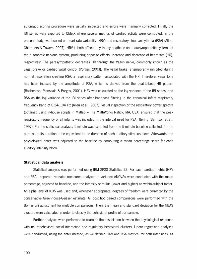
100
automatic scoring procedure were visually inspected and errors were manually corrected. Finally the
IBI series were exported to CMetX where several metrics of cardiac activity were computed. In the
present study, we focused on heart rate variability (HRV) and respiratory sinus arrhythmia (RSA) (Allen,
Chambers & Towers, 2007). HRV is both affected by the sympathetic and parasympathetic systems of
the autonomic nervous system, producing opposite effects: increase and decrease of heart rate (HR),
respectively. The parasympathetic decreases HR through the Vagus nerve, commonly known as the
vagal brake or cardiac vagal control (Porges, 2003). The vagal brake is temporarily inhibited during
normal respiration creating RSA, a respiratory pattern associated with the HR. Therefore, vagal tone
has been indexed by the amplitude of RSA, which is derived from the beat-to-beat HR pattern
(Bazhenova, Plonskaia & Porges, 2001). HRV was calculated as the log variance of the IBI series, and
RSA as the log variance of the IBI series after bandpass filtering in the canonical infant respiratory
frequency band of 0.24-1.04 Hz (Allen et al., 2007). Visual inspection of the respiratory power spectra
(obtained using in-house scripts in Matlab – The MathWorks Natick, MA, USA) ensured that the peak
respiratory frequency of all infants was included in the interval used for RSA filtering (Berntson et al.,
1997). For the statistical analysis, 1-minute was extracted from the 5-minute baseline collected, for the
purpose of its duration to be equivalent to the duration of each auditory stimulus block. Afterwards, the
physiological score was adjusted to the baseline by computing a mean percentage score for each
auditory intensity block.
Statistical data analysis
Statistical analysis was performed using IBM SPSS Statistics 22. For each cardiac metric (HRV
and RSA), separate repeated-measures analyses of variance ANOVAs were conducted with the mean
percentage, adjusted to baseline, and the intensity stimulus (lower and higher) as within-subject factor.
An alpha level of 0.05 was used and, whenever appropriate, degrees of freedom were corrected by the
conservative Greenhouse-Geisser estimate. All post hoc paired comparisons were performed with the
Bonferroni adjustment for multiple comparisons. Then, the mean and standard deviation for the NBAS
clusters were calculated in order to classify the behavioral profile of our sample.
Further analyses were performed to examine the association between the physiological response
with neurobehavioral social interaction and regulatory behavioral clusters. Linear regression analyses
were conducted, using the enter method, as we defined HRV and RSA metrics, for both intensities, as
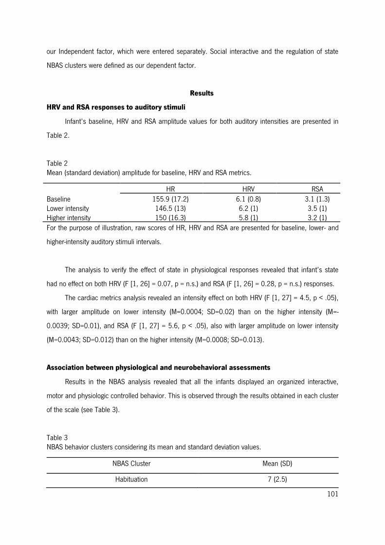
101
our Independent factor, which were entered separately. Social interactive and the regulation of state
NBAS clusters were defined as our dependent factor.
Results
HRV and RSA responses to auditory stimuli
Infant’s baseline, HRV and RSA amplitude values for both auditory intensities are presented in
Table 2.
Table 2 Mean (standard deviation) amplitude for baseline, HRV and RSA metrics.
HR HRV RSA
Baseline 155.9 (17.2) 6.1 (0.8) 3.1 (1.3) Lower intensity 146.5 (13) 6.2 (1) 3.5 (1) Higher intensity 150 (16.3) 5.8 (1) 3.2 (1)
For the purpose of illustration, raw scores of HR, HRV and RSA are presented for baseline, lower- and
higher-intensity auditory stimuli intervals.
The analysis to verify the effect of state in physiological responses revealed that infant’s state
had no effect on both HRV (F [1, 26] = 0.07, p = n.s.) and RSA (F [1, 26] = 0.28, p = n.s.) responses.
The cardiac metrics analysis revealed an intensity effect on both HRV (F [1, 27] = 4.5, p < .05),
with larger amplitude on lower intensity (M=0.0004; SD=0.02) than on the higher intensity (M=-
0.0039; SD=0.01), and RSA (F [1, 27] = 5.6, p < .05), also with larger amplitude on lower intensity
(M=0.0043; SD=0.012) than on the higher intensity (M=0.0008; SD=0.013).
Association between physiological and neurobehavioral assessments
Results in the NBAS analysis revealed that all the infants displayed an organized interactive,
motor and physiologic controlled behavior. This is observed through the results obtained in each cluster
of the scale (see Table 3).
Table 3 NBAS behavior clusters considering its mean and standard deviation values.
NBAS Cluster Mean (SD)
Habituation 7 (2.5)
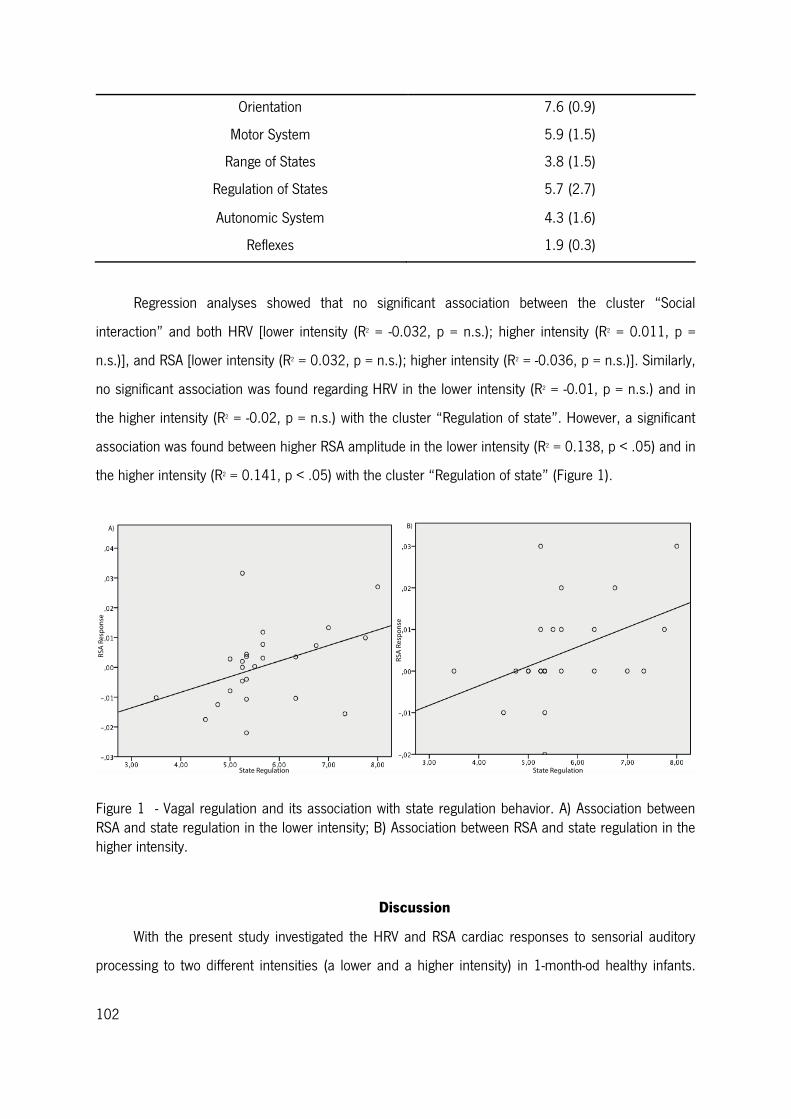
102
Orientation 7.6 (0.9)
Motor System 5.9 (1.5)
Range of States 3.8 (1.5)
Regulation of States 5.7 (2.7)
Autonomic System 4.3 (1.6)
Reflexes 1.9 (0.3)
Regression analyses showed that no significant association between the cluster “Social
interaction” and both HRV [lower intensity (R2 = -0.032, p = n.s.); higher intensity (R2 = 0.011, p =
n.s.)], and RSA [lower intensity (R2 = 0.032, p = n.s.); higher intensity (R2 = -0.036, p = n.s.)]. Similarly,
no significant association was found regarding HRV in the lower intensity (R2 = -0.01, p = n.s.) and in
the higher intensity (R2 = -0.02, p = n.s.) with the cluster “Regulation of state”. However, a significant
association was found between higher RSA amplitude in the lower intensity (R2 = 0.138, p < .05) and in
the higher intensity (R2 = 0.141, p < .05) with the cluster “Regulation of state” (Figure 1).
Figure 1 - Vagal regulation and its association with state regulation behavior. A) Association between RSA and state regulation in the lower intensity; B) Association between RSA and state regulation in the higher intensity.
Discussion
With the present study investigated the HRV and RSA cardiac responses to sensorial auditory
processing to two different intensities (a lower and a higher intensity) in 1-month-od healthy infants.
103
Furthermore, we analyzed the relation between these cardiac responses to neurodevelopmental
measures, specifically social orientation and regulatory abilities.
Our results evidence that, at 1-month of age, infants are displaying different cardiac response
patterns to both lower- and higher-intensity auditory stimuli, showing higher HRV and RSA amplitudes
to lower intensities (decrease in HR) and, on the contrary lower HRV and RSA amplitudes to higher
intensities (increase in HR). These results suggest that infants display a regulated vagal response to the
lower-intensity auditory stimuli, which is dependent on visceral state regulation by the autonomic
system (ref). These results are consistent with previous studies (Bazhenova, Plonskaia & Porges, 2001;
Stifter & Corey, 2001), showing that stimuli perceived as more appealing/engaging are associated with
more attentive states in infants, which is a behavioral characteristic associated with less physiological
reactivity. Differently, the higher intensity stimuli is likely to produce an adjustment of the autonomic
functions (i.e., characterized by lower HRV and RSA amplitudes), suggesting that infants may be
processing these type of auditory stimuli as more aversive. In fact, stimulus that is associated with
more discomfort in infants tends to be translated into an increase in the physiological reactivity,
represented by lower HRV and RSA (i.e. increased heart rate) (Porges & Lewis, 2010; Porges, 2003).
Overall, an increase in the RSA amplitude, and thus a regulated vagal tone, is associated to the
lower intensity auditory processing may be mirroring more attentive states in young infants. A decrease
in RSA amplitude, observed to the higher intensity, suggests discomfort in processing higher auditory
intensities in young infants. As shown originally by Porges (2001), vagal activity represents a stable
physiological state playing a crucial role in successful social interaction. However, the argument about
measuring HRV or any of its measures such as the RSA, to explore regulatory processes has various
precursors. Actually, the vagal activity interacts with other regulating systems, namely the
neuroendocrine and immune system (Field & Diego, 2008). That interaction between those reactivity
systems allows a better understanding of how the vagal regulation can be associated with
temperament, social interaction and infant development.
From other perspective, despite at first being seen as completely unrelated, recent finding has
showed that state regulation and auditory processing are theoretically and anatomically interconnected
through neural efferent pathways. Interestingly, those neural pathways underly the social engagement
system, as described in the polyvagal theory, which shows an association between changes in RSA and
functions of the middle ear structures influencing infant’s attending behaviors to noise (Porges et al.,
2013).
104
Regarding the association between HRV and RSA responses with neurobehavioral assessment,
our results suggest that higher RSA amplitudes, in both intensities, are correlated with adjusted scores
obtained in regulation of state cluster, implying that cardiac vagal response is underling regulatory
abilities in young infants. Indeed, when associating the physiological response with behavioral
characteristics, several studies have showed that engagement or mobilization behaviors seem to have
a physiological basis, as vagal regulation is associated with different behavioral outcomes across
infancy (Hastings et al., 2008; Beauchaine, Gatzke-Kopp & Mead, 2007; Calkins, 1997). The fact that
higher RSA amplitude is associated with state regulation abilities, seem to suggest that the 1-month-old
infants are able to regulate themselves in order to attend to stimuli, which may be translated into
engagement behaviors, although the higher intensity may be producing more discomfort (Moore &
Calkins, 2004).
Several studies (Calkins, Graziano & Keane, 2007; Bazhenova, Plonskaia & Porges, 2001;
Doussard‐Roosevelt et al., 1997) have demonstrated that vagal tone seems to be underlying interactive
and social orientation behaviors to different stimuli. Infants’ regulated vagal tone (i.e., increase RSA) is
underlying engagement and attending behaviors and, in opposite, high vagal tone (i.e., decrease RSA)
seems to be associated with avoidance, tantrums and mobilization behaviors in young infants (Porges,
2011). Overall, our results seem to suggest that 1-month-old infants regulated vagal tone is associated
with the their ability to regulate their state in order to attend to the presented stimuli (both intensities).
Although no positive or negative states behavioral analyses were preformed in regards to the
auditory stimulation, our results seem consistent with the Polyvagal theory, indicating that one-month
old infants are already displaying a cardiac vagal response that may indicate whether a particular
stimulus may be associated with adjusted behavioral outcomes (Porges, 2001, 2003). Indeed,
increased vagal reactivity seems to be modulating engagement strategies, which may be verified by the
associated found between higher RSA amplitude to both auditory intensities.
Different studies have evidenced that vagal regulation is essential for promoting social
interactive behavior in young infants as it has an important role in attenuating sympathetic system
influences at the level of the heart and allowing social communication. Therefore, it seems evident that
infants with poor regulatory abilities would present lower thresholds for environmental stimulation, as
they are not able to reduce the sympathetic reactivity in response to the stimuli (Porges, 2011).
Behaviorally, we will assist at infants who would not be able shut down negative affect, translated into
105
tantrums and avoidant contact, which would be associated with less organized behavior, and, thus,
positive engagement with social environment would be difficult to establish (Porges, 2001).
Particularly, with the present study, we evidence that vagal regulation abilities are present very
early and that they are associated with neurobehavioral regulatory abilities that underlying social
interaction. As Porges (2011) described, Social Engagement System is sustained on self-regulatory
abilities that allow the infant to interact with the environment, and this abilities are sustained by the
vagal brake acting over sympathetic influences over the heart to environmental stimulation. Indeed,
studies have been demonstrating that children’s less cardiac vagal withdrawal seems to be associated
with externalizing problems (Perry et al., 2014; Calkins, Graziano & Kean, 2007). Therefore,
deregulated vagal tone seems to be a key component implied in state regulation and, consequently,
involved in invalidating positive social engagement. Our study suggest that regulated vagal tone may be
used as a physiological marker of stimuli processing in young infants, as it suggests that vagal tone is
associated with adjusted regulatory abilities in healthy 1-month-old infants. These results may offer a
physiological basis for an early identification of stimuli processing thresholds in young infants (ability
commonly impaired in developmental disorders) and, consequently, an early diagnostic of
developmental-related problems.
Future studies should assess threshold discrimination in young infants in regards to different
stimuli, as visual or olfactory processing. Furthermore, it is of interest to investigate if the vagal
regulation identified in young infants may be predicting future development such as cognitive or
language abilities.
Acknowledgements
We would like to thank the families and infants for their availability to participate in this study.
Also, we would like to thank Pedro Hispano Hospital, particularly to Dr. J. Lopes dos Santos, for all the
kindness throughout the time that we were there for the data collection process. We thank to Maria de
Góis-Eanes for all the help and knowledge entailed throughout this study. Furthermore, we thank
Associação Viver a Ciência for the Simbiontes prize in 2013. This work was supported by Fundação
Bial (grant number 42/08) and the Fundação para a Ciência e Tecnologia (FCT) under a PhD grant
(reference SFRH/BD/68263/2010).

106
107
References
Allen, J. J. B., Chambers, A. S., & Towers, D. N. (2007). The many metrics of cardiac chronotropy: A
pragmatic primer and a brief comparison of metrics. Biol Psychol, 74(2), 243-262.
Bazhenova, O. V., Plonskaia, O., & Porges, S. W. (2001). Vagal reactivity and affective adjustment in
infants during interaction challenges. Child Dev, 72(5), 1314-1326.
Beauchaine, T. P., Gatzke-Kopp, L., & Mead, H. K. (2007). Polyvagal theory and developmental
psychopathology: Emotion dysregulation and conduct problems from preschool to
adolescence. Biol Psychol, 74(2), 174-184.
Berntson, G. G., Bigger, J. T., Jr., Eckberg, D. L., Grossman, P., Kaufmann, P. G., Malik, M., . . . van
der Molen, M. W. (1997). Heart rate variability: origins, methods, and interpretive caveats.
Psychophysiology, 34(6), 623-648.
Brazelton, T. B., & Nugent, J. K. (1995). Neonatal Behavioral Assessment Scale. (3rd Edition ed. Vol.
137). London: MacKeith Press.
Calkins, S. D. (1997). Cardiac vagal tone indices of temperamental reactivity and behavioral regulation
in young children. Dev Psychobiol, 31(2), 125-135.
Calkins, S. D., Graziano, P. A., & Keane, S. P. (2007). Cardiac vagal regulation differentiates among
children at risk for behavior problems. Biol Psychol, 74(2), 144-153.
Doussard‐Roosevelt, J. A., Porges, S. W., Scanlon, J. W., Alemi, B., & Scanlon, K. B. (1997). Vagal
regulation of heart rate in the prediction of developmental outcome for very low birth weight
preterm infants. Child Dev, 68(2), 173-186.
Field, T., & Diego, M. (2008). Vagal activity, early growth and emotional development. Infant Behav
Dev, 31(3), 361-373. doi: 10.1016/j.infbeh.2007.12.008
Field, T. M., Dempsey, J. R., Hatch, J., Ting, G., & Clifton, R. K. (1979). Cardiac and behavioral
responses to repeated tactile and auditory stimulation by preterm and term neonates. Dev
Psychol, 15(4), 406.
Geva, R., & Feldman, R. (2008). A neurobiological model for the effects of early brainstem functioning
on the development of behavior and emotion regulation in infants: implications for prenatal and
perinatal risk. J Child Psychol Psychiatry, 49(10), 1031-1041. doi: 10.1111/j.1469-
7610.2008.01918.x
108
Graham, A. M., Ablow, J. C., & Measelle, J. R. (2010). Interparental relationship dynamics and cardiac
vagal functioning in infancy. Infant Behavior and Development, 33(4), 530-544.
Hastings, P. D., Nuselovici, J. N., Utendale, W. T., Coutya, J., McShane, K. E., & Sullivan, C. (2008).
Applying the polyvagal theory to children's emotion regulation: Social context, socialization, and
adjustment. Biol Psychol, 79(3), 299-306.
Moore, D. R., & Hunter, L. L. (2013). Auditory processing disorder (APD) in children: A marker of
neurodevelopmental syndrome. Hearing, Balance and Communication, 11(3), 160-167.
Moore, G. A., & Calkins, S. D. (2004). Infants' vagal regulation in the still-face paradigm is related to
dyadic coordination of mother-infant interaction. Dev Psychol, 40(6), 1068-1080. doi:
10.1037/0012-1649.40.6.1068
Perry, N. B., Nelson, J. A., Swingler, M. M., Leerkes, E. M., Calkins, S. D., Marcovitch, S., & O'Brien,
M. (2013). The relation between maternal emotional support and child physiological regulation
across the preschool years. Dev Psychobiol, 55(4), 382-394.
Pizur-Barnekow, K., Kraemer, G. W., & Winters, J. M. (2008). Pilot study investigating infant vagal
reactivity and visual behavior during object perception. Am J Occup Ther, 62(2), 198-205.
Pomerleau, A., & Malcuit, G. (1981). State effects on concomitant cardiac and behavioral responses to
a rocking stimulus in human newborns. Infant Behavior and Development, 4, 163-174.
Porges, S. W. (1995). Cardiac vagal tone: a physiological index of stress. Neuroscience & Biobehavioral
Reviews, 19(2), 225-233.
Porges, S. W. (2001). The polyvagal theory: phylogenetic substrates of a social nervous system.
International Journal of Psychophysiology, 42(2), 123-146.
Porges, S. W. (2003). The Polyvagal Theory: phylogenetic contributions to social behavior. Physiol
Behav, 79(3), 503-513.
Porges, S. W., Arnold, W. R., & Forbes, E. J. (1973). Heart rate variability: An index of attentional
responsivity in human newborns. Dev Psychol, 8(1), 85.
Porges, S. W., & Furman, S. A. (2011). The early development of the autonomic nervous system
provides a neural platform for social behaviour: A polyvagal perspective. Infant and child
development, 20(1), 106-118.
Porges, S. W., & Lewis, G. F. (2010). .2-The polyvagal hypothesis: common mechanisms mediating
autonomic regulation, vocalizations and listening. Handbook of Behavioral Neuroscience, 19,
255-264.
109
Porges, S. W., Macellaio, M., Stanfill, S. D., McCue, K., Lewis, G. F., Harden, E. R., . . . Heilman, K. J.
(2013). Respiratory sinus arrhythmia and auditory processing in autism: modifiable deficits of
an integrated social engagement system? Int J Psychophysiol, 88(3), 261-270. doi:
10.1016/j.ijpsycho.2012.11.009
Reulecke, S., Schulz, S., & Voss, A. (2012). Autonomic regulation during quiet and active sleep states
in very preterm neonates. Integrative Physiology, 3, 61.
Sameroff, A. J., Cashmore, T. F., & Dykes, A. C. (1973). Heart rate deceleration during visual fixation in
human newborns. Dev Psychol, 8(1), 117.
Schmidt, L. A., & Segalowitz, S. J. (2008). Developmental psychophysiology: theory, systems, and
methods: Cambridge University Press New York.
Sprangler, G., Fremmer-Bombik, E., & Grossmann, K. (1996). Social and individual determinants of
infant attachment security and disorganization. Infant Mental Health Journal, 17(2), 127-139.
doi: 10.1002/(SICI)1097-0355(199622)17:2<127::AID-IMHJ3>3.0.CO;2-N
Stifter, C. A., & Corey, J. M. (2001). Vagal regulation and observed social behavior in infancy. Social
Development, 10(2), 189-201.
Stifter, C. A., & Fox, N. A. (1990). Infant reactivity: Physiological correlates of newborn and 5-month
temperament. Dev Psychol, 26(4), 582.
Trapanotto, M., Benini, F., Farina, M., Gobber, D., Magnavita, V., & Zacchello, F. (2004). Behavioural
and physiological reactivity to noise in the newborn. J Paediatr Child Health, 40(5-6), 275-281.
doi: 10.1111/j.1440-1754.2004.00363.x
Vranekovic, G., Hock, E., Isaac, P., & Cordero, L. (1974). Heart rate variability and cardiac response to
an auditory stimulus. Biol Neonate, 24(1), 66-73.

110
111
Conclusion
Interactive behavior is a core aspect that is involved in adequate developmental outcomes and,
later, with healthy psychological adjustment. According to the literature review and research studies
presented in the current dissertation, it seems evident that neurophysiological processes are underlying
social behaviors in young infants. With the present dissertation we aimed to identify, in 1-month-old
infants, neurophysiological markers of the sensory processing and their relation with both social
behavior; and later at 12-months of age, with developmental cognitive, language and motor outcomes.
To date, evidence has been presented on the importance of neural and physiological
mechanisms underlying interactive behavior (Grossmann & Johnson, 2007; Striano & Reid, 2006;
Porges, 2003). With the present dissertation we intend to contribute and expand the knowledge
regarding the neurophysiological signatures underlying social behavior
Regarding the developmental studies, our results evidence that 1-month-old infants show a
specific neural and physiological response underlying visual and auditory sensory processing.
Moreover, these neurophysiological markers are associated with infant’s interactive abilities.
Additionally, specific auditory ERPs were associated with language milestones at 12-months of age.
Concerning the first study, focusing on visual processing in young infants, we observed that two
main neural VEP components were display in regards to two unpatterned flashlight stimuli intensities (a
lower and a higher one): P2 and N3. Both components have been found to be present to visual
stimulation in infants, whether the infants were awake or sleeping, being associated with maturational
processes (McGlone et al., 2013; Benavente, Tamargo, Tajada, Yuste, & Olivan, 2005; Kraemer,
Abrahamsson & Sjostrom, 1999). Additionally, our study evidenced that infant’s state seems crucial for
stimuli intensities processing, as P2 amplitude was revealed to be higher in infants that were awake
during the stimuli presentation than in the sleeping infants. In fact, several studies have showed the
implications of sleep-awake states in visual stimulation (Shepherd, Saunders & McCulloch, 1999; Meek
et al., 1998; Mercuri, von Siebenthal, Tutuncuoglu, Guzzetta, & Casaer, 1995; Whyte, Pearce & Taylor,
1987) and our study reveals that awake and sleep states have an effect in visual thresholds processing
in young infants. Moreover, our results show that infant’s state seems crucial when associated with
behavioral interactive abilities, as we found a positive association between N3 component and social
interaction and state regulation behaviors only in awake infants.
Social interaction is essentially occurring by the infant’s ability to attend to stimuli produced by
others, especially through visual but also through auditory processing, in particular by attending to face
112
and voice cues (Grossmann, Striano, & Friederici, 2006). Likewise, in the second study, we studied
auditory ERPs in respect to two stimuli intensities. Overall, we found that P2 and N2 components were
present to both auditory intensities, regardless of infants’ state. Similarly to visual processing, these
two AEP’s components have been associated with cortical maturational processes, as they have been
consistently found in healthy full-term infants auditory neural response (Lippé, Martinez-Montes,
Arcand, & Lassonde, 2009; Wunderlich, Cone-Wesson & Shepherd, 2006). Moreover, an intensity
effect was found in the P2 component, with larger amplitude in the higher intensity. Indeed, P2
component has been found to be associated with auditory discrimination (Marcoux, 2011; Ceponiene
et al., 2002) and, therefore, our results seem consistent with previous findings. When associating this
neural response with interactive competences at 1-month-old, our results evidence that P2 positive
amplitude in the lower intensity is associated with social interactive behavior and regulatory abilities
and, later, in infants aged 12-month-old, with language competences. These results are consistent with
previous studies (Kushnerenko et al., 2013; Benasich et al., 2006; Benasich & Tallal, 2002; Benasich,
Thomas, Choudhury, & Leppanen, 2002; Molfese & Molfese, 1997) and reveal that early at a postnatal
age, neural markers of auditory processing can be already identified in order to predict social behavior
in 1-month-old infants and language competences at 12-months of age. Similarly, an increased P2
latency in lower intensity was also correlated with adjusted regulatory behavior. As explained in the
second study, we hypothesized that the neural responses is possibly characterized by employing more
time at a given stimulus, so that is possible that infant’s with better regulatory abilities attend
repeatedly to the stimuli, spending more attentional resources and, therefore, taking more time in
auditory stimuli processing (Kushnerenko, Van den Bergh & Winkler, 2013; Atkinson, 2002).
In both studies, P2 component was found to be present in visual and auditory processing and,
therefore, seems to be associated with the physical properties of the stimuli. Similarly, P2 amplitude
was found to be larger in the higher intensity, indicating that this component seems sensible to detect
threshold differences regarding sensorial stimulation. P2 component was found to be an auditory
neural marker of interactive behavior and language outcome. However, the same was not verified in
visual processing, as N3 component was associated with adjusted behavioral outcomes. These
differences may be resultant of myelination and synaptogenesis processes occurring around this age.
Indeed, myelination and synaptogenesis are crucial for brain function and, although, at term we
evidence the burst of synaptic formation (e.g. pruning) and myelination, differences regarding these
processes have been noticed in the cortex (Huttenlocher, 2009; Fogel, 2009; Nelson & Luciana,
113
2008). Dendritic growth seems to vary when comparing maturation processes in frontal, temporal and
visual cortices. The time that these synaptic changes happen differs as the burst of synaptogenesis in
the visual cortex only occurs around 2-4 months postnatal age (Dubois et al., 2004). In the auditory
cortex, these processes emerge at birth (Huttenlocher, 2009). Moreover, N3 component seem to be
associated with attentional processes, which are required for visual abilities to follow objects and to
interact with other (Luo, Feng, He, Wang, & Luo, 2010; McCulloch & de Haan, 2007). Therefore,
regarding visual processing and its association with behavioral outcome, we believe that in 1-month-old
infants P2 component is correlated with of physical stimuli processing and N3 associated with mature
and adjusted social behavioral outcomes.
Similarly, in these two studies, the association between the neural response and behavior was
found in regards to the lower intensity. This may be occurring due to the fact that during the first
months of life (until circa 3 month of age) infants present increased sensitivity to lower sensorial
information (Kushnerenko et al., 2013; Atkinson, 2002). Thus, we hypothesize that neural response to
the lower intensities underlies the infant’s ability to perceive lower sensory information, which is
associated with coherent and focused interactive and regulatory characteristics, and enables infants to
attend to different social and sensorial cues from the surrounding environment (Kushnerenko et al.,
2013; Brazelton & Nugent, 1995).
Neural and physiological correlates are related processes, as central nervous system influences
autonomic nervous system through neural circuits with afferent pathways characterized by adaptive
reactivity (Porges, 2009). Therefore, considering the third study we present physiological reactivity
correlates in infants to two auditory stimuli intensities. We found that higher HRV and RSA amplitudes
were displayed in regards to the lower intensity. On the contrary, lower HRV and RSA amplitudes were
observed to the higher auditory intensity. Indeed, as previous studies have suggested (Bazhenova,
Plonskaia & Porges, 2001; Stifter & Corey, 2001), stimuli that are perceived as more
appealing/engaging are associated with more attentive states in infants, which is a behavioral
characteristic associated with less physiological reactivity. On the contrary, stimulus that is associated
with more discomfort in infants tends to be translated into an increased physiological reactivity,
represented by lower HRV and RSA (i.e. increase heart rate) (Perry et al., 2013; Hastings et al., 2008;
Calkins, Graziano & Keane, 2007; Calkins, 1997). Furthermore, we found a positive association
between higher RSA amplitude, in both auditory intensities, and adjusted state regulation competences
in these infants. This result suggests the regulation of vagal response implications in self-regulatory
114
behavior, allowing 1-month-old infants to attend to different stimuli intensities. Similarly, these results
are consistent with previous studies (Beauchaine, Gatzke-Kopp & Mead, 2007; Moore & Calkins, 2004;
Bazhenova, Plonskaia & Porges, 2001) indicating that regulated vagal response seems to be an
indicator of physiological basis to display engagement/disengagement behaviors in response to the
environment. Higher RSA amplitudes suggested regulated vagal tone, which is essential for displaying
engagement behaviors to stimulation. This ability seems to be closely related with regulatory abilities
displayed by these infants, enabling them to attend to both auditory stimuli intensities.
Overall, with the present studies we were able to identify in 1-month-old infants
neurophysiological and psychophysiological correlates of sensory processing and it’s association with
social behavior and development. Differences regarding neurophysiological processing of different
sensorial modalities seem evident, however both seem to be correlate with interactive behavior. We
expect to enlarge the knowledge regarding this field by highlighting the contributions of the present
studies to better understand brain development and physiological reactivity in young infants. Some
studies, have largely contributed to uncover the neural and physiological processes involved in this
issue (Porges, 2011; Grossmann, Oberecker, Koch, & Friederici, 2010; Grossmann & Johnson, 2010;
Grossmann, Striano & Friederici, 2005). With the present research we wanted to enlighten the
emergence of such processes in younger infants and its consequences for interactive behaviors, from
an early postnatal age. Doing so, clinical implications drawn from this work seem evident. Indeed, an
early identification of sensory thresholds in young infants can be useful to adequate social interactions
to the infants’ necessities. As every infant responds differently to the surrounding environment, the
neurobehavioral assessment can provide valuable information regarding the infants’ behavior, their
stronger abilities and adjust interactive routines to physiological responses. Moreover, social
impairments are associated with different developmental disorders (i.e., autism spectrum disorders),
and an early identification of neurophysiological signatures associated with adjusted social behavior can
provide clinical information in regards to development-related problems. Therefore, an early
identification of such impairments can be used to implement early intervention programs, as early
interventions are associated with better therapeutic results.
Nevertheless, some limitations of the presented work must be addressed. First, the great intra-
and inter-variability observed in infants neural and physiological sensory processing responses, similar
to previously reports (Snyder, Webb & Nelson, 2002). This may be occurring as brain development and
maturational processes are still ongoing processes in this age, which difficult data analysis and
115
interpretation. Moreover, the equipment used in the ERP data collection may not be the most suitable,
as scalp cleaning to minimize impedances revealed to be stressful for young infants and, sometimes,
compromising data collection. Another limitation relies on the state dependence for the
neurobehavioral assessment. Indeed, in order to assess some NBAS clusters (i.e. habituation) the
infant was need to be in a specific state. Sometimes this was not possible, what had implications
completing the neurobehavioral scale.
Therefore, future studies should increase the number of participants for the purpose of
replication of the described results. Similarly, neurophysiological evidence regarding stimuli processing
should be collected across the infancy period, in a longitudinal fashion, in order to detect maturational
processes and to identify alterations in regards to VEP, AEP and HRV and RSA responses and its
association with social behavior and development outcomes. Moreover, such studies should include
infants at risk (i.e. premature infants) in order to understand if neurophysiological aspects involved in
social behavior differ in such infants.
116
References
Atkinson, J. (2002). The developing visual brain. (Vol. 32). New York, United States: Oxford University
Press.
Bazhenova, O. V., Plonskaia, O., & Porges, S. W. (2001). Vagal reactivity and affective adjustment in
infants during interaction challenges. Child Dev, 72(5), 1314-1326.
Beauchaine, T. P., Gatzke-Kopp, L., & Mead, H. K. (2007). Polyvagal theory and developmental
psychopathology: Emotion dysregulation and conduct problems from preschool to
adolescence. Biol Psychol, 74(2), 174-184.
Benasich, A. A., Choudhury, N., Friedman, J. T., Realpe-Bonilla, T., Chojnowska, C., & Gou, Z. (2006).
The infant as a prelinguistic model for language learning impairments: predicting from event-
related potentials to behavior. Neuropsychologia, 44(3), 396-411. doi:
10.1016/j.neuropsychologia.2005.06.004
Benasich, A. A., & Tallal, P. (2002). Infant discrimination of rapid auditory cues predicts later language
impairment. Behav Brain Res, 136(1), 31-49.
Benasich, A. A., Thomas, J. J., Choudhury, N., & Leppanen, P. H. (2002). The importance of rapid
auditory processing abilities to early language development: evidence from converging
methodologies. Dev Psychobiol, 40(3), 278-292.
Benavente, I., Tamargo, P., Tajada, N., Yuste, V., & Olivan, M. J. (2005). Flash visually evoked
potentials in the newborn and their maturation during the first six months of life. Doc
Ophthalmol, 110(2-3), 255-263. doi: 10.1007/s10633-005-0818-0
Brazelton, T. B., & Nugent, J. K. (1995). Neonatal Behavioral Assessment Scale. (3rd Edition ed. Vol.
137). London: MacKeith Press.
Calkins, S. D. (1997). Cardiac vagal tone indices of temperamental reactivity and behavioral regulation
in young children. Dev Psychobiol, 31(2), 125-135.
Calkins, S. D., Graziano, P. A., & Keane, S. P. (2007). Cardiac vagal regulation differentiates among
children at risk for behavior problems. Biol Psychol, 74(2), 144-153.
Ceponiene, R., Kushnerenko, E., Fellman, V., Renlund, M., Suominen, K., & Naatanen, R. (2002).
Event-related potential features indexing central auditory discrimination by newborns. Brain Res
Cogn Brain Res, 13(1), 101-113.
117
Fogel, A. (2009). Infancy: Infant, family, and society (Fifth ed.): Sloan Publishing.
Grossmann, T., & Johnson, M. H. (2007). The development of the social brain in human infancy. Eur J
Neurosci, 25(4), 909-919. doi: 10.1111/j.1460-9568.2007.05379.x
Grossmann, T., & Johnson, M. H. (2010). Selective prefrontal cortex responses to joint attention in
early infancy. Biol Lett, 6(4), 540-543. doi: 10.1098/rsbl.2009.1069
Grossmann, T., Oberecker, R., Koch, S. P., & Friederici, A. D. (2010). The Developmental Origins of
Voice Processing in the Human Brain. Neuron, 65(6), 852-858. doi:
http://dx.doi.org/10.1016/j.neuron.2010.03.001
Grossmann, T., Striano, T., & Friederici, A. D. (2005). Infants' electric brain responses to emotional
prosody. Neuroreport, 16(16), 1825-1828.
Hastings, P. D., Nuselovici, J. N., Utendale, W. T., Coutya, J., McShane, K. E., & Sullivan, C. (2008).
Applying the polyvagal theory to children's emotion regulation: Social context, socialization, and
adjustment. Biol Psychol, 79(3), 299-306.
Huttenlocher, P. R. (2009). Neural plasticity: Harvard University Press.
Kraemer, M., Abrahamsson, M., & Sjostrom, A. (1999). The neonatal development of the light flash
visual evoked potential. Doc Ophthalmol, 99(1), 21-39.
Kushnerenko, E., Tomalski, P., Ballieux, H., Ribeiro, H., Potton, A., Axelsson, E. L., . . . Moore, D. G.
(2013). Brain responses to audiovisual speech mismatch in infants are associated with
individual differences in looking behaviour. Eur J Neurosci, 38(9), 3363-3369. doi:
10.1111/ejn.12317
Kushnerenko, E. V., Van den Bergh, B. R., & Winkler, I. (2013). Separating acoustic deviance from
novelty during the first year of life: a review of event-related potential evidence. Front Psychol,
4, 595. doi: 10.3389/fpsyg.2013.00595
Lippé, S., Martinez-Montes, E., Arcand, C., & Lassonde, M. (2009). Electrophysiological study of
auditory development. Neuroscience, 164(3), 1108-1118. doi:
http://dx.doi.org/10.1016/j.neuroscience.2009.07.066
Luo, W., Feng, W., He, W., Wang, N.-Y., & Luo, Y.-J. (2010). Three stages of facial expression
processing: ERP study with rapid serial visual presentation. Neuroimage, 49(2), 1857-1867.
Marcoux, A. M. (2011). Maturation of auditory function related to hearing threshold estimations using
the auditory brainstem response during infancy. Int J Pediatr Otorhinolaryngol, 75(2), 163-170.
doi: 10.1016/j.ijporl.2010.10.027
118
McCulloch, D. L., & de Haan, M. (2007). Visual evoked potentials in infants. Infant EEG and event-
related potentials, 39-63.
McGlone, L., Hamilton, R., McCulloch, D. L., Boulton, R., Bradnam, M. S., Weaver, L. T., & Mactier, H.
(2013). Neonatal visual evoked potentials in infants born to mothers prescribed methadone.
Pediatrics, 131(3), e857-863. doi: 10.1542/peds.2012-2113
Meek, J. H., Firbank, M., Elwell, C. E., Atkinson, J., Braddick, O., & Wyatt, J. S. (1998). Regional
hemodynamic responses to visual stimulation in awake infants. Pediatr Res, 43(6), 840-843.
Mercuri, E., von Siebenthal, K., Tutuncuoglu, S., Guzzetta, F., & Casaer, P. (1995). The effect of
behavioural states on visual evoked responses in preterm and full-term newborns.
Neuropediatrics, 26(4), 211-213. doi: 10.1055/s-2007-979756
Molfese, D. F., & Molfese, V. J. (1997). Discrimination of language skills at five years of age using
event-related potentials recorded at birth. Dev Neuropsychol, 13(2), 135-156.
Moore, G. A., & Calkins, S. D. (2004). Infants' vagal regulation in the still-face paradigm is related to
dyadic coordination of mother-infant interaction. Dev Psychol, 40(6), 1068.
Nelson, C. A., & Luciana, M. (2008). Handbook of developmental cognitive neuroscience: MIT Press.
Perry, N. B., Nelson, J. A., Swingler, M. M., Leerkes, E. M., Calkins, S. D., Marcovitch, S., & O'Brien,
M. (2013). The relation between maternal emotional support and child physiological regulation
across the preschool years. Dev Psychobiol, 55(4), 382-394.
Porges, S. W. (2003). The Polyvagal Theory: phylogenetic contributions to social behavior. Physiol
Behav, 79(3), 503-513.
Porges, S. W. (2009). The polyvagal theory: new insights into adaptive reactions of the autonomic
nervous system. Cleve Clin J Med, 76 Suppl 2, S86-90. doi: 10.3949/ccjm.76.s2.17
Porges, S. W. (2011). The Polyvagal Theory: Neurophysiological Foundations of Emotions, Attachment,
Communication, and Self-regulation (Norton Series on Interpersonal Neurobiology): WW Norton
& Company.
Shepherd, A., Saunders, K., & McCulloch, D. (1999). Effect of sleep state on the flash visual evoked
potential. A case study. Doc Ophthalmol, 98(3), 247-256.
Snyder, K., Webb, S. J., & Nelson, C. A. (2002). Theoretical and methodological implications of
variability in infant brain response during a recognition memory paradigm. Infant Behavior and
Development, 25(4), 466-494.
119
Stifter, C. A., & Corey, J. M. (2001). Vagal regulation and observed social behavior in infancy. Social
Development, 10(2), 189-201.
Striano, T., & Reid, V. M. (2006). Social cognition in the first year. Trends Cogn Sci, 10(10), 471-476.
Whyte, H. E., Pearce, J. M., & Taylor, M. J. (1987). Changes in the VEP in preterm neonates with
arousal states, as assessed by EEG monitoring. Electroencephalogr Clin Neurophysiol, 68(3),
223-225.
Wunderlich, J. L., Cone-Wesson, B. K., & Shepherd, R. (2006). Maturation of the cortical auditory
evoked potential in infants and young children. Hear Res, 212(1-2), 185-202. doi:
10.1016/j.heares.2005.11.010

120
121
CHAPTER 2 Neuropsychophysiological correlates of interactive behavior: implications for therapeutic
relationship

122
123
Physiological correlates of therapeutic relationship
The equivalent efficacy results among different psychotherapy models are commonly known as
the Dodo Bird Verdict (from Lewis Carroll´s tale Alice in the Wonderland) stating, “Everybody has won,
and all must have prizes”. This verdict became the representation for the common factors that
contribute to good therapeutic outcomes despite the different theoretical and technical backgrounds
and approaches that seem to produce good results (Hodgetts & Wright, 2007).
It is largely known that different factors are contributing for the success of psychotherapy
process. Therapeutic alliance, as a common factor to different therapeutic approaches, has been of
great interest among different researchers and has been mostly studied in order to uncover how the
dynamics between client and therapist are underlying the therapeutic outcome (Norcross, 2010).
Indeed, different theoretical therapeutic models assume the therapeutic alliance as the common
element that emerges among them and plays a crucial role in the therapeutic treatment plan. Such
importance had been described in Freud’s work as the establishment of a relation between therapist
and client was viewed as a treatment objective. A positive relation with the therapist, considered as an
ally, would help the client to combat his problems (Horvath, 2001). Later, Luborsky, Auerbach,
Chandler, Cohen, and Bachrach (1971) defended that the therapist should be able to provide the client
the right environment for him to accomplish his treatment plans and, therefore, assumed the
therapeutic alliance as a help connection for the client (Gaston, 1990).
Probably the author that most focused on the therapeutic relationship subject was Rogers
(1951) through his therapeutic model Client-Centered Therapy (CCT). Rogers defended that the main
way that the therapist had to help the client with his problems was to offer him a good relation, by
being someone in whom the client sense empathy and psychological availability to establish a relation.
Moreover, the author stated that the therapist should experience positive and unconditional
consideration for the client, as well as emphatic comprehension over his problems, and should always
communicate his experience, regarding the client’s problems, to the client (Rogers, 1992). Therefore,
the relation between therapist and client was a necessary and crucial characteristic for the therapeutic
process and should be considered as the main concept regarding therapy (Rogers, 1992).
Similarly, Bordin (1979) argued that the efficacy verified in different therapeutic models was due
to the connection that was established between the person who seeks help and the one who offers
help. This connection was considered as the main agent for the change process. To establish this
124
relation, both therapist and client should be in accordance in regards to the problem that is present by
the client and regarding the objectives for the treatment plan. Therefore, the therapeutic dyad should
work together to achieve the therapeutic work objectives and, in addition, the client should compromise
to execute tasks in the therapy. These two components would favor the establishment of the
therapeutic relation, based on a mutual concordance regarding the therapeutic work and relied on a
better communication process (Bordin, 1979).
Thus, the concepts stressed out by Bordin (1979) draw the basis of therapeutic alliance process
as understood nowadays, once therapeutic alliance has been largely associated to the strength and
quality of the relationship that is established between therapist and client (Horvath, Del Re, Flückiger, &
Symonds, 2011; Horvath, 2006). Additionally, therapeutic alliance has been assumed as a negotiation
process once it is constantly changing regarding the needs of the therapeutic relationship at a given
moment in the therapy (Safran & Muran, 2006, 2000). Therapeutic alliance strength has been
considered as one of the most important factors contributing to the therapeutic process and has been
largely accepted as the most robust and reliable predictor of therapy outcomes, been positively
associated to good therapeutic results (Crits-Christoph, Gibbons & Hearon, 2006; Martin, Garske &
Davis, 2000; Horvath & Symonds, 1991). Most remarkably, it is the fact that several studies have
demonstrated that the initial phase of the therapeutic process is of extremely importance for alliance
formation and development as its positive evaluation in this phase is positively associated with the
client’s permanence in therapy, as well as with better therapeutic outcomes (Horvath, 2006)
(Hilsenroth, Peters & Ackerman, 2004; Bachelor & Salamé, 2000).
Therapeutic alliance is based on the quality of the collaborative relationship that unfolds between
therapist and client, as the therapeutic dyad is actively contributing to its formation and development
(Hatcher & Barends, 2006; Hatcher, 1999). Recently, it has been of great interest to better understand
the micro processes that are underlying the therapeutic alliance (Horvath, 2006, 2005; Ackerman &
Hilsenroth, 2003). This is true for the therapeutic collaboration. Being accepted as contributing
significantly to the alliance and, consequently, to the therapeutic process, therapeutic collaboration
refers to the interactive exchanges happening between therapist and client in a moment-by-moment
basis (Ribeiro, Ribeiro, Gonçalves, Horvath, & Stiles, 2013).
Indeed, therapeutic collaboration has been largely accepted as a core dimension of the
therapeutic alliance. Is commonly described as the contributions of therapist and client to the therapy
that cannot be reduced to the inputs of each element of the therapeutic dyad alone (Hatcher, 1999).
125
Generally, is characterized by the therapeutic dyad shared responsibility and participation in the
therapy objectives and multiple proposals or activities that happen within the therapeutic context, as
well as by affinitive, cooperative and engagement behaviors (Tryon & Winograd, 2011; Colli & Lingiardi,
2009; Boardman, Catley, Grobe, Little, & Ahluwalia, 2006).
In a review study, Tryon and Winograd (2011), focused on the therapeutic collaboration,
assessed clients’ involvement and homework task during therapy. The authors found that when clients’
were defensive or resistant, it was associated with a decrease in their improvement. On the contrary,
those who worked with the therapist regarding the resolution of their problems, felt better about the
treatment and experienced good outcomes.
Different studies focusing on the collaborative process have agreed that communication between
the dyad is of particular importance for clinical engagement, identifying a relation between interactive
situations and outcomes of the therapy (Strong, Sutherland & Ness, 2011; Lepper & Mergenthaler,
2007). Early studies have demonstrated that the communicative behavior of each element of the dyad
influences the behavior of the other element (Henry & Strupp, 1994; Tracey, 1993). For instance, it
has been demonstrated that when the therapist presented more interpretative intervention, the client
responses were mainly emotional or produced more insightful connections (Milbrath et al., 1999). On
the other hand, according to the concept of coordination a pattern of client’s responses characterized
as “basic features”, were found to be contributing to the therapeutic bond (Westerman, 1998).
Moreover, a relation was found between topic duration and communicative variables (such as
elaboration or dyselaboration) regarding the client’s discourse, demonstrating that these variables
seem to vary with topics and not with time along the speech (Horowitz et al., 1993).
Communicative actions between therapist and client seem to play an important role in the
collaborative process and client’s experience seems to improve within a positive collaborative
relationship with the therapist (Tryon & Winograd, 2011). However, more recently, uncovering what
happens in turn-by-turn interactions between therapist and client’s has been addressed (Ribeiro et al.,
2013; Lepper & Mergenthaler 2007).
In order to study turn-by-turn phenomena and its relation with change in psychotherapy, Lepper
and Mergenthaler (2007) developed a study based on their Therapeutic Cycles Model and on
conversation analysis in order to understand how therapeutic interactions are contributing to the
change cycles in therapy (Lepper & Mergenthaler, 2007, 2005; Mergenthaler, 1996). Through this
analytic method, and studying multiple case studies, the authors identified a marker of collaborative
126
rapport – topic coherence – happening between therapist and client communicative interaction,
underlying a correlation between topic coherence and periods of affective and cognitive engagement
during therapy (Lepper & Mergenthaler, 2008, 2007, 2005).
More recently, Ribeiro and colleagues (2013) have addressed the question of collaboration
focusing on moment-by-moment exchanges within therapeutic sessions and across therapeutic
processes. To do so, the authors developed a coding system that allows the identification and coding of
communicative exchanges, happening between therapist and client, and that characterizes the
collaboration involvement in the dyad. Therapeutic Collaboration Coding System (TCCS) unveils
moment-by-moment therapeutic interactions assessed through communicative patterns that are
mirroring collaborative or non-collaborative exchanges. The rational behind the TCCS builds on the
developmental concept Zone of proximal developmental (Vygotsky, 1978) and two theories of change in
psychotherapy, the assimilation model (Stiles, 2011, 2001) and the narrative framework, particularly
the innovative moments model (Gonçalves & Stiles, 2011; Goncalves, Matos & Santos, 2009), to
explain the therapeutic dyad communication exchanges and how change happens in psychotherapy.
In the therapeutic context, therapist must be able to create an environment where the client feels
confortable to tolerate and accept the new meanings that are being created. Therefore, and based on
the concept of Zone of Proximal Development (ZPD; Vygotsky, 1924, 1978), the TCCS conceptual
approach builds on the concept of Therapeutic Zone of Proximal Development (TZPD) (Ribeiro et al.,
2013; Leiman & Stiles, 2001), as therapy is understood as a developmental continuum, i.e. the space
between the client’s actual developmental level and a potential developmental level that can be
reached. TZPD, thus, is a developmental region the client goes through during successful therapy
(Figure 1). Therapy context and dialogue set the client within the TZPD and as therapeutic work
progresses the client moves to higher levels of the therapeutic zone. Therapeutic intervention
formulated within the TZPD are likely to success and, on the contrary, interventions formulated outside
the zone are likely to fail.
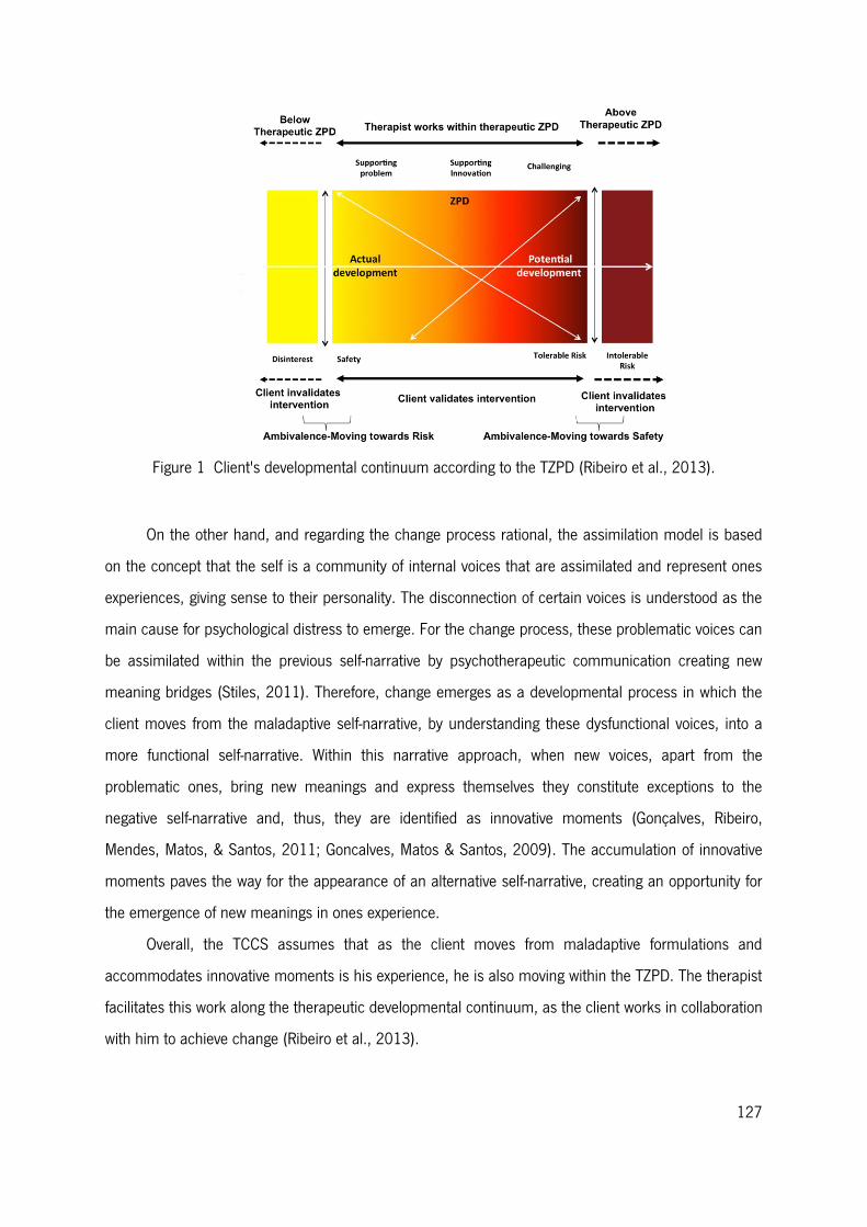
127
Figure 1 Client's developmental continuum according to the TZPD (Ribeiro et al., 2013).
On the other hand, and regarding the change process rational, the assimilation model is based
on the concept that the self is a community of internal voices that are assimilated and represent ones
experiences, giving sense to their personality. The disconnection of certain voices is understood as the
main cause for psychological distress to emerge. For the change process, these problematic voices can
be assimilated within the previous self-narrative by psychotherapeutic communication creating new
meaning bridges (Stiles, 2011). Therefore, change emerges as a developmental process in which the
client moves from the maladaptive self-narrative, by understanding these dysfunctional voices, into a
more functional self-narrative. Within this narrative approach, when new voices, apart from the
problematic ones, bring new meanings and express themselves they constitute exceptions to the
negative self-narrative and, thus, they are identified as innovative moments (Gonçalves, Ribeiro,
Mendes, Matos, & Santos, 2011; Goncalves, Matos & Santos, 2009). The accumulation of innovative
moments paves the way for the appearance of an alternative self-narrative, creating an opportunity for
the emergence of new meanings in ones experience.
Overall, the TCCS assumes that as the client moves from maladaptive formulations and
accommodates innovative moments is his experience, he is also moving within the TZPD. The therapist
facilitates this work along the therapeutic developmental continuum, as the client works in collaboration
with him to achieve change (Ribeiro et al., 2013).
128
According to the TCCS conceptualization, the therapeutic activities can be divided into two main
components: a) supporting and helping the client to feel safe, usually involving an expression of
understanding the client’s experience within his or her usual perspective; and b) challenging the
maladaptive framework by using strategies that stimulate the occurrence of innovation. These
components of collaboration happen in a balanced way once the therapist must keep working within a
therapeutic climate in which the client feels comfortable, but also must be able to help the client to
experience and consider new perspectives (Ribeiro et al, 2013).
The client’s response to the therapist’s intervention indicates whether the therapist worked
within the TZPD (validation), or instead, worked out of TZPD (invalidation), or even at the limit of the
TZPD (ambivalence). Interventions occurring within the TZPD require that the therapist addresses
empathically to the client’s TZPD, particularly the client’s current stage of change and the client’s
capacity to make sense of his own mental states or the therapist’s comments. Hence, supporting or
challenging interventions from the therapist side and validation, ambivalence or invalidation responses
from the client side constitute the communication actions that are coded by the TCCS. Theoretically, by
the articulation between therapist and client’s actions, 18 types of therapeutic exchanges can be
identified: 6 of them are collaborative exchanges, i.e., occurring within the TZPD, 6 of them are in the
limit of the TZPD and the others 6 are non-collaborative ones (out of the TZPD) (refer to tables 1, 2 and
3 presented on the first article of this chapter) (Ribeiro et. al, 2013).
More recently, physiological reactions occurring within therapy context and between therapist
and client have been addressed (Marci, Ham, Moran, & Orr, 2007; Marci & Orr, 2006). Indeed,
physiological processes have been accepted as an interpersonal reactivity aspect that is largely
influenced by intrapersonal interactive exchanges happening in a social context (Heaphy & Dutton,
2008; Roy, Steptoe & Kirschbaum, 1998). Therefore, understanding the physiological alterations
underlying the collaborative exchanges seems of particularly importance as social interactions seem to
be influenced by physiological aspects.
According to Porges (1995, 2001, 2003, 2009) underlying social interactions are physiological
changes mirroring behavioral outcomes. It is through the collaborative process that a social relationship
is established and emotional responses emerge (Porges & Furman, 2011). Indeed, therapeutic
sessions emerge as a context where social end emotional processes are being experienced. Thus,
collaboration exchanges become important moments to uncover physiological changes that are
happening once they are associated with social, emotional and cognitive outcomes during therapy
129
(Porges, 2003). Therefore, considering the psychotherapy process as a social encounter between
therapist and client, physiological reactivity is associated with behavioral outcomes happening within
and throughout therapy.
The Polyvagal theory (Porges, 2009) has built on the fact that social development and interactive
exchanges, as well as the expression of cognitive and emotional processes, are sustained by
physiological reactivity, which have different neural basis, and are phylogenetic organized reflecting
more complex and adaptive social strategies. According to that theory, neuroanatomical structures and
neurophysiological reactions control social behaviors and emotional processes (Porges, 1995). Indeed,
the author (2003) states that brain structures regulate social and defensive behaviors and, similarly,
neurophysiological states of emotional and affective processes, which are major outcomes in social
involvement. Moreover, this theory emphasizes that psychological, behavioral and physiological
processes are associated with emotional regulation and social behavior. These neurophysiological
states translate social availability to initiate communicative and interactive behaviors within an
environmental context (Porges, 2001).
It is through this neural functionality that physiological availability emerges supporting social
interactions. These social interactions are mainly characterized by reactions to different contexts and
challenges that are sustained by changings in physiological states that are, consequently, molding
sensory awareness, motor behavior and cognitive processes (Porges, 2011; Porges, 2003). This way,
regulatory behaviors assume an important role in adaptation processes to environment challenges.
These social competences, according to Porges (1995), are based on a social engagement
system that controls interactive and stress-related functions and have different connections to reactivity
behavior. This system focuses on the neural regulation of different afferent muscles (for instance face,
head and eyelid opening, involved in emotional and looking behavior; gestures and orientation;
vocalization and language) and autonomic functions, mediated by the Vagus nerve (see chapter 2, on
the Part I), that together control behavioral outcomes involved in social interactions. Furthermore, these
neuroanatomical structures have neurophysiological interactions with the hypothalamic-pituitary-adrenal
(HPA) axis (involving neuropeptides of oxytocin and vasopressin) also associated with diverse stress
reactions such as body regulation (i.e. digestion) or emotional processes. Positive social contact with
others is established by modulating physiological state produced by an inhibitory effect on the SNS and
on the HPA axis.
130
In addition, the Polyvagal theory is based on the functions of the myelinated Vagus Nerve on the
heart rate activity, either preparing for social engagement or producing defensive response
mechanisms (Porges, Doussard‐Roosevelt & Maiti, 1994). As referred before, the myelinated Vagus
action on cardiac activity is responsible for two processes, either 1) increasing the metabolic output
and, thus, producing mobilization behaviors (e.g., fight or flight response); or 2) regulating the cardiac
output to enable engagement or disengagement behaviors with the environment. When vagal tone is
high (increase in metabolic output) the Vagus can act as a brake, restraining the HR and producing
calm behaviors; when the vagal tone is considered to be low, then the Vagus nerve increases the hear
rate (Porges, 2001). Therefore, these two vagal pathways mediate the social reactions to the
environment, reducing cardiac output and promoting calm states, or increasing cardiac output and
promoting mobilization behaviors.
Moreover, according to Porges (2001) stress and emotional arousal states seem to be under the
influence of the sympathetic nervous system activity. As emotional issues are frequently addressed in
therapeutic sessions, it is clear that within this context, different physiological reactions are also
occurring. Although the study of physiological activity within the therapeutic context is scarce, some
studies have been contributing to better understand the physiological phenomenon underlying different
therapeutic factors, such as empathy (Decety, Norman, Berntson, & Cacioppo, 2012). Indeed, in a
study conducted by Oliveira-Silva and Gonçalves (2011), the authors have showed that high-level
empathic responses are associated with a significant decrease in interbeat interval measure, therefore
representing a greater heart rate acceleration. Similarly, different physiological signatures have been
studied in different psychological disorders as depression (Udupa et al., 2007) and anxiety (Hoehn-
Saric & McLeod, 2000), reflecting on the physiological states that are impressed in such disorders.
Caspar (2003) defended that in order to fully understand the clinical phenomena brought by the
client, both psychological and biological mechanisms must be considered for the problems´
comprehension and for its resolution. Indeed, as the collaboration process is established, physiological
modifications are occurring along the therapeutic process between the dyad. Therefore, the client’s
problems not only involve a combination of emotion, cognition and behavioral aspects but also
biological variables, and all these aspects account for understanding both the client’s problem as well
as his change process (Ham & Tronick, 2009; Caspar, 2003).
Some studies focusing on psychophysiological reactions occurring within and throughout the
therapeutic process have showed that different therapeutic exchanges are accompanied by
131
physiological responses that reflect internal and behavioral states of both therapist and client (Dittes,
1957; Dimascio, Boyd & Greenblatt, 1957; Malmo, 1957; Coleman, Greenblatt & Solomon, 1956).
Additionally, studies focusing on client and therapist collaboration have found that the
therapeutic dyad is reacting to each other and that there are producing physiological responses that
can be considered as “concordant” or “discordant” along the therapeutic process (Marci et al., 2007;
Marci & Orr, 2006). Indeed, skin conductance concordance between the dyad was found to be lower in
an emotionally distant context compared to an emotionally neutral context, indicating that increased
emotional distance is associated with decrease psychophysiological concordance (Mari & Orr, 2006).
Similarly Marci and colleagues (2007) have evidenced that skin conductance concordance was
associated with perceived ratings of therapist empathy and with more positive social-emotional
interactions for both clients and therapists. Physiological concordance seems to be present between
the dyad when therapist and client are actively attending to each other and being responsive to the
therapy goals and activities, thus, when they are working collaboratively (Ham & Tronick, 2009).
In conclusion, therapeutic collaboration, as micro process contributing to the emergence and
development of therapeutic alliance, is one of the most important concepts to better understand
therapist and client’s relationship throughout the therapeutic process and its implications for the
change process throughout therapy. Several factors are underlying the formation and maintenance of
therapeutic collaboration, particularly physiological reactivity. As previously described, physiological
activity is underlying different social, emotional and cognitive process, dimensions involved in
psychological problems and explored within therapy. Indeed, being the therapeutic process a social
context, different physiological patterns can be observed between therapist and client, as they are
being reactive to each other. Thereby, a better understanding about the physiological basis contributing
to therapeutic collaboration can be assumed as a different approach to clarify the therapeutic
collaboration process.
132
References
Ackerman, S. J., & Hilsenroth, M. J. (2003). A review of therapist characteristics and techniques
positively impacting the therapeutic alliance. Clinical Psychology Review, 23(1), 1-33.
Bachelor, A., & Salamé, R. (2000). Participants’ perceptions of dimensions of the therapeutic alliance
over the course of therapy. Journal of Psychotherapy Practice and Research, 9(1), 39-53.
Boardman, T., Catley, D., Grobe, J. E., Little, T. D., & Ahluwalia, J. S. (2006). Using motivational
interviewing with smokers: Do therapist behaviors relate to engagement and therapeutic
alliance? Journal of Substance Abuse Treatment, 31(4), 329-339.
Bordin, E. S. (1979). The generalizability of the psychoanalytic concept of the working alliance.
Psychotherapy: Theory, research & practice, 16(3), 252.
Caspar, F. (2003). Psychotherapy research and neurobiology: Challenge, chance, or enrichment?
Psychotherapy Research, 13(1), 1-23.
Coleman, R., Greenblatt, M., & Solomon, H. C. (1956). Physiological evidence of rapport during
psychotherapeutic interviews. Diseases of the nervous system, 17(3), 71.
Colli, A., & Lingiardi, V. (2009). The Collaborative Interactions Scale: a new transcript-based method for
the assessment of therapeutic alliance ruptures and resolutions in psychotherapy.
Psychotherapy Research, 19(6), 718-734.
Crits-Christoph, P., Gibbons, M. B. C., & Hearon, B. (2006). Does the alliance cause good outcome?
Recommendations for future research on the alliance. Psychotherapy: Theory, research,
practice, training, 43(3), 280.
Decety, J., Norman, G. J., Berntson, G. G., & Cacioppo, J. T. (2012). A neurobehavioral evolutionary
perspective on the mechanisms underlying empathy. Prog Neurobiol, 98(1), 38-48. doi:
10.1016/j.pneurobio.2012.05.001
Dimascio, A., Boyd, R. W., & Greenblatt, M. (1957). Physiological Correlates of Tension and
Antagonism During Psychotherapy A Study of" Interpersonal Physiology". Psychosomatic
Medicine, 19(2), 99-104.
Dittes, J. E. (1957). Galvanic skin response as a measure of patient's reaction to therapist's
permissiveness. The Journal of Abnormal and Social Psychology, 55(3), 295.
Gaston, L. (1990). The concept of the alliance and its role in psychotherapy: Theoretical and empirical
considerations. Psychotherapy: Theory, research, practice, training, 27(2), 143.
133
Goncalves, M. M., Matos, M., & Santos, A. (2009). Narrative therapy and the nature of “innovative
moments” in the construction of change. Journal of Constructivist Psychology, 22(1), 1-23.
Gonçalves, M. M., Ribeiro, A. P., Mendes, I., Matos, M., & Santos, A. (2011). Tracking novelties in
psychotherapy process research: The innovative moments coding system. Psychotherapy
Research, 21(5), 497-509.
Gonçalves, M. M., & Stiles, W. B. (2011). Introducing the special section on narrative and
psychotherapy.
Ham, J., & Tronick, E. (2009). Relational psychophysiology: Lessons from mother–infant physiology
research on dyadically expanded states of consciousness. Psychotherapy Research, 19(6),
619-632.
Hatcher, R. (1999). Therapists' views of treatment Alliance and collaboration in Therapy.
Psychotherapy Research, 9(4), 405-423.
Hatcher, R. L., & Barends, A. W. (2006). How a return to theory could help alliance research.
Psychotherapy: Theory, research, practice, training, 43(3), 292.
Heaphy, E. D., & Dutton, J. E. (2008). Positive social interactions and the human body at work: Linking
organizations and physiology. Academy of Management Review, 33(1), 137-162.
Henry, W. P., & Strupp, H. H. (1994). The therapeutic alliance as interpersonal process. The working
alliance: Theory, research, and practice, 51-84.
Hilsenroth, M. J., Peters, E. J., & Ackerman, S. J. (2004). The development of therapeutic alliance
during psychological assessment: Patient and therapist perspectives across treatment. Journal
of Personality Assessment, 83(3), 332-344.
Hodgetts, A., & Wright, J. (2007). Researching clients' experiences: A review of qualitative studies.
Clinical Psychology & Psychotherapy, 14(3), 157-163.
Hoehn-Saric, R., & McLeod, D. R. (2000). Anxiety and arousal: physiological changes and their
perception. Journal of affective disorders, 61(3), 217-224.
Horowitz, M. J., Stinson, C., Curtis, D., Ewert, M., Redington, D., Singer, J., . . . Hartley, D. (1993).
Topics and signs: Defensive control of emotional expression. Journal of Consulting and Clinical
Psychology, 61(3), 421.
Horvath, A. O. (2001). The alliance. Psychotherapy: Theory, research, practice, training, 38(4), 365.
Horvath, A. O. (2005). The therapeutic relationship: Research and theory: An introduction to the special
issue. Psychotherapy Research, 15(1-2), 3-7.
134
Horvath, A. O. (2006). The alliance in context: Accomplishments, challenges, and future directions.
Psychotherapy: Theory, research, practice, training, 43(3), 258.
Horvath, A. O., Del Re, A. C., Flückiger, C., & Symonds, D. (2011). Alliance in individual
psychotherapy. Psychotherapy, 48(1), 9.
Horvath, A. O., & Symonds, B. D. (1991). Relation between working alliance and outcome in
psychotherapy: A meta-analysis. Journal of counseling psychology, 38(2), 139.
Leiman, M., & Stiles, W. B. (2001). Dialogical sequence analysis and the zone of proximal development
as conceptual enhancements to the assimilation model: The case of Jan revisited.
Psychotherapy Research, 11(3), 311-330.
Lepper, G., & Mergenthaler, E. (2005). Exploring group process. Psychotherapy Research, 15(4), 433-
444.
Lepper, G., & Mergenthaler, E. (2007). Therapeutic collaboration: How does it work? Psychotherapy
Research, 17(5), 576-587.
Lepper, G., & Mergenthaler, E. (2008). Observing therapeutic interaction in the “Lisa” case.
Psychotherapy Research, 18(6), 634-644.
Luborsky, L., Auerbach, A. H., Chandler, M., Cohen, J., & Bachrach, H. M. (1971). Factors influencing
the outcome of psychotherapy: A review of quantitative research. Psychological bulletin, 75(3),
145.
Malmo, R. B. (1957). Anxiety and behavioral arousal. Psychological Review, 64(5), 276.
Marci, C. D., Ham, J., Moran, E., & Orr, S. P. (2007). Physiologic correlates of perceived therapist
empathy and social-emotional process during psychotherapy. The Journal of nervous and
mental disease, 195(2), 103-111.
Marci, C. D., & Orr, S. P. (2006). The effect of emotional distance on psychophysiologic concordance
and perceived empathy between patient and interviewer. Applied psychophysiology and
biofeedback, 31(2), 115-128.
Martin, D. J., Garske, J. P., & Davis, M. K. (2000). Relation of the therapeutic alliance with outcome
and other variables: a meta-analytic review. Journal of Consulting and Clinical Psychology,
68(3), 438.
Mergenthaler, E. (1996). Emotion–abstraction patterns in verbatim protocols: A new way of describing
psychotherapeutic processes. Journal of Consulting and Clinical Psychology, 64(6), 1306.
135
Milbrath, C., Bond, M., Cooper, S., Znoj, H. J., Horowitz, M. J., & Perry, J. C. (1999). Sequential
consequences of therapists' interventions. Journal of Psychotherapy Practice and Research, 8,
40-54.
Norcross, J. C. (2010). The therapeutic relationship.
Oliveira-Silva, P., & Gonçalves, Ó. F. (2011). Responding empathically: A question of heart, not a
question of skin. Applied psychophysiology and biofeedback, 36(3), 201-207.
Porges, S. W. (1995). Cardiac vagal tone: a physiological index of stress. Neuroscience & Biobehavioral
Reviews, 19(2), 225-233.
Porges, S. W. (2001). The polyvagal theory: phylogenetic substrates of a social nervous system.
International Journal of Psychophysiology, 42(2), 123-146.
Porges, S. W. (2003). The Polyvagal Theory: phylogenetic contributions to social behavior. Physiol
Behav, 79(3), 503-513.
Porges, S. W. (2009). The polyvagal theory: new insights into adaptive reactions of the autonomic
nervous system. Cleve Clin J Med, 76 Suppl 2, S86-90. doi: 10.3949/ccjm.76.s2.17
Porges, S. W. (2011). The Polyvagal Theory: Neurophysiological Foundations of Emotions, Attachment,
Communication, and Self-regulation (Norton Series on Interpersonal Neurobiology): WW Norton
& Company.
Porges, S. W., Doussard‐Roosevelt, J. A., & Maiti, A. K. (1994). Vagal tone and the physiological
regulation of emotion. Monographs of the Society for Research in Child Development, 59(2‐3),
167-186.
Porges, S. W., & Furman, S. A. (2011). The early development of the autonomic nervous system
provides a neural platform for social behaviour: A polyvagal perspective. Infant and child
development, 20(1), 106-118.
Ribeiro, E., Ribeiro, A. P., Gonçalves, M. M., Horvath, A. O., & Stiles, W. B. (2013). How collaboration
in therapy becomes therapeutic: The therapeutic collaboration coding system. Psychology and
Psychotherapy: Theory, Research and Practice, 86(3), 294-314.
Rogers, C. R. (1951). Client-centered therapy: Its current practice, implications and theory. London:
Constable.
Rogers, C. R. (1992). The necessary and sufficient conditions of therapeutic personality change.
Journal of Consulting and Clinical Psychology, 60(6), 827.
136
Roy, M. P., Steptoe, A., & Kirschbaum, C. (1998). Life events and social support as moderators of
individual differences in cardiovascular and cortisol reactivity. Journal of personality and social
psychology, 75(5), 1273.
Safran, J. D., & Muran, J. C. (2000). Negotiating the therapeutic alliance: A relational treatment guide:
Guilford Press.
Safran, J. D., & Muran, J. C. (2006). Has the concept of the therapeutic alliance outlived its
usefulness? Psychotherapy: Theory, research, practice, training, 43(3), 286.
Stiles, W. B. (2001). Assimilation of problematic experiences. Psychotherapy: Theory, research,
practice, training, 38(4), 462.
Stiles, W. B. (2011). Coming to terms. Psychotherapy Research, 21(4), 367-384.
Strong, T., Sutherland, O., & Ness, O. (2011). Considerations for a discourse of collaboration in
counseling. Asia Pacific Journal of Counselling and Psychotherapy, 2(1), 25-40.
Tracey, T. J. (1993). An interpersonal stage model of the therapeutic process. Journal of counseling
psychology, 40(4), 396.
Tryon, G. S., & Winograd, G. (2011). Goal consensus and collaboration.
Udupa, K., Sathyaprabha, T. N., Thirthalli, J., Kishore, K. R., Lavekar, G. S., Raju, T. R., & Gangadhar,
B. N. (2007). Alteration of cardiac autonomic functions in patients with major depression: a
study using heart rate variability measures. Journal of affective disorders, 100(1), 137-141.
Vygotsky, L. S. (1978). Mind and society: The development of higher mental processes: Cambridge,
MA: Harvard University Press.
Westerman, M. A. (1998). Curvilinear trajectory in patient coordination over the course of short-term
psychotherapy. Psychotherapy: Theory, research, practice, training, 35(2), 206.
137
Therapeutic collaboration and the underlying physiological profile in the first session of psychotherapy6
Eugénia Ribeiro*
Sara Cruz*
Patrícia Oliveira-Silva
Dulce Pinto
Catarina Fernandes
Adriana Sampaio
Centre of Research in Psychology (CIPsi), School of Psychology, University of Minho
*These authors contributed equally to this work.
Address for all authors: School of Psychology, University of Minho, Campus de Gualtar, 4710-057
Braga, Portugal
Telephone number for all authors: +351 253 601 398
Corresponding author: Eugénia Ribeiro, School of Psychology, University of Minho, Campus de
Gualtar,4710-057 Braga, Portugal; Telephone number: +351 253 601 398; E-mail:
6 This study was submitted to: Psychotherapy Research
138
Abstract
We understand therapeutic collaboration as a mutual contribution of therapist and client to the
therapeutic work. We assume that underlying therapeutic exchanges, physiological alterations are
occurring in the therapeutic dyad either being in physiological concordance or discordance, considering
the collaborative or non-collaborative exchanges. This study focused on the analysis of the first session
of a good outcome case in Cognitive Behavior Therapy, using the Therapeutic Collaboration Coding
System and the physiological concordance, as measured by the Heart Rate. Results showed that when
therapist and client are being collaborative at the same developmental level, the dyad showed
physiological concordance. The results are discussed in terms of collaboration, quality of the session
and the role of physiological reactivity for the therapeutic process.
Keywords: Collaboration, TCCS, Physiology, Heart Rate
139
Introduction
The concept of therapeutic collaboration has been considered as the core dimension of the
therapeutic alliance, which is one of the most important common factors, and it has been reported as a
good predictor of therapeutic outcomes. Therapeutic collaboration is understood as the contributions of
the therapeutic dyad to the therapy that cannot be reduced either to the therapist, or to the client
inputs alone (Hatcher 1999). It is characterized as a shared responsibility process being characterized
by a mutual participation in the different goals, proposals or activities that happen inside the therapy
context (Boardman, Catley, Grobe, Little, & Ahluwalia, 2006; Lingiardi, 2009; Tryon & Winograd,
2011). Nevertheless, little is known about the mechanisms that allow this collaboration process to
become therapeutic. In order to contribute to answer this question (Ribeiro, E., Ribeiro, A.P.,
Gonçalves, Horvath, & Stiles, 2013) developed the Therapeutic Collaboration Coding System (TCCS)
that allows understanding the interactions that occur between therapist and client, at a moment-by-
moment level, analyzing its connections to both collaboration’s development as well as the changing
process.
According to the TCCS conceptualization, the therapeutic activities can be divided into two main
components: 1) supporting and helping the client to feel safe, usually involving an expression of
understanding the client’s experience within his or her usual perspective; and 2) challenging the
maladaptive framework by using strategies that stimulates the occurrence of innovations 7 . These
components of collaboration occur in a balanced way once the therapist must keep working within a
therapeutic climate in which the client feels comfortable, but also must be able to help the client to
experience and consider new perspectives (Ribeiro et al, 2013). Following the proposal of Leiman &
Stiles (2001) for a Therapeutic Zone of Proximal Development (TZPD) (Vigotsky, 1924), Ribeiro and
colleagues (2013) suggested that the therapist facilitates change by working along the therapeutic
developmental continuum, i.e. the space between the client’s actual developmental level and a potential
developmental level that can be reached in collaboration with the therapist. The client’s response to the
therapist’s intervention indicates whether the therapist worked within the TZPD (validation), or instead,
worked out of TZPD (invalidation), or at the limit of the TZPD (ambivalence). We assume that
interventions occurring within the TZPD require that the therapist addresses empathically to the client’s
TZPD at a given moment, specifically the client’s current stage of change and the client’s capacity to
make sense of their own mental states and of the therapist’s comments. Hence, supporting or
7 Ribeiro and colleagues (2013) used the concept of innovation by Gonçalves and colleagues (2009).
140
challenging interventions from the therapist side and validation, ambivalence or invalidation responses
from the client side constitute the communication actions that are coded by the TCCS. Theoretically, by
the intersection between therapist and client’s actions, 18 types of therapeutic exchanges can be
identified: 6 collaborative exchanges, i.e., occurring within the TZPD, 6 at the limit of the TZPD and
another 6 coded as non-collaborative (out of the TZPD) (Ribeiro et. al, 2013).
Different therapeutic approaches define specific contexts of therapeutic interaction in which
therapist and client assume different roles, contributing to specific collaborative balances. Studies with
TCCS have permitted the validation of a transversal perspective underlying conceptualization of the
TCCS, showing consistency between theoretical assumptions and the characterization of therapeutic
interaction of clinical cases of different therapeutic approaches (Ribeiro et al , 2013; Ribeiro, A.P. ,
2014 ; Ribeiro et al 2014; Ferreira, et al., submitted; Pinto, Ribeiro, Sousa, Pinheiro & Freitas,
submitted). Moreover, the results of case studies and studies focused on the early phases of therapy
have showed consistency with the theoretical assumption that in the initial stages of therapy the main
focus of therapeutic interaction relies on understanding the problem that motivates the client asking for
help. Therefore, according the cognitive-behavioral therapy it is expected that, in the first therapy
session, the therapist be active in understanding the client perspective on the problem that motivated
him to ask for help. However, several factors seem to contribute to a balance between support on
problem and challenging interventions, aimed at exploring and defining changes. Underlying
therapeutic goals or to explore the client’s expectations towards therapy or change to achieve at the
end of therapy, are therapeutic actions that require from the both participants active and collaborative
attitudes. This therapeutic work involves both therapist and client’s cognitive and emotional processes
that need to be considered and regulated.
In line with the developmental and collaborative rationale underlying the TCCS, from the first
therapy session the therapist should be aware and responsive to both client’s cognitive and emotional
experiences, to her own internal experiences, as well as how these internal personal experiences
manifest and are co-regulated in the context of moment by moment interaction. In the first session we
may expect that the client manifest cognitive and emotional deregulation and, in this case, it is
expected that the therapist is able to regulate her own internal experiences, helping the client to
regulate his therapeutic experience in a climate of comfort and interpersonal safety.
Recently, physiological reactivity has been accepted as an interpersonal aspect that is essential
influenced by intrapersonal interactive exchanges occurring within a social context (Heaphy & Dutton,
141
2008; Roy et al., 1998). According to Porges (1995, 2001, 2003, 2011) social interactive exchanges,
as well as cognitive and emotional processes, are phylogenetic organized and have a neural basis,
reflecting more complex and adaptive social strategies. Indeed, the Polyvagal theory (Porges, 1995,
2003) reflects on the neurophysiological reactions that are controlled through the Vagus nerve
functions, specifically the cardiac activity. Vagus actions on cardiac activity is responsible for two
processes, either 1) increasing the metabolic output and, thus, producing mobilization behaviors or
defensive responses (e.g., fight or flight response); or 2) regulating the cardiac output to enable social
engagement behaviors with the environment (Porges, 2001). Therefore, physiological responses,
particularly through cardiac output, seem to be underlying social behaviors and emotional processes by
influencing the perception of safety and, consequently, allowing the engagement in social interactions.
We assume that the expressions of emotional and cognitive behaviors are occurring in the therapy
process and between the therapeutic dyad, as both therapist and client join in social interactions
through the form of communication patterns within a social context, that is, therapy.
Moreover, the heart rate is recognized as a sensitive physiological marker of both affective and
cognitive states, as well as of the interaction between sympathetic and parasympathetic control
(Porges, 2011). On the basis of the evidences currently available, the interplay between the two
autonomic branches seems to represent how easily individuals can shift their arousal states between
high and low levels of reactivity. This physiological adjustment is a crucial ability for emotional
regulation, specifically for the discrimination between appetitive (approaching attitude) and defensive
responses (avoiding attitude) (Appelhans & Luecken, 2006). In fact, the assessment of cardiovascular
system was adopted as the cardiac activity has a central role on psychophysiological research,
essentially because it is under control of both sympathetic and parasympathetic branches of the
autonomic nervous system Cacioppo, Tassinary, & Berntson, 2007). This particular anatomical feature
has helped to explain findings that suggest the close relationship between the cardiac reactivity pattern
and the emotional and cognitive reactions to stressful events at a specific moment (Oliveira-Silva &
Gonçalves, 2011).
In fact, it has been suggested that in order to fully understand the clinical phenomena brought
by the client, both psychological and biological mechanisms must be considered for the problems´
comprehension and for its resolution (Caspar, 2003). Therefore, the client’s problem is expressed as a
combination of emotion, cognition, behavioral and biological variables that account both for
understanding the problem and for the changing process (Caspar, 2003; Ham & Tronick, 2009).
142
Psychotherapy, or therapeutic actions, is the process that facilitates the client’s self-organization in
order to improve his level of behavioral and physiological function (Ham & Tronick, 2009). This is
achieved through the mutual therapeutic exchanges that happen between therapist and client and are
accompanied by several psychophysiological responses that reflect distinct internal and behavioral
states of the therapist-client dyad (Coleman, Greenblatt, &Solomon, 1956; Dimascio, Boyd, &
Greenblatt, 1957). Stolorow (1997) pointed out that the therapeutic change is based on the shared
emotional regulation between the client and the therapist, and this dyadic level should be the target
rather than the individual level, to understanding the psychotherapeutic process. Different studies have
shown that both client and therapist are reactive to each other producing psychophysiological
responses that can be in “concordance” or in “discordance” between them and throughout the
psychotherapy process (Marci & Orr, 2006). The psychophysiological interaction between the client
and the therapist can be called in concordance when the biological outputs covariate in the same
direction over time. On the contrary, the coordination is said to be in discordance when the same
outputs move away from each other, or in opposite direction (Butler, 2011). Marci, Ham, Moran, and
Orr (2007) found that psychophysiological concordance is lower when the therapist behaves as
“emotionally distant”. Additionally, the authors showed that skin conductance concordance was
associated with perceived ratings of therapist empathy and with more positive social-emotional
interactions for both clients and therapists. This physiological concordance is achieved when the
therapeutic dyad is actively attending to each other and being responsive to the therapy goals and
activities (Ham & Tronick, 2009).
Therefore, with the present study we aimed to assess the therapeutic exchanges (coded
according to the TCCS) and underlying heart rate responses in both the therapist as client, as we
hypothsesize that these psychophysiological responses would reflect their internal and behavioral
states. Thus, we assume that client and therapist, being reactive to each other, can be either at the
same psychophysiological level (that is, in physiological concordance) or at different
psychophysiological level (that is, in physiological discordance). Hence, the main aim of the present
study is to understand how the psychophysiological processes at a moment-by-moment level, that
underlie the therapeutic exchanges, can be characterized in the first session of a therapy process of a
client diagnosed with Major Depression Disorder. Additionally, we want to understand how the
psychophysiological concordance between the therapeutic dyad, and within the therapeutic exchanges,
behave at a moment-by-moment level.
143
Method
Participants
The client.
When John (a pseudonym) asked for help, he was 22 years old and a student at the university.
He was diagnosed with Major Depression Disorder according to the Diagnostic and Statistical Manual
of Mental Disorders (DSM-IV; American Psychiatric Association, 2000), he was recruited from the
research program investigating therapeutic collaboration and their physiological correlates (BIAL,
178/12). The main problem reported was interpersonal conflicts, both with his family and friends. He
presented a central belief of need to be a good person, caring of others and being always available. He
avoided to be in conflict with family and friends and had difficult to say “no” to their requests. However
he believed his friends did not recognize him as valuable person, thus he felt sad and, consequently,
isolated himself. Although his girlfriend left him some weeks before the beginning of therapy, he
believed strongly that he will be needing her.
Based on the pre therapy evaluation of his readiness for change, using the URICA
(McConnaughy, Prochaska, & Velicer, 1983), John was classified as being in the contemplation stage
of change. Based on the pre and post therapy evaluation of symptoms, John’s case was considered a
good outcome case. At the intake the client presented a Global Assessment Functioning (GAF) of 60,
changing to a GAF of 85 at the termination. His pre-therapy OQ-45 score of 89 dropped to 50 at
therapy termination. At the follow-up sessions he dropped to an OQ-45 score of 43. A Reliable Change
Index (RCI) analysis of his OQ-45 pre-to post-test change scores classified John as having met criteria
for recovered (i.e., passed both a OQ45 cut-off score of 62 and the RCI criteria of 18 points) at
treatment termination and follow-up as well (see Jacobson & Truax, 1991).
The therapist and the therapy process.
As for the therapist, female, 49 years old, is a well-trained professional psychologist, with 23
years of professional experience. The therapist followed a Cognitive-Behavioral Therapy approach.
Therapeutic intervention did not follow a rigid structure and was conducted in a flexible way in order to
select the more appropriate strategies to the specific problems and needs presented by the client
throughout the therapy. Typical therapeutic strategies included cognitive and behavioral self-
observation, identification of automatic and alternative thoughts, analysis of evidence, beliefs and
144
meanings discussion and relapse prevention. Both the participants were asked to refrain from caffeine,
physical exercise, and nicotine for at least 4 hours before each session, and were also quired whether
they were taking any prescription or nonprescription medication, in order to avoid potential
cardiovascular effects. Furthermore, psychiatric and/or physical co-morbidities were assessed to
excluded the possibility of confounding the results concerning with the cardiac function.
Measures
Pre-therapy variables measures.
Structured Clinical Interview for DSM-IV-TR, Axis-I (SCID-I; First, Spitzer, Gibbon, & William,
2002). In the current study, the diagnosis of depression and global functioning were assessed at the
intake. The DSM Axis V Global Assessment of Functioning Score (GAF) was used as a global functioning
measure.
Stages of Change Scale (URICA; McConnaughy, Prochaska, & Velicer, 1983). The URICA is a
self-report measure which uses a 5-point Likert scale to assess 32 items divided by 4 subscales – 1)
precontemplation, 2) contemplation, 3) actions and 4) maintenance – in accordance with the
transtheoretical stages change model (Prochaska & DiClemente, 1983). The internal consistency of
each subscale has been high (Cronbach’s alpha ranging from .71 to .87). In the current study, we
used the URICA as a continuous measure.
Outcome measure.
Outcome Questionnaire - 45.2 (OQ-45.2; (Lambert et al, 1996). The OQ-45.2 is self-report
measure, constituted by 45 items designed to assess the client’s symptomatology, interpersonal
functioning, and social role performance. It presents substantial evidence for validity and reliability, as
well as good internal consistency (Machado & Fassnacht, submitted for publication).
Process Measures.
Working Alliance Inventory (WAI; Horvtah & Greenberg, 1989). The WAI consisted of a 36-item
questionnaire, which uses a 7-point Likert scale to assess the quality of the therapeutic alliance. The
Portuguese version (Machado & Horvath, 1999) has good internal consistency (Cronbach’s alpha .95).
145
The current study used the Portuguese adaptation of the WAI short revised versions in client and
therapist’s forms (Machado & Ribeiro, in preparation).
Session Evaluation Questionnaire (SEQ; Stiles, 1980). The SEQ consists of a 21-items
questionnaire, which uses a 7-point bipolar adjective format. This questionnaire evaluates two
dimensions in which the items are divided - the session evaluation, including 11 items, and the post-
session mood including 10 items. Higher scores indicate greater smoothness or depth session, and
greater positivity or arousal. Each dimension score is calculated as the mean of the respective items
ratings. Thus, the midpoint of the SEQ scales is 4 and the possible ranges of the bipolar dimensions
are 1 to 7. SEQ internal consistency has been high across different conditions and settings (Stiles,
Gordon, & Lani, 2002).
TCCS
The TCCS is a transcript-based method that micro-analyzes the therapeutic collaboration at a
moment-by-moment level, considering the therapist’s interventions and the client’s responses, during
the therapeutic session. For the codification, one must consider the therapist and client’s adjacent
pairs of speaking turns, as well as the immediate context before and after the pair that is being coded,
taking into consideration the overall sense of the session. This way, therapist may support the problem,
support the innovation, or challenge the intervention (table 1).
Table 1 Therapist's interventions.
Supporting Subcategories Definition Reflecting Therapist reflects the content, meaning or feeling present in the client’s
words. He or she uses his/her or client’s words but doesn’t add any content in the reflection, asking for an implicit or explicit feedback.
Confirming Therapist makes sure he/she understood the content of the client’s speech, asking the client in an explicit and direct mode.
Summarizing Therapist synthesizes the client’s discourse, using his/her own and client’s words, asking for feedback (implicit or explicit)
Demonstrating interest/attention
Therapist shows/affirms interest on client’s discourse
Open Questioning Therapist explores client’s experience using open questioning. The question opens to a variety of answers, not anticipated and/or linked to contents that the client doesn’t report or only reported briefly. This includes therapist asking for feedback of the session or of the therapeutic task.
146
Minimal encouragement Therapist makes minimal encouragement of client’s speech, repeating client’s words, in an affirmative or interrogative mode. (Ambiguous expressions with different possible meanings aren’t coded, like a simple “Hum…hum” or “Ok”).
Specifying information Therapist asks for concretization or clarification of the (imprecise) information given by the client, using closed, specific focused questions, asking for examples.
Challenging markers Definition Interpreting Therapist proposes to the client a new perspective over his or her
perspective, by using his or her own words (instead of client words). There is, although, a sense of continuity in relation to the client’s previous speaking turn.
Confronting Therapist proposes to the client a new perspective over his or her perspective or questions the client about a new perspective over his or her perspective. There is a clear discontinuity (i.e., opposition) with in relation to the client’s speaking turn.
Inviting to adopt a new perspective
Therapist invites (implicitly or explicitly) the client to understand a given experience in an alternative.
Inviting to put into practice a new action
Therapist invites the client to act in a different way, in the session or out of the session.
Inviting to explore hypothetical scenarios
Therapist invites the client to imagine hypothetical scenarios, that is, cognitive, emotional, and/or behavioral possibilities that are different from client’s usual way of understanding and experiencing.
Changing level of analysis Therapist changes the level of the analysis of the client’s experience from the descriptive a concrete level to a amore abstract one or vice versa.
Emphasizing novelty Therapist invites the client to elaborate upon the emergence of novelty. Debating client’s beliefs Therapist debates the evidence or logic of the client’s believes and
thoughts. Tracking change evidence Therapist searches for markers of change and tries to highlight them.
Regarding the client’s responses, we can have validation, invalidation or ambivalence towards
the therapist’s interventions (table 2).
Table 2 Client's responses.
Validation sub-categories Definition Confirming Client agrees with the therapist’s interventions, but does not extend it. Giving information Client provides information according to therapist’s specific request. Extending Client not only agrees with the therapist intervention, but expands it (i.e.,
going further). Reformulating oneself perspective
Client answers therapist’s question or reflects upon the therapist’s prior affirmation and, in doing so, reformulates his or her perspective over the experience being explored.
Clarifying Client attempts to clarify the sense of his or her response to the
147
therapist prior intervention or clarify the sense of the therapist’s intervention itself.
Invalidation sub-categories Definition Expressing confusion Client feels confused and/or states his or her incapacity to answer the
therapist’s question. Focusing/persisting on the dominant maladaptive self-narrative
Client persists in looking at a specific experience or topic from his or her standpoint.
Defending oneself perspective and/or disagreeing with therapist’s intervention
Client defends his/her thoughts, feelings, or behavior by using self-enhancing strategies or self-justifying statements.
Denying progress Client states the absence of change (novelty) or progress. Self-criticism and/or hopelessness
Client is self-critical or self-blaming and becomes absorbed in a process of hopelessness (e.g., client doubts about the progress that can be made).
Lack of involvement in response
Client gives minimal responses to therapist’s efforts to explore and understand client’s experience.
Shift topic Client changes topic or tangentially answers the therapist. Topic/focus disconnection Client persists in elaborating upon a given topic despite the therapist’s
efforts to engage in the discussion of a new one. Non meaningful storytelling and/or focusing on other’s reactions
Client talks in a wordy manner or overly elaborates non-significant stories to explain an experience and/or spends inordinate amount of time talking about other people.
Sarcastic answer Client questions therapist’s intervention or is ironic towards therapist’s interventions.
The intersection between therapist’s interventions and clients’ responses allows for the
identification of 18 possible therapeutic exchanges (table 3). Previous studies using the TCCS (Ribeiro,
E., et al, 2013; Ribeiro, A.P., et al. 2013) showed good reliability with mean Cohen’s Kappa values of
.92 for therapist interventions and .93 for client’s responses.
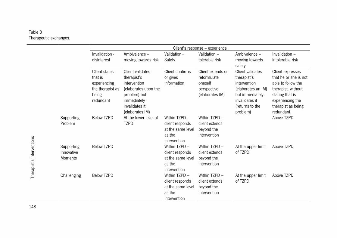
148
Table 3 Therapeutic exchanges.
Client’s response – experience Invalidation -
disinterest Ambivalence – moving towards risk
Validation - Safety
Validation – tolerable risk
Ambivalence – moving towards safety
Invalidation – intolerable risk
Client states that is experiencing the therapist as being redundant
Client validates therapist’s intervention (elaborates upon the problem) but immediately invalidates it (elaborates IM)
Client confirms or gives information
Client extends or reformulate oneself perspective (elaborates IM)
Client validates therapist’s intervention (elaborates an IM) but immediately invalidates it (returns to the problem)
Client expresses that he or she is not able to follow the therapist, without stating that is experiencing the therapist as being redundant.
Ther
apis
t’s in
terv
entio
ns
Supporting Problem
Below TZPD At the lower level of TZPD
Within TZPD – client responds at the same level as the intervention
Within TZPD – client extends beyond the intervention
Above TZPD
Supporting Innovative Moments
Below TZPD Within TZPD – client responds at the same level as the intervention
Within TZPD – client extends beyond the intervention
At the upper limit of TZPD
Above TZPD
Challenging Below TZPD Within TZPD – client responds at the same level as the intervention
Within TZPD – client extends beyond the intervention
At the upper limit of TZPD
Above TZPD
149
Collecting Physiological data
The cardiac activity from both client and therapist was recorded simultaneously using the
modular BIOPAC System MP150 (Santa Barbara, CA, USA) coupled to two amplifiers ECG 100C, one
connected to the client and the other to the therapist. This system was connected to a computer
equipped with AcqKnowledge (AcK) software (Biopac Systems, Santa Barbara, CA, USA), used to
define the acquisition parameters, record, filters and analyze the physiological data. For the acquisition
parameters, 1-second epochs were defined using a band pass filter and the Blackman Algorithm was
set at -61dB. Before the signal recording, left and right clavicles and the left inferior frontal tibia were
cleaned with alcohol and, according to a modified Lead II electrode configuration, three pre-gelled
Ag/AgCl electrodes were positioned at those places. To ensure the quality of the signal, the register
was visually examined before starting the session.
Baseline task
For the purpose of assessing the clients’ current physiological state and individual differences in
physiological reactivity, a 10-minute minimally demanding baseline task was performed before the
beginning of the therapeutic process. Based on recommendations by Jennings, Kamarck, Stewart,
Eddy, & Johnson (1992), this control task established the standard values against which the HR values
during the therapeutic exchanges were compared. The therapist instructed the client that some images
would be presented to them in the center of a laptop screen and together they should describe as
many details as possible of each image. This process was made in collaboration with the therapist,
who did not know the images and guided the client to keep him talking during the full 10 minutes. The
stimuli were colored objects on a white background randomly selected from the Web. While they
performed the task, the cardiac baseline scores from both therapist and client were recorded on a
second laptop. Along the therapeutic session, the cardiac activity was recorded simultaneously for the
dyad. Manual markers along the register were inserted in order to synchronize the different therapeutic
session moments (baseline task, fill in the questionnaires, and the therapeutic session) and cardiac
activity. The therapist was responsible for providing a signal that would represent the beginning of each
therapeutic moment. Furthermore, therapist and client were invited to report any discomfort or
subjective effects related to the psychophysiological recording devices.
150
Procedure
The following study was reviewed and accepted by the ethical committee from University of
Minho, Portugal. Also, both therapist and client accepted all the procedures involved in the data
collection and gave permission for their data to be used in this study. The client searched for the
Clinical Service of the School of Psychology at the university where the research was conducted.
Informed consent was obtained after the procedure has been orally and written fully explained to the
participants, in a meeting previous to the beginning of the therapeutic process. The therapy and the
research protocols were shared with the client at the pre-therapy meeting. Psychotherapeutic process
was established according to the guidelines of a clinical trial, including 16 weekly sessions and two
monthly follow-up sessions. All session were video recorded and transcribed by the research team
members. For the client´s participation in the study, the therapeutic process was offered to him free of
charge and it was assured that if he wanted to withdraw his participation in the research project, the
therapy process would continue, if necessary. Also, the client could be referred for further therapy after
the 18 sessions, if appropriate. At the intake, the therapist, using the SCID-I, conducted an initial
evaluation session. At the end of this session the client filled in two questionnaires: URICA and OQ-45.
The intake session was not video recorded. Regarding the session in analysis in the current study (the
first of the 16 weekly sessions) the client filled the OQ-10 at the beginning of the session and
immediately after the baseline task. At the end of session both therapist and client filled up the
respective forms of the SEQ and the WAI-S.
When the client arrived, the therapist accompanied him to the room where the session took
place and a researcher prepared the dyad for the physiological assessment. In an adjacent room, the
computer for the cardiac activity recording was placed and a team was responsible for following the
record along the session. The research team allocated in the computer room did not have access to
any information related to the therapeutic session that was considered as confidential topic between
therapist and client.
Data Analyses
Coding procedures using TCCS.
The first therapeutic session was videotaped, then transcribed and analyzed. The session was
coded using the TCCS, by the fourth and fifth authors, two reliable judges, previously trained in the

151
system. The pair of judges met, after independently coding the all the session, to assess their rating
reliability (reliability for therapist interventions assessed by Cohen’s K was .76; percentage of
agreement was 88%; and reliability for client’s response assessed by Cohen’s K was .58; percentage of
agreement 83%) and to note any discrepancies in their codifications. When disagreements were
detected they were resolved by consensual discussion. Afterwards, the first author audited the
codification.
State Space grid analysis.
State Space Grid (SSGs; Lewis, Lamey, & Douglas, 1999; Lewis, Zimmerman, Hollenstein, &
Lamey, 2004) was used to describe the evolution of the therapeutic interactions throughout the
session. The SSGs is originally derived from the developmental psychology and aims to study two or
more series of synchronized data. The fundamental principle of the SSGs method is that these two
series constitute a dynamic system with a finite number of possible states, called State Space (Thelen
& Smith, 1994, cited in Lewis, 2000).
In the present study, two series of data were taken into special consideration – the therapist’s
interventions and the client’s responses, assuming the (non)existence of synchrony in each therapeutic
exchanges to which they contribute according to the TCCS (Ribeiro et al., 2013).
Physiological data analysis.
Once the different therapeutic exchanges of the therapeutic session were time synchronized with
the physiological recording, the cardiac reactivity from both therapist and client were analyzed using
the AcK software. More specifically, the decision for averaging the waveform was time-based, taking
into consideration the exact moment each therapeutic exchange episode happened in the therapeutic
session. Before analysis, all physiological data were visually inspected to ensure the quality of the data,
along with the use of the AcK software to detect values outside the expected physiological range. In
order to minimize muscle artefacts, a Low and High Frequency Cutoff were set at 0.5Hz and 35Hz,
respectively, after the session recording. The cardiac activity from both components of the dyad was
assessed through a standard electrocardiogram (ECG) protocol. Different authors have measured the
cardiac activity in a variety of ways, but in this study, the cardiac parameter presented is the Heart Rate
(HR; estimated through the number of cardiac beats per minute) (Berntson, Quigley, & Lozano, 2007).
Besides being widely used in recent psychophysiological studies, the analysis of the HR was chosen
152
because it is one of the most precise physiological parameters with which to measure phasic cardiac
activity, and a key method to evaluate the pattern of arousability (Andreassi, 2000). Additionally, the
HR measure allows an accurate assessment of the heart function, being the main determinant of the
cardiac output (Cacioppo & Tassinary, 1990). The HR measure was automatically performed using AcK
software, on the basis of continuous ECG recordings. Then the HR means values for each subject were
corrected to the individual baseline (Stern, Ray, & Quigley, 2001). For the HR pattern illustration
(Figure 5) since there are too many therapeutic exchanges to include in one graph, we calculated the
mean of the therapeutic exchanges that accured more than once and repeatedly across the session
(e.g. if supporting problem-safety was coded 3 times we assumed these as one episode). Moreover, we
divided the session in three equal parts.
Results
Characterization of the quality of the session and of the therapeutic alliance
The session in analysis was evaluated both by the client and the therapist as moderately smooth
(4.6; 4 respectively) and moderately deep (4.4; 3.8 respectively). Comparing with the average of the
first 4 sessions, this first session was evaluated as smoother and less deep. Regarding the impact of
the session, John showed a higher score on the positivity scale (4.2) than on the arousal scale (3.8),
and this impact was similar to the average of the first 4 sessions (4.35 and 4.25, respectively).
Similarly, the therapist showed a higher value on the positivity scale (3.4) than on the arousal (2.8),
which was also similar to the average of the first 4 sessions (3.6 and 3.25 respectively).
Regarding the therapeutic alliance, the client perceived the alliance at the end of the session as
moderate (3/5) while the therapist perceived it as moderately high (4.6/5).
Characterization of the therapeutic collaboration
The development of the therapeutic collaboration was analyzed according to the TCCS (Ribeiro et
al., 2013), which allowed the identification of the different therapist interventions, client’s responses
and therapeutic exchanges, within this session. The different categories indexes were, then, obtained
through the calculation of the ratio between their specific frequency and the total number of each
variable that have had occurred. They will be presented and discussed in terms of percentage.
Figure 1 shows that across the session, the therapist tended to perform, more frequently,
challenging (C) interventions (a little more than 48% of the time), although supporting problem
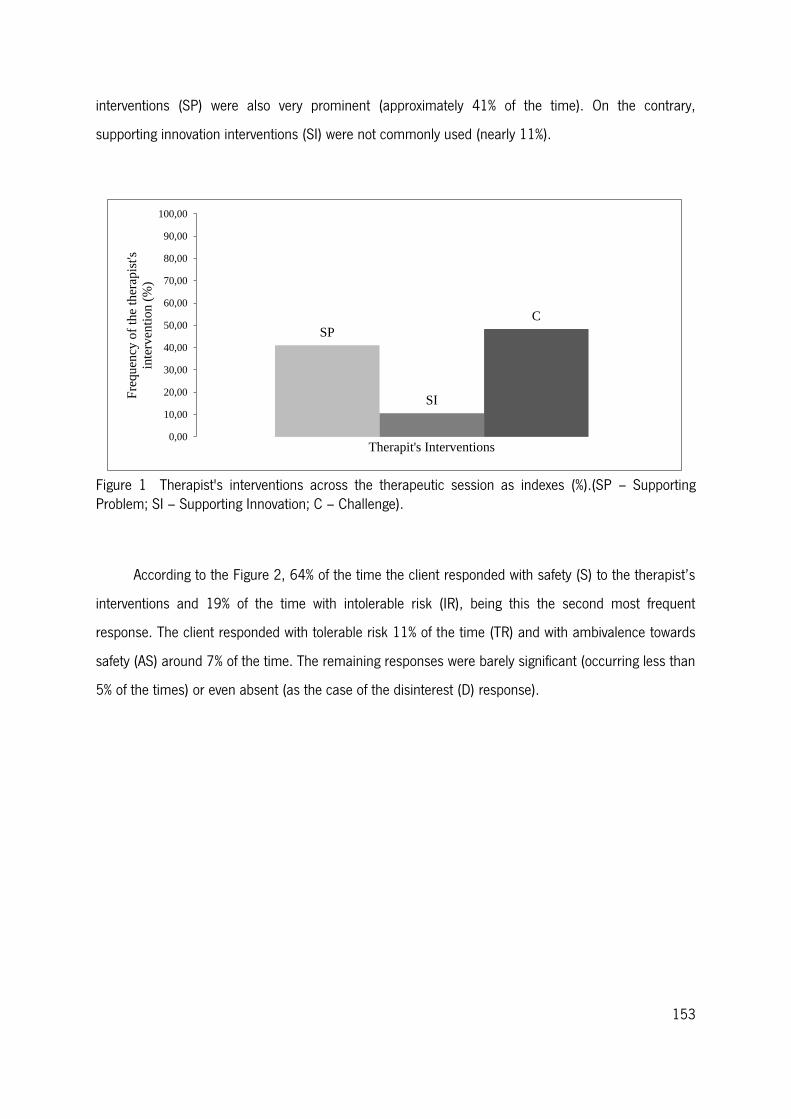
153
interventions (SP) were also very prominent (approximately 41% of the time). On the contrary,
supporting innovation interventions (SI) were not commonly used (nearly 11%).
Figure 1 Therapist's interventions across the therapeutic session as indexes (%).(SP – Supporting Problem; SI – Supporting Innovation; C – Challenge).
According to the Figure 2, 64% of the time the client responded with safety (S) to the therapist’s
interventions and 19% of the time with intolerable risk (IR), being this the second most frequent
response. The client responded with tolerable risk 11% of the time (TR) and with ambivalence towards
safety (AS) around 7% of the time. The remaining responses were barely significant (occurring less than
5% of the times) or even absent (as the case of the disinterest (D) response).
SP
SI
C
0,00
10,00
20,00
30,00
40,00
50,00
60,00
70,00
80,00
90,00
100,00
Fre
qu
ency
of
the
ther
apis
t's
inte
rven
tio
n (
%)
Therapit's Interventions
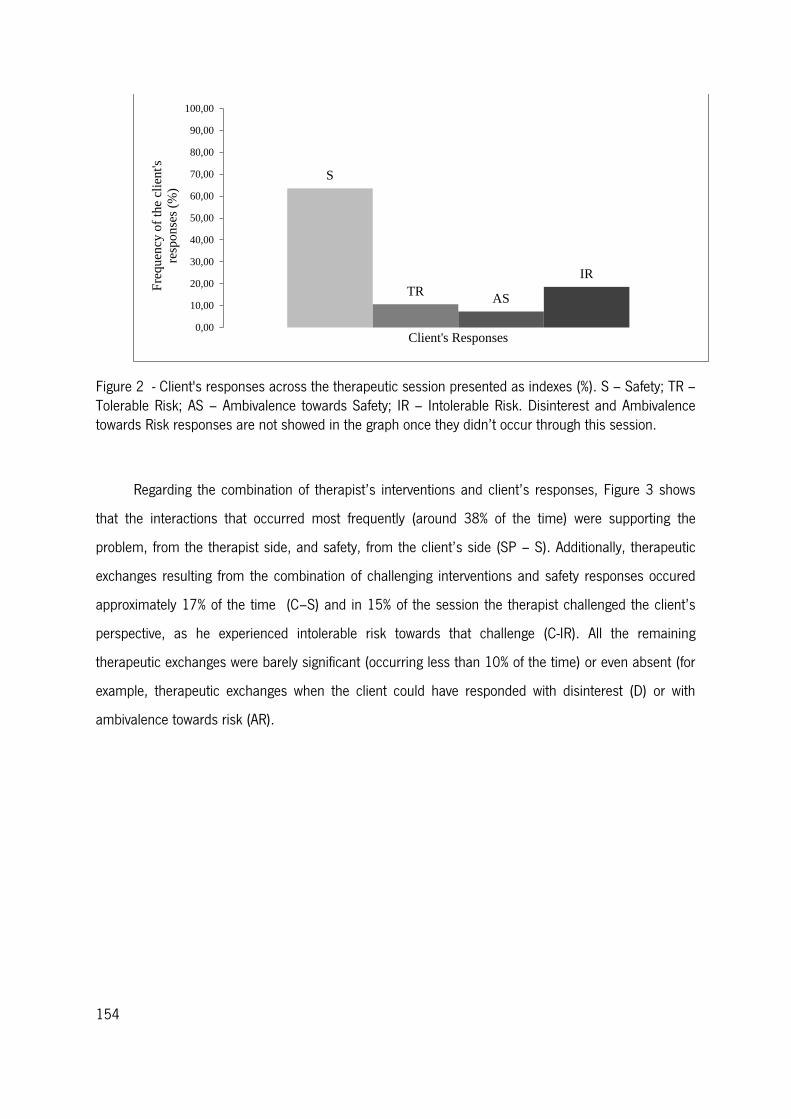
154
Figure 2 - Client's responses across the therapeutic session presented as indexes (%). S – Safety; TR – Tolerable Risk; AS – Ambivalence towards Safety; IR – Intolerable Risk. Disinterest and Ambivalence towards Risk responses are not showed in the graph once they didn’t occur through this session.
Regarding the combination of therapist’s interventions and client’s responses, Figure 3 shows
that the interactions that occurred most frequently (around 38% of the time) were supporting the
problem, from the therapist side, and safety, from the client’s side (SP – S). Additionally, therapeutic
exchanges resulting from the combination of challenging interventions and safety responses occured
approximately 17% of the time (C–S) and in 15% of the session the therapist challenged the client’s
perspective, as he experienced intolerable risk towards that challenge (C-IR). All the remaining
therapeutic exchanges were barely significant (occurring less than 10% of the time) or even absent (for
example, therapeutic exchanges when the client could have responded with disinterest (D) or with
ambivalence towards risk (AR).
S
TR AS
IR
0,00
10,00
20,00
30,00
40,00
50,00
60,00
70,00
80,00
90,00
100,00
Fre
qu
ency
of
the
clie
nt's
resp
on
ses
(%)
Client's Responses
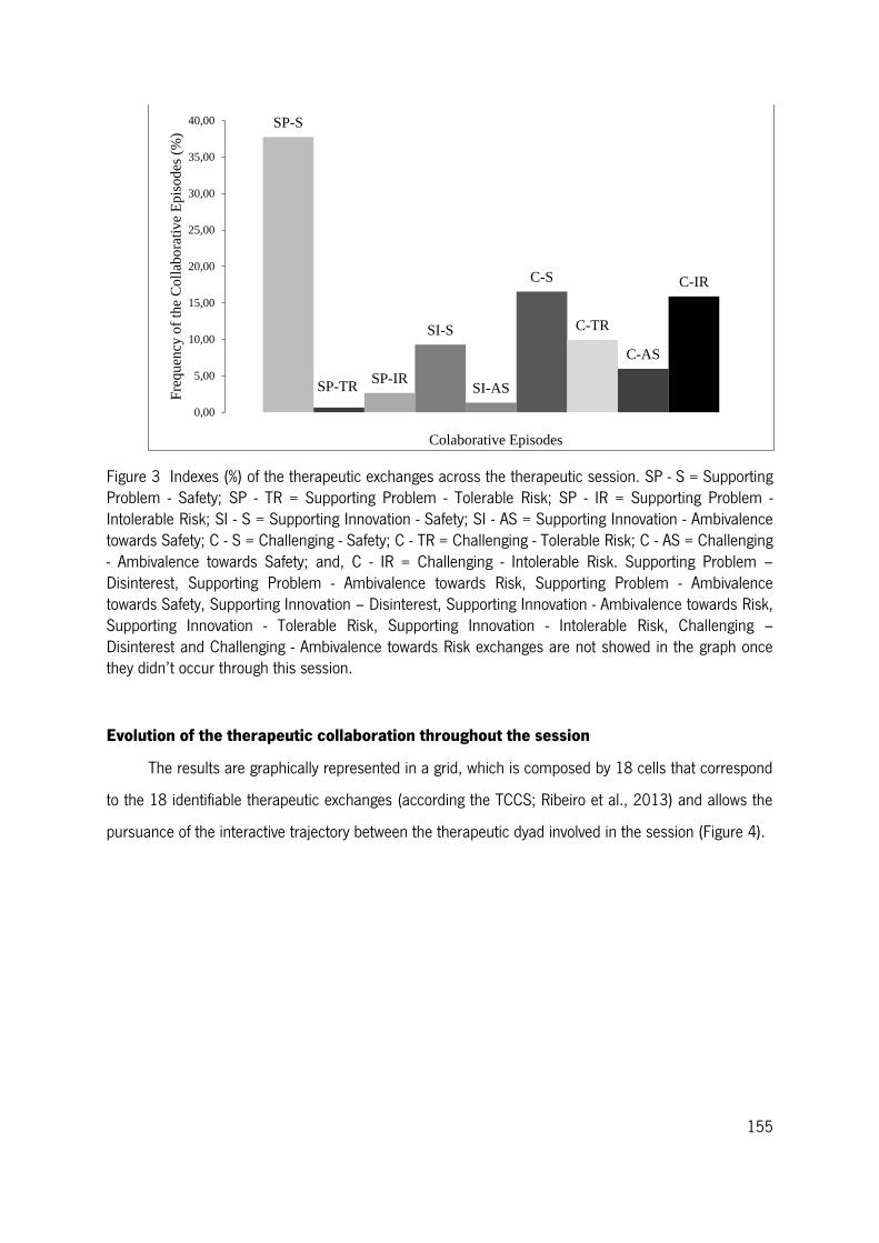
155
Figure 3 Indexes (%) of the therapeutic exchanges across the therapeutic session. SP - S = Supporting Problem - Safety; SP - TR = Supporting Problem - Tolerable Risk; SP - IR = Supporting Problem - Intolerable Risk; SI - S = Supporting Innovation - Safety; SI - AS = Supporting Innovation - Ambivalence towards Safety; C - S = Challenging - Safety; C - TR = Challenging - Tolerable Risk; C - AS = Challenging - Ambivalence towards Safety; and, C - IR = Challenging - Intolerable Risk. Supporting Problem – Disinterest, Supporting Problem - Ambivalence towards Risk, Supporting Problem - Ambivalence towards Safety, Supporting Innovation – Disinterest, Supporting Innovation - Ambivalence towards Risk, Supporting Innovation - Tolerable Risk, Supporting Innovation - Intolerable Risk, Challenging – Disinterest and Challenging - Ambivalence towards Risk exchanges are not showed in the graph once they didn’t occur through this session.
Evolution of the therapeutic collaboration throughout the session
The results are graphically represented in a grid, which is composed by 18 cells that correspond
to the 18 identifiable therapeutic exchanges (according the TCCS; Ribeiro et al., 2013) and allows the
pursuance of the interactive trajectory between the therapeutic dyad involved in the session (Figure 4).
SP-S
SP-TR SP-IR
SI-S
SI-AS
C-S
C-TR
C-AS
C-IR
0,00
5,00
10,00
15,00
20,00
25,00
30,00
35,00
40,00
Fre
qu
ency
of
the
Co
llab
ora
tiv
e E
pis
od
es (
%)
Colaborative Episodes
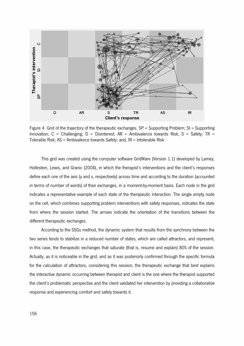
156
Figure 4 Grid of the trajectory of the therapeutic exchanges. SP = Supporting Problem; SI = Supporting Innovation; C = Challenging; D = Disinterest; AR = Ambivalence towards Risk; S = Safety; TR = Tolerable Risk; AS = Ambivalence towards Safety; and, IR = Intolerable Risk
This grid was created using the computer software GridWare (Version 1.1) developed by Lamey,
Hollestein, Lewis, and Granic (2004), in which the therapist’s interventions and the client’s responses
define each one of the axis (y and x, respectively) across time and according to the duration (accounted
in terms of number of words) of their exchanges, in a moment-by-moment basis. Each node in the grid
indicates a representative example of each state of the therapeutic interaction. The single empty node
on the cell, which combines supporting problem interventions with safety responses, indicates the state
from where the session started. The arrows indicate the orientation of the transitions between the
different therapeutic exchanges.
According to the SSGs method, the dynamic system that results from the synchrony between the
two series tends to stabilize in a reduced number of states, which are called attractors, and represent,
in this case, the therapeutic exchanges that saturate (that is, resume and explain) 80% of the session.
Actually, as it is noticeable in the grid, and as it was posteriorly confirmed through the specific formula
for the calculation of attractors, considering this session, the therapeutic exchange that best explains
the interactive dynamic occurring between therapist and client is the one where the therapist supported
the client’s problematic perspective and the client validated her intervention by providing a collaborative
response and experiencing comfort and safety towards it.
157
Indeed, the therapeutic exchange of supporting the problem – safety was, not only the most
frequent exchange occurring during this session, but also the one that the therapeutic dyad dedicated
more time, thus being assumed as the most representative of their encounter.
Characterization of the physiological concordance
Overall, regarding the physiological concordance analysis, across the session, the client showed
a higher HR pattern (M = 81.8 [SD = 12.5]) compared the therapist’s cardiac behavior (M = 78.8 [SD
= 8.3]). This pattern was clearer when the therapist challenges the client’s perspective. During these
therapeutic exchanges, the therapist and client were more often in different levels of development
regarding the client’s TZPD (e.g. Challenging-safety or Challenging – intolerable risk). Although some of
these therapeutic exchanges were collaborative, the therapist was beyond the client’s level. Namely,
the therapist challenged the client’s perspective and the client only accepted this proposal but doesn’t
elaborated or extended it, showing a safety experience (Challenging – Safety).
Most of the coincident points in the both participants’ HR patterns occurred in the collaborative
therapeutic exchanges (Supporting- problem; Challenging –Safety and Challenging – Tolerable risk)
(Figure 5).
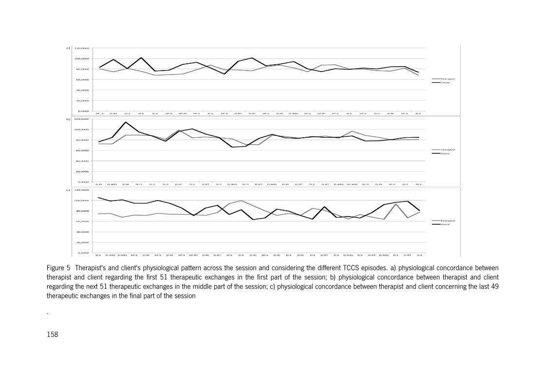
158
Figure 5 Therapist's and client's physiological pattern across the session and considering the different TCCS episodes. a) physiological concordance between therapist and client regarding the first 51 therapeutic exchanges in the first part of the session; b) physiological concordance between therapist and client regarding the next 51 therapeutic exchanges in the middle part of the session; c) physiological concordance between therapist and client concerning the last 49 therapeutic exchanges in the final part of the session
.
159
In most of non-collaborative therapeutic exchanges (e.g. Challenge – Intolerable Risk) the client
presented an increase in heart rate that is not accompanied by the therapist (the dyad is not at the
same physiological level) (Figure 5).
Statistical analyses regarding the physiological concordance between therapist and client were
performed. For these analyses we selected the following therapeutic exchanges: Supporting problem –
Safety, Supporting innovation – Safety, Challenge – Safety, Challenge – Tolerable risk and Challenge –
Intolerable risk. This was performed taking into account the therapeutic exchanges that happened more
frequently, that is, the ones that are presented and displayed more time across the session, (figure 4)
and based on the descriptive analysis of the HR concordance throughout the session considering the
therapeutic exchanges (Figure 5).
Regarding collaborative episodes the correlation between the therapist and client HR in
supporting problem – safety episode is statistically significant (r = 0.3, p < .05), what does not happen
in supporting innovation – safety (r = 0.3, p = 0.3), challenge – safety (r = 0.3, p = 0.1) or challenge -
tolerable risk (r = 0.2, p = 0.6) episodes. In non-collaborative episodes, no significant results were
found (challenge – intolerable risk: r = -0.1, p = 0.5) regarding the therapist and client’s HR.
Discussion
With this exploratory study, our main aim was to describe therapist and client’s physiological
concordance across the different therapeutic exchanges. The results indicated that throughout the first
therapeutic session, a large variety of TCCS therapeutic exchange is occurring, which was
accompanied by psychophysiological changes by both therapist and client cardiac activity.
Considering the TCCS therapeutic exchanges, the therapist interventions focused on supporting
the problem are the ones that occur more frequently in the beginning of the session as the therapist is
exploring and trying to understand the client’s problem. As the session develops, and by the end of it,
more challenge interventions emerge, it being these interventions that appear more often across the
session. Regarding the client’s response, he validated more frequently the supporting interventions
from the therapist, responding with safety, and invalidated more often the challenging interventions
(when the therapist works beyond his TZPD). Specifically, in this session, we verified that more
collaborative episodes occurred when the therapist supports on the problem, once the client validates
these interventions. On the contrary, the non-collaborative episodes appeared more commonly when
the client invalidates the challenge interventions. This therapeutic collaboration pattern is consistent
160
with the main goal of the first therapy session - to understand the problem that brought the client to
therapy –, and is also consistent with results of previous studies using the TCCS with cognitive behavior
therapy (Ribeiro, 2012), constructivist therapy (Ribeiro, Silveira, Senra, Azevedo, & Morais, 2014) and
narrative therapy (Ribeiro A.P., et al., 2013).
The increase of challenging interventions as the session develops may be perceived as resulting
from the use of the therapeutic strategies more focused on the novelty, as a way of negotiating the
therapy goals and tasks. The client’s most prevalent responses to these two kinds of therapist’s
interventions suggest that the therapist was responsive to his needs of being understood in his
experience, as indicated by safety responses following the supporting problem interventions. Moreover,
John’s responses to therapist interventions also showed his difficulties in anticipating change as
indicated by intolerable risk experiences following these challenging interventions. The anxiety John felt
following some of these therapists’ challenging interventions may explain the client´s difficulty to agree
with the therapist regarding the goals and tasks oriented to change. In light of the prevalence of these
two types of therapeutic exchanges it seems clear that the therapist has perceived the therapeutic
alliance as better than the client at the end of the session. Although the client presented a dependent
interpersonal style, which has been reported in the literature as favorable to more positive perceptions
of alliance in early sessions (Hilsenroth, Cromer, & Ackerman, 2012), the interpersonal nature of his
main problem (reflected in the difficulty to trust others) and the severity of the interpersonal conflicts
discussed in this first session may have influenced his less favorable perception of the alliance
(Hersoug, H⊘glend, Havik, vonder Lippe, & Monsen, 2009).
Since the most prevalent and lengthy therapeutic exchange was supporting problem-safety, the
dyad spent most of their time working within the John’s TZPD which seems coherent with both the
therapist and client’s evaluations of the session as moderately smoother and positive, suggesting an
agreement regarding their comfortable experience throughout the session.
At the psychophysiological level, both client and therapist’s HR pattern are very similar across
the collaborative episodes (compared to the baseline response), although, most of the time, the client
showed an increased mean HR compared with the therapist’s HR. However, considering the non-
collaborative episodes, that is, when the client invalidates the therapist interventions, the client shows
an increase in the HR when compared to the therapist physiological response (occurring more
frequently across challenging interventions). That is, the client, although is able to understand the
different perspective proposed by the therapist, invalidates this intervention, displaying a higher cardiac
161
activation, which is not observed in the therapist. This activation perhaps is related to perceiving a new
perspective, although not assuming it yet. It could be argued that this new understanding, which was
accompanied by a change in the client’s HR, seems to represent a close relationship between the
cardiac activity and the challenge involved in cognitive and emotional processes, being this information
perceived as more demanding (Stern et al., 2001; Cacioppo, Tassinary, & Berntson, 2007).
Both therapist and client show a higher cardiac activation when the challenging interventions are
validated by the client, which is observed most of the time. This result suggests that when the therapist
invites the client going further in his TZPD and the client accepts and elaborates this invitation, now
assuming this new perspective, it seems that physiological concordance tends to exist (Stern et al.,
2001; Ribeiro et al., 2013). Maybe the increasing in the therapist cardiac activity is related to the fact
that the client has accepted her suggestion and she was emotional satisfied for the client once he
moved beyond his problematic view. However, no statistically significant results support this HR
patterns in therapist and client cardiac response. This may be due to the fact that less challenge-
security or challenge-tolerable risk therapeutic exchanges happen along the session, a number that is
not enough to produce statistically significant results.
This way, considering this initial session, two important moments regarding the physiological
characterization were identified: 1) Physiological Concordance, which happens, when the dyad is at the
same developmental level regarding the TZPD and focused on the problem presented by the client: the
therapist supports on the problem and the client accepts it and both show a synchronized HR; the
same way, when the therapist introduces challenge to the client and he elaborates on it, validating the
therapist intervention, the dyad also shows, simultaneously, an increase in HR, although this synchrony
is not statistically significant; and 2) Non-Physiological Concordance: the non-physiological concordance
is especially evident in non-collaborative therapeutic exchanges, when challenging interventions are
invalidated by the client, that is, when the therapist conceptualizes the client’s problem differently from
his problematic view. When the client does not accept a new problematic formulation by the therapist,
it produces a moment of physiological discordance characterized by an increase in the client’s cardiac
activity, not followed by the therapist.
These results are consistent with previous studies (Marci et al., 2007; Ham & Tronick, 2009;
Stern et al., 2001) showing that when therapist and client are attending to each other (as expected into
collaborative episodes, in particular those that placed the dyad at the same developmental level, both
exhibit physiological concordance. This is evident when the therapist supports the client’s problem but
162
also when she challenges the client prespective and he elaborates on innovation. This suggests that the
dyad is therapeutically involved and the client is being responsive to the new information provided by
the therapist (Ham & Tronick, 2009). Additionally, it suggests that when therapist and client are being
physiological reactive to each other, the client is also being more responsive and collaborative with the
therapy goals (Ribeiro et al., 2013; Marci & Orr, 2006; Caspar, 2003). Furthermore, in this first
session, when the client validated the therapist’s challenging intervention by accepting and elaborating
the new perspective regarding his problem, we observed that these episodes were closely related to
physiological change (Caspar, 2003). This means that, along the first session of this therapeutic
process, the changes in the client’s perspective are already translated into physiological changes as
well. Furthermore, our results seem consistent with the Polyvagal theory (Porges, 1995, 2001, 2003,
2011) suggesting that cardiac acitivity influences social interactions. Indeed, in therapeutic exchanges
where the dyad is at the same TZPD level, producing validation responses from the client, their heart
rate is similar (either maintaining or increasing) and, therefore, allowing them to engage in social
communication. On the contrary, therapeutic exchanges where therapist and client are not at the same
TZPD level, and the client shows an increase in heart rate, he responds with invalidation markers not
following the therapist’s suggestions.
This work being an exploratory study, its main strength relies on the elaboration of a recording
protocol for the examination of the Heart Rate concordance between the therapeutic dyad and for its
analysis across the different therapeutic exchanges, occurring along one session. With the present work
we were able to study the combination of therapeutic interactions and the underlying physiological
response occurring between the therapeutic dyad. We could find a strong relation regarding supporting
problem-safety exchange and the physiological concordance which could be understood as a
physiological marker of the therapeutic collaboration when therapist and client are at the same
developmental level. However, future studies, should examine this physiological
concordance/discordence throughout a therapeutic process in order to find further physiological
markers of the TCCS therapeutic exchanges.
163
References
Andreassi, J. L. (2000). Psychophysiology: Human Behavior & Physiological Response (4th ed.). New
Jersey: Lawrence Erlbaum Associates.
Appelhans, B. M., & Luecken, L. J. (2006). Heart rate variability as an index of regulated emotional
responding. Review of general psychology, 10, 229-240. doi: 10.1037/1089-2680.10.3.229.
Berntson, G. G., Quigley, K. S., & Lozano, D. (2007). Cardiovascular Psychophysiology. In J. T.
Cacioppo, L. G. Tassinary & G. G. Berntson (Eds.), Handbook of Psychophysiology (3rd ed., pp.
182-210). New York: Cambridge University Press.
Boardman, T., Catley, D., Grobe, J. E., Little, T. D., & Ahluwalia, J. S. (2006). Using motivational
interviewing with smokers: do therapist behaviors relate to engagement and therapeutic alliance?
Journal of Substance Abuse Treatment, 31(4), 329-339. doi: 10.1016/j.jsat.2006.05.006.
Butler, E. A. (2011). Temporal interpersonal emotion systems: the "TIES" that form relationships.
Personality and Social Psychology Review, 15(4), 367-393. doi: 10.1177/1088868311411164.
Cacioppo, J. T., & Tassinary, L. G. (1990). Inferring psychological significance from physiological
signals. American Psychologist, 45(1), 16-28.
Cacioppo, J. T., Tassinary, L. G., & Berntson, G. G. (2007). Psychophysiological Science:
Interdisciplinary Approaches to Classic Questions About the Mind. In J. T. Cacioppo, L. G.
Tassinary & G. G. Berntson (Eds.), Handbook of Psychophysiology (3rd ed., pp. 1-16). New York:
Cambridge University Press.
Caspar, F. (2003). Psychotherapy research and neurobiology: challenge, chance, or enrichment?
Psychotherapy Research, 13(1), 1-23. doi: 10.1093/ptr/kpg013.
Coleman, R., Greenblatt, M., & Solomon, H. C. (1956). Physiological evidence of rapport during
psychotherapeutic interviews. Diseases of the Nervous System, 17(3), 71-77.
Colli, A., & Lingiardi, V. (2009). The Collaborative Interactions Scale: a new transcript-based method for
the assessment of therapeutic alliance ruptures and resolutions in psychotherapy.
Psychotherapy Research, 19(6), 718-734. doi: 10.1080/10503300903121098.
Dimascio, A., Boyd, R. W., & Greenblatt, M. (1957). Physiological correlates of tension and antagonism
during psychotherapy; a study of interpersonal physiology. Psychosomatic Medicine, 19(2), 99-
104.
164
First, M. B., Spitzer, R. L., Gibbon, M., & William, J. (2002). Structured Clinical Interview for DSM-IV-TR
Axis I Disorders, Research Version, Patient Edition (SCID-I/P). New York: Biometrics Research. :
New York State Psychiatric Institute.
Ham, J., & Tronick, E. (2009). Relational psychophysiology: lessons from mother-infant physiology
research on dyadically expanded states of consciousness. Psychotherapy Research, 19(6), 619-
632. doi: 10.1080/10503300802609672.
Hatcher, R. (1999). Therapists' Views of Treatment Alliance and Collaboration in Therapy.
Psychotherapy Research, 9(4), 405-423.
Hersoug, A. G., H⊘glend, P., Havik, O. E., von der Lippe, A., & Monsen, J. T. (2009). Pretreatment
patient characteristics related to the level and development of working alliance in long-term
psychotherapy. Psychotherapy Research, 19(2), 172-180. doi: 10.1080/10503300802657374.
Hilsenroth, M., Cromer, T., & Ackerman, S. (2012). How to Make Practical Use of Therapeutic Alliance
Research in Your Clinical Work. In R. A. Levy, J. S. Ablon & H. Kächele (Eds.), Psychodynamic
Psychotherapy Research (pp. 361-380): Human Press.
Horvath, A. O., & Greenberg, L. S. (1989). Development and validation of the Working Alliance
Inventory. Journal of Counseling Psychology, 36, 223-233. doi: 10.1037/0022-0167.36.2.223.
Jacobson, N. S., & Truax, P. (1991). Clinical significance: a statistical approach to defining meaningful
change in psychotherapy research. Journal of Consulting and Clinical Psychology, 59(1), 12-19.
Jennings, J. R., Kamarck, T., Stewart, C., Eddy, M., & Johnson, P. (1992). Alternate cardiovascular
baseline assessment techniques: vanilla or resting baseline. Psychophysiology, 29(6), 742-750.
Lambert, M. J., Burlingame, G. M., Umphress, V., Hansen, N. B., Vermeersch, D. A., Clouse, G. C., &
Yanchar, S. C. (1996). The Reliability and Validity of the Outcome Questionnaire. Clinical
Psychology & Psychotherapy, 3(4), 249-258. doi: 10.1002/(SICI)1099-
0879(199612)3:4<249::AID-CPP106>3.0.CO;2-S.
Lamey, A., Hollestein, T., Lewis, M., & Granic, I. (2004). The Gridware Manual (Version 1.1).
http://statespacegrids.org.
Leiman, M., & Stiles, W. B. (2001). Dialogical sequence analysis and the zone of proximal development
as conceptual enhancements to the assimilation model: The case of Jan revisited.
Psychotherapy Research, 11(3), 311-330. doi: 10.1093/ptr/11.3.311.
165
Lewis, M. D., Zimmerman, S., Hollenstein, T., & Lamey, A. V. (2004). Reorganization in coping
behavior at 1 1/2 years: dynamic systems and normative change. Developmental Science, 7(1),
56-73.
Machado, P. P., & Horvath, A. O. (1999). Inventário da Aliança Terapêutica - WAI. Testes e provas
psicológicas em Portugal., 2, 87-94.
Marci, C. D., Ham, J., Moran, E., & Orr, S. P. (2007). Physiologic correlates of perceived therapist
empathy and social-emotional process during psychotherapy. Journal of Nervous & Mental
Disease, 195(2), 103-111. doi:10.1097/01.nmd.0000253731.71025.fc.
Marci, C. D., & Orr, S. P. (2006). The effect of emotional distance on psychophysiologic concordance
and perceived empathy between patient and interviewer. Applied Psychophysiology and
Biofeedback, 31(2), 115-128. doi: 10.1007/s10484-006-9008-4.
McConnaughy, E. A., Prochaska, J. O., & Velicer, W. F. (1983). Stages of change in psychotherapy:
Measurement and sample profiles. Psychotherapy: Theory, Research & Practice, 20(3), 368-
375. doi: 10.1037/h0090198.
Oliveira-Silva, P., & Goncalves, O. F. (2011). Responding empathically: a question of heart, not a
question of skin. Applied Psychophysiology and Biofeedback, 36(3), 201-207. doi:
10.1007/s10484-011-9161-2.
Prochaska, J. O., & DiClemente, C. C. (1983). Stages and processes of self-change of smoking: toward
an integrative model of change. Journal of Consulting and Clinical Psychology, 51(3), 390-395.
Ribeiro, A. P., Ribeiro, E., Loura, J., Goncalves, M. M., Stiles, W. B., Horvath, A. O., & Sousa, I. (2013).
Therapeutic collaboration and resistance: Describing the nature and quality of the therapeutic
relationship within ambivalence events using the Therapeutic Collaboration Coding System.
Psychotherapy Research. doi: 10.1080/10503307.2013.856042.
Ribeiro, E. (2012). Therapeutic collaboration: communalities and specifics across therapeutic
approaches and therapy outcomes. . Paper presented at the Panel in 43th Annual Meeting of
the Society for Psychotherapy Research,, Virginia Beach, EUA. 24 June.
Ribeiro, E., Ribeiro, A. P., Goncalves, M. M., Horvath, A. O., & Stiles, W. B. (2013). How collaboration
in therapy becomes therapeutic: the therapeutic collaboration coding system. Psychology and
Psychotherapy: Theory, Research and Practice, 86(3), 294-314. doi: 10.1111/j.2044-
8341.2012.02066.x.
166
Ribeiro, E., Silveira, J., Senra, J., Azevedo, A., & Morais, J. (2014). Therapeutic Collaboration in
Constructivist Therapy: a comparative study of a good and a poor outcome cases. Manuscript
submitted for publication.
Stern, R. M., Ray, W. J., & Quigley, K. S. (2001). Psychophysiological recording. New York: Oxford
University Press.
Stiles, W. B. (1980). Measurement of the impact of psychotherapy sessions. Jounal of Consulting and
Clinical Psychology, 48(2), 176-185. doi: 10.1037/0022-006X.48.2.176.
Stiles, W. B., Gordon, L. E., & Lani, J. A. (2002). Session evaluation and the Session Evaluation
Questionnaire. In G. S. Tryon (Ed.), Counseling based on process research: Applying what we
know. (pp. 325-343). Boston, MA: Allyn & Bacon.
Stolorow, R. D. (1997). Dynamic, dyadic, intersubjective systems: An evolving paradigm for
psychoanalysis. Psychoanalytic Psychology, 14(3), 337-346. doi: 10.1037/h0079729.
Tryon, G. S., & Winograd, G. (2011). Goal consensus and collaboration. Psychotherapy, 48(1), 50-57.
doi: 10.1037/a0022061.
167
Therapeutic Collaboration and the underlying physiological profile: concordance and discordance in the early phase of a CBT good outcome case8
Eugénia Ribeiro*
Sara Cruz*
Patrícia Oliveira-Silva
Dulce Pinto
Catarina Fernandes
Adriana Sampaio
Centre of Research in Psychology (CIPsi), School of Psychology, University of Minho
*These authors contributed equally to this work.
Address for all authors: School of Psychology, University of Minho, Campus de Gualtar, 4710-057
Braga, Portugal
Telephone number for all authors: +351 253 601 398
Corresponding author: Eugénia Ribeiro, School of Psychology, University of Minho, Campus de Gualtar,
4710-057 Braga, Portugal; Telephone number: +351 253 601 398; E-mail: [email protected]
8 Article in preparation.
168
Abstract
Therapeutic alliance has been accounted as one of the most important factors contributing to
the therapeutic success. Similarly, the initial phase of the therapeutic process has been largely
accepted as determinant to client’s adhesion and permanence in therapy. Therefore, with the present
work we aim to characterize therapeutic collaboration development, a core dimension of alliance, along
the first four therapeutic sessions of a CBT good outcome case. Furthermore, we want to uncover
physiological correlates associated with the different (non) collaborative therapeutic exchanges, by
analyzing therapeutic dyad concordance and discordance heart rate levels. Overall, our results suggest
that when therapist and client interactions are collaborative, both show physiological concordance. On
the contrary, when the dyad produces non-collaborative exchanges they do not show physiological
concordance. Moreover, on the first sessions supporting problem are the most frequent therapist’s
interventions, followed by the client’s safety response. The 4th session seems to be crucial for the
change process, as it is in this session that therapist’s challenge interventions are more frequently
validated. Results are discussed in the light of cardiac response influences in interactive exchanges, as
well as the role of physiological reactivity for the therapeutic process.
Keywords: collaboration, TCCS, heart rate, physiological reactivity, therapeutic exchanges
169
Introduction
Therapeutic alliance has been considered as one of the most important factors contributing to
the therapeutic process and has been largely accepted as the most robust and reliable predictor of
therapy outcomes (Crits-Christoph, Gibbons & Hearon, 2006; Martin, Garske & Davis, 2000; Horvath &
Symonds, 1991). Different studies have demonstrated that therapeutic alliance assessment of the early
phase of the therapeutic process is positively associated with the client’s permanence in therapy, as
well as with better therapeutic outcomes (Horvath, 2006; Hilsenroth, Peters & Ackerman, 2004;
Bachelor & Salamé, 2000; Feltham, 1999).
Indeed, the investigating of the relation between alliance and outcome has consensually showed
that the therapeutic alliance plays a significant role on the therapy outcome, across many types of
theoretical orientations (Flückiger, Del Re, Wampold, Symonds, & Horvath, 2012). Several meta-
analyses (Horvath & Bedi, 2002; Horvath, Del Re, Flückiger, & Symonds, 2011; Horvath & Symonds,
1991; Martin, Garske, & Davis, 2000) estimated the relation between alliance and outcome in the
range of r = 0.25 to r = 0.27, or responsible for 6.5% of the outcome variance (Horvath, 2013).
Similarly, it has been widely demonstrated that the early phase of therapy is crucial for the alliance
development, generally establish within the first three/four sessions (de Roten et al., 2004; Principe,
Marci, Glick, & Ablon, 2006).
A core dimension largely accepted as contributing to therapeutic alliance formation and
development is therapeutic collaboration. As Hatcher (1999) defined, the collaboration process is
considered as the mutual contribution of the therapeutic dyad to therapy that cannot be reduced either
to the therapist, or to the client, input alone. Additionally, is assumed as the dyad shared responsibility
and participation in the therapy objectives and multiple proposals or activities within the therapeutic
context (Tryon & Winograd, 2011; Colli & Lingiardi, 2009; Boardman, Catley, Grobe, Little, &
Ahluwalia, 2006). Therapeutic collaboration can be understood as the therapist and client’s
interactions happening at a moment-by-moment level, which are occurring within a Therapeutic zone of
Proximal Development (TZPD) (Ribeiro, Ribeiro, Gonçalves, Horvath, & Stiles, 2013; Leiman & Stiles,
2001) (based on the adoption of the Zone of Proximal Development concept developed by Vigotsky,
1924, to therapy by Leiman & Stiles, 2001). TZPD is, therefore, a therapeutic developmental
continuum, assumed as a space between the client’s actual developmental level and a potential
developmental level that can be reached in collaboration with the therapist. On the other hand,
therapist facilitates change by working within this developmental continuum.
170
According to Ribeiro and colleagues (2013) therapeutic activities associated with the
collaborative process can be divided into two components: 1) supporting and helping the client to feel
safe, usually involving an expression of understanding the client’s experience within his or her usual
perspective; and 2) challenging the maladaptive framework by using strategies that stimulates the
occurrence of innovations (based on the concept of innovation developed by Gonçalves et al., 2009).
These components seem to occur in a balanced way in the therapy once the therapist must keep
working within a therapeutic climate in which the client feels comfortable, but also must be able to help
the client to experience and consider new perspectives (Ribeiro et al., 2013). Moreover, it is assumed
that the client’s response to the therapist’s intervention indicates whether the therapist worked within
the TZPD (validation), or instead, worked out of TZPD (invalidation), or at the limit of the TZPD
(ambivalence). It is suggested that interventions occurring within the TZPD require that the therapist
addresses empathically to the client’s TZPD at a given moment; specifically the client’s current stage of
change and the client’s capacity to make sense of their own mental states and of the therapist’s
comments. Hence, supporting or challenging interventions from the therapist side and validation,
ambivalence or invalidation responses from the client side constitute the communication actions that
are coded by the Therapeutic collaboration coding system – TCCS (Ribeiro et al., 2013). It has been
showed that in regards to the initial phase of therapy (initial four sessions) the collaboration process,
coded using TCCS, is mainly characterized by supporting-problem interventions, followed by safety
responses and, as the initial phase moves on, it is observed that validated challenging interventions are
occurring more often around the third/forth sessions (Pires et al., in preparation).
Additionally, we are assuming that beneath these therapeutic exchanges physiological responses
are happening, on both therapist and client, that reflect their internal and behavioural states inherent to
these different therapeutic exchanges. In fact, physiological reactivity has been assumed as an
interpersonal aspect that is essentially influenced by intrapersonal interactive exchanges occurring
within a social context (Heaphy & Dutton, 2008; Roy, Steptoe & Kirschbaum, 1998). Considering the
therapy context as social space, where communication patterns are mainly characterizing therapist-
client interactive exchanges, we assume that physiological correlates are underlying the interaction
process occurring in therapy.
As Porges (1995, 2001, 2003) documented, developmental and interactive exchanges, as well
as expression of cognitive and emotional processes, are sustained by physiological reactivity, which
have different neural basis, and are phylogenetically organized, reflecting more complex and adaptive
171
social strategies. The Polyvagal theory (Porges, 1995, 2001) builds on the fact that neuroanatomical
structures and neurophysiological reactions, through the Vagus nerve functions, are controlling social
behaviors and emotional processes, influencing the perception of safety. These processes are
occurring within the therapeutic sessions and between therapist and client (i.e. communicative
behavior). Thus, we assume that psychophysiological reactions are occurring throughout the
therapeutic process and, therefore, therapeutic exchanges are accompanied by several physiological
responses that reflect distinct internal and behavioral states of the therapist-client dyad (Dittes, 1957;
Dimascio, Boyd & Greenblatt, 1957; Malmo, 1957; Coleman, Greenblatt & Solomon, 1956).
Moreover, the assessment of cardiovascular system in social interactions have been largely
accepted as the cardiac activity has a central role on psychophysiological research, essentially because
it is under control of both sympathetic and parasympathetic branches of the autonomic nervous system
Cacioppo, Tassinary, & Berntson, 2007). Indeed, Oliveira-Silva and Gonçalves (2011) showed that
higher levels of additive empathy are associated with increased cardiac activity (i.e., decreased
Interbeat Interval). Furthermore, in a study conducted by Marci and Orr (2006), the authors showed
that the therapeutic dyad is being reactive to each other as they are producing psychophysiological
responses that can be in “concordance” or in “discordance” between them and across therapy.
Similarly, Marci, Ham, Moran, and Orr (2007) found that psychophysiological concordance is lower
when the therapist behaves as “emotionally distant”, showing that skin conductance concordance was
associated with perceived ratings of therapist empathy and with more positive social-emotional
interactions for both clients and therapists. Thus, physiological concordance is achieved when the
therapeutic dyad is actively attending to each other and being responsive to the therapy goals and
activities (Ham & Tronick, 2009).
Hence, and considering the importance of the early phase in the therapeutic process (Martin et
al., 1999; Horvath & Symonds, 1991), with the present work we aim to understand the therapeutic
collaboration dynamics throughout the early phase of a good outcome case of Cognitive-Behavioral
therapy (CBT) (first 4 therapeutic sessions) by coding therapeutic collaborations exchanges.
Furthermore, we aim to uncovering the physiological correlates between therapist and client within
these therapeutic exchanges, particularly concordance and discordance processes. In addition, we aim
to examine co-variation of momentary changes in the individual cardiac activity within the clinical
sessions.
172
Methods9
Participants
John (a pseudonym), 22-years-old and a student at the university, was diagnosed with Major
Depression Disorder, according to the DSM-IV-TR (2002), as he was recruited from the research
program investigating therapeutic collaboration and their physiological correlates (BIAL, 178/12).
John’s main problem was related with interpersonal conflicts, both with his family and friends. He
presented a central belief of need to be a good person, caring of others and being always available. He
avoided to be in conflict with them and had difficult to say “no” to their requests. Nevertheless, he
believed his friends did not recognize him as valuable person, thus he felt sad and, consequently,
isolated himself. Although, his girlfriend left him some weeks before the beginning of therapy, he
believed strongly that she was the one who he will need. Based on the pre therapy evaluation of his
readiness for change, using the URICA (McConnaughy, Prochaska & Velicer, 1983), John was
classified as being in the contemplation stage of change. Based on the pre and post therapy evaluation
of symptoms, John’s case was considered a good outcome case. He was selected for this study on the
basis of significant symptomatic change evidenced on pre–post assessment. At the intake the client
presented a GAF of 60, changing to a GAF of 85 at the termination. His pre-therapy OQ45 score of 89
dropped to 50 at therapy termination. At the dropout sessions he dropped his OQ45 score to 43. A
Reliable Change Index (RCI) analysis of his OQ45 pre-to post-test change scores classified John as
having met criteria for recovered (i.e., passed both a OQ45 cut-off score of 62 and the RCI criteria of
18 points) at treatment termination (Jacobson & Truax, 1991).
The therapist, female, 49 years old, is a well-trained professional psychologist, with 23 years of
professional experience. Although following a CBT approach, therapeutic interventions did not follow a
manualized structure and was conducted in a flexible way in order to select the more appropriate
strategies to specific problems and needs presented by the client throughout the therapy. Typical
therapeutic strategies include cognitive and behavioral self-observation, identifications of automatic and
alternative thoughts, analysis of evidence, believes and meanings discuss and relapse prevention.
TCCS
The TCCS is a transcript base method to micro-analyze the therapeutic collaboration at a
moment-by-moment level, considering the therapist’s interventions and the client’s responses, during
9 The 4 sessions used in this article were extracted from the same therapeutic case used the in the first study focusing on the first session. Therefore, information regarding methodology may sometimes be repeated.
173
the therapeutic session. For the codification, one must consider the therapist and client’s adjacent
pairs of speaking turns, as well as immediate context before and after the pair that is being coded,
taking into consideration the overall sense of the session. This way, we can have from the therapist
side supporting the problem or supporting the innovation, or challenging interventions. Regarding the
client’s responses, we can have validation, invalidation or ambivalence towards the therapist’s
interventions. The articulation between therapist’s interventions and clients’ responses allows for the
identification of 18 possible therapeutic exchanges. Previous studies using the TCCS (E. Ribeiro, et al,
2013; A.P. Ribeiro et al, 2013) showed good reliability with mean Cohen’s Kappa values of .92 for
therapist interventions and .93 for client’s responses.
Physiological data acquisition
The cardiac activity from both client and therapist was recorded simultaneously using the
modular and wireless BIOPAC System MP150 (Santa Barbara, CA, USA) coupled to two amplifiers ECG
100C, one connected to the client and the other to the therapist. This system was connected to a
computer equipped with AcqKnowledge (AcK) software (Biopac Systems, Santa Barbara, CA, USA),
used to define the acquisition parameters, record, filters and analyze the physiological data. For the
acquisition parameters, 1-second epochs were defined using a band pass filter and the Blackman
Algorithm was set at -61dB. Before the signal recording, left and right clavicles and the left inferior
frontal tibia were cleaned with alcohol and, according to a modified Lead II electrode configuration,
three pre-gelled Ag/AgCl electrodes were positioned at those places. To ensure the quality of the signal,
the recording was visually examined before starting each session.
Baseline task
For the purpose of assessing the clients’ physiological state and individual physiological reactivity
differences, before the therapeutic sessions began, a 10-minute minimally demanding baseline task
was performed. Based on recommendations by Jennings, Kamarck, Stewart, Eddy, and Johnson
(1992) this control task establishes standard values against which the heart rate values alterations
happening during the therapeutic exchanges were compared. Before each session begin, the therapist
instructed the client that some images would be presented to them in the center of a laptop screen and
together they would describe as many details as possible associated with each image. The therapist did
not know the images as she also guided the client to keep him talking during the full 10 minutes. The
174
stimuli were colored objects on a white background randomly selected from the Web. For each session,
the baseline task images were different. While they performed the task, the cardiac baseline scores,
from both therapist and client, were recorded on a second laptop. Similarly, along the therapeutic
sessions, cardiac activity was recorded simultaneously for the dyad. Manual markers in the recording
were inserted in order to synchronize the different therapeutic session moments (baseline task, fill in
the questionnaires, and the therapeutic session). The therapist was responsible for providing a signal
that would represent the beginning of each therapeutic moment.
Procedure
The following study was reviewed and accepted by the ethical committee from School of
Psychology, University of Minho, Braga, Portugal. Both therapist and client accepted all the procedures
involved in the data collection and gave permission to use it in the study. John searched for the Clinical
Service of the School of Psychology at the university where the research happened. Informed consent
was obtained after the procedure has been orally and in written form fully explained to the participants,
in a meeting previous to the beginning of the therapeutic process. Both participants were asked to
refrain from caffeine, physical exercise, and nicotine for at least 4 hours before each session, and were
also inquired whether were taking any prescription or nonprescription medication, for the sake of
evaluating potential cardiovascular effects. For this study subjects who presented psychiatric and/or
physical co-morbidities were excluded due to the possibility of confounding the results concerning with
the cardiac function. The therapy and the research protocols were shared with the client at a pre-
therapy meeting. Psychotherapeutic process was established, according to the guidelines of a clinical
trial, including 16 weekly sessions and two monthly follow-up sessions. All session were video recorded
and transcribed by the research team members. For the client´s participation in the study, therapeutic
process was free of charge and he was assured that if quit his participation in the research project, the
therapy process would continue, if necessary. Also, the client could be referred for further therapy after
the 18 sessions, if appropriate. At the intake, the therapist, using the SCID-I, conducted an initial
evaluation session. At the end of this session the client filled in two questionnaires: URICA and OQ-45,
if he matched the inclusion criteria for the research and do not match any of the exclusion criteria. This
intake session was not video recorded. Regarding the sessions in analysis in the current study, the first
four of a process off weekly 16 sessions, the client filled the OQ-10 at the beginning of the each
session and immediately after the baseline task. At the end of each session both therapist and client
175
filled up the respective forms of the SEQ, the WAI-S (to a detailed description of these questionnaires
refer to Methods section in the Therapeutic collaboration and the underlying physiological profile in the
first session of psychotherapy study).
When the client arrived the therapist accompanied him to the room where the sessions took
place and a researcher prepared the dyad for the physiological assessment. On a contingent room, the
computer for the cardiac activity recording was placed and a team was responsible for following the
recording along the session. The research team allocated in the computer room did not have access to
any aspect related to the therapeutic sessions.
Data Analyses
Coding procedures using TCCS.
The therapeutic sessions were videotaped, then transcript and analyzed. 50% of sessions (2)
were coded using the TCCS by the fourth and fifth authors, two reliable judges, previously trained in the
system, for effects of calculating the coding agreement. The pair of judges met, after independently
coding all the sessions, to assess their rating reliability (1st session: reliability for therapist interventions
assessed by Cohen’s K was 0,76; percentage of agreement was 88%; and reliability for client’s
response assessed by Cohen’s K was 0,58; percentage of agreement 83%; 2nd session: reliability for
therapist interventions assessed by Cohen’s K was 0,89; percentage of agreement was 93%; and
reliability for client’s response assessed by Cohen’s K was 0,85; percentage of agreement 92%;) and to
note any discrepancies in their codifications. When disagreements were detected they were resolved by
consensual discussion. Afterwards, the first author audited to the codification.
State Space grid analysis.
State Space Grid (SSG) (Lewis, Zimmerman, Hollenstein, & Lamey, 2004) was used to describe
the evolution of the therapeutic interaction throughout each session in four therapeutic sessions. The
State Space Grids method is originally from the developmental psychology and aims to studying two or
more series of synchronized data. The fundamental principle of the SSGs method is that these two
series constitute a dynamic system with a finite number of possible states, called State Space (Thelen
& Smith, 1994, cited in Lewis, 2000).
In the present study, two series of data were taken into special consideration – the therapist’s
interventions and the client’s responses, assuming the (in)existence of synchrony in each therapeutic
176
exchanges to which they contribute according to the TCCS (Ribeiro et al., 2013). This procedure was
calculated for the four therapeutic sessions analyzed.
Physiological data processing.
The different therapeutic exchanges of each therapeutic session were time synchronized with the
physiological recording and the cardiac activity from both therapist and client were analyzed using the
AcK software. Before analysis, all physiological data were visually inspected to ensure the quality of the
data, along with the use of the AcK software to detect values outside the expected physiological
range. In order to minimize muscle artefacts, a Low and High Frequency Cutoff were set at 0,5Hz and
35Hz, respectively, after the sessions recording. The cardiac activity from both components of the dyad
was assessed through a standard electrocardiogram (ECG) protocol. Different authors have measured
the cardiac activity in a variety of ways, but in this study, the cardiac parameter presented is the Heart
Rate (HR, estimated through the number of cardiac beats per minute) (Berntson, Quigley & Lozano,
2007). Besides being widely used in recent psychophysiological studies, the analysis of the heart rate
was chosen because is one of the most rigorous physiological parameter to measure phasic cardiac
activity, and a key method to evaluate the pattern of arousability (Andreassi, 2000). Additionally, HR
measure allows an accurate assessment of the heart function, being the main determinant of the
cardiac output (Cacioppo & Tassinary, 1990). The HR measure was automatically performed using AcK
software, on the basis of continuous ECG recordings. Then HR means values for each subject were
corrected to the individual baseline (Stern, Ray & Quigley, 2001). Specifically, the cardiac activity
waveform was time-based averaged taking into consideration the exact moment each therapeutic
exchange episode happened in the therapeutic session. Regarding the statistical analysis a Person’s
correlation between client and therapist’s cardiac activity was performed for each therapeutic
exchange.
Results
Averaging the first 4 sessions regarding the sessions’ quality assessment (SEQ), the client
presented a higher score on the positivity scale (4.35/5) than on the arousal scale (4.25/5). Similarly,
he presented higher punctuations on both deep scale (4.8/5) and smooth scale (4.2/5). Therapist’s
SEQ results reveal that she scored higher punctuations to smooth scale (4/5) and to deep scale
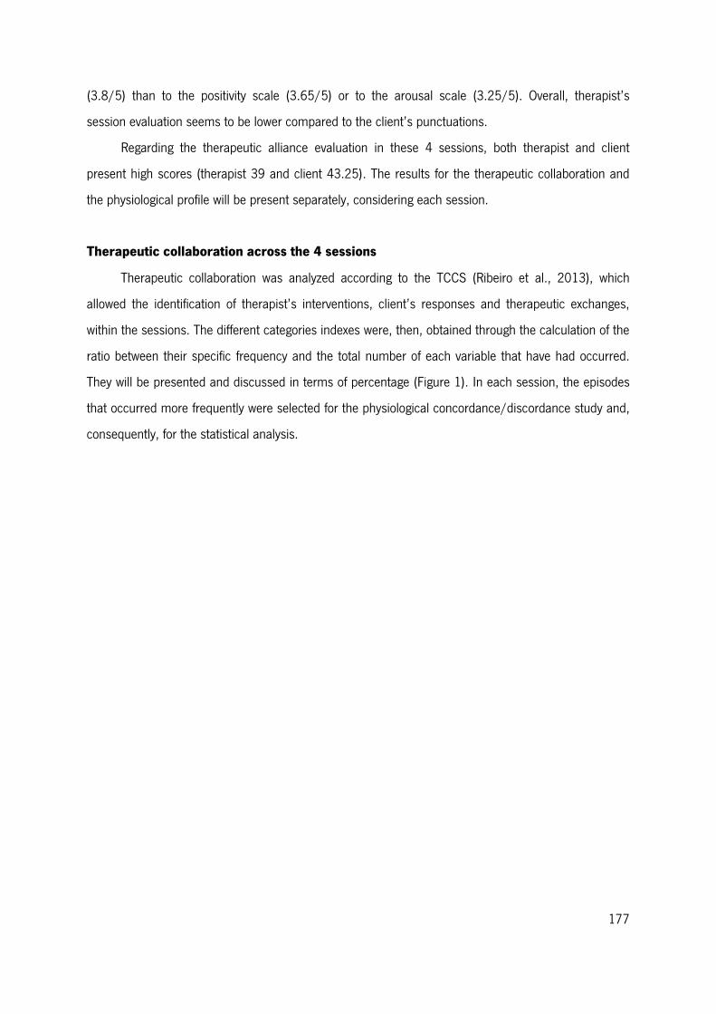
177
(3.8/5) than to the positivity scale (3.65/5) or to the arousal scale (3.25/5). Overall, therapist’s
session evaluation seems to be lower compared to the client’s punctuations.
Regarding the therapeutic alliance evaluation in these 4 sessions, both therapist and client
present high scores (therapist 39 and client 43.25). The results for the therapeutic collaboration and
the physiological profile will be present separately, considering each session.
Therapeutic collaboration across the 4 sessions
Therapeutic collaboration was analyzed according to the TCCS (Ribeiro et al., 2013), which
allowed the identification of therapist’s interventions, client’s responses and therapeutic exchanges,
within the sessions. The different categories indexes were, then, obtained through the calculation of the
ratio between their specific frequency and the total number of each variable that have had occurred.
They will be presented and discussed in terms of percentage (Figure 1). In each session, the episodes
that occurred more frequently were selected for the physiological concordance/discordance study and,
consequently, for the statistical analysis.
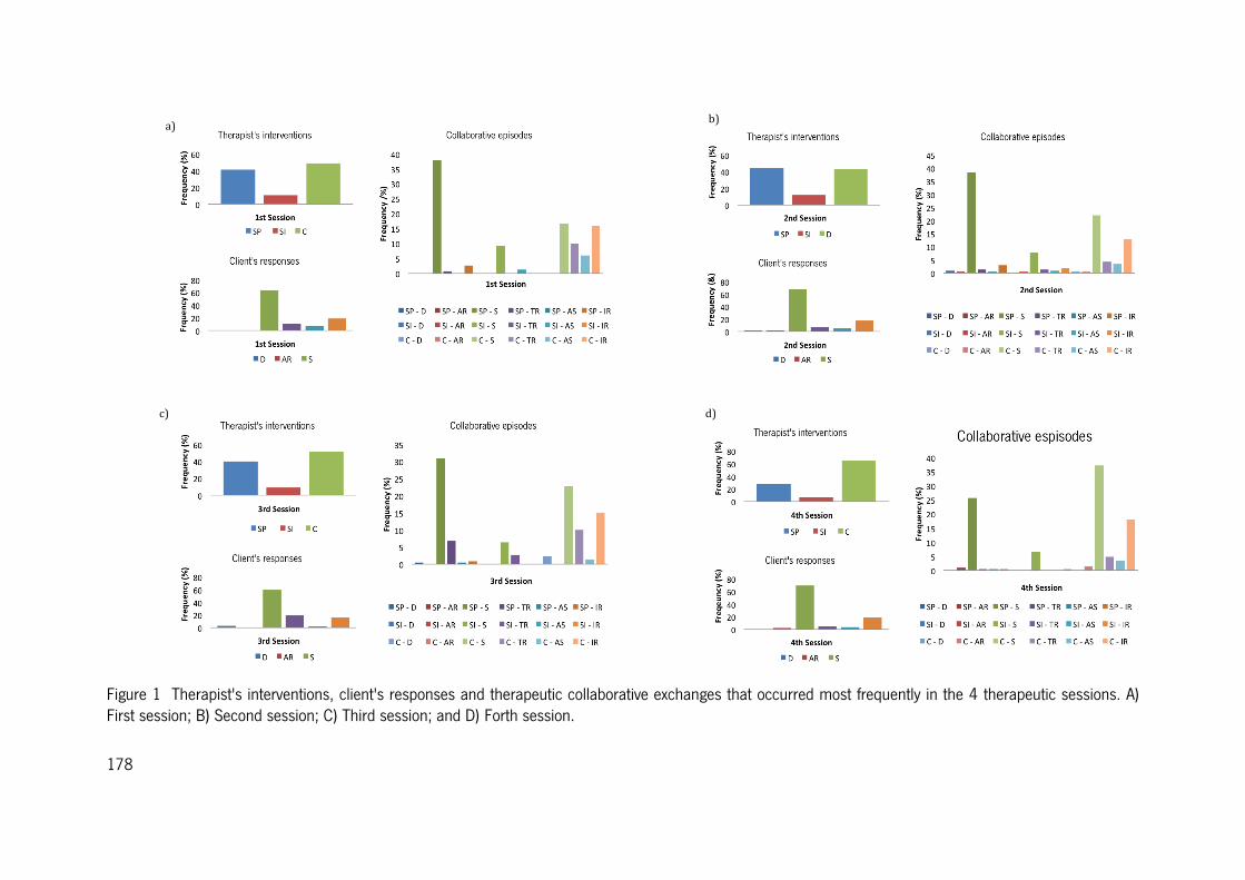
178
Figure 1 Therapist's interventions, client's responses and therapeutic collaborative exchanges that occurred most frequently in the 4 therapeutic sessions. A) First session; B) Second session; C) Third session; and D) Forth session.
a) b)
d) c)
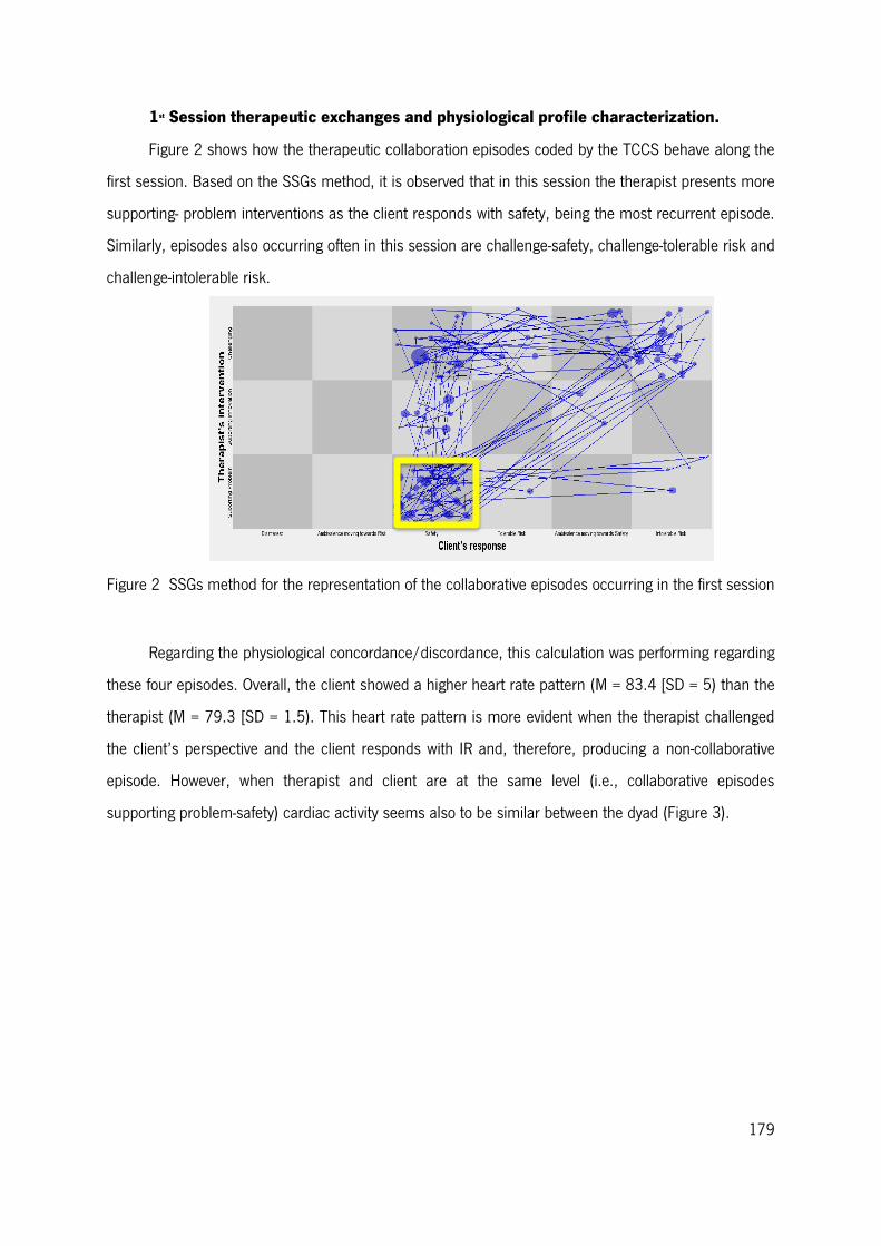
179
1st Session therapeutic exchanges and physiological profile characterization.
Figure 2 shows how the therapeutic collaboration episodes coded by the TCCS behave along the
first session. Based on the SSGs method, it is observed that in this session the therapist presents more
supporting- problem interventions as the client responds with safety, being the most recurrent episode.
Similarly, episodes also occurring often in this session are challenge-safety, challenge-tolerable risk and
challenge-intolerable risk.
Figure 2 SSGs method for the representation of the collaborative episodes occurring in the first session
Regarding the physiological concordance/discordance, this calculation was performing regarding
these four episodes. Overall, the client showed a higher heart rate pattern (M = 83.4 [SD = 5) than the
therapist (M = 79.3 [SD = 1.5). This heart rate pattern is more evident when the therapist challenged
the client’s perspective and the client responds with IR and, therefore, producing a non-collaborative
episode. However, when therapist and client are at the same level (i.e., collaborative episodes
supporting problem-safety) cardiac activity seems also to be similar between the dyad (Figure 3).
180
Figure 3 Heart rate (beats per minute) concordance/discordance between therapist and client regarding SP-S, C-S, C-IR and C-TR exchanges, in the first session.
Statistical analysis regarding collaborative episodes shows a correlation between the therapist
and client heart rate in supporting problem-safety episode that is statistically significant (r = 0.28, p <
.05), what does not happen in challenge-safety (r = .05 p = n.s.) or challenge-tolerable risk (r = .15, p =
n.s.). Considering the non-collaborative episode, challenge-intolerable risk, no statistical significant
results were found (r = .14, p = n.s.).
2nd Session therapeutic exchanges and physiological profile characterization.
Although in the second session more diverse therapist’s interventions and client’s responses are
happening, the most recurrent episodes occurring are supporting problem-safety, challenge-safety,
challenge-intolerable risk and supporting innovation-safety (Figure 4). Similarly to session 1, supporting
problem-safety continues to be the episode occurring more often.
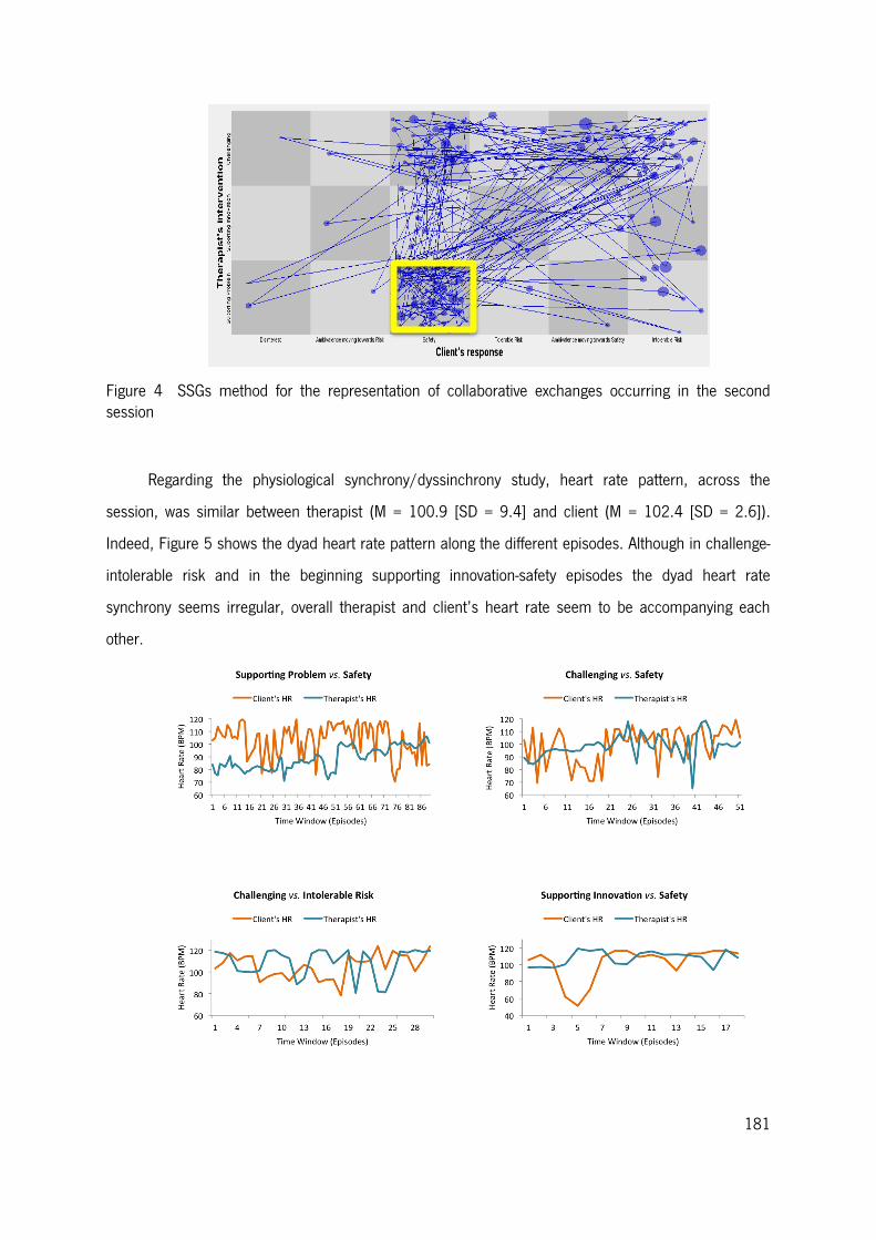
181
Figure 4 SSGs method for the representation of collaborative exchanges occurring in the second session
Regarding the physiological synchrony/dyssinchrony study, heart rate pattern, across the
session, was similar between therapist (M = 100.9 [SD = 9.4] and client (M = 102.4 [SD = 2.6]).
Indeed, Figure 5 shows the dyad heart rate pattern along the different episodes. Although in challenge-
intolerable risk and in the beginning supporting innovation-safety episodes the dyad heart rate
synchrony seems irregular, overall therapist and client’s heart rate seem to be accompanying each
other.
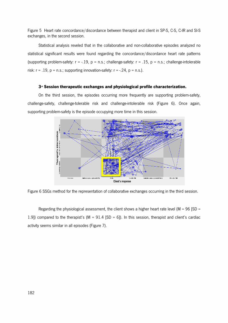
182
Figure 5 Heart rate concordance/discordance between therapist and client in SP-S, C-S, C-IR and SI-S exchanges, in the second session.
Statistical analysis reveled that in the collaborative and non-collaborative episodes analyzed no
statistical significant results were found regarding the concordance/discordance heart rate patterns
(supporting problem-safety: r = -.19, p = n.s.; challenge-safety: r = .15, p = n.s.; challenge-intolerable
risk: r = .19, p = n.s.; supporting innovation-safety: r = -.24, p = n.s.).
3rd Session therapeutic exchanges and physiological profile characterization.
On the third session, the episodes occurring more frequently are supporting problem-safety,
challenge-safety, challenge-tolerable risk and challenge-intolerable risk (Figure 6). Once again,
supporting problem-safety is the episode occupying more time in this session.
Figure 6 SSGs method for the representation of collaborative exchanges occurring in the third session.
Regarding the physiological assessment, the client shows a higher heart rate level (M = 96 [SD =
1.9]) compared to the therapist’s (M = 91.4 [SD = 6]). In this session, therapist and client’s cardiac
activity seems similar in all episodes (Figure 7).
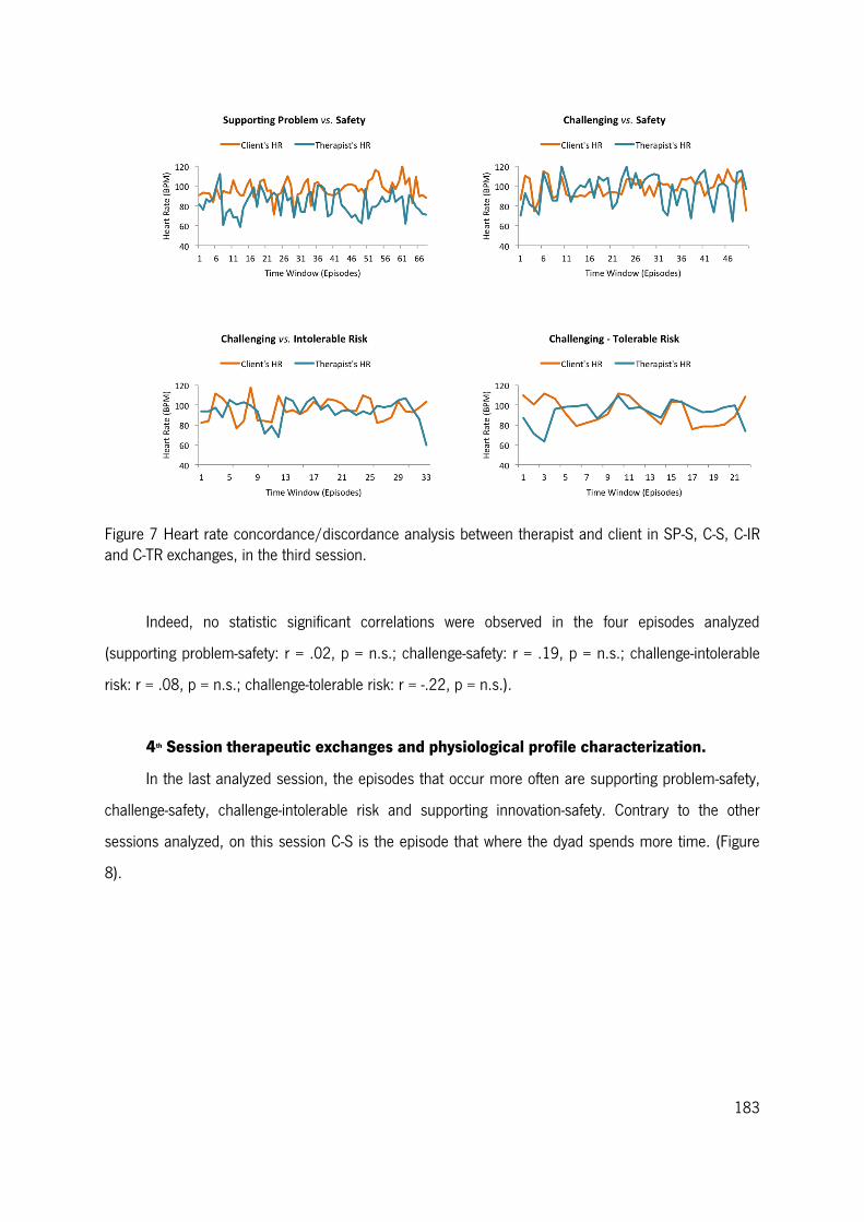
183
Figure 7 Heart rate concordance/discordance analysis between therapist and client in SP-S, C-S, C-IR and C-TR exchanges, in the third session.
Indeed, no statistic significant correlations were observed in the four episodes analyzed
(supporting problem-safety: r = .02, p = n.s.; challenge-safety: r = .19, p = n.s.; challenge-intolerable
risk: r = .08, p = n.s.; challenge-tolerable risk: r = -.22, p = n.s.).
4th Session therapeutic exchanges and physiological profile characterization.
In the last analyzed session, the episodes that occur more often are supporting problem-safety,
challenge-safety, challenge-intolerable risk and supporting innovation-safety. Contrary to the other
sessions analyzed, on this session C-S is the episode that where the dyad spends more time. (Figure
8).
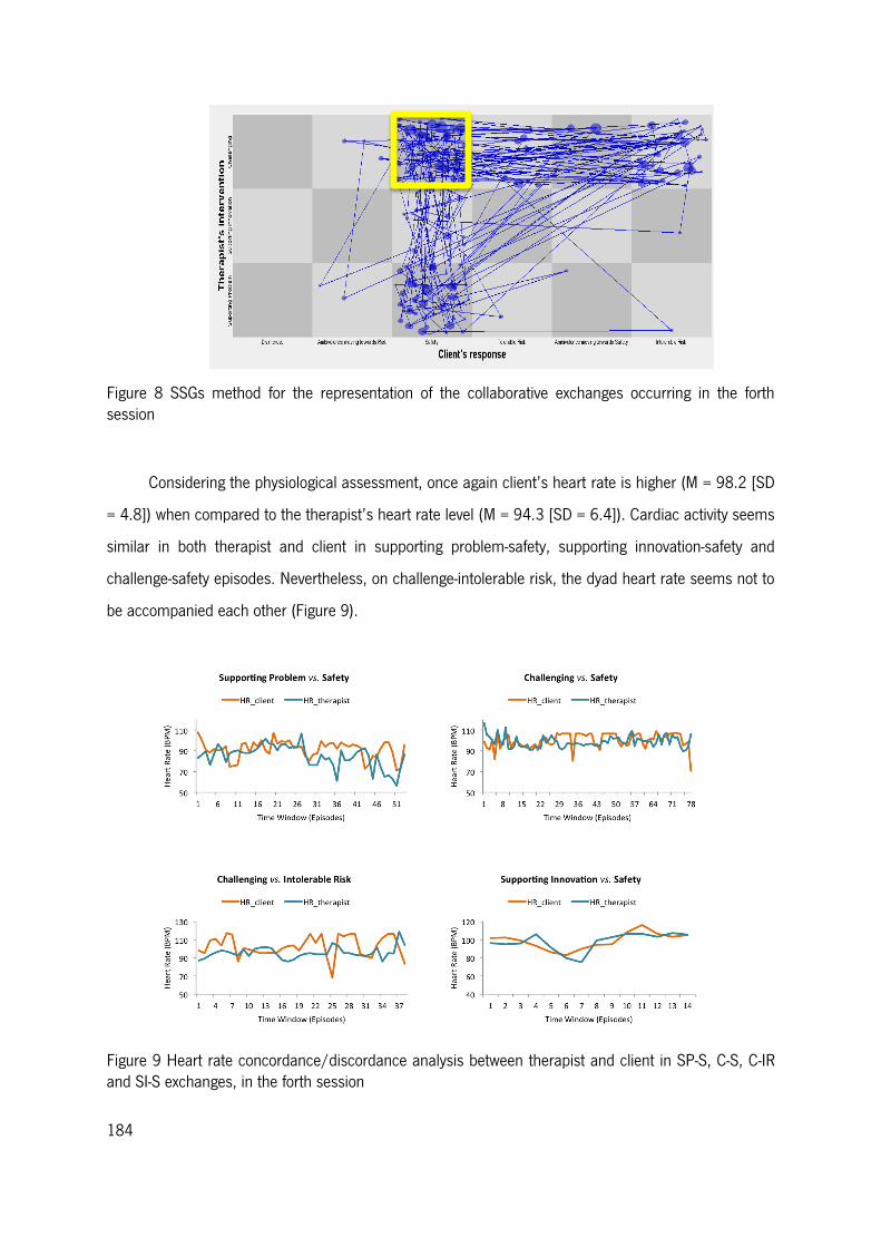
184
Figure 8 SSGs method for the representation of the collaborative exchanges occurring in the forth session
Considering the physiological assessment, once again client’s heart rate is higher (M = 98.2 [SD
= 4.8]) when compared to the therapist’s heart rate level (M = 94.3 [SD = 6.4]). Cardiac activity seems
similar in both therapist and client in supporting problem-safety, supporting innovation-safety and
challenge-safety episodes. Nevertheless, on challenge-intolerable risk, the dyad heart rate seems not to
be accompanied each other (Figure 9).
Figure 9 Heart rate concordance/discordance analysis between therapist and client in SP-S, C-S, C-IR and SI-S exchanges, in the forth session
185
Statistical analysis revealed that no significant results were found regarding challenge-safety (r
= .10, p = n.s.) and challenge-intolerable risk (r = -.19, p = n.s.) episodes. However, marginal
significant positive correlation was found regarding supporting problem-safety (r = .23, p < .1) episode
and a significant negative correlation in supporting innovation-safety (r = -.17, p < .05) when therapist
and client’s individual heart rate is compared.
Discussion
With the present study we aimed to characterize the therapeutic collaboration process in the first
four therapeutic sessions of a CBT good outcome case. Furthermore, we wanted to uncover the
therapist and client’s physiological profile by studying the momentary changes in the individual cardiac
activity within the clinical sessions as well as the heart rate concordance/discordance process inherent
to different therapeutic exchanges.
Regarding the therapeutic collaboration characterization, in the early phase of this particular
therapy process the therapeutic exchanges occurring more often were supporting problem-safety,
challenge-safety, challenge-tolerable risk, challenge-intolerable risk and supporting innovation-safety.
Although in the first three sessions the episode more frequently coded was supporting problem-safety,
on the forth session was challenge-safety. Indeed, the first sessions, the therapist’s focus relies on
knowing the problem presented by the client and, therefore, more collaborative episodes, where
therapist and client are at the same TZPD level, are occurring more frequently (Ribeiro et al., 2013;
Pires et al., in preparation). These collaborative exchanges are mainly coded as supporting problem
interventions and the client validates them with safety response. On the contrary, the non-collaborative
episodes are commonly identified as challenge-intolerable risk. This may be due to the fact that the
client is more resistant to change his problematic view and, consequently, invalidates the therapist’s
interventions with an intolerable risk response. Indeed John was classified as being in the
contemplation stage of change, which means that he recognize his problem but is not yet prompted to
anticipate or act toward change. Nevertheless, on the second session, supporting innovation-safety
episodes are now emerging, suggesting that the client becomes available to accept and incorporate
new perspectives regarding his problem.
Our results seem to suggest that the 4th therapeutic session can be a transition session which is
consistent with previous studies focusing on the importance of the quality of the therapeutic alliance in
186
the initial phase of the therapeutic process for the client’s permanence in therapy and prediction of
outcomes (Crits-Christoph, Gibbons & Hearon, 2006; Martin, Garske & Davis, 2000; Horvath &
Symonds, 2001). Indeed, with the present study, and according to the therapeutic collaboration
rational presented, this transition is explained by the increase of challenge interventions, produced by
the therapist, followed by the client’s validation responses (more challenge-safety or challenge-tolerable
risk episodes). Our results suggest that, being a good outcome case, more validated challenge
interventions are implied in the client’s acceptance of new perspectives aside from his problem and,
therefore, associated with the change process (Ribeiro et al., 2013).
Regarding the individual physiological activity, in the therapy initial phase, overall, therapist and
client show similar cardiac activity along the therapeutic sessions (compared to the baseline response),
although, most of the time, the client showed an increased mean heart rate compared with the
therapist’s heart rate. Moreover, the different TZPD positions (being or not ate the same developmental
level) seem to be accompanied by different physiological pattern, once when therapist and client are
not at the same TZPD level, the client shows an increase in heart rate activity, which is not observed in
the therapist. Although no statistical significant results support this, the graphic representation of the
heart rate patterns seems to indicate to these differences. The increase in the client’s heart rate may
be explained by the fact that he is able to understand the new conceptualization proposed by the
therapist about his problem, although, he seems not to be accepting it. It could be argued that this new
understanding, which was accompanied by a change in the client’s heart rate, seems to represent a
close relationship between the cardiac activity and the challenge involved in cognitive and emotional
processes, being this information perceived as more demanding (Stern et al., 2001;
Cacioppo, Tassinary, & Berntson, 2007; Porges, 2011).
However, as the client validates more frequently the challenge interventions, heart rate level
seems to increase simultaneously in the therapeutic dyad (challenge-tolerable risk in the third session).
This seems evident in the episodes supporting innovation-safety, occurring in the forth session, as new
perspectives introduced by therapist seem to be accepted by the client, therefore, integrating new
information in his problematic view. Positive heart rate correlation in supporting problem-safety
episodes in session 1 and 4 are observed, contrary to session 2. Once therapist and client are at the
same TZPD level positive correlations would be expected. Nevertheless, differences in these results
may be due to the influence of the sequence of previous episodes as they can produce different
expectations regarding the other’s responses. Indeed, in the second session the client’s physiological
187
activation seems higher, compared to the first session, which may be related with supporting problem
interventions exploring cognitive/emotional processes that may be more difficult to the client.
Therefore, although the focus in this session is on the client’s problematic questions, this may be
perceived as more demanding to him (Ribeiro et al., 2013).
Similarly, although no statistical significant results were found during the episodes in which the
therapist challenges the client’s perspective, we can observe an increase of the therapist’s cardiac
reactivity. This may be explained by emotional and cognitive response associated with cardiac arousal
associated with the acceptance of a new idea or also may be demanding to the therapist as empathic
challenge involves cognitive processes such interpretation, confrontation, thinking and to propose
different perspectives (Porges, 2009, Marci et al., 2007; Marci & Orr, 2006; Heaphy & Dutton, 2008;
Roy et al., 1998).
Our results seem consistent with Porges theory (2003, 2009, 2011) highlighting the cardiac
influence on social interactions. Indeed, this study suggests that different cardiac outputs are
underlying therapeutic exchanges, as interactions that are characterized as more demanding for the
client (according to the TCCS) seem to be producing an increased in heart rate pattern, as evidenced
by the visual analysis of the physiological data. On the contrary, interactions assumed as more
comprehensive and aiming to understand the problematic questions are producing safety responses
from the client, which are accompanied by heart rate signatures allowing the involvement in these
interactive exchanges. Moreover, it seems that the interpersonal aspect of the interactive exchanges is
influencing cardiac activity (Heaphy & Dutton, 2008; Roy et al., 1998) as it seems that therapist’s heart
rate is, occasionally (i.e., challenging interventions validated with tolerable risk), changing according to
the client’s responses. Therefore, physiological correlates seem to play an important role in the
different interactive therapeutic exchanges.
Moreover, in the initial phase of the therapeutic process, it seems important that therapist and
client work at the same TZPD level, as the involvement and progress in the therapy seems to be relying
on the dyad working together on the changing process (Ham & Tronick, 2009). As the therapeutic
process evolves, the forth session emerges as a transition phase as new perspectives are accepted by
the client, proven by the validated challenged and supporting innovations interventions.
With the present work (part of a bigger project to identify physiological markers of the therapeutic
collaboration process) we present preliminary results in tempting to identify physiological markers of
collaborative and non-collaborative exchanges regarding the initial phase of a good outcome case,

188
between the therapeutic dyad. It seems clear that in the initial sessions of a good outcome case,
supporting problem-safety episodes are more frequently occurring, indicating that therapist and client
interact at the same TZPD level. However, by the forth session challenge-safety or supporting
innovation-safety are more often happening, probably indicating an important moment for the change
process. This action coordination (according to the TCCS) along these sessions the therapist acts
based on the client’s readiness for change and, moreover, seems to reflect on physiological
concordance/discordance processes in the dyad.
Future studies, should examine this physiological concordance/discordance regarding the initial
phase of the therapy with more good outcome cases, in order to try to establish physiological markers
associated with the collaboration process. Moreover, these aspects should be address in, both
successful and unsuccessful cases, and regarding different therapeutic approaches.
189
References
Andreassi, J. L. (2000). Psychophysiology: Human Behavior & Physiological Response (4th ed.). New
Jersey: Lawrence Eribaum Associates.
Bachelor, A., & Salamé, R. (2000). Participants’ perceptions of dimensions of the therapeutic alliance
over the course of therapy. Journal of Psychotherapy Practice and Research, 9(1), 39-53.
Berntson, G. G., Quigley, K. S., & Lozano, D. (2007). Cardiovascular Psychophysiology. In J. T.
Cacioppo, L. G. Tassinary & G. G. Berntson (Eds.), Handbook of Psychophysiology (3rd ed.,
pp. 182-210). New York: Cambridge University Press.
Boardman, T., Catley, D., Grobe, J. E., Little, T. D., & Ahluwalia, J. S. (2006). Using motivational
interviewing with smokers: Do therapist behaviors relate to engagement and therapeutic
alliance? Journal of Substance Abuse Treatment, 31(4), 329-339.
Cacioppo, J. T., & Tassinary, L. G. (1990). Inferring psychological significance from physiological
signals. Am Psychol, 45(1), 16-28.
Coleman, R., Greenblatt, M., & Solomon, H. C. (1956). Physiological evidence of rapport during
psychotherapeutic interviews. Diseases of the nervous system, 17(3), 71.
Colli, A., & Lingiardi, V. (2009). The Collaborative Interactions Scale: a new transcript-based method for
the assessment of therapeutic alliance ruptures and resolutions in psychotherapy.
Psychotherapy Research, 19(6), 718-734.
Crits-Christoph, P., Gibbons, M. B. C., & Hearon, B. (2006). Does the alliance cause good outcome?
Recommendations for future research on the alliance. Psychotherapy: Theory, research,
practice, training, 43(3), 280.
Dimascio, A., Boyd, R. W., & Greenblatt, M. (1957). Physiological Correlates of Tension and
Antagonism During Psychotherapy A Study of" Interpersonal Physiology". Psychosomatic
Medicine, 19(2), 99-104.
Dittes, J. E. (1957). Galvanic skin response as a measure of patient's reaction to therapist's
permissiveness. The Journal of Abnormal and Social Psychology, 55(3), 295.
Feltham, C. (1999). Understanding the counselling relationship: Sage.
First, M. B., Spitzer, R. L., Gibbon, M., & William, J. (2002). Structured Clinical Interview for DSM-IV-TR
Axis I Disorders, Research Version, Patient Edition (SCID-I/P). New York: Biometrics Research.
: New York State Psychiatric Institute.
190
Ham, J., & Tronick, E. (2009). Relational psychophysiology: lessons from mother-infant physiology
research on dyadically expanded states of consciousness. Psychother Res, 19(6), 619-632.
doi: 10.1080/10503300802609672
Hatcher, R. (1999). Therapists' views of treatment Alliance and collaboration in Therapy.
Psychotherapy Research, 9(4), 405-423.
Heaphy, E. D., & Dutton, J. E. (2008). Positive social interactions and the human body at work: Linking
organizations and physiology. Academy of Management Review, 33(1), 137-162.
Hilsenroth, M. J., Peters, E. J., & Ackerman, S. J. (2004). The development of therapeutic alliance
during psychological assessment: Patient and therapist perspectives across treatment. Journal
of Personality Assessment, 83(3), 332-344.
Horvath, A. O. (2006). The alliance in context: Accomplishments, challenges, and future directions.
Psychotherapy: Theory, research, practice, training, 43(3), 258.
Horvath, A. O., & Greenberg, L. S. (1989). Development and validation of the Working Alliance
Inventory. Journal of Counseling Psychology, 36, 223-233. doi: 10.1037/0022-
0167.36.2.223
Horvath, A. O., & Symonds, B. D. (1991). Relation between working alliance and outcome in
psychotherapy: A meta-analysis. Journal of counseling psychology, 38(2), 139.
Jacobson, N. S., & Truax, P. (1991). Clinical significance: a statistical approach to defining meaningful
change in psychotherapy research. J Consult Clin Psychol, 59(1), 12-19.
Jennings, J. R., Kamarck, T., Stewart, C., Eddy, M., & Johnson, P. (1992). Alternate cardiovascular
baseline assessment techniques: vanilla or resting baseline. Psychophysiology, 29(6), 742-
750.
Lambert, M. J., Burlingame, G. M., Umphress, V., Hansen, N. B., Vermeersch, D. A., Clouse, G. C., &
Yanchar, S. C. (1996). The Reliability and Validity of the Outcome Questionnaire. Clinical
Psychology & Psychotherapy, 3(4), 249-258. doi: 10.1002/(SICI)1099-
0879(199612)3:4<249::AID-CPP106>3.0.CO;2-S
Leiman, M., & Stiles, W. B. (2001). Dialogical sequence analysis and the zone of proximal development
as conceptual enhancements to the assimilation model: The case of Jan revisited.
Psychotherapy Research, 11(3), 311-330.
Lewis, M. D. (2000). The promise of dynamic systems approaches for an integrated account of human
development. Child Dev, 71(1), 36-43.

191
Lewis, M. D., Zimmerman, S., Hollenstein, T., & Lamey, A. V. (2004). Reorganization in coping
behavior at 1 1/2 years: dynamic systems and normative change. Dev Sci, 7(1), 56-73.
Machado, P. P., & Fassnacht, D. B. (2014). The Portuguese version of the Outcome Questionnaire
(OQ-45): Normative data,
reliability and clinical significance cut-offs scores. . Manuscript submitted for publication.
Malmo, R. B. (1957). Anxiety and behavioral arousal. Psychological Review, 64(5), 276.
Marci, C. D., Ham, J., Moran, E., & Orr, S. P. (2007). Physiologic correlates of perceived therapist
empathy and social-emotional process during psychotherapy. The Journal of nervous and
mental disease, 195(2), 103-111.
Marci, C. D., & Orr, S. P. (2006). The effect of emotional distance on psychophysiologic concordance
and perceived empathy between patient and interviewer. Applied psychophysiology and
biofeedback, 31(2), 115-128.
Martin, D. J., Garske, J. P., & Davis, M. K. (2000). Relation of the therapeutic alliance with outcome
and other variables: a meta-analytic review. Journal of Consulting and Clinical Psychology,
68(3), 438.
Martin, E., Joeri, P., Loenneker, T., Ekatodramis, D., Vitacco, D., Hennig, J., & Marcar, V. L. (1999).
Visual processing in infants and children studied using functional MRI. Pediatr Res, 46(2), 135-
140.
McConnaughy, E. A., Prochaska, J. O., & Velicer, W. F. (1983). Stages of change in psychotherapy:
Measurement and sample profiles. Psychotherapy: Theory, Research & Practice, 20(3), 368-
375. doi: 10.1037/h0090198
Oliveira-Silva, P., & Gonçalves, Ó. F. (2011). Responding empathically: A question of heart, not a
question of skin. Applied psychophysiology and biofeedback, 36(3), 201-207.
Prochaska, J. O., & DiClemente, C. C. (1983). Stages and processes of self-change of smoking: toward
an integrative model of change. J Consult Clin Psychol, 51(3), 390-395.
Ribeiro, E., Ribeiro, A. P., Gonçalves, M. M., Horvath, A. O., & Stiles, W. B. (2013). How collaboration
in therapy becomes therapeutic: The therapeutic collaboration coding system. Psychology and
Psychotherapy: Theory, Research and Practice, 86(3), 294-314.
Roy, M. P., Steptoe, A., & Kirschbaum, C. (1998). Life events and social support as moderators of
individual differences in cardiovascular and cortisol reactivity. Journal of personality and social
psychology, 75(5), 1273.

192
Stern, R. M., Ray, W. J., & Quigley, K. S. (2001). Psychophysiological recording. New York: Oxford
University Press.
Stiles, W. B. (1980). Measurement of the impact of psychotherapy sessions. J Consult Clin Psychol,
48(2), 176-185. doi: 10.1037/0022-006X.48.2.176
Stiles, W. B., Gordon, L. E., & Lani, J. A. (2002). Session evaluation and the Session Evaluation
Questionnaire. In G. S. Tryon (Ed.), Counseling based on process research: Applying what we
know. (pp. 325-343). Boston, MA: Allyn & Bacon.
Tryon, G. S., & Winograd, G. (2011). Goal consensus and collaboration.
193
Conclusion
Interactive behavior is presented as a core dimension associated to the therapeutic dyad
collaboration exchanges in the therapy context and seems to be involved in adequate psychological
adjustment. Evidence has been presented of physiological mechanisms underlying interactive behavior
in the therapy context (Marci, Ham, Moran, & Orr, 2007; Marci & Orr, 2006). Therefore, with the
present dissertation we intend to contribute to expand the knowledge regarding physiological correlates
associated with interactive behavior and, furthermore, understanding its implications for therapeutic
dyad relationship.
The studies presented on the physiological correlates underlying therapeutic collaboration
process have showed that physiological reactivity patterns seem to be associated with therapeutic dyad
collaborative/non-collaborative exchanges in the therapy context. Indeed, by studying heart rate
patterns between therapist and client across the (non)collaborative exchanges identified according to
the TCCS, it seems that underlying these different exchanges physiological reactivity patterns are
occurring.
Regarding the first study, we aimed to characterize the first therapy session, by studying the
collaborative process, and identify concordance or discordance heart rate patterns in the collaborative
and non-collaborative exchanges. Our results evidenced that in this session, the first of a CBT good
outcome case, both supporting problem and challenging interventions produced by the therapist are
happening. Indeed, at the beginning of the session we verified that supporting problem are the
interventions occurring more frequently and, as the session develops, challenging interventions are
occurring more often. The client tends to validate more the supporting problem interventions with
safety response, producing collaborative episodes; on the contrary, tends to invalidate challenging
interventions (therapist works beyond TZPD) and, therefore, non-collaborative episodes are identified.
These results seem consistent with the expectations for the initial phase of a therapeutic process, as
therapist focus more on exploring and understanding the client’s problem. Regarding the physiological
results, our study evidences that heart rate between therapist and client is similar in collaborative
episodes. However, in non-collaborative episodes, when the client invalidates therapist intervention, an
increase in the client’s heart rate is observed. When the client validates challenging interventions,
producing a collaborative episode, both therapist and client’s heart rate increases. In summary,
194
physiological concordance is observed in collaborative episodes and, on the contrary, physiological
discordance in non-collaborative episodes.
On the second study, similarly to the first one, we aimed to characterize the therapeutic
collaboration process and the dyad physiological concordance and discordance physiological correlates
in the initial phase (first 4 therapeutic sessions) of a CBT good outcome case. Considering the TCCS
categorization, differences in the sessions were found as in the first one more supporting problem
interventions followed by safety response were occurring. However, in the second session supporting
innovation interventions followed by safety responses are emerging and, by the 4th session more
challenging interventions are being validated by the client (client responds with safety or tolerable risk).
Therefore, the 4th session seems to emerge as a transition session, as challenging interventions are
validated more often, mirroring the fact that the client is open to accept new perspectives presented by
the therapist (Ribeiro, Ribeiro, Gonçalves, Horvath, & Stiles, 2013). On the physiological level,
collaborative episodes seem to be accompanied by physiological concordance as a positive correlation
was found regarding therapist’s supporting problem intervention and client’s safety response. More
specifically, physiological concordance occurs when therapist and client are working at the same level
of the TZPD, which indicates that the therapist was able to emphatically understand the client’s
experience and identify his needs at a specific moment. Moreover, when the therapeutic dyad is not
working at the same TZPD level, the client shows an increase in heart rate, also observed in the first
study. By the 4th session, as challenging interventions are validated both therapist and client present an
increase in heart rate activity.
Regarding the therapeutic collaboration characterization, both studies seem to evidence that the
first therapy session, which the therapist mostly dedicates to explore and understand the problem
brought by the client, therapeutic exchanges are mainly characterized by therapist’s supporting
problem interventions, followed by safety response by the client’s side. Furthermore, challenging
interventions that are occurring in this session seem to be frequently invalidated. Therefore, the initial
session seems to be mainly characterized by collaborative episodes, mainly focused on supporting
problem – safety, happening more often. The non-collaborative episodes are frequently coded as
challenging – intolerable risk (Ribeiro et al., 2013).
Similarly, as referred before, the initial phase of a therapeutic process has been proven to be
important for the success of a therapeutic process (Crits-Christoph, Gibbons & Hearon, 2006;
Hilsenroth, Peters & Ackerman, 2004; Martin, Garske & Davis, 2000; Horvath & Symonds, 1991). The
195
collaboration characterization in the first 4 sessions seem to reveal this as well, considering that those
sessions are part of a CBT good outcome case. We verified that early in the process the therapist
presented challenging interventions that were mostly invalidated by the client in the first one. However,
in the second session we observe that the client validated supporting innovation interventions with
safety response. By the 4th session, the therapist’s interventions occurring more often are challenging,
being validated by the client, who responded either with safety or tolerable risk. This may indicated that
the client is accepting new perspectives and incorporate alternative information in his problematic view.
This may indicate that for a successful therapeutic process, challenging interventions emerge early in
the therapy and, although being largely invalidated in the first session, by the 4th one challenging
interventions are highly accepted. This results are consistent with previous studies using the TCCS,
showing that in good outcome cases, usually the therapist use challenging interventions since the
beginning of therapy, that are most likely validated by the client throughout the therapy (Ribeiro, 2012).
In regards to the physiological processes, on both studies, it seems clear that physiological
reactivity patterns seem to be associated with specific collaborative and non-collaborative episodes.
Indeed, collaborative episodes seem to be accompanied by similar heart rate activity in the dyad.
Supporting the problem interventions followed by safety response seem to not imply an increase in
heart rate, which is consistent with the TCCS rational (Ribeiro et al., 2013) and the Polyvagal theory
(Porges, 2011; Porges, 2009), as the therapist is exploring the problem and the client is explaining it,
which may suggest that as the client is feeling supported heart rate activity is underlying engagement
behaviors and therefore the dyad is at a collaborative level. However, when therapist’s challenging
interventions are validated by the client, an increase in the dyad heart rate is observed. This may be
occurring due to the fact that the client is accepting a new perspective presented by the therapist,
producing an incorporation of new information, which may involve a restructuring of cognitive and
emotional processes (Cacioppo, Tassinary & Berntson, 2007; Stern, Ray & Quigley, 2001). It has been
showed that heart rate activity is related with cognitive, emotional and social processes and, similarly,
producing mobilization behaviors (i.e. fight or flight) (Porges & Furman, 2011; Porges, 2001), which
supports the evidence that underlying the change process physiological reactivity processes are
occurring.
Regarding the non-collaborative episodes, mostly characterized by invalidation of challenging
interventions, physiological evidence suggest that as the client invalidates these interventions he
presents an increase in heart rate, not accompanied by therapist. Therefore, this may suggest that as

196
the therapist is presenting a new perspective, the client may not be prepared to accept it and,
therefore, presents a flight response, congruent with the Polyvagal theory reports (Porges, 1995). The
increase in heart rate may be associated with the fact that the client perceives the new information
provided, which is likely associated with restructuring of cognitive and emotional processes, however he
does not accepted it yet.
With the present studies, regarding physiological processes involved in the therapeutic
relationship, we aimed to further validate the therapeutic collaboration exchanges coding by
understanding if physiological reactivity responses were associated to them. In fact, we verified that
when considering collaborative and non-collaborative exchanges, differences regarding heart rate
patterns are observed in the dyad. Therefore, we believe that clinical implications can be derived from
these results. Foremost, from an interventional point of view, and considering the TCCS contributions
to a better understanding about the collaboration process, it seems it can contribute to the therapist’s
awareness regarding his or hers interventions, which may be a valuable tool to adequate his or her
interventions accordingly to the client’s responses, in order to be responsive accordingly to the client’s
needs at the therapy and, consequently, increase adhesion to therapeutic process. Furthermore, we
want to identify reliable and consistent physiological markers (i.e., heart rate patterns) that may be
associated with collaborative and non-collaborative exchanges. Nevertheless, some limitations may be
reported. In fact, the results presented must be considered as preliminary, as further investigation
regarding these processes must be carried out. Moreover, TCCS coding and physiological signal
processing are time-consuming tasks that require a large amount of time for data to be analyzed.
Future studies must address several questions, for a clear understanding about the development
of collaboration and its association with physiological reactivity. Therefore, collaboration and its
association with physiological processes should be investigated considering different variables such as
successful, unsuccessful and dropout cases, in regards to different psychological disorders (i.e. anxiety
disorders), assess different therapists and regarding multiple theoretical approaches. This way, TCCS
exchanges coding and its association with physiological signatures can be validated.
197
References
Cacioppo, J. T., Tassinary, L. G., & Berntson, G. G. (2007). Handbook of psychophysiology (Vol. 2):
Cambridge University Press New York.
Crits-Christoph, P., Gibbons, M. B. C., & Hearon, B. (2006). Does the alliance cause good outcome?
Recommendations for future research on the alliance. Psychotherapy: Theory, research,
practice, training, 43(3), 280.
Hilsenroth, M. J., Peters, E. J., & Ackerman, S. J. (2004). The development of therapeutic alliance
during psychological assessment: Patient and therapist perspectives across treatment. Journal
of Personality Assessment, 83(3), 332-344.
Horvath, A. O., & Symonds, B. D. (1991). Relation between working alliance and outcome in
psychotherapy: A meta-analysis. Journal of counseling psychology, 38(2), 139.
Marci, C. D., Ham, J., Moran, E., & Orr, S. P. (2007). Physiologic correlates of perceived therapist
empathy and social-emotional process during psychotherapy. The Journal of nervous and
mental disease, 195(2), 103-111.
Marci, C. D., & Orr, S. P. (2006). The effect of emotional distance on psychophysiologic concordance
and perceived empathy between patient and interviewer. Applied psychophysiology and
biofeedback, 31(2), 115-128.
Martin, D. J., Garske, J. P., & Davis, M. K. (2000). Relation of the therapeutic alliance with outcome
and other variables: a meta-analytic review. Journal of Consulting and Clinical Psychology,
68(3), 438.
Porges, S. W. (1995). Cardiac vagal tone: a physiological index of stress. Neuroscience & Biobehavioral
Reviews, 19(2), 225-233.
Porges, S. W. (2001). The polyvagal theory: phylogenetic substrates of a social nervous system.
International Journal of Psychophysiology, 42(2), 123-146.
Porges, S. W. (2009). The polyvagal theory: new insights into adaptive reactions of the autonomic
nervous system. Cleve Clin J Med, 76 Suppl 2, S86-90. doi: 10.3949/ccjm.76.s2.17
Porges, S. W. (2011). The Polyvagal Theory: Neurophysiological Foundations of Emotions, Attachment,
Communication, and Self-regulation (Norton Series on Interpersonal Neurobiology): WW Norton
& Company.
198
Porges, S. W., & Furman, S. A. (2011). The early development of the autonomic nervous system
provides a neural platform for social behaviour: A polyvagal perspective. Infant and child
development, 20(1), 106-118.
Ribeiro, E. (2012). Therapeutic collaboration: communalities and specifics across therapeutic
approaches and therapy outcomes. Paper presented at the Panel in 43rd Annual Meeting of
Society for Psychotherapy Research, Virginia Beach, EUA.
Ribeiro, E., Ribeiro, A. P., Gonçalves, M. M., Horvath, A. O., & Stiles, W. B. (2013). How collaboration
in therapy becomes therapeutic: The therapeutic collaboration coding system. Psychology and
Psychotherapy: Theory, Research and Practice, 86(3), 294-314.
Stern, R. M., Ray, W. J., & Quigley, K. S. (2001). Psychophysiological recording: Oxford University
Press.
199
General Conclusion
With this dissertation we focused on describing the neural and psychophysiological correlates
that are associated with interactive behavior in two main research topics: 1) infant’s development and
2) therapeutic relationship. Although these two topics are in its core distinct, it is assumed that social
involvement plays a crucial role in the developmental process and, similarly, in healthy psychological
adjustment. Indeed, neuropsychophysiological processes seem to be underlying both infant’s social
development, as well as, therapist and client’s relationship within the therapeutic context.
Particularly, regarding the infant’s developmental research topic, evidence suggests that neural
mechanisms and physiological signatures are accompanying behavioral outcomes across the infancy
period. As presented before, it seems clear that underlying the emergence of cognitive and socio-
emotional behaviors are central and peripheral responses that seems to support these developmental
outcomes. Additionally, neurophysiological correlates seem to be predicting behavioral, social and
cognitive developmental outcomes in infants.
From birth until circa 3-months of age, infants responses to the environment are mainly
occurring through visual and auditory stimuli processing. Therefore, in order to determine early
neurophysiological correlates of infant’s development we wanted to identify visual and auditory
neuropsychophysiological correlates that could be associated with infant’s interactive behavior. As
revealed by our results, early at a postnatal age, infant’s visual and auditory sensory processing
abilities are associated with neurobehavioral social orientation and state regulation behaviors, which
are characteristics involved in interactive routines, described in healthy young infants. Therefore, neural
and physiological markers of social development can be identified early in the development and,
furthermore, can be used as predictors of developmental outcomes. Such results convey clinical
implications, as these neural and psychophysiological correlates can be used as a tool for an early
diagnosis of developmental-related problems.
Similarly, neurophysiological evidence have showed that underlying different psychological
disorders seem to be specific physiological reactivity patterns that may be associated with the
emergence and development of diverse symptoms. Nevertheless, physiological correlates associated
with therapeutic relationship and, furthermore, with the change process within the therapy context
remain to clarify.
200
Our studies focusing on the therapeutic relationship, revealed that specific therapist and client’s
heart rate signatures seem to be associated with the collaboration process. In fact, preliminary
evidence seem to sustain the indication that physiological markers may be associated with specific
therapeutic exchanges and, additionally, may be used as predictors of (un)successful change process.
Clinical implications are draw from this evidence, as physiological markers of collaborative or non-
collaborative relationship may provide important information regarding the client’s readiness to change
and adhesion to therapy.
Overall, this dissertation shows that neuropsychophysiological processes seem crucial for the
development of interactive abilities in young infants and, similarly, for the establishment of a
collaborative relationship in therapy, as the identification of these neuropsychophysiological correlates
may be a useful tool in predicting future behavioral outcomes. Indeed, being these research topics
distinct, infant’s neural and physiological associated with interactive behavior may offer a framework to
understand the dynamic of the physiological processes regarding interactive behaviors in the
therapeutic relationship, as particular reactivity profiles (i.e., hypo- and hyper-reactivity) seem to
emerge early in the development and are maintained as stable characteristics across lifetime period
with influences over diverse contexts of interactive behaviors.


