

UNIVERSIDADE DE LISBOA
FACULDADE DE CIÊNCIAS
DEPARTAMENTO DE BIOLOGIA VEGETAL
STUDIES ON THE INVOLVEMENT OF QUORUM SENSING IN THE
REGULATION OF EXOPOLYSACCHARIDE BIOSYNTHESIS BY
BURKHOLDERIA CEPACIA COMPLEX ISOLATES
Vítor Hugo Jorge de Oliveira
MESTRADO EM MICROBIOLOGIA APLICADA
2012

ii
UNIVERSIDADE DE LISBOA
FACULDADE DE CIÊNCIAS
DEPARTAMENTO DE BIOLOGIA VEGETAL
STUDIES ON THE INVOLVEMENT OF QUORUM SENSING IN THE
REGULATION OF EXOPOLYSACCHARIDE BIOSYNTHESIS BY
BURKHOLDERIA CEPACIA COMPLEX ISOLATES
Dissertação orientada pela Profª. Doutora Leonilde Moreira (IBB-IST)
e pelo Prof. Doutor Mário Santos (FCUL)
Vítor Hugo Jorge de Oliveira
MESTRADO EM MICROBIOLOGIA APLICADA
2012

iii
STUDIES ON THE INVOLVEMENT OF QUORUM SENSING IN THE
REGULATION OF EXOPOLYSACCHARIDE BIOSYNTHESIS BY
BURKHOLDERIA CEPACIA COMPLEX ISOLATES
Vítor Hugo Jorge de Oliveira
MASTER THESIS
2012
This thesis was fully performed at the Institute for Biotechnology and
Bioengineering of the Instituto Superior Técnico under the direct
supervision of Prof. Dr. Leonilde Moreira and Dr. Ana Sofia Ferreira.
Prof. Dr. Mário Santos was the internal designated supervisor in the
scope of the Master in Applied Microbiology of the Faculty of
Sciences of the University of Lisbon.

iv
Acknowledgments
This master thesis reflects the support of many people who influenced the work
in different ways and who I would like to thank.
First, I am very grateful to Prof. Leonilde Moreira from Instituto Superior
Técnico, where my work was developed. I am most thankful for the careful support and
guidance she gave me during the experimental work and during the writing of this
thesis.
I would also like to thank Prof. Mário Santos, who was designated to be my
internal supervisor in the scope of the Master in Applied Microbiology, and that from the
beginning proved to be available.
I would like to address a special word to Doctor Ana Sofia Ferreira, co-
supervisor of this work. Thank you for the advice and support throughout this work and
for being always present to help me.
Furthermore, I would like to thank all members of BSRG group by their
welcoming and a special acknowledge to PhD students Inês Silva and Mário Santos for
all the support and suggestions they gave me during this work.
Finally, thanks also to all my friends and family for the patience and
unconditional support.

v
Abstract
Quorum sensing (QS) is a cell-to-cell communication mechanism used by
bacteria that produce and recognize signaling molecules named autoinducers. It is
known to be involved in the regulation of different virulence factors including
exopolysaccharide (EPS) biosynthesis in several pathogens. Bacteria belonging to
Burkholderia cepacia complex (Bcc) are a group of related species that had emerged
as important opportunistic pathogens mainly in cystic fibrosis (CF) patients. EPS
produced by these bacteria plays an important role in the development of Bcc
infections. Like in other bacteria, Bcc possess several QS-regulated phenotypes but
little is known about the effect of this regulation mechanism in the Bcc EPS
biosynthesis. Therefore, to evaluate the role of QS in such regulation, we tested the
effects of acyl-homoserine lactones (AHLs) degradation by AiiA lactonase and the
presence of a quorum sensing inhibitor (QSI) 4-nitro-pyridine-1-oxide (4NPO) in EPS
biosynthesis inhibition. The results indicate that indeed QS regulates EPS biosynthesis
in Bcc. However the signaling molecules involved are not dependent on the CepI/R QS
system since an insertion mutant in the cepI gene did not inhibit the EPS production.
Thus, bioinformatics analyses led to the identification of protein CbsI as another
putative AHL synthase of Burkholderia.
Bcc bacteria are resistant to most of the conventional antibiotic treatments
making its eradication a challenge. Thus, it has become essential to identify and
develop alternative therapies to deal with these infections. The use of QSI seems a
promising field to be explored. Since 4NPO was able to inhibit EPS production in
Burkholderia, it was decided test its effect in several other Bcc QS-dependent
phenotypes, such as motility, production of extracellular proteases and siderophores,
and biofilm formation. Furthermore, its potential use as co-adjuvant of antibiotics in
different Bcc species was also evaluated. The results confirmed that 4NPO is indeed
affecting the tested phenotypes and increases the susceptibility to the antibiotics tested
in both Bcc planktonic and sessile cells. The results also showed that 4NPO
potentiates the activity of detergents such as Triton X100 and Tween 20. Since the
clinical application of this compound is limited, its usage to clean and disinfect abiotic
surfaces could be a possibility.
Keywords: Burkholderia complex cepacia (Bcc), virulence factors, exopolysaccharide
(EPS), quorum sensing (QS), quorum sensing inhibitors (QSI), 4-nitro-pyridine-1-oxide
(4NPO)

vi
Resumo
As bactérias pertences ao complexo Burkholderia cepacia (Bcc) estão divididas
em 17 espécies e são consideradas patogénicas oportunistas infectando sobretudo
doentes com fibrose quística (FQ). Apesar de representarem apenas uma pequena
percentagem da totalidade das infecções em doentes com FQ, os danos causadas por
estas bactérias apresentam uma grande heterogeneidade, uma vez que as infecções
que causam podem ser infecções crónicas ou podem estar associadas a quadros
clínicos mais graves, levando muitas vezes à falência das funções pulmonares,
desenvolvimento de pneumonia e de septicémia, designada síndrome da cepacia. A
severidade destas infecções é agravada pela possibilidade de transmissão cruzada
entre pacientes e pela partilha de equipamentos de terapêutica inalatória.
Consequentemente, a maioria dos centros de FQ em todo o mundo teve que
implementar regras especiais de higiene hospitalar e medidas severas de isolamento
de indivíduos colonizados e indivíduos não colonizados com Bcc. Adicionalmente,
estas bactérias são intrinsecamente resistentes à maioria dos antibióticos clinicamente
usados e à maioria dos desinfectantes. Os compostos antibacterianos, com acção
bacteriostática ou bacteriocida, têm-se assim revelado pouco eficazes no tratamento
destas infecções, sendo necessário recorrer a elevadas concentrações de antibióticos
e ao uso combinado desses mesmos antibióticos. Torna-se, portanto, fundamental o
desenvolvimento de novas estratégias que permitam um tratamento mais rápido e
eficaz desta infecções.
Umas das estratégias que tem sido fortemente estudada e que se tem revelado
promissora é a interferência dos mecanismos de quorum sensing (QS). Este
mecanismo é dependente de uma densidade populacional elevada, permitindo a
comunicação entre as bactérias de uma população através da libertação de sinais
químicos, sendo os mais comuns em bactérias Gram-negativa as acil-homoserina
lactonas (AHLs). Quando esses sinais se acumulam no meio e atingem um
determinado nível, ocorre a activação de proteínas reguladoras que vão activar ou
reprimir a expressão de genes regulados por QS, levando a comportamentos
sincronizados dentro dessa população. Tem sido demonstrado que o QS está
envolvido na regulação da expressão de diversos genes relacionados com a
virulência. No caso das bactérias do complexo Bcc, o sistema mais conservado é
constituído pela sintase cepI, que produz C6-HSL e C8-HSL, e pelo regulador de
transcrição cepR, que está envolvido na expressão de genes codificantes para
proteases e sideróforos, motilidade e formação de biofilmes, entre outros. Ao contrário
do que ocorre em outras espécies, pouco se sabe sobre os efeitos do QS, e em

vii
concreto do sistema CepI/R ao nível da regulação da síntese de exopolissacárido, um
importante factor de virulência em Bcc. Trabalhos feitos anteriormente no nosso
laboratório sugerem que a biossíntese deste polímero deverá ser regulada por QS.
Assim, um dos objectivos deste trabalho foi compreender qual o papel do mecanismo
de QS na regulação do exopolissacárido em diferentes espécies de Burkholderia. Para
tal, estudaram-se os efeitos da degradação de AHLs pela lactonase AiiA e do inibidor
de QS, 1-óxido- 4-nitro-piridina (4NPO) na síntese de exopolissacárido. Os resultados
obtidos, sugerem que o QS está efectivamente a regular a produção de
exopolissacárido em Bcc mas as moléculas sinalizadoras não são as dependentes do
sistema CepI/R. Isto porque a construção de um mutante de inserção no gene cepI
não aboliu a síntese do exopolissacárido. Assim sendo, utilizaram-se ferramentas
bioinformáticas para identifcar outro possível sistema de QS em Burkholderia.
O segundo objectivo deste trabalho foi determinar os efeitos de um inibidor de
QS descrito em Pseudomas aeruginosa, o 4NPO, em diferentes espécies do complexo
Bcc, ao nível de diversos fenótipos regulados por QS, nomeadamente a mobilidade,
produção de proteases extracelulares e de sideróforos e a formação de biofilmes. Uma
vez que se confirmou que efectivamente o 4NPO inibe o QS em Burkholderia, decidiu-
se estudar a potencial utilização deste composto como co-adjuvante de antibióticos.
Para tal, testaram-se diferentes antibióticos e os resultados mostraram que o 4NPO
aumenta a capacidade antibacteriana desses antimicrobioanos, tanto ao nível da
inibição do crescimento das células planctónicas como ao nível da inibição da
formação de biofilmes. Embora o uso clínico deste produto esteja limitado devido às
suas propriedades mutagénicas, o uso de 4NPO em superfícies abióticas poderá ser
uma potencial aplicação deste composto, uma vez que, também se verificou potenciar
a actividade de detergentes como o Triton X100 e o Tween 20, ao nível do
crescimento planctónico e do desenvolvimento de biofilmes. De acordo com estes
resultados, os inibidores de QS, e o 4NPO em particular, poderão de facto ter
aplicações a nível da desinfecção de superfícies e de material clínico, prevenindo o
aparecimento de casos de contaminações hospitalares, que já levaram a graves surtos
de Bcc em doentes com FQ.

Index
Acknowledgments ..................................................................................................... iv
Abstract ....................................................................................................................... v
Resumo ...................................................................................................................... vi
1 Introduction .......................................................................................................... 1
1.1 The Burkholderia genus .................................................................................. 1
1.1.1 Burkholderia cepacia complex: a group of opportunistic CF pathogens ... 1
1.1.2 Virulence factores in Burkholderia ........................................................... 2
1.2 Insights into quorum sensing mechanism and functions ................................. 5
1.3 Quorum sensing inhibitors as antimicrobial compounds ................................. 7
1.4 Objectives ....................................................................................................... 9
2 Material and Methods ....................................................................................... 11
2.1 Bacterial strains, plasmids and oligonucleotides ........................................... 11
2.2 Bacterial growth conditions ........................................................................... 13
2.3 DNA manipulation techniques ....................................................................... 13
2.3.1 DNA extraction ...................................................................................... 13
2.3.2 DNA amplification by PCR ..................................................................... 14
2.4 Construction of unmarked deletion mutants .................................................. 15
2.5 Construction of a marked deletion mutant on cbsI gene ............................... 15
2.6 Burkholderia triparental mating ..................................................................... 16
2.7 RNA manipulation techniques ....................................................................... 16
2.7.1 RNA extraction and purification ............................................................. 16
2.7.2 Quantitative Real-Time PCR .................................................................. 17
2.8 Determination of 4NPO sub-lethal concentration .......................................... 17
2.9 Determination of Minimal Inhibitory Concentration (MIC) .............................. 18
2.10 Phenotypic tests ........................................................................................... 18
2.10.1 EPS precipitation and quantification ...................................................... 18
2.10.2 Motility assays: Swimming and Swarming ............................................. 19
2.10.3 Proteases and Siderophores production ................................................ 19
2.10.4 Antibiotic and disinfectant susceptibility in the presence of 4NPO .......... 19
2.10.5 Biofilm formation assays ........................................................................ 20
2.11 Bioinformatic analyses .................................................................................. 20
3 Results and Discussion .................................................................................... 22

ii
3.1 Role of quorum-sensing in cepacian biosynthesis regulation ........................ 22
3.1.1 Role of a quorum sensing inhibitor in preventing EPS biosynthesis ....... 23
3.1.2 Search of other AHL synthases that might control EPS biosynthesis ..... 25
3.2 4NPO as a QSI in Bcc species ..................................................................... 27
3.2.1 Evaluation of the effect of 4NPO in QS-dependent phenotypes ............. 28
3.2.2 Effect of 4NPO as enhancer of Bcc susceptibility against antibiotics ..... 30
3.2.3 Effect of 4NPO as co-adjuvant of detergents and disinfectants .............. 36
4 Final remarks ..................................................................................................... 39
5 Bibliography ....................................................................................................... 40
6 Supplementary data .......................................................................................... 49

1
1 Introduction
1.1 The Burkholderia genus
Bacteria belonging to the Burkholderia genus are motile, metabolically diverse
Gram-negative β-proteobacteria that occupy a wide range of ecological niches
including soil, industrial waste, water, clinical sources and many others [1, 2].
Burkholderia species have unusual large genomes, which contribute to the high
plasticity, adaptability and capacity to colonize different environments and to survive
under several stress conditions. These bacteria are able to use many unusual carbon
and energy sources, being highly versatile from the metabolic point of view [3]. Some
beneficial Burkholderia species establish symbiotic rhizospheric interactions with fungi,
bring benefits to crops due to their ability to produce several antimicrobial compounds;
promote plant growth by fixing atmospheric nitrogen in symbiosis with several plants;
and can degrade natural and man-made pollutants (reviewed in [4]). On the other
hand, some Burkholderia can be pathogens of plants, animals and humans [5, 6].
Among the pathogenic Burkholderia species are included B. mallei, which causes
glanders in horses; B. pseudomallei that causes melioidosis; and the species belonging
to Burkholderia cepacia complex (Bcc) which have emerged as important opportunistic
human pathogens, especially in patients with chronic granulomatous disease (CGD), of
immunocompromised individuals, and most importantly in cystic fibrosis (CF) patients
[7]. Burkholderia cepacia complex comprises a group of related species that share a
high level of similarity at 16S rRNA gene sequence (>97.5%) and of recA gene
sequence (94% to 95%), and moderate levels of DNA-DNA hybridization (30% to 60%)
[8-11].
1.1.1 Burkholderia cepacia complex: a group of opportunistic CF pathogens
Cystic fibrosis is a genetic disease caused by mutations in a gene encoding the
cystic fibrosis transmembrane conductance regulator (CFTR) that leads to the
accumulation of a thick mucus in different organs including lungs, favoring bacterial
colonization of several pathogens like Burkholderia [12, 13]. Even though Burkholderia
infections affect less than 10% of the CF patients [14], it has become an extremely
important opportunistic pathogen, which is associated with a worst clinical outcome and
lower life expectancy. Furthermore, lung transplants are usually denied to these
patients, due to the possibility of the development of cepacia syndrome characterized
by necrotizing pneumonia and septicemia [15]. Bcc are highly transmissible from
patient-to-patient and by contact with contaminated clinical devices, such as respiratory

2
therapy equipment, reusable temperature probes and catheters, being these bacteria
resistant to the most commonly used disinfectants [16-19]. Burkholderia is also
intrinsically resistant to antibiotics. Although poorly understood, the antibiotic resistance
mechanisms in Bcc strains can be divided into three categories: enzyme modification,
alteration of drug targets and limited permeability [20]. Permeability alterations at the
membrane level play a key role in the Bcc defense mechanisms against antimicrobial
agents, due to the existence of modified LPS, porins and efflux pumps, as well as the
ability to produce EPS and/or form biofilms, which are thought to limit drugs access to
the cell [20]. Bcc are able to cause enzyme drug modifications and target changes.
These organisms are intrinsically resistant to aminoglycoside antibiotics and have high
levels of β-lactam resistance due to the production of inducible chromosomal β-
lactamases and altered penicillin-binding proteins [21]. Moreover, the capacity of
antibiotic resistance in the Bcc can be highlighted by its capacity to use penicillin G as
a sole carbon source [22]. Accordingly, the majority of Bcc strains are multidrug
resistant and conventional antimicrobial therapies which includes the combination of
two or sometimes three different antibiotics are often ineffective [19].
1.1.2 Virulence factores in Burkholderia
Multiple factors contribute to the pathogenicity of Bcc bacteria giving them the
capacity to overwhelm the host defenses, to establish chronic infections that are rarely
eradicated, to invade the epithelial cells, causing their necrosis, or to cross the
epithelium paracelularly, enabling their dissemination into the blood stream [23]. These
proprieties are due to the production of numerous virulence factors, the intrinsic
resistance to antibiotics and the ability to form biofilms.
Among the virulence factors produced by Bcc bacteria there is the synthesis of
lipopolysaccharide (LPS), which contains particular structural properties that neutralize
the anionic charge of cell surface, being involved in the resistance to polymyxin,
cationic antimicrobial peptides like protegin-1 [24] and in the prevention of bacterial
phagocytosis by macrophages [25-28]. Other virulence factors are the expression of a
cable pili and 22 kDa adhesin, which allow the binding to epithelial cells, induce
cytotoxicity and initiate cellular apoptosis [29]; flagella, which are involved in bacteria
dissemination, epithelial cells invasion, biofilm formation and induction of host
responses [30]; and the biosynthesis and secretion of haemolysins, lipases,
siderophores and extracellular proteases (eg. ZmpA and ZmpB), which were shown to
be controlled by quorum sensing (QS) [31-33]. In particular, ZmpA and ZmpB are

3
metalloproteases involved in the disruption of host tissues and the enhancement of the
host immune system, contributing to an increase of inflammation [32, 34].
The ability of Bcc bacteria to form biofilms has been shown in both
environmental and clinical Bcc isolates grown in abiotic and biotic surfaces [35].
Biofilms are defined as a sessile community of bacterial cells in the stationary phase
irreversibly attached to a surface, being embedded by a matrix of extracellular
polymeric substances. The matrix of mature biofilms is composed of diverse substrates
that include polysaccharides, proteins, nucleic acids and lipids. Biofilm communities
exhibit gradients of nutrients and oxygen through the different layers of the biofilm,
having the cells of the bottom lower availability of nutrients and therefore less metabolic
activity [36]. These characteristics protect bacteria-forming biofilm from many
environmental factors, including antibiotics, disinfectant chemicals and the host
immune system. The reason it could be the limited drug diffusion and inactivation of
compounds by the biofilm matrix [37, 38]. Biofilm formation involves several temporal
phases as resumed in the Figure 1. The role of extracellular polymeric substances on
biofilm matrix cohesiveness and biofilm disruption have been an interesting target to
find chemical compounds that acting at that level, could promote biofilm detachment
[39].
Figure 1 - Steps required for biofilm formation. [1] initial attachment, [2] irreversible attachment, [3-4] maturation, [5] dispersion. Each stage of development in the diagram is paired with a photomicrograph of a developing P. aeruginosa biofilm. All photomicrographs are shown to same scale. The planktonic bacteria start to swim towards the substratum using flagella, to form loose attachments [1] on the surface forming microcolonies [2]. The microcolonies multiply and differentiate into mature biofilms where cells are
embedded into a thick extracellular polymeric matrix. The biofilm can acquire mushroom- or tower-like structures [3-4]. Subsequently, it is observed a detachment of bacterial cells that can be spread to other places giving origin to new biofilms [5] (adapted from [40])
Besides being involved in biofilm formation, extracellular polysaccharides or
exopolysaccharides (EPS) are considered to be important virulence features for
several pathogens. In the case of Burkholderia the role of EPS in infection is not well

4
established, as some authors point out that it might have a virulence role and others
consider EPS a persistence factor. Still the importance of EPS in Burkholderia
adaptation to different environments, stress conditions, and to overcome the immune
system has been demonstrated experimentally (reviewed in [41]), as summarized in
Figure 2. Several Burkholderia EPSs have been identified and structurally
characterized, but the EPS cepacian is the most common one, being produced by both
Bcc and non-Bcc Burkholderia strains of clinical and environmental origin [42, 43].
Cepacian biosynthesis involves many genes, most of them encoded by bce-I and bce-II
gene clusters [43-46]. Still, little is known about the mechanisms involved in the
regulation of cepacian biosynthesis, with the exception of the BY-kinase BceF and the
phosphotyrosine phosphatase BceD by exerting regulation at the post-translational
level [47].
Figure 2 – Summary of the role of Burkholderia EPS in the adaptation to different niches (adapted from [41]). Cepacian was shown to interact with antimicrobial peptides [48]; to scavenge reactive oxygen
species (ROS), to interfere with neutrophil chemotaxis [49], to be required for the formation of mature biofilms structures [50] and to be involved in the resistance to desiccation and metal ion stress, contributing to Bcc capacity to thrive in adverse environments [43].
Quorum sensing (QS) mechanisms are known to control EPS production in
other bacterial species, including nonpathogenic Sinorhizobium meliloti and the plant
pathogens Erwinia stewartii and Pseudomonas syringae [51-53]. Recently, Soarez-
Moreno and collaborators had shown that QS mechanisms also control EPS
biosynthesis in non-Bcc plant-associated Burkholderia [54]. Accordingly, we had
hypothesized that QS may also regulate cepacian biosynthesis in Bcc by acting at the
transcriptional level, and that will be one of the aims of this work.

5
1.2 Insights into quorum sensing mechanism and functions
Cell-to-cell communication can be mediated by different chemical signals that
are secreted, diffused and detected throughout the community. In response to such
factors, signal transduction cascades are activated and induce alterations in gene
expression which enable bacteria to survive in different stress conditions and
consequently adjust to the surrounding environment [55]. These mechanisms are
mediated by a cell-density-dependent regulatory mechanism designated by quorum
sensing.
QS signaling is based on the intracellular production of low molecular weight
molecules, known as autoinducers, that are either passively or actively released to
external milieu. When these signal molecules reach a specific threshold, which is
usually dependent on bacterial population density they bind to specific receptors
(regulatory proteins) that trigger signal transduction cascades, resulting in the alteration
of gene expression [56]. QS plays a central role in both symbiotic and pathogenic
interactions. For instance, it regulates several virulence phenotypes and allows a
coordinated “attack” to the host that is triggered only at high population density,
increasing the bacterial ability to overwhelm host defenses and to establish an infection
[57]. In symbiotic associations, QS has also crucial roles in root nodulation by
legumes/Rhizobiaceae associations and the production of bioluminescence in
squid/Vibrio fisheri symbiosis [58, 59].
The autoinducers chemical structure is highly diverse, being the most common
structures presented by Gram-negative bacteria based on N-acylated-L-homoserine
lactones (AHLs), while in Gram-positive bacteria QS is based on small peptides
synthesis and detection [60].
The first QS system was described in the marine bacterium Vibrio fischeri,
which establishes symbiotic associations with the squid Euprymna scolopes and is
involved in the activation of the luciferase operon that enables bacterial
bioluminescence [59, 61]. This system is composed by two proteins: LuxI, a N-
acylhomoserine lactone synthase, and LuxR, a cytoplasmatic transcriptional regulator
which recognizes the signal molecules constituting the LuxI/R QS-system [62]. This
system represents the paradigm of QS systems that are usually composed by a
synthase and a regulator protein. Several types of QS systems have been described in
Gram-negative bacteria, each one presenting distinct class of signal molecules, such
as N-acyl homoserine lactones, quinolones (AQs), long-chain fatty acids and fatty acid
methyl esters as well as autoinducer-2 (AI-2), a group of furanone derivatives [63].
Due to substrate specificity of the LuxI-homologues and conformation
restrictions of the LuxR-homologues, only specific AHLs can bind to the regulator

6
protein [64, 65]. Therefore, AHL autoinducers tend to be species specific, as only
particular acyl-chains are recognized by the species producing them being associated
to intraspecies communication [64, 65]. Interspecies communication is usually
associated with another autoinducer class, known as autoinducer-2 (AI-2), which is
present in both Gram-negative and Gram-positive bacteria being proposed as an
“universal signal” for interspecies communication (reviewed in [66]).
In the case of Bcc bacteria, the most well conserved QS system identified so far
is based on CepI, the synthase LuxI-homologue, and CepR, the LuxR-like protein [67,
68]. The CepI protein synthesizes two AHL molecules: N-octanoyl-L-homoserine
lactone (C8-HSL) and, in lower amounts, N-hexanoyl-L-homoserine lactone (C6-HSL)
[69] (Figure 3). This system was shown to play a crucial role in B. cenocepacia
virulence being essential for full pathogenicity in several infection models [70, 71].
CepI/R system positively regulates expression of extracellular proteases, chitinases
and a polygalactunorase, as well as the swarming motility and biofilm formation; and
negatively regulates the biosynthesis of the siderophore ornibactin [72-74]. Besides
CepI/R homologue systems, an orphan LuxR homologue was also described in B.
cenocepacia [75]. Orphan LuxR homologues are QS regulators that do not have an
associated AHL synthase but respond to endogenous or exogenous synthesized AHLs
[76] (Figure 3).
Figure 3 - Schematic representation of CepI/R Burkholderia QS system and some QS-regulated phenotypes (adapted from http://botserv1.uzh.ch/microbio/site/research/introduction/Burkholderia
Cepacia.php).
Other AHL- based QS systems have been identified in several Burkholderia
species. For instance, the B. cenocepacia CciI/R system produces and is activated by
C6-HSL and C8-HSL [77]. B. vietnamiensis BviI/R system produces and responds to
C10-HSL [78] and the BraI/R system from B. kururiensis synthesizes and responds to
3-oxo-C12-HSL [54]. System BraI/R was shown to control EPS biosynthesis in the
cep box
C6-HSL
C8-HSL
CepR
cepI
cepR

7
plant-associated Burkholderia species [79]. Even though no BraI/R homologues can be
found in the sequenced Bcc species and the Bcc EPS is slightly different from the one
produced by the plant-associated Burkholderia, the results obtained by Suarez-Moreno
and co-authors give a further indication that QS may control EPS production in Bcc
species. Besides AHLs, other molecules have been described to be involved in QS
within the Burkholderia genus such as 4-hydroxy-2-alkylquinolines (HAQ) derivative.
These compounds are thought to function as iron chelators, immune modulators and
antimicrobial compounds, able to inhibit bacterial growth [80]. B. cenocepacia is able to
produce a diverse set of 2-alkyl-4(1H)-quinolones shown be involved in colony
morphology and elastase production, and cis-2-dodecenoic acid (BDSF), a molecule
structurally related to the diffusible signal factor found in Xanthomonas campestris
contributing to interspecies and intraspecies communication [81-83].
1.3 Quorum sensing inhibitors as antimicrobial compounds
Considering the rapid growth of bacterial resistance against many antimicrobial
compounds and the link between QS and virulence, the discovery of QS antagonists
may provide a possible means to achieve new antimicrobial strategies and it has thus
attracted significant attention in recent years. Many QS inhibitors (QSIs) have been
described and there are a growing number of studies showing their ability to prevent
and/or disrupt biofilm formation, diminish the production of virulence factors, impair the
responses to oxidative stress and increase neutrophils activity against bacteria [84-88].
Accordingly, these studies point out that the use of QSIs might be the next generation
of drugs against bacterial infections, as QS is directly related with the production of
virulence factors that are not essential for bacterial survival. Therefore, with the
selective disruption of QS, pathogens can no longer adapt to the host environment
being eliminated by innate host defenses without the selective pressure associated
with the conventional antibiotic treatments, which have biocide activity [86, 89].
QSI can act at several levels. For instance, the modulation of QS can occur by
interfering with the synthase, with the regulator protein or with the signal itself (Figure
4) [86, 90]. Regarding QSI that act by preventing the production of autoinducers, there
are studies that use small-molecule agents such as 59-methylthioadenosine (MTA) and
S-adenosylmethionine (SAM) analogs to target LuxI-type synthase proteins [91]; others
that use substitutes of SAM, one of the AHL’s precursors [86, 92]; and others that use
autoinducer synthase blocking compounds, such as thiol derivatives and homoserine
lactone derivatives [93]. Another approach used is to block the signal at the receptor
level, which can be achieved by the use of antagonist compounds, capable of

8
competing or interfering with the cognate AHL signal for binding to LuxR-type receptors
and both designed synthetic and natural naturally occurring substances, such as
extracts from plants and food, have been tested to modulate QS at LuxR-homologues
levels [94, 95]. Finally, QS inhibition can occur by manipulation of the QS signal itself.
Some plants and bacteria present defense mechanisms that destroy the invading or
competing bacteria autoinducers to protect themselves. For instance, some plants
increase the pH at infection sites, causing hydrolysis of the lactone ring; others secrete
of oxidized halogenated compounds that are capable of reacting with the 3-oxo-AHLs;
some bacterial species are capable of using AHLs as a carbon and nitrogen source,
others are able to breakdown AHL by secreting lactonases and acylases enzymes,
others produce secondary metabolites, such as brominated furanones, that can block
the action of AHLs [86, 96, 97].
Figure 4 – Different levels of quorum-sensing (QS) disrupting strategies in bacteria using quorum sensing inhibitors (QSI) (adapted from [98]).
The use of QSI as co-adjuvant of antibiotic action has been widely studied,
particularly against P. aeruginosa causing CF lung infections where halogenated
furanone compounds were able to inhibit biofilm formation [99] and garlic extract in
combination with tobramycin enhanced the clearance of infecting bacteria in the mice
pulmonary infection model [100]. Still, little is known about the potential use of QSIs
against Bcc, probably due to the fact that these bacteria are able to degrade a wide
variety of compounds, as some of the QSIs known to be efficient in P. aeruginosa did

9
not seem to have any effect on Bcc strains [101]. Nevertheless, Brackman and co-
authors (2009, 2011) showed that same QSIs can interfere with biofilm formation and
maturation in B. multivorans and B. cenocepacia [84, 102].
1.4 Objectives
Quorum sensing is known to regulate the expression of several genes that are
involved in the production of virulence factors. Among the phenotypes controlled by
QS, the biosynthesis of exopolysaccharides was shown to be regulated at the
transcriptional level in several bacterial systems, including the plant pathogens Erwinia
stewartii and Pseudomonas syringae [51, 52] and nonpathogenic species of
Sinorhizobium meliloti [53]. Regarding Burkholderia species, the work of Suarez-
Moreno and co-authors (2008, 2010) developed in plant-associated Burkholderia,
which are non-Bcc and non-virulent species, showed that the QS system BraI/R,
directing the production of oxo-C12-HSL and oxo-C14-HSL, is involved in the control of
EPS biosynthesis in these species [43, 44].
Previous results from our laboratory also indicate that cepacian biosynthesis is
most likely regulated by QS. In the presence of the lactonase Aiia from Bacillus subtilis
which is able to hydrolyze the lactone ring of AHLs and sub-lethal concentrations of a
QSI, 4-nitro-pyridine-1-oxide (4NPO), cepacian production by B. cepacia IST408 was
inhibited. Therefore, one of the goals of this work was to determine if QS is indeed
controlling cepacian biosynthesis in Bcc bacteria and if CepI/R system, the most well
conserved QS system among Bcc species, was involved in such control or there might
be another conserved system controlling this feature. To accomplish this goal we
tested lactonase expression in different Bcc and non-Bcc species and evaluate
whether the QSI 4NPO is also able to inhibit EPS biosynthesis. In parallel we
constructed unmarked cepI and cepR genes deletion mutants to evaluate whether the
AHLs produced by CepI/R QS system are the one controlling EPS expression. If these
mutants still produce EPS, it is our aim to identify other QS system in a strain with the
genome sequenced such as B. multivorans ATCC 17616.
The second major goal of this work is to determine if the QSI 4NPO can be
used to control biofilm formation by Bcc strains, if applied as a co-adjuvant of
antibiotics or detergent/disinfectants. Our previous results had shown that EPS
biosynthesis by B. cepacia IST408 was inhibited by the P. aeruginosa QSI 4NPO [85]
when used in sub-lethal concentrations which do not affect bacteria growth. Since
4NPO seemed to be an efficient QSI against this strain, further work was done to
determine its potential as an antibiotic adjuvant. The results indicate that indeed B.
cepacia IST408 susceptibility to trimethoprim, kanamycin, amikacin and piperacillin

10
was enhanced in vitro as well as in Galleria mellonella virulence model of infection,
where significant survival differences were observed between the presence and
absence of 4NPO supplementation with trimethoprim and kanamycin (Ferreira et. al.
unpublished results). Even though the results obtained were quite promising, the
application of 4NPO to humans is compromised, as previous studies in E. coli and
fibroblasts showed that this compound has mutagenic activity [103, 104]. Still, 4NPO
could have other biotechnological applications such as being used as disinfectant of
surfaces or anti-biofouling agent, being used together with detergents or other
antimicrobial compounds. The work of Vanoyan et al. (2010) showed that 4NPO has
interesting physico-chemical properties that can reduce the extent of bacterial adhesion
to surfaces [105]. Currently, a CF patient infected with Bcc needs to be isolated from
other CF patients, requiring special treatment rooms, equipment and nursing teams,
leading to an increase of costs [106]. The potential use of 4NPO or others QSI as co-
adjuvant of detergents or disinfectants could prevent cross-contaminations in CF
centers and hospitals. Thus, in this work, different classes of antibiotic and detergents
were used to test the efficiency of 4NPO as a QSI. The chosen antibiotics are the two
aminoglycosides kanamycin and amikacin (inhibitors of protein synthesis); the
sulfonamide trimethoprim (folate pathway inhibitor) and the β-lactam piperacillin and
ceftazidime (inhibitors of cell wall synthesis). As disinfectant agent it were used bleach
solution and as detergents the anionic sodium dodeyl sulphate (SDS) and non-anionic
Triton X100 (TX100) and Tween 20.

11
2 Material and Methods
2.1 Bacterial strains, plasmids and oligonucleotides
Bacterial strains and plasmid used in this work are listed on Table 1.
Table 1 – List of strains and plasmids used in this work.
Strain Relevant Characteristics Reference
Burkholderia strains
B. cepacia IST408
Cystic fibrosis clinical isolate
[107]
B. cenocepacia K56-2 Cystic fibrosis clinical isolate [108]
B. multivorans ATCC 17616 Soil isolate [9]
B. multivorans D2095 Mucoid cystic fibrosis clinical isolate [109]
B. dolosa AUO158 Cystic fibrosis clinical isolate [110]
B. ambifaria AMMD Root-colonizing bacterium [111]
B. lata 383 Soil isolate [112]
B. xenovorans LB400 Soil isolate [113]
B. phymatum STM815 Soil isolate; nitrogen fixation [114]
B. phytofirmans PsJN Soil isolate; plant growth-promoting bacterium [115]
B. multivorans ATCC 17616 cepI::pVO1105-1
pVO1105-1 integrated into cepI gene region This work
B. multivorans ATCC 17616 cepR::pVO1106-1
pVO1106-1 integrated into cepR gene region This work
B. multivorans ATCC 17616 cepI::pVO1105-1+pDAI-SceI
B. multivorans ATCC 17616 derivative containing pVO1105-1 integrated into cepI gene region and the
replicative plasmid pDAI-SceI
This work
B. multivorans ATCC 17616 cepR::pVO1106-1+pDAI-SceI
B. multivorans ATCC 17616 derivative containing pVO1106-1 integrated into cepR gene region and the
replicative plasmid pDAI-SceI
This work
B. cepacia IST 408 cepI::pIS410-1
pIS410-1 integrated into cepI gene region Ferreira et al. unpublished
results
E.coli strains
E. coli αDH5 supE44 (ф80 lacZΔM15) hsdR17(rK- mK
+) recA1
endA1 gyrA96 thi-1 relA1 deoR Δ(lacZYA-argF)U169 Invitrogen
E. coli HB101 thi-1 hsdS20(rB -, mB-) supE44 recA13 ara-14 leuB6 proA2 lacY1 galK 2rpsL20 (Str
R) xyl-5 mti-1
Promega

12
Abbreviations: TpR trimethoprim resistance; Km
R: kanamycin resistance; Amp
R: ampicillin resistance;
CmR, chloramphenicol resistance.
The list of oligonucleotides used in PCR and a RT-PCR amplifications is shown
in Table 2. Oligonucleotides primers were designed using AmplifX 1.5.4, available at
http://ifrjr.nord.univ-mrs.fr/AmplifX and synthesized by MWG Biotech AG (Germany).
Table 2 - Oligonucleotides used for PCR amplification. Abbreviations: A, Adenine; C, Cytosine; G,
Guanine; T, Thymine.
Primer name
Sequence (5`- 3´) Primer name
Sequence (5`- 3´)
1997A-fw
AAGATTCAGTCTGAGATGAAGGCACGAGT RTbceH-rev
CGATGTCGTCGCCTTTCC
1997A-rev
AAGGTACCTCGGCAGTTCTCGCATTAG RTbceI-fw
AAGTTTCGAGCGTGACCAGTTC
1997B-fw
GTCTCTAGACTTCCAGACCTTCATGGCGTA RTbceI-rev
AACAGCGACTTCAGCAGATACG
1997B-rev
TAGGTACCTGTTCCGACTGTCCGACATC RTbceP-fw
GGACAAAGGCATACTCAAGAACGT
catKpn-up
ATGGTACCTATCACGAGGCCCTTTCGTCTTC RTbceP-rev
CGAAGGTCGGCAGGATCA
catKpn-low
CTGGTACCTGTCGTGCCAGCTGCATTA RTbceQ-fw
TTCGGCGAGGACGACTATG
RTbceB-fw
TTCGTGAACATCCGCTTCATT RTbceQ-rev
TGGAACCCGAGGAAATGC
RTbceB-rev
CCGAGCACCTCGACCACTT proC-fw GTCGGCGAGATCGTATGGTT
RTbceE-fw
CCGAGACCTATCCGGTTCATT proC-rev CTGCAGCGCTTCGATGAAA
Plasmids
pMLBAD pBBR1 ori, araC-PBAD, TpR mob
+ [116]
pMLBAD-aiiA Broad-host-range vector carrying araC-PBAD-aiiA
for expression of AiiA; TpR [117]
pGPI-SceI oriR6K TPR, mob
+, carries I-SceI cut site [118]
pDAI-SceI pDA17 carrying the I-SceI gene [118]
pVO1105-1 pGPI-SceI derivative containing ΔcepI gene This work
pVO1106-1 pGPI-SceI derivative containing ΔcepR gene This work
pDrive Cloning vector, AmpR Km
R Qiagen
pK18mob Cloning vector, KmR [119]
pUC18 Cloning vector, Ampr
[120]
pBBRIMCS Cloning vector, containing cat gene, Cmr
[121]
pVO412-1 pK18mob derivative containing cbsI gene (upstream
region) This work
pVO412-2 pDrive derivative containing cbsI gene (downstream
region) This work
pVO412-3 pK18mob derivative containing cbsI gene regions This work

13
RTbceE-rev
CTTTCTGCAGCTGGTCCATCA cepRa-fw
TCGAATTCTGTTCCTCGGCGTGACGATTCC
RTbceF-fw
AAACACTCCTACGCGGATCTGT cepRa-rev
ATGGATCCATCGAAGCACCCTGACGCAA
RTbceF-rev
CAGCCAGATGTCGTCCATGA cepRb-fw
ATGGATCCTATACCGAATGGCATCGCA
RtbceH-fw
ACGAAAGTCCACGTCCATCTG cepRb-rev
CATCTAGAGTGCCACAGCAATTCGTCA
cepIa-fw TGAATTCTTGCGTCAGGGTGCTTCGAT cepIb-fw ATGGATCCTCGATCCGCAAACGTTTGCT
cepIa-rev
TAGGATCCTCGTGAACGAAGGTCTGCAT cepIb-rev
GCGTCTAGAGTAGGGAACTGACGAATGGGTA
2.2 Bacterial growth conditions
Burkholderia fresh cultures were obtained by inoculating a portion of the frozen
material at -80oC into plates of Pseudomonas isolation agar media (PIA, Difco),
followed by incubation at 37oC overnight. The cultures were then maintained at 4oC
until further use. Pre-inocula required for all assays were done as followed: overnight
liquid cultures were prepared by transferring one isolated bacterial colony of
Burkholderia, previously grown on solid media, into LB liquid medium followed by
incubating overnight at 30oC or 37oC with orbital agitation (250 rpm). Then, these
cultures would be used to inoculate working cultures in the proper conditions.
Burkholderia strains were cultured in S medium or MM medium (see below), at 30ºC
with orbital agitation (250 rpm). E. coli strains were always grown in LB medium (Difco)
and incubated at 37ºC with orbital agitation (250 rpm).
S liquid medium – 12.5 g/l Na2HPO4, 3 g/l KH2PO4, 1 g/l K2SO4, 1 g/l NaCl, 0.2 g/l
MgSO4.7H2O, 0.001 g/l FeSO4.7H2O, 0.01 g/l CaCl2 2H2O, 20 g/l glucose, 1 g/l yeast
extract, 1 g/l casamino acids
MM liquid medium – 2 g/l of yeast extract (Difco), 20 g/l of mannitol (Merck)
2.3 DNA manipulation techniques
2.3.1 DNA extraction
Plasmid DNA was extracted from overnight cultures of E. coli host strains
growing in LB medium supplemented with appropriate antibiotics, by a QIAprep Spin
Miniprep kit (QIAGEN), following the manufacturer’s instructions. Total genomic DNA
from Burkholderia strains was extracted according standard protocol [122].
Concentration of genomic and plasmid DNA solutions was determined on a
Spectrophotometer ND-1000 (NanoDrop).
14
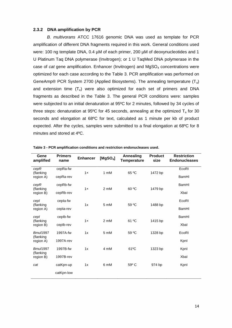
2.3.2 DNA amplification by PCR
B. multivorans ATCC 17616 genomic DNA was used as template for PCR
amplification of different DNA fragments required in this work. General conditions used
were: 100 ng template DNA, 0.4 µM of each primer, 200 µM of deoxynucleotides and 1
U Platinum Taq DNA polymerase (Invitrogen); or 1 U TaqMed DNA polymerase in the
case of cat gene amplification. Enhancer (Invitrogen) and MgSO4 concentrations were
optimized for each case according to the Table 3. PCR amplification was performed on
GeneAmp® PCR System 2700 (Applied Biosystems). The annealing temperature (Ta)
and extension time (Te) were also optimized for each set of primers and DNA
fragments as described in the Table 3. The general PCR conditions were: samples
were subjected to an initial denaturation at 95ºC for 2 minutes, followed by 34 cycles of
three steps: denaturation at 95ºC for 45 seconds, annealing at the optimized Ta for 30
seconds and elongation at 68ºC for text, calculated as 1 minute per kb of product
expected. After the cycles, samples were submitted to a final elongation at 68ºC for 8
minutes and stored at 4ºC.
Table 3 - PCR amplification conditions and restriction endonucleases used.
Gene amplified
Primers name
Enhancer [MgSO4] Annealing
Temperature Product
size Restriction
Endonucleases
cepR (flanking region A)
cepRa-fw
cepRa-rev 1× 1 mM 65 ºC 1472 bp
EcoRI
BamHI
cepR (flanking region B)
cepRb-fw
cepRb-rev 1× 2 mM 60 ºC 1479 bp
BamHI
XbaI
cepI (flanking region A)
cepIa-fw
cepIa-rev 1x 5 mM 59 ºC 1488 bp
EcoRI
BamHI
cepI
(flanking region B)
cepIb-fw
cepIb-rev 1× 2 mM 61 ºC 1415 bp
BamHI
XbaI
Bmul1997
(flanking region A)
1997A-fw
1997A-rev
1x 5 mM 59 ºC 1328 bp EcoRI
KpnI
Bmul1997
(flanking region B)
1997B-fw
1997B-rev
1x 4 mM 61ºC 1323 bp KpnI
XbaI
cat catKpn-up
catKpn-low
1x 6 mM 59º C 974 bp KpnI

15
2.4 Construction of unmarked deletion mutants
Burkholderia deletion mutations were designed according to Flannagan et al.
[118]. Fragments of around 700 bp of each flanking regions of cepI and cepR genes
were obtained by PCR amplification, digested with the appropriate restriction
endonucleases (Table 3) and inserted into pK18mob and pUC18 cloning vectors to
increase their number of copies, and selected according to white/blue selection in the
respective selection marker. The digested PCR fragments were subsequently ligated
into pGPI-SceI (a suicide plasmid that cannot replicate in Burkholderia and contains the
I-SceI recognition site) digested with EcoRI and XbaI in a triple ligation mixture, giving
rise to the plasmids pVO1105-1 (deletion of cepI) and pVO1106-1 (deletion of cepR).
Selection of vectors containing the inserted regions was made on E. coli αDH5 by
electrotransformation and candidates were selected based on pGPI-SceI selection
marker (trimethoprim). Then, the vectors obtained were introduced into B. multivorans
ATCC 17616 by triparental mating and candidates were selected based on
trimethoprim resistance phenotype.
Next, pDAI-SceI, constitutively expressing the I-SceI nuclease, was introduced
by triparental mating into the strains carrying the integrated mutagenic plasmid. I-SceI
will causes a double strand break into the inserted plasmid sequence, stimulating the
intramolecular recombination between the mutant and parental alleles. From this
recombination it can be observed either the restoration of the parental allele or gene
deletion. Thus, exconjungants were selected for tetracycline resistance (to select for
pDAI-ISceI) and trimethoprim sensitivity (indicating the loss of the integrated pGPI-SceI
plasmid). Then, the colonies were picked to LB agar in the absence to tetracycline until
the loss of pDAI-SceI occurs and the mutants confirmed by PCR and Southern blot
hybridization.
2.5 Construction of a marked deletion mutant on cbsI gene
Fragments of around 1400 bp of each flanking regions of cbsI gene were
obtained by PCR amplification, digested with the proper restriction endonucleases
(Table 3) and inserted into pK18mob and pUC18 cloning vectors to increase its number
of copies and selected according to white/blue selection in the respective selection
marker. Two constructions were obtained, pVO412-1 and pVO412-2. Subsequently,
pVO412-2 was digested with EcoRI and KpnI, and the fragment of interest was then
inserted in the digested pVO412-1, originating the pVO412-3 plasmid, carrying the
flanking areas of cbsI. To insert the Cmr cassette between cbsI flanking regions,
pBBRIMCS plasmid was used as template to amplify by PCR the cat gene encoding
chloramphenicol acetyltransferase that was subsequently digested with KpnI. The PCR

16
fragment with around 980 bp was obtained and it was then inserted in the digested
pVO412-3 plasmid. Selection of vectors containing the inserted regions and the Cmr
cassette were made on E. coli αDH5 by electrotransformation and candidates were
selected based on plasmid selection markers. The obtained construction was
subsequently, inserted into B. multivorans ATCC 17616 by triparental mating and
candidates selected by the Cmr phenotype.
2.6 Burkholderia triparental mating
Triparental conjugation was used to transfer plasmids of interest from E. coli to
Burkholderia strains. The E. coli donor strain and the helper strain E. coli HB101
(pRK2013) were grown overnight at 37ºC in 30 ml of LB supplemented with appropriate
antibiotics. The recipient Bcc strain was grown overnight at 30ºC in 30 ml of LB. Cells
from 0.6 ml of Burkholderia culture, of donor and helper strains were harvested by
centrifugation, washed with sterile 0.9% (wt/v) NaCl twice and suspended in saline
solution. The three bacterial cultures were mixed together and harvested by low speed
centrifugation for 5 minutes. The pellet was suspended in 80 µl of 0.9% (wt/v) NaCl and
spot-inoculated on the surface of a filter Supor®-200 (13 mm diameter; 0.2 µm pore
size, Pall corporation), placed onto the surface of an LB agar plate. After overnight
incubation at 30ºC, the bacterial layer on the surface of the filter was suspended in 1 ml
of saline solution and appropriate serial dilutions were plated on PIA supplemented
with the proper antibiotic.
2.7 RNA manipulation techniques
2.7.1 RNA extraction and purification
The time point chosen to extract RNA was the 24th hour of growth that is the
time where EPS production is detected in B. multivorans ATCC 17616. Therefore,
strains under study were inoculated with an initial OD640 nm 0.1 in MM medium
supplemented with 1% (wt/v) of arabinose to promote the expression of Aiia lactonase,
and incubated at 30ºC with orbital agitation. Similar approach was assayed with 4NPO
at sub-lethal concentration. Strains carrying pMLBAD-aiiA that allows the expression of
the lactonase and the addition of 4NPO to the pre-inocula were performed to evaluate
their effects by comparison with the wild type strain (without lactonase/4NPO). RNA
was extracted with RNeasy Mini Kit (Qiagen) following an optimized procedure. Briefly,
samples of 400 µl of culture were taken and two volumes of RNA protect reagent were
added, promoting mRNA stabilization. After 5 minutes of incubation at room
temperature, the mixture was centrifuged and the pellet was suspended in 200 µl of TE
buffer (30 mM Tris-HCl, 1 mM EDTA, pH 8.0) containing 15 mg/ml lysozyme and 15 µl

17
of proteinase K (Qiagen) were added to the suspension. After 10 min of enzymatic lysis
700 µl of RLT buffer were added, the mixture was vortexed for 10 s and 500 µl of 100%
ethanol were added, mixed and transferred to a RNeasy Mini spin column. The
mixture, in the columns, was then centrifuged and washed with RW1. To avoid
contamination with genomic DNA, a step of DNA digestion using RNase-free DNA
digestion kit (Qiagen) was introduced after wash with RW1 buffer. RNA elution was
made after a second wash with RW1 buffer and a two-step RPE wash.
All steps described above were executed using RNase-free material. RNA
concentration was estimated using a UV spectrophotometer (ND-1000 UV-Vis,
NanoDrop Technologies, USA). RNA samples were stored at -80ºC, immediately after
the extraction.
2.7.2 Quantitative Real-Time PCR
qRT-PCR was performed using a relative quantification method based on a two
steps protocol. In the first step RNA was converted into cDNA and in the second step
the cDNA formed was quantified. TaqManR Reverse Transcription Reagents (Applied
Biosystems) were used to convert 1000 ng of total RNA to cDNA, according to the
manufacturer’s instructions. The cDNA samples obtained were diluted to a proper
concentration and mixtures containing 400 ng of template cDNA, 2x SYBR Green PCR
Master Mix (Applied Biosystems) and 0.4 mM of reverse and forward primer for each
gene under study, in a total amount of 25 µl, were prepared. Reactions were run on
7500 Instrument from Applied Biosystem. The expression ratio of the target gene was
determined relative to a reference gene, proC, encoding L-proline oxidase, which did
not show variation in the transcription under the conditions tested. Results were
obtained from the average of three technical and three biological replicates.
2.8 Determination of 4NPO sub-lethal concentration
To determine the highest concentration of 4NPO that can be added to each
Burkholderia species under study without affecting their growth, a 96-well plate
microtiter dish was inoculated with the bacteria and different 4NPO concentrations and
grown for 15 hours. Briefly, a stock solution of 50 mM 4NPO was prepared and 150 µl
of S medium were added to each well. Two-fold serial dilutions of 4NPO were made
from 500 µM to 0 µM. The wells were then inoculated with 50 µl of each bacterial
species to a final OD640 nm of 0.2. Growth was followed on a Spectro star nano (BMG
Labtech) at 640 nm using an appropriate kinetic program. The microtiter plate was
thermostatized at 30ºC during the growth. Rows containing only S medium were used

18
as blank and negative controls to show that wells were not contaminated during the
growth period.
2.9 Determination of Minimal Inhibitory Concentration (MIC)
MICs of antibiotics and disinfectant agents, in presence or absence of 4NPO,
used in this study were determined in triplicate using a microdilution assay in 96-well
microtiter plates. The proper amount of the disinfectant compounds, stock solutions of
each antibiotic and 4NPO sub-lethal concentrations were added to the respective wells
containing 300 μl of S medium and subsequently, two-fold serial dilutions of each
compound were carried to test the concentrations ranging from 3300 mg/l to 6.4 mg/l,
in the case of antibiotics tested, and 50% (v/v) to 0.1 % (v/v) for the disinfectants. Two
rows of dilutions were made for each condition (each compound alone and in
combination with 4NPO sub-lethal concentrations) being the final volume of each
sample 150 μl. Wells were then inoculated with more 150 µl of the respective
Burkholderia cultures to a final OD640 nm of 0.05. Rows containing only S medium or
Burkholderia culture were used as blank and positive controls, respectively. The plates
were incubated for 24 h at 30oC and absorbance at 590 nm was measuring in a
Spectro star nano (BMG Labtech). The results were standardized by subtraction of the
negative controls absorbance from those of the corresponding inoculated wells. To
each condition, the lowest concentration for which no growth was observed it was
recorded as the MIC.
2.10 Phenotypic tests
Several phenotypic properties of B. multivorans ATCC 17616, B. cepacia
IST408 and B. cenocepacia K56-2, in 4NPO presence and absence, were tested, as
will be described.
2.10.1 EPS precipitation and quantification
To test the EPS production, strains under study were grown in S medium for 3
days, at 30ºC with orbital agitation. Samples of 2 ml were taken overtime for EPS
quantification.
EPS quantification was based on the dry weight of ethanol-precipitated
polysaccharide [50]. Samples were taken from Burkholderia cultures and centrifuged at
8000 rpm for 15 min to separate bacterial cells. Supernatant was then added to 2.5
volumes of cold ethanol. The EPS precipitates obtained was dried and weighted.

19
2.10.2 Motility assays: Swimming and Swarming
Several optimizations had to be performed to infer how 4NPO could affect
Burkholderia motility. These included testing different media, which would allow
following Bcc growth over 2 days; incubation at 30oC or 37oC; and 4NPO
supplementation over the solid media surface or by incorporating the QSI into the
media under test. All assays were done using 3 μl of overnight Bcc cultures
standardized to an OD640 nm of 0.3 and were inoculated in swimming and swarming
media with or without 4NPO supplementation. Plates were then incubated for 2 days
and the halos were measured at 24 h and 48 h of incubation. The best results were
obtained using the following media:
Swarming medium – 20 g/l of LB medium (Difco) supplemented with agar 0.5% (wt/v)
and 5 g/l glucose
Swimming medium – 20 g/l of LB medium (Difco) supplemented with agar 0.3% (wt/v)
and 5 g/l glucose
2.10.3 Proteases and Siderophores production
To test the 4NPO effect in the production of proteases and siderophores,
different media were prepared. Thus, to proteases production, AL medium (12 g/l skin
milk (Difco), 10 g/l peptone (Difco) and 25 g/l agar) and to siderophores production,
Chrome azurol S (CAS) agar diffusion, were made. In the latter case, the modified CAS
agar diffusion assay was carried out. A CAS agar diffusion assay made according to
the method described by Shin et al. [123] was modified adding 20 g/l of mannitol, 2 g/l
yeast extract and 15 g/l of agar (Noble, Difco) and pH adjusted to 7.0. Then, 5 µl of B.
cepacia IST408 strains overnight liquid cultures standardized to an OD640 nm of 1, were
inoculated into plates of AL medium or MM medium supplemented with CAS. From
those plates, only half of them were supplemented with 4NPO sub-lethal concentration.
During incubation 30oC for 3 days, halos were measured every 24h.
2.10.4 Antibiotic and disinfectant susceptibility in the presence of 4NPO
To evaluate whether 4NPO could be used to enhance Burkholderia
susceptibility to antibiotics and disinfectant agents, growth differences with and without
4NPO were measured. The antibiotics and disinfectant/detergents tested were
inoculated in 96-well plates with B. mulitovorans D2095 B. cepacia IST408 and B.
cenocepacia K6-2 to a final OD640 nm of 0.2 using LB medium. Half of the inoculum was
supplemented with 4NPO sub-lethal concentrations. Sequential two-fold dilutions of
each compound were carried to test the concentrations ranging from 1000 mg/l to 8
mg/l, for each antibiotic tested and 10 % (v/v) to 0 % (v/v) in the case of the
disinfectants. Rows containing only bacteria with and without 4NPO were used as

20
controls to guarantee that 4NPO did not affect bacteria growth within each assay; as
well as rows containing only medium to account for possible contaminations. Growth
was followed on a Spectro star nano (BMG Labtech) read at 640 nm using an
appropriate kinetic program.
2.10.5 Biofilm formation assays
Biofilm assays were conducted to determine 4NPO possible role as anti-
biofouling agent. Differences in biofilm formation upon antibiotic and disinfectant
addition and in presence or absence of 4NPO sub-lethal concentrations were analyzed.
The antibiotic and disinfectant concentrations tested correspond to those where
different growth behaviors were observed due to 4NPO presence.
Biofilm assays were performed at least in triplicate and based on the
methodology described by O’Toole and Kolter [124]. LB overnight liquid cultures of Bcc
species were performed and grown at 30 or 37oC with orbital agitation until the mid-
exponential phase was reached. The cultures were then diluted to a standardized
culture OD640 nm of 0.1, and 20 µl of this cell suspension were used to inoculate the
wells of a 96-well microtiter plate containing 180 µl of liquid medium. The compounds
under study were supplemented in the medium at appropriate concentrations. Plates
were incubated at 30 (disinfectants) or 37°C (antibiotics) for 48 hours without agitation.
Wells containing sterile medium were used as negative controls.
For biofilm quantification, the culture medium and unattached bacterial cells
were removed by washing the wells with 200 µl of distilled water, three times. Adherent
bacteria were stained with 200 µl of a 1% (wt/v) crystal violet solution for 20 minutes at
room temperature. After three gentle rinses with 200 µl of distilled water, the dye
associated with the attached cells was solubilized in 200 µl of 95% ethanol and the
biofilm was quantified by measuring the absorbance of the ethanol solution at 590 nm
in a Spectro star nano (BMG Labtech) reader.
Crystal violet solution 1% was prepared as described by dissolving 0.5 g crystal
violet in 10 ml of 95% (v/v) ethanol and 40 ml of water containing 0.4 g of ammonium
oxalate.
2.11 Bioinformatic analyses
BLAST [125] algorithm was used to compare sequences of the deduced amino
acids to database sequences available at NCBI (http://blast.ncbi.nlm.nih.gov/Blast.cgi)
and Expasy (http://www.expasy.org/). Alignments were performed using the program
CLUSTALW [126]. Swiss model (http://swissmodel.expasy.org/) was used to predict
protein 3D-structure Protein function was predicted using Tm-align [127]. SWISSDOCK

21
prediction (http://swissdock.vital-it.ch/docking) was performed do study molecular
interactions between a protein target and its ligands.

22
3 Results and Discussion
3.1 Role of quorum-sensing in cepacian biosynthesis regulation
The first step to test the hypothesis that
QS may regulate EPS biosynthesis in Bcc
strains was to introduce the plasmid pMLBAD-
aiiA into several isolates of Bcc (B. cepacia, B.
multivorans, B. dolosa, B. ambifaria and B. lata)
and non-Bcc species (B. xerovorans, B.
phytofirmans, B. phymatum) and verify if
polymer production was inhibited. This plasmid
contains the aiiA gene encoding a lactonase
enzyme under the control of an arabinose
inducible promoter and this enzyme is
responsible for AHLs degradation. The pLMBAD
vector alone was introduced into the same strains as a negative control. The results
shown in Figures 5 and 6 indicate that lactonase expression inhibited EPS production
in all tested strains, which indeed confirms that QS mediated AHL molecules are
involved in such regulation.
In order to confirm these results at the transcriptional level, quantitative real-
time RT-PCR assays were performed. Expression of some bce genes involved in EPS
biosynthesis was studied by growing B. multivorans ATCC 17616 harbouring pMLBAD
02468
10121416
EPS
qu
anti
fica
tio
n (
g/l)
WT
pMLBAD
pMLBAD-AiiA
Non-Bcc species
Figure 5 – Ethanol- precipitated exopolysaccharide production by B. multivorans ATCC 17616 in presence of pMLBAD-aiiA (left); vector pMLBAD only (centre) and wild-type strain (right). This result is representative of the behavior of the other strains studied.
Bcc species
* * * * * *
B. c.
IST408 B. m.
ATCC
17616
B. d.
AU158
B. a.
AMMD
B. l.
sp.
383
B. x.
LB
400
B. p.
PsIN
B. ph.
STM
815
Figure 6 – EPS production by the Bcc and non-Bcc species in presence or absence of the AiiA
lactonase. Error bars represent the standard error of the mean. ANOVA analysis was performed. A P
value of <0.05 was considered significant compared with the condition of pMLBAD only (*). Abbreviations:
B. c. IST408: B. cepacia IST408; B. m. ATCC 17616: B. multivorans ATCC 17616; B. d. AU158: B. dolosa
AU158; B. a. AMMD: B. ambifaria AMMD; B. lata sp 383; B. x. LB400: B. xenovorans LB400; B. p. PsIN: B.
phytofimans PsIN; B. ph. STM815: B. phymatum STM815.

23
or pMLBAD-aiiA in MM medium supplemented with 1% (wt/v) of arabinose. Cultures
were grown at 30ºC for 24 hours and total RNA was extracted. The relative expression
of genes belonging to the bce-I and bce-II gene clusters under the two tested
conditions indicate that lactonase production caused a repression in the expression of
all genes studied, with exception for the bceE gene (Figure 7). Consequently, these
results support the hypothesis of EPS biosynthesis in Burkholderia being regulated by
QS at a transcriptional level.
Figure 7 - Quantitative real-time RT-PCR analysis of the relative transcript abundance of B. multivorans ATCC 17616 containing pMLBAD-aiiA plasmid relatively to the parental strain harbouring pMLBAD growing in MM medium supplemented with 0.1% (wt/v) arabinose. Data was standardized to the internal control gene proC. The results were obtained from three independent experiments. Error bars represent
the standard error of the mean.
3.1.1 Role of a quorum sensing inhibitor in preventing EPS biosynthesis
As mentioned before, Bcc species are intrinsically resistant to many toxic
compounds, being able to degrade them and sometimes to use them as alternative
carbon sources. Due to this feature, little is known about QSI that can efficiently
interfere with Burkholderia QS. Previous work from our laboratory led to the
identification of a QSI that was able to inhibit EPS production by B. cepacia IST408.
Such QSI, 4-nitro-pyridine-N-oxide (4NPO), was firstly identified by random screening
of pure compound libraries using QSI selector systems, based on killing/survival of
reporter bacteria upon AHL presence/absence, respectively [85]. Expression studies
using microarrays showed that sub-lethal concentrations of 4NPO down regulated 37%
of the genes known to be dependent on QS in Pseudomonas aeruginosa [85].
Even though 4NPO was able to inhibit EPS production in B. cepacia IST408,
nothing was known for other EPS-producer Bcc species, namely for B. multivorans
ATCC 17616 whose genome sequence is freely available. To test that, we had to find
the highest 4NPO concentration that does not affect the growth of B. multivorans and
this value was 50 μM (Figure 8A). Therefore, the supplementation of the EPS-
-5
-4
-3
-2
-1
0
1
2
3
Exp
ress
ion
fo
ld
bceE
bceQ bceP bceI bceH bceF bceB

24
producing medium with this sub-lethal concentration of 4NPO confirmed that no EPS
was present in the culture supernatant of B. multivorans ATCC 17616 (Figure 8 B).
The next step was to determine if CepI/R, the most well conserved QS system
in Bcc species was involved in EPS biosynthesis regulation. To accomplish this we
followed the strategy described by Flannagan et al. to construct unmarked deletion
mutants in cepI and cepR genes [118]. This strategy is based on the use of the
endonuclease I-SceI to promote recombination events after double strand DNA
breakage. The sequences flanking B. multivorans ATCC 17616 chromosomal regions
of cepI and cepR genes were cloned into the suicide plasmid (pGPI-SceI) that is
unable to replicate in Burkholderia. The plasmids obtained, pVO1105-1 and pVO1106-
1 (see Annex A), were introduced into B. multivorans ATCC 17616 by triparental
mating and recombinants with the plasmid inserted into the genome selected. These
single recombinant strains were named B. multivorans ATCC 17616 cepI::pVO1105-1
and B. multivorans ATCC 17616 cepR::pVO1106-1. The next step was the introduction
of plasmid pDAI-SceI, that constitutively expresses the I-SceI nuclease into the single
recombinant strains. The nuclease recognizes a 15bp sequence present in vector
pVO1105-1 or pVO1106-1, causing a double strand break in the bacterial replicon,
stimulating the recombination events, mediated by the DNA repair system, between the
mutant and parental alleles. This recombination can originate either the gene deletion
or restore the parental allele depending on the site of the cross-over [118]. Although we
successfully introduced pGPI-SceI into each strain, we were unable to obtain the
unmarked deletion mutants. In spite of that, the few candidates obtained had the
parental allele. Accordingly, it was hypothesized that perhaps the nuclease was not be
expressed in B. multivorans. To test this, RNA was extracted from B. multivorans
ATCC 17616 cepI::pVO1105-1 and B. multivorans ATCC 17616 cepR::pVO1106-1 with
or without pGPI-SceI and I-SceI gene expression was evaluated by qRT-PCR
B A
50 μM
4NPO
0 μM
4NPO
0
0,1
0,2
0,3
0,4
0,5
0,6
0 1 2 3 4 5 6
OD
64
0 n
m
time (h)
75 μM
50 μM
0 μM
Figure 8 - Determination of 4NPO sub-lethal concentration of 4NPO (A) and EPS precipitation by addition of cold ethanol by B. multivorans ATCC 17616 growing in 4NPO absence or presence (B).

25
techniques. The results indicate that I-SceI is being expressed (data not shown) and
therefore the for the failure in obtaining the deletion mutants, should be another one..
As an alternative to the previous strategy, it was used an available insertion
mutant for the cepI gene encoding the AHL synthase in B. cepacia IST 408 (Ferreira et
al, unpublished results). After growing this cepI mutant strain in EPS-producing
medium, we were still able to recover EPS (data not shown), suggesting that C6- and
C8-HSL are not the signals regulating cepacian biosynthesis in B. cepacia IST 408 and
another synthase encoding gene must exist in this strain.
3.1.2 Search of other AHL synthases that might control EPS biosynthesis
Due to the previous results, it was decided to use bioinformatic approaches to
find new candidates of AHL synthase proteins that might be responsible for cepacian
biosynthesis regulation. For that, the sequences of several experimentally
characterized QS synthases (see Annex B) were collected and used in a blast search
against the genome B. multivorans ATCC 17616, available on line. As expected, the
best score was obtained for B. multivorans CepI protein and no other obvious one was
identified. Then, we looked for proteins with lower sequence identity and the results
included diverse proteins, such as a metelloprotease (E:4e-1); a type VI secretion
protein (E:5e-1); a flavin-containing monooxygenase (E:7e-1) and a hemolysin-like
protein (E:6e-3). Since these proteins are unrelated and their putative function is
different from the one we were looking for, we searched the literature for a common
motif of the AHLs synthases. In fact, according to Fuqua et al. (1996), the homology
between LuxI homologues has low scores that are often not higher than 28-35% [128].
However, the AHL synthase proteins have conserved regions composed by ten
conserved residues [129]. Among the genes identified through our BLAST search, such
local conservation was only identified in a putative protein annotated as a hemolysin-
like protein from B. multivorans ATCC 17616, encoded by the gene that we named cbsI
(Figure 9). This protein has six conserved amino acids among the conserved ten of
these proteins.
LuxI
LasI BraI
MrhI TraI
CepI
SolI BviI RhI
CciI ExpI HanI
CbsI
CepI1

26
Figure 9 - Conserved domains of autoinducer synthases. Alignment of cbsI protein from B. multivorans ATCC 17616 with other N-acylhomoserine lactone proteins from Vibrio fischeri ES114 (AAQ90197), Pseudomonas aeruginosa PAO1 (NP_250123 and ACI26688), Burkholderia kururiensis (CAP91066), Bradyrhizobium sp. BTAi1 (ABQ39897), Mesorhizobium huakuii (ABY91284), Burkholderia cenocepacia J2315 (YP_002232872 and CAR55728), B. multivorans ATCC 17616 (YP_001948920)
1, Ralstonia
solanacearum PSI07 (YP_003750860), Burkholderia vietnamiensis (ABK32015), Erwinia amylovora CFBP1430 (YP_003530770) and Halomonas anticariensis (ADN33402) was performed using CLUSTALW [130]. Asterisks and coloured bars indicate amino acid residues that are identical in the analysed proteins; two dots indicate conserved substitutions.
Further in silico analyses were made to characterize the protein encoded by
cbsI gene and access its possible role as an AHL synthase. Like P. aeruginosa LasI,
the CbsI protein is predicted to have cytoplasmic localization, using to the PSORTb tool
prediction. Through SWISS-Model tool was possible to obtain the predicted 3D-
structure of CbsI (Figure 10) revealing overlap between CbsI and LasI from P.
aeruginosa PAO1 with an Evalue: 8.8e-26 that corresponds to a 20% of homology. In
order to confirm these results, the TM-align algorithm was also used to predict protein
structure and function [131]. The output from TM-align is the result of an optimal
alignment between two proteins based in the TM-score, which calculates the similarity
of topologies of two protein structures. TM-score >0.5 indicates that the proteins share
the same fold. From the results obtained using this model, two experimentally
characterized AHL synthases were identified as being structural analogues of CbsI.
These enzymes were the P. aeruginosa LasI (TM-score: 0.615) and Burkholderia
glumae TofI (TM-score: 0.567).
Figure 10 - Predicted 3D-structure of CbsI performed by SWISS-Model (A). The crystal structure of CbsI presents 20% homology with Pseudomonas aeruginosa PAO1 LasI synthase (B). Resolution: 2.30Ӓ and Evalue: 8.8e
-26.
Additionally, SWISSDOCK program was used to predict molecular interactions
between the candidate protein and possible subtracts. If CbsI is indeed an AHL
synthase it will bind to SAM, which is an AHL precursor to form the QS molecules. The
results obtained in SWISSDOCK indicate that indeed the protein encoded by cbsI has
high affinity to the SAM molecule (data not shown).
Taken all together, even though no significant homology was observed between
the CbsI protein and the other AHL synthases at the amino acid level, the in silico
results, and particularly the ones obtained using 3D predictions approaches, indicate
A B

27
that this candidate protein could be indeed a N-acyl homoserine lactone synthase.
Furthermore, a search of cbsI gene homologues within the available Burkholderia
genomes, indicate that the gene and its genetic neighborhood is highly conserved,
which makes the protein a good candidate for being involved in EPS biosynthesis.
To confirm if cbsI gene encodes an AHL synthase involved in cepacian
biosynthesis, it was decided to construct a deletion mutant on this gene. Briefly, the
vector pVO412-3 was obtained by cloning the flanking regions of cbsI into pk18mob
vector. Next, an interposon cassette harbouring the cat gene (for chloramphenicol
resistance) was amplified using catKpn_up and catKpn_low primers, digested with
KpnI restriction enzyme, to be introduced into the middle of cbsI flanking regions. This
construction should be then introduced into a mucoid Bcc strains to evaluate AHL
production and EPS biosynthesis. Due to time limitation of this study, it was only
possible to obtain plasmid pVO412-3, harbouring cbsI flanking regions.
3.2 4NPO as a QSI in Bcc species
As early mentioned, Bcc bacteria are intrinsically resistant to several
compounds, including antibiotics and disinfectants. Consequently, there is a strong
need to develop alternative approaches to target those pathogenic bacteria. One of the
most promising approaches to cope with multi-drug
resistance in bacteria is the use of antimicrobial
compounds that attenuate pathogens virulence
features, making them more susceptible to the host
immune system, to antibiotics and to toxic
compounds. The main drawback to the use of
antibiotic therapies is the selective pressure imposed,
affecting cellular growth. Since QS is known to control
the expression of several virulence factors but not
affecting bacterial growth, it became a good target to
develop antimicrobial drugs that could be administrated together with other drugs to
prevent or to eliminate infections. Several compounds were shown to affect QS, being
generally designated as QS inhibitors (QSI). A well known QSI in P. aeruginosa is
4NPO which was identified by screening systems in natural and synthetic compound
libraries [85]. This compound has unique physico-chemical characteristics (Figure 11)
and it was shown to reduce the extent of bacterial adhesion in abiotic surfaces [105].
4NPO also seems to interfere with QS in B. cepacia IST408 as its supplementation in
sub-lethal concentration inhibited the expression of both cepI and cepR genes [132].
Accordingly to this, it was decided to evaluate the potential use of 4NPO in Bcc
Figure 11 - Molecular structure of the quorum sensing inhibitor 4-nitro-pyridine-N-oxide (4NPO). This compound is hydrophilic, do not serve as hydrogen bond donor and is electrically neutral. These properties make this QSI a good candidate to be used against bacterial infections.

28
bacteria by analyzing its effect against several QS-dependent phenotypes. All assays
described were done using sub-lethal concentrations of 4NPO, so that it does not affect
the bacterial growth, lowering the selective pressure on the bacteria.
3.2.1 Evaluation of the effect of 4NPO in QS-dependent phenotypes
To determine the effect of 4NPO in QS-dependent phenotypes there was the
need to estimate the 4NPO sub-lethal concentration for each of the Bcc strains under
study. This concentration is defined as the highest concentration of 4NPO that did not
affect cell growth. To do that, each Burkholderia species was grown in S medium
supplemented with increasing 4NPO concentrations (a range between 0 and 1600 µM)
at 30ºC. The results indicate that B. cenocepacia K56-2 4NPO sub-lethal concentration
was 25 μM and for B. cepacia IST408, B. multivorans ATCC 17616 and B. multivorans
D2095 was 50 μM (data not shown). After determination of 4NPO sub-lethal
concentration for the different species several QS-regulated phenotypes associated
with Bcc virulence, such as motility, extracellular proteases secretion and siderophore
production were evaluated in the presence of this compound.
Figure 12 - B. cepacia IST408 swimming (A) and swarming (B) motilities in the absence or in the presence of 4NPO sub-lethal concentration, respectively. Error bars represent the standard error of the mean. P value < 0.01 was considered significant using ANOVA analysis compared with the condition of no 4NPO supplementation (**).
Motility allows bacteria to move toward a surface, to colonize broader spaces
and move to locations where higher substrate concentrations are available. In this work
we tested changes in swimming and swarming motility of several Burkholderia strains
caused by 4NPO supplementation to the tested media. The technique required several
optimization steps, such as, best medium; best temperature for growth; 4NPO
incorporation into the hot medium before platting; spreading onto the medium surface
after solidification; and incorporation into the pre-inocula. The best results were
obtained using LB medium supplement with glucose and incorporation of 4NPO in the
0
1
2
3
4
5
0µM 4NPO 50µM 4NPO
Hal
o d
iam
ete
r (c
m)
A
0
1
2
3
4
5
0µM 4NPO 50µM 4NPO
Hal
o d
iam
ete
r (c
m)
B
** **

29
pre-inoculum and into the agar. Motility was tested at 37oC, with exception of B.
cepacia IST408 that was only motile at 30oC. Figure 12 shows the 48 h motility results
obtained for B. cepacia IST408. Both swimming (A) and swarming (B) motilities
decreased after adding 4NPO to the medium, suggesting that the compound affects
Bcc motility. Similar results were obtained for B. cenocepacia K56 and B. multivorans
D2095 (data not shown).
Secretion of proteases such as ZmpA and ZmpB and siderophores by Bcc
bacteria are known to be important in host/pathogen interaction and for CF lung
infections [32, 133]. Siderophores are involved in iron uptake from the environment and
therefore have an important role in adaptation, contributing to the severity of Bcc lung
infections [134]. Both proteases and siderophores production is known to be controlled
by QS at the transcriptional level. To confirm the efficiency of 4NPO in the disruption of
QS communication in Bcc, proteases and siderophores secretion were tested in the
presence of the QSI compound. To test this, B. cepacia IST408 and B. cenocepacia
K56-2 were inoculated into appropriate medium in the presence or absence of 4NPO.
AL medium was used to follow proteases activity over time. Proteases secretion was
evaluated by measuring the formation of a transparent halo around the bacterial colony
spot. Chrome azurol S modified medium was used to observe siderophore production,
which can be observed by the formation of an orange halo formed due to iron
chelation. The results showed that both proteases and siderophores production by B.
cepacia IST408 (Figure 13) and B. cenocepacia K56-2 (data not shown) were lower
upon 4NPO supplementation. The proteolytic activity showed the highest differences in
the diameter of the halos with 120h (Figure 13 A), while the differences in siderophore
production were visible after 24h of incubation (Figure 13 B). The decrease of
siderophore biosynthesis when QS is inhibited seems to be in disagreement with
previous results found in the literature where synthesis of the siderophore ornibactin
was negatively regulated by QS [73]. One possible explanation for this is that
Burkholderia species produce several siderophores, besides ornibactin, such as
pyochelin, salicylic acid, cepaciachelin, among other [135] and their biosynthesis might
become up-regulated by the indirect effect of 4NPO.

30
0
0,5
1
1,5
2
2,5
3
3,5
24h 48h 120h
Hal
o d
iam
ete
r (c
m)
time
50 μM 4NPO
0 μM 4NPO
Taken together, the results obtained for swimming and swarming motilities and
for proteases secretion, phenotypes that are positively controlled by QS indicate that
indeed 4NPO interferes efficiently with Bcc QS mechanisms. Still, how 4NPO inhibits
QS in these bacteria remains to be elucidated.
3.2.2 Effect of 4NPO as enhancer of Bcc susceptibility against antibiotics
Bcc species are among the most resistant bacteria to antibiotics being
intrinsically resistant due to the presence of β-lactamases, permeability changes of the
membrane due to modified LPS, reduction of the porin number and size and the
existence of efflux pumps [19]. This obligates the application of high doses of
antibiotics but even so, it is almost impossible to eradicate these bacteria.
Considering the observed anti-QS properties of 4NPO, the next stage of this
work was evaluate whether the use of 4NPO in sub-lethal concentrations induced
changes in Burkholderia antibiotic resistance profile. The first step to accomplish this
objective was to determine if the addition of 4NPO changed Bcc strains minimal
inhibitory concentrations (MICs) of several antibiotics. The antibiotics chosen belong to
different classes: kanamycin and amikacin are aminoglycosides, trimethoprim is a
sulfonamide and piperacillin and ceftazidime are β-lactam antibiotics. Three Bcc
species were tested, namely: B. cenocepacia K56-2, B. multivorans D2095 and B.
cepacia IST408. B. cenocepacia and B. multivorans are the most common species
infecting CF patients in the world, Portugal being an exception, where B. cepacia is the
most common species [136]. The MIC values were determined in 96-well plates using
S medium with two-fold dilutions of each antibiotic ranging from 3300 mg/l to 6.4 mg/l,
in the presence or absence of 4NPO added in the sub-lethal concentration. The MIC
concentration corresponded to the lowest concentration of antibiotic that inhibited the
bacterial growth. The results obtained are summarized in the table 4, showing that no
00,5
11,5
22,5
33,5
44,5
5
24h 48h 120h
Hal
o d
iam
ete
r (c
m)
time
B A
** **
Figure 13 – Extracellular proteases (A) and siderophores (B) secretion by B. cepacia IST408 upon 4NPO supplementation (dark grey bars) and without (light grey bars). Error bars represent the standard error of the mean. A P value < 0.01 was considered significant compared with the condition of no 4NPO supplementation (**).

31
MICs differences were observed due to 4NPO supplementation. These results are
similar to the ones obtained by Brackman and co-authors while analyzing the effect of
different QSIs in several B. multivorans and B. cenocepacia strains [102]. These
authors evaluated the effects of baicalin hydrate and cinnamaldehyde in combination
with some antibiotics against Burkholderia planktonic and biofilm cells and similarly to
our data they did not observed differences in the MIC values due to supplementation
with the QSI´s used.
Table 4 – Minimal inhibitory concentration (MIC) values in absence or presence of 4NPO for B. multivorans D2095, B. cepacia IST408 and B. cenocepacia K6-2. Strains not inhibited by 3300 mg/l are
depicted as an MIC of >3300 mg/l
Even though there were no differences in MIC concentrations, there could be
differences during bacterial growth. Accordingly, several antibiotic concentrations lower
than the MIC were tested to determine whether the 4NPO supplementation in sub-
lethal concentrations did interfere with the bacterial growth behavior. All antibiotics from
Table 4 were tested with B. multivorans D2095, B. cepacia IST408 and B. cenocepacia
K56-2 (Figure 14).
Antibiotic 4NPO
0 µM 4NPO 50 µM 4NPO
B. multivorans D2095 Kanamycin 1650 mg/l 1650 mg/l 412 mg/l Amikacin 412 mg/l
Trimethoprim 25,8 mg/l 25,8 mg/l >3300 mg/l Piperacillin >3300 mg/l
Ceftazidime 25,8 mg/l 25,8 mg/l B. cepacia IST408 Kanamycin 1650 mg/l 1650 mg/l
Amikacin 412 mg/l 412 mg/l Trimethoprim 25,8 mg/l 25,8 mg/l Piperacillin >3300 mg/l >3300 mg/ Ceftazidime 25,8 mg/l 25,8 mg/l
B. cenocepacia K56-2 Kanamycin 1650 mg/l 1650 mg/l Amikacin 412 mg/l 412 mg/l Trimethoprim 103,2 mg/l 103,2 mg/l Piperacillin >3300 mg/l >3300 mg/l Ceftazidime 103,1 mg/l 103,1 mg/l
B. multivorans D2095 B. cepacia IST408 B. cenocepacia K56-2
Km

32
Figure 14 – Antibiotic susceptibility in presence of 4NPO of different Bcc strains. The antibiotic
concentration used were for B. multivorans D2095: kanamycin (400 mg/l), amikacin (400 mg/l), trimethoprim (7.8 mg/l), piperacillin (31.25 mg/l) and ceftazidime (31.25 mg/l); B. cepacia IST408: kanamycin (250 mg/l), amikacin (50 mg/l) trimethoprim (15.6 mg/l), piperacilin (12.5mg/l) and ceftazidime (15.6 mg/l); and B. cenocepacia K56-2: kanamycin (500 mg/l), amikacin (250 mg/l), trimethoprim (31.25 mg/l), piperacillin (31.25 mg/l) and ceftazidime (1.9 mg/l). Growth was in presence of 4NPO sub-lethal concentrations. Abbreviations: Km- kanamycin; Amik- amikacin; Tp- trimethoprim, Pip- piperacillin; Ceft –
ceftazidime
In the case of B. multivorans D2095, the strongest growth differences due to the
supplementation of 4NPO and an antibiotic simultaneously were observed for
ceftazidime. In the case of B. cepacia IST408, 4NPO supplementation seemed
enhance the activity of trimethoprim, amikacin, piperacillin but no significant differences
were found using kanamycin. Finally for B. cenocepacia K56-2, the growth inhibition
was higher in the assays done with amikacin, piperacillin and ceftazidime and no
differences were observed when using trimethoprim. Overall, the results from Figure 14
Amik
Tp
Pip
Ceft
Growth with antibiotic and 4NPO sub-lethal concentration
Growth with antibiotic only
Growth with sub-lethal concentration of 4NPO

33
indicate a general increased susceptibility to antibiotics in the presence of 4NPO by all
three Burkholderia species tested. These five antimicrobial compounds, belonging to
different families of antibiotics, when used in combination with the QSI enhanced
bacterial growth inhibition when compared to their individual use. Similarly to P.
aeruginosa, Bcc are intrinsically resistant to aminoglycosides. However, while in P.
aeruginosa the aminoglycosides affect the outer membrane, promoting its partial
disruption and leaving the membrane partially permeabilized, in Bcc these compounds
seem to have no effect on the membrane probably due to differences in its
composition, structure and/or function [137]. Furthermore, cationic drugs self-promote
the entry of these antibiotics by disrupting the outer membrane structure as described
for E. coli and P. aeruginosa. However, the susceptibility of Bcc to aminoglycosides is
not enhanced by cationic drugs [138]. This resistance to aminoglycosides was
confirmed in these experiments. Even upon 4NPO supplementation, high doses of
kanamycin and amikacin were still necessary to observe a substantial growth inhibition
in the three Bcc species tested. Nevertheless, among these species, B. cepacia
IST408 was the most susceptible to amikacin in the presence of 4NPO, as the use of
relatively low concentrations (50 mg/l) inhibited almost completely bacterial growth. In
the case of Bcc, studies on clinical isolates indicate that very low membrane
permeability is the main mechanism of resistance to amikacin [139]. Bcc bacteria are
also intrinsically resistant to β-lactams due to the presence of β-lactamase proteins that
degrade these antibiotics. This can explain why we were unable to determine
piperacillin MIC values. Interestingly, the presence of 4NPO seemed to reduce this
resistance in all the three studied Bcc species as can be observed in Figure 14
showing that 4NPO supplementation in conjunction with the piperacillin had more effect
in bacterial growth of the three species than the administration of piperacillin alone.
Some studies showed a significant susceptibility of Bcc bacteria to trimethoprim
and ceftazidime, being extensively used in CF patients therapies [18, 140].
Furthermore, antibiotic combinations are used many times to efficiently treat CF
patients with Bcc infections, such as, the combination of trimethoprim-
sulfamethoxazole [140]. Our experiments, confirmed that B. multivorans D2095 and B.
cepacia IST408 have a higher susceptibility to trimethoprim and ceftazidime then B.
cenocepacia K56-2, which according to the MIC values determined is more tolerant to
these antibiotics (Table 4). The resistance to trimethoprim might be due to alterations in
outer membrane porin level and/or specific LPS characteristics which are responsible
for neutralization of cell anionic charge [141]. Regarding to ceftazidime, previous work
in P. aeruginosa showed that in low concentrations this antibiotic has QSI activity [142].
Skindersoe and colleagues have hypothesized that ceftazidime and other antibiotics

34
might exert its action on QS by interfering with PprB, a multifunctional protein that
controls the influx of AHLs in P. aeruginosa, thereby globally influencing the expression
of QS-regulated genes, and is also involved in the regulation of sensitivity to antibiotics,
probably due to a decrease in membrane permeability. Thus, even though we cannot
exclude the possibility that 4NPO may directly modify membrane permeability, leaving
the cells more susceptible to antibiotics, it might also be that this QSI could be acting at
the transcriptional level to down-regulate the expression of membrane proteins or
transporters that would reduce membrane permeability against the toxic effects of the
antibiotics. This is a possible explanation for the increased susceptibility to ceftazidime
and other antibiotics observed in our experiments in the presence of 4NPO (Figure 14).
Taken together, these data suggest that 4NPO can increase the antimicrobial
potential of some antibiotics against planktonic cells. Still, further studies are needed to
understand the role of this QSI in Burkholderia physiology. However, one of the major
factors leading to antibiotic resistance is the bacterial capacity to form biofilms. Thus,
our next aim was to evaluate if 4NPO had a role in biofilm formation.
3.2.2.1 Effect of the QSI 4NPO in biofilm formation
Bacterial biofilms are an important source of persistent bacterial infection,
contributing to a continuous inflammation and tissue damage [143]. After entering the
respiratory airways, Bcc is known to attach epithelial cells, forming microcolonies that
become extremely difficult to eradicate. Furthermore, Bcc species can also colonize
abiotic surfaces, forming biofilm structures that difficult the material cleaning and
disinfection and might represent a vehicle for infection propagation. Consequently,
conventional therapies, such as antibiotic treatments and disinfectants were shown to
be useless against biofilm populations due to their special physiology and physical
characteristics. Furthermore, biofilms are a concern since billions of dollars are spent
every year worldwide to deal with equipment damage, contamination of products and
infections in human beings as a result of microbial growth as biofilms [144].
Biofilm formation has been shown to be dependent on QS in several pathogens
including Burkholderia [74]. Thus, the application of QSI seems to be an interesting
field to inhibit biofilm formation. As a first approach, the effect of 4NPO itself in Bcc
biofilm formation was evaluated by testing how its supplementation in sub-lethal
concentrations would affect the biofilm biomass of the three species under study. The
results obtained showed that 4NPO has no significant effects on biofilm formation
(Figure 15). Therefore, the next step would be to study the additive effect of 4NPO

35
supplementation and antibiotics, especially the ones that were shown to have some
inhibitory effect in planktonic bacterial growth.
Figure 15 – Burkholderia biofilm formation in presence of 4NPO sub-lethal concentration. Biofilm
formation was evaluated in static 96-well plates after 48 h of growth in S medium, at 30º C. The 4NPO sub-lethal concentrations used were 50 μM to B. multivorans D2095 and B. cepacia IST408 and 25 μM to B. cenocepacia K6-2. Error bars represent the standard error of the mean Abbreviations: D2095: B. multivorans D2095; IST408: B. cepacia IST408; K56-2: B. cenocepacia K6-2.
B. cepacia IST408 was used as a test strain and the results showed that the
size of the biofilm formed in the presence of antibiotics and 4NPO is significantly
reduced when compared to the biofilm formed with antibiotic supplementation only
(Figure 16). However, these differences in biofilm formation were only observed when
higher doses of antibiotic other than the ones used to obtain bacterial growth inhibition
against planktonic cells were used. These observations are in agreement with the
current knowledge that biofilm formation by Burkholderia plays a key role in the
persistence of Bcc infections, since this form of growth is known to protect cells against
several stress conditions, including antibiotics. The only exception was observed for
ceftazidime, where a lower dose of antibiotic was required to reduce the size of the
biofilm formed, compared to the concentration required to observe inhibition of
planktonic growth. A possible explanation could be the fact that ceftazidime may affect
the cell envelope structure/function, interfering with the flux of AHLs and therefore,
affecting biofilm formation as shown in P. aeruginosa [142].
0
0,5
1
1,5
2
2,5
3
D2095 IST408 K56-2
OD
59
0 n
m
50 μM 4NPO
0 μM 4NPO

36
Figure 16 - B. cepacia IST408 biofilm formation in static 96-well plates after 48 h of growth in S medium, at 30ºC. Supplementation of 4NPO at sub-lethal concentration reduces the biofilm formation
when used in combination with trimethoprim (25 mg/l), kanamycim (400 mg/l) and ceftazidime (8 mg/l). Error bars represent the standard error of the mean. Abbreviations: IST408, B. cepacia IST408; Tp, Trimethoprim; Km, Kanamycin; Ceft, Ceftazidime. P value < 0.05 was considered significant compared
with the condition of no 4NPO supplementation (*).
3.2.3 Effect of 4NPO as co-adjuvant of detergents and disinfectants
Even considering that clinical application of 4NPO might be restricted due to its
possible mutagenic activity this QSI might still present important characteristics that
would allow its industrial use as co-adjuvant of detergents and other disinfectant
agents. Thus, it was decided to analyze the effects of 4NPO together with three
detergents and the commercial disinfectant agent bleach. For that, we started by
determining if the MIC values for each compound varied due to QSI presence at sub-
lethal concentrations for the three bacterial species under study. Two-fold dilutions to
test concentrations ranging from 50 % (v/v) to 0% (v/v) for each compound were
assayed. The results obtained are summarized in the Table 5 and, contrastingly to the
observed for the antibiotics, there were some MICs differences. B. cepacia IST408 was
more susceptible to sodium dodecyl sulfate (SDS) and B. cenocepacia K56-2 was
more susceptible triton X100 in presence of 4NPO, suggesting a synergy between
these compounds and 4NPO. Nevertheless, it was not possible to determine the MIC
values of tween 20 for the three Bcc species neither of bleach for B. cepacia IST408
revealing the ineffectiveness of these commercial compounds in Bcc clearance.
0
0,5
1
1,5
2
IST408 IST408+Tp IST408+Km IST408+Ceft
OD
59
0
50 μM 4NPO
0 μM 4NPO
*
*
*

37
Table 5 – Minimal inhibitory concentration (MIC) values in the absence or presence of 4NPO for B.multivorans D2095, B. cepacia IST408 and B. cenocepacia K6-2. Strains not inhibited by 50% (v/v) are depicted as an MIC of >50% (v/v).
To perceive the combined effects of 4NPO and the surfactant agents, different
sub-MIC concentrations of these compounds were used to determine differences in
biofilm formation of the three species duo to 4NPO activity. Cultures were grown in S
medium statically at 30oC, and biofilm quantification was assessed after 48 h of growth.
The results indicate that the effect of 4NPO combined with the compounds studied is
species-dependent. However, the same tendency was observed for the three Bcc
species, where 4NPO enhanced the antimicrobial activity of all the compounds tested.
Figure 17 describes the results obtained for B. multivorans D2095, being
representative of the results obtained for the other species under study.
The usage of commercial bleach solution at sub-MIC concentration revealed to
be completely inefficient against Bcc biofilms (Figure 17 A). Even, in presence of 4NPO
there were no significant differences in biofilm formation for the three tested species
using bleach. This resistance might be due to transport limitations of the disinfectant
through the biofilm due to reaction-diffusion interactions [145]. Although the basis for
biofilm resistance to NaOCl remains unknown, there are evidences that chlorine
penetration (a substance released from the reaction of hypochlorous acid and the
organic material) into biofilms can be retarded due to quicker consumption of the active
chlorine in the biofilm surface layers compared to its rate of diffusion into the biofilm
[146]. Moreover, a study in B. cenocepacia J2315 revealed that the presence of NaOCl
resulted in a significant up-regulation of genes involved in the oxidative stress response
as well as in the general stress response [147]. Accordingly, bleach, one of the most
common disinfectants used, is inefficient against Bcc.
Stress 4NPO
0 µM 50 µM
B. multivorans D2095 Bleach 1,56% 1,56%
SDS 0.47% 0.47%
Triton X100 25% 25%
Tween 20 >50% >50%
B. cepacia IST408 Bleach >50% >50%
SDS 1.88% 0.47%
Triton X100 25% 25%
Tween 20 >50% >50%
B. cenocepacia K56 Bleach 50% 50%
SDS 7.5% 7.5%
Triton X100 50% 25%
Tween 20 >50% >50%

38
Figure 17 – B. multivorans D2095 biofilm formation in presence of 4NPO sub-lethal concentration.
Bleach, SDS, TritonX100 and Tween 20 at concentration 0.17% (v/v) were evaluated in combination with 4NPO, to test the efficacy of this QSI in biofilm formation. A P value < 0.05 was considered significant
compared with condition of no 4NPO supplementation but with the detergent/disinfectant (*).
Also, the use of 4NPO sub-lethal concentrations in combination with SDS does
not seem to increase the efficiency of this detergent against B. multivorans D2095
biofilm (Figure 17 B). SDS is an anionic detergent commonly used to disrupt bacterial
membrane. Contrastingly, 4NPO decreased biofilm formation when used in
combination with the non-anionic detergents triton X100 and tween 20 (Figures 17 C
and D), suggesting that their combined application might bring benefits in the
disinfection process.
The high impact of biofilm formation in a broad range of surfaces is undoubted.
Although it was desirable a total biofilm formation inhibition, this is rarely accomplished.
Thus, using the detergents that exhibited a greater antibacterial activity upon 4NPO
supplementation we assayed for disruption of pre-established biofilm. After 24 h of
biofilm formation in 96-well plates at 30ºC, Bcc biofilms were rinsed and consequently
treated with a detergent alone; sub-lethal of 4NPO alone (control) or a combination of
the QSI and the detergent. However, no significant biofilm formation was observed
either using triton X100 or tween 20 only or in combination with 4NPO in the three Bcc
species under study (data not shown) being only observed biofilm formation in the
control conditions. Nevertheless, these are preliminary results that need further
confirmation and optimization.
0
0,5
1
1,5
2
2,5
SDS SDS + 4NPO D2095
OD
59
0 n
m
0
0,5
1
1,5
2
2,5
3
Triton Triton + 4NPO D2095
OD
59
0 n
m
0
0,5
1
1,5
2
2,5
3
Tween Tween +4NPO
D2095O
D 5
90
nm
0
0,5
1
1,5
2
2,5
Bleach Bleach + 4NPO D2095
OD
59
0n
m
D
B A
C
* *

39
4 Final remarks
One of the goals of this work was to study the influence of QS in Burkholderia
EPS biosynthesis regulation. Here we present evidence that EPS is regulated by AHL-
based QS systems since the presence of the lactonase enzyme AiiA from Bacillus sp.
inhibits EPS production in several Bcc and non-Bcc species. Moreover, the presence of
a known QSI also inhibited cepacian production in the tested Bcc strains. Even though
CepI/R system does not seem to be involved in EPS biosynthesis in Bcc, the
identification of a new putative autoinducer gene in B. multivorans ATCC 17616 will
allow us to get additional insights into the underlying mechanism of cepacian
biosynthesis regulation.
The second goal of this research work was the evaluation of the QSI 4NPO as a
possible agent to decrease Bcc bacteria virulence. It was first demonstrated that 4NPO
interferes with Bcc QS since several phenotypes known to be positively regulated by
QS (motility, biofilm formation, EPS production, extracellular proteases production
showed decreased levels in the presence of this inhibitor. These results are promising
since we are looking for compounds that do not interfere directly with bacterial growth,
but instead affect virulence factor expression and expose unprotected bacteria to killing
by for example the host immune system.
Another great problem of Bcc bacteria is their intrinsic resistance to most of the
clinically used antibiotics, making eradication of infections (namely, lung infections of
CF patients) very difficult. Here we tested whether 4NPO could be co-adjuvant of
antibiotics to decrease Bcc resistance to these last antimicrobials. Indeed, we showed
that 4NPO potentiate the antimicrobial action of the tested antibiotics against both
planktonic and sessile (biofilm) Bcc cells. Although the use of 4NPO for therapeutic
purposes is limited due to the mutagenic properties of this compound, its use to
prevent biofilm formation in abiotic surfaces of clinical or industrial origin it is perhaps a
possibility to explore. Similarly, we had shown that 4NPO combined with some
detergents such as Triton X100 or Tween 20 also affects biofilm development. Since
the use of antibiotics to prevent Bcc biofilm formation is not feasible/ desirable, these
detergents could be a good alternative that should be considered in future studies.

40
5 Bibliography
1. Vandamme, P., Holmes, B., Coenye, T., Goris, J., Mahenthiralingam, E., LiPuma, J. J. & Govan, J. R. (2003) Burkholderia cenocepacia sp. nov.-a new twist to an old story, Research in Microbiology. 154, 91 - 96. 2. Compant, S., Nowak, J., Coenye, T., Clement, C. & Ait Barka, E. (2008) Diversity and occurrence of Burkholderia spp. in the natural environment, FEMS Microbiology Reviews. 32, 607-626. 3. Lessie, T. G., Hendrickson, W., Manning, B. D. & Devereux, R. (1996) Genomic complexity and plasticity of Burkholderia cepacia, FEMS Microbiology Letters. 144, 117-128. 4. Vial, L., Chapalain, A., Groleau, M. C. & Déziel, E. (2011) The various lifestyles of the Burkholderia cepacia complex species: a tribute to adaptation, Environmental Microbiology. 13, 1-12. 5. Jeong, Y., Kim, J., Kim, S., Kang, Y., Nagamatsu, T. & Hwang, I. (2003) Toxoflavin produced by Burkholderia glumae causing rice grain rot is responsible for inducing bacterial wilt in many field crops, Plant disease. 87, 890-895. 6. Biddick, R., Spilker, T., Martin, A. & LiPuma, J. J. (2003) Evidence of transmission of Burkholderia cepacia, Burkholderia multivorans and Burkholderia dolosa among persons with cystic fibrosis, FEMS Microbiology Letters. 228, 57-62. 7. LiPuma, J. J. (2010) The changing microbial epidemiology in cystic fibrosis, Clinical microbiology reviews. 23, 299-323. 8. Vermis, K., Coenye, T., LiPuma, J. J., Mahenthiralingam, E., Nelis, H. J. & Vandamme, P. (2004) Proposal to accommodate Burkholderia cepacia genomovar VI as Burkholderia dolosa sp. nov, International Journal of Systematic and Evolutionary Microbiology. 54, 689-691. 9. Vandamme, P., Holmes, B., Vancanneyt, M., Coenye, T., Hoste, B., Coopman, R., Revets, H., Lauwers, S., Gillis, M., Kersters, K. & Govan, J. R. W. (1997) Occurrence of multiple genomovars of Burkholderia cepacia in cystic fibrosis patients and proposal of Burkholderia multivorans sp. nov, International Journal of Systematic and Evolutionary Microbiology. 47, 1188-1200. 10. Vandamme, P., Holmes, B., Coenye, T., Goris, J., Mahenthiralingam, E., LiPuma, J. J. & Govan, J. R. W. (2003) Burkholderia cenocepacia sp. nov.-a new twist to an old story, Research in Microbiology. 154, 91-96. 11. Coenye, T., Vandamme, P., Govan, J. R. W. & LiPuma, J. J. (2001) Taxonomy and identification of the Burkholderia cepacia complex, Journal Clinical of Microbiology. 39, 3427-3436. 12. Kerem, B., Rommens, J. M., Buchanan, J. A., Markiewicz, D., Cox, T. K., Chakravarti, A., Buchwald, M. & Tsui, L. C. (1989) Identification of the cystic fibrosis gene: genetic analysis, Science. 245, 1073-1080. 13. Gibson, R. L., Burns, J. L. & Ramsey, B. W. (2003) Pathophysiology and management of pulmonary infections in cystic fibrosis, American journal of respiratory and critical care medicine. 168, 918-951. 14. Razvi, S., Quittell, L., Sewall, A., Quinton, H., Marshall, B. & Saiman, L. (2009) Respiratory Microbiology of Patients With Cystic Fibrosis in the United States, 1995 to 2005, CHEST Journal. 136, 1554-1560. 15. Isles, A., Maclusky, I., Corey, M., Gold, R., Prober, C., Fleming, P. & Levison, H. (1984) Pseudomonas cepacia infection in cystic fibrosis: An emerging problem, Journal of Pediatrics. 104, 206-210. 16. Govan, J. R. W., Doherty, C. J., Nelson, J. W., Brown, P. H., Greening, A. P., Maddison, J., Dodd, M. & Webb, A. K. (1993) Evidence for transmission of Pseudomonas cepacia by social contact in cystic fibrosis, The Lancet. 342, 15-19. 17. LiPuma, J. J., Dasen, S. E., Stull, T. L., Nielson, D. W. & Stern, R. C. (1990) Person-to-person transmission of Pseudomonas cepacia between patients with cystic fibrosis, The Lancet. 336, 1094-1096.

41
18. Nzula, S., Vandamme, P. & Govan, J. R. W. (2002) Influence of taxonomic status on the in vitro antimicrobial susceptibility of the Burkholderia cepacia complex, Journal of Antimicrobial Chemotherapy. 50, 265-269. 19. Aaron, S. D., Ferris, W., Henry, D. A., Speert, D. P. & Macdonald, N. E. (2000) Multiple combination bactericidal antibiotic testing for patients with cystic fibrosis infected with Burkholderia cepacia, American Journal of Respiratory and Critical Care Medicine. 161, 1206-1212. 20. Sass, A., Marchbank, A., Tullis, E., LiPuma, J. & Mahenthiralingam, E. (2011) Spontaneous and evolutionary changes in the antibiotic resistance of Burkholderia cenocepacia observed by global gene expression analysis, BMC Genomics. 12, 373. 21. Hancock, R. E. W. (1998) Resistance mechanisms in Pseudomonas aeruginosa and other nonfermentative Gram-negative bacteria, Clinical Infectious Diseases. 27, S93-S99. 22. Beckman, W. & Lessie, T. G. (1979) Response of Pseudomonas cepacia to beta-Lactam antibiotics: utilization of penicillin G as the carbon source, Journal of Bacteriology. 140, 1126-1128. 23. Baird, R. M., Brown, H., Smith, A. W. & Watson, M. L. (1999) Burkholderia cepacia is resistant to the antimicrobial activity of airway epithelial cells, Immunopharmacology. 44, 267-272. 24. Albrecht, M., Wang, W., Shamova, O., Lehrer, R. & Schiller, N. (2002) Binding of protegrin-1 to Pseudomonas aeruginosa and Burkholderia cepacia, Respiratory Research. 3, 18. 25. Isshiki, Y., Zähringer, U. & Kawahara, K. (2003) Structure of the core-oligosaccharide with a
characteristic glycero--talo-oct-2-ulosylonate-(2→4)-3-deoxy--manno-oct-2-ulosonate [-Ko-(2→4)-Kdo] disaccharide in the lipopolysaccharide from Burkholderia cepacia, Carbohydrate Research. 338, 2659-2666. 26. Shimomura, H., Matsuura, M., Saito, S., Hirai, Y., Isshiki, Y. & Kawahara, K. (2003) Unusual Interaction of a Lipopolysaccharide Isolated from Burkholderia cepacia with Polymyxin B, Infection and Immunity. 71, 5225-5230. 27. Loutet, S. A., Flannagan, R. S., Kooi, C., Sokol, P. A. & Valvano, M. A. (2006) A complete lipopolysaccharide inner core oligosaccharide is required for resistance of Burkholderia cenocepacia to antimicrobial peptides and bacterial survival in vivo, Journal of Bacteriology. 188, 2073-2080. 28. Kotrange, S., Kopp, B., Akhter, A., Abdelaziz, D., Khweek, A. A., Caution, K., Abdulrahman, B., Wewers, M. D., McCoy, K. & Marsh, C. (2011) Burkholderia cenocepacia O polysaccharide chain contributes to caspase-1-dependent IL-1β production in macrophages, Journal of Leukocyte Biology. 89, 481-488. 29. Chung, J. W. & Speert, D. P. (2007) Proteomic identification and characterization of bacterial factors associated with Burkholderia cenocepacia survival in a murine host, Microbiology. 153, 206-214. 30. Tomich, M., Herfst, C. A., Golden, J. W. & Mohr, C. D. (2002) Role of flagella in host cell invasion by Burkholderia cepacia, Infection and Immunity. 70, 1799-1806. 31. Sokol, P. A., Sajjan, U., Visser, M. B., Gingues, S., Forstner, J. & Kooi, C. (2003) The CepIR quorum-sensing system contributes to the virulence of Burkholderia cenocepacia respiratory infections, Microbiology. 149, 3649 - 3658. 32. Kooi, C., Subsin, B., Chen, R., Pohorelic, B. & Sokol, P. A. (2006) Burkholderia cenocepacia ZmpB is a broad-specificity zinc metalloprotease involved in virulence, Infection and Immunity. 74, 4083-4093. 33. Mullen, T., Markey, K., Murphy, P., McClean, S. & Callaghan, M. (2007) Role of lipase in Burkholderia cepacia complex (Bcc) invasion of lung epithelial cells, European Journal of Clinical Microbiology & Infectious Diseases. 26, 869-77. 34. Corbett, C. R., Burtnick, M. N., Kooi, C., Woods, D. E. & Sokol, P. A. (2003) An extracellular zinc metalloprotease gene of Burkholderia cepacia, Microbiology. 149, 2263-2271.

42
35. Conway, B.-A. D., Venu, V. & Speert, D. P. (2002) Biofilm formation and acyl homoserine lactone production in the Burkholderia cepacia complex, Journal of Bacteriology 184, 5678-5685. 36. Fux, C. A., Costerton, J. W., Stewart, P. S. & Stoodley, P. (2005) Survival strategies of infectious biofilms, Trends in Microbiology. 13, 34-40. 37. Werner, E., Roe, F., Bugnicourt, A., Franklin, M. J., Heydorn, A., Molin, S., Pitts, B. & Stewart, P. S. (2004) Stratified growth in Pseudomonas aeruginosa biofilms, Applied and Environmental Microbiology. 70, 6188-6196. 38. Høiby, N., Bjarnsholt, T., Givskov, M., Molin, S. & Ciofu, O. (2010) Antibiotic resistance of bacterial biofilms, International journal of antimicrobial agents. 35, 322-332. 39. Xavier, J. B., Picioreanu, C., Rani, S. A., van Loosdrecht, M. C. M. & Stewart, P. S. (2005) Biofilm-control strategies based on enzymic disruption of the extracellular polymeric substance matrix - a modelling study, Microbiology. 151, 3817-3832. 40. Monroe, D. (2007) Looking for chinks in the armor of bacterial biofilms, PLoS Biology. 5, e307. 41. Ferreira, A. S., Silva, I. s. N., Oliveira, V. t. H., Cunha, R. & Moreira, L. M. (2011) Insights into the role of extracellular polysaccharides in Burkholderia adaptation to different environments, Frontiers in Cellular and Infection Microbiology. 1. 42. Herasimenka, Y., Cescutti, P., Impallomeni, G., Campana, S., Taccetti, G., Ravenni, N., Zanetti, F. & Rizzo, R. (2007) Exopolysaccharides produced by clinical strains belonging to the Burkholderia cepacia complex, J Cyst Fibros. 6, 145-152. 43. Ferreira, A. S., Leitao, J. H., Silva, I. N., Pinheiro, P. F., Sousa, S. A., Ramos, C. G. & Moreira, L. M. (2010) Distribution of cepacian biosynthesis genes among environmental and clinical Burkholderia strains and role of cepacian exopolysaccharide in resistance to stress conditions, Applied and Environmental Microbiology 76, 441-50. 44. Hallack, L. F., Passos, D. S., Mattos, K. A., Agrellos, O. A., Jones, C., Mendonça-Previato, L., Previato, J. O. & Todeschini, A. R. (2010) Structural elucidation of the repeat unit in highly branched acidic exopolysaccharides produced by nitrogen fixing Burkholderia, Glycobiology. 20, 338-347. 45. Moreira, L. M., Videira, P. A., Sousa, S. A., Leitão, J. H., Cunha, M. V. & Sá-Correia, I. (2003) Identification and physical organization of the gene cluster involved in the biosynthesis of Burkholderia cepacia complex exopolysaccharide, Biochemical and Biophysical Research Communications. 312, 323-333. 46. Cescutti, P., Impallomeni, G., Garozzo, D., Sturiale, L., Herasimenka, Y., Lagatolla, C. & Rizzo, R. (2003) Exopolysaccharides produced by a clinical strain of Burkholderia cepacia isolated from a cystic fibrosis patient, Carbohydrate Research. 338, 2687-2695. 47. Ferreira, A. S., Leitao, J. H., Sousa, S. A., Cosme, A. M., Sa-Correia, I. & Moreira, L. M. (2007) Functional analysis of Burkholderia cepacia genes bceD and bceF, encoding a phosphotyrosine phosphatase and a tyrosine autokinase, respectively: role in exopolysaccharide biosynthesis and biofilm formation, Applied and Environmental Microbiology 73, 524-34. 48. Herasimenka, Y., Benincasa, M., Mattiuzzo, M., Cescutti, P., Gennaro, R. & Rizzo, R. (2005) Interaction of antimicrobial peptides with bacterial polysaccharides from lung pathogens, Peptides. 26, 1127-1132. 49. Bylund, J., Burgess, L.-A., Cescutti, P., Ernst, R. K. & Speert, D. P. (2006) Exopolysaccharides from Burkholderia cenocepacia inhibit neutrophil chemotaxis and scavenge reactive oxygen species, Journal of Biological Chemistry. 281, 2526-2532. 50. Ferreira, A. S., Leitão, J. H., Sousa, S. A., Cosme, A. M., Sá-Correia, I. & Moreira, L. M. (2007) Functional analysis of Burkholderia cepacia genes bceD and bceF, encoding a phosphotyrosine phosphatase and a tyrosine autokinase, respectively: role in exopolysaccharide biosynthesis and biofilm formation, Applied and Environmental Microbiology 73, 524-534.

43
51. Beck von Bodman, S. & Farrand, S. K. (1995) Capsular polysaccharide biosynthesis and pathogenicity in Erwinia stewartii require induction by an N-acylhomoserine lactone autoinducer, Journal of Bacteriology. 177, 5000-8. 52. Quiñones, B., Dulla, G. & Lindow, S. E. (2005) Quorum sensing regulates exopolysaccharide production, motility, and virulence in Pseudomonas syringae, Molecular Plant-Microbe Interactions. 18, 682-693. 53. Marketon, M. M., Glenn, S. A., Eberhard, A. & González, J. E. (2003) Quorum sensing controls exopolysaccharide production in Sinorhizobium meliloti, Journal of Bacteriology. 185, 325-331. 54. Suarez-Moreno, Z. R., Caballero-Mellado, J. & Venturi, V. (2008) The new group of non-pathogenic plant-associated nitrogen-fixing Burkholderia spp. shares a conserved quorum-sensing system, which is tightly regulated by the RsaL repressor, Microbiology. 154, 2048-2059. 55. Bhattacharyya, R. P., Reményi, A., Yeh, B. J. & Lim, W. A. (2006) Domains, Motifs, and Scaffolds: The Role of Modular Interactions in the Evolution and Wiring of Cell Signaling Circuits, Annual Review of Biochemistry. 75, 655-680. 56. Ng, W. L. & Bassler, B. L. (2009) Bacterial quorum-sensing network architectures, Annual Review of Genetics. 43, 197-222. 57. de Kievit, T. R. & Iglewski, B. H. (2000) Bacterial Quorum Sensing in Pathogenic Relationships, Infection and Immunity. 68, 4839-4849. 58. Ramey, B. E., Koutsoudis, M., Bodman, S. B. v. & Fuqua, C. (2004) Biofilm formation in plant–microbe associations, Current Opinion in Microbiology. 7, 602-609. 59. Nealson, K. & Hastings, J. (1979) Bacterial bioluminescence: its control and ecological significance, Microbiological reviews. 43, 496. 60. Miller, M. B. & Bassler, B. L. (2001) Quorum Sensing in Bacteria, Annual Review of Microbiology. 55, 165-199. 61. Ruby, E. G. (1996) Lessons from a cooperative, bacterial-animal association: The Vibrio fischeri–Euprymna scolopes Light Organ Symbiosis, Annual Review of Microbiology. 50, 591-624. 62. Engebrecht, J. & Silverman, M. (1984) Identification of genes and gene products necessary for bacterial bioluminescence, Proceedings of the National Academy of Sciences. 81, 4154-4158. 63. Williams, P. (2007) Quorum sensing, communication and cross-kingdom signalling in the bacterial world, Microbiology. 153, 3923-3938. 64. Schuster, M., Urbanowski, M. & Greenberg, E. (2004) Promoter specificity in Pseudomonas aeruginosa quorum sensing revealed by DNA binding of purified LasR, Proceedings of the National Academy of Sciences of the United States of America. 101, 15833. 65. Urbanowski, M., Lostroh, C. & Greenberg, E. (2004) Reversible acyl-homoserine lactone binding to purified Vibrio fischeri LuxR protein, Journal of Bacteriology. 186, 631-637. 66. Xavier, K. B. & Bassler, B. L. (2003) LuxS quorum sensing: more than just a numbers game, Current Opinion in Microbiology. 6, 191-197. 67. Gotschlich, A., Huber, B., Geisenberger, O., Togl, A., Steidle, A., Riedel, K., Hill, P., Tummler, B., Vandamme, P. & Middleton, B. (2001) Synthesis of multiple N-acylhomoserine lactones is wide-spread among the members of the Burkholderia cepacia complex, Systematic and Applied Microbiology. 24, 1 - 14. 68. Venturi, V., Friscina, A., Bertani, I., Devescovi, G. & Aguilar, C. (2004) Quorum sensing in the Burkholderia cepacia complex, Research in Microbiology. 155, 238-244. 69. Gotschlich, A., Huber, B., Geisenberger, O., Tögl, A., Steidle, A., Riedel, K., Hill, P., Tümmler, B., Vandamme, P. & Middleton, B. (2001) Synthesis of multiple N-acylhomoserine lactones is wide-spread among the members of the Burkholderia cepacia complex, Systematic and Applied Microbiology. 24, 1-14.

44
70. Sokol, P. A., Sajjan, U., Visser, M. B., Gingues, S., Forstner, J. & Kooi, C. (2003) The CepIR quorum-sensing system contributes to the virulence of Burkholderia cenocepacia respiratory infections, Microbiology. 149, 3649-3658. 71. Uehlinger, S., Schwager, S., Bernier, S. P., Riedel, K., Nguyen, D. T., Sokol, P. A. & Eberl, L. (2009) Identification of specific and universal virulence factors in Burkholderia cenocepacia strains by using multiple infection hosts, Infection and Immunity 77, 4102 - 4110. 72. Lewenza, S., Conway, B., Greenberg, E. P. & Sokol, P. A. (1999) Quorum sensing in Burkholderia cepacia: Identification of the LuxRI homologs CepRI, Journal of Bacteriology. 181, 748-756. 73. Lewenza, S. & Sokol, P. A. (2001) Regulation of ornibactin biosynthesis and N-acyl-L-homoserine lactone production by CepR in Burkholderia cepacia, Journal of Bacteriology. 183, 2212-2218. 74. Huber, B., Riedel, K., Hentzer, M., Heydorn, A., Gotschlich, A., Givskov, M., Molin, S. & Eberl, L. (2001) The cep quorum-sensing system of Burkholderia cepacia H111 controls biofilm formation and swarming motility, Microbiology. 147, 2517-2528. 75. Malott, R. J., O'Grady, E. P., Toller, J., Inhulsen, S., Eberl, L. & Sokol, P. A. (2009) A Burkholderia cenocepacia orphan LuxR homolog is involved in quorum-sensing regulation, Journal of Bacteriology. 191, 2447-2460. 76. Fuqua, C. (2006) The QscR quorum-sensing regulon of Pseudomonas aeruginosa: an orphan claims its identity, Journal of Bacteriology. 188, 3169-3171. 77. Malott, R. J., Baldwin, A., Mahenthiralingam, E. & Sokol, P. A. (2005) Characterization of the cciIR quorum-sensing system in Burkholderia cenocepacia, Infection and Immunity. 73, 4982-4992. 78. Malott, R. J. & Sokol, P. A. (2007) Expression of the bviIR and cepIR quorum-sensing systems of Burkholderia vietnamiensis, Journal of Bacteriology. 189, 3006-3016. 79. Suárez-Moreno, Z. R., Devescovi, G., Myers, M., Hallack, L., Mendonça-Previato, L., Caballero-Mellado, J. & Venturi, V. (2010) Commonalities and differences in regulation of N-acyl homoserine lactone quorum sensing in the beneficial plant-associated Burkholderia species cluster, Applied and Environmental Microbiology. 76, 4302-4317. 80. Vial, L., Lepine, F., Milot, S., Groleau, M.-C., Dekimpe, V., Woods, D. E. & Deziel, E. (2008) Burkholderia pseudomallei, B. thailandensis, and B. ambifaria produce 4-hydroxy-2-alkylquinoline analogues with a methyl group at the 3 position that is required for quorum-sensing regulation, Journal of Bacteriology. 190, 5339-5352. 81. Diggle, S. P., Lumjiaktase, P., Dipilato, F., Winzer, K., Kunakorn, M., Barrett, D. A., Chhabra, S. R., Cámara, M. & Williams, P. (2006) Functional Genetic Analysis Reveals a 2-Alkyl-4-Quinolone Signaling System in the Human Pathogen Burkholderia pseudomallei and Related Bacteria, Chemistry & Biology. 13, 701-710. 82. Boon, C., Deng, Y., Wang, L.-H., He, Y., Xu, J.-L., Fan, Y., Pan, S. Q. & Zhang, L.-H. (2007) A novel DSF-like signal from Burkholderia cenocepacia interferes with Candida albicans morphological transition, ISME Journal 2, 27-36. 83. Ryan, R. P., McCarthy, Y., Watt, S. A., Niehaus, K. & Dow, J. M. (2009) Intraspecies Signaling Involving the Diffusible Signal Factor BDSF (cis-2-Dodecenoic Acid) Influences Virulence in Burkholderia cenocepacia, Journal of Bacteriology. 191, 5013-5019. 84. Brackman, G., Hillaert, U., Van Calenbergh, S., Nelis, H. J. & Coenye, T. (2009) Use of quorum sensing inhibitors to interfere with biofilm formation and development in Burkholderia multivorans and Burkholderia cenocepacia, Research in Microbiology. 160, 144-151. 85. Rasmussen, T. B., Bjarnsholt, T., Skindersoe, M. E., Hentzer, M., Kristoffersen, P., Kote, M., Nielsen, J., Eberl, L. & Givskov, M. (2005) Screening for quorum-sensing inhibitors (QSI) by use of a novel genetic system, the QSI selector, Journal of Bacteriology. 187, 1799-1814. 86. Rasmussen, T. B. & Givskov, M. (2006) Quorum-sensing inhibitors as anti-pathogenic drugs, International Journal of Medical Microbiology. 296, 149-161.

45
87. Rasmussen, T. B., Skindersoe, M. E., Bjarnsholt, T., Phipps, R. K., Christensen, K. B., Jensen, P. O., Andersen, J. B., Koch, B., Larsen, T. O., Hentzer, M., Eberl, L., Hoiby, N. & Givskov, M. (2005) Identity and effects of quorum-sensing inhibitors produced by Penicillium species, Microbiology. 151, 1325-1340. 88. Hoffmann, N., Lee, B., Hentzer, M., Rasmussen, T. B., Song, Z., Johansen, H. K., Givskov, M. & Hoiby, N. (2007) Azithromycin blocks quorum sensing and alginate polymer formation and increases the sensitivity to serum and stationary-growth-phase killing of Pseudomonas aeruginosa and attenuates chronic P. aeruginosa lung infection in Cftr/Mice, Antimicrobial Agents and Chemotherapy. 51, 3677-3687. 89. Suga, H. & Smith, K. M. (2003) Molecular mechanisms of bacterial quorum sensing as a new drug target, Current opinion in chemical biology. 7, 586-591. 90. Geske, G. D., O'Neill, J. C. & Blackwell, H. E. (2008) Expanding dialogues: from natural autoinducers to non-natural analogues that modulate quorum sensing in Gram-negative bacteria, Chemical Society Reviews. 37, 1432-1447. 91. Parsek, M. R., Val, D. L., Hanzelka, B. L., Cronan, J. E. & Greenberg, E. P. (1999) Acyl homoserine-lactone quorum-sensing signal generation, Proceedings of the National Academy of Sciences. 96, 4360-4365. 92. Hentzer, M., Wu, H., Andersen, J. B., Riedel, K., Rasmussen, T. B., Bagge, N., Kumar, N., Schembri, M. A., Song, Z. J., Kristoffersen, P., Manefield, M., Costerton, J. W., Molin, S., Eberl, L., Steinberg, P., Kjelleberg, S., Hoiby, N. & Givskov, M. (2003) Attenuation of Pseudomonas aeruginosa virulence by quorum sensing inhibitors, EMBO Journal. 22, 3803-3815. 93. (2000) Autoinducer synthase modulating compounds and uses therefor in, WO Patent WO/2000/006,177, 94. Koch, B., Liljefors, T., Persson, T., Nielsen, J., Kjelleberg, S. & Givskov, M. (2005) The LuxR receptor: the sites of interaction with quorum-sensing signals and inhibitors, Microbiology. 151, 3589-3602. 5 ruchado, , i - uierdo, , om s- ar er n, ende, (200 ) nhi ition by Chestnut Honey of N-Acyl-l-homoserine Lactones and Biofilm Formation in Erwinia carotovora, Yersinia enterocolitica, and Aeromonas hydrophila, Journal of Agricultural and Food Chemistry. 57, 11186-11193. 96. González, J. E. & Keshavan, N. D. (2006) Messing with Bacterial Quorum Sensing, Microbiology and Molecular Biology Reviews. 70, 859-875. 97. Givskov, M., de Nys, R., Manefield, M., Gram, L., Maximilien, R., Eberl, L., Molin, S., Steinberg, P. D. & Kjelleberg, S. (1996) Eukaryotic interference with homoserine lactone-mediated prokaryotic signalling, Journal of Bacteriology. 178, 6618-22. 98. Whitehead, N. A., Welch, M. & Salmond, G. P. C. (2001) Silencing the majority, Nature Biotechnology. 19, 735-736. 99. Hentzer, M., Riedel, K., Rasmussen, T. B., Heydorn, A., Andersen, J. B., Parsek, M. R., Rice, S. A., Eberl, L., Molin, S., Høiby, N., Kjelleberg, S. & Givskov, M. (2002) Inhibition of quorum sensing in Pseudomonas aeruginosa biofilm bacteria by a halogenated furanone compound, Microbiology. 148, 87-102. 100. Bjarnsholt, T., Jensen, P. Ø., Rasmussen, T. B., Christophersen, L., Calum, H., Hentzer, M., Hougen, H.-P., Rygaard, J., Moser, C., Eberl, L., Høiby, N. & Givskov, M. (2005) Garlic blocks quorum sensing and promotes rapid clearing of pulmonary Pseudomonas aeruginosa infections, Microbiology. 151, 3873-3880. 101. Riedel, K., Kothe, M., Kramer, B., Saeb, W., Gotschlich, A., Ammendola, A. & Eberl, L. (2006) Computer-aided design of agents that inhibit the cep quorum-sensing system of Burkholderia cenocepacia, Antimicrob Agents Chemother. 50, 318-323. 102. Brackman, G., Cos, P., Maes, L., Nelis, H. J. & Coenye, T. (2011) Quorum sensing inhibitors increase the susceptibility of bacterial biofilms to antibiotics in vitro and in vivo, Antimicrobial Agents and Chemotherapy. 55, 2655-2661.

46
103. Inuzuka, M., Toyama, H., Miyano, H. & Tomoeda, M. (1976) Specific Action of 4-Nitropyridine 1-Oxide on Escherichia coli K-12 Pro+ Strains Leading to the Isolation of Proline-Requiring Mutants: Mechanism of Action of 4-Nitropyridine 1-Oxide, Antimicrobial Agents and Chemotherapy. 10, 333-343. 104. Andoh, T., Ide, T., Saito, M. & Kawazoe, Y. (1975) Breakage of a DNA-Protein Complex Induced by 4-Nitroquinoline 1-Oxide, 4-Nitropyridine 1-Oxide, and Their Derivatives in Cultured Mouse Fibroblasts, Cancer Research. 35, 521-527. 105. Vanoyan, N., Walker, S. L., Gillor, O. & Herzberg, M. (2010) Reduced Bacterial Deposition and Attachment by Quorum-Sensing Inhibitor 4-Nitro-pyridine-N-oxide: The Role of Physicochemical Effects, Langmuir. 26, 12089-12094. 106. LiPuma, J. J. (1998) Burkholderia cepacia: Management Issues and New Insights, Clinics in Chest Medicine. 19, 473-486. 107. Richau, J. A., Leitão, J. H. & Sá-Correia, I. (2000) Enzymes leading to the nucleotide sugar precursors for exopolysaccharide synthesis in Burkholderia cepacia, Biochem Biophys Res Commun. 276, 71-76. 108. Mahenthiralingam, E., Coenye, T., Chung, J. W., Speert, D. P., Govan, J. R. W., Taylor, P. & Vandamme, P. (2000) Diagnostically and experimentally useful panel of strains from the Burkholderia cepacia complex, J Clin Microbiol. 38, 910-913. 109. Speert, D. P. (2001) Understanding Burkholderia cepacia : epidemiology, genomovars, and virulence, Infect Med. 18, 49 - 56. 110. Chiarini, L., Cescutti, P., Drigo, L., Impallomeni, G., Herasimenka, Y., Bevivino, A., Dalmastri, C., Tabacchioni, S., Manno, G., Zanetti, F. & Rizzo, R. (2004) Exopolysaccharides produced by Burkholderia cenocepacia recA lineages IIIA and IIIB, Journal of Cystic Fibrosis. 3, 165-172. 111. Coenye, T., LiPuma, J. J., Henry, D., Hoste, B., Vandemeulebroecke, K., Gillis, M., Speert, D. P. & Vandamme, P. (2001) Burkholderia cepacia genomovar VI, a new member of the Burkholderia cepacia complex isolated from cystic fibrosis patients, International Journal of Systemic and Evolutionary Microbiology. 51, 271-279. 112. Stanier, R. Y., Palleroni, N. J. & Doudoroff, M. (1966) The aerobic Pseudomonads a taxonomic study, Journal of General Microbiology. 43, 159-271. 113. Sessitsch, A., Coenye, T., Sturz, A. V., Vandamme, P., Barka, E. A., Salles, J. F., Van Elsas, J. D., Faure, D., Reiter, B., Glick, B. R., Wang-Pruski, G. & Nowak, J. (2005) Burkholderia phytofirmans sp. nov., a novel plant-associated bacterium with plant-beneficial properties, International Journal of Systemic and Evolutionary Microbiology. 55, 1187-1192. 114. Vandamme, P., Henry, D., Coenye, T., Nzula, S., Vancanneyt, M., LiPuma, J. J., Speert, D. P., Govan, J. R. W. & Mahenthiralingam, E. (2002) Burkholderia anthina sp. nov. and Burkholderia pyrrocinia, two additional Burkholderia cepacia complex bacteria, may confound results of new molecular diagnostic tools, FEMS Immunology & Medical Microbiology 33, 143-149. 115. Goris, J., De Vos, P., Caballero-Mellado, J., Park, J., Falsen, E., Quensen, J. F., III, Tiedje, J. M. & Vandamme, P. (2004) Classification of the biphenyl- and polychlorinated biphenyl-degrading strain LB400T and relatives as Burkholderia xenovorans sp. nov, International Journal of Systematic and Evolutionary Microbiology 54, 1677-1681. 116. Lefebre, M. D. & Valvano, M. A. (2002) Construction and evaluation of plasmid vectors optimized for constitutive and regulated gene expression in Burkholderia cepacia complex isolates, Appl Environ Microbiol. 68, 5956-5964. 117. Wopperer, J., Cardona, S. T., Huber, B., Jacobi, C. A., Valvano, M. A. & Eberl, L. (2006) A quorum-quenching approach to investigate the conservation of quorum-sensing-regulated functions within the Burkholderia cepacia complex, Appl Environ Microbiol. 72, 1579-1587. 118. Flannagan, R. S., Linn, T. & Valvano, M. A. (2008) A system for the construction of targeted unmarked gene deletions in the genus Burkholderia, Environmental Microbiology. 10, 1652 - 1660.

47
119. Katzen, F., Becker, A., Ielmini, M. V., Oddo, C. G. & Ielpi, L. (1999) New Mobilizable Vectors Suitable for Gene Replacement in Gram-Negative Bacteria and Their Use in Mapping of the 3′ End of theXanthomonas campestris pv campestrisgum Operon, Applied and Environmental Microbiology. 65, 278-282. 120. Vieira, J. & Messing, J. (1982) The pUC plasmids, an M13mp7-derived system for insertion mutagenesis and sequencing with synthetic universal primers, Gene. 121. Kovach, M. E., Phillips, R. W., Elzer, P. H., Roop, R. M. & Peterson, K. M. (1994) pBBR1MCS: A broad-host-range cloning vector Biotechniques. vol 16 no. 5, 800-802. 122. Sambrook, J., Fritsch, T. & T, M. (1989) Molecular cloning: a laboratory manual, 2nd ed edn, Cold Spring Harbor Laboratory Press, 2nd ed., Cold Spring Harbor, N.Y. 123. Shin, S. H., Lim, Y., Lee, S. E., Yang, N. W. & Rhee, J. H. (2001) CAS agar diffusion assay for the measurement of siderophores in biological fluids, Journal of Microbiological Methods. 44, 89-95. 124. O'Toole, G. A. & Kolter, R. (1998) Initiation of biofilm formation in Pseudomonas fluorescens WCS365 proceeds via multiple, convergent signalling pathways: a genetic analysis, Mol Microbiol. 28, 449-461. 125. Altschul, S. F., Gish, W., Miller, W., Myers, E. W. & Lipman, D. J. (1990) Basic Local Alignment Search Tool, J Mol Biol. 215, 403 - 410. 126. Sievers, F., Wilm, A., Dineen, D., Gibson, T. J., Karplus, K., Li, W., Lopez, R., McWilliam, H., Remmert, M. & Söding, J. (2011) Fast, scalable generation of high-quality protein multiple sequence alignments using Clustal Omega, Molecular systems biology. 7. 127 uex, N eitsch, M C (1 7) SW SS‐MODEL and the Swiss‐ d Viewer: an environment for comparative protein modeling, Electrophoresis. 18, 2714-2723. 128. Fuqua, C., Winans, S. C. & Greenberg, E. P. (1996) Census and consensus in bacterial ecosystems: the LuxR-LuxI family of quorum-sensing transcriptional regulators, Annual Reviews in Microbiology. 50, 727-751. 129. Fuqua, C. & Greenberg, E. P. (2002) Listening in on bacteria: acyl-homoserine lactone signalling, Nature Reviews Molecular Cell Biology. 3, 685 - 695. 130. Thompson, J. D., Higgins, D. G. & Gibson, T. J. (1994) CLUSTAL W: improving the sensitivity of progressive multiple sequence alignment through sequence weighting, position-specific gap penalties and weight matrix choice, Nucleic Acids Research. 22, 4673-4680. 131. Zhang, Y. & Skolnick, J. (2005) TM-align: a protein structure alignment algorithm based on the TM-score, Nucleic acids research. 33, 2302-2309. 132. Ferreira, A. S. (2010) Regulation and biosynthesis of the exopolysaccharide cepacian by Burkholderia strains: role in biofilm formation, virulence and adaptation to stress conditions, IST,UTL,Lisbon. 133. Visser, M. B., Majumdar, S., Hani, E. & Sokol, P. A. (2004) Importance of the ornibactin and pyochelin siderophore transport systems in Burkholderia cenocepacia lung infections, Infection and Immunity. 72, 2850-2857. 134. Thomas, M. (2007) Iron acquisition mechanisms of the Burkholderia cepacia complex, BioMetals. 20, 431-452. 135. Vial, L., Groleau, M.-C., Dekimpe, V. & Deziel, E. (2007) Burkholderia diversity and versatility : An inventory of the extracellular products, Korean Society for Applied Microbiology, Seoul, COREE, REPUBLIQUE DE. 136. Correia, S., Nascimento, C., Pereira, L., Cunha, M. V., Sá-Correia, I. & Barreto, C. (2008) Infecção respiratória por bactérias do complexo cepacia: Evolução clínica em doentes com fibrose quística, Revista Portuguesa de Pneumologia. 14, 5-26. 137. Moore, R. A. & Hancock, R. E. (1986) Involvement of outer membrane of Pseudomonas cepacia in aminoglycoside and polymyxin resistance, Antimicrobial Agents and Chemotherapy. 30, 923-926. 138. Rajyaguru, J. M. & Muszynski, M. J. (1998) Sensitization of Burkholderia cepacia to antibiotics by cationic drugs, Journal of Antimicrobial Chemotherapy. 41, 277-280.

48
139. MacLeod, D. L., Nelson, L. E., Shawar, R. M., Lin, B. B., Lockwood, L. G., Dirks, J. E., Miller, G. H., Burns, J. L. & Garber, R. L. (2000) Aminoglycoside-resistance mechanisms for cystic fibrosis Pseudomonas aeruginosa isolates are unchanged by long-term, intermittent, inhaled tobramycin treatment, Journal of Infectious Diseases. 181, 1180-1184. 140. Zhou, J., Chen, Y., Tabibi, S., Alba, L., Garber, E. & Saiman, L. (2007) Antimicrobial Susceptibility and Synergy Studies of Burkholderia cepacia Complex Isolated from Patients with Cystic Fibrosis, Antimicrobial Agents and Chemotherapy. 51, 1085-1088. 141. Rajyaguru, J. M. & Muszynski, M. J. (1997) Association of resistance to trimethoprim/sulphamethoxazole, chloramphenicol and quinolones with changes in major outer membrane proteins and lipopolysaccharide in Burkholderia cepacia, Journal of Antimicrobial Chemotherapy. 40, 803-809. 142. Skindersoe, M. E., Alhede, M., Phipps, R., Yang, L., Jensen, P. O., Rasmussen, T. B., Bjarnsholt, T., Tolker-Nielsen, T., Hoiby, N. & Givskov, M. (2008) Effects of antibiotics on quorum sensing in Pseudomonas aeruginosa, Antimicrobial Agents and Chemotherapy 52, 3648-3663. 143. Costerton, J. W., Stewart, P. S. & Greenberg, E. P. (1999) Bacterial biofilms: a common cause of persistent infections, Science. 284, 1318-1322. 144. Reid, G. (1999) Biofilms in infectious disease and on medical devices, International journal of antimicrobial agents. 11, 223-226. 145. Bridier, A., Briandet, R., Thomas, V. & Dubois-Brissonnet, F. (2011) Resistance of bacterial biofilms to disinfectants: a review, Biofouling. 27, 1017-1032. 146. Stewart, P., Grab, L. & Diemer, J. (1998) Analysis of biocide transport limitation in an artificial biofilm system, Journal of Applied Microbiology. 85, 495-500. 147. Peeters, E., Sass, A., Mahenthiralingam, E., Nelis, H. & Coenye, T. (2010) Transcriptional response of Burkholderia cenocepacia J2315 sessile cells to treatments with high doses of hydrogen peroxide and sodium hypochlorite, BMC Genomics. 11, 90.

49
6 Supplementary data
Annex A
Schematic representation of the cloning strategy to construct unmarked deletion
mutants in cepI and cepR genes (only the strategy for cepR is exemplified)
Annex B
AHL synthases used to search B. multivorans ATCC 17616 genome.
Strain Protein name ID number
Vibrio fischeri ES114 LuxI VF_AO924
Erwinia amylovora CFBP1430 ExpI YP_003530770
Ralstonia solanacearum PSI07 SolI YP_003750860
Burkholderia kururiensis BraI CAP91066
Bradyrhizobium sp. BTAi1 TraI ABQ39897
Burkholderia cenocepacia J2315 CciI YP_002232872
Burkholderia cenocepacia J2315 CepI CAR55728
Pseudomonas aeruginosa PAO1 LasI NP_250123
Pseudomonas aeruginosa PAO1 RhiI PA3476
cepI A´
B´
cepR A B
B. multivorans ATCC 17616
genome
A
B
pk18mob pUC18
Kmr
Ampr
A
B
pVO1106-1 Tmp
r
First recombination
ǁ
cepR A B
cepI B A
Second recombination
Introduction of pDAI-SceI
Recombination between B and B
Recombination between A and A
wild type cepR deletion mutant


