

Wound Bed Assessment Using Calibrated Images and Representation in OpenEHR
Bernadette Anne Gallagher
A dissertation submitted to the University of Dublin in partial fulfilment of the
requirements for the degree of Master of Science in Health Informatics
2012

i
Author Declaration
I declare that the work described in this dissertation is, except where otherwise
stated, entirely my own work, and has not been submitted as an exercise for a
degree at this or any other university
Signed:
Bernadette Anne Gallagher
2012

ii
Permission to Lend and/or Copy
I agree that Trinity College Library may lend or copy this dissertation upon
request
Signed: ------------------------------------------
Bernadette Anne Gallagher
2012

iii
Acknowledgements
I wish to acknowledge and thank the following people:
My supervisor, Dr. Damon Berry for his support and guidance throughout
the dissertation.
My course director, Dr Lucy Hederman for imparting her health informatics
knowledge.
Professor Sean Tierney for giving me the opportunity to conduct the
research.
The wound care clinicians at Tallaght Hospital, whose support enabled the
research to take place.
The Tissue Viability Nurses and the academic wound care clinician for their
willingness to participate in the research
The patients who kindly consented to participate in the research
Tommy Walsh for taking the photographs and Dr Yves Vander Haeghen
for calibrating them.
Finally, thank you to my family for their support when preparing this
dissertation.

iv
Summary
Wound bed Red-Yellow-Black-Pink (RYBP) assessment is used by clinicians to
classify tissue types by colour. Calibrated digital wound images can be data
mined for wound bed RYBP tissue classification.
Representation of wound bed RYBP assessment in the wound care electronic
health record (EHR) is needed to enable standardization in wound care. The
OpenEHR archetype is a computable representation of clinical information which
can be bound to terminologies to maintain interoperability between systems.The
OpenEHR draft archetype inspection of an open wound does represents wound
bed tissue types, but does not assign proportion or colour to these tissue types.
This research aimed to evaluate wound bed RYBP assessment, using calibrated
wound images and to present a research based proposal to develop the draft
archetype inspection of an open wound.
Wound assessment forms from 17 wound care centres, were surveyed. 65%
used wound bed RYBP assessment in their clinical practice.
19 wound care clinicians were surveyed regarding suitability of calibrated wound
images for treatment recommendations using a Likert type scale. 41% believed
that the images were probably suitable for treatment recommendations. A
further 39% thought that they were definitely suitable.
A study was conducted to measure inter-rater agreement on wound bed RYBP
assessment between 14 wound care clinicians, using calibrated wound images
and the Medical Reference Standard. Moderate to good agreement was found
using weighted kappa statistic, kw = 0.58 – 0.80.
The result of this research, along with wound care knowledge was used to
present a proposal to develop the OpenEHR draft archetype inspection of an
open wound. The OpenEHR Foundation has accepted the proposal for inclusion
into the archetype development process.

v
Table of Contents
Wound Bed Assessment Using Calibrated Images and Representation in
OpenEHR ................................................................................................. 1
Author Declaration ................................................................................... i
Permission to Lend and/or Copy ............................................................. ii
Acknowledgements ................................................................................ iii
Summary ............................................................................................... iv
List of Figures ......................................................................................... x
List of Tables .......................................................................................... xi
List of Abbreviations ............................................................................ xiii
Glossary of key terms and phrases ....................................................... xv
Chapter 1 Introduction ........................................................................ 1
1.1 Introduction ................................................................................... 2
1.2 Aims and objectives of this dissertation ............................................. 2
1.3 Research questions ......................................................................... 3
1.4 Research methodology .................................................................... 4
1.5 Overview of the dissertation ............................................................. 4
Chapter 2 Literature Review ................................................................ 6
2.1 Introduction to literature review ....................................................... 7
2.2 Search strategy .............................................................................. 7
2.3 Clinical wound care ......................................................................... 7
2.3.1 Introduction to clinical wound care .............................................. 7
2.3.2 Definition ................................................................................. 8
2.3.3 Incidence, prevalence and burden of disease ................................ 8
2.3.4 Wound classification .................................................................. 8
2.3.5 Clinicians .................................................................................. 9
2.3.6 Clinical guidelines ...................................................................... 9

vi
2.3.7 Wound assessment documentation ............................................ 10
2.3.8 Pathophysiology of wound healing ............................................. 10
2.3.9 Clinical assessment tools .......................................................... 12
2.3.10 Summary of clinical wound care ............................................. 14
2.4 Digital Imaging in Wound Care ....................................................... 15
2.4.1 Introduction to digital imaging .................................................. 15
2.4.2 Colour perception and colour models ......................................... 15
2.4.3 Digital imaging technology ....................................................... 17
2.4.4 Applications of digital imaging in wound care .............................. 18
2.4.5 Summary of digital imaging ...................................................... 23
2.5 Health Informatics in wound care .................................................... 24
2.5.1 Introduction to health informatics .............................................. 24
2.5.2 Terminology ........................................................................... 24
2.5.3 Ontology ................................................................................ 27
2.5.4 Open EHR archetype ................................................................ 28
2.5.5 Clinical template ..................................................................... 30
2.5.6 Electronic health record (EHR) .................................................. 30
2.5.7 Messaging standards ............................................................... 32
2.5.8 Health Informatics in Ireland .................................................... 33
2.5.9 Summary of health informatics ................................................. 35
2.6 Statistical methods in quantitative wound care research .................... 36
2.6.1 Introduction to statistical methods ............................................ 36
2.6.2 Medical Reference Standard ...................................................... 36
2.6.3 Cohen’s kappa statistic (k) - A coefficient of agreement for nominal
scales 37
2.6.4 Summary of statistical methods ................................................ 42
2.7 Conclusion to literature review ....................................................... 42
Chapter 3 Research Design and Methodology .................................... 45

vii
3.1 Introduction to Research Design and Methodology ............................ 46
3.2 Survey on wound assessment clinical practice .................................. 48
3.2.1 Objective ................................................................................ 48
3.2.2 Participants ............................................................................ 48
3.2.3 Data collection procedure ......................................................... 48
3.2.4 Statistical methods .................................................................. 48
3.3 Survey on suitability of calibrated wound images for treatment
recommendations .................................................................................. 50
3.3.1 Objective ................................................................................ 50
3.3.2 Participants ............................................................................ 50
3.3.3 Data collection procedures ........................................................ 50
3.3.4 Statistical methods .................................................................. 51
3.4 Study of wound bed RYBP assessment using calibrated wound images 52
3.4.1 Objective ................................................................................ 52
3.4.2 Participants ............................................................................ 52
3.4.3 Data collection instruments ...................................................... 53
3.4.4 Data collection procedure ......................................................... 55
3.4.5 Data loss ................................................................................ 60
3.4.6 Statistical methods .................................................................. 61
3.5 Ethical considerations .................................................................... 65
3.6 Conclusion to Research Design and Methodology .............................. 66
Chapter 4 Results .............................................................................. 68
4.1 Introduction to Results .................................................................. 69
4.2 Wound bed assessment clinical practice ........................................... 70
4.3 Wound bed RYBP assessment and treatment recommendations using
calibrated wound images ........................................................................ 73
4.4 Inter-rater agreement on wound bed RYBP assessment using calibrated
wound images ...................................................................................... 77

viii
4.5 Conclusion to Results .................................................................... 81
Chapter 5 Evaluation and Analysis ..................................................... 82
5.1 Introduction to Evaluation and Analysis ........................................... 83
5.2 Wound bed assessment: clinical practice, existing OpenEHR draft
archetype and related terminology .......................................................... 84
5.3 Suitability of calibrated wound images for treatment recommendations 86
5.4 Study of wound bed RYBP assessment using calibrated wound images 87
5.5 Conclusion to analysis and evaluation .............................................. 88
Chapter 6 Proposal to develop the OpenEHR draft archetype
inspection of an open wound ................................................................ 89
6.1 Introduction to the proposal ........................................................... 90
6.2 Justification for developing the openEHR draft archetype inspection of an
open wound .......................................................................................... 92
6.3 Existing OpenEHR draft archetype inspection of an open wound .......... 94
6.4 Proposal to develop the OpenEHR draft archetype inspection of an open
wound ................................................................................................. 96
6.4.1 Submitting the proposals to change the archetype....................... 98
6.5 Response from OpenEHR to the proposal ....................................... 101
6.6 Conclusion ................................................................................. 101
Chapter 7 Conclusion and Future Work ............................................ 102
7.1 Introduction ............................................................................... 102
7.2 Calibrated wound image RYBP assessment ..................................... 102
7.3 Research based proposal to develop wound bed findings in the OpenEHR
archetype ........................................................................................... 102
7.4 Limitations of the study ............................................................... 103
7.5 Implications for clinical wound care ............................................... 103
7.6 Recommendations for Future Work ............................................... 103
Appendix A Sample wound assessment form.......................................................... 112
Appendix B Wound bed terminology in UMLS Metathesaurus Browser ................ 113

ix
Appendix C Information and consent forms ............................................................ 114
Appendix D Data instruments .................................................................................. 119
Appendix E Calibration Report ................................................................................. 125
Appendix F Calibrated wound images 1 - 12 ........................................................... 159
Appendix G Inter-rater agreement weighted kappa ............................................... 171
Appendix H OpenEHR Foundation correspondence ................................................ 187
Appendix I Data Protection correspondence ........................................................... 188

x
List of Figures
Figure 1-1 Overview of dissertation illustrated ............................................... 5
Figure 2-1 Wound Healing Continuum (WHC) .............................................. 13
Figure 2-2 Simulated Macbeth colorchecker chart (Pascale) ........................... 17
Figure 3-1 Question on suitability for treatment recommendations ................. 51
Figure 3-2 Algorithm for Medical Reference Standard .................................... 62
Figure 4-1 Wound bed assessment clinical practice - Pie chart ....................... 72
Figure 4-2 Calibrated image suitability – bar chart ....................................... 74
Figure 4-3 Calibrated image suitability – Pie chart ........................................ 75
Figure 6-1 Archetype development ............................................................. 90
Figure 6-2 Existing OpenEHR cluster archetype inspection of an open wound –
mindmap ................................................................................................ 94
Figure 6-3 Existing OpenEHR draft archetype inspection of an open wound ..... 95
Figure 6-4 Change data value to proportion ................................................. 96
Figure 6-5 Map colour to wound bed tissue type ......................................... 97
Figure 6-6 Existing hierarchical levels for proposed cluster ............................ 98
Figure 6-7 Proposal to change OpenEHR draft archetype inspection of an open
wound – mindmap ................................................................................... 99
Figure 6-8 OpenEHR-EHR-CLUSTER-Inspection-skin-wound.v1 .................... 100

xi
List of Tables
Table 2-1 Nursing terminology representation of wound bed tissue ................ 25
Table 2-2 Wound bed tissue types represented in SNOMED CT Browser .......... 26
Table 2-3 Wound bed tissue semantic types in UMLS Metathesaurus Browser .. 27
Table 2-4 OpenEHR draft archetype inspection of an open wound .................. 29
Table 2-5 Cohen’s kappa statistic contingency table ..................................... 38
Table 3-1 Wound bed RYBP assessment contingency table with weighting ....... 64
Table 3-2 Interpretation of Kappa .............................................................. 65
Table 4-1 Wound bed assessment clinical practice ........................................ 71
Table 4-2 Kappa inter-rater agreement – wound bed RYBP assessment .......... 79

xii
List of Equations
Equation 2-1 Cohen's kappa statistic .......................................................... 37
Equation 2-2 Linear weighting of Cohen’s kappa statistic .............................. 39
Equation 2-3 Quadratic weighting of Cohen’s kappa statistic .......................... 40
Equation 2-4 Weighted Cohen’s kappa statistic ............................................ 40

xiii
List of Abbreviations
ADL Archetype Definition Language
ADP Archetype Development Process
ANA American Nurses Association
ANSI American National Standards
AWM Applied Wound Management
CCC Clinical Care Classification (previously Home Health Care
Classification)
CDSS Clinical Decision Support System
CEN Comité Européen de Normalisation
CH Connected Health
CHOBIC Canadian Health Outcomes for Better Information and Care
CIE Commission Internationale de l’Eclairage
CKM Clinical Knowledge Manager
CSP Computer Retrieval of Information on Scientific Projects
(Source vocabulary in UMLS)
EHR Electronic Health Record
EHRCom Electronic Health Record Communication
EPR Electronic Patient Record
GO GO Gene Ontology (source vocabulary in UMLS)
GP General Practitioner
HL7 Health Level 7
HSI Hue Saturation Intensity
ICD International Classification of Diseases
ICNP International Classification Nursing Practice
ICT Information Communications Technology
IHI Individual Health Identifier
ISO International Standards Organization
IT IT Information Technology
JPEG Joint Photographic Experts Group
LOINC Logical Observation Identifier Names and Codes
LUT Look Up Table
MCCC Macbeth Color Checker Chart

xiv
MH MeSH MeSH Heading (medical subject heading)
MSH Medical Subject Headings (in UMLS)
NANDA North American Nursing Diagnoses Association
NHS National Health Service
NIC National Cancer Institute (Source vocabulary in UMLS)
NIC Nursing Interventions Classification
NIC-GLOSSPT National Cancer Institute dictionary of cancer preferred terms
NLM NLM National Library of Medicine
OMAHA Omaha nursing classification system
OP Obsolete Preferred Name ( In UMLS)
OWL Web Ontology Language
PT Preferred Name ( designated preferred name in UMLS)
RCT Randomized Controlled Trial
RGB Red Green Blue colour model
RM Reference Model
ROI ROI Region of Interest
RYB Red Yellow Black
RYBP Red Yellow Black Pink
SNOMED CT Systematized Nomenclature of Medicine Clinical Terms
sRGB Standard Red Green Blue colour model
UMLS UMLS Unified Medical Language System
WBP Wound Bed Preparation
WHC Wound Healing Continuum
TVN Tissue Viability Nurse

xv
Glossary of key terms and phrases
Debridement Removal of unhealthy necrotic and
slough tissue from the wound bed to
promote healing
Direct wound assessment Direct visual assessment, looking at
the wound
Epithelialization / Epithelial
tissue
Pink wound bed tissue that
advances from the wound edge over
granulation tissue
Granulation tissue Red wound bed tissue formed from
red blood vessels, with granular
appearance
Indirect wound assessment
Indirect visual assessment, looking
at a digital image of the wound
Medical Reference Standard
(MRS)
The approximated true value for
wound bed tissue colour
characteristics based on algorithm in
Figure 3-2
Necrosis / Necrotic tissue
Black dead wound bed tissue
RYBP assessment Red Yellow Black Pink wound bed
tissue colour classification
Slough / Sloughy tissue
Yellow wound bed fibrous tissue that
impairs healing

xvi
Tissue Viability Nurse (TVN) Clinical nurse specialist in tissue
viability (CNS)
Wound care clinician Health care professional involved in
wound care, usually nurses and
doctors
Wound Healing Continuum
(WHC) scale
Wound healing continuum tissue
colour classification scale

1
Chapter 1 Introduction

2
1.1 Introduction
Wound care encompasses all aspects of clinical care provided to patients with
wounds. It involves shared care between the community and the hospital.
Wound care is prevalent and resource consuming.
The accurate visual assessment of the wound is an essential component of this
care. An understanding of the pathophysiology of wound healing has informed
clinical assessment tools. Wound bed tissue colour classification and its clinical
interpretation guide best practice in wound care. The Red-Yellow-Black-Pink
(RYBP) tissue colour classification is widely practiced.
Colour is common to the visual assessment of both wounds and wound images.
The calibrated digital wound image provides permanent accurate and
reproducible wound documentation, suitable for evaluation (Van Poucke et al.,
2010a). It can be data mined for size, volume and tissue classification.
Automated tissue colour classification of calibrated digital wound images, using
artificial intelligence has been developed (Oduncu et al., 2004, Belem, 2004,
Wannous et al., 2011).
The wound bed RYBP assessment needs to be represented in the wound care
electronic health record (EHR). This is a requirement irrespective of whether the
assessment is performed directly with the patient or indirectly using a calibrated
digital wound image. This representation in the EHR enables standardized wound
care.
The OpenEHR Foundation (Beale and Heard) is developing archetypes with
clinical domain specialists using the archetype development process (Madsen et
al., 2010). The archetype is a computable representation of clinical information
which can be bound to terminologies to maintain interoperability between
systems.The draft archetype inspection of an open wound does represent wound
bed tissue types, but not colour or proportion.
1.2 Aims and objectives of this dissertation
Aims of dissertation
There are two aims to this dissertation:
1. To explore calibrated digital wound images in wound bed assessment

3
2. To present a research based proposal to OpenEHR to develop the draft
archetype inspection of an open wound
Objectives of dissertation
The objectives that need to be met in order to fulfil the aims of this dissertation
are:
1. To identify current wound bed assessment clinical practice in Ireland
2. To identify opinion on the suitability of calibrated digital wound images in
wound care
3. To identify the level of agreement among clinicians in the assessment of
the wound bed using calibrated digital wound images.
4. To apply the research findings to the archetype development process
1.3 Research questions
By answering the following questions the aims and objectives of this dissertation
will be met:
1. What is current wound bed assessment clinical practice in Ireland?
2. Calibrated digital wound images -
a. Are calibrated digital images of wounds suitable for wound bed
RYBP assessment and treatment options?
b. What is the level of agreement between wound care clinicians when
completing wound bed RYBP assessment, using calibrated wound
images?
3. Open draft archetype development -
a. Should the data values for wound bed tissue in the OpenEHR draft
archetype inspection of an open wound be converted to proportion?
b. Should wound bed tissue types be mapped to colour in the
OpenEHR draft archetype inspection of an open wound?

4
1.4 Research methodology
The research methods used in this dissertation are:
1. Survey of wound care centres to measure current wound bed assessment
clinical practice in Ireland.
o Results will be presented with descriptive statistics
2. Survey of wound care clinicians to measure suitability of calibrated digital
wound images for wound bed RYBP assessment and treatment options
o Results will be presented with descriptive statistics
3. Inter-rater agreement study to measure the level of agreement between
wound care clinicians when completing wound bed RYBP assessment using
calibrated wound images
o Results will be presented with Cohen’s weighted kappa statistic
(section 2.6.3)
1.5 Overview of the dissertation
Following this introduction:
Chapter 2 presents the literature review and background.
Chapter 3 presents the design and implementation of the research.
Chapter 4 presents the results of the research.
Chapter 5 presents an analysis and evaluation of the research results.
Chapter 6 presents a research based proposal to develop the OpenEHR draft
archetype inspection of an open wound.
Chapter 7 concludes the dissertation
This overview is illustrated in Figure 1-1.

5
Chapter 2
WOUND CARE KNOWLEDGE
Chapter 3,4 ,5
SURVEY
Chapter 6
Archetype Development
Process
Chapter 3,4,5
STUDY
Necrosis
%
Black
Slough
%
Yellow
Granulation
%
Red
Epithelialization
%
Pink
Figure 1-1 Overview of dissertation illustrated
Wound care knowledge – Clinical; digital imaging; health informatics;
statistical methods(Chapter 2)
Survey of wound bed RYPB assessment clinical practice (Chapters 3,4,5)
Survey of calibrated digital wound image suitability for RYBP assessment
and treatment recommendations (Chapters 3,4,5)
Study on wound bed RYBP assessment using calibrated digital wound
images (Chapter 3,4,5)
Archetype development process to develop draft archetype inspection of
an open wound to represent wound bed RYBP assessment (Chapter 6)

6
Chapter 2 Literature Review

7
2.1 Introduction to literature review
The purpose of this literature review is to identify and critically appraise state of
the art research in relation to the application of digital imaging and health
informatics to clinical wound care. In addition, research methodology employed
by experts in these scientific domains is analyzed. Where appropriate,
background knowledge is provided to contextualize state of the art research.
This literature review encompasses four scientific disciplines. These are:
Clinical wound care
Colour and imaging science in wound care
Health informatics in wound care
Quantitative statistical analysis in wound care studies
2.2 Search strategy
The following resources have been utilized to obtain a comprehensive review of
literature in the scientific disciplines that inform this dissertation:
On-line searching; Google Scholar, IEEE, websites
Journals
Reference Texts, see bibliography
Personal communications with experts in the related scientific disciplines
2.3 Clinical wound care
2.3.1 Introduction to clinical wound care
The skin is the largest organ in the body. It provides a protective barrier against
the surrounding environment. A wound to the skin compromises its protective
ability. Wound care encompasses all aspects of clinical care provided to patients
with wounds. Wound care has advanced from the practice of cover and conceal
to active wound management in the last 25 years (Harding et al., 2007).
Clinical skin assessment is a visual and descriptive process. It is necessary to
have an understanding of current clinical wound care, to appreciate the role of
digital imaging and health informatics in this clinical domain.

8
Wound care is reviewed in the context of definition; incidence; classification;
clinicians; guidelines; documentation; pathophysiology and assessment tools.
2.3.2 Definition
A wound is a cut or break in the continuity of the skin(Schultz et al., 2003).
2.3.3 Incidence, prevalence and burden of disease
1.5% of the population is affected by a wound at any one time(Gottrup, 2004).
Studies in the UK estimate that wound management accounts for up to 4% of
total health care expenditure (Bennett et al., 2004, HSE, 2009). This is
anticipated to rise with increasing life expectancy and chronic co-morbidity such
as diabetes mellitus. The clinical domain of wound care has significant social,
psychological and economic consequences for the individual and society.
Wounds significantly affect the quality of life of the individual. In severe cases,
they can result in loss of limb or death. Wounds result in loss of productivity
and increased economic costs (Zhan and Miller, 2003). In Ireland, it is
estimated that 67% of community nursing time is spent on the provision of
wound care (HSE, 2009).
2.3.4 Wound classification
Wounds are commonly classified according to their aetiology. They may have a
single or mixed aetiology. More than one wound can be present at any given
time. The aetiology of wounds impacts on their management. Causes of wounds
are:
Venous disease
Arterial disease
Diabetes mellitus
Pressure
Trauma
Surgery
Neoplasm
Infection

9
Wounds are also classified as acute or chronic, based on the expected timeframe
of healing.
Acute wound healing progresses in accordance with the phases of healing
over 21 days.
Chronic wound healing can take months or years, characterized by
impaired healing and recurrent infections.
A more in-depth description of wound healing is outlined below in the sections
on pathophysiology of wound healing and clinical wound assessment tools
(section 2.3.8 and 2.3.9).
Leg ulcers are an important subclass of wounds because of their prevalence,
varied aetiologies and tendency towards chronicity.
2.3.5 Clinicians
Wound care involves interdisciplinary collaborative shared care between
clinicians. It is predominately a nursing domain. In Ireland the primary care
management of wounds is provided by the community nursing service. This is
delivered at the wound clinic in the local HSE Health Centre and through
domiciliary visits, to those unable to attend the clinic. Others involved are the
Family Practice Nurse, General Practitioner, Clinical Nurse Specialist (CNS) in
Tissue Viability, Vascular Surgeon, Plastic Surgeon, General Surgeon,
Dermatologist, Diabetologist and Podiatrist. This makes wound care a good
domain for implementing connected health initiatives. Specialist referral is
sought for complicated wounds and for patients with complicating co-
morbidities.
2.3.6 Clinical guidelines
Clinical guidelines are designed to support standardisation of care, in line with
evidence based practice. Specific HSE guidelines are limited to the management
of venous ulcers, arterial ulcers, diabetic ulcers and pressure ulcers. Guidelines
provide a framework to facilitate clinical decision support in the management of
leg ulcers (HSE, 2009).
Clinical guidelines, for best practice in wound care, give high priority to the
accurate assessment of wounds. This assessment of wounds informs wound
management decisions, such as dressing choice and specialist referral.

10
2.3.7 Wound assessment documentation
Clinical documentation in wound care is required for:
Recording clinical information
Communicating clinical information
Treatment planning
Standardizing care, compliant with clinical guidelines
Quality assurance
Accreditation
Billing
Medico-legal reasons
Current wound assessment practice, in compliance with HSE Guidelines, uses a
paper chart. This chart records details on wound size, wound bed, exudate, and
infection, surrounding skin, oedema and pain severity. The wound surface area
is measured by marking a trace on a sterile contact. Surface area is sometimes
calculated using a Visitrack System (Nephew).
There is no national standardised wound assessment form in use in Ireland. The
HSE Guidelines contain a sample wound assessment form (HSE, 2009).
2.3.8 Pathophysiology of wound healing
State of the art scientific research into the pathophysiology of wound healing has
altered understanding of this process. It has informed assessment and
management. Physiology applies to acute wound healing and pathology applies
to chronic wound healing.
Acute wound healing
Acute wound healing is the normal physiological response of the body to skin
injury (Cherry et al., 2001). The three phases of acute wound healing are
inflammation, proliferation and maturation. These phases may overlap. A
general description of the phases of acute wound healing is an important prelude
to the discussion on visual wound assessment. An understanding of this
molecular and cellular pathophysiology has been utilised in the development of
clinical assessment tools and treatment planning.

11
Inflammation (duration <5 days):
Following skin injury, there is blood vessel constriction and clot formation. Once
bleeding has ceased blood vessels dilate, to allow inflammatory cells, chemical
mediators and nutrients to reach the wound bed. This produces exudate in the
wound bed, which is necessary for moist acute wound healing (Romanelli et al.,
2010). The inflammatory response brings together chemical mediators and
inflammatory cells that will stimulate the proliferation of the three wound healing
cells (epithelial cells, vascular endothelial cells and fibroblasts) in the
proliferative phase of healing.
Proliferation (duration 21 days):
Three types of proliferation occur in this phase of acute wound healing.
Vascular endothelial cells proliferate to form new blood vessels
(angiogenesis). These give the visual appearance of red granules and the
clinical description of granulation.
Epithelial cells proliferate to form the new surface layer of cells
(epithelialization). Epithelial cells grow into the centre of the wound.
Epithelial tissue is pink.
Fibroblasts proliferate and form the new collagen-fibrin extracellular
matrix to support the new blood vessels and epithelium.
Maturation and remodeling (duration 2 years):
Contraction of the scar occurs during the maturation phase of acute wound
healing. Remodeling of the scar continues for up to two years (Dealey, 2007).
Chronic wound healing
Chronic wound healing does not follow the progression described above. In
chronic wounds, inflammatory cells and chemical mediators are defective and
incapable of orchestrating wound repair. This results in chronic hard to heal
wounds with defective re-modelling of extra cellular matrix, failure of re-
epitheliazation and chronic inflammation. If a wound becomes infected during
the proliferation phase of wound healing, chronic inflammation and tissue
damage occur. This results in black necrosis and yellow slough tissue in the
wound bed.

12
Older people are particularly vulnerable to chronic wounds. In addition, co-
morbidities can delay healing, resulting in wound chronicity (Ashcroft et al.,
1998).
Wound exudate has a significant role to play in the rate of wound healing. A
moist wound environment promotes healing. However, chronic wounds contain
excess exudate, with altered composition, that retards healing.
2.3.9 Clinical assessment tools
Clinicians use assessment tools to assist them in describing the status of the
wound. These provide a rationale for decision making regarding the clinical care
of the wound. Clinical tools are an accepted part of wound management. They
assess where a wound is on the spectrum of wound healing, at a point in time.
These assessment tools have evolved over time to current state of the art wound
assessment. Four such assessment tools are described below:
The Red-Yellow-Black colour classification
Applied Wound Management
Wound Bed Preparation assessment tools
ConvaTec Solutions
Red-Yellow-Black (RYB) classification (Cuzzell, 1988, Krasner, 1995)
Hellgren, a Danish dermatologist, developed the RYB wound colour classification
in 1983 (Hellgren and Vincent, 1986). Cuzzell introduced it to the US in 1988, as
a simple practical method of assessing wounds. The colours are descriptive of
tissue types in the wound bed:
Red wounds are usually granulating and healing.
Yellow wounds have sloughy tissue adherent to the wound bed.
Black wounds have necrotic devitalised tissue.
However, Cuzzell recognised the limitations of this colour classification of tissue.
Red wounds can be healing (granulated), over-granulated or infected. Yellow
wounds can contain slough or infected discharge. Cuzzell described the healing
wound progressing from black to red over time with appropriate management.
Good to moderate inter observer agreement has been found using RYB wound
bed assessment in clinical practice (Lorentzen et al., 1999, Vermeulen et al.,
2007).

13
Pink epithelialization was subsequently added to the colour classification
(Hellgren and Vincent, 1986)
The wound bed RYBP assessment is central to the research presented in
chapters 3, 4 and 5 of this dissertation
Applied Wound Management
Applied Wound Management describes three continuums, relating to wounds.
These are:
The wound healing continuum
The infection continuum
The exudate continuum
The Wound Healing Continuum (WHC) has been developed using the principles
of RYBP classification (Gray et al., 2005). This is a more in-depth colour
classification of tissue within the wound bed, recognising 7 colour types. Colour
moves, from left to right along a continuum. To the left is the unhealthy black
necrotic wound and to the right is the pink healthy epithelialized wound.
The infection continuum and the exudate continuum complete Applied Wound
Management. When the WHC is complimented by the infection and exudate
continuums, then more accurate interpretation of tissue colour and treatment
choice is achieved. Assessment of the infection and exudate continuums also
informs treatment choices. Bacterial bioburden is controlled. Exudate is
managed.
6
Black
5
Black
Yellow
4
Yellow
3
Yellow
Red
2
Red
1
Red Pink
0
Pink
Figure 2-1 Wound Healing Continuum (WHC)

14
Wound Bed Preparation (WBP) (Schultz et al., 2003, Ayello and Dowsett,
2004)
The objective of WBP is to optimize the healing environment in chronic wounds.
It identifies and manages the factors that have caused wound healing to be
delayed. It aims to promote healthy red granulation tissue, required for wound
closure. It has been developed from an understanding of the cellular and
molecular pathophysiology of chronic wounds, described previously. The focus is
on debridement of black necrotic and yellow slough tissue, correction of bacterial
imbalance and management of chronic wound exudate. Healthy red and pink
tissue is protected. In addition it identifies and manages patient factors that
contribute to wound chronicity.
ConvaTec Solutions
These are recommendations for wound care presented in the form of 8
algorithms at the ConvaTec Web site (ConvaTec, 2012). Quantity of exudate and
unhealthy (black necrotic and yellow slough) tissue in the wound bed determines
management.
2.3.10 Summary of clinical wound care
Wound care is a prevalent and resource consuming clinical domain. It involves
interdisciplinary collaborative shared care. Communication of wound status over
time and between medical personnel is an integral part of clinical wound care.
The accurate visual assessment of the wound is essential to wound
management.
An understanding of the pathophysiology of wound healing has informed the
clinical assessment tools that have been developed over the last decade. Wound
bed tissue colour classification and its clinical interpretation guide best practice
in wound bed RYBP assessment.

15
2.4 Digital Imaging in Wound Care
2.4.1 Introduction to digital imaging
Wound care requires frequent clinical assessments by interdisciplinary clinicians
over time. Colour tissue classification is fundamental to accurate wound
assessment, providing relevant clinical diagnostic information.
Colour digital imaging in wound care potentially offers:
Wound documentation
Non-invasive means of wound evaluation
A means of communication between clinicians
Colour digital imaging in wound care has been the focus of much research in the
last 25 years, but it has not found its way into daily clinical practice in Ireland.
Medical photography is available in specialized centres. The advent of relatively
cheap commercially available compact digital cameras has paved the way for the
general adoption of digital images in routine clinical wound care. An outline of
colour science and digital imaging is presented, prior to reviewing its application
to wound care. Limitations of imaging technology in wound care are also
addressed.
2.4.2 Colour perception and colour models
Colour is represented by colour models. A colour model is a mathematical model
that describes the way colour is represented by a set of numbers. The
Commission Internationale de l’Eclairage (CIE) colour model is human colour
perception, this is the perception used in the direct bed-side assessment of
wounds. The CIE colour model is a reference model, termed the “standard
colorimetric observer”.
The Red Green Blue (RGB) colour model is used in the indirect assessment of
wound images. Thus, the visual assessment of a wound image cannot convey
the same perceptual meaning as the direct visual assessment of the same
wound.
Human colour perception
Colour is the human perception of a section of the electromagnetic spectrum.
Two types of photoreceptors determine what we see. Rods are achromatic and

16
are most responsive in dim light. Cones require more light and are chromatic
(responsive to colour). Interpreting signals from these receptors in the brain is
both physiological and psychological, resulting in subjective colour perception. In
the context of wound bed colour classification, colour interpretation is influenced
by other sensory input such as smell (Belem, 2004).
Red Green Blue colour model (RGB)
The Red Green Blue (RGB) colour model is utilized in computer monitors and
colour digital imaging. This facilitates the viewing and transfer of colour digital
images electronically (Vander Haeghen and Naeyaert, 2006).This is an additive
model, meaning that colour is represented by the addition of three primary
colours (red green and blue). The RGB colour model is implemented in monitor
devices with 24 (3 x 8) bits. Each of the 3 primary colours has 8 bits with 255
discrete levels per colour channel. This model represents 16 million colours.
Two colour spaces within the RGB colour model are the standard RGB (s-RGB)
colour space and the Hue-Saturation-Intensity (HSI) colour space. Both these
colour spaces are utilized in colour digital image calibration or standardization.
Colour calibrated digital wound images will be viewed on a laptop computer with
sRGB settings in the research presented in chapters 3, 4 and 5 of this
dissertation.
Gretag Macbeth ColorChecker Colour Rendition Chart (MCCC)
The Gretag Macbeth ColorChecker Colour Rendition Chart (McCamy et al., 1976,
Pascale, 2006) is a scientifically prepared 24 grid colour card of standardized
colours Figure 2-2.
The ColorChecker chart contains:
12 colours representative of natural objects (skin tones, foliage, flowers,
sky, fruit, etc.).
6 primary colours (red, green, blue, cyan, magenta and yellow)
6 grey scales

17
Figure 2-2 Simulated Macbeth colorchecker chart (Pascale)
(Used with permission from D. Pascale)
Colour coordinates are defined for all the colour patches of the MCCC in gamma-
corrected standard Red Green Blue (s-RGB) colour space (Pascale, 2006). The
MCCC allows the relationship between s-RGB images and human visual
perception to be mapped.
A ColorChecker chart is used in the research presented in chapters 3, 4 and 5 of
this dissertation
2.4.3 Digital imaging technology
Image calibration
A colour digital wound image provides a 2-D representation of the wound. The
acquisition of the digital image is influenced by environmental conditions. These
include illumination settings, camera distance from the wound and camera
settings. This digital image is in device dependent Red Green Blue (RGB) colour
space and is unsuitable for meaningful clinical evaluation (Van Poucke et al.,
2010a).
In order for a wound image to be useful for clinical evaluation, it needs to be
colour calibrated and standardized. The resulting image is independent of

18
camera settings and illumination (white balance). Colour calibration is achieved
using a digital camera (>3 megapixels) and a Gretag Macbeth ColorChecker
Chart (MCCC). The MCCC is placed next to the wound and incorporated into the
digital image with the wound. Thus, a profile of the acquisition system is
determined.
The digital wound image, which includes the MCCC, is calibrated using a
calibration algorithm. This algorithm (a java plugin) scales the image using a
multi-point Look-up Table (LUT) (gray-balance). Calculations are used to
transform the tristimulus colour data in the image to the well-defined gamma
corrected Standard Red Green Blue (s-RGB) colour space, with known primaries
and white-point (Haeghen et al., 1999).
Reproducibility and accuracy of automatic colorimetric calibrated skin images, for
evaluation, has been validated (Haeghen and Naeyaert, 2006, Van Poucke et al.,
2010a)
Reproducibility precision has been confirmed with repeated colour
measurement, taken under different calibration profiles.
Accuracy has been confirmed by comparing colour measurements of the
imaging system to measurements made with a reference
spectrophotometer.
A new improved colour calibration chart and a more sophisticated calibration
algorithm is currently being prototyped (see Appendix E Vander Haeghen
personal communication).
Calibration of digital wound images is performed in the research presented in
chapters 3, 4 and 5 of this dissertation.
2.4.4 Applications of digital imaging in wound care
Digital imaging in wound care has found practical clinical applications in the
context of telemedicine, education and research. It has not been adopted
routinely in clinical care. This is despite the fact that planimetry (size of wound
measurements) and tissue analysis have proven accuracy (Oduncu et al., 2004,
Van Poucke et al., 2010b). Developments in health informatics and digital
imaging, combined with clinical assessment tools, have the potential to develop
electronic wound care.

19
Imaging application studies in wound care are critically reviewed in the context
of telemedicine, education and research, planimetric / volumetric analysis and
tissue classification.
Telemedicine using digital imaging
Research relating to wound bed RYBP assessment using calibrated digital
images, is presented in chapters 3, 4 and 5 of this dissertation.
Telemedicine refers to the use of ICT to facilitate health care (Coiera, 2003,
Taylor, 2006). Connected health is a further development where care is centred
on the patient in the community preventing disease progression and acute care
episodes. ICT can facilitate changes in care models from centralised acute care
to distributed networks of care. In this context, wound digital images have a role
to play in telemedicine. ‘Store and forward’ is the method employed when using
images. Images are captured and forwarded to the clinician for viewing at their
convenience. Sometimes live video conferencing is necessary for urgent
specialist consultation. Telemedicine and wound care is developed where
geographic distance prevents access to specialist consultation.
Nurse to nurse wound care telephone consultations are common practice. The
addition of a digital wound image, to a verbal report, has been evaluated.
The conclusion of one study was that expert clinicians were at risk of over-
treating or under-treating wounds, in the absence of indirect digital image visual
assessment (Buckley, 2009).
The provision of an image in addition to clinical data was found to be sufficient
for a correct diagnosis in the care of leg ulcers. This was found to reduce the
need for patients to travel long distances for medical consultation (Salmhofer et
al., 2005).
These two studies have identified the benefit of a digital wound image, in
addition to clinical information, in a telemedicine consultation.
Telemedicine can ensure quality of care and more efficient use of healthcare
resources. The Alfred Medseed Wound Imaging System (AMWIS) in Kimberly,
Western Australia (WA) is a successful implementation of telemedicine wound

20
care with evidence of improvements in clinical outcomes and cost effectiveness
(Santamaria et al., 2004, Flowers et al., 2008).
Another example of resource management using telemedicine and digital images
concerns the initial treatment of burns. This resulted in more appropriate
emergency referral, with 10% more patients being diverted to day surgery.
(Wallace et al., 2008).
More studies are required to justify the use of telemedicine to reduce costs
(Bergmo, 2009).
The importance of information technology project management is crucial when
introducing new systems. This applies to telemedicine in wound care not all
implementations are successful (Barrett et al., 2010).
Digital imaging in education and research
Research using calibrated wound images is presented in chapters 3, 4 and 5 of
this dissertation.
Published studies relating to education and research in wound care commonly
use digital images to represent wounds. In this context, the image for clinical
assessment is not the primary endpoint being measured. Studies that validate
the use of digital images for education or research cannot be presumed to
endorse their use for the clinical assessment of wounds. Most of the studies that
use digital images have no mention of digital image colorimetric calibration, prior
to use in education and research.
The use of digital wound images in education and research facilitates:
Adequate sample size (image collection over time and image rotation)
Distribution to observers / raters / students (on-line, slide show, hard
copy)
Intra-rater assessment (rotated images in random order over time)
Elimination of bias (isolation of the wound from the clinical environment)
Anoymization of wounds
Digital imaging in wound care education
Researchers in Canada used digital images as a pictorial guide, portraying
wound characteristics, to educate nurses prior to the use of the Bates Wound

21
Assessment Tool (BWAT) (Harris et al., 2010). Similarly, wound images have
been used to validate the ConvaTec Solutions algorithms referred to in section
2.3.9 (Beitz and van Rijswijk, 1999). Both these studies demonstrated the
advantages of using digital images in wound care education.
Digital imaging in wound care research
Digital imaging facilitates blinded assessment in randomized controlled trials
(RCT’s). The elimination of bias strengthens research (Baumgarten et al., 2009).
This is particularly important when assessing treatment modalities. The use of
imaging in research assists blinded trials in wound product evaluation. The effect
of Vacutex dressing on wound progress was assessed using direct wound
assessment and indirect wound image assessment. Image assessment was
concluded to be best because of the elimination of bias, despite losing some
finer detail of wound progress (Reynolds and Russell, 2004).
Digital imaging in planimetric and volumetric analysis
The wound boundary demarcates the wound bed and therefore the content of
the wound bed. Research relating to wound bed RYBP assessment using
calibrated digital images, is presented in chapters 3, 4 and 5 of this dissertation.
Consequently, studies relating to planimetric and volumetric analysis in digital
wound images are relevant to this dissertation.
The size of a wound and its progress over time gives an indication of the status
of wound healing. Digital image planimetry utilizes software to calculate wound
area from digital images. This helps to determine the extent of the wound bed.
Planimetric measurement of wound images has been validated in a number of
studies and was found to be superior to manual delineation in most cases (Jones
and Plassmann, 2000, Van Poucke et al., 2010b, Wendelken et al., 2011).
A limiting factor in two of the studies was identifying the wound boundary.
Planimetric measurement gives no indication of wound depth or volume.
Digital images can also be used for volumetric measurement.
MAVIS and MAVIS 11 have been developed using the principle of colour coded
structured light (Plassmann et al., 1995).

22
In other research 3D wound models have been obtained from a series of 2D
images in different planes, using computational algorithms. Good volumetric
accuracy has been recorded. (Wannous et al., 2011).
Digital imaging in tissue classification / segmentation analysis
Research relating to wound bed RYBP assessment using calibrated digital
images, is presented in chapters 3, 4 and 5 of this dissertation.
The clinical wound care literature review has outlined the importance of wound
bed tissue classification (section 2.3.9). It gives an indication of the status of
wound healing. Digital images can be used to classify tissue in the wound bed,
by clinicians. More recently, automated classification has been developed using
support vector machines. Wound bed tissue colour classification of digital wound
images has been validated in a number of studies.
Clinicians used images to evaluate the Red-Yellow-Black (RYB) tissue colour
classification system and exudate. Good to moderate inter-rater agreement was
found and RYB was found to be accurate and reliable. Three limitations of this
study were absence of direct wound assessment, absence of pink epithelial
tissue classification and the use of uncalibrated images (Vermeulen et al., 2007).
Support vector machines have been used in 3 studies and demonstrated good
results. They all assessed the wound bed in calibrated digital wound images.
One study assessed inflammation. (Belem, 2004). Another study assessed
slough (Oduncu et al., 2004). Yet another study assessed RYB (Wannous et al.,
2011). Wannous asserts that this technological advance provides inexpensive,
robust and accurate tissue classification. It has potential use in serial wound
assessment over time.
The widespread use of automatic wound bed tissue classification into routine
clinical practice is dependent on costs, wound EHR and clinician acceptance. In
the research presented in chapters 3, 4 and 5 of this dissertation, wound care
clinicians use calibrated wound images. This is an important first step in the
introduction of automated tissue classifiers into clinical practice.

23
Limitations with digital imaging of the skin
There are limitations to the use of images in wound care. Digital imaging of
circumferential wounds (leg, heel, toe, elbow and ankle) can be problematic.
Deep sinus wounds do not illuminate properly. In the home, lighting conditions
are variable and may challenge the calibration process. Some therapeutic
dressings affect the appearance of the wound bed. Iodine can produce a brown
stain. Silver dressings can produce a black stain. Some alginates also discolour
the wound bed.
2.4.5 Summary of digital imaging
An un-calibrated digital wound image is documentation without definition in
colour space. It is inaccurate and not reproducible. As such, it is unsuitable for
interpretation. The calibrated digital wound image provides permanent accurate
and reproducible wound documentation, suitable for evaluation. It can be data
mined for planimetric analysis, volumetric analysis and tissue classification. It
facilitates wound comparison over time and communication between clinicians.
It is used in telemedicine, education and research. It facilitates audit of wound
care interventions and outcomes.

24
2.5 Health Informatics in wound care
2.5.1 Introduction to health informatics
Health informatics literature review looks at wound care terminology, ontology,
archetype, template, electronic health record (EHR) and messaging standards.
Background information is given, where required, to contextualize the current
published literature. In addition, relevant aspects of health informatics in
Ireland are discussed.
2.5.2 Terminology
The pathophysiology in the wound bed is described using clinical terms. These
include slough, necrosis, granulation and epithelialisation. The ability of this data
to retain its integrity depends on the standards used to express these clinical
terms and concepts. Controlled vocabulary facilitates integration of computerised
clinical information. A recurring theme in the literature is the need for definitions
of wound bed tissue characteristics (Flowers et al., 2008, Van Poucke et al.,
2009). It has been identified in the literature that colour is a core concept in the
wound bed.
Nursing terminology
Wound care is primarily a nursing clinical domain. Consequently, the
representation of wound bed terms in nursing terminologies is relevant to their
representation in an EHR. Representation of wound bed terms in nursing
terminologies is shown in Table 2-1. There is a lack of term definitions in the
nursing terminologies

25
Necrosis Slough Granulation Epithelial
-ization
Other
ICNP
definition
10012482
no definition no definition no definition
OMAHA
no definition no definition no definition no definition wound care
NANDA
no definition no definition no definition no definition skin tissue
integrity 44
and 47
NIC
no definition no definition no definition no definition wound care
3660
CCC
no definition no definition no definition no definition wound care
R55.0
C-HOBIC
no definition no definition no definition no definition pressure
ulcer
Table 2-1 Nursing terminology representation of wound bed tissue
The International Classification of Nursing Practice (ICNP) Version 1.0 is a
classification of nursing practice. It is designed to be machine readable and is
maintained in the Web Ontology Language (OWL).
One research study looked at ICNP coverage for nursing assessment
documentation (Dykes et al., 2009). Within the domain of wound care, wound
bed characteristics and wound bed planimetry were not represented as concepts.
The Canadian Health Outcomes for Better Information and Care (C-HOBIC)
project introduced systematic use of standardized clinical nursing terminology
for patient assessment, to be incorporated into electronic health records
(Hannah et al., 2009). C-HOBIC identifies eight clinical outcomes with associated
assessment data elements. One such outcome is pressure ulcers with the
associated assessment data elements being ‘number by stage’. Staging refers to
the most dominant tissue affected by the pressure ulcer; epidermis, dermis,
subcutaneous fat, muscle or bone (Defloor and Schoonhoven, 2004). Staging
only applies to ulcers caused by pressure. This assessment does not provide

26
information on the wound bed characteristics. Nursing assessment and outcomes
concepts in C-HOBIC have been mapped to the ICNP.
Nursing terminologies, such as ICNP and C-HOBIC, provide little granularity
relating to wound bed assessment. They do not meet the atomic level of data
capture required.
SNOMED CT
SNOMED CT (UMLS) is a comprehensive clinical terminology. It represents the
four wound bed tissue types, but does not define them. It provides preferred
terms and synonyms for these four wound bed tissue types Table 2-2.
Tissue Type Wound bed terminology in SNOMED CT Browser
Necrosis
6574001
Not defined PT: Necrosis Synonyms :Cellular necrosis, tissue
devitalisation
Slough
449746002
Not defined
Granulation
225541009
Not defined PT: Granulation of Tissue Synonym: Tissue
Granulation
Epithelialization
449743005
Not defined
Table 2-2 Wound bed tissue types represented in SNOMED CT Browser
UMLS Metathesaurus
The UMLS Metathesaurus (UMLS) is a collection of over 100 source vocabularies.
The UMLS has the American Nursing Association (ANA) recognised terminologies
integrated into their system. SNOMED CT is also integrated into UMLS.
The UMLS represents all four tissue types Table 2-3

27
It defines the terms necrosis and granulation (see Appendix B). Definitions are
needed for unambiguous recording of wound bed characteristics in a wound care
EHR.
Tissue type Semantic type in UMLS Metathesaurus
Necrosis Organ or tissue function
Slough Finding
Granulation Tissue
Epithelialization Finding
Table 2-3 Wound bed tissue semantic types in UMLS Metathesaurus Browser
There is a need to develop the concept of wound bed assessment within the
UMLS.
2.5.3 Ontology
An ontology is an explicit formal specification of terms and concepts in a domain
and the relationships between them (Gruber, 1995). It is terminology with
reasoning capability.
Open EHR has used formal ontology engineering to design a logical record
architecture for a universal EHR (Madsen et al., 2010). The OpenEHR ontology
is core ontology of health care data. The Clinical Knowledge Manager (CKM) is a
web application which contains a repository of archetypes. It has an ontological
structure and it enables ontological based searching. The draft archetype
inspection of an open wound is contained within this repository. The proposal to
develop this draft archetype is presented in chapter 6 of this dissertation.

28
The Woundontology Consortium
The Woundontology Consortium is a community of working groups interested in
advancing the practice of wound assessment by digital image feature analysis,
ontology, semantic interpretation and knowledge extraction. It is a semi-open
international virtual community, collaborating through discussion groups and
wiki web site. A Woundontology is currently under construction using OWL, the
web ontology language. It proposes the development of a library of wound
images with associated data. This library will be available for research, education
and clinical decision support (Van Poucke, 2008). Members of this consortium
have contributed to the study concept and implementation presented in chapters
3, 4 and 5 of this dissertation.
2.5.4 Open EHR archetype
The OpenEHR is an international not for profit foundation, founded by University
of London UK and Ocean Informatics Australia. It is an open community
dedicated to the realisation of the EHR. Open source software, specifications and
tools are devised to create an information model to achieve semantic and
technical interoperability. There are two levels in the structure of the openEHR.
These are the reference information model and the archetype model. Reusable
clinical models, the archetypes, are developed by domain experts. Archetypes
and templates are defined using the Archetype Definition Language (ADL), and
mapped to terminologies.
The openEHR draft archetype inspection of an open wound represents the four
wound bed tissue types - necrosis, slough, granulation and epithelialization.
However, it does not map colour to tissue type. Furthermore, the values are not
proportion (Table 2-4)
There is current development activity on the draft wound archetype. This is
represented as August 2012 in column (c) of the table. These developments do
not appear in the draft archetype on the CKM web application. One such
development is a proposal to assign proportion to the four wound bed tissue
types.

29
Wound bed
Tissue type
Maximal wound bed
assessment in
clinical practice
(a)
OpenEHR Draft
Archetype
(b)
Archetype
(August 2012)
(c)
Necrosis Tissue type present
+ colour
+ Percentage
Tissue type present
(ordinal)
Tissue type present
(proportion)
Slough Tissue type present
+ Colour
+ Percentage
Tissue type present
(ordinal)
Tissue type present
(proportion)
Granulation Tissue type present
+ colour
+ Percentage
Tissue type present
(text)
Tissue type present
(proportion)
Epithelialization Tissue type present
+ colour
+ Percentage
Tissue type present
(Boolean)
Tissue type present
(proportion)
Table 2-4 OpenEHR draft archetype inspection of an open wound
Table 2-4 describes:
a. Maximal wound bed assessment in clinical practice.
b. OpenEHR draft archetype as documented on the openEHR archetype
repository
c. OpenEHR archetype August 2012 not yet on the CKM obtained from Dr.
Ian McNicoll (see Appendix H personal communication).
The proposal to develop this draft archetype is presented in chapter 6 of this
dissertation.

30
2.5.5 Clinical template
A clinical template is a collection of data items that facilitates a specific
healthcare application. It is modelled on the information content of a clinical
form. It is designed to facilitate the recording of standardized clinical
information, along with the maintenance of clinical and interoperability
standards.
The NHS in Scotland commissioned a feasibility study into a national library of
electronic clinical templates for community nursing. One such collaboration was
with Clinical Nurse Specialists in Tissue Viability, resulting in the development of
a wound assessment template (Hoy, 2007, Hoy et al., 2009) . The NHS in
Scotland did not implement the clinical templates and they were finally
withdrawn in January 2012. A new open source collaborative framework,
ClinicalTemplates.org, has been launched. This promotes the development of
clinical templates using OpenEHR (Hoy)
In Freiburg University Hospital researchers converted 2 clinical forms, with
approximately 200 data items, to an electronic clinical template (Schuler et al.,
2007). This template was developed to implement a generic web-based clinical
information system architecture in a wound care clinic. This will be described
further in the section on EHR’s (2.5.6).
The proposal to develop of the OpenEHR draft archetype inspection of an open
wound is presented in chapter 6 of this dissertation. This draft archetype will be
a component of an electronic wound template that will record wound
assessment.
2.5.6 Electronic health record (EHR)
The Electronic Health Record (EHR) is the sum of all the useful clinical
information that has been collected and stored by different people in different
locations about a patient over their lifetime. It is a patient centred record of all
relevant information that can be accessed from one place, independent of the
location of that information.
The EHR aims to produce and maintain a common record architecture to:
Record the clinical process (history, examination, assessment and plan)
Document communication about this care process.

31
Facilitate the safe and unambiguous recording, viewing and
communication of current and planned care.
Provide a record structure with consistent unambiguous semantics, which
enables the provision of consistent clinical decision support.
Improve quality of data for secondary use purposes (Sato, 2007).
Information and communication standards are required for the development of a
wound EHR. Shared care involves sharing patient records. This requires
technical, semantic and process interoperability (Gibbons et al., 2007):
Technical interoperability - The transmission of data, including access and
security. The transmission of data from image files requires effective
internet services (Lowery et al., 2002).
Semantic interoperability - The ability of information to be understood by
and shared between systems. This facilitates clinical decision support.
Process interoperability - The implementation of information systems
within work settings
A generic clinical information system architecture was designed and
implemented in the wound care outpatient department at Freiburg University
Hospital (Schuler et al., 2007). A hospital information system (HIS) was already
in place. Communication between distributed components was with HL7 V2. The
authors described the stages in the iterative development of the system.
Interviews were conducted with wound care clinicians. Two forms with 200 data
items were identified. This in-house development was successfully implemented
and established a proof of concept.
The proposal to develop of the OpenEHR draft archetype inspection of an open
wound is presented in chapter 6 of this dissertation. This draft archetype will be
a component of a wound care EHR that will fulfil the aims described above (Sato,
2007).
EHR and wound care in the United States of America
In the United States of America proprietary electronic wound care management
systems, incorporating digital wound images, are prevalent. These are well
developed and expensive to procure. It is important to analyse the reason for

32
their prevalence, as it has relevance to the adoption of a wound care EHR in
Ireland. Billing of health insurance providers has been a major driving force in
their development. Medical insurance companies require wound planimetric data
to determine the amount to pay for specialized advanced treatments.
Reimbursement for wound debridement is based on the total surface area and
not on the number of wounds debrided. Documentation and coding of clinical
wound care is central to the billing process. Medical organisations must be
compliant with International Classification of Disease (ICD) 10 by 2013.
2.5.7 Messaging standards
A wound care EHR, incorporating digital wound images, will have telemedicine
and clinical decision support applications. Messaging standards are relevant to
this dissertation.
The ISO standards, HL7 CDA and EHRcom / EN13606, are designed to structure
and code the clinical content of the EHR. Standards are required to transfer data
between health information systems. Standards preserve the context
information and provide comprehensive semantic definition of information.
EHRcom / EN13606
The Technical Committee 251 of the European Committee for Standardization
(CEN 251) developed EN 13606 for EHR communication (EHRcom). EN 13606
has also been published as an ISO standard under the name ISO 13606. It seeks
to provide a common platform between EN13606 compliant EHR systems. This is
a dual model approach, which differentiates between information (Reference
Model) and knowledge (Archetype Model). The CEN/TC251 EN 13606 is in five
parts:
Part 1: Reference Model – Information statements about specific entities.
It reflects the stable characteristics of an EHR and the context
information. It is made up of clinical data (composition, entry, element)
and organizational data (folder, section, cluster)
Part 2: Archetype Model – Knowledge statements which apply to all
entities of a class. This is a formal framework to define semantically rich
definitions of health concepts. An archetype is expressed in the form of
constraints on the Reference Model
Part 3: Reference archetypes and term lists

33
Part 4: Security
Part 5: Interface specification
Health Level Seven International (HL7)
HL7 is another standards organization. It is accredited by the American National
Standards Institute (ANSI). It aims to provide standards for all aspects of
electronic health information within health services. HL7 v3 Clinical Document
Architecture (CDA) is an XML-based messaging standard that is used to
exchange clinical documents.
2.5.8 Health Informatics in Ireland
The research presented in chapters 3, 4 and 5 of this dissertation relates to
wound care clinicians in Ireland. It uses calibrated wound images in wound bed
RYBP assessment. It is in this context that the EHRland project, HIQA and Data
Protection are described.
The EHRland project
The EHRland project in Ireland is evaluating EN 13606. It is analysing user
archetypes. These allow domain experts to agree on the information to be
exchanged and the context of that information. It also aims to integrate EHRcom
into existing electronic patient record (EPR) systems. The EHRland project will
facilitate the development and implementation of wound care archetypes,
templates and EHR.
The PARTNERS Project is one component of the EHRland project, focusing on
shared community care (EHRland). The project developed and evaluated a
shared electronic assessment tool, focusing on care of older people in the
community. It is anticipated that this tool will be further developed for use by
multi-disciplinary teams engaged in primary care, acute care and continuing care
as a shared summary assessment record. This will facilitate wound care in the
community.
Health Information Quality Authority (HIQA)
Established in 2007, HIQA has responsibility for standards on safety and quality
in health and social care services in Ireland (with the exception of mental health
services). HIQA are developing health information technical standards to support

34
consistency in recording health information, interoperability between systems
and meaningful communication between systems. These standards include data
definitions, clinical concepts and terminologies, coding and classifications,
messaging specifications, the EHR and security (HIQA). The development of an
Individual Health Identifier (IHI) and the proposed Health Information Bill are
intrinsic to the process of standards development. Common and consistent data
definitions are required to compare and measure health information. This will
facilitate patient safety and quality of wound care.
Data Protection
The Data Protection Acts 1988 and 2003 set out the security and privacy
obligations of data controllers. The collection, storage and transmission of wound
images extend the security and privacy concerns for patients relating to their
personal data. The wound image is considered personal data unless it is
completely anonymised. The same obligations apply to images, as with all
personal data collected.
Consent is required prior to capturing a digital image. This is informed consent
outlining:
The reason why the image is required
Where the image will be stored in their file
What the image will be used for
As with all stored data (hardcopy and electronic), appropriate security measures
are required to protect data. Transmission of data via a wireless network
connection is more vulnerable to security breaches. The use of portable devices
is envisaged in wound care. Measures to protect data include:
Encryption
Whole disk encryption
Strong password use
Remote memory wipe facility (in case of theft or loss)
Logs and Audit Trails
Intruder detection system
Currently, consent is required for the use of personal health data, including
images, for research purposes. Where possible the patient image should be
irrevocably anonymised for use in research or education. This places it outside
data protection requirements, as it is no longer personal data. When research

35
requires the linking of patient identity with the research data then a code is
used, this in known as pseudonymisation (Commissioner, 2007) .
Pseudonymisation is utilized in the research presented in chapters 3, 4 and 5 of
this dissertation.
2.5.9 Summary of health informatics
Wound care is shared between the community and the hospital. Information and
communication standards are required to provide an EHR with technical,
semantic and process interoperability.
The limitation of existing wound assessment terminology has been identified in
the literature review. The Woundontology Consortium is advancing the practice
of wound assessment by digital image feature analysis, ontology, semantic
interpretation and knowledge extraction.
A number of wound care specific health informatics initiatives have been
identified in the literature. These include the OpenEHR draft archetype inspection
of an open wound, wound assessment clinical templates and wound clinic
information system.
The EHRland project and HIQA have an important role to play in facilitating the
development and implementation of an EHR, incorporating wound care, in
Ireland.

36
2.6 Statistical methods in quantitative wound care research
2.6.1 Introduction to statistical methods
Research is presented in chapters 3, 4 and 5 of this dissertation. This involves a
study on inter-rater agreement between wound care clinicians and the Medical
Reference Standard when completing wound bed RYBP assessment, using
calibrated wound images. This section presents a background and literature
review to the statistical analysis related to this research. Statistical analysis is
outlined with respect to the Medical Reference Standard and Cohen’s Kappa
statistic.
2.6.2 Medical Reference Standard
A number of synonyms exist for this concept in the literature. These are
biological marker; gold standard; ground truth; absolute truth; true value;
identifiable true value; unequivocal correct value; reference standard; reference
clinician; artificial median clinician and super-clinician.
There are no unequivocal correct values or biological markers for wound bed
RYBP assessment. The wound bed assessment is subjective.
Colour analysis of pixels in a calibrated wound does not provide an absolute
medical reference with regard to wound bed RYBP assessment. Limiting the pixel
colour analysis to the wound bed within the wound image requires planimetric
analysis that can only approximate the wound boundary (Jones and Plassmann,
2000, Van Poucke et al., 2010a). Furthermore, a study on Red-Yellow-Black
(RYB) pixel colour analysis within the wound bed reported that 46% of the
wound bed had colour other than those three. There was also 24% overlap in
colour in the pixel analysis, i.e. pixels that were classified as consisting of two
colours. Thus, no absolute medical reference is found in pixel analysis of colour
within the wound bed. Further colour semantic descriptors are required (Van
Poucke et al., 2009).
Artificial intelligence support vector machines (SVM) do not provide an absolute
medical reference either. The SVM is trained with a set of images whose tissue
colour content has been determined by subjective expert clinicians. Thus, the

37
SVM does not have an absolute medical reference values from which to compile
its ‘knowledge’. (Belem, 2004, Wannous et al., 2011, Oduncu et al., 2004).
It is common to find in medical method comparison and inter-rater studies that
no absolute medical reference values are available (Oduncu et al., 2004, Belem,
2004, Wannous et al., 2011). When there are no absolute medical values, then
the mean of two methods of assessment most accurately represents these
values (Bland and Altman, 1986). This process is described in chapter 3 Figure
3-2.
2.6.3 Cohen’s kappa statistic (k)
Jacob Cohen published this statistical method in the journal ‘Educational and
Psychological Measurement’ in 1960 (Cohen, 1960). It is further described in
statistics texts (Fleiss et al., 2003, Agresti, 2002, Altman, 1991). This method
measures agreement between clinicians, in the absence of absolute medical
reference values (i.e. when judgements are complex and no judgement is
correct). It seeks to differentiate agreement from association.
It assumes that all clinicians have ‘equal’ competency and operate
independently. There is no restriction on the distribution of judgements. All
discrepancies between judgements are treated equally.
This method recognises that agreement may occur by chance. It measures the
proportion of agreement between clinicians after chance agreement has been
removed.
Cohen’s Kappa (k) is expressed as:
Equation 2-1 Cohen's kappa statistic
Po = Proportion of units in which judges agree (observed agreement)
Pe = Proportion of units for which agreement is expected by chance
(expected chance agreement)

38
1-Pe = Absolute agreement less expected chance agreement (prediction
of disagreement)
Po-Pe = beyond chance agreement
Recorded values between 2 Judges are plotted on a contingency table, from
which Po and Pe are calculated Table 2-5.
JUDGE B
ꜜ
JUDGE A
ꜜ
Category 1 Category 2 Category 3 Category 4
Category 1
Agreement
-
-
-
Marginal sum
Category 1
JUDGE A
Category 2
-
Agreement
-
-
Marginal sum
Category 2
JUDGE A
Category 3
-
-
Agreement
-
Marginal sum
Category 3
JUDGE A
Category 4
-
-
-
Agreement
Marginal sum
Category 4
JUDGE A
Marginal sum
Category 1
JUDGE B
Marginal sum
Category2
JUDGE B
Marginal sum
Category 3
JUDGE B
Marginal sum
Category 4
JUDGE B
N subjects
Table 2-5 Cohen’s kappa statistic contingency table
Po is calculated from the line of exact agreement, represented in red.
Pe is calculated from marginal values in each category by both judges
(Category1 Judge A x Category 1 Judge B) + (Category 2 Judge A x Category 2
Judge B) etc.. This is represented in pink.
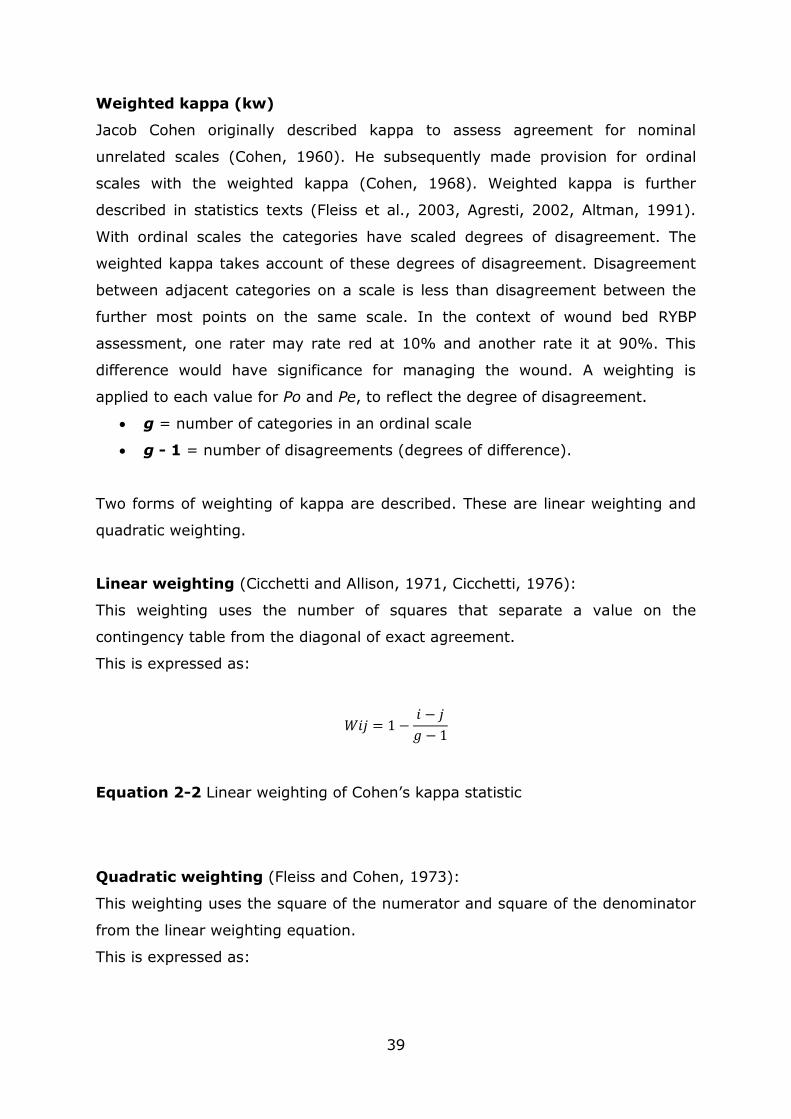
39
Weighted kappa (kw)
Jacob Cohen originally described kappa to assess agreement for nominal
unrelated scales (Cohen, 1960). He subsequently made provision for ordinal
scales with the weighted kappa (Cohen, 1968). Weighted kappa is further
described in statistics texts (Fleiss et al., 2003, Agresti, 2002, Altman, 1991).
With ordinal scales the categories have scaled degrees of disagreement. The
weighted kappa takes account of these degrees of disagreement. Disagreement
between adjacent categories on a scale is less than disagreement between the
further most points on the same scale. In the context of wound bed RYBP
assessment, one rater may rate red at 10% and another rate it at 90%. This
difference would have significance for managing the wound. A weighting is
applied to each value for Po and Pe, to reflect the degree of disagreement.
g = number of categories in an ordinal scale
g - 1 = number of disagreements (degrees of difference).
Two forms of weighting of kappa are described. These are linear weighting and
quadratic weighting.
Linear weighting (Cicchetti and Allison, 1971, Cicchetti, 1976):
This weighting uses the number of squares that separate a value on the
contingency table from the diagonal of exact agreement.
This is expressed as:
Equation 2-2 Linear weighting of Cohen’s kappa statistic
Quadratic weighting (Fleiss and Cohen, 1973):
This weighting uses the square of the numerator and square of the denominator
from the linear weighting equation.
This is expressed as:

40
Equation 2-3 Quadratic weighting of Cohen’s kappa statistic
Cohen’s weighted kappa (kw) is expressed as:
Equation 2-4 Weighted Cohen’s kappa statistic
When all categories other than absolute agreement are rated 0, then kw=k.
Weighting kappa does not always improve measurement of agreement (Cohen,
1968).
Weighted kappa - Comparing a judge with the Medical Reference
Standard
Weighted kappa may be calculated for an individual judge against the Medical
Reference Standard (Light, 1971b).
However, a different statistic (G) is required to measure agreement between a
group of judges collectively and a Medical Reference Standard. This is because
the marginal values for the Medical Reference Standard in the contingency table
will remain the same. This is not allowed for in the weighted kappa statistic.
These marginal values are used to calculate the expected chance agreement
(Light, 1971b, Fleiss et al., 2003)
Weighted kappa in wound bed assessment studies
The weighted kappa has been used in wound bed tissue colour classification
studies (Oduncu et al., 2004, Belem, 2004, Wannous et al., 2011). In addition,
these studies used a medical reference standard.
41
Multi-rater kappa (group kappa)
Multi-rater kappa has been described (Fleiss et al., 2003, Agresti, 2002, Landis
and Koch, 1977, Light, 1971b).
This is used when more than 2 raters (m) are judging n subjects in g categories.
Not all raters are required to rate all subjects in all categories.
Weighted kappa in IBM SPSS statistical software.
SPSS output produces a contingency table and an unweighted kappa value.
Linear and quadratic kappa is obtained by using the unweighted contingency
table output matrix as a new input matrix in SPSS. Then SPSS syntax MATRIX–
END MATRIX is used to generate the linear and quadratic weighted kappa value
(IBM)
Interpretation of kappa, weighted kappa and multi-rater kappa
The values for k are defined thus:
K = 0 - chance agreement
K = +1.00 - perfect agreement
K <0 - less than chance agreement
Interpretation of Kappa, Weighted Kappa and Multi-rater Kappa have been
defined (Landis and Koch, 1977, Altman, 1991)
K = 0 – 0.4 – fair agreement
K = >0.4 – 0.6 – moderate agreement
K = >0.6 – 0.8 – good agreement
K = >0.8 – 1 – very good agreement
Limitations with kappa agreement
Raters are required to be independent and equally experienced (Cohen,
1960, Fleiss et al., 2003). However, inter-rater agreement between a
single rater and a medical reference standard is allowed (Light, 1971a)
The information contained in a contingency table is reduced to a single
value. This can result in significant reduction in information (Agresti,
2002). The complete contingency table provides more information on
agreement.

42
Interpretation scale for kappa is open to question. The scale is arbitrary.
(Landis and Koch, 1977)
The value of kappa is dependent on marginal values that are used to
calculate chance agreement. The distribution of marginal values may
significantly alter the value of kappa.
Null hypothesis and confidence intervals are not so relevant when
measuring kappa.
Kappa range is from -1 to +1. Values <0 indicate agreement less than
chance agreement and are not worth analysing further (Cohen, 1960).
Kappa values cannot be compared between studies (Altman, 1991).
SPSS does not weight kappa. However, IBM has published the syntax
MATRIX-ENDMATRIX that can be applied to a kappa contingency table to
yield a weighted kappa result.
2.6.4 Summary of statistical methods
Research is presented in chapters 3, 4 and 5 of this dissertation. Study data will
be obtained relating to wound bed RYBP assessment from wound care clinicians
(using calibrated wound images) and the Medical Reference Standard. Weighted
Kappa statistic will be used to measure inter-rater agreement. This statistical
measure is used in comparable studies. Furthermore, it is identified from
statistical texts that this is a suitable measure to use in this study.
2.7 Conclusion to literature review
Literature was critically appraised in relation to clinical assessment, digital
imaging, health informatics and quantitative statistical analysis.
Wound care involves interdisciplinary collaborative shared care. Communication
of wound status over time between medical personnel is an integral part of
clinical wound care. The accurate visual assessment of the wound is essential to
effective wound management. An understanding of the pathophysiology of
wound healing has informed clinical assessment tools. Wound bed RYBP

43
assessment and its clinical interpretation guide best practice in wound
management.
Colour is common to the visual assessment of both wounds and wound images.
The calibrated digital wound image provides permanent accurate and
reproducible wound documentation, suitable for evaluation. It can be data mined
for planimetric analysis, volumetric analysis and tissue classification. It facilitates
wound comparison over time and telemedicine. It facilitates audit of wound care
interventions and outcomes. It facilitates wound care education and research.
Automated tissue colour classification of digital images, using artificial
intelligence, has been developed.
Wound care involves shared care between the community and the hospital.
Information and communication standards are required to provide an EHR with
technical, semantic and process interoperability. The limitation of existing wound
bed terminology has been identified in the literature review. A number of wound
care specific health informatics initiatives have been identified. The openEHR
draft archetype inspection of an open wound, the wound assessment clinical
template and wound clinic information system architecture are the foundation for
an EHR, facilitating standardized wound care. The Woundontology Consortium
aims to facilitate semantic interoperability and clinical decision support.
Cohen’s kappa statistic is frequently used in wound care studies. The Medical
Reference Standard is used in the absence of a biological marker for wound bed
tissue quantities. This statistical method is identified in statistical texts as being
appropriate for use in this type of research.
Literature review has identified that:
Clinical wound care utilizes wound bed RYBP assessment.
Calibrated digital wound images can be data mined for wound bed RYBP
assessment
The UMLS Metathesaurus under-represents wound bed RYBP assessment
The OpenEHR archetype inspection of an open wound does not use
proportion data value for wound bed RYBP assessment. It does not map
colour to wound bed tissue type

44
Cohen’s kappa statistic is a suitable measure in an inter-rater wound bed
RYBP assessment study
The Medical Reference Standard provides the reference value in wound
bed RYBP assessment
The review of wound care knowledge has informed the research presented in
chapters 3, 4 and 5 of this dissertation:
Survey of wound care centres - to identify current wound bed assessment
in clinical practice
Survey of wound care clinicians - to measure suitability of calibrated
wound images for treatment recommendations
Study using calibrated wound images - to measure wound bed RYBP
assessment inter-rater agreement between fourteen Tissue Viability
Nurses (TVN) and the Medical Reference Standard
Wound care knowledge presented in this chapter along with the research will
combine to form the basis of a proposal to develop the Open EHR draft
archetype inspection of an open wound presented in chapter 6.
Chapters 3, 4 and 5 will present the research. Chapter 6 will present the
research based proposal.

45
Chapter 3 Research Design and Methodology

46
3.1 Introduction to Research Design and Methodology
Chapter 2 presented the literature review relating to clinical wound bed
assessment, calibrated wound image assessment and health informatics in
wound bed assessment.
From the literature review the aims of this research have been developed:
To explore the use calibrated images in wound bed assessment.
To present a proposal to develop the OpenEHR draft archetype inspection
of an open wound to reflect wound bed RYBP assessment.
These aims will be met through the research objectives and research questions.
This chapter describes the research design and methodology. Chapter 4 will
present the results of this research. Chapter 5 will evaluate and analyze the
research results.
The research design and methodology of wound bed assessment relate to:
Survey of wound care centres - to identify current wound bed assessment
in clinical practice
Survey of wound care clinicians - to measure suitability of calibrated
wound images for treatment recommendations
Study using calibrated wound images - to measure wound bed RYBP
assessment inter-rater agreement between fourteen Tissue Viability
Nurses (TVN) and the Medical Reference Standard
RYBP is granulation (Red), slough (Yellow,) necrosis (Black) and epithelialization
(Pink) tissue types in the wound bed.
Medical Reference Standard is an approximated true value for the four wound
bed tissue types in the 11 wounds included in the study, based on an algorithm
(Figure 3-2).
TVN is Tissue Viability Nurse

47
Research design and methodology are described in terms of:
Objective
Participants
Data collection procedures
Statistical methods

48
3.2 Survey on wound assessment clinical practice
3.2.1 Objective
Sixteen wound care centres in Ireland, were surveyed to identify how they
record wound bed assessment.
3.2.2 Participants
Paper wound assessment forms in use at the major hospitals in the Republic of Ireland were collected.
The following criteria were used when selecting the sample of 14 hospitals for
the survey:
Regional location
Number of inpatient beds (>300) Presence of a TVN
3.2.3 Data collection procedure
Clinical Nurse Specialists in Tissue Viability (TVN’s) were contacted at the
hospitals by post. They were requested to forward the wound assessment form
currently in use in their centre. At this point they were also invited to participate
in a study using calibrated wound images.
In addition, two community wound assessment forms were collected. Finally the
sample wound assessment contained within the HSE wound management
guidelines (HSE, 2009) was included.
In total, seventeen Irish paper based wound assessment forms were collected
for inclusion in the survey.
3.2.4 Statistical methods
Data input
Raw data collected from the survey was recorded in a codebook, immediately
following data collection. Data entry was checked twice in the presence of an
independent observer. The statistical software package IBM SPSS was used for
data analysis.
Subsequently, the raw data was input into SPSS input files, again being checked
twice in the presence of an independent observer.

49
SPSS input files were screened for errors in accordance with the methods
described by Pallant (Pallant, 2010).
SPSS input files
Wound bed RYBP assessment - input file
The variable (column) was the RYBP assessment (n=1). This comprised an
ordinal variable 1-5.
The cases (rows) were the woundcare centres (n=17). This comprised 14
hospital forms, 2 community centre forms and HSE Wound Guideline
sample form.
Statistical analysis
Descriptive statistics were used to analyse the data collected in the survey.
These were generated as outputs in IBM SPSS. They included:
frequency table
pie chart
Scale
A scale was developed to categorize wound bed RYBP assessment in the forms.
This scale was ordinal and contained five items:
1. No tissue type recorded
2. Tissue type recorded – without colour or percentage
3. Tissue type recorded – colour without percentage
4. Tissue type recorded – percentage without colour
5. Tissue type recorded – percentage and colour

50
3.3 Survey on suitability of calibrated wound images
3.3.1 Objective
The objective of this survey was to measure the suitability of calibrated wound
images for treatment recommendations.
3.3.2 Participants
19 wound care clinicians were asked to rate the suitability of 12 calibrated
wound images for treatment recommendations, given all relevant clinical data.
They completed wound bed RYBP assessment on the wound images prior to
rating them.
3.3.3 Data collection procedures
Data was collected on a questionnaire on suitability of calibrated wound images
for treatment recommendations using a Likert type scale Figure 3-1. This was a
five point rating scale, ranging from definitely not to definitely yes. This data
instrument was developed using study design methods (Pallant, 2010).
Data instrument
If you are supplied with all other relevant clinical details, do you believe that this
wound image is suitable for TREATMENT RECOMMENDATIONS?
Please circle one number that reflects your
opinion
1.
DEFINITELY NOT
2.
PROBABLY NOT
3.
EQUAL PROBABILITY
4.
PROBABLY
5.
DEFINITELY
Suitable for treatment
Recommendations
1
2
3
4
5
1. DEFINITELY NOT: Clinician is certain that the wound image is not
suitable for treatment recommendations.
2. PROBABLY NOT: Clinician thinks that the wound image is not suitable
for treatment recommendations, but is not 100% certain
3. EQUAL PROBABILITY: Clinician thinks that the wound image may or
may not be suitable for treatment recommendations.

51
4. PROBABLY: Clinician thinks that the wound image is suitable for
treatment recommendations, but is not 100% certain
5. DEFINITELY: Clinician is certain that the wound image is suitable for
treatment recommendations.
Figure 3-1 Question on suitability for treatment recommendations
3.3.4 Statistical methods
Statistical data input
Raw data was input into SPSS input files following the same protocol described
for the survey on wound bed assessment in clinical practice (section3.2.4). SPSS
input files were screened for errors in accordance with the methods described by
Pallant (Pallant, 2010).
Statistical input file
Calibrated wound image suitability for treatment recommendation - input file
The variables (columns) were the wound images (n=11). These were
ordinal variables 1-5
The cases (rows) were four wound care clinicians at Tallaght Hospital, 14
TVN’s and academic wound care clinician (n=19)
Statistical analysis
Descriptive statistics were used to analyse the data collected. These were
generated as outputs from the IBM SPSS analysis. They included:
pie chart
bar chart
Scale
Suitability of calibrated wound images for treatment recommendations Likert
type scale Figure 3-1 above.

52
3.4 Study of wound bed RYBP assessment using calibrated
wound images
3.4.1 Objective
The objective of this study was to measure inter-rater agreement on wound bed
RYBP assessment between fourteen TVN’s and the Medical Reference Standard,
using calibrated wound images.
The study involved the selection of patients with wounds attending the
outpatient vascular clinic at Tallaght Hospital. The wounds were assessed by four
wound care clinicians and photographic images were obtained. The images were
calibrated. The calibrated wound images were subsequently assessed by the
same four wound care clinicians, along with fourteen TVN’s and an academic
wound care clinician.
3.4.2 Participants
Patients
Patients with lower limb wounds attending Tallaght Hospital outpatient vascular
clinic over two days were selected. Nine patients (n=9) with fifteen wounds
(n=15) were included in the study. These were mainly venous, arterial, and
diabetic ulcers.
Informed patient consent was obtained for:
Participation in the study
Wound image capture
Use of the wound image in education and research.
Clinicians
There were four wound care clinicians in Tallaght Hospital, fourteen TVN’s
country-wide and one academic wound care clinician.
A vascular specialist at Tallaght Hospital agreed to conduct the study in the
outpatient vascular clinic. He and three of his wound care colleagues participated

53
in the study. Information sheets were given to the clinicians and consent for
participation in the study was obtained. They assessed both the wounds and
images of the same wounds six weeks later.
Fourteen TVN’s were contacted and agreed to participate in the study. As
outlined previously, these were selected from hospitals countrywide. These were
the same nurses that supplied the wound assessment forms. Information sheets
were provided and consent for participation in the study was obtained. They
assessed the calibrated wound images that were collected at Tallaght Hospital.
An academic wound care clinician also assessed the calibrated wound images.
Information was provided and consent for participation in the study was
obtained.
Imaging participants
Wound image capture – The medical photographer at Tallaght Hospital captured
the wound images in accordance with hospital protocol.
Wound image calibration – Dr. Yves Vander Haeghen, referred to in section
2.4.3, has developed wound image calibration software. He calibrated the wound
images in Belgium. In addition, he provided instructions relating to camera flash,
camera settings and X-rite mini colour checker chart orientation.
3.4.3 Data collection instruments
There were four data collection instruments, which are described below (see
Appendix D). The clinicians were asked to complete data instrument 1. Data
instruments 2, 3 and 4 were completed by the researcher.
1. Wound bed assessment data instrument
This comprised four colour coded boxes linking colour with tissue type
Clinicians were requested to assign percentage to each tissue / colour
type, reflecting their opinion of each wound.
This data instrument was developed from the wound assessment forms
surveyed (see section 3.2)

54
2. Exudate assessment data instrument
This comprised two tables to record exudate volume and exudate viscosity
The researcher assigned values for each of these (low, medium, high),
based on wound and dressing assessment
This data instrument was developed from the Wound Exudate Continuum
referred to in the literature review see section 2.3.9(Gray et al., 2005)
3. Infection assessment data instrument
This comprised a table to record level of infection observed in the wound
The researcher circled one of four options on an ascending scale -
o Colonised; critically colonised; local infection; spreading infection
This data instrument was developed from the Wound Infection Continuum
referred to in the literature review see section 2.3.9(Gray et al., 2005)
4. Pain assessment data instrument
This comprised four tables:
o Pain frequency - ascending scale
o Pain location – ascending scale
o Analgesia use - ascending scale
o Pain severity - verbal rating ascending scale
The researcher recorded information obtained from interviewing the
patient.
This data instrument was developed from the wound assessment forms
surveyed in section 3.2
In addition to the data instruments described above, there was an information
and instruction sheet for each patient and clinician (see Appendix C).
Information and instruction was also given to patients and clinicians verbally.
A glossary of terms was compiled for the study, containing definitions of wound
bed terms as appear in terminologies and textbooks. However, the wound bed
tissue characteristics were so familiar to the clinicians that it was decided that

55
the glossary was not required. This wound bed glossary is incorporated in the
dissertation glossary to inform the reader.
A 5 point Likert type scale for wound edge and surrounding skin assessment was
also developed. This contained 30 items. It was decided to omit this data
instrument from the study; because of the time it would take clinicians to
complete the form.
3.4.4 Data collection procedure
Data collection for this study was in three stages:
STAGE 1 Direct wound assessment and wound image capture
STAGE 2 Wound image calibration
STAGE 3 Indirect wound image assessment using calibrated wound images.
Direct wound assessment refers to the assessment of the patients wound at the
clinic.
Indirect wound image assessment refers to the assessment of the calibrated
wound image of the wound.
STAGE 1 – Direct wound assessment and wound image capture
Study design and methodology was discussed at meetings with the four wound
care clinicians and the medical photographer, prior to data collection.
Data collection took place at the outpatient vascular clinic at Tallaght Hospital,
over two days. Hospital infection control policy was adhered to during data
collection.
Day One
Patients were selected from attendees at the clinic, with lower leg wounds
suitable for imaging. Written and verbal information was given to these patients
by the researcher. All patients who were approached agreed to participate in the
study. Informed consent for the capture of the images and for participation in
the study was obtained by the researcher. Each patient was assigned a folder
and a unique identifier. In patients with more than one wound, each wound had

56
a unique identifier. Each data instrument was labeled for that patient at this
time, with the exception of the wound assessment data instrument.
The researcher recorded information regarding exudate, infection and pain. This
was obtained from patient interview and clinical assessment.
The exudate assessment was informed by discussion with the patient and
attending nurse regarding the frequency of dressing changes and strike through
on the dressings.
In the infection assessment spreading infection was based on inflammation more
than two centimetres from the wound edge.
The pain assessment was based on a Verbal Rating Scale of 1-10, indicating
the patients’ subjective rating of pain severity. In addition, pain frequency, pain
location and analgesia use were recorded.
Each of the four wound care clinicians involved in the study was given a
clipboard containing information sheet, instruction sheet and wound bed
assessment data instruments. Each clinician assessed the wounds
independently. They were given a label to apply to the patient folder when their
assessment was completed. There was no clinician identifier on the wound bed
assessment data instrument. In doing so, anonymity of their assessment was
maintained.
The unique identifier assigned to the wound was incorporated into the wound
image. This was to link the image with its associated data instruments. The
signed photography consent form was photographed immediately preceding
capture of the wound image. This facilitated additional tracking of the images
with the data sets, during the data collection process.
The medical photographer adapted to imaging in the clinic environment, rather
than in the photographic studio. The workflow in the clinic required that patients
remain onsite for removal of dressings, treatment of wounds and application of
complex dressings. Furthermore, the profile of the patients attending the clinic
was such that their mobility was impaired.
A temporary photographic studio was set up in one of the treatment rooms in
the clinic. Patients were brought to this room, where black background material
and step up were placed on the floor. It proved to be impractical for patients to

57
stand posing for image capture. Most patients were not well enough to stand on
the step up. Subsequent patients were imaged in the room where they were
receiving their treatment. The researcher assisted the photographer by holding
the X-rite mini colour checker chart in a perpendicular plane to the wound. The
mini colour checker chart was incorporated into the wound image.
The usual protocol in wound care at the clinic was adhered to. Wound dressings
were removed and wounds were cleaned prior to assessment and imaging. The
routine clinical practice of applying paraffin gel to the legs was deferred until
imaging had been completed. This was necessary to avoid problems with
reflectance in the image.
Day Two
Based on the data collection procedure experience on day one, two changes
were made on day two.
Firstly, an anatomical drawing of the human body was attached to the wound
bed assessment data instruments. This was to prevent mislabeling, particularly
in the case of multiple wounds on a single patient.
Secondly, an assistant accompanied the researcher. The assistant ensured that
each patient dataset was complete.
In every other respect the data collection procedure was the same as on day
one.
STAGE 2 - Wound image calibration
As described above, the medical photographer captured fifteen wound images at
the outpatient clinic at Tallaght Hospital. The camera was the NIKON D3X (24.5
mega pixels). The flash was the NIKON RING FLASH SPEED LIGHT SBR 200. The
lens was a NIKOR 105 mm macro lens. The camera settings were ISO 800. The
aperture was adjusted between f8- f16 to suit the wound. The camera setting
was neutral, manual focus, RAW images, RGB colour space white balance.
It was originally planned to calibrate the images using C4Real, a software
application developed by Dr. Yves Vander Haeghen for this purpose. C4Real is
available on the internet. This software application was developed for use with
the Gretag Mini Macbeth Color Checker Chart (Mini MCCC). However, the Mini

58
MCCC is no longer available, since the company Gretag merged with X-rite.
Consequently, Dr. Vander Haeghen agreed to calibrate the images in Belgium,
using his latest calibration software that is compatible with the X-rite chart. The
annoymised wound images were sent to him via Dropbox. This method of
transfer was required because of the size of the wound image files.
The calibration process as described in section 2.4.3 of the literature review was
applied to fifteen wound images. The calibration was automated. Following a
batch run nine images were successfully calibrated. A further three images were
successfully calibrated manually (see Appendix E detailed calibration report).
Three of the images were unsuitable for calibration. This was because the
calibration colour checker chart was incorrectly positioned in the image capture
process or the image was overexposed. Twelve calibrated images were returned
via Dropbox. These were suitable for inclusion in the study.
STAGE 3 – Indirect wound image assessment using calibrated wound
images
The calibrated wound image assessment data was collected from:
Fourteen TVN’s countrywide. These were the same nurses that provided
the wound assessment forms for the survey described in section 3.2
Four wound care clinicians in Tallaght Hospital. These were the same four
clinicians that performed the direct wound assessment at the time of
image capture
One academic wound care clinician.
Clinical Nurse Specialists in Tissue Viability - TVN’s (n=14)
The researcher met with the fourteen TVN’s around the country, so that they
could view and assess the calibrated wound images. Other options for viewing
the images, such as postal and electronic transfer, were considered. However,
the approach taken was superior for the purpose of this research study. It
resulted in:
Maximum participation in the study.
Integrity of the data collected. Each wound bed assessment data set was
linked to its related calibrated wound image, at the time of recording.

59
Standardized calibrated image viewing conditions. The images were all
viewed on the same Sony VAIO Laptop computer, with the same sRGB
settings, to reduce bias. The viewing room environment in the different
centres remained a variable. This was minimized by ensuring that all
viewing took place under similar lighting and reflection conditions.
Assistance with calibrated wound image viewing. The magnification facility
on the Laptop proved to be very beneficial for visualization of the wound
bed.
Immediate response to unforeseen queries presented by the TVN’s, while
doing the image assessment.
Calibrated wound image assessment was done on a one to one basis, with the
exception of one hospital where 2 TVN’s assessed the images together,
simultaneously. In this case there was no collaboration with regard to their
assessments.
The data collection process took between 20-45 minutes. The time was divided
between:
Obtaining consent
Providing information and instruction
Recording responses to the 11 calibrated wound image assessments
Recording responses to questionnaire on calibrated wound image
suitability for treatment recommendations
Recording responses to demographic questionnaire
Wound care clinicians at Tallaght Hospital (n=4)
The researcher returned to Tallaght Hospital and presented the calibrated wound
images to the four wound care clinicians, who had performed the direct wound
assessments (section3.4).
More than six weeks had elapsed since their direct wound assessments had been
performed. Despite the time interval between the assessments, the clinicians
remembered some of the patients and described their wound progress in the
interim. This introduced potential bias to their indirect wound image
assessments.

60
These four clinicians performed their indirect wound image assessments
individually. The same protocol as described for the TVN’s above was used.
Academic wound care clinician (n=1)
An academic wound care clinician viewed and assessed the calibrated wound
images. The researcher followed the same protocol as described for the TVN’s,
when collecting the data.
3.4.5 Data loss
Stage 1 - Direct wound assessment and wound image capture
One wound was assessed by three clinicians prior to image capture. There was
eschar (scab) on this wound, which separated from the wound bed before
assessment by the fourth clinician. Consequently, the fourth direct wound
assessment data set was deemed to be invalid.
One wound was assessed by four clinicians, but the wound was debrided prior to
the image capture. This image was successfully calibrated, but was deemed
unsuitable for inclusion in the wound image study.
On day one, there were two wounds that had mislabeled data sets.
Consequently, one direct wound assessment data set from each of these wounds
was rendered invalid and omitted from the study.
All data sets were inspected on the day of their completion. The data was
checked twice and recorded in a study codebook in the presence of an
independent observer.
Stage 2 - Wound image calibration
Three of the wound images were unsuitable for calibration, either automatically
or manually. This resulted from misplacing of the colour checker chart or
overexposure of the image (see Appendix E for detailed calibration report).
These three wound images were not included in the study.

61
Stage 3 - Indirect calibrated wound image assessment
Eleven calibrated wound images (n=11) were suitable for inclusion in this part of
the study. A complete set of data was obtained from wound bed assessment of
these eleven calibrated images. All data sets were inspected immediately
following their completion. The data was checked twice and recorded in a study
codebook in the presence of an independent observer.
3.4.6 Statistical methods
Data input
Raw data collected from the study was recorded in a codebook immediately
following data collection. Data entry was checked twice in the presence of an
independent observer.
Subsequently, the raw data was input into IBM SPSS input files, again being
checked twice in the presence of an independent observer.
SPSS input files were screened for errors in accordance with the methods
described by Pallant (Pallant, 2010).
Data SPSS input file
Clinician inter-rater agreement wound bed RYBP assessment - input file
The variables (columns) were the clinicians assessments (n=23). This
comprised four wound care clinicians (direct and indirect assessments),
fourteen TVN’s and one academic wound care clinician. These were
continuous scale variables 0-100%
The cases (rows) were the wound tissue types. This comprised 11 images
with 4 tissue types in each image (n=44)
Statistical analysis methods
The research study involved the quantitative analysis of data obtained from
wound bed RYBP assessment. Cohen’s weighted kappa statistic was used to
measure inter-rater agreement between TVN’s and the Medical Reference
Standard, with respect to wound bed RYBP assessment.
Kappa inter-rater agreement measurement required categorical ordinal
variables. The wound bed RYBP data was input as continuous variables

62
(percentages) in SPSS. These continuous variables were collapsed into 10
ordinal percentage categories, using visual binning in SPSS (Pallant, 2010).
There were no absolute medical reference values for the wound bed RYBP
assessments. The Medical Reference Standard was obtained from the wound
care clinicians at Tallaght Hospital, who assessed both the wounds and the
wound images.
Figure 3-2 Algorithm for Medical Reference Standard
The mean value from both of their methods of assessments was assigned as the
Medical Reference Standard. The academic wound care clinician assessment was
used to adjudicate in the event of divergent assessments from the four wound
care clinicians. Wound infection continuum data, wound exudate continuum data
Does mean direct assessment and mean indirect assessment result
in the same wound management?
(Debride or Protect) YES / NO
YES
Use the mean of direct and
indirect assessments as
the Medical Reference
Standard
NO
Does mean direct assessment and academic
clinician assessment result in the same
management?
(Debride or Protect) YES/ NO
NO
Does mean indirect assessment and
academic clinician result in the same
management?
(Debride or Protect) YES / NO
YES
Use mean of indirect assessment and academic clinician
assessment as the Medical Reference Standard
YES
Use mean of direct
assessment and academic
clinician as the Medical
Reference Standard

63
and pain assessment data were also used in the adjudication process. Divergent
assessment meant that the mean values for each method of assessment would
result in a significant change in wound management (Figure 3-2).
Adjudication was required for three of the wounds. The method of assessment
by the four wound care clinicians that was in disagreement with the academic
wound care clinician and the related clinical data was not accepted. For these
three wounds, the Medical Reference Standard was determined from the more
consistent method of assessment by the four wound care clinicians and academic
wound care clinician.
Inter-rater agreement was analyzed using weighted Kappa statistic. Four tissue
types for eleven wounds were analyzed together.
A two-way contingency table was prepared, comprising the Medical Reference
Standard in the rows and each TVN in the columns. Firstly unweighted kappa
was calculated in IBM SPSS. Then the contingency table matrix output in IBM
SPSS was weighted using the MATRIX-ENDMATRIX syntax in the syntax editor
(IBM). Linear and quadratic weighting was applied to the categorical ordinal
variable scales, representing degrees of difference (Fleiss and Cohen, 1973),
(Cicchetti, 1976) see section 2.6.3 and Table 3-1.

64
Scale
Wound bed RYBP assessment ordinal scale.
This scale had 10 categorical variables between 0% and 100%
TVN
ꜜ
Medical
Reference
Standard
ꜜ
0-
9%
10-
19%
20-
29%
30-
39%
40-
49%
50-
59%
60-
69%
70-
79%
80-
89%
90-
100%
0-9% 1 0
10-19% 1
20-29% 1
30-39% 1
40-49% 1
50-59% 1
60-69% 1
70-79% 1
80-89% 1
90-100% 0 1
Table 3-1 Wound bed RYBP assessment contingency table with weighting
1. Red cells represent the diagonal of exact agreement and its weighting is 1
2. Blue cells represent weighted distance from the diagonal of exact
agreement (different shades represent degrees of difference)
3. Black cells represent maximum distance from the diagonal of exact
agreement. This is disagreement and its weighting is 0

65
Interpretation of weighted kappa result (see section 2.6.3)
Value of K Strength of agreement
<0.20 Poor
0.21-0.40 Fair
0.41-0.60 Moderate
0.61-0.80 Good
0.81-1.00 Very good
Table 3-2 Interpretation of Kappa
3.5 Ethical considerations
Tallaght and St. James’s Hospitals’ research ethics committee use a common
application form for academic and investigator led research studies that do not
involve medicinal products, covered by the statutory instrument 190. This
conforms to the World Medical Association Declaration of Helsinki
2008(Association, 2008). The participation of patients and inclusion of patient
data in this research study required ethical approval. Ethical approval was
obtained from this joint hospitals ethics committee. In addition ethical approval
was obtained from Trinity College Dublin ethics committee. This study conformed
to the conditions of the ethical approvals obtained.
The Data Protection Acts (Ireland, 1988 , Ireland, 2003) provide the legislative
basis for the protection of personal data in Ireland. The Data Commissioner
Guidelines on Research in the Health Sector provide additional information on
the protection of patient data (Commissioner, 2007).
The nature of this study involved sensitive patient data in the form of images.
The Data Commissioner was contacted for specific advice (see Appendix I).
Publication level patient consent was obtained from patient participants:
Consent to capture the image
Consent for use of the image in education and research
Consent to publish the image

66
The study did not involve access to patient healthcare records. The clinical
assessments needed to be linked to the wound image. The clinical assessments
and images were pseudonymised, using a code. This assigned the assessments
and images a code which did not identify the patient. The code linking the
images to the clinical data was held by the researcher. The electronically stored
images were irrevocably anonymised. The images were not linked to an
individual and could not be considered personal data. Consequently the images
were outside the remit of data protection requirements.
The assessment forms obtained for the survey remain the property of the
hospitals and centres from where they originated. The purpose of the survey was
to identify how wound bed is assessed. There was no intention to publish the
forms and consent to do so was not sought. (See Appendix A for sample form).
No patient information was recorded in these forms. Hence there were no ethical
issues in this regard.
3.6 Conclusion to Research Design and Methodology
This chapter presented research design and methodology relating to wound bed
assessment:
Survey of wound care centres to identify wound bed assessment clinical
practice
Survey of wound care clinicians on suitability of calibrated wound images
for treatment recommendations.
Study measuring inter-rater agreement between TVN’s completing wound
bed RYBP assessment using calibrated wound images and the Medical
Reference Standard.
The research was described in terms of participants, data collection procedure
and statistical methods
The statistical methods used were:
Descriptive statistics for survey of wound bed assessment clinical practice
and suitability of calibrated image for treatment recommendations

67
Cohen’s weighted kappa statistic to measure inter rater agreement
between the TVN’s and the Medical Reference Standard
The ethical considerations, including ethical approval and data protection
requirements, were discussed.
Research and analysis are presented in chapter 4 and chapter 5, respectively.

68
Chapter 4 Results

69
4.1 Introduction to Results
Chapter 3 described the methodology and implementation of the research
relating to wound bed assessment. This chapter will now present the results of
this research.
1. Survey results
Current wound bed assessment clinical practice
Suitability of calibrated wound images for wound bed assessment
and treatment recommendations
2. Study results
Wound care clinician agreement on wound bed assessment, using
calibrated wound images
The research results are presented in terms of:
Motivation
Objective
Methodology
Results
Analysis

70
4.2 Wound bed assessment clinical practice
Motivation
This research aims to present a rationale for developing the openEHR wound
archetype. A fundamental reason for doing so is to reflect current clinical
practice. It is in this regard, that a survey is undertaken to investigate current
clinical practice.
Objective
To measure wound bed assessment clinical practice
Question
What is wound bed assessment clinical practice in the survey sample?
Methodology
A sample of wound assessment forms from 14 major hospitals, 2 community
based centres and HSE wound care guidelines is selected.
Recording of wound bed tissue type with associated colour and percentage is
measured.
Descriptive statistic is used to analyze the data.
Results
Of the 17 forms surveyed:
One recorded tissue type without reference to colour or quantity.
Two forms recorded tissue type and colour e.g. Necrosis (black)
Three forms recorded tissue type and quantity e.g. Necrosis (%)
Eleven forms recorded tissue type with colour and percentage e.g.
Necrosis (black / %)

71
Table 4-1 presents wound bed assessment clinical practice in the sample
surveyed. The data is displayed in an ascending hierarchy.
Wound bed assessment documentation
Frequency
%
1 Tissue type not recorded
2 Tissue type recorded – without colour or percentage
0
1
0
5.9
3 Tissue type recorded - colour without percentage
2
11.8
4 Tissue type recorded - percentage without colour
3
17.6
5 Tissue type recorded - percentage and colour
11
64.7
Total (n=17)
17
100.0
Table 4-1 Wound bed assessment clinical practice

72
Figure 4-1 illustrates the distribution of wound bed assessment clinical practice.
Tissue classification referred to in the Pie chart is wound bed RYBP assessment.
Figure 4-1 Wound bed assessment clinical practice - Pie chart
Analysis
This survey demonstrates that all participating centres record wound bed tissue
type. In 65% of centres, colour and percentage are also included in their wound
assessment forms. Consequently, necrosis (Black), slough (Yellow), granulation
(Red) and epithelialization (Pink) are important components of wound bed
assessment. These are referred to as wound bed RYBP assessment.
Furthermore, wound bed RYBP assessment needs to be represented in the
OpenEHR draft wound archetype and related terminology.
73
4.3 Wound bed RYBP assessment and treatment
recommendations using calibrated wound images
Motivation
This research aims to develop the openEHR draft wound archetype and to
introduce calibrated wound images to wound care in Ireland.
The survey on clinical practice has identified that wound bed RYBP assessment is
prevalent (section 4.2). Literature review has identified that wound images are
used in telemedicine. Furthermore, automated wound bed tissue classification,
using calibrated wound images, has been developed. Hence, wound bed RYBP
assessment can be obtained either by clinicians or automatically, using
calibrated wound images. The OpenEHR draft archetype inspection of an open
wound needs to represent the calibrated wound image wound bed RYBP
assessment to facilitate telemedicine, connected health and clinical decision
support in a wound care EHR.
It is in this context that this research into wound care clinicians’ opinions
regarding the suitability of calibrated wound images for treatment
recommendations has been undertaken.
Objective
Measurement of suitability of calibrated wound images for treatment
recommendation.
Questions
Do wound care clinicians believe that calibrated wound images are
suitable for treatment recommendations?
Methodology
19 wound care clinicians were asked to score the suitability of each
calibrated wound image, using a five point Likert type scale (Figure 3-1).
These clinicians were asked to perform wound bed RYBP assessment
before being asked about image suitability. Thus, they had given
consideration to the content of the image prior to making their decision.

74
Results
Of the 19 wound care clinicians surveyed:
The mode (most frequent) response was that 41% of the calibrated
wound images were probably suitable for treatment recommendations, if
the clinician was given all other relevant clinical information.
39% of clinicians believed that the calibrated wound images were
definitely suitable for treatment recommendations
Figure 4-2 Calibrated image suitability – bar chart
Likert type scale applied to 12 wounds by 19 wound care clinicians
The x-axis represents the 12 wounds (case numbers).
The y-axis represents the frequency of responses by the 19 wound care
clinicians (values).
The colour coded bars represent the Likert scale for each wound ranging
from blue (definitely not suitable) to yellow (definitely suitable)

75
1 Definitely not 3% 2 Probably not
8%
3 Equal probability 9%
4 Probably 41%
5 Definitely 39%
Image suitability for treatment recommendations
Figure 4-3 Calibrated image suitability – Pie chart
The cumulative image suitability (12 wounds together), by the 19 clinicians is
represented in the pie chart. The same chart colour coding is used to represent
the Likert scale ranging from blue (definitely not suitable) to yellow (definitely
suitable).
Analysis
The wound images used in this study have been calibrated using a software
application that is newly developed. Consequently, these calibrated wound
images have never before been used for assessment by Irish wound care
clinicians. This survey indicates that this important group of clinicians are
positively disposed to using these images. They have the knowledge, skills and
experience to decide on the quality of a calibrated wound image in order to
make a treatment recommendation and deliver telemedicine in wound care.

76
This survey on calibrated wound images has identified that wound care clinicians
believe they are probably or definitely suitable for treatment recommendations,
if the clinician was given all other relevant clinical information.
The survey on clinical practice has identified that wound bed RYBP assessment is
prevalent (see section 4.2). The question then arises:
What is the inter-rater agreement between wound care clinicians, when
completing wound bed RYBP assessment, using calibrated wound images? This is
the focus of the research study presented in the next section (see section 4.4).

77
4.4 Inter-rater agreement on wound bed RYBP assessment
using calibrated wound images
Motivation
This research aims to develop the openEHR draft archetype inspection of an
open wound and to introduce calibrated wound images to wound care in Ireland.
The surveys and literature review have identified that:
Wound bed RYBP assessment is integral to clinical practice (section 4.2).
The majority of clinicians who were surveyed are of the opinion that
calibrated wound images are either probably or definitely suitable for
treatment recommendations (section 4.3).
Wound bed RYBP is underrepresented in the openEHR draft archetype
inspection of an open wound and related terminology.
Wound images are used in telemedicine. Calibrated wound images have
been automatically classified, using SVM tissue classifiers (Oduncu et al.,
2004, Belem, 2004, Wannous et al., 2011).
Thus, it is important to see the level of agreement between clinicians and the
Medical Reference Standard on wound bed RYBP assessment, when using
calibrated wound images.
Objective
Measurement of inter-rater agreement between TVN’s and the Medical Reference
Standard, using calibrated wound images for wound bed RYBP assessment
Question
What is the inter-rater agreement between TVN’s and the Medical Reference
Standard, using calibrated wound images for wound bed RYBP assessment?
Methodology
Wound bed RYBP Medical Reference Standard has been obtained for the 11
wounds that are included in this study. There are four tissue types in each
wound. Consequently, the Medical Reference Standard for the 11 wounds
comprises 44 measurements. There are no absolute true values for these wound
bed RYBP assessments. This is a common difficulty encountered in medical

78
studies (see section 2.6.2). An algorithm has been developed to establish the
Medical Reference Standard (see Figure 3-2). This has been calculated from the
mean of direct and indirect wound bed RYBP assessment by four wound care
clinicians at Tallaght Hospital. The additional assessment of an academic wound
clinician and ancillary clinical information has been utilized for three of the
wounds, when establishing the Medical Reference Standard.
Cohen’s weighted kappa statistic is used to measure inter-rater agreement in
this study, because of its use in similar wound studies (Oduncu et al., 2004,
Belem, 2004). It is well described in statistic texts (Altman, 1991, Fleiss et al.,
2003, Agresti, 2002) as being suitable for assessing inter-rater agreement. The
continuous percentage scale for wound bed RYBP assessment has been
transformed into ten categories between 0% and 100%, using SPSS. Degrees of
difference from the Medical Reference Standard are taken into account. These
are represented by degrees of difference from the diagonal of exact agreement.
These degrees of difference are used to calculate the weighting.
A multi-rater kappa value has not been calculated because The Medical
Reference Standard has been used. Hence, a range of kappa values is reported,
rather than a single multi-rater or group kappa. This approach is described in
the literature (Light, 1971a).
The kappa value is interpreted according to the scale of Landis and Koch (see
section 2.6.3).
Results
Quadratic weighted kappa ranged from moderately good (0.56) to good (0.80).
The lowest and highest agreements are highlighted (Table 4-2).

79
Tissue Viability
Nurses (TVN’s)
Unweighted
Kappa
(a)
Linear weighted
Kappa
(b)
Quadratic
weighted Kappa
(C)
TVN A 0.20 0.54 0.71
TVN B 0.29 0.55 0.72
TVN C 0.24 0.52 0.69
TVN D 0.37 0.65 0.80
TVN E 0.22 0.53 0.70
TVN F 0.11 0.39 0.56
TVN G 0.19 0.39 0.58
TVN H 0.30 0.53 0.70
TVN I 0.23 0.45 0.58
TVN J 0.17 0.43 0.50
TVN K 0.22 0.51 0.72
TVN L 0.09 0.44 0.60
TVN M 0.29 0.55 0.73
TVN N 0.42 0.64 0.80
Table 4-2 Kappa inter-rater agreement – wound bed RYBP assessment
Table 4-2 reports unweighted kappa, linear weighted kappa and quadratic
weighted kappa for the 14 TVN’s when rated with the Medical Reference
Standard. 4 tissue types in 11 wounds are compared in 14 different contingency
tables.
a. Unweighted kappa – This measurement only takes account of values on
the diagonal of exact agreement between the TVN’s and the Medical
Reference Standard in the contingency table. Values that are not in
diagonal of exact agreement are given a zero value.
b. Linear weighted kappa – This measurement takes account of the number
of squares from the diagonal of exact agreement in the contingency table
(Cicchetti, 1976).

80
c. Quadratic weighted kappa – This applies a quadratic weighting to the
degrees of difference from the diagonal of exact agreement in the
contingency table (Fleiss and Cohen, 1973).
Analysis
The quadratic weighted kappa represents agreement between individual TVN’s
and the Medical Reference Standard, when chance agreement has been
removed. The inter-rater agreement between a TVN and the Medical Reference
Standard for 4 tissue types in 11 wounds is presented on a contingency table.
The matrix of values is condensed to a single kappa measurement.
This study indicates moderate to good agreement between this cohort of TVN’s
and the Medical Reference Standard, when performing wound bed RYBP
assessment in 11 calibrated wound images.
The data shows differences in the recording of red (granulation) and pink
(epithelialization) by clinicians. Some clinicians described wounds as
predominantly red, while others described the same wounds as predominantly
pink. This may reflect difficulty distinguishing between these two colours in
calibrated wound images. Alternatively, it may reflect differences in definition of
the area covered in the wound bed applied by these clinicians. It is interesting to
note that the same difference in recording red and pink in the wound bed
occurred with direct wound assessment. Thus indicating that this is not an issue
related to the images but relates to clinical interpretation. These colours are
adjacent on the continuum of wound healing. Furthermore, management of red
and pink wounds is the same, in that they are both protected.
This study indicates that wound bed RYBP assessment using calibrated wound
images shows moderate to good agreement when compared to the Medical
Reference Standard, measured with quadratic weighted kappa. Consequently,
the OpenEHR draft archetype inspection of an open wound should be developed
to represent wound bed RYBP assessment and facilitate telemedicine, connected
health and clinical decision support in a wound care EHR.
The strength of inter-rater agreement with the Medical Reference Standard
indicates that the method of using calibrated wound images for wound bed RYBP

81
assessment is reliable (Light, 1971a). The use of calibrated wound images in
wound care in Ireland would also contribute to telemedicine, connected health
and clinical decision support.
4.5 Conclusion to Results
This chapter presents the results related to research on wound bed RYBP
assessment.
The survey on wound care clinical practice indicates that 65% of wound
assessment forms record tissue type with percentage and colour.
In the survey on the suitability of calibrated wound images for treatment
recommendations, 41% of clinicians stated that the calibrated wound images
were suitable for treatment recommendations. A further 39% of clinicians stated
that they were definitely suitability for treatment recommendations.
The study on wound bed RYBP assessment inter-rater agreement between TVN’s
and the Medical Reference Standard indicates moderate to good agreement,
using calibrated wound images.
The OpenEHR draft archetype inspection of an open wound needs to represent
wound bed RYBP assessment and facilitate telemedicine, connected health and
clinical decision support in a wound care EHR.
Calibrated wound image assessment will also contribute to these innovations in
wound care.
Chapter 5 will present an analysis and evaluation of the research in the context
of current knowledge

82
Chapter 5 Evaluation and Analysis

83
5.1 Introduction to Evaluation and Analysis
This dissertation has two aims. These are to explore calibrated wound image
assessment in wound care and to present a proposal to OpenEHR to develop the
draft archetype inspection of an open wound.
The design, methodology and results of research into wound bed assessment
and calibrated wound image assessment have been presented in Chapter 3 and
Chapter 4, respectively.
This chapter presents an evaluation and analysis of the research. The evaluation
will form the rationale of a research based proposal to develop the OpenEHR
draft archetype inspection of an open wound. The details of this proposal will be
presented in chapter 6.
The research results are evaluated and analysed, in the context of current
knowledge. These are described in terms of:
Survey of wound bed RYBP assessment in clinical practice, OpenEHR draft
archetype and related terminology
Survey on the suitability of calibrated wound images for treatment
recommendations
Study of wound bed RYBP assessment, using calibrated wound images

84
5.2 Wound bed assessment: clinical practice, existing
OpenEHR draft archetype and related terminology
Survey of wound bed assessment in clinical practice
A representative sample of wound care centres has been surveyed to identify
current wound bed assessment in clinical practice. This survey demonstrates
that all participating centres record wound bed tissue type. In 65% of centres
colour and percentage are also included in their wound assessment forms.
Wound bed RYBP assessment has been used in clinical practice since the 1980’s
(Cuzzell, 1988, Krasner, 1995). Other studies have validated its use (Lorentzen
et al., 1999, Vermeulen et al., 2007) as an assessment tool. The wound bed
RYBP assessment maps four colours to four tissue types. This is a simplification
in so far as red can represent inflammation as well as granulation. Furthermore,
slough may be yellow or green. However, the simplicity and is of use of this
assessment is also its strength. When used in conjunction with other clinical
information it identifies where the wound is on the continuum of healing and
guides appropriate management.
The openEHR draft archetype – inspection of an open wound
The existing openEHR draft archetype inspection of an open wound is contained
in the archetype repository on the Clinical Knowledge Manager (CKM) online web
application.
The archetype represents the four wound bed RYBP tissue types identified in the
survey as being used to clinical practice. These are necrosis, slough, granulation
and epithelialization.
Granulation data value is Text. Free text does not convey the
progress of the wound on a continuum of healing in a quantifiable
way
Necrosis and slough data values are expressed on a graded ordinal
scale 1-4. This scale, while attempting to quantify pathology within
the wound bed, lacks internal consistency. The scale is arbitrary.
Epithelialization data value is Boolean. This does not convey enough
information to identify healing progress
Choosing the most appropriate data type maximizes precision in recording and
retrieval of wound bed assessment. In this case assigning proportion to wound

85
bed tissue will achieve this result. Furthermore, it is required to support
comparisons in wound assessment.
There is no mapping of colour to these tissue types within the archetype. Thus,
the OpenEHR draft archetype inspection of an open wound on the CKM provides
no means for recording wound bed RYBP assessment.
Wound bed assessment in terminology systems
The National Library of Medicine UMLS Metathesaurus incorporates 137
vocabularies, including SNOMED CT. The American Nursing Association
terminologies are also included. Nursing terminologies focus on intervention and
diagnosis. They do not have the atomic level of detail required to cover wound
bed assessment (Henry and Mead, 1997, Dykes et al., 2009).
The UMLS contains definitions for two of the wound bed tissue types contained in
the wound bed RYBP assessment. These are granulation and necrosis. Slough
and epithelialization require further development.
Binding from the OpenEHR archetype to terminology at the development stage
facilitates coding of clinical assessment. This standardised clinical content
enables semantic interoperability. The four wound bed tissue terms need to be
defined in the UMLS and nursing terminologies. Definitions and further
expansion are required for precision in recording and retrieval of wound care
assessment. Furthermore, wound care is a multidisciplinary clinical domain.
Consequently, wound bed RYBP assessment needs to be accessible in
terminologies used by all participating clinicians.
Colour is central to wound bed RYBP assessment. Colour is a non-clinical
concept. Thus, it presents a particular challenge for the terminologist (Hardiker
et al., 2002). In the survey of clinical practice it has been identified that
clinicians map colour to tissue type. This same process of mapping the wound
bed colour to its related tissue type, in formal terminology systems, is required
to properly represent wound bed RYBP assessment electronically.
Binding of the OpenEHR draft archetype inspection of an open wound to
terminology that represents wound bed RYBP assessment is required to develop

86
a wound care EHR, with semantic interoperability and standardisation. This will
enable telemedicine, connected health and clinical decision support.
5.3 Suitability of calibrated wound images for treatment
recommendations
All wound care clinicians who participated in this research were asked to
comment on the suitability of the calibrated wound images for treatment
recommendations. These clinicians were asked to complete wound bed RYBP
assessment before commenting on image suitability. Thus, they had given
consideration to the content of the image prior to making their decision. They
were asked to score the suitability of each calibrated wound image, using a five
point Likert type scale (Figure 3-1). The most frequent response (41%) was that
the calibrated wound images were probably suitable for treatment
recommendations. A further 39% of clinicians believed that they were definitely
suitable.
Calibrated wound images offer accuracy and reproducibility (Haeghen and
Naeyaert, 2006, Van Poucke et al., 2010a). Standardisation of these images is
not primarily to improve image quality, but to facilitate evaluation of the healing
wound over time. A longitudinal study with the same clinicians would yield more
information on the value of calibrated wound images.
The wound care clinicians who participated in this research are in prime position
to utilise calibrated wound images in their clinical practice and to deliver
telemedicine in wound care in Ireland. Indeed, they are currently providing
clinical decision support and education to non-expert clinicians. The positive
response obtained in this calibrated wound image survey reinforces the need to
include these images and related wound image data in a wound care EHR.

87
5.4 Study of wound bed RYBP assessment using calibrated
wound images
The survey on wound care clinical practice has identified that the majority of
clinicians use wound bed RYBP assessment. Furthermore, the majority believe
that the same assessment can be applied to calibrated wound images.
The study compares the wound bed RYBP assessments of these clinicians, using
calibrated wound images, with the Medical Reference Standard. Measurement of
inter-rater agreement with the Medical Reference Standard using Cohen’s
weighted kappa has been discussed in the literature review (section 2.6.2) and
when presenting study results (section 4.4). This study indicates that these
clinicians have moderate to good agreement with the Medical Reference
Standard when completing wound bed RYBP assessment on calibrated wound
images.
Limitations of this study are the small sample size and range of wounds
represented. Originally 15 wound images were captured for the study, but data
loss occurred as described in section 3.4.5.
Differences recording red (granulation) and pink (epithelialization) in the wound
bed have been commented on (section 4.4). This may relate to difficulty with
distinguishing these two colours in calibrated wound images. Alternatively, it
may reflect differences in interpretation of the area covered within the wound
bed. The same difference in recording red and pink in the wound bed occurred
with direct wound assessment. Planimetric studies have identified differences in
recording the wound border by clinicians (Van Poucke et al., 2010b, Jones and
Plassmann, 2000). Further study is required to establish if clinicians accept pink
epithelialization tissue as being a component of the wound bed or new
surrounding skin.
The wound care clinicians who participated in this research are poised to use
calibrated wound images in their clinical practice and record their findings in a
wound care EHR. All clinicians who were approached participated in this
research. This is encouraging because user engagement is a key component to
the adoption of health informatics initiatives.

88
The literature review has identified that calibrated wound images are suitable for
clinical assessment (Vander Haeghen and Naeyaert, 2006, Van Poucke et al.,
2010a). Automated wound bed tissue colour classification in calibrated wound
images using support vector machines (SVM) has been described (Wannous et
al., 2011) (Oduncu et al., 2004) (Belem, 2004). Thus, the evidence from this
research and related literature indicates that wound bed RYBP assessment,
needs to be represented in the OpenEHR draft archetype inspection of an open
wound and related terminology. This is the case irrespective of whether the
assessment is performed directly or indirectly, clinically or automatically. The
development of the OpenEHR draft archetype inspection of an open wound to
represent wound bed RYBP assessment will provide a framework for
telemedicine, connected health and clinical decision support in a wound care
EHR. Furthermore, calibrated wound images in their own right will contribute to
the advancement of wound care.
5.5 Conclusion to analysis and evaluation
The research results have been evaluated and analyzed in the context of current
knowledge as presented in the literature review.
Wound bed RYBP assessment is integral to clinical practice for wound
management. It is accessible in calibrated wound images, either by clinicians or
automated tissue classifiers. It is not represented in the existing OpenEHR draft
archetype inspection of an open wound and related terminology.
The research results, along with existing knowledge in wound care, present a
justification for developing the OpenEHR draft archetype inspection of an open
wound. This will facilitate telemedicine, connected health and clinical decision
support in a wound care EHR.
Calibrated wound images will also contribute to the advancement of wound care.
Chapter 6 will outline a research based proposal presented to the OpenEHR
Foundation. This is a proposal to develop the OpenEHR draft archetype
inspection of an open wound in order to represent wound bed RYBP assessment.

89
Chapter 6 Proposal to develop the OpenEHR draft
archetype inspection of an open wound

90
WOUND CARE KNOWLEDGE
SURVEY
Archetype Development
Process
(ADP)
STUDY
Necrosis
%
BLACK
Slough
%
YELLOW
Granulation
%
RED
Epithelialization
%
PINK
6.1 Introduction to the proposal
Chapter 5 has evaluated and analyzed research results on wound bed
assessment in the context of current knowledge. This evaluation and analysis
formed the basis of a research based proposal to develop the openEHR draft
archetype inspection of an open wound in the archetype repository on the
Clinical Knowledge Manager (CKM) online web application (Figure 6-1).
Based on the research findings in this dissertation and current knowledge it was
proposed to incorporate wound bed RYBP assessment in the draft archetype.
The changes proposed were:
To quantify the four wound bed tissue types, using proportion in the draft
archetype
To map colour with the four wound bed tissue types in the draft archetype
Figure 6-1 Archetype development

91
This chapter describes the proposal:
1. The justification for and benefits of developing the archetype are restated
2. Existing OpenEHR draft archetype inspection of an open wound is
described
3. Outline of proposal
4. Submission of proposal to OpenEHR is described
5. Response from OpenEHR is documented

92
6.2 Justification for developing the openEHR draft archetype
inspection of an open wound
The proposal to develop the openEHR draft archetype inspection of an open
wound has arisen from:
The survey of clinical practice in wound bed assessment
o In the sample of wound care centres surveyed, it was identified that
the majority of wound care clinicians complete wound bed RYBP
assessment.
The survey on suitability of calibrated wound bed images for treatment
recommendations
o In the survey on suitability of wound images, it was identified that
the majority of wound care clinicians are of the opinion that
calibrated wound images are probably or definitely suitable for
treatment recommendations
The study on wound bed RYBP assessment, using calibrated wound
images
o The study indicated moderate to good agreement between TVN’s
and the Medical Reference Standard, when completing wound bed
RYBP assessment using calibrated wound images.
Literature review on wound care clinical practice
o Wound bed assessment is used to evaluate the progress of wounds
on the continuum of healing and informs wound management.
Literature review on the use of wound images in wound care
o Wound images are used for clinical assessment, education and
research. Automated tissue classification using calibrated wound
images has been developed.
Literature review on the existing representation of wound bed assessment
in OpenEHR draft archetype inspection of an open wound and related
terminology
o The openEHR draft archetype inspection of an open wound, as
presented in the archetype repository on the Clinical Knowledge
Manager (CKM), represents the four wound bed tissue types. The
values are not proportion. Colour is not mapped to these tissue
types. Two of the tissue types are defined in UMLS Metathesaurus.

93
Benefits of developing the openEHR draft archetype inspection of an
open wound
Matching wound bed data values with the most appropriate data type is required
to:
Plot the wound on the continuum of healing
Communicate wound information between clinicians over time – continuity
of care.
Monitor response of the wound to therapeutic interventions – sequential
wound assessments.
Research advanced therapeutic interventions - use of EHR for data
extraction
Facilitate automated tissue classification of wounds.
Facilitate clinical decision support (e g. Applied Wound Management and
ConvaTec Solutions)
Conform to the standards of best clinical practice
Medico legal protection.

94
6.3 Existing OpenEHR draft archetype inspection of an open
wound
The openEHR draft archetype inspection of an open wound, in the archetype
repository on the CKM, is presented below.
Necrosis, slough, granulation and epithelialization are represented in the
archetype. However, these four tissue types are not mapped to colour and the
associated data values are not proportion.
Figure 6-2 Existing OpenEHR cluster archetype inspection of an open wound – mindmap

95
Necrosis
(ordinal)
Slough
(ordinal)
Granulation
(text)
Epithelialization
(boolean)
The existing values for the four wound bed tissue types are (Figure 6-3):
Granulation is TEXT
Necrosis is ORDINAL – graded 0 – 3
Slough is ORDINAL – graded 0 – 3
Epithelialization is BOOLEAN – present or absent
Figure 6-3 Existing OpenEHR draft archetype inspection of an open wound

96
Necrosis
(%)
Slough
(%)
Granulation
(%)
Epithelialization
(%)
6.4 Proposal to develop the OpenEHR draft archetype
inspection of an open wound
1. Assign proportion data values to the four wound bed tissue types in
openEHR draft archetype inspection of an open wound
It was proposed to assign proportion / percentage data values to the four wound
bed tissue types, within the draft archetype, on a scale 0% 100% (Figure
6-4).
Figure 6-4 Change data value to proportion

97
Necrosis
(%)
Black
Slough
(%)
Yellow
Granulation
(%)
Red
Epithelialization
(%)
Pink
2. Map of RYBP colour to the four wound bed tissue types in the
openEHR draft archetype inspection of an open wound
It was proposed to map the four colours in RYBP to the four wound bed tissue
types, within the draft archetype (Figure 6-5)
Figure 6-5 Map colour to wound bed tissue type

98
6.4.1 Submitting the proposals to change the archetype
The CKM is used to manage developments in the archetype. The archetype is
downloaded using CKM online web application. Suggested changes in the
archetype are made in response to clinical knowledge. These suggested changes
are made using LinkEHR or another archetype editor. LinkEHR is an open source
software platform. It allows the clinical domain specialist to model their subject
knowledge. The revised archetype, incorporating the suggested changes, is
resubmitted for other contributors to comment. Thus, the archetype evolves
iteratively. This is the archetype development process (Madsen et al., 2010). In
theory, no technical ability is required, when submitting proposals to develop the
archetype. However, it has been the experience in this research that some
informatics guidance is necessary.
The hierarchical levels of the proposed cluster for changes were as follows:
OpenEHR cluster-inspection of an open skin wound-item-findings-wound bed-
item-data value-proportion (Figure 6-6)
Figure 6-6 Existing hierarchical levels for proposed cluster
(Beale, 2012)

99
Following the protocol described above, this proposal was submitted to the
OpenEHR Foundation including an archetype that had been remodelled using
LinkEHR (Figure 6-7 and Figure 6-8).
Figure 6-7 Proposal to change OpenEHR draft archetype inspection of an open
wound – mindmap

100
Figure 6-8 OpenEHR-EHR-CLUSTER-Inspection-skin-wound.v1

101
6.5 Response from OpenEHR to the proposal
Dr. Ian McNicoll from the OpenEHR Foundation has responded positively to the
proposal to develop the OpenEHR draft archetype inspection of an open wound
(see Appendix H). Independently, two other domain experts have submitted
proposals to develop the OpenEHR draft archetype. They have also proposed
changing data value of wound bed tissue types to proportion. In this way
collaboration between domain experts means that the proposal is more likely to
be adopted. Active participation as an archetype editor and contributor is
ongoing.
6.6 Conclusion
Justification for and the benefits of developing the OpenEHR draft archetype
inspection of an open wound have been outlined.
The existing draft archetype wound bed findings have been described.
A research based proposal was submitted, via LinkEHR, to develop the openEHR
draft archetype. Health informatics guidance was found to be necessary. This
proposal was to map colour to the four tissue types in the wound bed and
change the data values to proportion. Other domain experts are also proposing
to develop the wound bed finding in the archetype. The proposal presented in
this dissertation is being actively considered and engagement with the archetype
development process is ongoing.

102
Chapter 7 Conclusion and Future Work
7.1 Introduction
This dissertation defined two aims. These aims related to wound bed RYBP
assessment. One aim was to explore this assessment through the medium of
calibrated wound images. A complementary aim was to represent RYBP
assessment in the OpenEHR draft archetype inspection of an open wound.
Research objectives and questions were formulated to fulfil these aims.
The next section analyses how these aims were achieved.
7.2 Calibrated wound image RYBP assessment
The results of this research indicate that wound care clinicians are positively
disposed to the use of calibrated wound images for assessment and treatment
recommendations. They had moderate to good agreement compared with a
Medical Reference Standard, when using these images for assessment.
The design and implementation of a survey and a study using calibrated wound
images for wound bed RYBP assessment was successfully achieved.
7.3 Research based proposal to develop wound bed findings
in the OpenEHR archetype
This research indicated that the majority of wound care clinicians complete
wound bed RYBP assessment in their clinical practice. The majority of them
believed that calibrated wound images were suitable for assessment and
treatment recommendations. Inter rater agreement with Medical Reference
Standard was moderate to good, indicating that their opinion was justified.
These research conclusions along with wound care knowledge presented in the
literature review form the basis of a proposal to develop the OpenEHR draft
archetype inspection of an open wound to include wound bed RYBP assessment.
The positive response obtained from the OpenEHR Foundation and invitation to
become an archetype editor or contributor, indicates that this aim was achieved.

103
7.4 Limitations of the study
The sample size of wound images was smaller than planned. This resulted from
data loss during data collection and image calibration. However, the sample size
was at the lower limit of requirements for Cohen’s kappa statistical
measurement.
The range of wounds was limited by the attendees at the wound care clinic.
Inclusion of a completely necrotic wound would have been interesting to
evaluate. However mixed tissue / colour wounds are probably the most
challenging for clinicians.
Longitudinal study to evaluate wound progress over time, using calibrated
wound images would reveal more information about their role in wound care.
7.5 Implications for clinical wound care
Calibrated wound images were presented to nineteen wound care clinicians for
evaluation. These clinicians are well positioned to incorporate calibrated wound
images in their clinical practice. In doing so, telemedicine and connected health
in wound care would be enhanced.
Calibrated wound images can be data mined for wound bed RYBP assessment,
using automated tissue classifiers. In doing so, clinical decision support for non-
expert clinicians would be enhanced.
Representation of wound bed RYBP assessment in the draft archetype inspection
of an open wound would provide the means of recording an assessment that is
integral to wound care clinical practice. It would allow this assessment to be
communicated between wound care clinicians in this multidisciplinary clinical
domain. It would make available data for secondary use in research to enhance
wound care knowledge.
7.6 Recommendations for Future Work
Longitudinal research, using calibrated wound images, would further explore
their properties of accuracy and reproducibility.
Research into the Wound Healing Continuum colour classification of wounds,
would explore it as a potential source of new colour semantic terms.

104
REFERENCES
AGRESTI, A. (ed.) 2002. Categorical Data Analysis, New Jersey: Wiley-
Interscience.
ALTMAN, D. G. (ed.) 1991. Practical Statistics for Medical Research, London:
Chapman & Hall.
ASHCROFT, G. S., HORAN, M. A. & FERGUSON, M. W. 1998. Aging alters the
inflammatory and endothelial cell adhesion molecule profiles during
human cutaneous wound healing. Laboratory investigation; a journal of
technical methods and pathology, 78, 47-58.
ASSOCIATION, W. M. 2008. Declaration of Helsinki [Online]. Available:
http://www.wma.net/en/30publications/10policies/b3/ [Accessed
September 2, 2012]
AYELLO, E. & DOWSETT, C. 2004. TIME heals all wounds. Nursing, 2004 Apr;34,
36-41; quiz.
BARRETT, M., LARSON, A., CARVILLE, K. & ELLIS, I. 2010. Chalanges faced in
implementation of a telehealth enabled chronic wound care system. Rural
and Remote Health, 9.
BAUMGARTEN, M., MARGOLIS, D. J., SELEKOF, J. L., MOYE, N., JONES, P. S. &
SHARDELL, M. 2009. Validity of pressure ulcer diagnosis using digital
photography. Wound Repair & Regeneration, 17, 287-290.
BEALE, T. 2012. OpenEHR 2.xRM proposals - lower information model [Online].
Available:
http://www.openehr.org/wiki/display/spec/openEHR+2.x+RM+proposals+
-+lower+information+model#openEHR2.xRMproposals-
lowerinformationmodel-CurrentstateRelease1.0.2 [Accessed September 4,
2012]
BEALE, T. & HEARD, S. Archetype Definition and Principles [Online]. London: The
openEHR foundation. [Accessed November, 2011]
BEITZ, J. M. & VAN RIJSWIJK, L. 1999. Using wound care algorithms: A content
validation study. Journal of WOCN, 26, 238-249.
BELEM, B. 2004. Non-invasive Wound Assessment by Image Analysis. Doctor of
Philosophy, University of Glamorgan.
BENNETT, G., DEALEY, C. & POSNETT, J. 2004. The cost of pressure ulcers in the
UK. Age and Ageing, 33, 230-235.
105
BERGMO, T. 2009. Can economic evaluation in telemedicine be trusted? A
systematic review of the literature %U http://www.resource-
allocation.com/content/7/1/18. Cost Effectiveness and Resource
Allocation, 7, 18.
BLAND, M. J. & ALTMAN, D. G. 1986. STATISTICAL METHODS FOR ASSESSING
AGREEMENT BETWEEN TWO METHODS OF CLINICAL MEASUREMENT. The
Lancet, 327, 307-310.
BUCKLEY, K. M. 2009. Reducing the risks of wound consultation: adding digital
images to verbal reports. J Wound Ostomy Continence Nurs., 36, 163-70.
CHERRY, G., HUGHES, M., LEAPER, D. & FERGUSON, W. 2001. Wound Healing.
In: MORRIS, P. & WOOD, W. (eds.) Oxford Textbook of Surgery. 2nd ed.:
Oxford University Press.
CICCHETTI, D. V. 1976. Assessing inter-rater reliability for rating scales:
resolving some basic issues. The British Journal of Psychiatry, 129, 452-6.
CICCHETTI, D. V. & ALLISON, T. 1971. A new procedure for assessing reliability
of scoring EEG sleep recordings. Am. J. EEG Technol., 11, 101-109.
Clinical Templates [Online]. Available: http://www.clinicaltemplates.org/
[Accessed September 2, 2012].
COHEN, J. 1960. A Coefficient of agreement for nominal scales. Educational and
Psychological Measurement, 20, 37-46.
COHEN, J. 1968. Nominal scale agreement with provision for scaled
disagreement or partial credit. Psychological Bulletin, 70, 213-220.
COIERA, E. (ed.) 2003. Guide to health informatics, London: Hodder Arnold.
COMMISSIONER, D. P. 2007. Data protection guidelines on research in the
health sector [Online]. Available:
http://www.dataprotection.ie/docs/Guidelines_on_research_in_the_Health
_Sector/573.htm [Accessed September 2, 2012]
CONVATEC. 2012. Solutions [Online]. [Accessed 05.08.2012]
CUZZELL, J. Z. 1988. Wound Care Forum: The New RYB Color Code. The
American Journal of Nursing, 88, 1342-1346.
DEALEY, C. 2007. The care of wounds: a guide for nurses, Oxford, Wiley
Blackwell.
DEFLOOR, T. & SCHOONHOVEN, L. 2004. Inter-rater reliability of the EPUAP
pressure ulcer classification system using photographs. Journal of Clinical
Nursing, 13, 952-959.

106
DYKES, P. C., KIM, H.-E., GOLDSMITH, D. M., CHOI, J., ESUMI, K. & GOLDBERG,
H. S. 2009. The Adequacy of ICNP Version 1.0 as a Representational
Model for Electronic Nursing Assessment Documentation. Journal of the
American Medical Informatics Association, 16, 238-246.
EHRLAND. The Partners Project [Online]. Available:
http://www.ehrland.ie/partners.html [Accessed September 2, 2012.
FLEISS, J. & COHEN, J. 1973. The equivalence of weighted kappa and the
intraclass correlation coefficient as measures of reliability. Educational and
Psychological Measurement, 33, 613-619.
FLEISS, J., LEVIN, B. & PAIK, M. 2003. Statistical methods for rates and
proportions. Wiley series in probability and statistice. Third ed. New
Jersey: Wiley-Interscience.
FLOWERS, C., NEWALL, N., KAPP, S., LEWIN, G., GLIDDON, T., CARVILLE, K.,
MARTINELLI, D. & SANTAMARIA, N. 2008. Clinician interrater reliability
using a medical wound imaging system. Wound Practice and Research,
16, 22-31.
GIBBONS, P., ARZT, N., BURKE-BEEBE, S., CHUTE, C., DICKINSON, G.,
FLEWELLING, T., JEPSEN, T., KAMENS, D., LARSON, J., RITTER, J.,
ROZEN, M., SELOVER, S. & STANFORD, J. 2007. Coming to Terms Scoping
Interoperability for Healthcare. In: GROUP, H. L. S. E. I. W. (ed.). Health
Level Seven.
GOTTRUP, F. 2004. A specialized wound-healing center concept: importance of a
multidisciplinary department structure and surgical treatment facilities in
the treatment of chronic wounds. The American Journal of Surgery, 187,
S38-S43.
GRAY, D., WHITE, R., COOPER, P. & KINGSLEY, A. 2005. Understanding Applied
Wound Management. Wounds UK.
GRUBER, T. 1995. Toward principles for the design of ontologies used for
knowledge sharing. Int. J . Human-Computer Studies, 43, 907-9228.
HAEGHEN, Y. & NAEYAERT, J. 2006. Consistent cutaneous imaging with
commercial digital cameras. Arch Dermatol, 142, 42 - 46.
HAEGHEN, Y., NAEYAERT, J., LEMAHIEU, I. & PHILIPS, W. 1999. Multimedia
systems and equipment - Colour measurement and management -Part 2-
1: Colour management - Default RGB colour space - sRGB

107
An imaging system with calibrated colour image acquisition for use in
dermatology. IEEE Trans Med Imaging, 19, 722 - 730.
HANNAH, K. J., WHITE, P. A., NAGLE, L. M. & PRINGLE, D. M. 2009.
Standardizing Nursing Information in Canada for Inclusion in Electronic
Health Records: C-HOBIC. Journal of the American Medical Informatics
Association, 16, 524-530.
HARDIKER, N. R., BAKKEN, S., CASEY, A. & HOY, D. 2002. Formal nursing
terminology systems: a means to an end. Journal of Biomedical
Informatics, 35, 298-305.
HARDING, K., GRAY, D., TIMMONS, J. & HURD, T. 2007. Evolution or revolution?
Adapting to complexity in wound management. International Wound
Journal, 4, 1-12.
HARRIS, C., BATES-JENSEN, B., PARSLOW, N., RAIZMAN, R., SINGH, M. &
KETCHEN, R. 2010. Bates-Jensen Wound Assessment Tool: Pictorial Guide
Validation Project. Journal of Wound Ostomy & Continence Nursing, 37,
253-259 10.1097/WON.0b013e3181d73aab.
HELLGREN & VINCENT 1986. A Classification of dressings and preparations for
the treatment of wounds by second intention based on stages in the
healing process. Care Sci Pract, 13-17.
HENRY, S. B. & MEAD, C. N. 1997. Nursing Classification Systems. Journal of the
American Medical Informatics Association, 4, 222-232.
HIQA. Available: http://www.hiqa.ie/ [Accessed September 2, 2012]
HOY, D. Clinical Templates.org [Online]. Available:
http://www.clinicaltemplates.org/ [Accessed September 2, 2012]
HOY, D. 2007. A National Library of Electronic Clinical Templates for Nursing in
the Community - a Feasibility Study. NHS Scotland.
HOY, D., HARDIKER, N. R., MCNICOLL, I. T., WESTWELL, P. & BRYANS, A. 2009.
Collaborative development of clinical templates as a national resource.
International Journal of Medical Informatics, 78, Supplement 1, S3-S8.
HSE 2009. National best practice and evidence based guidelines for wound
management.
IBM. Available: http://www-01.ibm.com/software/analytics/spss/ [Accessed
September 2, 2012]

108
IBM. Available: http://www-
01.ibm.com/support/docview.wss?uid=swg21477357 [Accessed
September 1,2012]
IRELAND, G. O. 1988 Data Protection Act. Stationery Office, Dublin.
IRELAND, G. O. 2003. Data Protection Act. Statonery Office, Dublin.
JONES, T. D. & PLASSMANN, P. 2000. An active contour model for measuring the
area of leg ulcers. Medical Imaging, IEEE Transactions on, 19, 1202-1210.
KRASNER, D. 1995. Wound Care How to Use the Red-Yellow-Black System. The
American Journal of Nursing, 95, 44-47.
LANDIS, J. R. & KOCH, G. G. 1977. The Measurement of Observer Agreement for
Categorical Data. Biometrics, 33, 159-174.
LIGHT, R. 1971a. Measures of response agreement for qualitative data: some
generalisations and alternatives. Psychological Bulletin, 76, 365-377.
LIGHT, R. 1971b. Measures of response agreement for qualitative data:some
generalizations and alternatives. Psychological Bulletin, 76, 365-377.
LORENTZEN, H. F., HOLSTEIN, P. & GOTTRUP, F. 1999. Interobserver variation
in the Red-Yellow-Black wound classification system. Ugeskr Laeger., 161,
6045-8.
LOWERY, J. C., HAMILL, J. B., WILKINS, E. G. & CLEMENTS, E. 2002. Technical
overview of a Web-based telemedicine system for wound assessment.
Advances in Skin & Wound Care, 15, 165-165.
MADSEN, M., LESLIE, H., EJ, H. & HEARD, S. 2010. Sustainable clinical
knowledge management: an archetype development life cycle. Stud
Health Technol Inform, 151, 115-32.
MCCAMY, C. S., MARCUS, H. & DAVIDSON, J. D. 1976. A Color-Rendition Chart.
J. Appl. Phot. Eng., 2, 95-99.
NEPHEW, S. Visitrak wound measurement system.
ODUNCU, H., HOPPE, A., CLARK, M., WILLIAMS, R. & HARDING, K. 2004.
Analysis of skin wound images using digital colour image processing: a
preliminary communication. Int J Low Extrem Wounds, 3, 151 - 156.
PALLANT, J. 2010. SPSS Survival Manual. 4th ed.: Open University Press.
PASCALE, D. Simulation of the ColorChecker Chart from Gretag Macbeth
[Online]. Available:
http://www.babelcolor.com/main_level/ColorChecker.htm#ColorChecker_i
mages [Accessed September 3, 2012]

109
PASCALE, D. 2006. RGB coordinates of the Macbeth ColorChecker [Online].
Available: http://www.babelcolor.com [Accessed August 29, 2012]
PLASSMANN, P., JONES, B. F. & RING, E. F. J. 1995. A Structured Light System
For Measuring Wounds. The Photogrammetric Record, 15, 197-204.
REYNOLDS, T. & RUSSELL, L. 2004. Evaluation of a would dressing using
different research methods. British Journal of Nursing (BJN), 13, S21-S24.
ROMANELLI, M., VOWDEN, K. & WEIR, D. 2010. Exudate Management made
easy. Wounds International, 1.
SALMHOFER, W., HOFMANN-WELLENHOF, R., GABLER, G., RIEGER-
ENGELBOGEN, K., GUNEGGER, D., BINDER, B., KERN, T., KERL, H. &
SOYER, H. P. 2005. Wound Teleconsultation in Patients with Chronic Leg
Ulcers. Dermatology, 210, 211-7.
SANTAMARIA, N., ELLIS, I., CARVILLE, K. & PRENTICE, J. 2004. The
effectiveness of digital imaging and remote wound consultation on healing
rates in chronic lower leg ulcers in the Kimberley region of Western
Australia.
SATO, L. 2007. Investigating implementing CEN 13606 with HL7 V3 and
SNOMED CT - Final Report. NHS Connecting for Health.
SCHULER, T., BOEKER, M., KLAR, R. & MULLER, M. 2007. A Generic, Web-Based
Clinical Information System Architecture Using HL7 CDA: Successful
Implementation in Dermatological Routine Care. STUDIES IN HEALTH
TECHNOLOGY AND INFORMATICS, 129, 439-443.
SCHULTZ, G. S., SIBBALD, R. G., FALANGA, V., AYELLO, E. A., DOWSETT, C.,
HARDING, K., ROMANELLI, M., STACEY, M. C., TEOT, L. & VANSCHEIDT,
W. 2003. Wound bed preparation: a systematic approach to wound
management. Wound Repair and Regeneration, 11, S1-S28.
TAYLOR, P. (ed.) 2006. From patient data to medical knowledge: the principles
and practice of health informatics, Malden Blackwell.
UMLS. SNOMED CT [Online]. Available:
http://www.nlm.nih.gov/research/umls/Snomed/snomed_main.html
[Accessed August 29, 2012]
UMLS. UMLS Methathesaurus [Online]. Available:
http://www.nlm.nih.gov/research/umls/ [Accessed August 29, 2012.

110
VAN POUCKE, S. 2008. Introduction to the wound ontology consortium [Online].
Available: http://www.slideshare.net/SvenVanPoucke/woundontology1
[Accessed September 1, 2012]
VAN POUCKE, S., DE JONGH, R., NELISSEN, R., VANDER HAEGHEN, Y. &
JORENS, P. 2009. The red-yellow-black system: a colorimetric analysis of
convex hulls in the sRGB color space. EWMA. Helsinki.
VAN POUCKE, S., HAEGHEN, Y., VISSERS, K., MEERT, T. & JORENS, P. 2010a.
Automatic colorimetric calibration of human wounds. BMC Medical
Imaging, 10, 7.
VAN POUCKE, S., NELISSEN, R., JORENS, P. & VANDER HAEGHEN, Y. 2010b.
Comparative analysis of two methods for wound bed area measurement.
International Wound Journal, 7, 366-377.
VANDER HAEGHEN, Y. & NAEYAERT, J. M. 2006. Consistent Cutaneous Imaging
With Commercial Digital Cameras. Arch Dermatol, 142, 42-46.
VERMEULEN, H., UBBINK, D. T., SCHRENDER, S. M. & LUBBERS, M. J. 2007.
Inter- and intra-observer (dis)agreement among nurses and doctors to
classify colour and exudation of open surgical wounds according to the
Red-Yellow-Black scheme. J Clin Nurs., 16, 1270-7.
WALLACE, D. L., JONES, S. M., MILROY, C. & PICKFORD, M. A. 2008.
Telemedicine for acute plastic surgical trauma and burns. Journal of
Plastic, Reconstructive & Aesthetic Surgery, 61, 31-36.
WANNOUS, H., LUCAS, Y. & TREUILLET, S. 2011. Enhanced assessment of the
wound-healing process by accurate multiview tissue classification. IEEE
Transactions of Medical Imaging, 30, 315-326.
WENDELKEN, M., BERG, W., LICHTENSTEIN, P., MARKOWITZ, L., COMFORT, C.
& ALVAREZ, O. 2011. Wounds measured from digital photographs using
photo-digital planimetry software: validation and rater reliability. Wounds,
23, 267-275.
ZHAN, C. & MILLER, M. R. 2003. Excess Length of Stay, Charges, and Mortality
Attributable to Medical Injuries During Hospitalization. JAMA: The Journal
of the American Medical Association, 290, 1868-1874.

111

112
Appendix A Sample wound assessment form
THE ADELAIDE & MEATH HOSPITAL, DUBLIN INCORPORATING
THE NATIONAL CHILDREN’S HOSPITAL WOUND ASSESSMENT CHART
To be completed at each dressing change/measure weekly *VRS= Verbal Rating Scale Surname: ................................................................................
Forenames: ............................................................................. Address: ..................................................................................
................................................................................................ Hospital No.: ................................ D.O.B.: .......................... Consultant: .............................................................................
Date WOUND DIMENSIONS
Max. length mm. Max. width mm. Depth mm.
Undermining mm. WOUND BED ESTIMATE IN %
Necrotic (black) Sloughy (yellow) Granulating (red)
Epithelialising (pink) Other
Exudate—Content: High/Moderate/Low Condition of
surrounding skin Intact
Blisters Erythema
Macerated/Excoriated Eczema Odema
Odour: Pain in wound:
Intermittent Continuous At Dressing
*VRS:1-10 Infection:
Yes/No Wound swab taken Signature:
Ward:

113
Appendix B Wound bed terminology in UMLS
Metathesaurus Browser
Tissue
type
Wound bed terminology in UMLS Metathesaurus
Browser
Necrosis
Concept
C0027540
Semantic type: Organ or tissue function
Definition:
CSP/PT | sum of the morphological changes indicative of cell death and
caused by the progressive degradation action of enzymes; may affect
groups of cells or part of a structure or an organ.
GO/PT | A permanent cessation of all vital functions of a tissue.
[GOC:dph]
GO/PT | A cell death process that is morphologically characterized by a
gain in cell volume (oncosis), swelling of organelles, plasma membrane
rupture and subsequent loss of intracellular contents. [PMID:18846107]
GO/OP | OBSOLETE. The processes that cause necrosis, the death of
tissues, in another organism.
[GOC:ma]
MSH/MH | The pathological process occurring in cells that are dying
from irreparable injuries. It is caused by the progressive, uncontrolled
action of degradative ENZYMES, leading to MITOCHONDRIAL
SWELLING, nuclear flocculation, and cell lysis. Distinguish it from
APOPTOSIS which is a normal, regulated cellular process.
NCI/PT Relating to or affected by necrosis
NCI/PT | A cell death process that is morphologically characterized by
a gain in cell volume (oncosis), swelling of organelles, plasma
membrane rupture and subsequent loss of intracellular contents.
NCI/PT | Relating to or affected by necrosis.
NCI/NCI-GLOSSPT | Refers to the death of tissues.
Wound Slough
Concept
C3266040
Semantic Type: Finding
Not defined
Granulation
Tissue
Concept
C0018180
Semantic Type: Tissue
Definition:
MSH/MH | A vascular connective tissue formed on the surface of a
healing wound, ulcer, or inflamed tissue. It consists of new capillaries
and an infiltrate containing lymphoid cells, macrophages, and plasma
cells.
Wound
Epithelial
-ization
Concept
C3266038
(finding)
Semantic Type: Finding
Not defined

114
Appendix C Information and consent forms
TRINITY COLLEGE DUBLIN and AMNCH
PATIENT INFORMATION SHEET
LEAD RESEARCHERS: Bernie Gallagher; Professor Sean Tierney; Helen Strapp; Thomas Walsh.
BACKGROUND OF RESEARCH: This research seeks to identify the correlation between wound
assessment and wound image assessment, by mapping the description of the wound image to the
terminology of the wound. The wound image is an artefact representation of wound. The pre-
processed and calibrated wound image is valid for wound evaluation. This study will facilitate
remote expert wound assessment and clinical decision support in wound care.
PROCEDURES OF THIS STUDY: Wounds will be assessed. Wound images will be acquired. Wound
images will be assessed. The study will take 6 months. There will be no risks to participants
PUBLICATION: M.Sc. Thesis in Health Informatics at Trinity College Dublin.
Individual results will be aggregated anonymously and research reported on aggregate results.
Declaration of conflicts of interest: There is no known conflict of interest
Voluntary nature of participation: Participation is voluntary. You have the right to withdraw and to
omit responses without penalty.
Expected duration of participant’s involvement: 30 minutes
Anticipated risks / benefits to participant: There are no anticipated risks or benefits to the
participant.
The provision of debriefing after participation: Researcher will be available to deal with any issues
that might arise.
Anonymity: Preservation of participant and third party anonymity, in analysis, publication and
presentation of resulting data and findings, will be maintained.
Illicit activity: Inadvertent discovery of illicit activities will be reported to authorities.
Direct quotations: Provision will be made for verifying direct quotations and their contextual
appropriateness.
Audio and video recordings: No audio or video recordings will be made available to anyone other
than the research / research team, nor will any such recordings be replayed in any public forum or
presentation of the research.
RESEARCHER’S CONTACT DETAILS: Bernie Gallagher
INVESTIGATORS SIGNATURE: Date:

115
TRINITY COLLEGE DUBLIN and AMNCH
PATIENT INFORMED CONSENT FORM
LEAD RESEARCHERS: Bernie Gallagher; Professor Sean Tierney; Helen Strapp; Thomas Walsh.
BACKGROUND OF RESEARCH: This research seeks to identify the correlation between wound assessment and wound image assessment, by mapping the description of the wound image to the terminology of the wound. The wound image is an artefact representation of wound. The pre-processed and calibrated wound image is valid for wound evaluation. This study will facilitate remote expert wound assessment and clinical decision support in wound care. PROCEDURES OF THIS STUDY: Wounds will be assessed. Wound images will be acquired. Wound images will be assessed. The study will take 6 months. There will be no risks to participants PUBLICATION: M.Sc. Thesis in Health Informatics at Trinity College Dublin. Individual results will be aggregated anonymously and research reported on aggregate results. DECLARATION:
I am 18 years or older and am competent to provide consent.
I have read, or have had read to me, a document providing information about this research and this consent form. I have had the opportunity to ask questions and all my questions have been answered to my satisfaction and understand the description of the research that is being provided to me.
I agree that my data is used for scientific purposes and I have no objection that my data is published in scientific publications in a way that does not reveal my identity.
I understand that if I make illicit activities known, these will be reported to appropriate authorities.
I understand that I may stop electronic recordings at any time and that I may, at any time, even subsequent to my participation have recordings destroyed (except in situations such as above).
I understand that, subject to the constraints above, no recordings will be replayed in any public forum or made available to any audience other than the current researchers / research team.
I freely and voluntarily agree to be part of this research study, though without prejudice to my legal and ethical rights.
I understand that I may refuse to answer any question and that I may withdraw at any time without penalty.
I understand that my participation is fully anonymous and that no personal details about me will be recorded.
I have received a copy of this agreement. PARTICIPANT’S NAME: PARTICIPANT’S SIGNATURE: Date: Statement of investigator’s responsibility: I have explained the nature and purpose of this research study, the procedures to be undertaken and the risks that may be involved. I have offered to answer any questions and fully answered such questions. I believe that the participant understands my explanation and has freely given informed consent RESEARCHER’S CONTACT DETAILS: Bernie Gallagher INVESTIGATORS SIGNATURE: Date:

116
TRINITY COLLEGE DUBLIN and AMNCH
CLINICIAN INFORMATION SHEET
LEAD RESEARCHERS: Bernie Gallagher; Professor Sean Tierney; Helen Strapp; Thomas Walsh.
BACKGROUND OF RESEARCH: This research seeks to identify the correlation between wound
assessment and wound image assessment, with respect to wound bed tissue colour characteristics.
This study will facilitate remote expert wound assessment and clinical decision support in wound
care.
PROCEDURES OF THIS STUDY: Wounds will be assessed. Wound images will be acquired and
calibrated. Wound images will be assessed. The study will take 6 months. There will be no risks to
participants
PUBLICATION: M.Sc. Thesis in Health Informatics at Trinity College Dublin.
Individual results will be aggregated anonymously and research reported on aggregate results.
Declaration of conflicts of interest: There is no known conflict of interest.
Voluntary nature of participation: Participation is voluntary. You have the right to withdraw and to
omit responses without penalty.
Expected duration of participant’s involvement: 30 – 40 minutes.
Anticipated risks / benefits to participant: There are no anticipated risks the participant. The
participant will benefit by being informed of the results of the study.
The provision of debriefing after participation: Researcher will be available to deal with any issues
that might arise.
Anonymity: Preservation of participant and third party anonymity, in analysis, publication and
presentation of resulting data and findings, will be maintained.
Illicit activity: Inadvertent discovery of illicit activities will be reported to authorities.
Direct quotations: Provision will be made for verifying direct quotations and their contextual
appropriateness.
Audio and video recordings: No audio or video recordings will be made available to anyone other
than the research / research team, nor will any such recordings be replayed in any public forum or
presentation of the research.
RESEARCHER’S CONTACT DETAILS: Bernie Gallagher
INVESTIGATORS SIGNATURE: Date:

117
TRINITY COLLEGE DUBLIN and AMNCH
CLINICIAN INFORMED CONSENT FORM
LEAD RESEARCHERS: Bernie Gallagher; Professor Sean Tierney; Helen Strapp; Thomas Walsh.
BACKGROUND OF RESEARCH: This research seeks to identify the correlation between wound assessment and wound image assessment, with respect to wound bed tissue colour characteristics. This study will facilitate remote expert wound assessment and clinical decision support in wound care. PROCEDURES OF THIS STUDY: Wounds will be assessed. Wound images will be acquired and calibrated. Wound images will be assessed. The study will take 6 months. There will be no risks to participants PUBLICATION: M.Sc. Thesis in Health Informatics at Trinity College Dublin. Individual results will be aggregated anonymously and research reported on aggregate results. DECLARATION:
I am 18 years or older and am competent to provide consent.
I have read, or have had read to me, a document providing information about this research and this consent form. I have had the opportunity to ask questions and all my questions have been answered to my satisfaction and understand the description of the research that is being provided to me.
I agree that my data is used for scientific purposes and I have no objection that my data is published in scientific publications in a way that does not reveal my identity.
I understand that if I make illicit activities known, these will be reported to appropriate authorities.
I understand that I may stop electronic recordings at any time and that I may, at any time, even subsequent to my participation have recordings destroyed (except in situations such as above).
I understand that, subject to the constraints above, no recordings will be replayed in any public forum or made available to any audience other than the current researchers / research team.
I freely and voluntarily agree to be part of this research study, though without prejudice to my legal and ethical rights.
I understand that I may refuse to answer any question and that I may withdraw at any time without penalty.
I understand that my participation is fully anonymous and that no personal details about me will be recorded.
I have received a copy of this agreement. PARTICIPANT’S NAME: PARTICIPANT’S SIGNATURE: Date: Statement of investigator’s responsibility: I have explained the nature and purpose of this research study, the procedures to be undertaken and the risks that may be involved. I have offered to answer any questions and fully answered such questions. I believe that the participant understands my explanation and has freely given informed consent RESEARCHER’S CONTACT DETAILS: Bernie Gallagher INVESTIGATORS SIGNATURE: Date:

118
TRINITY COLLEGE DUBLIN and AMNCH
CLINICIAN INSTRUCTION SHEET – WOUND IMAGE ASSESSMENT
WOUND BED TISSUE COLOUR CLASSIFICATION
Please record the percentage of each tissue type that reflects your opinion for each wound image
Black - Necrosis (%)
Yellow - Slough (%)
Red - Granulation (%
Pink - Epithelialization (%)
WOUND IMAGE QUALITATIVE QUESTIONNAIRE
If you were supplied with all other relevant clinical details, do you believe that the wound image is
suitable for treatment recommendations?
DEMOGRAPHIC INFORMATION FORM
1. Years of wound care nursing experience.
2. Wound care patients encountered per week on average.
3. Qualification in wound care.
RESEARCHER’S CONTACT DETAILS: Bernie Gallagher
INVESTIGATORS SIGNATURE: Date:

119
Appendix D Data instruments
WOUND BED ASSESSMENT
TISSUE TYPE / COLOUR CHARACTERISTICS
(Red / Yellow / Black / Pink)
Please assign a percentage for each tissue type that reflects your opinion
Granulation (%)
%
Slough (%)
%
Necrosis (%)
%
Epithelialization (%)
%

120
WOUND IMAGE SUITABILITY QUESTIONNAIRE
If you are supplied with all other relevant clinical details, do you believe that
this wound image is suitable for TREATMENT RECOMMENDATIONS?
Please circle one number that reflects your opinion
1.
DEFINITELY NOT
2.
PROBABLY NOT
3.
EQUAL PROBABILITY
4.
PROBABLY
5.
DEFINITELY
Suitable for treatment
Recommendations
1
2
3
4
5
1. DEFINITELY NOT: Clinician is certain that the wound image is not suitable for treatment
recommendations.
2. PROBABLY NOT: Clinician thinks that the wound image is not suitable for treatment
recommendations, but is not 100% certain
3. EQUAL PROBABILITY: Clinician thinks that the wound image may or may not be suitable for
treatment recommendations.
4. PROBABLY: Clinician thinks that the wound image is suitable for treatment
recommendations, but is not 100% certain
5. DEFINITELY: Clinician is certain that the wound image is suitable for treatment
recommendations.

121
WOUND EXUDATE ASSESSMENT
Please circle the response that reflects your opinion
EXUDATE VOLUME
LOW VOLUME
MEDIUM VOLUME
HIGH VOLUME
EXUDATE VISCOSITY
LOW VISCOSITY
MEDIUM VISCOSITY
HIGH VISCOSITY
WOUND INFECTION ASSESSMENT
Please circle the response that reflects your opinion
INFECTION STATUS
COLONISED
CRITICALLY COLONISED
LOCAL INFECTION
SPREADING INFECTION

122
WOUND EXUDATE CONTINUUM
HIGH VISCOSITY
5
MEDIUM VICOSITY
3
LOW VISCOSITY
1
HIGH VOLUME
5
MEDIUM VOLUME
3
LOW VOLUME
1
6. WOUND INFECTION CONTINUUM
SPREADING INFECTION
LOCAL
INFECTION
CRITICALLY COLONISED
COLONISED

123
PAIN
Please circle one number that reflects patient’s response
PAIN FREQUENCY NONE INTERMITTENT AT DRESSING CHANGE
CONTINUOUS
Circle one number
1
2
3
4
PAIN LOCATION NONE AT WOUND SITE LIMB PAIN
Circle one number
1
2
3
ANALGESIA USE NONE PRE DRESSING CHANGE
CONTINUOUS
Circle one number
1
2
3
PAIN SEVERITY - VERBAL RATING SCALE Circle one number
1
2
3
4
5
6
7
8
9
10

124
Wound care clinician demographic questionnaire
1.
YEARS OF
WOUNDCARE
NURSING
EXPERIENCE
0 – 4 YEARS
5 – 9 YEARS
10 – 14
YEARS
15 – 20
YEARS
20 – 24
YEARS
≥ 25
YEARS
Tick one box
2.
AVERAGE
NUMBER OF
WOUNDCARE
PATIENTS
ENCOUNTERED
PER WEEK
0 – 19
PATIENTS
20 – 39
PATIENTS
40 – 59
PATIENTS
60 – 79
PATIENTS
80 – 99
PATIENTS
≥ 100
PATIENTS
Tick one box
3.
QUALIFICATION
IN WOUND
CARE
INDUSTRY
SPONSORED
EDUCATION
IN WOUND
CARE
INMO
EDUCATION
IN WOUND
CARE
POST –
GRADUATE
DIPLOMA IN
TISSUE
VIABILITY
M.Sc. IN
TISSUE
VIABILITY
OTHER
QUALIFICATION IN
TISSUE VIABILITY
(please specify)
Tick all relevant
boxes

125
Appendix E Calibration Report
Calibration performed by Dr. Yves Vander Haeghen in Ghent, Belgium
Wound No. 1 – calibration successful 23:13:35: D:\Users\yvdh\Dropbox\Wound Study uncalibrated images\Uncalibrated Wound Images 20.04.2012\Un-calibrated wound images 1 - 16 April 22\UC Wound No.1.jpg 1182x1800x24 - 23:13:38: Automatic determination of profile Shaper and CLUT (slower, but best quality), Color Checker Passport, DSC Labs spectro dataset (28/08/2010) - 23:13:38: Looking for chart Color Checker Passport - 23:13:38: Search is unrestricted - 23:13:38: First patch search (White) - 23:13:38: Real first patch rectangularity is 1.000 - 23:13:38: Real first patch major axis has angle -1.53433798428428 in normally oriented chart - 23:13:38: Real first patch compactness is 0.882 - 23:13:38: Patch candidate must have an area between 532 and 106380 - 23:13:38: Patch candidate must have uniformity error < 70 - 23:13:38: Patch candidate must have rectangularity error < 50 - 23:13:38: Patch candidate must have compactness error < 50 - 23:13:38: Found 3844 first patch candidates using a threshold at 60% - 23:13:38: There are 3844 patch candidates - 23:13:38: Patch candidate at {X=846.0107, Y=92.92604} is selected with fitness = 65.99 - 23:13:38: Patch candidate at {X=449.705, Y=106.7529} is selected with fitness = 69.35 - 23:13:38: Patch candidate at {X=1087.46, Y=405.3669} is rejected based on shape (rectangularity error = 100.00, compactness error = 97.69) - 23:13:38: Patch candidate at {X=730.8746, Y=483.5819} is selected with fitness = 64.51 - 23:13:38: Patch candidate at {X=601.0971, Y=487.4784} is selected with fitness = 70.13 - 23:13:38: Patch candidate at {X=471.2108, Y=491.2834} is selected with fitness = 79.28 - 23:13:38: Patch candidate at {X=341.0605, Y=495.7713} is selected with fitness = 80.31 - 23:13:38: Patch candidate at {X=1090.665, Y=486.4711} is rejected based on shape (rectangularity error = 99.99, compactness error = 97.75) - 23:13:38: Patch candidate at {X=595.5252, Y=738.8889} is rejected based on shape (rectangularity error = 99.72, compactness error = 92.35) - 23:13:38: Patch candidate at {X=568.9728, Y=770.5417} is rejected based on shape (rectangularity error = 55.60, compactness error = 92.95) - 23:13:38: Patch candidate at {X=703.4694, Y=782.2292} is rejected based on shape (rectangularity error = 99.84, compactness error = 84.60) - 23:13:38: Patch candidate at {X=622.5498, Y=885.3495} is rejected based on shape (rectangularity error = 99.95, compactness error = 99.38) - 23:13:38: Patch candidate at {X=810.2283, Y=884.55} is rejected based on shape (rectangularity error = 99.98, compactness error = 84.71) - 23:13:38: Patch candidate at {X=440.8802, Y=1189.119} is rejected based on shape (rectangularity error = 100.00, compactness error = 99.07) - 23:13:38: Patch candidate at {X=450.8453, Y=1355.341} is rejected based on shape (rectangularity error = 94.19, compactness error = 90.37) - 23:13:38: There are 3829 patch(es) rejected based on area or with less than 3 points - 23:13:38: Selected 6 first patch candidates - 23:13:38: First patch detection took 0.35 s - 23:13:38: Second patch search

126
- 23:13:38: First patch (White) at {X=341.0605, Y=495.7713} - 23:13:38: Rough second patch located at {X=5284.896, Y=2637.431} - 23:13:38: Optimized second patch located at {X=1006.551, Y=476.5424} in image - 23:13:38: Image pseudo-luminance range is (0) - (242) - 23:13:38: Image color range: (0 0 0) - (255 250 247) - 23:13:38: Real distance between the first and second patch is 7.500 cm - 23:13:38: Found chart at {X=329.5232, Y=96.47711} - {X=1006.55, Y=476.5423} that calibrates image - 23:13:38: Chart detection took 0.00 s - 23:13:38: Computing profile Shaper and CLUT (slower, but best quality), Color Checker Passport, DSC Labs spectro dataset (28/08/2010) - 23:13:38: Excluded patch Orange: over or under-exposed, R*G*B* (255 148 36). - 23:13:38: Excluded patch Orange yellow: over or under-exposed, R*G*B* (255 199 29). - 23:13:38: Excluded patch Yellow: over or under-exposed, R*G*B* (255 234 16). - 23:13:38: Excluded patch Cyan: out of gamut in sRGB (0.0 0.2 0.4). - 23:13:38: Excluded patch White: over or under-exposed, R*G*B* (255 255 255). - 23:13:38: Number of invalid patches: 5 < 6 - 23:13:38: Errors for valid patches prior to calibration (CIE dE*2000): median = 11.8, IQR = 10 - 13.1, maximum = 18.1 (Purplish blue) - 23:13:38: Errors for valid patches after calibration (CIE dE*2000): median = 0.0831, IQR = 0.0528 - 0.136, maximum = 0.289 (Dark gray) - 23:13:38: Errors for all patches after calibration (CIE dE*2000): median = 0.111, IQR = 0.0573 - 0.267, maximum = 8.66 (White - Input saturated) - 23:13:38: Image resolution is 225.473 dpi - 23:13:38: Computation of profile took 0.00 s - 23:13:38: Chart detection and profile computation took 3.74 s - 23:13:38: Applying profile Shaper and CLUT (slower, but best quality), Color Checker Passport, DSC Labs spectro dataset (28/08/2010) to 1182 x 1800 image - 23:13:39: Profile application took 0.58 s - 23:13:39: Saved calibrated image in,C:\temp\Bernie\UC Wound No.1.jpg - 23:13:39: Automatic calibration took 4.51 s Wound No. 2 image No. 1 calibration unsuccessful (second image of this wound was successfully calibrated) 23:13:40: D:\Users\yvdh\Dropbox\Wound Study uncalibrated images\Uncalibrated Wound Images 20.04.2012\Un-calibrated wound images 1 - 16 April 22\UC Wound No.2 Image 1.jpg 1200x1800x24 - 23:13:47: Automatic determination of profile Shaper and CLUT (slower, but best quality), Color Checker Passport, DSC Labs spectro dataset (28/08/2010) - 23:13:47: Looking for chart Color Checker Passport - 23:13:47: Search is unrestricted - 23:13:47: First patch search (White) - 23:13:47: Real first patch rectangularity is 1.000 - 23:13:47: Real first patch major axis has angle -1.53433798428428 in normally oriented chart - 23:13:47: Real first patch compactness is 0.882 - 23:13:47: Patch candidate must have an area between 540 and 108000 - 23:13:47: Patch candidate must have uniformity error < 70 - 23:13:47: Patch candidate must have rectangularity error < 50 - 23:13:47: Patch candidate must have compactness error < 50 - 23:13:47: Found 4514 first patch candidates using a threshold at 60% - 23:13:47: There are 4514 patch candidates - 23:13:47: Patch candidate at {X=455.9144, Y=68.24956} is selected with fitness = 73.47

127
- 23:13:47: Patch candidate at {X=804.1686, Y=71.15591} is selected with fitness = 72.29 - 23:13:47: Patch candidate at {X=266.515, Y=65.19816} is rejected based on shape (rectangularity error = 100.00, compactness error = 82.23) - 23:13:47: Patch candidate at {X=570.7422, Y=69.41626} is selected with fitness = 67.97 - 23:13:47: Patch candidate at {X=920.4199, Y=72.08287} is selected with fitness = 81.15 - 23:13:47: Patch candidate at {X=1010.652, Y=93.49119} is rejected based on shape (rectangularity error = 99.94, compactness error = 64.18) - 23:13:47: Patch candidate at {X=1004.854, Y=339.5784} is rejected based on shape (rectangularity error = 100.00, compactness error = 95.71) - 23:13:47: Patch candidate at {X=803.3721, Y=299.1615} is selected with fitness = 67.76 - 23:13:47: Patch candidate at {X=348.6703, Y=402.4982} is selected with fitness = 78.94 - 23:13:47: Patch candidate at {X=462.2476, Y=404.0802} is selected with fitness = 78.13 - 23:13:47: Patch candidate at {X=576.1575, Y=405.6667} is selected with fitness = 75.57 - 23:13:47: Patch candidate at {X=689.8277, Y=407.4963} is selected with fitness = 66.78 - 23:13:47: Patch candidate at {X=532.5593, Y=608.5894} is rejected based on shape (rectangularity error = 99.99, compactness error = 94.59) - 23:13:47: Patch candidate at {X=843.0496, Y=786.7643} is rejected based on shape (rectangularity error = 99.97, compactness error = 98.92) - 23:13:47: Patch candidate at {X=521.3781, Y=1058.508} is rejected based on shape (rectangularity error = 99.80, compactness error = 98.44) - 23:13:47: Patch candidate at {X=823.5904, Y=1067.748} is rejected based on shape (rectangularity error = 99.99, compactness error = 98.61) - 23:13:47: Patch candidate at {X=318.7564, Y=1418.576} is rejected based on shape (rectangularity error = 100.00, compactness error = 99.64) - 23:13:47: Patch candidate at {X=249.7446, Y=1250.519} is rejected based on shape (rectangularity error = 97.03, compactness error = 97.87) - 23:13:47: Patch candidate at {X=802.6242, Y=1251.943} is rejected based on shape (rectangularity error = 98.36, compactness error = 90.71) - 23:13:47: Patch candidate at {X=364.4269, Y=1318.128} is rejected based on shape (rectangularity error = 100.00, compactness error = 93.66) - 23:13:47: Patch candidate at {X=71.20789, Y=1340.711} is rejected based on shape (rectangularity error = 83.89, compactness error = 98.15) - 23:13:47: Patch candidate at {X=596.5659, Y=1388.59} is rejected based on shape (rectangularity error = 99.39, compactness error = 98.88) - 23:13:47: Patch candidate at {X=26.80675, Y=1417.17} is rejected based on shape (rectangularity error = 99.94, compactness error = 98.26) - 23:13:47: Patch candidate at {X=73.54682, Y=1454.771} is rejected based on shape (rectangularity error = 47.52, compactness error = 98.03) - 23:13:47: Patch candidate at {X=542.1575, Y=1470.118} is rejected based on shape (rectangularity error = 99.76, compactness error = 95.81) - 23:13:47: Patch candidate at {X=614.8612, Y=1489.55} is rejected based on shape (rectangularity error = 88.43, compactness error = 93.51) - 23:13:47: There are 4488 patch(es) rejected based on area or with less than 3 points - 23:13:47: Selected 9 first patch candidates - 23:13:47: First patch detection took 0.26 s - 23:13:47: Second patch search - 23:13:47: First patch (White) at {X=920.4199, Y=72.08287} - 23:13:47: Rough second patch located at {X=4494.838, Y=3159.549} - 23:13:47: No optimized second patch found in the image - 23:13:47: First patch (White) at {X=348.6703, Y=402.4982} - 23:13:47: Rough second patch located at {X=4407.165, Y=2616.874}

128
- 23:13:47: No optimized second patch found in the image - 23:13:47: First patch (White) at {X=462.2476, Y=404.0802} - 23:13:47: Rough second patch located at {X=4953.514, Y=2153.808} - 23:13:47: Optimized second patch located at {X=1056.399, Y=439.2327} in image - 23:13:47: Excluded patch Dark skin: uniformity is too low, R*G*B* std (68 52 46) - 23:13:47: Excluded patch Light skin: uniformity is too low, R*G*B* std (27 45 62) - 23:13:47: Excluded patch Blue sky: uniformity is too low, R*G*B* std (23 31 14) - 23:13:47: Excluded patch Foliage: uniformity is too low, R*G*B* std (47 49 68) - 23:13:47: Excluded patch Blue flower: uniformity is too low, R*G*B* std (39 75 75) - 23:13:47: Excluded patch Bluish flower: uniformity is too low, R*G*B* std (30 30 30) - 23:13:47: Excluded patch Orange: uniformity is too low, R*G*B* std (9 17 37) - 23:13:47: Excluded patch Purplish blue: uniformity is too low, R*G*B* std (47 17 23) - 23:13:47: Excluded patch Moderate red: uniformity is too low, R*G*B* std (19 6 30) - 23:13:47: Excluded patch Purple: uniformity is too low, R*G*B* std (55 64 4) - 23:13:47: Excluded patch Yellow green: uniformity is too low, R*G*B* std (68 59 17) - 23:13:47: Excluded patch Orange yellow: uniformity is too low, R*G*B* std (28 29 29) - 23:13:47: Excluded patch Green: over or under-exposed, R*G*B* (254 97 87). - 23:13:47: Excluded patch Red: uniformity is too low, R*G*B* std (28 32 14) - 23:13:47: Excluded patch Yellow: uniformity is too low, R*G*B* std (46 20 42) - 23:13:47: Excluded patch Magenta: uniformity is too low, R*G*B* std (9 48 58) - 23:13:47: Excluded patch Cyan: out of gamut in sRGB (0.0 0.2 0.4). - 23:13:47: Excluded patch White: over or under-exposed, R*G*B* (255 255 255). - 23:13:47: Too many invalid patches 18 > 6 - 23:13:47: Optimized second patch does not lead to satisfactory calibration! - 23:13:47: First patch (White) at {X=576.1575, Y=405.6667} - 23:13:47: Rough second patch located at {X=366.3102, Y=3623.873} - 23:13:47: Optimized second patch located at {X=102.8052, Y=809.1661} in image - 23:13:47: Excluded patch Blue: uniformity is too low, R*G*B* std (35 34 34) - 23:13:47: Excluded patch Yellow: uniformity is too low, R*G*B* std (26 23 22) - 23:13:47: Excluded patch Magenta: uniformity is too low, R*G*B* std (43 28 29) - 23:13:47: Excluded patch Cyan: out of gamut in sRGB (0.0 0.2 0.4). - 23:13:47: Excluded patch Light gray: uniformity is too low, R*G*B* std (40 40 40) - 23:13:47: Excluded patch Light medium gray: uniformity is too low, R*G*B* std (39 32 30) - 23:13:47: Number of invalid patches: 6 < 6 - 23:13:47: Median patch CIE dE*2000 is too high: 20 > 10 - 23:13:47: Maximal patch CIE dE*2000 error is high: 44 (Purple) - 23:13:47: Maximal patch CIE dE*2000 error including discarded patches is high: 61 (Blue - High non uniformity) - 23:13:47: Optimized second patch does not lead to satisfactory calibration! - 23:13:47: First patch (White) at {X=455.9144, Y=68.24956} - 23:13:47: Rough second patch located at {X=647.8599, Y=2608.294} - 23:13:47: Optimized second patch located at {X=129.4011, Y=558.1304} in image - 23:13:47: Excluded patch Dark skin: uniformity is too low, R*G*B* std (76 61 73) - 23:13:47: Excluded patch Light skin: uniformity is too low, R*G*B* std (55 55 57) - 23:13:47: Excluded patch Blue sky: uniformity is too low, R*G*B* std (60 62 62) - 23:13:47: Excluded patch Foliage: uniformity is too low, R*G*B* std (59 37 39) - 23:13:47: Excluded patch Orange: uniformity is too low, R*G*B* std (35 16 36) - 23:13:47: Excluded patch Moderate red: uniformity is too low, R*G*B* std (71 72 72) - 23:13:47: Excluded patch Purple: uniformity is too low, R*G*B* std (31 31 31) - 23:13:47: Excluded patch Yellow green: uniformity is too low, R*G*B* std (28 23 24) - 23:13:47: Excluded patch Orange yellow: uniformity is too low, R*G*B* std (38 26 27)

129
- 23:13:47: Excluded patch Blue: uniformity is too low, R*G*B* std (68 39 53) - 23:13:47: Excluded patch Green: uniformity is too low, R*G*B* std (27 50 73) - 23:13:47: Excluded patch Red: uniformity is too low, R*G*B* std (40 65 46) - 23:13:47: Excluded patch Yellow: uniformity is too low, R*G*B* std (57 58 57) - 23:13:47: Excluded patch Magenta: uniformity is too low, R*G*B* std (28 26 25) - 23:13:47: Excluded patch Cyan: out of gamut in sRGB (0.0 0.2 0.4). - 23:13:47: Excluded patch White: over or under-exposed, R*G*B* (255 214 199). - 23:13:47: Excluded patch Light gray: uniformity is too low, R*G*B* std (73 41 68) - 23:13:47: Excluded patch Light medium gray: uniformity is too low, R*G*B* std (24 20 67) - 23:13:47: Excluded patch Dark gray: uniformity is too low, R*G*B* std (29 29 29) - 23:13:47: Too many invalid patches 19 > 6 - 23:13:47: Optimized second patch does not lead to satisfactory calibration! - 23:13:47: First patch (White) at {X=804.1686, Y=71.15591} - 23:13:47: Rough second patch located at {X=4752.131, Y=3165.541} - 23:13:47: No optimized second patch found in the image - 23:13:47: First patch (White) at {X=570.7422, Y=69.41626} - 23:13:47: Rough second patch located at {X=426.5913, Y=2174.226} - 23:13:47: Optimized second patch located at {X=73.55844, Y=462.9194} in image - 23:13:47: Excluded patch Dark skin: uniformity is too low, R*G*B* std (49 24 43) - 23:13:47: Excluded patch Light skin: uniformity is too low, R*G*B* std (47 48 49) - 23:13:47: Excluded patch Blue sky: uniformity is too low, R*G*B* std (66 44 46) - 23:13:47: Excluded patch Bluish flower: uniformity is too low, R*G*B* std (53 35 36) - 23:13:47: Excluded patch Orange: uniformity is too low, R*G*B* std (73 73 58) - 23:13:47: Excluded patch Purplish blue: uniformity is too low, R*G*B* std (70 60 50) - 23:13:47: Excluded patch Moderate red: uniformity is too low, R*G*B* std (71 72 72) - 23:13:47: Excluded patch Purple: uniformity is too low, R*G*B* std (33 32 32) - 23:13:47: Excluded patch Blue: uniformity is too low, R*G*B* std (42 17 37) - 23:13:47: Excluded patch Green: uniformity is too low, R*G*B* std (75 24 32) - 23:13:47: Excluded patch Red: uniformity is too low, R*G*B* std (36 55 37) - 23:13:47: Excluded patch Yellow: over or under-exposed, R*G*B* (253 253 253). - 23:13:47: Excluded patch Cyan: out of gamut in sRGB (0.0 0.2 0.4). - 23:13:47: Excluded patch Light gray: uniformity is too low, R*G*B* std (35 34 66) - 23:13:47: Excluded patch Light medium gray: uniformity is too low, R*G*B* std (81 42 63) - 23:13:47: Excluded patch Medium Gray: uniformity is too low, R*G*B* std (15 20 50) - 23:13:47: Excluded patch Dark gray: uniformity is too low, R*G*B* std (37 37 37) - 23:13:47: Too many invalid patches 17 > 6 - 23:13:47: Optimized second patch does not lead to satisfactory calibration! - 23:13:47: First patch (White) at {X=803.3721, Y=299.1615} - 23:13:47: Rough second patch located at {X=1086.855, Y=168.6341} - 23:13:47: Optimized second patch located at {X=215.4538, Y=45.87827} in image - 23:13:47: Excluded patch Blue sky: uniformity is too low, R*G*B* std (46 31 30) - 23:13:47: Excluded patch Blue flower: uniformity is too low, R*G*B* std (34 34 34) - 23:13:47: Excluded patch Orange: uniformity is too low, R*G*B* std (70 46 49) - 23:13:47: Excluded patch Purplish blue: uniformity is too low, R*G*B* std (43 43 43) - 23:13:47: Excluded patch Moderate red: uniformity is too low, R*G*B* std (69 70 69) - 23:13:47: Excluded patch Purple: uniformity is too low, R*G*B* std (51 52 52) - 23:13:47: Excluded patch Yellow green: uniformity is too low, R*G*B* std (31 31 29) - 23:13:47: Excluded patch Blue: uniformity is too low, R*G*B* std (35 36 36) - 23:13:47: Excluded patch Green: uniformity is too low, R*G*B* std (62 61 60) - 23:13:47: Excluded patch Red: uniformity is too low, R*G*B* std (66 59 16) - 23:13:47: Excluded patch Yellow: uniformity is too low, R*G*B* std (28 52 66)

130
- 23:13:47: Excluded patch Magenta: uniformity is too low, R*G*B* std (69 39 23) - 23:13:47: Excluded patch Cyan: uniformity is too low, R*G*B* std (41 41 41) - 23:13:47: Excluded patch Cyan: out of gamut in sRGB (0.0 0.2 0.4). - 23:13:47: Excluded patch White: over or under-exposed, R*G*B* (255 157 236). - 23:13:47: Excluded patch Light gray: uniformity is too low, R*G*B* std (67 74 55) - 23:13:47: Excluded patch Light medium gray: uniformity is too low, R*G*B* std (38 14 18) - 23:13:47: Excluded patch Medium Gray: uniformity is too low, R*G*B* std (35 34 66) - 23:13:47: Excluded patch Dark gray: uniformity is too low, R*G*B* std (36 18 15) - 23:13:47: Too many invalid patches 18 > 6 - 23:13:47: Optimized second patch does not lead to satisfactory calibration! - 23:13:47: First patch (White) at {X=689.8277, Y=407.4963} - 23:13:47: Rough second patch located at {X=5241.663, Y=121.0127} - 23:13:47: No optimized second patch found in the image - 23:13:47: Image pseudo-luminance range is (0) - (245) - 23:13:47: Image color range: (3 3 3) - (255 255 255) - 23:13:47: Real distance between the first and second patch is 7.500 cm - 23:13:47: Could not find chart! - 23:13:47: Chart detection took 0.00 s - 23:13:47: Automatic calibration took 7.40 s Wound No. 2 image No. 2 – calibration successful 23:13:47: D:\Users\yvdh\Dropbox\Wound Study uncalibrated images\Uncalibrated Wound Images 20.04.2012\Un-calibrated wound images 1 - 16 April 22\UC Wound No.2 Image 2.jpg 1200x1800x24 - 23:13:53: Automatic determination of profile Shaper and CLUT (slower, but best quality), Color Checker Passport, DSC Labs spectro dataset (28/08/2010) - 23:13:53: Looking for chart Color Checker Passport - 23:13:53: Search is unrestricted - 23:13:53: First patch search (White) - 23:13:53: Real first patch rectangularity is 1.000 - 23:13:53: Real first patch major axis has angle -1.53433798428428 in normally oriented chart - 23:13:53: Real first patch compactness is 0.882 - 23:13:53: Patch candidate must have an area between 540 and 108000 - 23:13:53: Patch candidate must have uniformity error < 70 - 23:13:53: Patch candidate must have rectangularity error < 50 - 23:13:53: Patch candidate must have compactness error < 50 - 23:13:53: Found 4470 first patch candidates using a threshold at 60% - 23:13:53: There are 4470 patch candidates - 23:13:53: Patch candidate at {X=428.6616, Y=163.8735} is selected with fitness = 72.79 - 23:13:53: Patch candidate at {X=776.2487, Y=168.7406} is selected with fitness = 70.44 - 23:13:53: Patch candidate at {X=892.6632, Y=170.9908} is selected with fitness = 79.65 - 23:13:53: Patch candidate at {X=981.9692, Y=190.8893} is rejected based on shape (rectangularity error = 99.99, compactness error = 90.49) - 23:13:53: Patch candidate at {X=977.7936, Y=365.968} is rejected based on shape (rectangularity error = 100.00, compactness error = 84.09) - 23:13:53: Patch candidate at {X=774.8542, Y=394.8289} is selected with fitness = 67.31 - 23:13:53: Patch candidate at {X=975.3326, Y=483.1427} is rejected based on shape (rectangularity error = 100.00, compactness error = 92.64) - 23:13:53: Patch candidate at {X=319.4078, Y=496.2495} is selected with fitness = 79.30 - 23:13:53: Patch candidate at {X=433.0819, Y=498.2477} is selected with fitness = 79.63 - 23:13:53: Patch candidate at {X=546.7496, Y=500.4162} is selected with fitness = 73.72

131
- 23:13:53: Patch candidate at {X=660.2458, Y=502.9162} is selected with fitness = 66.62 - 23:13:53: Patch candidate at {X=613.046, Y=1105.083} is rejected based on shape (rectangularity error = 100.00, compactness error = 98.84) - 23:13:53: Patch candidate at {X=339.8384, Y=1163.047} is rejected based on shape (rectangularity error = 98.55, compactness error = 73.90) - 23:13:53: Patch candidate at {X=272.2769, Y=1479.924} is rejected based on shape (rectangularity error = 99.25, compactness error = 99.71) - 23:13:53: Patch candidate at {X=779.4034, Y=1347.579} is rejected based on shape (rectangularity error = 93.10, compactness error = 95.93) - 23:13:53: Patch candidate at {X=566.6564, Y=1480.114} is rejected based on shape (rectangularity error = 77.89, compactness error = 99.08) - 23:13:53: Patch candidate at {X=525.1141, Y=1566.398} is rejected based on shape (rectangularity error = 99.98, compactness error = 97.94) - 23:13:53: There are 4453 patch(es) rejected based on area or with less than 3 points - 23:13:53: Selected 8 first patch candidates - 23:13:53: First patch detection took 0.27 s - 23:13:53: Second patch search - 23:13:53: First patch (White) at {X=892.6632, Y=170.9908} - 23:13:53: Rough second patch located at {X=4331.84, Y=3565.608} - 23:13:53: No optimized second patch found in the image - 23:13:53: First patch (White) at {X=433.0819, Y=498.2477} - 23:13:53: Rough second patch located at {X=4690.861, Y=2591.923} - 23:13:53: Optimized second patch located at {X=1016.14, Y=541.3068} in image - 23:13:53: Excluded patch Dark skin: uniformity is too low, R*G*B* std (58 41 36) - 23:13:53: Excluded patch Light skin: uniformity is too low, R*G*B* std (23 36 50) - 23:13:53: Excluded patch Foliage: uniformity is too low, R*G*B* std (37 39 56) - 23:13:53: Excluded patch Blue flower: uniformity is too low, R*G*B* std (29 59 54) - 23:13:53: Excluded patch Orange: uniformity is too low, R*G*B* std (9 16 35) - 23:13:53: Excluded patch Purplish blue: uniformity is too low, R*G*B* std (43 15 18) - 23:13:53: Excluded patch Purple: uniformity is too low, R*G*B* std (37 43 4) - 23:13:53: Excluded patch Yellow green: uniformity is too low, R*G*B* std (56 39 17) - 23:13:53: Excluded patch Magenta: uniformity is too low, R*G*B* std (10 34 42) - 23:13:53: Excluded patch Cyan: out of gamut in sRGB (0.0 0.2 0.4). - 23:13:53: Too many invalid patches 10 > 6 - 23:13:53: Optimized second patch does not lead to satisfactory calibration! - 23:13:53: First patch (White) at {X=319.4078, Y=496.2495} - 23:13:53: Rough second patch located at {X=4062.967, Y=2415.973} - 23:13:53: Optimized second patch located at {X=891.0929, Y=513.2395} in image - 23:13:53: Image pseudo-luminance range is (0) - (193) - 23:13:53: Image color range: (5 5 5) - (247 227 224) - 23:13:53: Real distance between the first and second patch is 7.500 cm - 23:13:53: Found chart at {X=329.6017, Y=153.2385} - {X=891.0927, Y=513.2393} that calibrates image - 23:13:53: Chart detection took 0.00 s - 23:13:53: Computing profile Shaper and CLUT (slower, but best quality), Color Checker Passport, DSC Labs spectro dataset (28/08/2010) - 23:13:53: Excluded patch Orange: over or under-exposed, R*G*B* (254 160 63). - 23:13:53: Excluded patch Cyan: out of gamut in sRGB (0.0 0.2 0.4). - 23:13:53: Number of invalid patches: 2 < 6 - 23:13:53: Errors for valid patches prior to calibration (CIE dE*2000): median = 13.1, IQR = 9.69 - 15.6, maximum = 22 (Purplish blue)

132
- 23:13:53: Errors for valid patches after calibration (CIE dE*2000): median = 0.124, IQR = 0.0826 - 0.165, maximum = 0.232 (Moderate red) - 23:13:53: Errors for all patches after calibration (CIE dE*2000): median = 0.128, IQR = 0.093 - 0.171, maximum = 3.18 (Orange - Input saturated) - 23:13:53: Image resolution is 193.696 dpi - 23:13:53: Computation of profile took 0.00 s - 23:13:53: Chart detection and profile computation took 13.04 s - 23:13:53: Applying profile Shaper and CLUT (slower, but best quality), Color Checker Passport, DSC Labs spectro dataset (28/08/2010) to 1200 x 1800 image - 23:13:53: Profile application took 0.52 s - 23:13:53: Saved calibrated image in,C:\temp\Bernie\UC Wound No.2 Image 2.jpg - 23:13:53: Automatic calibration took 5.98 s Wound No. 3 – calibration successful manually (see manual report below) 23:13:54: D:\Users\yvdh\Dropbox\Wound Study uncalibrated images\Uncalibrated Wound Images 20.04.2012\Un-calibrated wound images 1 - 16 April 22\UC Wound No.3.jpg 1200x1800x24 - 23:13:56: Automatic determination of profile Shaper and CLUT (slower, but best quality), Color Checker Passport, DSC Labs spectro dataset (28/08/2010) - 23:13:56: Looking for chart Color Checker Passport - 23:13:56: Search is unrestricted - 23:13:56: First patch search (White) - 23:13:56: Real first patch rectangularity is 1.000 - 23:13:56: Real first patch major axis has angle -1.53433798428428 in normally oriented chart - 23:13:56: Real first patch compactness is 0.882 - 23:13:56: Patch candidate must have an area between 540 and 108000 - 23:13:56: Patch candidate must have uniformity error < 70 - 23:13:56: Patch candidate must have rectangularity error < 50 - 23:13:56: Patch candidate must have compactness error < 50 - 23:13:56: Found 2030 first patch candidates using a threshold at 60% - 23:13:56: There are 2030 patch candidates - 23:13:56: Patch candidate at {X=250.8789, Y=9.581315} is rejected based on shape (rectangularity error = 100.00, compactness error = 95.17) - 23:13:56: Patch candidate at {X=378.0785, Y=225.7348} is selected with fitness = 68.32 - 23:13:56: Patch candidate at {X=802.3366, Y=230.9873} is selected with fitness = 62.79 - 23:13:56: Patch candidate at {X=1048.324, Y=256.6191} is rejected based on shape (rectangularity error = 99.99, compactness error = 96.68) - 23:13:56: Patch candidate at {X=1027.008, Y=454.7838} is rejected based on shape (rectangularity error = 100.00, compactness error = 95.00) - 23:13:56: Patch candidate at {X=1016.191, Y=583.2977} is rejected based on shape (rectangularity error = 100.00, compactness error = 97.71) - 23:13:56: Patch candidate at {X=225.6644, Y=605.3251} is selected with fitness = 71.11 - 23:13:56: Patch candidate at {X=364.1632, Y=606.1771} is rejected based on shape (rectangularity error = 50.12, compactness error = 18.26) - 23:13:56: Patch candidate at {X=501.9196, Y=607.2496} is rejected based on shape (rectangularity error = 50.49, compactness error = 18.28) - 23:13:56: Patch candidate at {X=626.384, Y=653.6029} is rejected based on shape (rectangularity error = 100.00, compactness error = 98.59) - 23:13:56: Patch candidate at {X=571.1693, Y=928.3536} is rejected based on shape (rectangularity error = 98.02, compactness error = 99.70) - 23:13:56: Patch candidate at {X=827.6218, Y=1012.913} is rejected based on shape (rectangularity error = 99.98, compactness error = 95.62)

133
- 23:13:56: Patch candidate at {X=749.3787, Y=1154.179} is rejected based on shape (rectangularity error = 99.96, compactness error = 98.34) - 23:13:56: Patch candidate at {X=484.6813, Y=1519.957} is rejected based on shape (rectangularity error = 99.38, compactness error = 82.29) - 23:13:56: There are 2016 patch(es) rejected based on area or with less than 3 points - 23:13:56: Selected 3 first patch candidates - 23:13:56: First patch detection took 0.20 s - 23:13:56: Second patch search - 23:13:56: First patch (White) at {X=225.6644, Y=605.3251} - 23:13:56: Rough second patch located at {X=5039.755, Y=3306.982} - 23:13:56: Optimized second patch located at {X=914.9104, Y=621.0413} in image - 23:13:56: Excluded patch Dark skin: uniformity is too low, R*G*B* std (31 12 5) - 23:13:56: Excluded patch Light skin: uniformity is too low, R*G*B* std (57 41 36) - 23:13:56: Excluded patch Blue sky: uniformity is too low, R*G*B* std (21 33 45) - 23:13:56: Excluded patch Blue flower: uniformity is too low, R*G*B* std (31 32 46) - 23:13:56: Excluded patch Bluish flower: uniformity is too low, R*G*B* std (24 46 42) - 23:13:56: Excluded patch Orange: uniformity is too low, R*G*B* std (55 24 8) - 23:13:56: Excluded patch Purplish blue: uniformity is too low, R*G*B* std (11 19 41) - 23:13:56: Excluded patch Moderate red: uniformity is too low, R*G*B* std (36 12 16) - 23:13:56: Excluded patch Cyan: out of gamut in sRGB (0.0 0.2 0.4). - 23:13:56: Too many invalid patches 9 > 6 - 23:13:56: Optimized second patch does not lead to satisfactory calibration! - 23:13:56: First patch (White) at {X=378.0785, Y=225.7348} - 23:13:56: Rough second patch located at {X=5835.718, Y=605.9098} - 23:13:56: No optimized second patch found in the image - 23:13:56: First patch (White) at {X=802.3366, Y=230.9873} - 23:13:56: Rough second patch located at {X=489.9234, Y=427.3267} - 23:13:56: No optimized second patch found in the image - 23:13:56: Image pseudo-luminance range is (0) - (183) - 23:13:56: Image color range: (3 3 3) - (247 222 222) - 23:13:56: Real distance between the first and second patch is 7.500 cm - 23:13:56: Could not find chart! - 23:13:56: Chart detection took 0.00 s - 23:13:56: Automatic calibration took 2.12 s Image No. 4 – calibration successful 23:13:56: D:\Users\yvdh\Dropbox\Wound Study uncalibrated images\Uncalibrated Wound Images 20.04.2012\Un-calibrated wound images 1 - 16 April 22\UC Wound No.4.jpg 1200x1800x24 - 23:14:00: Automatic determination of profile Shaper and CLUT (slower, but best quality), Color Checker Passport, DSC Labs spectro dataset (28/08/2010) - 23:14:00: Looking for chart Color Checker Passport - 23:14:00: Search is unrestricted - 23:14:00: First patch search (White) - 23:14:00: Real first patch rectangularity is 1.000 - 23:14:00: Real first patch major axis has angle -1.53433798428428 in normally oriented chart - 23:14:00: Real first patch compactness is 0.882 - 23:14:00: Patch candidate must have an area between 540 and 108000 - 23:14:00: Patch candidate must have uniformity error < 70 - 23:14:00: Patch candidate must have rectangularity error < 50 - 23:14:00: Patch candidate must have compactness error < 50 - 23:14:00: Found 1914 first patch candidates using a threshold at 60%

134
- 23:14:00: There are 1914 patch candidates - 23:14:00: Patch candidate at {X=994.414, Y=35.67479} is rejected based on uniformity (error = 99.24) - 23:14:00: Patch candidate at {X=710.8983, Y=90.22197} is selected with fitness = 65.95 - 23:14:00: Patch candidate at {X=222.3634, Y=121.4733} is selected with fitness = 68.62 - 23:14:00: Patch candidate at {X=998.5634, Y=101.791} is rejected based on shape (rectangularity error = 99.99, compactness error = 84.24) - 23:14:00: Patch candidate at {X=998.7061, Y=331.7058} is rejected based on shape (rectangularity error = 100.00, compactness error = 83.77) - 23:14:00: Patch candidate at {X=1000.661, Y=491.5906} is rejected based on shape (rectangularity error = 100.00, compactness error = 93.11) - 23:14:00: Patch candidate at {X=563.3188, Y=544.058} is rejected based on shape (rectangularity error = 27.36, compactness error = 99.58) - 23:14:00: Patch candidate at {X=411.1324, Y=565.8073} is selected with fitness = 67.42 - 23:14:00: Patch candidate at {X=251.9522, Y=575.8082} is rejected based on shape (rectangularity error = 50.52, compactness error = 19.16) - 23:14:00: Patch candidate at {X=92.22485, Y=586.4264} is selected with fitness = 69.51 - 23:14:00: Patch candidate at {X=467.3265, Y=1486.491} is rejected based on shape (rectangularity error = 98.75, compactness error = 95.63) - 23:14:00: There are 1903 patch(es) rejected based on area or with less than 3 points - 23:14:00: Selected 4 first patch candidates - 23:14:00: First patch detection took 0.22 s - 23:14:00: Second patch search - 23:14:00: First patch (White) at {X=92.22485, Y=586.4264} - 23:14:00: Rough second patch located at {X=5676.716, Y=3633.842} - 23:14:00: Optimized second patch located at {X=884.8604, Y=543.2256} in image - 23:14:00: Image pseudo-luminance range is (0) - (188) - 23:14:00: Image color range: (0 0 0) - (250 224 224) - 23:14:00: Real distance between the first and second patch is 7.500 cm - 23:14:00: Found chart at {X=66.30433, Y=110.845} - {X=884.8602, Y=543.2254} that calibrates image - 23:14:00: Chart detection took 0.00 s - 23:14:00: Computing profile Shaper and CLUT (slower, but best quality), Color Checker Passport, DSC Labs spectro dataset (28/08/2010) - 23:14:00: Excluded patch Orange: over or under-exposed, R*G*B* (255 145 45). - 23:14:00: Excluded patch Cyan: out of gamut in sRGB (0.0 0.2 0.4). - 23:14:00: Number of invalid patches: 2 < 6 - 23:14:00: Errors for valid patches prior to calibration (CIE dE*2000): median = 9.02, IQR = 6.65 - 11.1, maximum = 17.2 (Purplish blue) - 23:14:00: Errors for valid patches after calibration (CIE dE*2000): median = 0.0878, IQR = 0.0507 - 0.132, maximum = 0.176 (Purple) - 23:14:00: Errors for all patches after calibration (CIE dE*2000): median = 0.0932, IQR = 0.0587 - 0.136, maximum = 2.3 (Orange - Input saturated) - 23:14:00: Image resolution is 268.838 dpi - 23:14:00: Computation of profile took 0.00 s - 23:14:00: Chart detection and profile computation took 6.14 s - 23:14:00: Applying profile Shaper and CLUT (slower, but best quality), Color Checker Passport, DSC Labs spectro dataset (28/08/2010) to 1200 x 1800 image - 23:14:00: Profile application took 0.56 s - 23:14:00: Saved calibrated image in,C:\temp\Bernie\UC Wound No.4.jpg - 23:14:01: Automatic calibration took 4.38 s

135
Wound No. 5 – calibration successful manually (see manual report below) 23:14:01: D:\Users\yvdh\Dropbox\Wound Study uncalibrated images\Uncalibrated Wound Images 20.04.2012\Un-calibrated wound images 1 - 16 April 22\UC Wound No.5.jpg 1200x1800x24 - 23:14:06: Automatic determination of profile Shaper and CLUT (slower, but best quality), Color Checker Passport, DSC Labs spectro dataset (28/08/2010) - 23:14:06: Looking for chart Color Checker Passport - 23:14:06: Search is unrestricted - 23:14:06: First patch search (White) - 23:14:06: Real first patch rectangularity is 1.000 - 23:14:06: Real first patch major axis has angle -1.53433798428428 in normally oriented chart - 23:14:06: Real first patch compactness is 0.882 - 23:14:06: Patch candidate must have an area between 540 and 108000 - 23:14:06: Patch candidate must have uniformity error < 70 - 23:14:06: Patch candidate must have rectangularity error < 50 - 23:14:06: Patch candidate must have compactness error < 50 - 23:14:06: Found 1840 first patch candidates using a threshold at 60% - 23:14:06: There are 1840 patch candidates - 23:14:06: Patch candidate at {X=934.6898, Y=113.116} is selected with fitness = 75.31 - 23:14:06: Patch candidate at {X=791.1542, Y=115.9054} is selected with fitness = 67.63 - 23:14:06: Patch candidate at {X=1048.266, Y=114.2865} is rejected based on shape (rectangularity error = 100.00, compactness error = 81.91) - 23:14:06: Patch candidate at {X=367.8664, Y=125.9158} is selected with fitness = 65.41 - 23:14:06: Patch candidate at {X=1065.196, Y=451.3516} is rejected based on shape (rectangularity error = 100.00, compactness error = 95.36) - 23:14:06: Patch candidate at {X=810.6457, Y=406.0039} is selected with fitness = 63.71 - 23:14:06: Patch candidate at {X=408.508, Y=546.1773} is selected with fitness = 73.12 - 23:14:06: Patch candidate at {X=545.3378, Y=545.6833} is selected with fitness = 67.29 - 23:14:06: Patch candidate at {X=273.3472, Y=546.7291} is selected with fitness = 74.67 - 23:14:06: Patch candidate at {X=682.8024, Y=546.3365} is selected with fitness = 62.15 - 23:14:06: Patch candidate at {X=516.5688, Y=1320.876} is rejected based on shape (rectangularity error = 100.00, compactness error = 98.85) - 23:14:06: There are 1829 patch(es) rejected based on area or with less than 3 points - 23:14:06: Selected 8 first patch candidates - 23:14:06: First patch detection took 0.22 s - 23:14:06: Second patch search - 23:14:06: First patch (White) at {X=934.6898, Y=113.116} - 23:14:06: Rough second patch located at {X=1282.637, Y=118.4603} - 23:14:06: No optimized second patch found in the image - 23:14:06: First patch (White) at {X=273.3472, Y=546.7291} - 23:14:06: Rough second patch located at {X=5538.95, Y=3173.047} - 23:14:06: Optimized second patch located at {X=967.9957, Y=536.9574} in image - 23:14:06: Excluded patch Dark skin: uniformity is too low, R*G*B* std (32 14 6) - 23:14:06: Excluded patch Light skin: uniformity is too low, R*G*B* std (62 45 40) - 23:14:06: Excluded patch Blue sky: uniformity is too low, R*G*B* std (24 38 51) - 23:14:06: Excluded patch Blue flower: uniformity is too low, R*G*B* std (31 32 45) - 23:14:06: Excluded patch Bluish flower: uniformity is too low, R*G*B* std (15 28 26) - 23:14:06: Excluded patch Orange: uniformity is too low, R*G*B* std (41 21 10) - 23:14:06: Excluded patch Purplish blue: uniformity is too low, R*G*B* std (12 20 42) - 23:14:06: Excluded patch Moderate red: uniformity is too low, R*G*B* std (43 15 19) - 23:14:06: Excluded patch Orange yellow: over or under-exposed, R*G*B* (254 210 55).

136
- 23:14:06: Excluded patch Yellow: over or under-exposed, R*G*B* (255 233 38). - 23:14:06: Excluded patch Cyan: out of gamut in sRGB (0.0 0.2 0.4). - 23:14:06: Excluded patch White: over or under-exposed, R*G*B* (255 255 255). - 23:14:06: Too many invalid patches 12 > 6 - 23:14:06: Optimized second patch does not lead to satisfactory calibration! - 23:14:06: First patch (White) at {X=408.508, Y=546.1773} - 23:14:06: Rough second patch located at {X=6403.083, Y=3688.831} - 23:14:06: No optimized second patch found in the image - 23:14:06: First patch (White) at {X=791.1542, Y=115.9054} - 23:14:06: Rough second patch located at {X=175.9939, Y=1706.247} - 23:14:06: Optimized second patch located at {X=49.07314, Y=284.252} in image - 23:14:06: Excluded patch Dark skin: uniformity is too low, R*G*B* std (25 26 26) - 23:14:06: Excluded patch Light skin: uniformity is too low, R*G*B* std (41 41 42) - 23:14:06: Excluded patch Blue sky: uniformity is too low, R*G*B* std (49 49 49) - 23:14:06: Excluded patch Blue flower: uniformity is too low, R*G*B* std (33 27 27) - 23:14:06: Excluded patch Orange: uniformity is too low, R*G*B* std (65 29 51) - 23:14:06: Excluded patch Purplish blue: uniformity is too low, R*G*B* std (77 68 31) - 23:14:06: Excluded patch Moderate red: uniformity is too low, R*G*B* std (60 56 58) - 23:14:06: Excluded patch Purple: uniformity is too low, R*G*B* std (61 61 61) - 23:14:06: Excluded patch Yellow green: over or under-exposed, R*G*B* (255 255 255). - 23:14:06: Excluded patch Orange yellow: uniformity is too low, R*G*B* std (38 37 36) - 23:14:06: Excluded patch Blue: uniformity is too low, R*G*B* std (27 30 3) - 23:14:06: Excluded patch Green: uniformity is too low, R*G*B* std (20 12 28) - 23:14:06: Excluded patch Red: uniformity is too low, R*G*B* std (65 24 33) - 23:14:06: Excluded patch Yellow: uniformity is too low, R*G*B* std (17 50 28) - 23:14:06: Excluded patch Magenta: uniformity is too low, R*G*B* std (11 16 49) - 23:14:06: Excluded patch Cyan: uniformity is too low, R*G*B* std (36 35 34) - 23:14:06: Excluded patch Cyan: out of gamut in sRGB (0.0 0.2 0.4). - 23:14:06: Excluded patch Light medium gray: uniformity is too low, R*G*B* std (52 40 53) - 23:14:06: Excluded patch Medium Gray: uniformity is too low, R*G*B* std (44 39 68) - 23:14:06: Excluded patch Dark gray: uniformity is too low, R*G*B* std (83 41 24) - 23:14:06: Excluded patch Black: uniformity is too low, R*G*B* std (27 27 27) - 23:14:06: Too many invalid patches 20 > 6 - 23:14:06: Optimized second patch does not lead to satisfactory calibration! - 23:14:06: First patch (White) at {X=545.3378, Y=545.6833} - 23:14:06: Rough second patch located at {X=6273.653, Y=613.6409} - 23:14:06: No optimized second patch found in the image - 23:14:06: First patch (White) at {X=367.8664, Y=125.9158} - 23:14:06: Rough second patch located at {X=436.9991, Y=4485.235} - 23:14:06: Optimized second patch located at {X=100.9835, Y=819.2738} in image - 23:14:06: Excluded patch Dark skin: uniformity is too low, R*G*B* std (36 42 9) - 23:14:06: Excluded patch Light skin: uniformity is too low, R*G*B* std (70 61 62) - 23:14:06: Excluded patch Foliage: uniformity is too low, R*G*B* std (75 62 64) - 23:14:06: Excluded patch Purplish blue: uniformity is too low, R*G*B* std (68 56 30) - 23:14:06: Excluded patch Moderate red: uniformity is too low, R*G*B* std (49 51 51) - 23:14:06: Excluded patch Purple: uniformity is too low, R*G*B* std (28 27 27) - 23:14:06: Excluded patch Blue: uniformity is too low, R*G*B* std (47 41 54) - 23:14:06: Excluded patch Green: uniformity is too low, R*G*B* std (60 33 58) - 23:14:06: Excluded patch Red: uniformity is too low, R*G*B* std (40 54 41) - 23:14:06: Excluded patch Yellow: uniformity is too low, R*G*B* std (81 81 82) - 23:14:06: Excluded patch Magenta: uniformity is too low, R*G*B* std (36 28 28)

137
- 23:14:06: Excluded patch Cyan: uniformity is too low, R*G*B* std (28 25 24) - 23:14:06: Excluded patch Cyan: out of gamut in sRGB (0.0 0.2 0.4). - 23:14:06: Excluded patch Light gray: uniformity is too low, R*G*B* std (68 34 79) - 23:14:06: Excluded patch Medium Gray: uniformity is too low, R*G*B* std (84 85 85) - 23:14:06: Too many invalid patches 14 > 6 - 23:14:06: Optimized second patch does not lead to satisfactory calibration! - 23:14:06: First patch (White) at {X=810.6457, Y=406.0039} - 23:14:06: Rough second patch located at {X=598.0712, Y=575.0894} - 23:14:06: No optimized second patch found in the image - 23:14:06: First patch (White) at {X=682.8024, Y=546.3365} - 23:14:06: Rough second patch located at {X=483.0432, Y=399.8847} - 23:14:06: No optimized second patch found in the image - 23:14:06: Image pseudo-luminance range is (0) - (224) - 23:14:06: Image color range: (3 3 3) - (255 242 240) - 23:14:06: Real distance between the first and second patch is 7.500 cm - 23:14:06: Could not find chart! - 23:14:06: Chart detection took 0.00 s - 23:14:06: Automatic calibration took 4.96 s Wound No. 6 calibration successful 23:14:06: D:\Users\yvdh\Dropbox\Wound Study uncalibrated images\Uncalibrated Wound Images 20.04.2012\Un-calibrated wound images 1 - 16 April 22\UC Wound No.6.jpg 1200x1800x24 - 23:14:11: Automatic determination of profile Shaper and CLUT (slower, but best quality), Color Checker Passport, DSC Labs spectro dataset (28/08/2010) - 23:14:11: Looking for chart Color Checker Passport - 23:14:11: Search is unrestricted - 23:14:11: First patch search (White) - 23:14:11: Real first patch rectangularity is 1.000 - 23:14:11: Real first patch major axis has angle -1.53433798428428 in normally oriented chart - 23:14:11: Real first patch compactness is 0.882 - 23:14:11: Patch candidate must have an area between 540 and 108000 - 23:14:11: Patch candidate must have uniformity error < 70 - 23:14:11: Patch candidate must have rectangularity error < 50 - 23:14:11: Patch candidate must have compactness error < 50 - 23:14:11: Found 5605 first patch candidates using a threshold at 60% - 23:14:11: There are 5605 patch candidates - 23:14:11: Patch candidate at {X=1171.17, Y=111.6411} is rejected based on shape (rectangularity error = 99.99, compactness error = 76.10) - 23:14:11: Patch candidate at {X=883.7851, Y=210.1344} is selected with fitness = 64.08 - 23:14:11: Patch candidate at {X=411.0418, Y=225.5622} is selected with fitness = 67.51 - 23:14:11: Patch candidate at {X=1038.517, Y=264.3264} is rejected based on shape (rectangularity error = 100.00, compactness error = 99.28) - 23:14:11: Patch candidate at {X=1137.108, Y=423.1591} is rejected based on shape (rectangularity error = 100.00, compactness error = 89.38) - 23:14:11: Patch candidate at {X=710.85, Y=668.306} is selected with fitness = 59.38 - 23:14:11: Patch candidate at {X=557.3022, Y=675.3088} is selected with fitness = 67.06 - 23:14:11: Patch candidate at {X=400.9406, Y=683.0539} is selected with fitness = 77.40 - 23:14:11: Patch candidate at {X=244.4332, Y=690.8813} is selected with fitness = 76.15 - 23:14:11: Patch candidate at {X=1121.195, Y=678.3497} is rejected based on shape (rectangularity error = 100.00, compactness error = 97.98)

138
- 23:14:11: Patch candidate at {X=811.2833, Y=1158.246} is rejected based on shape (rectangularity error = 99.99, compactness error = 98.77) - 23:14:11: Patch candidate at {X=705.2013, Y=1419.012} is rejected based on shape (rectangularity error = 99.81, compactness error = 99.71) - 23:14:11: Patch candidate at {X=397.4644, Y=1229.37} is rejected based on shape (rectangularity error = 99.95, compactness error = 98.31) - 23:14:11: Patch candidate at {X=902.9825, Y=1289.448} is rejected based on shape (rectangularity error = 100.00, compactness error = 98.10) - 23:14:11: Patch candidate at {X=220.4645, Y=1297.279} is rejected based on shape (rectangularity error = 99.93, compactness error = 98.47) - 23:14:11: Patch candidate at {X=482.7776, Y=1376.518} is rejected based on shape (rectangularity error = 97.47, compactness error = 90.42) - 23:14:11: Patch candidate at {X=323.0349, Y=1490.624} is rejected based on shape (rectangularity error = 100.00, compactness error = 99.19) - 23:14:11: Patch candidate at {X=625.23, Y=1586.385} is rejected based on shape (rectangularity error = 99.97, compactness error = 92.54) - 23:14:11: There are 5587 patch(es) rejected based on area or with less than 3 points - 23:14:11: Selected 6 first patch candidates - 23:14:11: First patch detection took 0.30 s - 23:14:11: Second patch search - 23:14:11: First patch (White) at {X=400.9406, Y=683.0539} - 23:14:11: Rough second patch located at {X=7153.867, Y=5271.518} - 23:14:11: No optimized second patch found in the image - 23:14:11: First patch (White) at {X=244.4332, Y=690.8813} - 23:14:11: Rough second patch located at {X=6527.962, Y=4346.006} - 23:14:11: Optimized second patch located at {X=1030.112, Y=668.2534} in image - 23:14:11: Image pseudo-luminance range is (0) - (196) - 23:14:11: Image color range: (3 3 3) - (250 227 227) - 23:14:11: Real distance between the first and second patch is 7.500 cm - 23:14:11: Found chart at {X=230.8565, Y=219.4741} - {X=1030.111, Y=668.2532} that calibrates image - 23:14:11: Chart detection took 0.00 s - 23:14:11: Computing profile Shaper and CLUT (slower, but best quality), Color Checker Passport, DSC Labs spectro dataset (28/08/2010) - 23:14:11: Excluded patch Blue flower: uniformity is too low, R*G*B* std (18 19 28) - 23:14:11: Excluded patch Orange: over or under-exposed, R*G*B* (254 156 53). - 23:14:11: Excluded patch Cyan: out of gamut in sRGB (0.0 0.2 0.4). - 23:14:11: Number of invalid patches: 3 < 6 - 23:14:11: Errors for valid patches prior to calibration (CIE dE*2000): median = 10.6, IQR = 7.85 - 12.6, maximum = 20.7 (Purplish blue) - 23:14:11: Errors for valid patches after calibration (CIE dE*2000): median = 0.0912, IQR = 0.069 - 0.113, maximum = 0.295 (Moderate red) - 23:14:11: Errors for all patches after calibration (CIE dE*2000): median = 0.0977, IQR = 0.0717 - 0.131, maximum = 5.14 (Orange - Input saturated) - 23:14:11: Image resolution is 266.193 dpi - 23:14:11: Computation of profile took 0.00 s - 23:14:11: Chart detection and profile computation took 9.58 s - 23:14:11: Applying profile Shaper and CLUT (slower, but best quality), Color Checker Passport, DSC Labs spectro dataset (28/08/2010) to 1200 x 1800 image - 23:14:11: Profile application took 0.59 s - 23:14:11: Saved calibrated image in,C:\temp\Bernie\UC Wound No.6.jpg

139
- 23:14:11: Automatic calibration took 4.99 s Wound No. 7 – calibration successful 23:14:12: D:\Users\yvdh\Dropbox\Wound Study uncalibrated images\Uncalibrated Wound Images 20.04.2012\Un-calibrated wound images 1 - 16 April 22\UC Wound No.7.jpg 1277x1800x24 - 23:14:16: Automatic determination of profile Shaper and CLUT (slower, but best quality), Color Checker Passport, DSC Labs spectro dataset (28/08/2010) - 23:14:16: Looking for chart Color Checker Passport - 23:14:16: Search is unrestricted - 23:14:16: First patch search (White) - 23:14:16: Real first patch rectangularity is 1.000 - 23:14:16: Real first patch major axis has angle -1.53433798428428 in normally oriented chart - 23:14:16: Real first patch compactness is 0.882 - 23:14:16: Patch candidate must have an area between 575 and 114930 - 23:14:16: Patch candidate must have uniformity error < 70 - 23:14:16: Patch candidate must have rectangularity error < 50 - 23:14:16: Patch candidate must have compactness error < 50 - 23:14:16: Found 3202 first patch candidates using a threshold at 60% - 23:14:16: There are 3202 patch candidates - 23:14:16: Patch candidate at {X=1223.472, Y=34.75377} is rejected based on shape (rectangularity error = 99.33, compactness error = 83.25) - 23:14:16: Patch candidate at {X=885.709, Y=121.3632} is selected with fitness = 65.71 - 23:14:16: Patch candidate at {X=360.0378, Y=144.4152} is selected with fitness = 68.51 - 23:14:16: Patch candidate at {X=1191.615, Y=136.7333} is rejected based on shape (rectangularity error = 99.99, compactness error = 95.46) - 23:14:16: Patch candidate at {X=1184.363, Y=397.8247} is rejected based on shape (rectangularity error = 100.00, compactness error = 96.13) - 23:14:16: Patch candidate at {X=1184.445, Y=570.7996} is rejected based on shape (rectangularity error = 100.00, compactness error = 98.09) - 23:14:16: Patch candidate at {X=717.6549, Y=633.793} is selected with fitness = 62.78 - 23:14:16: Patch candidate at {X=546.1935, Y=640.8528} is selected with fitness = 68.52 - 23:14:16: Patch candidate at {X=373.5178, Y=649.7997} is selected with fitness = 73.37 - 23:14:16: Patch candidate at {X=199.3531, Y=659.3936} is selected with fitness = 68.94 - 23:14:16: Patch candidate at {X=772.6781, Y=858.6382} is rejected based on shape (rectangularity error = 99.99, compactness error = 97.08) - 23:14:16: Patch candidate at {X=626.6689, Y=1021.931} is rejected based on shape (rectangularity error = 94.72, compactness error = 99.36) - 23:14:16: Patch candidate at {X=1089.047, Y=1031.466} is rejected based on shape (rectangularity error = 99.81, compactness error = 89.00) - 23:14:16: Patch candidate at {X=815.9417, Y=1029.958} is rejected based on shape (rectangularity error = 99.99, compactness error = 91.26) - 23:14:16: Patch candidate at {X=495.1416, Y=1104.571} is rejected based on shape (rectangularity error = 98.34, compactness error = 40.23) - 23:14:16: Patch candidate at {X=1223.963, Y=1299.521} is rejected based on shape (rectangularity error = 97.70, compactness error = 93.20) - 23:14:16: Patch candidate at {X=444.4338, Y=1559.867} is rejected based on shape (rectangularity error = 94.99, compactness error = 93.95) - 23:14:16: Patch candidate at {X=387.1065, Y=1591.415} is rejected based on shape (rectangularity error = 100.00, compactness error = 98.93) - 23:14:16: There are 3184 patch(es) rejected based on area or with less than 3 points - 23:14:16: Selected 6 first patch candidates

140
- 23:14:16: First patch detection took 0.25 s - 23:14:16: Second patch search - 23:14:16: First patch (White) at {X=373.5178, Y=649.7997} - 23:14:16: Rough second patch located at {X=8732.549, Y=4718.75} - 23:14:16: No optimized second patch found in the image - 23:14:16: First patch (White) at {X=199.3531, Y=659.3936} - 23:14:16: Rough second patch located at {X=7763.733, Y=4376.602} - 23:14:16: Optimized second patch located at {X=1077.491, Y=628.4594} in image - 23:14:16: Image pseudo-luminance range is (0) - (206) - 23:14:16: Image color range: (5 5 5) - (255 232 232) - 23:14:16: Real distance between the first and second patch is 7.500 cm - 23:14:16: Found chart at {X=180.7926, Y=132.5108} - {X=1077.491, Y=628.4592} that calibrates image - 23:14:16: Chart detection took 0.00 s - 23:14:16: Computing profile Shaper and CLUT (slower, but best quality), Color Checker Passport, DSC Labs spectro dataset (28/08/2010) - 23:14:16: Excluded patch Orange: over or under-exposed, R*G*B* (255 156 55). - 23:14:16: Excluded patch Yellow: over or under-exposed, R*G*B* (253 217 33). - 23:14:16: Excluded patch Cyan: out of gamut in sRGB (0.0 0.2 0.4). - 23:14:16: Number of invalid patches: 3 < 6 - 23:14:16: Errors for valid patches prior to calibration (CIE dE*2000): median = 11.7, IQR = 9.92 - 13.5, maximum = 20.5 (Purplish blue) - 23:14:16: Errors for valid patches after calibration (CIE dE*2000): median = 0.0807, IQR = 0.0521 - 0.122, maximum = 0.234 (Moderate red) - 23:14:16: Errors for all patches after calibration (CIE dE*2000): median = 0.0885, IQR = 0.0545 - 0.149, maximum = 4.12 (Orange - Input saturated) - 23:14:16: Image resolution is 297.580 dpi - 23:14:16: Computation of profile took 0.00 s - 23:14:16: Chart detection and profile computation took 3.90 s - 23:14:16: Applying profile Shaper and CLUT (slower, but best quality), Color Checker Passport, DSC Labs spectro dataset (28/08/2010) to 1277 x 1800 image - 23:14:16: Profile application took 0.56 s - 23:14:16: Saved calibrated image in,C:\temp\Bernie\UC Wound No.7.jpg - 23:14:16: Automatic calibration took 4.64 s Wound No. 8 – calibration successful 23:14:17: D:\Users\yvdh\Dropbox\Wound Study uncalibrated images\Uncalibrated Wound Images 20.04.2012\Un-calibrated wound images 1 - 16 April 22\UC Wound No.8.jpg 1738x1800x24 - 23:14:21: Automatic determination of profile Shaper and CLUT (slower, but best quality), Color Checker Passport, DSC Labs spectro dataset (28/08/2010) - 23:14:21: Looking for chart Color Checker Passport - 23:14:21: Search is unrestricted - 23:14:21: First patch search (White) - 23:14:21: Real first patch rectangularity is 1.000 - 23:14:21: Real first patch major axis has angle -1.53433798428428 in normally oriented chart - 23:14:21: Real first patch compactness is 0.882 - 23:14:21: Patch candidate must have an area between 782 and 156420 - 23:14:21: Patch candidate must have uniformity error < 70 - 23:14:21: Patch candidate must have rectangularity error < 50 - 23:14:21: Patch candidate must have compactness error < 50 - 23:14:21: Found 8749 first patch candidates using a threshold at 60%

141
- 23:14:21: There are 8749 patch candidates - 23:14:21: Patch candidate at {X=438.757, Y=166.2891} is rejected based on shape (rectangularity error = 100.00, compactness error = 99.87) - 23:14:21: Patch candidate at {X=821.3344, Y=160} is selected with fitness = 71.19 - 23:14:21: Patch candidate at {X=1393.251, Y=161.9142} is selected with fitness = 67.85 - 23:14:21: Patch candidate at {X=372.1932, Y=362.7148} is rejected based on shape (rectangularity error = 98.33, compactness error = 99.53) - 23:14:21: Patch candidate at {X=1723.443, Y=446.516} is rejected based on shape (rectangularity error = 99.92, compactness error = 91.25) - 23:14:21: Patch candidate at {X=1722.818, Y=507.0022} is rejected based on shape (rectangularity error = 99.79, compactness error = 81.72) - 23:14:21: Patch candidate at {X=371.042, Y=561.0751} is rejected based on shape (rectangularity error = 99.98, compactness error = 99.26) - 23:14:21: Patch candidate at {X=1722.436, Y=669.6323} is rejected based on shape (rectangularity error = 99.82, compactness error = 97.79) - 23:14:21: Patch candidate at {X=628.6688, Y=734.8312} is selected with fitness = 84.14 - 23:14:21: Patch candidate at {X=820.6677, Y=734.6667} is selected with fitness = 82.39 - 23:14:21: Patch candidate at {X=1012.084, Y=734.9137} is selected with fitness = 72.55 - 23:14:21: Patch candidate at {X=1723.33, Y=774.4999} is rejected based on shape (rectangularity error = 100.00, compactness error = 97.87) - 23:14:21: Patch candidate at {X=865.3848, Y=857.4495} is rejected based on shape (rectangularity error = 100.00, compactness error = 99.87) - 23:14:21: Patch candidate at {X=178.0446, Y=1192.621} is rejected based on shape (rectangularity error = 99.91, compactness error = 53.69) - 23:14:21: Patch candidate at {X=228.3554, Y=1400.744} is rejected based on shape (rectangularity error = 100.00, compactness error = 98.45) - 23:14:21: Patch candidate at {X=493.0349, Y=1297.321} is rejected based on shape (rectangularity error = 96.23, compactness error = 59.19) - 23:14:21: Patch candidate at {X=364.2299, Y=1358.886} is rejected based on shape (rectangularity error = 99.99, compactness error = 54.66) - 23:14:21: Patch candidate at {X=633.9046, Y=1375.889} is rejected based on shape (rectangularity error = 100.00, compactness error = 92.80) - 23:14:21: Patch candidate at {X=356.3276, Y=1474.959} is rejected based on shape (rectangularity error = 97.17, compactness error = 86.27) - 23:14:21: Patch candidate at {X=716.0032, Y=1488.219} is rejected based on shape (rectangularity error = 99.96, compactness error = 90.43) - 23:14:21: There are 8729 patch(es) rejected based on area or with less than 3 points - 23:14:21: Selected 5 first patch candidates - 23:14:21: First patch detection took 0.35 s - 23:14:21: Second patch search - 23:14:21: First patch (White) at {X=628.6688, Y=734.8312} - 23:14:21: Rough second patch located at {X=12690.19, Y=5742.303} - 23:14:21: Optimized second patch located at {X=1598.873, Y=743.1994} in image - 23:14:21: Image pseudo-luminance range is (0) - (183) - 23:14:21: Image color range: (0 0 0) - (242 219 216) - 23:14:21: Real distance between the first and second patch is 7.500 cm - 23:14:21: Found chart at {X=633.6896, Y=152.7084} - {X=1598.873, Y=743.1991} that calibrates image - 23:14:21: Chart detection took 0.00 s - 23:14:21: Computing profile Shaper and CLUT (slower, but best quality), Color Checker Passport, DSC Labs spectro dataset (28/08/2010)

142
- 23:14:21: Excluded patch Cyan: out of gamut in sRGB (0.0 0.2 0.4). - 23:14:21: Number of invalid patches: 1 < 6 - 23:14:21: Errors for valid patches prior to calibration (CIE dE*2000): median = 6.02, IQR = 4.48 - 7.72, maximum = 13.5 (Purplish blue) - 23:14:21: Errors for valid patches after calibration (CIE dE*2000): median = 0.118, IQR = 0.0863 - 0.154, maximum = 0.259 (Orange yellow) - 23:14:21: Errors for all patches after calibration (CIE dE*2000): median = 0.12, IQR = 0.0864 - 0.158, maximum = 0.282 (Cyan - Output out of gamut) - 23:14:21: Image resolution is 328.588 dpi - 23:14:21: Computation of profile took 0.00 s - 23:14:21: Chart detection and profile computation took 3.92 s - 23:14:21: Applying profile Shaper and CLUT (slower, but best quality), Color Checker Passport, DSC Labs spectro dataset (28/08/2010) to 1738 x 1800 image - 23:14:22: Profile application took 0.82 s - 23:14:22: Saved calibrated image in,C:\temp\Bernie\UC Wound No.8.jpg - 23:14:22: Automatic calibration took 4.96 s Wound No. 9 – calibration successful 23:14:22: D:\Users\yvdh\Dropbox\Wound Study uncalibrated images\Uncalibrated Wound Images 20.04.2012\Un-calibrated wound images 1 - 16 April 22\UC Wound No.9.jpg 1344x1800x24 - 23:14:26: Automatic determination of profile Shaper and CLUT (slower, but best quality), Color Checker Passport, DSC Labs spectro dataset (28/08/2010) - 23:14:26: Looking for chart Color Checker Passport - 23:14:26: Search is unrestricted - 23:14:26: First patch search (White) - 23:14:26: Real first patch rectangularity is 1.000 - 23:14:26: Real first patch major axis has angle -1.53433798428428 in normally oriented chart - 23:14:26: Real first patch compactness is 0.882 - 23:14:26: Patch candidate must have an area between 605 and 120960 - 23:14:26: Patch candidate must have uniformity error < 70 - 23:14:26: Patch candidate must have rectangularity error < 50 - 23:14:26: Patch candidate must have compactness error < 50 - 23:14:26: Found 2879 first patch candidates using a threshold at 60% - 23:14:26: There are 2879 patch candidates - 23:14:26: Patch candidate at {X=1240.443, Y=55.89575} is rejected based on shape (rectangularity error = 29.23, compactness error = 90.92) - 23:14:26: Patch candidate at {X=355.9988, Y=114.8258} is selected with fitness = 70.47 - 23:14:26: Patch candidate at {X=860.7446, Y=125.8949} is selected with fitness = 67.15 - 23:14:26: Patch candidate at {X=1024.714, Y=130.7516} is rejected based on shape (rectangularity error = 19.30, compactness error = 93.92) - 23:14:26: Patch candidate at {X=1158.896, Y=161.4} is rejected based on shape (rectangularity error = 99.99, compactness error = 94.48) - 23:14:26: Patch candidate at {X=1146.32, Y=413.4458} is rejected based on shape (rectangularity error = 100.00, compactness error = 95.50) - 23:14:26: Patch candidate at {X=1141.867, Y=583.8096} is rejected based on shape (rectangularity error = 100.00, compactness error = 95.62) - 23:14:26: Patch candidate at {X=185.7442, Y=602.2347} is selected with fitness = 79.93 - 23:14:26: Patch candidate at {X=351.4165, Y=605.7398} is selected with fitness = 77.13 - 23:14:26: Patch candidate at {X=517.0831, Y=609.9082} is selected with fitness = 71.02 - 23:14:26: Patch candidate at {X=1017.697, Y=1037.516} is rejected based on shape (rectangularity error = 100.00, compactness error = 98.87)

143
- 23:14:26: Patch candidate at {X=1018.804, Y=1150.38} is rejected based on shape (rectangularity error = 99.93, compactness error = 99.01) - 23:14:26: Patch candidate at {X=483.1459, Y=1133.768} is rejected based on shape (rectangularity error = 99.64, compactness error = 93.50) - 23:14:26: Patch candidate at {X=261.9579, Y=1144.8} is rejected based on shape (rectangularity error = 99.89, compactness error = 66.97) - 23:14:26: Patch candidate at {X=1031.592, Y=1377.488} is rejected based on shape (rectangularity error = 99.96, compactness error = 97.60) - 23:14:26: Patch candidate at {X=584.5347, Y=1441.955} is rejected based on shape (rectangularity error = 100.00, compactness error = 95.83) - 23:14:26: Patch candidate at {X=640.0279, Y=1464.747} is rejected based on shape (rectangularity error = 97.13, compactness error = 71.87) - 23:14:26: There are 2862 patch(es) rejected based on area or with less than 3 points - 23:14:26: Selected 5 first patch candidates - 23:14:26: First patch detection took 0.26 s - 23:14:26: Second patch search - 23:14:26: First patch (White) at {X=185.7442, Y=602.2347} - 23:14:26: Rough second patch located at {X=6783.492, Y=4445.432} - 23:14:26: Optimized second patch located at {X=1027.484, Y=634.3597} in image - 23:14:26: Image pseudo-luminance range is (0) - (209) - 23:14:26: Image color range: (0 0 0) - (252 232 232) - 23:14:26: Real distance between the first and second patch is 7.500 cm - 23:14:26: Found chart at {X=205.0191, Y=97.19102} - {X=1027.483, Y=634.3595} that calibrates image - 23:14:26: Chart detection took 0.00 s - 23:14:26: Computing profile Shaper and CLUT (slower, but best quality), Color Checker Passport, DSC Labs spectro dataset (28/08/2010) - 23:14:26: Excluded patch Blue: over or under-exposed, R*G*B* (1 90 178). - 23:14:26: Excluded patch Yellow: over or under-exposed, R*G*B* (255 215 0). - 23:14:26: Excluded patch Cyan: out of gamut in sRGB (0.0 0.2 0.4). - 23:14:26: Number of invalid patches: 3 < 6 - 23:14:26: Errors for valid patches prior to calibration (CIE dE*2000): median = 8.56, IQR = 6.14 - 9.86, maximum = 13.1 (Purplish blue) - 23:14:26: Errors for valid patches after calibration (CIE dE*2000): median = 0.0729, IQR = 0.0381 - 0.176, maximum = 0.305 (Dark gray) - 23:14:26: Errors for all patches after calibration (CIE dE*2000): median = 0.101, IQR = 0.0434 - 0.191, maximum = 1.66 (Blue - Input saturated) - 23:14:26: Image resolution is 285.277 dpi - 23:14:26: Computation of profile took 0.00 s - 23:14:26: Chart detection and profile computation took 3.62 s - 23:14:26: Applying profile Shaper and CLUT (slower, but best quality), Color Checker Passport, DSC Labs spectro dataset (28/08/2010) to 1344 x 1800 image - 23:14:26: Profile application took 0.60 s - 23:14:26: Saved calibrated image in,C:\temp\Bernie\UC Wound No.9.jpg - 23:14:26: Automatic calibration took 4.39 s Wound No. 10 – calibration successful manually (see manual report below) 23:14:27: D:\Users\yvdh\Dropbox\Wound Study uncalibrated images\Uncalibrated Wound Images 20.04.2012\Un-calibrated wound images 1 - 16 April 22\UC Wound No.10.jpg 1699x1800x24 - 23:14:29: Automatic determination of profile Shaper and CLUT (slower, but best quality), Color Checker Passport, DSC Labs spectro dataset (28/08/2010)

144
- 23:14:29: Looking for chart Color Checker Passport - 23:14:29: Search is unrestricted - 23:14:29: First patch search (White) - 23:14:29: Real first patch rectangularity is 1.000 - 23:14:29: Real first patch major axis has angle -1.53433798428428 in normally oriented chart - 23:14:29: Real first patch compactness is 0.882 - 23:14:29: Patch candidate must have an area between 765 and 152910 - 23:14:29: Patch candidate must have uniformity error < 70 - 23:14:29: Patch candidate must have rectangularity error < 50 - 23:14:29: Patch candidate must have compactness error < 50 - 23:14:29: Found 9440 first patch candidates using a threshold at 60% - 23:14:29: There are 9440 patch candidates - 23:14:29: Patch candidate at {X=1587.768, Y=85.86794} is rejected based on shape (rectangularity error = 99.02, compactness error = 18.96) - 23:14:29: Patch candidate at {X=532.4144, Y=191.4084} is selected with fitness = 67.14 - 23:14:29: Patch candidate at {X=1515.499, Y=495.2069} is rejected based on shape (rectangularity error = 99.97, compactness error = 92.40) - 23:14:29: Patch candidate at {X=1516.047, Y=557.5613} is rejected based on shape (rectangularity error = 99.80, compactness error = 82.99) - 23:14:29: Patch candidate at {X=1522.738, Y=658.7986} is rejected based on shape (rectangularity error = 99.87, compactness error = 96.88) - 23:14:29: Patch candidate at {X=1520.563, Y=731.7445} is rejected based on shape (rectangularity error = 100.00, compactness error = 97.53) - 23:14:29: Patch candidate at {X=318.5821, Y=797.0816} is selected with fitness = 82.87 - 23:14:29: Patch candidate at {X=529.9159, Y=797.7498} is selected with fitness = 79.97 - 23:14:29: Patch candidate at {X=740.7502, Y=798.4808} is selected with fitness = 71.06 - 23:14:29: Patch candidate at {X=1523.964, Y=840.4246} is rejected based on shape (rectangularity error = 99.98, compactness error = 97.94) - 23:14:29: Patch candidate at {X=712.8661, Y=1157.414} is rejected based on shape (rectangularity error = 62.96, compactness error = 98.94) - 23:14:29: Patch candidate at {X=646.4403, Y=1272.146} is rejected based on shape (rectangularity error = 93.96, compactness error = 96.42) - 23:14:29: Patch candidate at {X=748.6288, Y=1317.767} is rejected based on shape (rectangularity error = 99.98, compactness error = 95.77) - 23:14:29: Patch candidate at {X=907.1412, Y=1406.281} is rejected based on shape (rectangularity error = 99.70, compactness error = 99.18) - 23:14:29: Patch candidate at {X=1101.599, Y=1588.646} is rejected based on shape (rectangularity error = 91.09, compactness error = 99.55) - 23:14:29: Patch candidate at {X=1370.477, Y=1466.025} is rejected based on shape (rectangularity error = 99.59, compactness error = 88.54) - 23:14:29: Patch candidate at {X=1400.193, Y=1585.113} is rejected based on shape (rectangularity error = 99.50, compactness error = 95.14) - 23:14:29: Patch candidate at {X=1600.502, Y=1559.289} is rejected based on shape (rectangularity error = 43.05, compactness error = 82.13) - 23:14:29: Patch candidate at {X=678.7045, Y=1596.489} is rejected based on shape (rectangularity error = 99.98, compactness error = 63.15) - 23:14:29: Patch candidate at {X=1235.36, Y=1756.014} is rejected based on shape (rectangularity error = 100.00, compactness error = 99.26) - 23:14:29: There are 9420 patch(es) rejected based on area or with less than 3 points - 23:14:29: Selected 4 first patch candidates - 23:14:29: First patch detection took 0.33 s

145
- 23:14:29: Second patch search - 23:14:29: First patch (White) at {X=318.5821, Y=797.0816} - 23:14:29: Rough second patch located at {X=11985.34, Y=6758.834} - 23:14:29: Optimized second patch located at {X=1377.282, Y=806.3222} in image - 23:14:29: Excluded patch Light skin: uniformity is too low, R*G*B* std (35 24 23) - 23:14:29: Excluded patch Blue sky: uniformity is too low, R*G*B* std (11 18 26) - 23:14:29: Excluded patch Blue flower: uniformity is too low, R*G*B* std (18 20 28) - 23:14:29: Excluded patch Bluish flower: uniformity is too low, R*G*B* std (20 35 33) - 23:14:29: Excluded patch Blue: over or under-exposed, R*G*B* (3 78 174). - 23:14:29: Excluded patch Yellow: over or under-exposed, R*G*B* (255 213 0). - 23:14:29: Excluded patch Cyan: over or under-exposed, R*G*B* (1 157 193). - 23:14:29: Excluded patch Cyan: out of gamut in sRGB (0.0 0.2 0.4). - 23:14:29: Too many invalid patches 7 > 6 - 23:14:29: Optimized second patch does not lead to satisfactory calibration! - 23:14:29: First patch (White) at {X=529.9159, Y=797.7498} - 23:14:29: Rough second patch located at {X=13256.36, Y=6086.064} - 23:14:29: Optimized second patch located at {X=1611.923, Y=758.5173} in image - 23:14:29: Excluded patch Dark skin: uniformity is too low, R*G*B* std (65 47 43) - 23:14:29: Excluded patch Light skin: uniformity is too low, R*G*B* std (21 42 59) - 23:14:29: Excluded patch Blue sky: uniformity is too low, R*G*B* std (25 33 11) - 23:14:29: Excluded patch Foliage: uniformity is too low, R*G*B* std (48 53 74) - 23:14:29: Excluded patch Blue flower: uniformity is too low, R*G*B* std (45 82 76) - 23:14:29: Excluded patch Bluish flower: uniformity is too low, R*G*B* std (75 76 76) - 23:14:29: Excluded patch Orange: uniformity is too low, R*G*B* std (10 11 27) - 23:14:29: Excluded patch Purplish blue: uniformity is too low, R*G*B* std (56 16 22) - 23:14:29: Excluded patch Moderate red: uniformity is too low, R*G*B* std (18 11 28) - 23:14:29: Excluded patch Purple: uniformity is too low, R*G*B* std (59 71 16) - 23:14:29: Excluded patch Yellow green: uniformity is too low, R*G*B* std (89 61 41) - 23:14:29: Excluded patch Orange yellow: uniformity is too low, R*G*B* std (27 26 27) - 23:14:29: Excluded patch Red: uniformity is too low, R*G*B* std (45 37 19) - 23:14:29: Excluded patch Yellow: uniformity is too low, R*G*B* std (55 21 45) - 23:14:29: Excluded patch Magenta: uniformity is too low, R*G*B* std (25 45 59) - 23:14:29: Excluded patch Cyan: uniformity is too low, R*G*B* std (28 27 28) - 23:14:29: Excluded patch Cyan: out of gamut in sRGB (0.0 0.2 0.4). - 23:14:29: Excluded patch Black: uniformity is too low, R*G*B* std (30 29 30) - 23:14:29: Too many invalid patches 17 > 6 - 23:14:29: Optimized second patch does not lead to satisfactory calibration! - 23:14:29: First patch (White) at {X=740.7502, Y=798.4808} - 23:14:29: Rough second patch located at {X=13512.23, Y=1609.181} - 23:14:29: No optimized second patch found in the image - 23:14:29: First patch (White) at {X=532.4144, Y=191.4084} - 23:14:29: No rough second patch found in the image - 23:14:29: Image pseudo-luminance range is (0) - (209) - 23:14:29: Image color range: (0 0 0) - (252 232 229) - 23:14:29: Real distance between the first and second patch is 7.500 cm - 23:14:29: Could not find chart! - 23:14:29: Chart detection took 0.00 s - 23:14:29: Automatic calibration took 2.28 s

146
Wound No. 11 – calibration successful 23:14:30: D:\Users\yvdh\Dropbox\Wound Study uncalibrated images\Uncalibrated Wound Images 20.04.2012\Un-calibrated wound images 1 - 16 April 22\UC Wound No.11.jpg 1170x1800x24 - 23:14:34: Automatic determination of profile Shaper and CLUT (slower, but best quality), Color Checker Passport, DSC Labs spectro dataset (28/08/2010) - 23:14:34: Looking for chart Color Checker Passport - 23:14:34: Search is unrestricted - 23:14:34: First patch search (White) - 23:14:34: Real first patch rectangularity is 1.000 - 23:14:34: Real first patch major axis has angle -1.53433798428428 in normally oriented chart - 23:14:34: Real first patch compactness is 0.882 - 23:14:34: Patch candidate must have an area between 526 and 105300 - 23:14:34: Patch candidate must have uniformity error < 70 - 23:14:34: Patch candidate must have rectangularity error < 50 - 23:14:34: Patch candidate must have compactness error < 50 - 23:14:34: Found 1667 first patch candidates using a threshold at 60% - 23:14:34: There are 1667 patch candidates - 23:14:34: Patch candidate at {X=1119.589, Y=65.90418} is rejected based on shape (rectangularity error = 51.55, compactness error = 88.16) - 23:14:34: Patch candidate at {X=346.5843, Y=88.73196} is selected with fitness = 66.33 - 23:14:34: Patch candidate at {X=1001.218, Y=354.0741} is rejected based on shape (rectangularity error = 100.00, compactness error = 96.60) - 23:14:34: Patch candidate at {X=992.772, Y=501.2448} is rejected based on shape (rectangularity error = 100.00, compactness error = 97.21) - 23:14:34: Patch candidate at {X=191.0062, Y=497.9958} is selected with fitness = 76.99 - 23:14:34: Patch candidate at {X=331.5512, Y=503.8044} is selected with fitness = 79.12 - 23:14:34: Patch candidate at {X=471.2348, Y=509.3335} is selected with fitness = 69.60 - 23:14:34: Patch candidate at {X=406.7768, Y=883.5862} is rejected based on shape (rectangularity error = 99.99, compactness error = 98.31) - 23:14:34: Patch candidate at {X=528.7827, Y=950.3026} is rejected based on shape (rectangularity error = 99.89, compactness error = 75.50) - 23:14:34: Patch candidate at {X=347.6523, Y=1089.578} is rejected based on shape (rectangularity error = 99.98, compactness error = 96.38) - 23:14:34: Patch candidate at {X=427.2825, Y=1087.412} is rejected based on shape (rectangularity error = 100.00, compactness error = 91.84) - 23:14:34: Patch candidate at {X=375.2761, Y=1117.338} is rejected based on shape (rectangularity error = 99.99, compactness error = 95.75) - 23:14:34: Patch candidate at {X=408.7492, Y=1352.188} is rejected based on shape (rectangularity error = 97.14, compactness error = 99.39) - 23:14:34: There are 1654 patch(es) rejected based on area or with less than 3 points - 23:14:34: Selected 4 first patch candidates - 23:14:34: First patch detection took 0.16 s - 23:14:34: Second patch search - 23:14:34: First patch (White) at {X=331.5512, Y=503.8044} - 23:14:34: Rough second patch located at {X=6150.001, Y=3341.69} - 23:14:34: Optimized second patch located at {X=1071.914, Y=559.1832} in image - 23:14:34: Excluded patch Dark skin: uniformity is too low, R*G*B* std (54 35 30) - 23:14:34: Excluded patch Light skin: uniformity is too low, R*G*B* std (18 30 43) - 23:14:34: Excluded patch Foliage: uniformity is too low, R*G*B* std (40 40 61) - 23:14:34: Excluded patch Blue flower: uniformity is too low, R*G*B* std (34 73 65) - 23:14:34: Excluded patch Bluish flower: uniformity is too low, R*G*B* std (77 76 75)

147
- 23:14:34: Excluded patch Orange: uniformity is too low, R*G*B* std (7 11 31) - 23:14:34: Excluded patch Purple: uniformity is too low, R*G*B* std (50 58 5) - 23:14:34: Excluded patch Yellow green: uniformity is too low, R*G*B* std (84 54 14) - 23:14:34: Excluded patch Red: uniformity is too low, R*G*B* std (48 38 13) - 23:14:34: Excluded patch Yellow: uniformity is too low, R*G*B* std (60 18 44) - 23:14:34: Excluded patch Magenta: uniformity is too low, R*G*B* std (6 50 64) - 23:14:34: Excluded patch Cyan: out of gamut in sRGB (0.0 0.2 0.4). - 23:14:34: Too many invalid patches 12 > 6 - 23:14:34: Optimized second patch does not lead to satisfactory calibration! - 23:14:34: First patch (White) at {X=191.0062, Y=497.9958} - 23:14:34: Rough second patch located at {X=5051.459, Y=3110.192} - 23:14:34: Optimized second patch located at {X=899.3132, Y=535.2155} in image - 23:14:34: Image pseudo-luminance range is (0) - (188) - 23:14:34: Image color range: (5 5 3) - (245 222 219) - 23:14:34: Real distance between the first and second patch is 7.500 cm - 23:14:34: Found chart at {X=213.338, Y=73.01161} - {X=899.3129, Y=535.2153} that calibrates image - 23:14:34: Chart detection took 0.00 s - 23:14:34: Computing profile Shaper and CLUT (slower, but best quality), Color Checker Passport, DSC Labs spectro dataset (28/08/2010) - 23:14:34: Excluded patch Cyan: out of gamut in sRGB (0.0 0.2 0.4). - 23:14:34: Number of invalid patches: 1 < 6 - 23:14:34: Errors for valid patches prior to calibration (CIE dE*2000): median = 4.35, IQR = 2.47 - 5.18, maximum = 7.13 (Purplish blue) - 23:14:34: Errors for valid patches after calibration (CIE dE*2000): median = 0.079, IQR = 0.0459 - 0.135, maximum = 0.449 (Dark gray) - 23:14:34: Errors for all patches after calibration (CIE dE*2000): median = 0.0806, IQR = 0.0465 - 0.164, maximum = 0.449 (Dark gray) - 23:14:34: Image resolution is 240.211 dpi - 23:14:34: Computation of profile took 0.00 s - 23:14:34: Chart detection and profile computation took 7.07 s - 23:14:34: Applying profile Shaper and CLUT (slower, but best quality), Color Checker Passport, DSC Labs spectro dataset (28/08/2010) to 1170 x 1800 image - 23:14:35: Profile application took 0.55 s - 23:14:35: Saved calibrated image in,C:\temp\Bernie\UC Wound No.11.jpg - 23:14:35: Automatic calibration took 5.10 s Wound No. 12 – calibration successful 23:14:35: D:\Users\yvdh\Dropbox\Wound Study uncalibrated images\Uncalibrated Wound Images 20.04.2012\Un-calibrated wound images 1 - 16 April 22\UC Wound No.12.jpg 1800x1200x24 - 23:14:39: Automatic determination of profile Shaper and CLUT (slower, but best quality), Color Checker Passport, DSC Labs spectro dataset (28/08/2010) - 23:14:39: Looking for chart Color Checker Passport - 23:14:39: Search is unrestricted - 23:14:39: First patch search (White) - 23:14:39: Real first patch rectangularity is 1.000 - 23:14:39: Real first patch major axis has angle -1.53433798428428 in normally oriented chart - 23:14:39: Real first patch compactness is 0.882 - 23:14:39: Patch candidate must have an area between 540 and 108000 - 23:14:39: Patch candidate must have uniformity error < 70 - 23:14:39: Patch candidate must have rectangularity error < 50

148
- 23:14:39: Patch candidate must have compactness error < 50 - 23:14:39: Found 3862 first patch candidates using a threshold at 60% - 23:14:39: There are 3862 patch candidates - 23:14:39: Patch candidate at {X=1617.015, Y=55.01075} is rejected based on shape (rectangularity error = 99.78, compactness error = 32.00) - 23:14:39: Patch candidate at {X=1499.901, Y=120.004} is rejected based on shape (rectangularity error = 99.81, compactness error = 42.16) - 23:14:39: Patch candidate at {X=776.2369, Y=123.2448} is selected with fitness = 71.36 - 23:14:39: Patch candidate at {X=552.6185, Y=114.2221} is rejected based on shape (rectangularity error = 100.00, compactness error = 94.01) - 23:14:39: Patch candidate at {X=1186.151, Y=134.6294} is selected with fitness = 68.48 - 23:14:39: Patch candidate at {X=1321.823, Y=137.7127} is selected with fitness = 74.16 - 23:14:39: Patch candidate at {X=1415.695, Y=438.6739} is rejected based on shape (rectangularity error = 100.00, compactness error = 96.45) - 23:14:39: Patch candidate at {X=1178.644, Y=379.4581} is rejected based on shape (rectangularity error = 41.89, compactness error = 99.69) - 23:14:39: Patch candidate at {X=645.6591, Y=498.4804} is selected with fitness = 76.51 - 23:14:39: Patch candidate at {X=778.6566, Y=501.8208} is selected with fitness = 76.05 - 23:14:39: Patch candidate at {X=912.16, Y=505.8222} is selected with fitness = 71.47 - 23:14:39: Patch candidate at {X=1045.825, Y=510.487} is selected with fitness = 63.44 - 23:14:39: Patch candidate at {X=1643.801, Y=491.9367} is rejected based on shape (rectangularity error = 68.09, compactness error = 99.11) - 23:14:39: Patch candidate at {X=1637.746, Y=559.3798} is rejected based on shape (rectangularity error = 99.96, compactness error = 99.03) - 23:14:39: Patch candidate at {X=1643.129, Y=629.375} is rejected based on shape (rectangularity error = 99.85, compactness error = 97.20) - 23:14:39: Patch candidate at {X=518.5143, Y=690.9603} is rejected based on shape (rectangularity error = 66.17, compactness error = 98.67) - 23:14:39: Patch candidate at {X=499.2569, Y=775.6572} is rejected based on shape (rectangularity error = 99.99, compactness error = 99.19) - 23:14:39: Patch candidate at {X=581.9541, Y=777.0724} is rejected based on shape (rectangularity error = 100.00, compactness error = 86.62) - 23:14:39: Patch candidate at {X=616.5437, Y=767.0988} is rejected based on shape (rectangularity error = 99.99, compactness error = 93.98) - 23:14:39: Patch candidate at {X=36.77957, Y=824.7738} is rejected based on shape (rectangularity error = 99.69, compactness error = 49.17) - 23:14:39: Patch candidate at {X=1049.043, Y=867.7853} is rejected based on shape (rectangularity error = 100.00, compactness error = 98.44) - 23:14:39: There are 3841 patch(es) rejected based on area or with less than 3 points - 23:14:39: Selected 7 first patch candidates - 23:14:39: First patch detection took 0.22 s - 23:14:39: Second patch search - 23:14:39: First patch (White) at {X=645.6591, Y=498.4804} - 23:14:39: Rough second patch located at {X=6877.653, Y=2822.365} - 23:14:39: Optimized second patch located at {X=1299.625, Y=523.0117} in image - 23:14:39: Image pseudo-luminance range is (0) - (201) - 23:14:39: Image color range: (0 0 0) - (255 229 232) - 23:14:39: Real distance between the first and second patch is 7.500 cm - 23:14:39: Found chart at {X=660.3777, Y=106.1006} - {X=1299.625, Y=523.0114} that calibrates image - 23:14:39: Chart detection took 0.00 s

149
- 23:14:39: Computing profile Shaper and CLUT (slower, but best quality), Color Checker Passport, DSC Labs spectro dataset (28/08/2010) - 23:14:39: Excluded patch Orange: over or under-exposed, R*G*B* (255 159 42). - 23:14:39: Excluded patch Orange yellow: over or under-exposed, R*G*B* (255 199 10). - 23:14:39: Excluded patch Blue: over or under-exposed, R*G*B* (0 101 192). - 23:14:39: Excluded patch Yellow: over or under-exposed, R*G*B* (255 224 0). - 23:14:39: Excluded patch Cyan: out of gamut in sRGB (0.0 0.2 0.4). - 23:14:39: Number of invalid patches: 5 < 6 - 23:14:39: Errors for valid patches prior to calibration (CIE dE*2000): median = 11.7, IQR = 9.59 - 14.3, maximum = 18.1 (Purplish blue) - 23:14:39: Errors for valid patches after calibration (CIE dE*2000): median = 0.11, IQR = 0.0748 - 0.196, maximum = 0.258 (Dark gray) - 23:14:39: Errors for all patches after calibration (CIE dE*2000): median = 0.164, IQR = 0.0813 - 0.241, maximum = 3.26 (Orange yellow - Input saturated) - 23:14:39: Image resolution is 221.632 dpi - 23:14:39: Computation of profile took 0.00 s - 23:14:39: Chart detection and profile computation took 3.64 s - 23:14:39: Applying profile Shaper and CLUT (slower, but best quality), Color Checker Passport, DSC Labs spectro dataset (28/08/2010) to 1800 x 1200 image - 23:14:39: Profile application took 0.53 s - 23:14:39: Saved calibrated image in,C:\temp\Bernie\UC Wound No.12.jpg - 23:14:39: Automatic calibration took 4.34 s Wound No. 13 – calibration unsucessful 23:14:40: D:\Users\yvdh\Dropbox\Wound Study uncalibrated images\Uncalibrated Wound Images 20.04.2012\Un-calibrated wound images 1 - 16 April 22\UC Wound No.13 Image 1 upper.jpg 1200x1800x24 - 23:14:40: Automatic determination of profile Shaper and CLUT (slower, but best quality), Color Checker Passport, DSC Labs spectro dataset (28/08/2010) - 23:14:40: Looking for chart Color Checker Passport - 23:14:40: Search is unrestricted - 23:14:40: First patch search (White) - 23:14:40: Real first patch rectangularity is 1.000 - 23:14:40: Real first patch major axis has angle -1.53433798428428 in normally oriented chart - 23:14:40: Real first patch compactness is 0.882 - 23:14:40: Patch candidate must have an area between 540 and 108000 - 23:14:40: Patch candidate must have uniformity error < 70 - 23:14:40: Patch candidate must have rectangularity error < 50 - 23:14:40: Patch candidate must have compactness error < 50 - 23:14:40: Found 3789 first patch candidates using a threshold at 60% - 23:14:40: There are 3789 patch candidates - 23:14:40: Patch candidate at {X=1169.926, Y=92.56523} is rejected based on shape (rectangularity error = 91.09, compactness error = 22.74) - 23:14:40: Patch candidate at {X=52.26858, Y=110.5022} is rejected based on shape (rectangularity error = 100.00, compactness error = 65.03) - 23:14:40: Patch candidate at {X=317.2918, Y=123.3636} is rejected based on shape (rectangularity error = 86.54, compactness error = 18.25) - 23:14:40: Patch candidate at {X=802.4311, Y=140.7439} is rejected based on shape (rectangularity error = 87.45, compactness error = 18.39) - 23:14:40: Patch candidate at {X=965.9458, Y=149.8032} is rejected based on shape (rectangularity error = 82.78, compactness error = 18.68)

150
- 23:14:40: Patch candidate at {X=1089.169, Y=154.9062} is rejected based on shape (rectangularity error = 100.00, compactness error = 59.68) - 23:14:40: Patch candidate at {X=1155.688, Y=194.7298} is rejected based on shape (rectangularity error = 21.62, compactness error = 52.93) - 23:14:40: Patch candidate at {X=157.9181, Y=166.6036} is rejected based on shape (rectangularity error = 100.00, compactness error = 96.44) - 23:14:40: Patch candidate at {X=474.0073, Y=174.9353} is rejected based on shape (rectangularity error = 100.00, compactness error = 95.83) - 23:14:40: Patch candidate at {X=640.0276, Y=183.4111} is rejected based on shape (rectangularity error = 100.00, compactness error = 95.73) - 23:14:40: Patch candidate at {X=471.7691, Y=293.8524} is rejected based on shape (rectangularity error = 100.00, compactness error = 96.68) - 23:14:40: Patch candidate at {X=1057.688, Y=421.912} is rejected based on shape (rectangularity error = 100.00, compactness error = 95.01) - 23:14:40: Patch candidate at {X=788.2418, Y=379.2118} is rejected based on shape (rectangularity error = 92.94, compactness error = 19.28) - 23:14:40: Patch candidate at {X=167.6185, Y=466.5572} is rejected based on shape (rectangularity error = 93.65, compactness error = 19.92) - 23:14:40: Patch candidate at {X=321.9008, Y=471.6732} is rejected based on shape (rectangularity error = 93.36, compactness error = 19.83) - 23:14:40: Patch candidate at {X=475.5237, Y=477.4232} is rejected based on shape (rectangularity error = 93.00, compactness error = 19.76) - 23:14:40: Patch candidate at {X=628.7603, Y=483.7255} is rejected based on shape (rectangularity error = 92.99, compactness error = 19.78) - 23:14:40: Patch candidate at {X=480.8013, Y=700.1971} is rejected based on shape (rectangularity error = 98.26, compactness error = 99.14) - 23:14:40: Patch candidate at {X=515.0536, Y=968.7656} is rejected based on shape (rectangularity error = 99.96, compactness error = 99.69) - 23:14:40: Patch candidate at {X=437.8856, Y=1056.664} is rejected based on shape (rectangularity error = 99.99, compactness error = 97.71) - 23:14:40: Patch candidate at {X=516.0679, Y=1248.157} is rejected based on shape (rectangularity error = 99.94, compactness error = 98.57) - 23:14:40: Patch candidate at {X=569.632, Y=1252.461} is rejected based on shape (rectangularity error = 99.98, compactness error = 96.89) - 23:14:40: Patch candidate at {X=828.0165, Y=1513.479} is rejected based on shape (rectangularity error = 99.71, compactness error = 81.05) - 23:14:40: There are 3766 patch(es) rejected based on area or with less than 3 points - 23:14:40: Selected 0 first patch candidates - 23:14:40: Found 3048 first patch candidates using a threshold at 54% - 23:14:40: There are 3048 patch candidates - 23:14:40: Patch candidate at {X=1169.908, Y=92.34045} is rejected based on shape (rectangularity error = 94.82, compactness error = 22.67) - 23:14:40: Patch candidate at {X=55.08779, Y=132.6312} is rejected based on shape (rectangularity error = 100.00, compactness error = 91.41) - 23:14:40: Patch candidate at {X=317.4208, Y=123.2749} is rejected based on shape (rectangularity error = 87.40, compactness error = 18.90) - 23:14:40: Patch candidate at {X=480.8132, Y=131.3047} is rejected based on shape (rectangularity error = 85.97, compactness error = 19.14) - 23:14:40: Patch candidate at {X=802.2892, Y=141.0873} is rejected based on shape (rectangularity error = 84.50, compactness error = 18.82)

151
- 23:14:40: Patch candidate at {X=963.9755, Y=148.7795} is rejected based on shape (rectangularity error = 84.44, compactness error = 20.05) - 23:14:40: Patch candidate at {X=1089.572, Y=154.123} is rejected based on shape (rectangularity error = 100.00, compactness error = 56.06) - 23:14:40: Patch candidate at {X=1155.47, Y=195.2104} is rejected based on uniformity (error = 99.99) - 23:14:40: Patch candidate at {X=155.0962, Y=165.9154} is rejected based on shape (rectangularity error = 100.00, compactness error = 95.91) - 23:14:40: Patch candidate at {X=638.126, Y=182.6111} is rejected based on shape (rectangularity error = 100.00, compactness error = 95.26) - 23:14:40: Patch candidate at {X=477.9681, Y=250.9076} is rejected based on shape (rectangularity error = 89.45, compactness error = 18.83) - 23:14:40: Patch candidate at {X=633.0536, Y=300.8558} is rejected based on shape (rectangularity error = 100.00, compactness error = 96.57) - 23:14:40: Patch candidate at {X=773.7397, Y=306.9525} is rejected based on shape (rectangularity error = 100.00, compactness error = 98.36) - 23:14:40: Patch candidate at {X=1057.679, Y=420.4} is rejected based on shape (rectangularity error = 100.00, compactness error = 94.95) - 23:14:40: Patch candidate at {X=788.5305, Y=377.5438} is rejected based on shape (rectangularity error = 91.76, compactness error = 19.60) - 23:14:40: Patch candidate at {X=167.3714, Y=466.6189} is rejected based on shape (rectangularity error = 93.80, compactness error = 19.93) - 23:14:40: Patch candidate at {X=321.6288, Y=471.679} is rejected based on shape (rectangularity error = 92.75, compactness error = 19.74) - 23:14:40: Patch candidate at {X=475.2757, Y=477.3635} is rejected based on shape (rectangularity error = 93.76, compactness error = 19.89) - 23:14:40: Patch candidate at {X=628.7672, Y=483.5988} is rejected based on shape (rectangularity error = 93.33, compactness error = 19.82) - 23:14:40: There are 3029 patch(es) rejected based on area or with less than 3 points - 23:14:40: Selected 0 first patch candidates - 23:14:40: First patch detection took 0.43 s - 23:14:40: Image pseudo-luminance range is (0) - (216) - 23:14:40: Image color range: (0 0 0) - (255 237 240) - 23:14:40: Real distance between the first and second patch is 7.500 cm - 23:14:40: No first patch candidates for the chart could be found! - 23:14:40: Could not find chart! - 23:14:40: Chart detection took 0.00 s - 23:14:40: Automatic calibration took 0.61 s Wound No. 14 calibration unsuccessful 23:14:41: D:\Users\yvdh\Dropbox\Wound Study uncalibrated images\Uncalibrated Wound Images 20.04.2012\Un-calibrated wound images 1 - 16 April 22\UC Wound No.14 Image 2 lower.jpg 1200x1800x24 - 23:14:41: Automatic determination of profile Shaper and CLUT (slower, but best quality), Color Checker Passport, DSC Labs spectro dataset (28/08/2010) - 23:14:41: Looking for chart Color Checker Passport - 23:14:41: Search is unrestricted - 23:14:41: First patch search (White) - 23:14:41: Real first patch rectangularity is 1.000 - 23:14:41: Real first patch major axis has angle -1.53433798428428 in normally oriented chart - 23:14:41: Real first patch compactness is 0.882

152
- 23:14:41: Patch candidate must have an area between 540 and 108000 - 23:14:41: Patch candidate must have uniformity error < 70 - 23:14:41: Patch candidate must have rectangularity error < 50 - 23:14:41: Patch candidate must have compactness error < 50 - 23:14:41: Found 3789 first patch candidates using a threshold at 60% - 23:14:41: There are 3789 patch candidates - 23:14:41: Patch candidate at {X=1169.926, Y=92.56523} is rejected based on shape (rectangularity error = 91.09, compactness error = 22.74) - 23:14:41: Patch candidate at {X=52.26858, Y=110.5022} is rejected based on shape (rectangularity error = 100.00, compactness error = 65.03) - 23:14:41: Patch candidate at {X=317.2918, Y=123.3636} is rejected based on shape (rectangularity error = 86.54, compactness error = 18.25) - 23:14:41: Patch candidate at {X=802.4311, Y=140.7439} is rejected based on shape (rectangularity error = 87.45, compactness error = 18.39) - 23:14:41: Patch candidate at {X=965.9458, Y=149.8032} is rejected based on shape (rectangularity error = 82.78, compactness error = 18.68) - 23:14:41: Patch candidate at {X=1089.169, Y=154.9062} is rejected based on shape (rectangularity error = 100.00, compactness error = 59.68) - 23:14:41: Patch candidate at {X=1155.688, Y=194.7298} is rejected based on shape (rectangularity error = 21.62, compactness error = 52.93) - 23:14:41: Patch candidate at {X=157.9181, Y=166.6036} is rejected based on shape (rectangularity error = 100.00, compactness error = 96.44) - 23:14:41: Patch candidate at {X=474.0073, Y=174.9353} is rejected based on shape (rectangularity error = 100.00, compactness error = 95.83) - 23:14:41: Patch candidate at {X=640.0276, Y=183.4111} is rejected based on shape (rectangularity error = 100.00, compactness error = 95.73) - 23:14:41: Patch candidate at {X=471.7691, Y=293.8524} is rejected based on shape (rectangularity error = 100.00, compactness error = 96.68) - 23:14:41: Patch candidate at {X=1057.688, Y=421.912} is rejected based on shape (rectangularity error = 100.00, compactness error = 95.01) - 23:14:41: Patch candidate at {X=788.2418, Y=379.2118} is rejected based on shape (rectangularity error = 92.94, compactness error = 19.28) - 23:14:41: Patch candidate at {X=167.6185, Y=466.5572} is rejected based on shape (rectangularity error = 93.65, compactness error = 19.92) - 23:14:41: Patch candidate at {X=321.9008, Y=471.6732} is rejected based on shape (rectangularity error = 93.36, compactness error = 19.83) - 23:14:41: Patch candidate at {X=475.5237, Y=477.4232} is rejected based on shape (rectangularity error = 93.00, compactness error = 19.76) - 23:14:41: Patch candidate at {X=628.7603, Y=483.7255} is rejected based on shape (rectangularity error = 92.99, compactness error = 19.78) - 23:14:41: Patch candidate at {X=480.8013, Y=700.1971} is rejected based on shape (rectangularity error = 98.26, compactness error = 99.14) - 23:14:41: Patch candidate at {X=515.0536, Y=968.7656} is rejected based on shape (rectangularity error = 99.96, compactness error = 99.69) - 23:14:41: Patch candidate at {X=437.8856, Y=1056.664} is rejected based on shape (rectangularity error = 99.99, compactness error = 97.71) - 23:14:41: Patch candidate at {X=516.0679, Y=1248.157} is rejected based on shape (rectangularity error = 99.94, compactness error = 98.57) - 23:14:41: Patch candidate at {X=569.632, Y=1252.461} is rejected based on shape (rectangularity error = 99.98, compactness error = 96.89)

153
- 23:14:41: Patch candidate at {X=828.0165, Y=1513.479} is rejected based on shape (rectangularity error = 99.71, compactness error = 81.05) - 23:14:41: There are 3766 patch(es) rejected based on area or with less than 3 points - 23:14:41: Selected 0 first patch candidates - 23:14:41: Found 3048 first patch candidates using a threshold at 54% - 23:14:41: There are 3048 patch candidates - 23:14:41: Patch candidate at {X=1169.908, Y=92.34045} is rejected based on shape (rectangularity error = 94.82, compactness error = 22.67) - 23:14:41: Patch candidate at {X=55.08779, Y=132.6312} is rejected based on shape (rectangularity error = 100.00, compactness error = 91.41) - 23:14:41: Patch candidate at {X=317.4208, Y=123.2749} is rejected based on shape (rectangularity error = 87.40, compactness error = 18.90) - 23:14:41: Patch candidate at {X=480.8132, Y=131.3047} is rejected based on shape (rectangularity error = 85.97, compactness error = 19.14) - 23:14:41: Patch candidate at {X=802.2892, Y=141.0873} is rejected based on shape (rectangularity error = 84.50, compactness error = 18.82) - 23:14:41: Patch candidate at {X=963.9755, Y=148.7795} is rejected based on shape (rectangularity error = 84.44, compactness error = 20.05) - 23:14:41: Patch candidate at {X=1089.572, Y=154.123} is rejected based on shape (rectangularity error = 100.00, compactness error = 56.06) - 23:14:41: Patch candidate at {X=1155.47, Y=195.2104} is rejected based on uniformity (error = 99.99) - 23:14:41: Patch candidate at {X=155.0962, Y=165.9154} is rejected based on shape (rectangularity error = 100.00, compactness error = 95.91) - 23:14:41: Patch candidate at {X=638.126, Y=182.6111} is rejected based on shape (rectangularity error = 100.00, compactness error = 95.26) - 23:14:41: Patch candidate at {X=477.9681, Y=250.9076} is rejected based on shape (rectangularity error = 89.45, compactness error = 18.83) - 23:14:41: Patch candidate at {X=633.0536, Y=300.8558} is rejected based on shape (rectangularity error = 100.00, compactness error = 96.57) - 23:14:41: Patch candidate at {X=773.7397, Y=306.9525} is rejected based on shape (rectangularity error = 100.00, compactness error = 98.36) - 23:14:41: Patch candidate at {X=1057.679, Y=420.4} is rejected based on shape (rectangularity error = 100.00, compactness error = 94.95) - 23:14:41: Patch candidate at {X=788.5305, Y=377.5438} is rejected based on shape (rectangularity error = 91.76, compactness error = 19.60) - 23:14:41: Patch candidate at {X=167.3714, Y=466.6189} is rejected based on shape (rectangularity error = 93.80, compactness error = 19.93) - 23:14:41: Patch candidate at {X=321.6288, Y=471.679} is rejected based on shape (rectangularity error = 92.75, compactness error = 19.74) - 23:14:41: Patch candidate at {X=475.2757, Y=477.3635} is rejected based on shape (rectangularity error = 93.76, compactness error = 19.89) - 23:14:41: Patch candidate at {X=628.7672, Y=483.5988} is rejected based on shape (rectangularity error = 93.33, compactness error = 19.82) - 23:14:41: There are 3029 patch(es) rejected based on area or with less than 3 points - 23:14:41: Selected 0 first patch candidates - 23:14:41: First patch detection took 0.42 s - 23:14:41: Image pseudo-luminance range is (0) - (216) - 23:14:41: Image color range: (0 0 0) - (255 237 240) - 23:14:41: Real distance between the first and second patch is 7.500 cm - 23:14:41: No first patch candidates for the chart could be found!

154
- 23:14:41: Could not find chart! - 23:14:41: Chart detection took 0.00 s - 23:14:41: Automatic calibration took 0.60 s Wound No. 15 - calibration unsuccessful 23:14:42: D:\Users\yvdh\Dropbox\Wound Study uncalibrated images\Uncalibrated Wound Images 20.04.2012\Un-calibrated wound images 1 - 16 April 22\UC Wound No.15.jpg 1473x1800x24 - 23:14:43: Automatic determination of profile Shaper and CLUT (slower, but best quality), Color Checker Passport, DSC Labs spectro dataset (28/08/2010) - 23:14:43: Looking for chart Color Checker Passport - 23:14:43: Search is unrestricted - 23:14:43: First patch search (White) - 23:14:43: Real first patch rectangularity is 1.000 - 23:14:43: Real first patch major axis has angle -1.53433798428428 in normally oriented chart - 23:14:43: Real first patch compactness is 0.882 - 23:14:43: Patch candidate must have an area between 663 and 132570 - 23:14:43: Patch candidate must have uniformity error < 70 - 23:14:43: Patch candidate must have rectangularity error < 50 - 23:14:43: Patch candidate must have compactness error < 50 - 23:14:43: Found 11096 first patch candidates using a threshold at 60% - 23:14:43: There are 11096 patch candidates - 23:14:43: Patch candidate at {X=1333.029, Y=67.9825} is rejected based on shape (rectangularity error = 99.33, compactness error = 43.28) - 23:14:43: Patch candidate at {X=1344.905, Y=64.84731} is rejected based on shape (rectangularity error = 97.19, compactness error = 21.05) - 23:14:43: Patch candidate at {X=1103.183, Y=138.6667} is selected with fitness = 74.52 - 23:14:43: Patch candidate at {X=934.9626, Y=139.6749} is selected with fitness = 66.70 - 23:14:43: Patch candidate at {X=599.2552, Y=142.5369} is selected with fitness = 65.29 - 23:14:43: Patch candidate at {X=430.652, Y=144.4682} is selected with fitness = 70.11 - 23:14:43: Patch candidate at {X=940.1066, Y=470.5376} is selected with fitness = 62.73 - 23:14:43: Patch candidate at {X=1234.568, Y=435.3085} is rejected based on shape (rectangularity error = 99.65, compactness error = 82.62) - 23:14:43: Patch candidate at {X=1238.879, Y=580.0557} is rejected based on shape (rectangularity error = 99.99, compactness error = 97.02) - 23:14:43: Patch candidate at {X=772.8534, Y=639.8993} is selected with fitness = 66.91 - 23:14:43: Patch candidate at {X=602.779, Y=641.5873} is selected with fitness = 74.64 - 23:14:43: Patch candidate at {X=431.968, Y=642.3525} is selected with fitness = 77.43 - 23:14:43: Patch candidate at {X=261.2174, Y=644.1118} is selected with fitness = 82.22 - 23:14:43: Patch candidate at {X=1241.687, Y=672.0793} is rejected based on shape (rectangularity error = 100.00, compactness error = 98.09) - 23:14:43: Patch candidate at {X=851.4896, Y=747.6779} is rejected based on shape (rectangularity error = 100.00, compactness error = 99.72) - 23:14:43: Patch candidate at {X=773.7654, Y=1042.553} is rejected based on shape (rectangularity error = 99.98, compactness error = 55.70) - 23:14:43: Patch candidate at {X=531.4434, Y=1135.591} is rejected based on shape (rectangularity error = 99.97, compactness error = 92.11) - 23:14:43: Patch candidate at {X=737.6412, Y=1304.937} is rejected based on shape (rectangularity error = 80.10, compactness error = 92.95) - 23:14:43: Patch candidate at {X=318.7027, Y=1416.228} is rejected based on shape (rectangularity error = 100.00, compactness error = 95.12)

155
- 23:14:43: Patch candidate at {X=851.4561, Y=1420.951} is rejected based on shape (rectangularity error = 100.00, compactness error = 76.39) - 23:14:43: There are 11076 patch(es) rejected based on area or with less than 3 points - 23:14:43: Selected 9 first patch candidates - 23:14:43: First patch detection took 0.27 s - 23:14:43: Second patch search - 23:14:43: First patch (White) at {X=261.2174, Y=644.1118} - 23:14:43: Rough second patch located at {X=7922.34, Y=4259.733} - 23:14:43: Optimized second patch located at {X=1123.067, Y=642.8182} in image - 23:14:43: Image pseudo-luminance range is (0) - (242) - 23:14:43: Image color range: (0 0 0) - (255 247 242) - 23:14:43: Real distance between the first and second patch is 7.500 cm - 23:14:43: Excluded patch Light skin: over or under-exposed, R*G*B* (252 196 174). - 23:14:43: Excluded patch Orange: over or under-exposed, R*G*B* (255 164 23). - 23:14:43: Excluded patch Moderate red: over or under-exposed, R*G*B* (255 131 135). - 23:14:43: Excluded patch Orange yellow: over or under-exposed, R*G*B* (255 213 0). - 23:14:43: Excluded patch Red: over or under-exposed, R*G*B* (255 71 70). - 23:14:43: Excluded patch Yellow: over or under-exposed, R*G*B* (255 241 0). - 23:14:43: Excluded patch Magenta: over or under-exposed, R*G*B* (255 149 199). - 23:14:43: Excluded patch Cyan: out of gamut in sRGB (0.0 0.2 0.4). - 23:14:43: Excluded patch White: over or under-exposed, R*G*B* (255 255 251). - 23:14:43: Too many invalid patches 9 > 6 - 23:14:43: Found chart at {X=260.4412, Y=127.0022} - {X=1123.066, Y=642.818}, but it does not lead to a satisfactory calibration! - 23:14:43: Chart detection took 0.00 s - 23:14:43: Automatic calibration took 1.13 s 23:14:43: Finished batch calibration,9 out of 16 images calibrated in 67.25 s (4.20 s per image). 23:14:43: 1 out of 16 are bad images (over- or underexposure) that cannot be calibrated 23:14:43: 6 out of 16 are images were we could not locate the chart, but which might still be calibrated manually
Wound No. 3 – manual calibration successful 23:43:16: UC%20Wound%20No.3.jpg 1200x1800x24 - 23:43:16: Chart manually situated at {X=239.7901, Y=238.5426} - {X=938.8867, Y=624.0909} - 23:43:16: Computing profile Shaper and CLUT (slower, but best quality), Color Checker Passport, DSC Labs spectro dataset (28/08/2010) - 23:43:16: Excluded patch Cyan: out of gamut in sRGB (0.0 0.2 0.4). - 23:43:16: Excluded patch White: uniformity is too low, R*G*B* std (79 80 79) - 23:43:16: Excluded patch Light gray: uniformity is too low, R*G*B* std (50 51 51) - 23:43:16: Number of invalid patches: 3 < 6 - 23:43:19: Errors for valid patches prior to calibration (CIE dE*2000): median = 8.58, IQR = 6.76 - 10.4, maximum = 16.9 (Purplish blue) - 23:43:19: Errors for valid patches after calibration (CIE dE*2000): median = 0.196, IQR = 0.171 - 0.31, maximum = 0.81 (Light medium gray) - 23:43:19: Errors for all patches after calibration (CIE dE*2000): median = 0.213, IQR = 0.171 - 0.311, maximum = 0.81 (Light medium gray) - 23:43:19: Image resolution is 231.848 dpi - 23:43:19: Computation of profile took 0.00 s - 23:43:19: Applying profile Shaper and CLUT (slower, but best quality), Color Checker Passport, DSC Labs spectro dataset (28/08/2010) to 1200 x 1800 image

156
- 23:43:19: Profile application took 0.51 s - 23:43:19: Manual calibration took 3.28 s 23:44:13: UC%20Wound%20No.3.jpg 1200x1800x24 - 23:44:13: Chart manually situated at {X=237.932, Y=236.6846} - {X=913.8029, Y=608.2975} - 23:44:13: Computing profile Shaper and CLUT (slower, but best quality), Color Checker Passport, DSC Labs spectro dataset (28/08/2010) - 23:44:13: Excluded patch Bluish flower: uniformity is too low, R*G*B* std (24 48 43) - 23:44:13: Excluded patch Cyan: out of gamut in sRGB (0.0 0.2 0.4). - 23:44:13: Number of invalid patches: 2 < 6 - 23:44:15: Errors for valid patches prior to calibration (CIE dE*2000): median = 8.04, IQR = 6.44 - 10.4, maximum = 17 (Purplish blue) - 23:44:15: Errors for valid patches after calibration (CIE dE*2000): median = 0.101, IQR = 0.0913 - 0.187, maximum = 0.4 (Light medium gray) - 23:44:15: Errors for all patches after calibration (CIE dE*2000): median = 0.117, IQR = 0.0929 - 0.21, maximum = 0.585 (Cyan - Output out of gamut) - 23:44:15: Image resolution is 223.988 dpi - 23:44:15: Computation of profile took 0.00 s - 23:44:15: Applying profile Shaper and CLUT (slower, but best quality), Color Checker Passport, DSC Labs spectro dataset (28/08/2010) to 1200 x 1800 image - 23:44:16: Profile application took 0.53 s - 23:44:16: Manual calibration took 3.30 s Wound No. 5 - manual calibration successful 23:46:36: UC%20Wound%20No.5.jpg 1200x1800x24 - 23:46:36: Chart manually situated at {X=239.9814, Y=138.3711} - {X=969.2715, Y=537.8549} - 23:46:36: Computing profile Shaper and CLUT (slower, but best quality), Color Checker Passport, DSC Labs spectro dataset (28/08/2010) - 23:46:36: Excluded patch Orange: over or under-exposed, R*G*B* (255 163 51). - 23:46:36: Excluded patch Orange yellow: over or under-exposed, R*G*B* (255 210 54). - 23:46:36: Excluded patch Yellow: over or under-exposed, R*G*B* (255 233 38). - 23:46:36: Excluded patch Cyan: out of gamut in sRGB (0.0 0.2 0.4). - 23:46:36: Excluded patch White: over or under-exposed, R*G*B* (255 255 255). - 23:46:36: Number of invalid patches: 5 < 6 - 23:46:38: Errors for valid patches prior to calibration (CIE dE*2000): median = 16.6, IQR = 13.4 - 18.1, maximum = 24.4 (Purplish blue) - 23:46:38: Errors for valid patches after calibration (CIE dE*2000): median = 0.136, IQR = 0.0596 - 0.175, maximum = 0.276 (Dark gray) - 23:46:38: Errors for all patches after calibration (CIE dE*2000): median = 0.154, IQR = 0.0727 - 0.231, maximum = 7.04 (White - Input saturated) - 23:46:38: Image resolution is 241.481 dpi - 23:46:38: Computation of profile took 0.00 s - 23:46:38: Applying profile Shaper and CLUT (slower, but best quality), Color Checker Passport, DSC Labs spectro dataset (28/08/2010) to 1200 x 1800 image - 23:46:39: Profile application took 0.51 s - 23:46:39: Manual calibration took 2.90 s Wound No. 10 - manual calibration successful 23:47:36: UC%20Wound%20No.10.jpg 1699x1800x24 - 23:47:36: Chart manually situated at {X=323.9032, Y=191.8451} - {X=1366.742, Y=809.6514} - 23:47:36: Computing profile Shaper and CLUT (slower, but best quality), Color Checker Passport, DSC Labs spectro dataset (28/08/2010)

157
- 23:47:36: Excluded patch Yellow: over or under-exposed, R*G*B* (255 214 0). - 23:47:36: Excluded patch Cyan: over or under-exposed, R*G*B* (1 157 193). - 23:47:36: Excluded patch Cyan: out of gamut in sRGB (0.0 0.2 0.4). - 23:47:36: Number of invalid patches: 2 < 6 - 23:47:39: Errors for valid patches prior to calibration (CIE dE*2000): median = 6.37, IQR = 5.34 - 8, maximum = 9.74 (Purplish blue) - 23:47:39: Errors for valid patches after calibration (CIE dE*2000): median = 0.101, IQR = 0.0676 - 0.184, maximum = 0.351 (Dark gray) - 23:47:39: Errors for all patches after calibration (CIE dE*2000): median = 0.113, IQR = 0.0724 - 0.193, maximum = 4.63 (Cyan - Output out of gamut, input saturated) - 23:47:39: Image resolution is 352.000 dpi - 23:47:39: Computation of profile took 0.00 s - 23:47:39: Applying profile Shaper and CLUT (slower, but best quality), Color Checker Passport, DSC Labs spectro dataset (28/08/2010) to 1699 x 1800 image - 23:47:40: Profile application took 0.80 s - 23:47:40: Manual calibration took 3.48 s Image No. 15 – manual calibration unsuccessful – too many excluded patches 23:48:51: UC%20Wound%20No.15.jpg 1473x1800x24 - 23:48:51: Chart manually situated at {X=258.8576, Y=150.066} - {X=1113.567, Y=642.453} - 23:48:51: Computing profile Shaper and CLUT (slower, but best quality), Color Checker Passport, DSC Labs spectro dataset (28/08/2010) - 23:48:51: Excluded patch Light skin: over or under-exposed, R*G*B* (253 196 174). - 23:48:51: Excluded patch Orange: over or under-exposed, R*G*B* (255 164 23). - 23:48:51: Excluded patch Moderate red: over or under-exposed, R*G*B* (255 131 135). - 23:48:51: Excluded patch Orange yellow: over or under-exposed, R*G*B* (255 213 0). - 23:48:51: Excluded patch Red: over or under-exposed, R*G*B* (255 71 70). - 23:48:51: Excluded patch Yellow: over or under-exposed, R*G*B* (255 241 0). - 23:48:51: Excluded patch Magenta: over or under-exposed, R*G*B* (255 149 199). - 23:48:51: Excluded patch Cyan: out of gamut in sRGB (0.0 0.2 0.4). - 23:48:51: Excluded patch White: over or under-exposed, R*G*B* (255 255 251). - 23:48:51: Too many invalid patches 9 > 6 - 23:48:51: Computation of profile took 0.04 s - 23:48:51: Manual calibration took 0.23 s
Communication from Dr. Yves Vander Haeghen that accompanied the calibration report:
I had a go at the 15 images in the dropbox folder, at first without looking at the images. However, seeing that several could not be calibrated during my batch run I had a closer look at them (9 out 16 calibrated). Unfortunately, the photographer did not follow the guidelines on every image
- 1: saturated in all channels for the white patch, i.e. over-exposed. Good composition and layout - 2-1: completely saturated, even the light gray patch! Probably lost for calibration! - 2-2: good image (even if it looks a bit washed out) - 3: proper exposure, but the chart is slanted (i.e. not perpendicular to the optical axis) so it cannot be found automatically (well we even don’t want to find it because the lighting falls on it at an angle that is different from the lesion) - 4: chart is partly out of the image an not in focus, and slanted. Proper exposure. Amazingly this images was calibrated automatically!

158
- 5: chart is strongly slanted, and out of focus. Could not be calibrated
- 6: Proper exposure, slightly slanted. Nice
- 7: Proper exposure, slightly slanted and out of focus. - 8: Very good exposure, but lesion is in font of chart (I know this is though to do with an open lesion!). The original image actually looks better than the calibrated one on my screen, but this is partly because our screens have a though time creating proper blacks! - 9: Okay image
- 10: good exposure, but lesion is in front of chart (closer to the camera). Did not get calibrated automatically because there is a default limit to the portion the chart can take up in the image (to speed up searching for the chart). Once this limit was increased it calibrated just fine ... - 11: Fine image
- 12: Slanted chart, slightly out of focus, but fine otherwise (scary color balance though) - 13: very slanted chart, can never be calibrated! - 14: the same as 13
- 15: Strongly over-exposed (8 patches are actually saturated in one or more channels!), slightly slanted but lesion in front of chart There is a detailed calibration report of the batch run, and some individual report of images that were done manually. In the end we got about 12 images calibrated, some with some larger maximum errors due to saturation (look at the lines with the CIE dE2000 errors in the reports). In order to get even better results, the quality and consistency of images has to get even better. I know this is very hard to achieve with real patients and real lesions. In my case i found out that using a chart holding kit improves the quality (something like the the Cullmann FLEXX studio kit can help).
March, 2012 If you use professional flash equipment, and with a good photographer than this should be no problem (we used studio umbrella type flashes with diffusers in the past, and it works). The main thing is to have an even lighting over the scene. My experience is just that in the hands of less experienced photographers we were better off without flash …

159
Appendix F Calibrated wound images 1 - 12
Calibrated wound image No. 1

160
Calibrated wound image No. 2

161
Calibrated wound image No. 3

162
Calibrated wound image No. 4

163
Calibrated wound image No. 5

164
Calibrated wound image No. 6

165
Calibrated wound image No. 7

166
Calibrated wound image No. 8

167
Calibrated wound image No. 9

168
Calibrated wound image No. 10

169
Calibrated wound image No. 11

170
Calibrated wound image No. 12

171
Appendix G Inter-rater agreement weighted kappa
Contingency table explained:
MRS Medical Reference Standard
TVN Tissue Viability Nurse (A – N)
Binned
(visual binning in SPSS)
”Collapsed” continuous wound bed RYBP
assessment variables into categories
1 (=<9%)
2 (10-19%) etc.
Categories wound bed RYBP assessment
N = 44 4 tissue colour types in 11 wounds = 44
variables
Total column and
Total row
Marginals – to calculate expected agreement by
chance
Matrix values Degrees of agreement - from exact agreement
(weighting = 1) to maximum disagreement
(weighting = 0)
Red diagonal line Diagonal of exact agreement

172
MRS (Binned) * TVN A (Binned)
Crosstab
TVN A (Binned)
Total
1
<= 9
2
10 - 19
3 4 5
40 - 49
6
50 - 59
7 8
70 - 79
9
80 - 89
10
90+
MRS
(Binned)
1 <= 9 18 1 0 0 0 0 0 0 0 0 19
2 10 - 19 6 1 0 0 1 0 0 0 0 0 8
3 20 - 29 0 1 0 0 0 0 0 0 0 1 2
4 30 - 39 0 1 0 0 0 0 0 0 0 1 2
5 40 - 49 1 1 0 0 0 1 0 0 1 1 5
6 50 - 59 1 0 0 0 0 0 0 0 0 0 1
7 60 - 69 0 0 0 0 0 0 0 1 0 1 2
8 70 - 79 0 1 0 0 0 0 0 0 1 1 3
9 80 - 89 0 0 0 0 0 0 0 0 0 2 2
10 0 0 0 0 0 0 0 0 0 0 0
Total 26 6 0 0 1 1 0 1 2 7 44
Symmetric Measures
Value Asymp. Std. Errora Approx. T
b Approx. Sig.
Measure of Agreement Kappa .203 .057 3.055 .002
N of Valid Cases 44
a. Not assuming the null hypothesis.
b. Using the asymptotic standard error assuming the null hypothesis.
Run MATRIX procedure: TVNA
WK1 .5425742574
WK2 .7063078396
------ END MATRIX -----

173
MRS (Binned) * TVN B (Binned)
Crosstab
TVN B (Binned)
Total
1
<= 9
2
10 - 19
3
20 - 29
4 5
40 - 49
6
50 - 59
7
60 - 69
8
70 - 79
9
80 - 89
10
90+
MRS
(Binned)
1 <= 9 13 2 2 0 1 0 1 0 0 0 19
2 10 - 19 1 3 3 0 0 1 0 0 0 0 8
3 20 - 29 1 0 1 0 0 0 0 0 0 0 2
4 30 - 39 0 0 1 0 0 1 0 0 0 0 2
5 40 - 49 0 1 1 0 1 0 0 0 1 1 5
6 50 - 59 0 0 1 0 0 0 0 0 0 0 1
7 60 - 69 0 0 0 0 1 0 0 1 0 0 2
8 70 - 79 0 0 0 0 0 0 1 0 2 0 3
9 80 - 89 0 0 0 0 1 0 0 0 1 0 2
10 0 0 0 0 0 0 0 0 0 0 0
Total 15 6 9 0 4 2 2 1 4 1 44
Symmetric Measures
Value Asymp. Std. Errora Approx. T
b Approx. Sig.
Measure of Agreement Kappa .289 .074 4.555 .000
N of Valid Cases 44
a. Not assuming the null hypothesis.
b. Using the asymptotic standard error assuming the null hypothesis.
Run MATRIX procedure: TVNB
WK1 .5536231884
WK2 .7218750000
------ END MATRIX -----

174
MRS (Binned) * TVN C (Binned)
Crosstab
TVN C (Binned)
Total
1
<= 9
2
10 - 19
3
20 - 29
4
30 - 39
5
40 - 49
6
50 - 59
7
60 - 69
8
70 - 79
9 10
90+
MRS
(Binned)
1 <= 9 16 1 0 1 0 0 0 1 0 0 19
2 10 - 19 4 0 1 1 0 2 0 0 0 0 8
3 20 - 29 1 0 0 1 0 0 0 0 0 0 2
4 30 - 39 1 0 0 0 0 0 0 1 0 0 2
5 40 - 49 1 0 1 0 1 1 0 0 0 1 5
6 50 - 59 0 0 0 1 0 0 0 0 0 0 1
7 60 - 69 0 0 1 0 0 0 1 0 0 0 2
8 70 - 79 0 0 0 0 0 0 1 1 0 1 3
9 80 - 89 0 0 0 0 0 1 0 0 0 1 2
10 0 0 0 0 0 0 0 0 0 0 0
Total 23 1 3 4 1 4 2 3 0 3 44
Symmetric Measures
Value Asymp. Std. Errora Approx. T
b Approx. Sig.
Measure of Agreement Kappa .244 .069 4.020 .000
N of Valid Cases 44
a. Not assuming the null hypothesis.
b. Using the asymptotic standard error assuming the null hypothesis.
Run MATRIX procedure: TVNC
WK1 .5229292570
WK2 .6907395070
------ END MATRIX -----

175
MRS (Binned) * TVN D (Binned)
Crosstab
TVN D (Binned)
Total
1
<= 9
2
10 - 19
3
20 - 29
4
30 - 39
5 6
50 - 59
7
60 - 69
8
70 - 79
9
80 - 89
10
90+
MRS
(Binned)
1 <= 9 17 1 1 0 0 0 0 0 0 0 19
2 10 - 19 2 4 1 1 0 0 0 0 0 0 8
3 20 - 29 0 1 0 1 0 0 0 0 0 0 2
4 30 - 39 0 0 0 0 0 1 0 0 1 0 2
5 40 - 49 1 0 1 0 0 0 0 1 1 1 5
6 50 - 59 0 0 0 0 0 1 0 0 0 0 1
7 60 - 69 0 1 0 0 0 1 0 0 0 0 2
8 70 - 79 0 0 0 0 0 0 2 0 0 1 3
9 80 - 89 0 0 0 0 0 0 0 0 1 1 2
10 0 0 0 0 0 0 0 0 0 0 0
Total 20 7 3 2 0 3 2 1 3 3 44
Symmetric Measures
Value Asymp. Std. Errora Approx. T
b Approx. Sig.
Measure of Agreement Kappa .373 .074 5.672 .000
N of Valid Cases 44
a. Not assuming the null hypothesis.
b. Using the asymptotic standard error assuming the null hypothesis.
Run MATRIX procedure: TVND
WK1 .6450368292
WK2 .7969658660
------ END MATRIX -----

176
MRS (Binned) * TVN E (Binned)
Crosstab
TVN E (Binned)
Total
1
<= 9
2
10 - 19
3
20 - 29
4
30 - 39
5 6
50 - 59
7
60 - 69
8
70 - 79
9
80 - 89
10
90+
MRS
(Binned)
1 <= 9 12 4 2 0 0 1 0 0 0 0 19
2 10 - 19 2 3 2 0 0 0 0 1 0 0 8
3 20 - 29 1 0 0 1 0 0 0 0 0 0 2
4 30 - 39 0 1 0 0 0 0 0 0 0 1 2
5 40 - 49 1 0 1 1 0 0 1 0 0 1 5
6 50 - 59 0 1 0 0 0 0 0 0 0 0 1
7 60 - 69 0 0 0 1 0 0 1 0 0 0 2
8 70 - 79 0 0 0 0 0 1 1 0 1 0 3
9 80 - 89 0 0 0 0 0 0 0 0 1 1 2
10 0 0 0 0 0 0 0 0 0 0 0
Total 16 9 5 3 0 2 3 1 2 3 44
Symmetric Measures
Value Asymp. Std. Errora Approx. T
b Approx. Sig.
Measure of Agreement Kappa .223 .075 3.472 .001
N of Valid Cases 44
a. Not assuming the null hypothesis.
b. Using the asymptotic standard error assuming the null hypothesis.
Run MATRIX procedure: TVNE
WK1 .5267120832
WK2 .6985491671
------ END MATRIX -----

177
MRS (Binned) * TVN F (Binned)
Crosstab
Count
TVN F (Binned)
Total
1
<= 9
2
10 - 19
3
20 - 29
4
30 - 39
5
40 - 49
6
50 - 59
7
60 - 69
8
70 - 79
9
80 - 89
10
90+
MRS
(Binned)
1 <= 9 13 4 1 0 0 0 1 0 0 0 19
2 10 - 19 3 0 3 1 1 0 0 0 0 0 8
3 20 - 29 1 0 0 0 0 0 0 0 1 0 2
4 30 - 39 0 0 0 0 0 1 0 0 1 0 2
5 40 - 49 1 1 0 0 0 0 1 1 1 0 5
6 50 - 59 1 0 0 0 0 0 0 0 0 0 1
7 60 - 69 0 0 0 0 1 1 0 0 0 0 2
8 70 - 79 0 1 0 0 0 0 1 1 0 0 3
9 80 - 89 0 0 1 0 0 0 0 0 0 1 2
10 0 0 0 0 0 0 0 0 0 0 0
Total 19 6 5 1 2 2 3 2 3 1 44
Symmetric Measures
Value Asymp. Std. Errora Approx. T
b Approx. Sig.
Measure of Agreement Kappa .111 .062 1.664 .096
N of Valid Cases 44
a. Not assuming the null hypothesis.
b. Using the asymptotic standard error assuming the null hypothesis.
Run MATRIX procedure: TVNF
WK1 .3924418605
WK2 .5575589459
------ END MATRIX -----

178
MRS (Binned) * TVN G (Binned)
Crosstab
TVN G (Binned)
Total
1
<= 9
2
10 - 19
3
20 - 29
4
30 - 39
5
40 - 49
6
50 - 59
7
60 - 69
8 9
80 - 89
10
90+
MRS
(Binned)
1 <= 9 10 4 3 1 0 1 0 0 0 0 19
2 10 - 19 1 3 0 1 2 1 0 0 0 0 8
3 20 - 29 1 0 0 0 0 0 1 0 0 0 2
4 30 - 39 0 1 0 0 1 0 0 0 0 0 2
5 40 - 49 1 1 0 0 1 1 0 0 1 0 5
6 50 - 59 0 0 0 0 0 0 0 0 0 1 1
7 60 - 69 0 0 0 1 0 1 0 0 0 0 2
8 70 - 79 0 0 1 0 0 1 0 0 0 1 3
9 80 - 89 0 0 1 0 0 0 0 0 1 0 2
10 0 0 0 0 0 0 0 0 0 0 0
Total 13 9 5 3 4 5 1 0 2 2 44
Symmetric Measures
Value Asymp. Std. Errora Approx. T
b Approx. Sig.
Measure of Agreement Kappa .187 .072 2.987 .003
N of Valid Cases 44
a. Not assuming the null hypothesis.
b. Using the asymptotic standard error assuming the null hypothesis.
Run MATRIX procedure: TVNG
WK1 .3873004085
WK2 .5824890367
------ END MATRIX -----

179
MRS (Binned) * TVN H (Binned)
Crosstab
TVN H (Binned)
Total
1
<= 9
2
10 - 19
3
20 - 29
4
30 - 39
5
40 - 49
6 7
60 - 69
8
70 - 79
9
80 - 89
10
90+
MRS
(Binned)
1 <= 9 16 1 0 0 2 0 0 0 0 0 19
2 10 - 19 3 2 1 1 0 0 1 0 0 0 8
3 20 - 29 1 0 0 1 0 0 0 0 0 0 2
4 30 - 39 1 0 0 1 0 0 0 0 0 0 2
5 40 - 49 1 0 1 0 0 0 0 1 0 2 5
6 50 - 59 0 0 0 0 0 0 0 0 0 1 1
7 60 - 69 1 0 0 1 0 0 0 0 0 0 2
8 70 - 79 0 0 0 0 0 0 1 1 0 1 3
9 80 - 89 0 0 0 0 0 0 0 0 1 1 2
10 0 0 0 0 0 0 0 0 0 0 0
Total 23 3 2 4 2 0 2 2 1 5 44
Symmetric Measures
Value Asymp. Std. Errora Approx. T
b Approx. Sig.
Measure of Agreement Kappa .298 .078 4.641 .000
N of Valid Cases 44
a. Not assuming the null hypothesis.
b. Using the asymptotic standard error assuming the null hypothesis.
Run MATRIX procedure: TVNH
WK1 .5339938504
WK2 .7029158700
------ END MATRIX -----
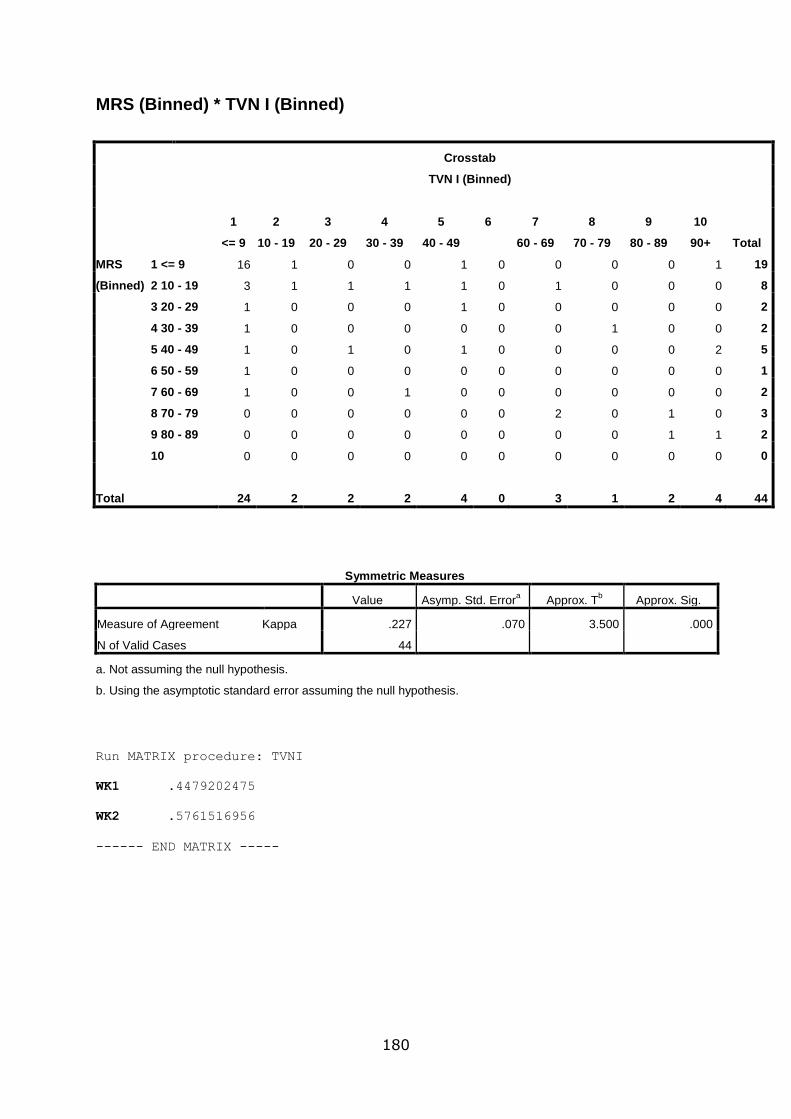
180
MRS (Binned) * TVN I (Binned)
Crosstab
TVN I (Binned)
Total
1
<= 9
2
10 - 19
3
20 - 29
4
30 - 39
5
40 - 49
6 7
60 - 69
8
70 - 79
9
80 - 89
10
90+
MRS
(Binned)
1 <= 9 16 1 0 0 1 0 0 0 0 1 19
2 10 - 19 3 1 1 1 1 0 1 0 0 0 8
3 20 - 29 1 0 0 0 1 0 0 0 0 0 2
4 30 - 39 1 0 0 0 0 0 0 1 0 0 2
5 40 - 49 1 0 1 0 1 0 0 0 0 2 5
6 50 - 59 1 0 0 0 0 0 0 0 0 0 1
7 60 - 69 1 0 0 1 0 0 0 0 0 0 2
8 70 - 79 0 0 0 0 0 0 2 0 1 0 3
9 80 - 89 0 0 0 0 0 0 0 0 1 1 2
10 0 0 0 0 0 0 0 0 0 0 0
Total 24 2 2 2 4 0 3 1 2 4 44
Symmetric Measures
Value Asymp. Std. Errora Approx. T
b Approx. Sig.
Measure of Agreement Kappa .227 .070 3.500 .000
N of Valid Cases 44
a. Not assuming the null hypothesis.
b. Using the asymptotic standard error assuming the null hypothesis.
Run MATRIX procedure: TVNI
WK1 .4479202475
WK2 .5761516956
------ END MATRIX -----

181
MRS (Binned) * TVN J (Binned)
Crosstab
TVN J (Binned)
Total
1
<= 9
2
10 - 19
3
20 - 29
4
30 - 39
5
40 - 49
6 7
60 - 69
8
70 - 79
9
80 - 89
10
90+
MRS
(Binned)
1 <= 9 14 3 0 0 0 0 0 0 0 2 19
2 10 - 19 5 1 2 0 0 0 0 0 0 0 8
3 20 - 29 0 1 0 1 0 0 0 0 0 0 2
4 30 - 39 1 0 0 0 0 0 0 0 1 0 2
5 40 - 49 1 0 0 1 1 0 0 0 1 1 5
6 50 - 59 1 0 0 0 0 0 0 0 0 0 1
7 60 - 69 0 1 0 0 0 0 0 0 1 0 2
8 70 - 79 0 0 0 0 0 0 1 1 0 1 3
9 80 - 89 1 0 0 0 0 0 0 0 0 1 2
10 0 0 0 0 0 0 0 0 0 0 0
Total 23 6 2 2 1 0 1 1 3 5 44
Symmetric Measures
Value Asymp. Std. Errora Approx. T
b Approx. Sig.
Measure of Agreement Kappa .167 .075 2.499 .012
N of Valid Cases 44
a. Not assuming the null hypothesis.
b. Using the asymptotic standard error assuming the null hypothesis.
Run MATRIX procedure: TVNJ
WK1 .4310990133
WK2 .5059880240
------ END MATRIX -----

182
MRS (Binned) * TVN K (Binned)
Crosstab
TVN K (Binned)
Total
1
<= 9
2
10 - 19
3
20 - 29
4
30 - 39
5
40 - 49
6 7
60 - 69
8
70 - 79
9
80 - 89
10
90+
MRS
(Binned)
1 <= 9 15 2 2 0 0 0 0 0 0 0 19
2 10 - 19 2 2 0 2 1 0 1 0 0 0 8
3 20 - 29 1 0 0 1 0 0 0 0 0 0 2
4 30 - 39 1 0 0 0 0 0 0 0 1 0 2
5 40 - 49 1 1 0 2 0 0 0 0 1 0 5
6 50 - 59 0 0 0 0 0 0 0 0 0 1 1
7 60 - 69 0 1 0 0 1 0 0 0 0 0 2
8 70 - 79 0 0 0 0 1 0 0 1 0 1 3
9 80 - 89 0 0 0 0 0 0 0 0 0 2 2
10 0 0 0 0 0 0 0 0 0 0 0
Total 20 6 2 5 3 0 1 1 2 4 44
Symmetric Measures
Value Asymp. Std. Errora Approx. T
b Approx. Sig.
Measure of Agreement Kappa .222 .069 3.309 .001
N of Valid Cases 44
a. Not assuming the null hypothesis.
b. Using the asymptotic standard error assuming the null hypothesis.
Run MATRIX procedure: TVNK
WK1 .5149359886
WK2 .7213750323
------ END MATRIX -----

183
MRS (Binned) * TVN L (Binned)
Crosstab
TVN L (Binned)
Total
1
<= 9
2
10 - 19
3
20 - 29
4
30 - 39
5
40 - 49
6
50 - 59
7 8
70 - 79
9
80 - 89
10
90+
MRS
(Binned)
1 <= 9 13 3 1 0 0 0 0 0 2 0 19
2 10 - 19 5 0 1 1 1 0 0 0 0 0 8
3 20 - 29 1 0 0 1 0 0 0 0 0 0 2
4 30 - 39 1 0 0 0 1 0 0 0 0 0 2
5 40 - 49 2 0 0 0 0 1 0 0 0 2 5
6 50 - 59 0 0 1 0 0 0 0 0 0 0 1
7 60 - 69 1 0 0 0 0 1 0 0 0 0 2
8 70 - 79 0 0 0 0 0 1 0 1 0 1 3
9 80 - 89 0 0 0 0 0 0 0 0 0 2 2
10 0 0 0 0 0 0 0 0 0 0 0
Total 23 3 3 2 2 3 0 1 2 5 44
Symmetric Measures
Value Asymp. Std. Errora Approx. T
b Approx. Sig.
Measure of Agreement Kappa .087 .063 1.371 .170
N of Valid Cases 44
a. Not assuming the null hypothesis.
b. Using the asymptotic standard error assuming the null hypothesis.
Run MATRIX procedure: TVNL
WK1 .4449369247
WK2 .5972420352
------ END MATRIX -----

184
MRS (Binned) * TVN M (Binned)
Crosstab
TVN M (Binned)
Total
1
<= 9
2
10 - 19
3
20 - 29
4
30 - 39
5
40 - 49
6
50 - 59
7
60 - 69
8
70 - 79
9
80 - 89
10
90+
MRS
(Binned)
1 <= 9 14 3 1 0 1 0 0 0 0 0 19
2 10 - 19 3 1 0 1 1 1 1 0 0 0 8
3 20 - 29 1 0 0 1 0 0 0 0 0 0 2
4 30 - 39 0 0 0 1 0 0 1 0 0 0 2
5 40 - 49 1 0 1 0 1 1 0 0 0 1 5
6 50 - 59 1 0 0 0 0 0 0 0 0 0 1
7 60 - 69 0 0 0 1 0 0 1 0 0 0 2
8 70 - 79 0 0 0 0 0 1 0 1 0 1 3
9 80 - 89 0 0 0 0 0 0 0 0 1 1 2
10 0 0 0 0 0 0 0 0 0 0 0
Total 20 4 2 4 3 3 3 1 1 3 44
Symmetric Measures
Value Asymp. Std. Errora Approx. T
b Approx. Sig.
Measure of Agreement Kappa .288 .083 4.464 .000
N of Valid Cases 44
a. Not assuming the null hypothesis.
b. Using the asymptotic standard error assuming the null hypothesis.
Run MATRIX procedure: TVNM
WK1 .5535777857
WK2 .7283034148
------ END MATRIX -----

185
MRS (Binned) * TVN N (Binned)
Crosstab
TVN N (Binned)
Total
1
<= 9
2
10 - 19
3
20 - 29
4
30 - 39
5 6 7
60 - 69
8
70 - 79
9
80 - 89
10
90+
MRS
(Binned)
1 <= 9 18 1 0 0 0 0 0 0 0 0 19
2 10 - 19 2 5 1 0 0 0 0 0 0 0 8
3 20 - 29 1 0 0 0 0 0 1 0 0 0 2
4 30 - 39 0 0 1 0 0 0 1 0 0 0 2
5 40 - 49 1 1 0 0 0 0 0 2 0 1 5
6 50 - 59 0 0 0 0 0 0 0 0 1 0 1
7 60 - 69 0 0 1 0 0 0 1 0 0 0 2
8 70 - 79 0 0 0 1 0 0 0 1 1 0 3
9 80 - 89 0 0 0 0 0 0 0 0 0 2 2
10 0 0 0 0 0 0 0 0 0 0 0
Total 22 7 3 1 0 0 3 3 2 3 44
Symmetric Measures
Value Asymp. Std. Errora Approx. T
b Approx. Sig.
Measure of Agreement Kappa .417 .076 6.103 .000
N of Valid Cases 44
a. Not assuming the null hypothesis.
b. Using the asymptotic standard error assuming the null hypothesis.
Run MATRIX procedure: TVNN
WK1 .6432851604
WK2 .8000498008
------ END MATRIX -----

186
IBM SPSS syntax MATRIX-ENDMATRIX
MATRIX-ENDMATRIX applied to the unweighted kappa contingency tables to
produce linear weighted kappa (WK1) and quadratic weighted kappa (WK2).
* Read in the IxI matrix of counts for I-level scale.
DATA LIST LIST / x1 to x3. BEGIN DATA
abc lmn xyz
END DATA. * Commands to compute weighted Kappa .
matrix. GET x /var=x1 to x3.
compute wt1=make(nrow(x),ncol(x),0). compute wt2=make(nrow(x),ncol(x),0). compute prop=x/msum(x).
loop i=1 to nrow(x). loop j=1 to ncol(x).
compute wt1(i,j)=1-(abs(i-j)/(nrow(x)-1)). compute wt2(i,j)=1-((i-j)/(nrow(x)-1))**2. end loop.
end loop. compute wk1num=msum(wt1&*prop)-
msum(mdiag(rsum(prop))*wt1*mdiag(csum(prop))). compute wk1den=1-msum(mdiag(rsum(prop))*wt1*mdiag(csum(prop))). compute wk1=wk1num/wk1den.
print wk1. compute wk2num=msum(wt2&*prop)-
msum(mdiag(rsum(prop))*wt2*mdiag(csum(prop))). compute wk2den=1-msum(mdiag(rsum(prop))*wt2*mdiag(csum(prop))). compute wk2=wk2num/wk2den.
print wk2. end matrix.

187
Appendix H OpenEHR Foundation correspondence
Response from Dr. Ian McNicoll to the proposal to develop the OpenEHR draft
archetype inspection of an open wound

188
Appendix I Data Protection correspondence
Data Protection query

189
Data Protection response - Page 1

190
Data Protection response – Page 2

191
Data Protection response – Page 3


