permitting all non-commercial use, distribution, and reproduction in any medium, provided the original work is properly cited.
Original Article
E-PodoFavalin-15999 (Atremorine®)-Induced Neurotransmitter and Hormonal Response in Parkinson’s Disease
Ramón Cacabelos*, Lucía Fernández-Novoa, Ramón Alejo, Lola Corzo, Susana Rodríguez, Margarita Alcaraz, Laura Nebril, Pablo Cacabelos, Carmen Fraile,
Iván Carrera and Juan C. Carril
EuroEspes Biomedical Research Center, Institute of Medical Science and Genomic Medicine, 15165-Bergondo, Corunna, Spain
Abstract
E-PodoFavalin-15999 (Atremorine®) is a novel bioproduct ob-tained by means of non-denaturing biotechnological procedures from structural components of the Vicia faba L. plant, for the pre-vention and treatment of Parkinsonian disorders. Preclinical stud-ies have revealed that Atremorine is a powerful neuroprotectant with specific activity on dopaminergic neurons, reversing neuro-degeneration and improving motor function in animal models of Parkinson’s disease (PD). Clinical studies indicate that Atremo-rine is a powerful catecholaminergic enhancer with time- and genotype-dependent effects in PD. In the present study, we inves-tigated the effects of Atremorine on the levels of neurotransmitters (dopamine, adrenaline, noradrenaline, serotonin) and hormones (adrenocorticotropic hormone (ACTH), growth hormone (GH), prolactin (PRL), follicle-stimulating hormone (FSH), luteinizing hormone (LH), cortisol, estrogens, testosterone) in Parkinsonian patients (n=119) 1 hour after Atremorine administration (5 g/day). These effects were also studied after stratification of PD patients according to their cardiovascular function. Atremorine induced a significant increase in the levels of dopamine (p<0.001), no-radrenaline (p=0.004) and adrenaline (p=0.01), and a significant decrease in the levels of PRL (p<0.001), cortisol (p<0.001), and GH (p=0.002), with no apparent changes in the levels of serotonin, ACTH, FSH, LH, testosterone or estrogen. The levels of dopamine were significantly higher in patients with normal EKG than in pa-tients with abnormal electrocardiogram (EKG); however, the lev-els of adrenaline, noradrenaline and serotonin tended to be lower in patients with normal EKG as compared to patients with abnor-mal EKG. There were also some differences in hormonal levels in patients with normal EKG, compared to abnormal EKG. These results clearly show that Atremorine is an effective enhancer of catecholaminergic neurotransmission, which contributes to opti-mization of hormonal regulation in PD.
Introduction
Alzheimer’s disease and Parkinson’s disease (PD) are the most important neurodegenerative disorders afflicting today’s elderly population. PD, in particular, shows a prevalence that ranges from 35.8 per 100,000 to 12,500 per 100,000, with an annual incidence ranging from 1.5 per 100,000 to 346 per 100,000 in different countries.1–7 The prevalence of PD gradually increases in paral-lel with age, following an age-dependent trend (41 per 100,000 at 40–49 years; 107 at 50–59 years; 173 at 55–64 years; 428 at 60–69 years; 425 at 65–74 years; 1,087 at 70–79 years; and 1,903 per 100,000 at older than age 80).6 PD is more prevalent in males (1,729 per 100,000, >65 yrs) than in females (1,644 per 100,000), with a peak prevalence in the age group of ≥90 years (4,633 cas-es per 100,000) and a mean prevalence of 1,680 per 100,000 in people >65 years of age.7,8 The geographic distribution of PD is also characteristic, with a prevalence of 1,601 per 100,000 in North America, Europe and Australia, and a prevalence of 646 per 100,000 in Asia.6
The clinical onset of PD, characterized by resting tremor, ri-gidity and bradykinesia, is likely to occur several decades af-ter the beginning of the progressive neurodegenerative process, which predominantly affects dopaminergic neurons in the sub-stantia nigra pars compacta, and is accompanied by widespread involvement of other CNS structures and peripheral tissues.9 PD is a form of multi-systemic α-synucleinopathy, with Lewy bodies deposits in the midbrain. Descriptive phenomena to explain, at least in part, this neuropathological phenotype include the fol-lowing: (i) genomic factors, (ii) epigenetic changes, (iii) toxic factors, (iv) oxidative stress anomalies, (v) neuroimmune/neu-roinflammatory reactions, (vi) hypoxic-ischemic conditions, and (vii) ubiquitin-proteasome system dysfunction; all these condi-tions lead to protein misfolding and aggregation and premature neuronal death.10–17 Recent evidence also suggests that PD might be a prion-like disease.12 Additionally, the catecholaldehyde hy-pothesis postulates an “autotoxicity”-inherent cytotoxicity of cat-echolamines and their metabolites in neurons, in which a long-term increase in 3,4-dihydroxyphenylacetaldehyde (DOPAL, the catecholaldehyde metabolite of dopamine) oligomerizes and ag-gregates alpha-synuclein, accelerating the eventual death of do-paminergic neurons.18
Classical therapeutic interventions for the symptomatic treat-ment of motor symptoms in PD include pharmacotherapy, deep brain stimulation, and physiotherapy.19 L-3,4-dihydroxypheny-lalanine [(2-amino-3-(3,4-dihydroxyphenyl) propanoic acid] (L-DOPA) has been the most representative treatment for PD since the 1960s.20,21 In addition to L-DOPA, as a dopamine precursor,
Keywords: Atremorine; Dopamine; Adrenaline; Noradrenaline; Serotonin; Growth hormone; Prolactin; ACTH; Cortisol; Parkinson’s disease.Abbreviations: 5HT, serotonin; ACTH, adrenocorticotropic hormone; AD, adrena-line; COR, cortisol; DA, dopamine; EKG, electrocardiogram; FSH, follicle stimu-lating hormone; GH, growth hormone; L-DOPA, L-3,4-dihydroxyphenylalanine [(2-amino-3-(3,4-dihydroxyphenyl) propanoic acid); LH, luteinizing hormone; NA, noradrenaline; PRL, prolactin; T4, thyroxine; TSH, thyroid stimulating hormone.Received: 11 October 2016; Accepted: 20 October 2016☆DOI: 10.14218/JERP.2016.00031*Correspondence to: Ramón Cacabelos, EuroEspes Biomedical Research Center, In-stitute of Medical Science and Genomic Medicine, 15165-Bergondo, Corunna, Spain. Tel: +34-981-780505, Fax: +34-981-780511, E-mail: [email protected]
Journal of Exploratory Research in Pharmacology 2016 vol. 1 | 1-122
Cacabelos R. et al: Atremorine in Parkinson’s Disease
other symptomatic treatments for PD include dopamine agonists, monoamine oxidase (MAO) inhibitors, and catechol-O-methyl-transferase (COMT) inhibitors.22 A common complication of L-DOPA and dopamine agonists, after long periods of treatment, is the “wearing-off” phenomenon23,24, with motor fluctuations and dyskinesia.20,25 Polypharmacy in PD and the co-administration of psychotropic drugs and/or any other drug category with potential interaction with dopaminergic neurotransmission may also con-tribute to severe complications.26 It is also common to find gas-trointestinal27, cardiovascular28, hormonal and neuropsychiatric problems associated with the chronic administration of conven-tional antiparkinsonian drugs.22,27
The present understanding of PD suggests the necessity for a change in the paradigm of PD management, including the follow-ing: (i) characterization of pathogenic factors (either genomic or environmental) and reliable biomarkers for early intervention in the population at risk, (ii) accelerating the search for novel mo-dalities of therapeutic intervention devoid of side effects and/or delayed deleterious consequences for the quality of life and well-being of PD patients, and (iii) optimization of the present therapeu-tic resources in order to optimize pharmacological treatments by means of a personalized, pharmacogenetic approach.10
We have developed a research program in search of new pre-ventive and/or therapeutic options for PD. The first bioproduct of this series is E-PodoFavalin-15999 (Atremorine®).29 Atremo-rine is a novel biopharmaceutical compound, obtained by means of non-denaturing biotechnological procedures from structural components of the Vicia faba L. plant, for the prevention and treatment of PD.29 Preclinical studies revealed that Atremorine is a powerful neuroprotectant in (i) cell cultures of human neu-roblastoma SH-SY5Y cells, (ii) hippocampal slices under condi-tions of oxygen and glucose deprivation, and (iii) striatal slices under conditions of 6-hydroxydopamine (OHDA)-induced neu-rotoxicity.
In vivo studies have shown that Atremorine (i) protects against 1-methyl-4-phenyl-1,2,3,6-tetrahydropyridine (MPTP)-induced dopaminergic neurodegeneration, (ii) inhibits MPTP-induced mi-croglia activation and neurotoxicity in substantia nigra, and (iii) improves motor function in mice with MPTP-induced neurode-generation.29,30 Clinical studies of untreated patients who received Atremorine for the first time (treatment-naïve for antiparkinsonian drugs) revealed that Atremorine can enhance dopaminergic neuro-transmission and increase plasma dopamine levels by 200- to 500-fold. In patients who have been chronically treated with L-DOPA or other antiparkinsonian drugs, Atremorine was found to induce a dopamine response of similar magnitude to that observed in pre-viously untreated patients. Moreover, this dopaminergic response was shown to be associated with the pharmacogenetic profile of the patients.29,31
In this paper we report, for the first time, the influence of Atrem-orine on different neurotransmitters (dopamine, noradrenaline, adrenaline, serotonin (5HT)), pituitary hormones (adrenocortico-tropic hormone (ACTH), growth hormone (GH), prolactin (PRL), luteinizing hormone (LH), follicle-stimulating hormone (FSH)) and peripheral hormones (cortisol (COR), testosterone, estrogen) in PD patients.
Material and methods
Patients and treatment
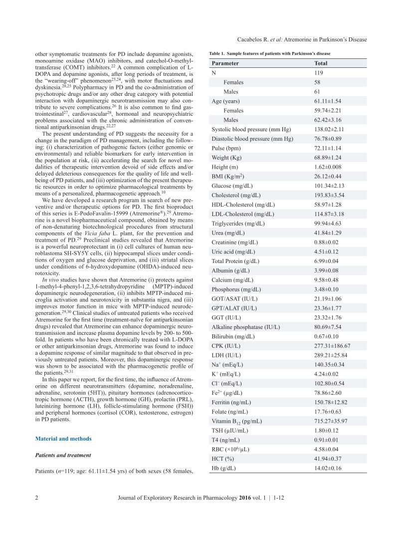
Patients (n=119; age: 61.11±1.54 yrs) of both sexes (58 females,
Table 1. Sample features of patients with Parkinson’s disease
Journal of Exploratory Research in Pharmacology 2016 vol. 1 | 1-12 3
Cacabelos R. et al: Atremorine in Parkinson’s Disease
age: 59.74±2.21; 61 males, age: 62.42±3.16 yrs) with PD were re-cruited for this study. All patients underwent, under informed con-sent, the following protocol: (i) clinical (neurologic, psychiatric) examination, (ii) blood and urine analyses (Table 1), (iii) neuropsy-chological assessment [Mini-Mental State Examination (MMSE), Alzheimer’s Disease Assessment Scale (ADAS), Hamilton-A/D, Geriatric Depression Scale (GDS), Unified Parkinson Disease Rat-ing Scale (UPDRS), Hoehn and Yahr staging, Schwab and England ‘activities of daily living’ (ADL) scale] (Table 1), (iv) cardiovascu-lar evaluation (EKG), (v) structural neuroimaging (brain magnetic resonance imaging), (vi) functional neuroimaging (brain mapping, brain optical topography), (vii) genetic assessment [apolipopro-tein E (APOE)], and (viii) pharmacogenetic profiling (CYP2D6, CYP2C19, CYP2C9, CYP3A4/5).
All patients received a single oral dose of 5 g of E-PodoFava-lin-15999 (Atremorine®) in the morning, to avoid circadian varia-tions in biochemical and hormonal parameters, and blood samples were obtained prior to Atremorine intake and 60 minutes later.
Analytical methods
Venous blood samples were taken after overnight fasting, with patients in supine position. Blood for the analysis of serum total testosterone, FSH, LH, PRL, COR, GH, estradiol (E2), 5HT and thyroid hormones [thyroid-stimulating hormone (TSH) and free-thyroxine (FT4)] was collected in BD Vacutainer serum separa-
tion tubes, while blood for analysis of plasma catecholamine (no-radrenaline, adrenaline and dopamine) and ACTH was collected in EDTA-containing tubes. Specimens for catecholamine and ACTH analyses were immediately placed on ice and centrifuged at 3000 rpm, at 4°C, for 10 minutes, soon after venipuncture.32 Serum tubes were allowed to clot at room temperature for 30 minutes be-fore processing, and centrifuged within 60 minutes of sampling under the same conditions as the EDTA tubes. After refrigerated centrifugation, serum and plasma were removed from blood cells33 and placed in a suitable sample container.
Serum levels of testosterone, FSH, LH, PRL, COR, GH and E2 and plasma levels of ACTH were measured by electrochemi-luminescence on the same day of venipuncture, using the Immu-lite System (Siemens, Malvern, PA, USA). The Immulite 1000 is an automated, random-access immunoassay analyzer with a solid-phase washing process and a chemiluminescent detection system.34–37 Access2 Immunoassay System (Beckman Coulter, Brea, CA, USA), an automated system with chemiluminescent signal, was used to analyze serum concentrations of ultrasensi-tive TSH (us-TSH; 3rd generation) and FT4.38 Plasma aliquots for fractionated catecholamine determinations were stored at −20°C (1) and purified with albumin until their analysis by High Perfor-mance Liquid Chromatography (HPLC) with electrochemical de-tection.39,40 The HPLC system consisted of a pump (515; Waters Corp., Milford, MA, USA), an autosampler (717; Waters Corp.), a chromatographic column (Resolve C18; Waters Corp.), an elec-trochemical detector (2465; Waters Corp.), and the Empower2 chromatography data software (Waters Corp.). Serum aliquots for serotonin analysis were stored at −20°C until measurement by a commercial ultrasensitive ELISA kit (Labor Diagnostika Nord, Nordhorn, Germany) after quantitative acylation41; the ELISA was performed according to the manufacturer’s instructions and each reaction was monitored at 450 nm using the Sunrise Microplate Absorbance Reader (Tecan, Grödig, Austria).
Statistical analysis
Data were analyzed by using IBM SPSS Statistics 20 and Sigma-Plot 10.0 software. Comparisons between groups were studied by t-test, Mann-Whitney rank sum test, chi-square test without Yates correction and Fisher’s exact test, and Pearson’s correlation analy-sis (nonlinear regression, Durbin-Watson statistic, normality test, constant variance test, 95% confidence). All values are expressed as mean ± SE, and the degree of significance was considered to have been met when p<0.05.
Results
Neurotransmitters
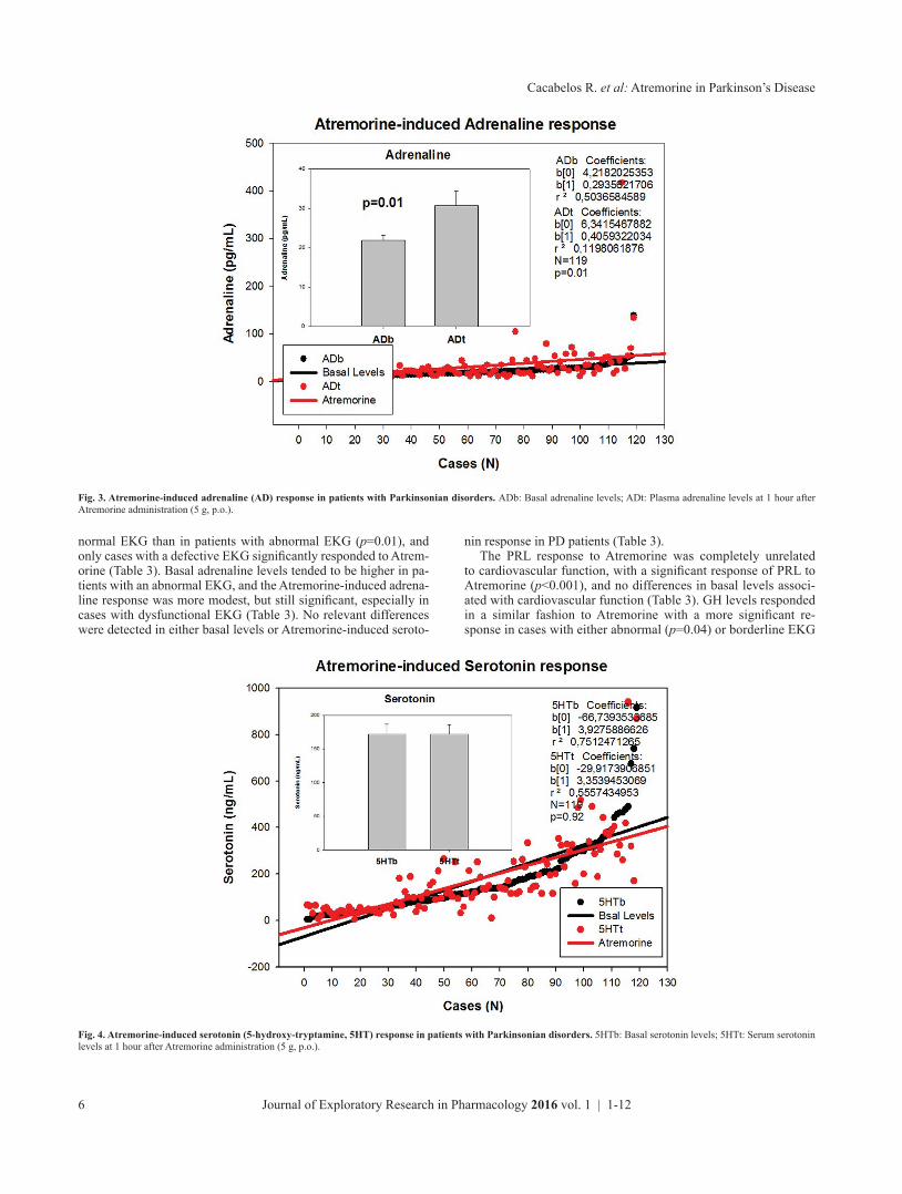
One hour after Atremorine administration, basal dopamine levels increased from 762.28±296.94 pg/mL (range: 8–30,318 pg/mL) to 4,556.51±678.95 (range: 54–40,603 pg/mL) (p<0.001) (Table 2; Fig. 1), with a similar response in females and males (p<0.001) (Table 2). A significant increase was observed in noradrenaline (286.52±21.88 pg/mL vs. 360.72±22.54 pg/mL) (p=0.004) (Ta-ble 2; Fig. 2) and adrenaline (21.83±1.31 pg/mL vs. 30.69±3.70) (p=0.01) (Table 2; Fig. 3), with no significant changes in seroto-nin (Table 2; Fig. 4). The noradrenaline response to Atremorine was similar in females (p=0.05) and males (p=0.01); however, although adrenaline responded to Atremorine in both females and
Table 1. Sample features of patients with Parkinson’s disease - (continued)
Journal of Exploratory Research in Pharmacology 2016 vol. 1 | 1-124
Cacabelos R. et al: Atremorine in Parkinson’s Disease
males, this response was only significant in males (p=0.05) (Table 2).
Hormones
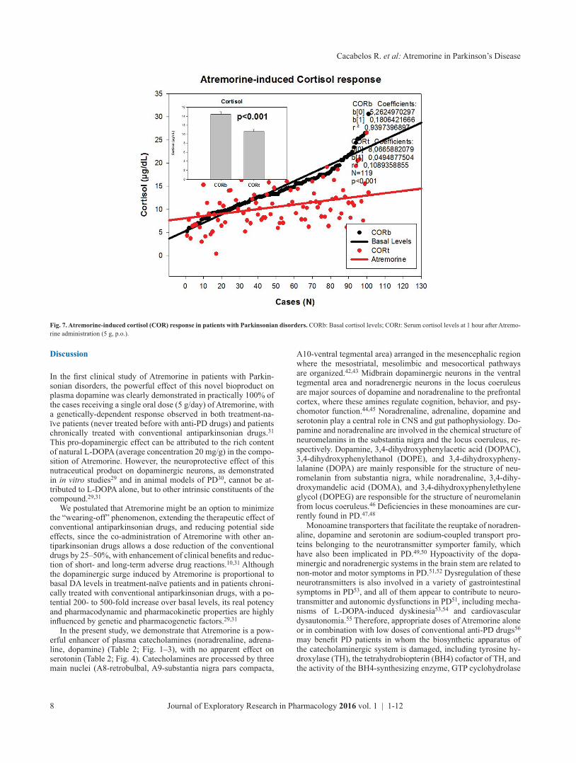
Atremorine induced a significant decrease in the levels of prolac-tin (11.94±1.76 ng/mL vs. 7.32±1.38 ng/mL) (p<0.001) (Table 2; Fig. 5), GH (1.24±0.20 vs. 0.43±0.08 ng/mL) (p=0.002) (Table 2; Fig. 6) and COR (14.47±0.54 vs. 10.64±0.46 µg/mL) (p<0.0001) (Table 2; Fig. 7). No significant changes were observed in ACTH,
FSH, LH, testosterone and estrogen levels in response to Atremo-rine (Table 2). Basal levels of PRL were higher in females than in males (p=0.05), and both sexes responded significantly to Atremo-rine in a similar manner (Table 2). GH levels were found to be higher in females than in males (p=0.003). In addition, females exhibited a significant response to Atremorine (p=0.003), while the response in males was poorer (Table 2). Basal COR levels were similar in females and males, and patients of both sexes re-sponded to Atremorine in an identical manner (p<0.001) (Table 2). As expected, both FSH and LH levels were significantly higher in females than in males (p<0.001), with no apparent response to
Table 2. Atremorine-induced neurotransmitter and hormonal changes
Parameter Cases Basal levels Atremorine pNoradrenaline (pg/mL) T 286.52±21.88 360.72±22.54 0.004 A vs B
F 281.19±26.75 371.70±31.15 0.01 A vs B; 0.91 BF vs BMM 295.50±34.48 350.27±32.70 0.05 A vs B; 0.48 AF vs AM
Adrenaline (pg/mL) T 21.83±1.31 30.69±3.70 0.01 A vs BF 18.62±0.89 28.93±6.97 0.22 A vs B; 0.04 BF vs BMM 24.88±2.35 32.37±2.95 0.03 A vs B; 0.008 AF vs AM
Dopamine (pg/mL) T 762.28±296.94 4556.51±678.95 <0.001 A vs BF 232.05±107.33 3431.03±676.71 <0.001 A vs B; 0.03 BF vs BMM 1266.44±564.98 5626.83±1147.08 <0.001 A vs B; 0.26 AF vs AM
Serotonin (ng/mL) T 171.71±14.43 171.31±14.22 0.92 A vs BF 158.10±18.07 157.84±19.64 0.98 A vs B; 0.58 BF vs BMM 182.10±22.29 184.13±20.55 0.77 A vs B; 0.32 AF vs AM
Prolactin (ng/mL) T 11.94±1.76 7.32±1.38 <0.001 A vs BF 12.89±2.27 7.60±1.74 <0.001 A vs B; 0.01 BF vs BMM 11.08±2.67 7.20±2.12 0.003 A vs B; 0.19 AF vs AM
Cortisol (µg/dL) T 14.47±0.54 10.64±0.46 <0.001 A vs BF 15.29±0.96 10.40±0.71 <0.001 A vs B; 0.29 BF vs BMM 13.73±0.54 10.35±0.62 <0.001 A vs B;<0.001 AF vs AM
ACTH (pg/mL) T 31.29±4.12 29.69±3.95 0.77 A vs BF 35.10±8.21 31.92±7.49 0.54 A vs B; 0.67 BF vs BMM 28.35±2.93 27.71±3.48 0.99 A vs B; 0.35 AF vs AM
GH (ng/mL) T 1.24±0.20 0.43±0.08 0.002 A vs BF 1.72±0.35 0.52±0.12 0.003 A vs B; 0.003 BF vs BMM 0.80±0.20 0.38±0.13 0.16 A vs B; 0.05 AF vs AM
FSH (mIU/mL) T 30.69±3.50 29.62±3.57 0.89 A vs BF 54.00±5.52 52.36±5.84 0.80 A vs B; <0.001 BF vs BMM 9.58±1.38 9.56±1.40 0.88 A vs B; <0.001 AF vs AM
LH (mIU/mL) T 14.78±1.61 13.63±1.46 0.68 A vs BF 24.52±2.71 22.05±2.48 0.54 A vs B; <0.001 BF vs BMM 5.96±0.65 6.21±0.74 0.90 A vs B; <0.001 AF vs AM
Testosterone (ng/dL) M 379.79±24.54 331.18±20.68 0.13 A vs BEstrogen (pg/mL) F 59.23±12.44 58.27±11.83 0.69 A vs B
Data: mean ± standard error.Abbreviations: A: atremorine (60 minutes after treatment with 5g Atremorine, p.o.); AF: Atremorine-females; AM: Atremorine-males; B: basal levels (prior to treatment with Atremorine); BF: basal-females; BM: basal-males; F: females; M: males; T: total sample.
Journal of Exploratory Research in Pharmacology 2016 vol. 1 | 1-12 5
Cacabelos R. et al: Atremorine in Parkinson’s Disease
Atremorine in either of the sexes (Table 2).
EKG-related Atremorine-induced changes in neurotransmitters and hormones
Important differences were identified in the peripheral concentra-tion of some neurotransmitters and hormones in PD patients ac-
cording to their cardiovascular condition, as assessed by EKG recording (Table 3). Basal dopamine levels were found to be significantly higher in patients with a normal EKG as compared with those patients with an abnormal EKG (p=0.01). However, the dopamine response to Atremorine was significantly higher in the three conditions (normal EKG, borderline EKG, abnormal EKG) as compared with their respective basal levels (p<0.001) (Table 3). In contrast, basal noradrenaline levels were lower in cases with
Fig. 1. Atremorine-induced dopamine (DA) response in patients with Parkinsonian disorders. DAb: Basal dopamine levels; DAt: Plasma dopamine levels at 1 hour after Atremorine administration (5 g, p.o.).
Fig. 2. Atremorine-induced noradrenaline (NA) response in patients with Parkinsonian disorders. NAb: Basal noradrenaline levels; NAt: Plasma noradrenaline levels at 1 hour after Atremorine administration (5 g, p.o.).
Journal of Exploratory Research in Pharmacology 2016 vol. 1 | 1-126
Cacabelos R. et al: Atremorine in Parkinson’s Disease
normal EKG than in patients with abnormal EKG (p=0.01), and only cases with a defective EKG significantly responded to Atrem-orine (Table 3). Basal adrenaline levels tended to be higher in pa-tients with an abnormal EKG, and the Atremorine-induced adrena-line response was more modest, but still significant, especially in cases with dysfunctional EKG (Table 3). No relevant differences were detected in either basal levels or Atremorine-induced seroto-
nin response in PD patients (Table 3).The PRL response to Atremorine was completely unrelated
to cardiovascular function, with a significant response of PRL to Atremorine (p<0.001), and no differences in basal levels associ-ated with cardiovascular function (Table 3). GH levels responded in a similar fashion to Atremorine with a more significant re-sponse in cases with either abnormal (p=0.04) or borderline EKG
Fig. 3. Atremorine-induced adrenaline (AD) response in patients with Parkinsonian disorders. ADb: Basal adrenaline levels; ADt: Plasma adrenaline levels at 1 hour after Atremorine administration (5 g, p.o.).
Fig. 4. Atremorine-induced serotonin (5-hydroxy-tryptamine, 5HT) response in patients with Parkinsonian disorders. 5HTb: Basal serotonin levels; 5HTt: Serum serotonin levels at 1 hour after Atremorine administration (5 g, p.o.).
Journal of Exploratory Research in Pharmacology 2016 vol. 1 | 1-12 7
Cacabelos R. et al: Atremorine in Parkinson’s Disease
(p=0.005) than in patients with a normal EKG (Table 3). Signifi-cant differences were found in the basal FSH levels of patients with abnormal EKG versus borderline EKG (p=0.006) and in those with normal EKG versus borderline EKG (p=0.003). These differences persisted after Atremorine administration (Table 3). A similar pattern was found regarding basal LH levels, which were found to be significantly higher in borderline EKG cases than in
patients with abnormal (p=0.04) or normal EKG (p=0.01) (Ta-ble 3). Atremorine did not alter these differences (Table 3). Both ACTH and testosterone levels did not show any relevant variabil-ity (Table 3); however, basal levels of estrogen were the highest in cases with normal EKG, intermediate in patients with borderline EKG, and the lowest in those cases exhibiting an abnormal EKG (Table 3).
Fig. 5. Atremorine-induced prolactin (PRL) response in patients with Parkinsonian disorders. PRLb: Basal prolactin levels; PRLt: Serum prolactin levels at 1 hour after Atremorine administration (5 g, p.o.).
Fig. 6. Atremorine-induced growth hormone (GH) response in patients with Parkinsonian disorders. GHb: Basal growth hormone levels; GHt: Serum growth hormone levels at 1 hour after Atremorine administration (5 g, p.o.).
Journal of Exploratory Research in Pharmacology 2016 vol. 1 | 1-128
Cacabelos R. et al: Atremorine in Parkinson’s Disease
Discussion
In the first clinical study of Atremorine in patients with Parkin-sonian disorders, the powerful effect of this novel bioproduct on plasma dopamine was clearly demonstrated in practically 100% of the cases receiving a single oral dose (5 g/day) of Atremorine, with a genetically-dependent response observed in both treatment-na-ïve patients (never treated before with anti-PD drugs) and patients chronically treated with conventional antiparkinsonian drugs.31 This pro-dopaminergic effect can be attributed to the rich content of natural L-DOPA (average concentration 20 mg/g) in the compo-sition of Atremorine. However, the neuroprotective effect of this nutraceutical product on dopaminergic neurons, as demonstrated in in vitro studies29 and in animal models of PD30, cannot be at-tributed to L-DOPA alone, but to other intrinsic constituents of the compound.29,31
We postulated that Atremorine might be an option to minimize the “wearing-off” phenomenon, extending the therapeutic effect of conventional antiparkinsonian drugs, and reducing potential side effects, since the co-administration of Atremorine with other an-tiparkinsonian drugs allows a dose reduction of the conventional drugs by 25–50%, with enhancement of clinical benefits and reduc-tion of short- and long-term adverse drug reactions.10,31 Although the dopaminergic surge induced by Atremorine is proportional to basal DA levels in treatment-naïve patients and in patients chroni-cally treated with conventional antiparkinsonian drugs, with a po-tential 200- to 500-fold increase over basal levels, its real potency and pharmacodynamic and pharmacokinetic properties are highly influenced by genetic and pharmacogenetic factors.29,31
In the present study, we demonstrate that Atremorine is a pow-erful enhancer of plasma catecholamines (noradrenaline, adrena-line, dopamine) (Table 2; Fig. 1–3), with no apparent effect on serotonin (Table 2; Fig. 4). Catecholamines are processed by three main nuclei (A8-retrobulbal, A9-substantia nigra pars compacta,
A10-ventral tegmental area) arranged in the mesencephalic region where the mesostriatal, mesolimbic and mesocortical pathways are organized.42,43 Midbrain dopaminergic neurons in the ventral tegmental area and noradrenergic neurons in the locus coeruleus are major sources of dopamine and noradrenaline to the prefrontal cortex, where these amines regulate cognition, behavior, and psy-chomotor function.44,45 Noradrenaline, adrenaline, dopamine and serotonin play a central role in CNS and gut pathophysiology. Do-pamine and noradrenaline are involved in the chemical structure of neuromelanins in the substantia nigra and the locus coeruleus, re-spectively. Dopamine, 3,4-dihydroxyphenylacetic acid (DOPAC), 3,4-dihydroxyphenylethanol (DOPE), and 3,4-dihydroxypheny-lalanine (DOPA) are mainly responsible for the structure of neu-romelanin from substantia nigra, while noradrenaline, 3,4-dihy-droxymandelic acid (DOMA), and 3,4-dihydroxyphenylethylene glycol (DOPEG) are responsible for the structure of neuromelanin from locus coeruleus.46 Deficiencies in these monoamines are cur-rently found in PD.47,48
Monoamine transporters that facilitate the reuptake of noradren-aline, dopamine and serotonin are sodium-coupled transport pro-teins belonging to the neurotransmitter symporter family, which have also been implicated in PD.49,50 Hypoactivity of the dopa-minergic and noradrenergic systems in the brain stem are related to non-motor and motor symptoms in PD.51,52 Dysregulation of these neurotransmitters is also involved in a variety of gastrointestinal symptoms in PD53, and all of them appear to contribute to neuro-transmitter and autonomic dysfunctions in PD51, including mecha-nisms of L-DOPA-induced dyskinesia53,54 and cardiovascular dysautonomia.55 Therefore, appropriate doses of Atremorine alone or in combination with low doses of conventional anti-PD drugs56 may benefit PD patients in whom the biosynthetic apparatus of the catecholaminergic system is damaged, including tyrosine hy-droxylase (TH), the tetrahydrobiopterin (BH4) cofactor of TH, and the activity of the BH4-synthesizing enzyme, GTP cyclohydrolase
Fig. 7. Atremorine-induced cortisol (COR) response in patients with Parkinsonian disorders. CORb: Basal cortisol levels; CORt: Serum cortisol levels at 1 hour after Atremo-rine administration (5 g, p.o.).
Journal of Exploratory Research in Pharmacology 2016 vol. 1 | 1-12 9
Cacabelos R. et al: Atremorine in Parkinson’s Disease
I, as well as the activities of aromatic L-amino acid decarboxylase (DOPA decarboxylase), dopamine beta hydroxylase, and pheny-lethanolamine N-methyltransferase, which synthesize dopamine, noradrenaline, and adrenaline, respectively.57 Atremorine may also neutralize the apoptosis of nigro-striatal dopamine neurons which, in postmortem studies, show increased levels of pro-inflammatory cytokines (TNF-α, IL-6), increased levels of apoptosis-related fac-tors (p55, soluble Fas, bcl-2, caspases 1-2), and decreased levels of neurotrophins (BDNF).57
The increase in noradrenaline induced by Atremorine may con-tribute to clinical improvement and neuroprotection, since noradr-energic neuronal loss in the locus coeruleus is exacerbated in PD. Noradrenaline exerts critical effects in the modulation of different types of behavior (sleep-wakefulness cycle, depression, anxiety), psychomotor function, anti-inflammatory responses in glial cells, neurotrophic activity, and neuroprotection against oxidative stress-related free radical formation.58,59 Preclinical studies showed that Atremorine displays powerful anti-oxidant, anti-inflammatory, and neuroprotective effects29,30, some of which might be associated with its effect as a noradrenergic enhancer. Premature noradrena-line deficiency resulting from selective degeneration of neurons of the locus coeruleus and sympathetic ganglia has also been postu-lated as a praecox event in PD, even prior to selective dopaminer-
gic neurodegeneration.60,61 In this regard, the noradrenergic effects of Atremorine may also explain, in part, its clinical and biochemi-cal benefits.
The midbrain dopaminergic system is regulated by the central adrenergic system.62 The moderate increase in adrenaline levels ob-served after Atremorine administration (Table 2; Fig. 3) may also contribute to enhancing dopaminergic neurotransmission in PD.
Neuroendocrine dysfunction and alterations in circadian rhythms are frequently seen in patients with PD, but most results are contradictory, with no clear definition between basal conditions and drug-induced modifications in hypothalamus-pituitary neuro-peptides and hormones.63–65 Furthermore, PRL and GH secretion are directly regulated by hypothalamic and supra-hypothalamic do-paminergic mechanisms.66–68 Some studies reported higher levels of PRL and GH in PD patients, as compared with controls.65,69 In patients with multiple system atrophy, in whom there is a reported loss of hypothalamic dopamine, basal PRL levels are elevated, L-DOPA increases GH secretion, and the neuroendocrine response to L-DOPA is unclear70, differing from endocrine responses in PD patients.71,72 6-Pyruvoyl-tetrahydropterin synthase deficiency is a BH4 deficiency with hyperphenylalaninemia, which is treated with L-DOPA/carbidopa, 5-hydroxytryptophan and BH4. In these pa-tients, serum PRL levels are elevated due to their hypodopamin-
Table 3. Atremorine-induced neurotransmitter and hormonal changes in parkinsonian patients with normal, borderline and abnormal EKG
Parameter Abnormal Borderline Normal PNoradrenaline (pg/mL) B 340.76±39.49 265.30±47.87 249.32±29.00 0.08 A vs B; 0.01 A vs N
T 405.89±32.61 386.87±56.18 305.12±35.58 0.04 Ab vs At; 0.04 Bb vs BtAdrenaline (pg/mL) B 24.42±2.86 18.60±1.42 20.85±1.40 0.02 Ab vs At
T 31.21±3.21 22.02±2.83 33.89±8.37 0.04 A vs BDopamine (pg/mL) B 676.57±253.94 824.91±614.33 815.10±621.06 0.01 A vs B; 0.008 A vs N
T 5984.76±1276.53 4533.43±1686.01 3197.63±750.64 <0.001 Ab vs At, Bb vs Bt, Nb vs NtSerotonin (ng/mL) B 167.80±24.95 189.91±30.72 160.12±21.08 NS
T 176.51±22.24 199.82±41.25 152.95±19.28 NSProlactin (ng/mL) B 10.04±1.91 11.61±3.93 13.81±3.36 0.002 Ab vs At; 0.05 Bb vs Bt
T 5.56±0.90 5.42±0.94 9.65±3.02 <0.001 Nb vs NtCortisol (µg/dL) B 10.93±0.93 14.00±1.24 14.24±0.79 0.009 Ab vs At; 0.05 Bb vs Bt
T 11.72±4.53 10.69±0.98 9.67±0.73 <0.001 Nb vs NtACTH (pg/mL) B 30.09±6.49 38.88±16.48 29.74±3.47 NS
T 31.11±7.41 36.55±13.53 25.65±3.22 NSGH (ng/mL) B 1.00±0.26 1.67±0.42 1.24±0.37 0.04 Ab vs At
T 0.53±0.18 0.44±0.18 0.34±0.09 0.05 Bb vs BtFSH (mIU/mL) B 25.86±5.09 51.08±9.00 26.08±5.16 0.006 A vs B; 0.003 B vs N
T 26.01±5.45 46.98±9.65 25.78±5.12 0.01 A vs B; 0.03 B vs NLH (mIU/mL) B 11.60±1.61 23.59±4.17 13.83±2.90 0.01 B vs N
T 11.24±1.55 23.01±4.59 11.95±2.26 0.04 A vs B; 0.03 B vs NTestosterone (ng/dL) B 380.36±37.93 407.16±61.97 390.60±41.12 NS
T 316.95±29.18 353.00±49.60 353.40±36.28 NSEstrogen (pg/mL) B 28.12±3.62 44.10±17.96 95.63±26.44 NS
T 28.13±3.88 48.15±20.52 86.33±23.40 NS
Data: mean ± standard error.Abbreviations: A: abnormal EKG; B: borderline EKG; b: basal levels; N: normal EKG; t: 60 minutes after treatment with Atremorine (5g); NS: non-significant differences.
Journal of Exploratory Research in Pharmacology 2016 vol. 1 | 1-1210
Cacabelos R. et al: Atremorine in Parkinson’s Disease
ergic condition, and the administration of L-DOPA reduces PRL secretion.73 In healthy subjects, acute L-DOPA and exercise release GH, but in PD patients this response is delayed.74 In animal studies, L-DOPA causes a decrease in PRL response, whereas COR levels tend to increase.75 It has also been postulated that peripheral no-radrenergic terminals may contribute to regulating PRL secretion.76
In our study, Atremorine induced a significant decrease in PRL and GH levels (Table 2; Fig. 5,6), and a significant decrease in COR levels (Table 2; Fig. 7). The PRL and GH response to Atremorine can be directly attributed to the effect of L-DOPA on dopamine and noradrenaline synthesis and release, with the consequent increase in central and peripheral dopamine and noradrenaline levels (Fig. 1). In contrast, the effect on COR might be primarily influenced by a direct effect of dopamine, noradrenaline and adrenaline on the adrenal gland, and secondarily by pituitary and/or hypothalamic regulation of ACTH, which in plasma did not show any significant changes (Table 2). In our opinion, neuroendocrine function in PD is still poorly understood and the investigation of differences in PD-related basal neuroendocrine conditions versus anti-PD-drug-induced neuroendocrine changes deserves further studies.
Cardiovascular dysfunction is a common finding in PD patients. Low plasma levels of noradrenaline and adrenaline, secondary to the loss of catecholaminergic neurons in the rostral ventrolateral medulla, together with loss of nigral dopaminergic neurons, may be responsible for reduced sympathetic activity77 and cardiovas-cular dysautonomia in PD.55 Our findings, in part, agree with this postulate. We found that PD patients with abnormal EKG have lower levels of dopamine, and the basal levels of noradrenaline are substantially different between cases with abnormal versus nor-mal EKG (Table 3). In any case, the administration of Atremorine tends to increase the plasma levels of the three catecholamines, with the highest impact on dopamine levels, and a minimum effect on adrenaline levels (Table 3). Other non-motor symptoms present in PD, such as constipation and other alterations in gastrointesti-nal motility mediated via catecholaminergic mechanisms78, might also be alleviated by Atremorine. However, further investigation is needed on the central and peripheral effects of Atremorine, especially taking into account that the effects of Atremorine are genotype-related, involving both pathogenic genes associated with neurodegeneration, and genes of the cytochrome P450 family as-sociated with drug metabolism.31
All these data together clearly indicate that Atremorine is a very safe bioproduct for PD patients, capable of exerting a powerful effect on catecholamines, especially dopamine and, to a lesser ex-tent, noradrenaline. The effect of Atremorine on adrenaline is very modest, and no effect on serotonin levels could be detected at 1 hour after oral administration. The effect of Atremorine on PRL and GH is likely to result from the primary consequence of hypo-thalamic regulation mediated via dopaminergic and noradrenergic neurotransmission, and secondarily as a direct effect on the pitui-tary gland. In contrast, the effect on COR might result primarily from a direct effect on the adrenal gland, and secondarily from the hypothalamus-hypophyseal regulation of ACTH. According to these results, Atremorine may help to optimize neuroendocrine function in PD, especially in those patients with somatotropiner-gic, lactotropinergic and corticotropinergic dysregulation. The ef-fects of Atremorine on plasma catecholamines might also be ben-eficial for PD patients with cardiovascular dysautonomia.
Conflict of interest
All authors are staff members of EuroEspes at EuroEspes Biomed-
ical Research Center (RC, MA, LN, CF, PC, JCC) and EuroEspes Biotechnology (LFN, RA, IC). RC is the inventor of the product, and EuroEspes Co. is the owner of the patent.
Author contributions
Writing the paper and acting as the IP in clinical studies (RC), monitoring and management of patients in the clinical trial (MA, LN, CF, PC), performing biochemical and analytical studies (LC, SR), in charge of genomic and pharmacogenomic studies (JCC), performing chemical studies and monitoring quality control of the product in preclinical and clinical studies (LFN, RA, IC).
References
[1] von Campenhausen S, Bornschein B, Wick R, Bötzel K, Sampaio C, Poewe W, et al. Prevalence and incidence of Parkinson’s disease in Europe. Eur Neuropsy-chopharmacol 2005;15(4):473–490. doi:10.1016/j.euroneuro.2005.04.007.
[2] Zou YM, Liu J, Tian ZY, Lu D, Zhou YY. Systematic review of the prevalence and incidence of Parkinson’s disease in the People’s Republic of China. Neu-ropsychiatr Dis Treat 2015;11:1467–1472. doi:10.2147/NDT.S85380.
[3] Muangpaisan W, Hori H, Brayne C. Systematic review of the prevalence and incidence of Parkinson’s disease in Asia. J Epidemiol 2009;19(6):281–293. doi:10.2188/jea.JE20081034.
[4] Hirsch L, Jette N, Frolkis A, Steeves T, Pringsheim T. The Incidence of Parkin-son’s Disease: A Systematic Review and Meta-Analysis. Neuroepidemiology 2016;46(4):292–300. doi:10.1159/000445751.
[5] Savica R, Grossardt BR, Bower JH, Ahlskog JE, Rocca WA. Time trends in the incidence of parkinson disease. JAMA Neurol 2016;73(8):981–989. doi:10.1001/jamaneurol.2016.0947.
[6] Pringsheim T, Jette N, Frolkis A, Steeves TD. The prevalence of Parkinson’s disease: a systematic review and meta-analysis. Mov Disord 2014;29(13):1583–1590. doi:10.1002/mds.25945.
[7] Riedel O, Bitters D, Amann U, Garbe E, Langner I. Estimating the prevalence of Parkinson’s disease (PD) and proportions of patients with associated demen-tia and depression among the older adults based on secondary claims data. Int J Geriatr Psychiatry 2016;31(8):938–943. doi:10.1002/gps.4414.
[8] Moisan F, Kab S, Mohamed F, Canonico M, Le Guern M, Quintin C, et al. Parkinson disease male-to-female ratios increase with age: French nation-wide study and meta-analysis. J Neurol Neurosurg Psychiatry 87(9):952–957. doi:10.1136/jnnp-2015-312283.
[9] Miller DB, O’Callaghan JP. Biomarkers of Parkinson’s disease: Present and future. Metabolism 2015;64:S40–S46. doi:10.1016/j.metabol.2014.10.030.
[10] Cacabelos R. Parkinson’s disease: Old concepts and new challenges. Scientific Pages Alzheimers Dis Dement 2016;1(1):1–3.
[11] Ritz BR, Paul KC, Bronstein JM. Of pesticides and men: a California story of genes and environment in Parkinson’s disease. Curr Environ Health Rep 2016;3(1):40–52. doi:10.1007/s40572-016-0083-2.
[12] Olanow CW, Brundin P. Parkinson’s disease and alpha synuclein: is Parkinson’s disease a prion-like disorder? Mov Disord 2013;28(1):31–40. doi:10.1002/mds.25373.
[13] Nuytemans K, Theuns J, Cruts M, Van Broeckhoven C. Genetic etiology of Parkinson disease associated with mutations in the SNCA, PARK2, PINK1, PARK7, and LRRK2 genes: a mutation update. Hum Mutat 2010;31(7):763–780. doi:10.1002/humu.21277.
[14] Lardenoije R, Iatrou A, Kenis G, Kompotis K, Steinbusch H, Mastroeni D, et al. The epigenetics of aging and neurodegeneration. Prog Neurobiol 2015;131:21–64. doi:10.1016/j.pneurobio.2015.05.002.
[15] Coppedè F. Genetics and epigenetics of Parkinson’s disease. Sci World J 2012;2012:489830. doi:10.1100/2012/489830.
[16] Hernandez DG, Reed X, Singleton AB. Genetics in Parkinson’s disease: Men-delian versus non-Mendelian inheritance. J Neurochem 2016;139(Suppl 1):59–74. doi:10.1111/jnc.13593.
[17] Darweesh SK, Verlinden VJ, Adams HH, Uitterlinden AG, Hofman A, Stricker BH, et al. Genetic risk of Parkinson’s disease in the general population. Parkin-sonism Relat Disord 2016;29:54–59. doi:10.1016/j.parkreldis.2016.05.030.
[18] Goldstein DS, Kopin IJ, Sharabi Y. Catecholamine autotoxicity. Implications for pharmacology and therapeutics of Parkinson disease and related disorders. Pharmacol Ther 2014;144(3):268–282. doi:10.1016/j.pharmthera.2014.06.006.
[19] Oertel W, Schulz JB. Current and experimental treatments of Parkinson dis-ease: A guide for neuroscientists. J Neurochem 2016;139(Suppl 1):325–337. doi:10.1111/jnc.13750.
[20] Katzenschlager R, Lees AJ. Treatment of Parkinson’s disease: levodopa as the
Journal of Exploratory Research in Pharmacology 2016 vol. 1 | 1-12 11
Cacabelos R. et al: Atremorine in Parkinson’s Disease
first choice. J Neurol 2002;249(Suppl 2):II19–24. doi:10.1007/s00415-002-1204-4.
[21] Bizzarri BM, Tortolini S, Rotelli L, Botta G, Saladino R. Current advances in L-DOPA and DOPA-Peptidomimetics: chemistry, applications and biological activity. Curr Med Chem 2015;22(36):4138–4165. doi:10.2174/0929867322666150625095748.
[22] Cacabelos R. World Guide for Drug Use and Pharmacogenomics. EuroEspes Publishing, Corunna, 2012.
[23] Pahwa R, Lyons KE. Levodopa-related wearing-off in Parkinson’s disease: identification and management. Curr Med Res Opin 2009;25(4):841–849. doi:10.1185/03007990902779319.
[24] Bhidayasiri R, Hattori N, Jeon B, Chen RS, Lee MK, Bajwa JA, et al. Asian perspectives on the recognition and management of levodopa ‘wearing-off’ in Parkinson’s disease. Expert Rev Neurother 2015;15(11):1285–1297. doi:10.1586/14737175.2015.1088783.
[25] Haaxma CA, Horstink MW, Zijlmans JC, Lemmens WA, Bloem BR, Borm GF. Risk of disabling response fluctuations and dyskinesias for dopamine agonists versus levodopa in Parkinson’s disease. J Parkinsons Dis 2015;5(4):847–853. doi:10.3233/JPD-150532.
[26] Lertxundi U, Isla A, Solinis MA, Domingo-Echaburu S, Hernandez R, Peral-Aguirregoitia J, et al. Anticholinergic burden in Parkinson’s disease inpatients. Eur J Clin Pharmacol 2016;71(10):1271–1277. doi:10.1007/s00228-015-1919-7.
[27] Owolabi LF, Samaila AA, Sunmonu T. Gastrointestinal complications in new-ly diagnosed Parkinson’s disease: A case-control study. Trop Gastroenterol 2014;35(4):227–231. doi:10.7869/tg.221.
[28] Tran T, Brophy JM, Suissa S, Renoux C. Risks of cardiac valve regurgitation and heart failure associated with ergot- and non-ergot-derived dopamine agonist use in patients with Parkinson’s Disease: a systematic review of observational studies. CNS Drugs 2015;29(12):985–998. doi:10.1007/s40263-015-0293-4.
[29] Cacabelos R. Bioactive extract obtained from Vicia faba and its use in the treatment and/or prevention of neurodegenerative diseases. European Patent EP16382138, 2016.
[30] Carrera I, Fernández-Novoa L, Sampedro C, Aliev G, Cacabelos R. Dopamin-ergic neuroprotection with atremorine in Parkinson’s disease. Curr Med Chem 2016 (in press).
[31] Cacabelos R, Fernández-Novoa L, Alejo R, Corzo L, Alcaraz M, Nebril L, et al. E-PodoFavalin-15999 (Atremorine®)-induced dopamine response in Parkin-son’s Disease: Pharmacogenetics-related effects. J Genomic Med Pharmacog-enomics 2016;1(1):1–26.
[32] Boomsma F, Alberts G, van Eijk L, Man in ‘t Veld AJ, Schalekamp MA. Opti-mal collection and storage conditions for catecholamine measurements in hu-man plasma and urine. Clin Chem 1993;39(12):2503–2508.
[33] Tuck MK, Chan DW, Chia D, Godwin AK, Grizzle WE, Krueger KE, et al. Standard operating procedures for serum and plasma collection: early detection research network consensus statement standard operating procedure integration working group. J Proteome Res 2009;8(1):113–117. doi:10.1021/pr800545q.
[35] Barth JH, Sibley PE. Standardization of the IMMULITE systems growth hormone assay with the recombinant IS 98/574. Ann Clin Biochem 2008;45(Pt6):598–600. doi:10.1258/acb.2008.008074.
[36] Schaap AP, Akhavan H, Romano LJ. Chemiluminescent substrates for alkaline phosphatase: application to ultrasensitive enzyme-linked immunoassays and DNA probes. Clin Chem 1989;35:1863–1864.
[37] Whitley RJ, Meikle AW, Watts NB. Endocrinology. Part 2: Protein hormones. In: Burtis CA, Ashwood ER, ed. Tietz textbook of clinical chemistry. 2nd ed. Philadelphia: Saunders, 1994;1665–1670.
[38] Baloch Z1, Carayon P, Conte-Devolx B, Demers LM, Feldt-Rasmussen U, Hen-ry JF, et al. Guidelines Committee, National Academy of Clinical Biochemistry. Laboratory medicine practice guidelines. Laboratory support for the diagnosis and monitoring of thyroid disease. Thyroid 2003;13(1):3–126.
[39] Bouloux P, Perrett D, Besser GM. Methodological considerations in the deter-mination of plasma catecholamines by high-performance liquid chromatogra-phy with electrochemical detection. Ann Clin Biochem 1985;22(Pt2):194–203.
[40] Foti A, Kimura S, DeQuattro V, Lee D. Liquid-chromatographic measure-ment of catecholamines and metabolites in plasma and urine. Clin Chem 1987;33(12):2209–2213.
[41] Lee GS, Simpson C, Sun BH, Yao C, Foer D, Sullivan B, et al. Measurement of plasma, serum, and platelet serotonin in individuals with high bone mass and mutations in LRP5. J Bone Miner Res 2014;29(4):976–981. doi:10.1002/jbmr.2086.
[42] Cavalcanti JR, Pontes AL, Fiuza FP, Silva KD, Guzen FP, Lucena EE, et al. Nuclear organization of the substantia nigra, ventral tegmental area and retroru-bral field of the common marmoset (Callithrix jacchus): A cytoarchitectonic and TH-immunohistochemistry study. J Chem Neuroanat 2016;77:100–109. doi:10.1016/j.jchemneu.2016.05.010.
[43] Sulzer D, Cragg SJ, Rice ME. Striatal dopamine neurotransmission: regula-tion of release and uptake. Basal Ganglia 2016;6(3):123–148. doi:10.1016/j.baga.2016.02.001.
[44] Chandler DJ, Waterhouse BD, Gao WJ. New perspectives on catecholaminergic regulation of executive circuits: evidence for independent modulation of pre-frontal functions by midbrain dopaminergic and noradrenergic neurons. Front Neural Circuits 2014;8:53. doi:10.3389/fncir.2014.00053.
[45] Xing B, Li YC, Gao WJ. Norepinephrine versus dopamine and their inter-action in modulating synaptic function in the prefrontal cortex. Brain Res 2016;1641(PtB):217–233. doi:10.1016/j.brainres.2016.01.005.
[46] Wakamatsu K, Tabuchi K, Ojika M, Zucca FA, Zecca L, Ito S. Norepineph-rine and its metabolites are involved in the synthesis of neuromelanin derived from the locus coeruleus. J Neurochem 2015;135(4):768–776. doi:10.1111/jnc.13237.
[47] Buddhala C, Loftin SK, Kuley BM, Cairns NJ, Campbell MC, Perlmutter JS, et al. Dopaminergic, serotonergic, and noradrenergic deficits in Parkinson disease. Ann Clin Transl Neurol 2015;2(10):949–959. doi:10.1002/acn3.246.
[48] Jellinger KA. Post mortem studies in Parkinson’s disease—is it possible to de-tect brain areas for specific symptoms? J Neural Transm Suppl 1999;56:1–29.
[49] Grouleff J, Ladefoged LK, Koldsø H, Schiøtt B. Monoamine transporters: insights from molecular dynamics simulations. Front Pharmacol 2015;6:235. doi:10.3389/fphar.2015.00235.
[50] Lohr KM, Bernstein AI, Stout KA, Dunn AR, Lazo CR, Alter SP, et al. In-creased vesicular monoamine transporter enhances dopamine release and op-poses Parkinson disease-related neurodegeneration in vivo. Proc Natl Acad Sci U S A 2014;111(27):9977–9982. doi:10.1073/pnas.1402134111.
[51] Nagatsu T, Nagatsu I. Tyrosine hydroxylase (TH), its cofactor tetrahydrobiop-terin (BH4), other catecholamine-related enzymes, and their human genes in relation to the drug and gene therapies of Parkinson’s disease (PD): historical overview and future prospects. J Neural Transm (Vienna) 2016;123(11):1255–1278. doi:10.1007/s00702-016-1596-4.
[52] Conti MM, Meadows SM, Melikhov-Sosin M, Lindenbach D, Hallmark J, Wer-ner DF, et al. Monoamine transporter contributions to l-DOPA effects in hemi-parkinsonian rats. Neuropharmacology 2016;110(PtA):125–134. doi:10.1016/j.neuropharm.2016.07.025.
[53] Mittal R, Debs LH, Patel AP, Nguyen D, Patel K, O’Connor G, et al. Neuro-transmitters: The Critical Modulators Regulating Gut-Brain Axis. J Cell Physiol Aug 11, 2016. doi:10.1002/jcp.25518.
[54] Cenci MA. Presynaptic mechanisms of l-DOPA-induced dyskinesia: The find-ings, the debate, and the therapeutic implications. Front Neurol 2014;5:242. doi:10.3389/fneur.2014.00242.
[55] Jain S, Goldstein DS. Cardiovascular dysautonomia in Parkinson disease: from pathophysiology to pathogenesis. Neurobiol Dis 2012;46(3):572–580. doi:10.1016/j.nbd.2011.10.025.
[56] Olanow CW1. Levodopa: effect on cell death and the natural history of Parkin-son’s disease. Mov Disord 2015;30(1):37–44. doi:10.1002/mds.26119.
[57] Nagatsu T, Sawada M. Biochemistry of postmortem brains in Parkinson’s disease: historical overview and future prospects. J Neural Transm Suppl 2007;72:113–120. doi:10.1007/978-3-211-73574-9_14.
[58] Feinstein DL, Kalinin S, Braun D. Causes, consequences, and cures for neuro-inflammation mediated via the locus coeruleus: noradrenergic signaling system. J Neurochem March 10, 2016. doi:10. 10.1111/jnc.13447.
[60] Espay AJ, LeWitt PA, Kaufmann H. Norepinephrine deficiency in Par-kinson’s disease: the case for noradrenergic enhancement. Mov Disord 2014;29(14):1710–1719. doi:10.1002/mds.26048.
[61] Rommelfanger KS, Weinshenker D. Norepinephrine: The redheaded step-child of Parkinson’s disease. Biochem Pharmacol 2007;74(2):177–190. doi:10.1016/j.bcp.2007.01.036.
[62] Mejias-Aponte CA. Specificity and impact of adrenergic projections to the mid-brain dopamine system. Brain Res 2016;1641(PtB):258–273. doi:10.1016/j.brainres.2016.01.036.
[63] Aziz NA, Pijl H, Frölich M, Roelfsema F, Roos RA. Diurnal secretion profiles of growth hormone, thyrotrophin and prolactin in Parkinson’s disease. J Neu-roendocrinol 2011;23(6):519–524. doi:10.1111/j.1365-2826.2011.02134.x.
[64] Willis GL. Parkinson’s disease as a neuroendocrine disorder of circadian func-tion: dopamine-melatonin imbalance and the visual system in the genesis and progression of the degenerative process. Rev Neurosci 2008;19(4-5):245–316. doi:10.1515/REVNEURO.2008.19.4-5.245.
[65] Schaefer S, Vogt T, Nowak T, Kann PH; German KIMS board. Pituitary func-tion and the somatotrophic system in patients with idiopathic Parkinson’s dis-ease under chronic dopaminergic therapy. J Neuroendocrinol 2008;20(1):104–109. doi:10.1111/j.1365-2826.2007.01622.x.
[66] Gruszka A, Ren SG, Dong J, Culler MD, Melmed S. Regulation of growth hor-mone and prolactin gene expression and secretion by chimeric somatostatin-dopamine molecules. Endocrinology 2007;148(12):6107–6114. doi:10.1210/en.2007-0378.
[67] Wells S, Murphy D. Transgenic studies on the regulation of the anterior pituitary gland function by the hypothalamus. Front Neuroendocrinol 2003;24(1):11–26. doi:10.1016/S0091-3022(02)00103-6.
[68] Jin J, Hara S, Sawai K, Fülöp F, Nagy GM, Hashizume T. Effects of hypotha-
Journal of Exploratory Research in Pharmacology 2016 vol. 1 | 1-1212
Cacabelos R. et al: Atremorine in Parkinson’s Disease
lamic dopamine (DA) on salsolinol (SAL)-induced prolactin (PRL) secretion in male goats. Anim Sci J 2014;85(4):461–467. doi:10.1111/asj.12157.
[69] Nitkowska M, Tomasiuk R, Czyżyk M, Friedman A. Prolactin and sex hormones levels in males with Parkinson’s disease. Acta Neurol Scand 2015;131(6):411–416. doi:10.1111/ane.12334.
[70] Kimber J, Watson L, Mathias CJ. Neuroendocrine responses to levodo-pa in multiple system atrophy (MSA). Mov Disord 1999;14(6):981–987. doi:10.1002/1531-8257(199911)14:6<981::AID-MDS1011>3.0.CO;2-W.
[71] Kimber JR, Watson L, Mathias CJ. Distinction of idiopathic Parkinson’s dis-ease from multiple-system atrophy by stimulation of growth-hormone release with clonidine. Lancet 1997;349(9069):1877–1881. doi:10.1016/S0140-6736(96)10168-9.
[72] Winkler AS, Landau S, Chaudhuri KR. Serum prolactin levels in Parkinson’s disease and multiple system atrophy. Clin Auton Res 2002;12(5):393–398. doi:10.1007/s10286-002-0025-y.
[73] Ogawa A, Kanazawa M, Takayanagi M, Kitani Y, Shintaku H, Kohno Y. A case of 6-pyruvoyl-tetrahydropterin synthase deficiency demonstrates a more sig-nificant correlation of L-Dopa dosage with serum prolactin levels than CSF homovanillic acid levels. Brain Dev 2008;30(1):82–85. doi:10.1016/j.brain-
levodopa induced growth hormone release in patients with Parkinson’s disease. Neurosci Lett 2007;422(2):119–122. doi:10.1016/j.neulet.2007.06.008.
[75] Kasuya E, Yayou K, Sutoh M. L-DOPA attenuates prolactin secretion in re-sponse to isolation stress in Holstein steers. Anim Sci J 2013;84(7):562–568. doi:10.1111/asj.12037.
[76] Székács D, Bodnár I, Mravec B, Kvetnansky R, Vizi ES, Nagy GM, et al. The peripheral noradrenergic terminal as possible site of action of salsolinol as prolactoliberin. Neurochem Int 2007;50(2):427–434. doi:10.1016/j.neu-int.2006.10.001.
[77] Zhang Z, Du X, Xu H, Xie J, Jiang H. Lesion of medullary catecholaminer-gic neurons is associated with cardiovascular dysfunction in rotenone-induced Parkinson’s disease rats. Eur J Neurosci 2015;42(6):2346–2355. doi:10.1111/ejn.13012.
[78] Levandis G, Balestra B, Siani F, Rizzo V, Ghezzi C, Ambrosi G, et al. Response of colonic motility to dopaminergic stimulation is subverted in rats with nigros-triatal lesion: relevance to gastrointestinal dysfunctions in Parkinson’s disease. Neurogastroenterol Motil 2015;27(12):1783–1795. doi:10.1111/nmo.12691.