he maxillary canine is one of the most fre-quently impacted teeth, second only to the
hird molar.1,2 Because the mandibular canine is0 times less likely to be impacted comparedith the maxillary canine, concerns regarding itsanagement are not as frequent,3,4 and thus the
bundance of literature regarding impacted ca-ines involves the maxillary canine rather than
he mandibular canine. The prevalence of im-acted maxillary canines varies and is reporteds follows: 2%,2 0.9% to 2%,4 1% to 2%,5 1.5% to%,6 1% to 3%,7 with a palatal location 85% ofhe time and a labial location 15% of the time.2-4
anagement Issues
anagement of impacted canines may be one ofhe most challenging problems for orthodontistsecause this type of problem is associated with
ncreased numbers and duration of office visits as
Associate Clinical Professor, Division of Craniofacial Sciencesnd Therapeutics, University of Southern California School of Den-istry, Los Angeles, CA; Associate Clinical Professor, Advancedpecialty Program in Orthodontics and Dentofacial Orthopedics,niversity of Nevada, Las Vegas, NV; Orthodontic Resident, Divi-
ion of Craniofacial Sciences and Therapeutics, University of South-rn California School of Dentistry, Los Angeles, CA.
Address correspondence to James Mah, 925 West 34th Street,EN 312, Los Angeles, CA 90089-0641; E-mail: [email protected]
ell as an overall prolonged treatment time.8 Aase report describes an 18-year-long failed at-empt at correcting an impacted canine.9 Criticaleview of this isolated report suggests possible er-ors in determining the tooth’s position, biome-hanical approach, and overall recognition of caseomplexity.
Various unique management issues are in-olved with the maxillary canine because of itselatively late development and long path of erup-ion that begins high up in the infraorbital region
axilla.10-13 Impactions may lead to resorption ofdjacent teeth, retention of primary canines, orther complications.14-16 Nute17 observed that theeported incidence of resorption depends on themaging technique used, and the authors of vari-us studies18-23 have reported incidences of resorp-ion between 6% and 67%. For example, it seemshat polytomography used in conjunction with in-raoral radiographs is more sensitive than intraoraladiographs alone for the evaluation of resorp-ion.18
iagnosis, Including Localization
ecause the maxillary canine develops high upn the maxilla,10-13 it may migrate to unexpectedocations and become impacted outside of theeld of view offered in the more conventional
maging modalities (Fig 1). A periapical, pan-ramic, or occlusal view will not reveal the pres-
iew. Also, superimposition in the anterior andalatal regions of the maxilla may mask the pres-nce of a canine (Fig 2).
In addition, the canine may not be adequatelyisualized with conventional imaging to correctlydentify its position (Fig 3). When one takes allhese considerations into account, 3-dimen-ional imaging modalities provide a volume ofnformation that can be used to assess and local-ze teeth within the entire maxilla and adjacentegions without the limitation of visualizationith superimposed structures.
Many investigations have focused on the fol-owing factors for managing impacted maxillaryuspids: resorption of incisors, proximity of ad-acent structures, alveolar width, and follicleize.23 A high incidence of resorption (82%) was
igure 1. Canine in ethmoid sinus. (Color version ofgure is available online.)
eported in the middle third of the adjacent s
ateral incisor root and 13% apically with only 5%n the cervical region.5 Another report showed0% resorption in the combined regions of thepical and middle thirds.24 Two-dimensional imag-ng modalities can mask the presence or absencef dental defects, and the severity of resorptionay be underreported because of superimposi-
ion. Indeed, a palatally impacted canine canesorb the adjacent lateral root in an oblique di-ection, which leaves the lateral apex intact androduces a 2-dimensional image on traditional ra-iographs that is difficult to distinguish and diag-ose when teeth are superimposed.25 It seems that
he severity and location of resorption with the usef conventional imaging techniques may not trulyeveal the resorption because of the limitations ofhese techniques. Many of the findings from stud-es using conventional imaging are being revisitedsing 3-dimensional imaging.
When one utilizes 3-dimensional imaging, theroximity of impacted canines to adjacent inci-ors and other neighboring structures, particu-arly in the bucco-lingual plane, can be relativelyasily evaluated, both qualitatively and quantita-ively. One study found that virtual linear mea-urements may be slightly different from thectual distances, but these virtual measurementsere determined to have sufficient clinical accu-acy in a range of �1.13% � 1.47%.26
Other relevant details, such as size and orienta-ion of the follicle, can be visualized. In-depthetails, such as the characteristics of the adjacentlveolar bone, can be determined.23 This informa-ion is of considerable clinical value to surgeons inssessment of the site for sufficient access and softissue versus hard tissue uncovering.27
igure 2. Canine in the anterior maxilla. (Color ver-
ion of figure is available online.)
S
Ctn
ccctsptim2pltt
raDa4ahsmmsrseo
sportltlctcttddtlouc
Fvwvo
201CBCT in the Management of Impacted Canines
ensitivity of Various Modalities
onventional computerized tomography (CT)echnology is not routinely accepted as a diag-
igure 3. Inverted canine. Note that the panoramiciew suggests the canine is in a horizontal positionhile the volumetric views show that it is in an in-erted position. (Color version of figure is availablenline.)
ostic modality for impacted cuspids because of r
oncerns regarding radiation dose, cost, and ac-ess. However, the development of cone beamomputed tomography (CBCT) for use in den-istry has to a large degree removed these issues asignificant barriers. Indeed, there seems to be vastotential for improved diagnostics with present
he use of CBCT. In an early report,23 the authorsndicated that 3-dimensional imaging techniques
ay be more sensitive compared with traditional-dimensional approaches and can accurately pin-oint the location, angulation, and proximity to
ateral roots. Also, there is a high correlation be-ween CBCT diagnosis and direct visual observa-ion of the roots of extracted teeth.28
Previous 2-dimensional methods have variouseported degrees of sensitivity and the parametersre reported in various forms. Van der Linden anduterloo10 report that the parallax technique haspalatal sensitivity of 89% and buccal sensitivity of6%. The authors of additional studies have beenble to establish vertical sensitivity at 83%11 andorizontal sensitivity at 68%.20 In a more recenttudy, Shirani et al29 report that the Sector Analysisethod, another 2-dimensional modality, for esti-ating the probability of lateral incisor root re-
orption adjacent to an impacted canine is not aseliable as 3-dimensional imaging with CBCT. Thistudy concluded that the use of CBCT imagesliminates false positives that may occur using pan-ramic radiographs.29
The authors of previous studies18,30,31 havehown that accuracy in diagnosis of the labio-alatal location of impacted maxillary caninesn the basis of magnification in the panoramicadiograph was 80% to 90%. However, diagnos-ic accuracy of panoramic radiographs seems toargely depend on the degree of canine impac-ion. Fox et al31 report that bucco-lingual canineocalization is more accurately realized when therown of the canine is vertically positioned nearhe middle and coronal thirds of the adjacententral incisor. However, impactions in whichhe canine crown is positioned near the apicalhird of the adjacent central incisor were moreifficult to localize.31 With respect to all theisplaced canines in this sample, ie, in all 3 ofhe vertical zones, accurate diagnosis of canineocation was possible only in approximately 88%f cases. By considering vertical positions andsing the mesiodistal width ratio of the caninerown to the central incisor crown, 100% accu-
acy could be achieved for all of those canines
ttm(wt
M
Otuoonat
cpcemopaaain
tnis
gwttebccctf(rsrpsptaa
iititdttihtat
Fcal
Fv
202 Mah and Alexandroni
hat overlapped the middle or coronal zones ofhe adjacent teeth. Thus, additional imaging
odalities are required for each of those cases12.5% of the cases examined in the study) inhich the canine overlapped the apical zone of
he adjacent incisor.32
anagement
ften, correct assessment of the clinical condi-ion may indicate simple extraction of the decid-ous canine. Frequently, extracting the decidu-us canine will allow for spontaneous eruptionf the impacted maxillary canine, reducing theeed for further surgery and orthodontics. Suchn intervention may mitigate resorptive damageo adjacent roots.33,34
Improved periodontal response with early un-overing and autonomous eruption yielded moreositive periodontal results when compared withlosed exposure and traction.34 The extent of lat-ral incisor root resorption secondary to impactedaxillary canines is comparable with both auton-
mous and closed exposure with traction ap-roaches.35 However, case reports demonstratingutonomous eruption of impacted canines gener-lly include canines that are fairly upright andpically located relative to adjacent teeth. Deeplympacted and horizontally impacted canines mayot be amenable to this approach.
A common biomechanical approach to erup-ion and alignment of impacted maxillary ca-ines by the use of fixed orthodontic appliances
nvolves use of coil springs to create adequatepace for the canine if indicated and the pro-
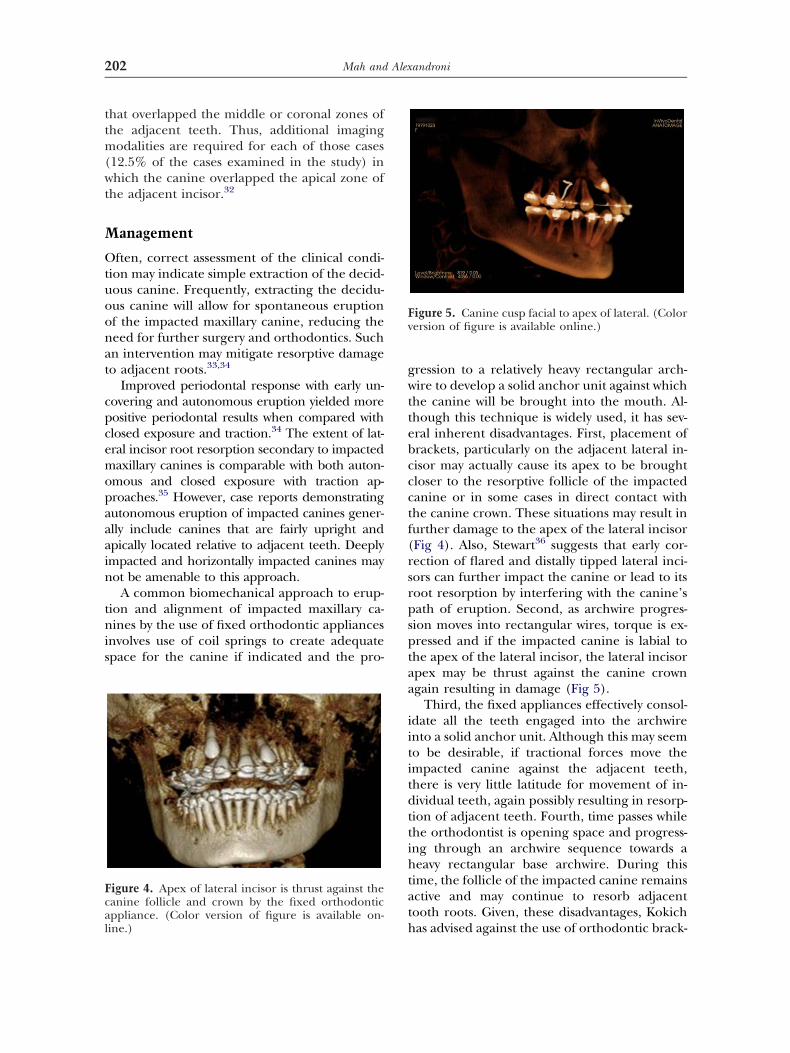
igure 4. Apex of lateral incisor is thrust against theanine follicle and crown by the fixed orthodonticppliance. (Color version of figure is available on-
hine.)
ression to a relatively heavy rectangular arch-ire to develop a solid anchor unit against which
he canine will be brought into the mouth. Al-hough this technique is widely used, it has sev-ral inherent disadvantages. First, placement ofrackets, particularly on the adjacent lateral in-isor may actually cause its apex to be broughtloser to the resorptive follicle of the impactedanine or in some cases in direct contact withhe canine crown. These situations may result inurther damage to the apex of the lateral incisorFig 4). Also, Stewart36 suggests that early cor-ection of flared and distally tipped lateral inci-ors can further impact the canine or lead to itsoot resorption by interfering with the canine’sath of eruption. Second, as archwire progres-ion moves into rectangular wires, torque is ex-ressed and if the impacted canine is labial tohe apex of the lateral incisor, the lateral incisorpex may be thrust against the canine crowngain resulting in damage (Fig 5).
Third, the fixed appliances effectively consol-date all the teeth engaged into the archwirento a solid anchor unit. Although this may seemo be desirable, if tractional forces move thempacted canine against the adjacent teeth,here is very little latitude for movement of in-ividual teeth, again possibly resulting in resorp-ion of adjacent teeth. Fourth, time passes whilehe orthodontist is opening space and progress-ng through an archwire sequence towards aeavy rectangular base archwire. During this
ime, the follicle of the impacted canine remainsctive and may continue to resorb adjacentooth roots. Given, these disadvantages, Kokich
igure 5. Canine cusp facial to apex of lateral. (Colorersion of figure is available online.)
as advised against the use of orthodontic brack-
eivlisaa
tmtfalvdoeoaopmcedcjtiittmt
rttcfpwmbp
otcctacmlpldccpi
C
Thaf
Fuc
Fi
203CBCT in the Management of Impacted Canines
ts and wires in the early phase of managingmpacted maxillary canines.35 This approach in-olves exposure of the impacted canine to theevel of the cementoenamel junction and allow-ng it to erupt autonomously. This techniqueeems to be effective for impacted canines thatre not too deeply impacted and have a favor-ble vertical angulation for unassisted eruption.
In consideration of the aforementioned fac-ors and the precise localization of impacted
axillary canines when CBCT is used, the au-hors have developed a technique that refrainsrom use of brackets in the early stage in favor of
custom-manufactured transpalatal arch. Afterocalization of the impacted canine in the CBCTolume, an occlusal view of the maxilla is used toesign a custom transpalatal arch with a wireutline that does not interfere with the path ofruption of the canine (Fig 6). The occlusal viewf the maxilla is used to design the shape of thenterior portion of the palatal arch to avoid the pathf exposure of the impacted canine and to plan theosition of eyelets. The eyelets are used for attach-ent of elastics and are soldered or laser-welded to
arefully selected locations along the anterior palatalxtension of the transpalatal arch to allow for moreirect lines of force in the traction of the impactedanine. Laser-welding does not create a bulky solderoint and allows for numerous eyelets to be attachedo the transpalatal wire (Fig 7). The transpalatal archs anchored to molar bands. After exposure of thempacted canine, elastic thread is used to directhe canine into a more desirable location. Oncehe canine has been adequately brought into the
outh, fixed appliances are placed. This approacho management of impacted canines allows for
igure 6. Transpalatal arch. (Color version of figure
ts available online.)
elatively immediate movement of the canine offhe apex of the lateral incisor. In general, resorp-ion of the lateral incisor root apex generallyeases when the impacted canine is moved awayrom it.37 In addition, if the tractional forces hap-en to bring the impacted canine into a collisionith the adjacent lateral incisor, the lateral incisoray drift slightly allowing the canine to pass by
ecause the lateral incisor is not rigidly held inlace by an appliance.
Indeed, this type of tooth contact and movementccurs in the development of the dentition duringhe “ugly duckling” stage, when the maxillary caninesontact the apex of the lateral incisors and assist inlosing the spaces between the incisors.38 In contrast,he conventional technique of using a heavy baserchwire to hold the lateral incisor in its place may beounterproductive by interfering with canine move-ent and increasing the likelihood of damage to the
ateral incisor. Furthermore, the total time that theatient is in fixed appliances is greatly reduced, al-
owing for increased efficiency of treatment and re-uced time for breakages, decalcification and otheromplications of fixed orthodontic appliances to oc-ur. Indeed, this approach addresses the issue ofrolonged treatment times in the management of
mpacted canines.8
onclusions
he use of CBCT in orthodontics greatly en-ances our understanding of impacted caninesnd offers unique, comprehensive informationor individual situations. Compared with conven-
igure 7. Custom eyelets on the transpalatal arch aresed to direct tractional eruptive forces to the impactedanines. (Color version of figure is available online.)
ional imaging approaches, the fidelity of this
ifdm
R
1
1
1
1
1
1
1
1
1
1
2
2
2
2
2
2
2
2
2
2
3
3
3
3
3
3
3
3
3
204 Mah and Alexandroni
nformation is unsurpassed. In addition, this in-ormation is invaluable for prognostication andevelopment of biomechanical approaches foranagement of the impacted teeth.
eferences1. Grover PS, Lorton L: The incidence of unerupted per-
manent teeth and related clinical cases. Oral Surg OralMed Oral Pathol 59:420-425, 1985
2. Elefteriadis JN, Athanasiou AE: Evaluation of impactedcanines by means of computerized tomography. Int JAdult Orthodon Orthognath Surg 11:257-264, 1996
3. Shapira Y, Kuftinec MM: Early diagnosis and intercep-tion of potential maxillary canine impaction. J Am DentAssoc 129:1450-1454, 1998
4. Ericson S, Kurol J: Resorption of maxillary lateral inci-sors caused by ectopic eruption of the canines. Am JOrthod Dentofac Orthop 94:503-513, 1988
6. Fox NA, Fletcher GA, Horner K: Localizing maxillarycanines using panoramic tomography. Br Dent J 179:416-420, 1995
7. Stewart JA, Heo G, Glover KE, et al: Factors that relate totreatment duration for patients with palatally impactedmaxillary canines. Am J Orthod Dentofac Orthop 119:216-225, 2001
8. Bishara SE: Impacted maxillary canines: A review. Am JOrthod Dentofac Orthop 101:159-171, 1992
9. Davidovitch Z, Krishnan V: Adverse effects of orthodon-tics: A report of 2 cases. World J Orthod 9:e64-e77, 2008
0. Van der Linden FPGM, Duterloo HS: Development of theHuman Dentition: An Atlas. Hagerstown, Md, Harper &Row, 1976
1. Jacoby H: The etiology of maxillary canine impactions.Am J Orthod 84:125-132, 1983
2. Thilander B, Jakobsson SO: Local factors in impaction ofmaxillary canines. Acta Odontol Scand 26:145-168, 1968
5. Sasakura H, Yoshida T, Murayama S, et al: Root resorptionof upper permanent incisor caused by impacted canine.An analysis of 23 cases. Int J Oral Surg 13:299-306, 1984
6. Leivesley WD: Minimizing the problem of impacted andectopic canines. ASDC J Dent Child 51:367-370, 1984
7. Nute SJ: Severe incisor resorption by impacted maxillarycanines: Case report and literature review. Int J PaediatrDent 14:451-454, 2004
8. Ericson S, Kurol J: Radiographic examination of ectopi-cally erupting maxillary canines. Am J Orthod DentofacOrthoped 91:483-492, 1987
9. Ericson S, Kurol J: Radiographic examination of ectopi-cally erupting maxillary canines. Am J Orthod DentofacOrthoped 91:483-492, 1987
0. Peene P, Lamoral Y, Plas H, et al: Resorption of thelateral maxillary incisor: Assessment by CT. J Comput
Assist Tomogr 14:427-429, 1990
1. Schmuth GPF, Freisfeld M, Koster O, et al: The applica-tion of computerized tomography (CT) in cases of im-pacted maxillary canines. Eur J Orthod 14:296-301, 1992
2. Ericson S, Kurol J: Incisor root resorptions due to ectopicmaxillary canines imaged by computerized tomography: Acomparative study in extracted teeth. Angle Orthod 70:276-283, 2000
3. Walker L, Enciso R, Mah J: Three-dimensional localizationof maxillary canines with cone beam computed tomogra-phy. Am J Orthod Dentofac Orthop 128:418-423, 2005
4. Rimes RJ, Mitchell CT, Willmot DR: Maxillary incisorresorption in relation to the ectopic canine: A review of26 patients. Eur J Orthod 19:79-84, 1997
5. Bodner L, Bar-Ziv J, Becker A: Image accuracy of plainfilm radiography and computerized tomography in as-sessing morphological abnormality of impacted teeth.Am J Orthod Dentofac Orthop 128:623-628, 2001
6. Periago DR, Scarfe WC, Moshiri M, et al: Linear accuracyand reliability of cone beam CT derived 3-dimensionalimages constructed using an orthodontic volumetricrendering program. Angle Orthod 78:387-395, 2008
7. Chaushu S, Chaushu G, Becker A: The role of digitalvolume tomography in the imaging of impacted teeth.World J Orthod 5:120-132, 2004
8. Liu Y, Olszewski R, Alexandroni ES, et al: The validity ofin vivo tooth volume determinations from cone-beamcomputed tomography. Angle Orthod, in press
9. Shirani S, Rawle C, Chrzan B, et al: Evaluation of lateralincisor resorption adjacent to maxillary impacted ca-nines using sector analysis with panoramic radiographsand cone-beam CT, in press
0. Wolf JE, Mattila K: Localization of impacted maxillary caninesby panoramic tomography. Dentomaxillofac Radiol 8:85-91,1979
1. Fox NA, Fletcher GA, Horner K: Localising maxillarycanines using dental panoramic tomography. Br Dent J179:416-420, 1995
2. Chaushu S, Chaushu G, Becker A: The use of panoramicradiographs to localize displaced maxillary canines. OralSurg Oral Med Oral Pathol 88:511-516, 1999
3. Ericson S, Kurol J: Early treatment of palatally eruptingmaxillary canines by extraction of the primary canines.Eur J Orthod 10:283-295, 1988
4. Jacobs SG: Reducing the incidence of unerupted pala-tally displaced canines by extraction of deciduous ca-nines: The history and application of this procedure withsome case reports. Aust Dent J 43:20-27, 1998
5. Schmidt AD, Kokich VG: Periodontal response to earlyuncovering, autonomous eruption, and orthodonticalignment of palatally impacted maxillary canines. Am JOrthod Dentofac Orthoped 131:449-455, 2007
6. Stewart J: Factors that relate to treatment duration forpatients with palatally impacted maxillary canines. Am JOrthod Dentofac Orthoped 119:216-225, 2001
7. Becker A, Chaushu S: Long-term follow-up of severelyresorbed maxillary incisors following resolution of etio-logically associated canine impaction. Am J OrthodDentofac Orthoped 127:650-654, 2005
8. Broadbent BH: Ontogentic development of occlusion.