Page 1/13 Clinical presentation and CT features in pediatric patients with COVID‐ 19 infection Huseyin Avni Solgun ( [email protected]) Altinbas Universitesi https://orcid.org/0000-0001-6811-4600 Isıl Yurdaısık Istinye Universitesi Research Keywords: Covid-19, viral pneumenia, chest computed tomography (CT), VRT (volumetric rendering technique), pediatric Posted Date: June 11th, 2021 DOI: https://doi.org/10.21203/rs.3.rs-595070/v1 License: This work is licensed under a Creative Commons Attribution 4.0 International License. Read Full License
Transcript
Page 1/13
Clinical presentation and CT features in pediatricpatients with COVID‐19 infectionHuseyin Avni Solgun ( [email protected] )
BackgroundThe aim of this study includes to discuss the clinical, laboratory, and chest computed tomography (CT) inpediatric patients with 2019 novel coronavirus (COVID-19) infection.
Material and MethodsThe clinical, laboratory, and chest CT features of 17 pediatric inpatients with COVID-19 infectioncon�rmed by pharyngeal swab COVID‐19 polymerase chain reaction(PCR). All clinical and laboratorydata have been recorded and analyzed during march-february 2021. Chest CT have been performed to allCovid 19 PCR con�rmed patients and radiologicall view have been noted.
ResultsSeventeen pediatric patients with a history of close contact with COVID-19 diagnosed family membersincluded to the study. Fever (10/17, 58%) and cough (13/17, 76%) were the most common symptoms. Forlaboratory �ndings, c reactive protein elevation (15/17, 88%) seem to be the most �nding. A total of 4patients presented with unilateral pulmonary lesions (4/17, 23%), 9 with bilateral pulmonary lesions(9/17, 52%) and 13 cases showed bilateral diffuse covid pattern on chest CT (13/17, 76%). Non-spesi�cconsolidation with was observed in 8 patients (8/17, 47%), ground‐glass opacities were observed in 11patients (11/17, 64%), nodules were observed in 7 patients (7/17, 41%), and tiny nodules were observedin 2 patients (2/17, 11%).
ConclusionIn pediatric patients with positive COVID-19 nucleic acid test from pharyngeal swab samples; the earlydetection of lesions by CT can be e�cient; in management and early treatment for pediatric patients.However; early chest CT screening and COVİD-19 PCR testing together can be more e�cent in diagnose.
IntroductionA pneumonia epidemic broke out in Wuhan and then spread to other Chinese cities and several countriesresectively in december 2019. A new type of coronavirus announced by the Chinese Center for DiseaseControl and Prevention on January 7, 2020 [1]. Finally; on february 11, 2020, the International Committeeon Taxonomy of Viruses (ICTV) proposed to name the new virus SARS-CoV-2 and the WHO named thedisease caused by SARS-CoV-2 infection COVID-19 [2, 3]. As of today, 8 months after the onset ofepidemic, China’s domestic COVID-19 epidemic has been well controlled contravarsiouly the epidemicspreaded to many countries worldwide[4]. Lately; the virus outbreak in countries of Europe and America
Page 3/13
are severely affected at this moment, which means that COVID-19 has evolved from an epidemic topandemic. From the early days of the outbreak to this moment; the disease showed that there were toless cases in children under the age of 15 [5]. Soon afterwards, laboratory-diagnosed cases from all overChina through January 29, 2020, indicated that 0.9% of patients were aged below 15 years, which meansthat COVID-19 can be spread within the whole age spectrum [6].
In this study; clinical and imaging features of pediatric patients with COVID-19 infection were presented ina series of 17 cases who have been identi�ed by the pharyngeal swab COVID‐19 nucleic acid test.
Material And MethodsSeventeen pediatric inpatients with COVID-19 infection con�rmed by pharyngeal swab COVID‐19 nucleicacid test from march to february 2021 in our university hospital were included in this study. All thepatients are in accordance to the Diagnosis and Treatment Protocol for COVID‐19 by the National HealthCommission.
Clinical data including demography information, contact history, previous history, clinical symptoms,laboratory �ndings, and coinfection which de�ned as a concurrent infection of a patient with two or morepathogens simultaneously.
The chest CT were obtained from all subjects, as the plain chest X-ray cannot exclude the existence ofpulmonary lesions, especially for the patients without symptoms and mild cases. For all the patients,noncontrast chest CT studies were performed on SOMATOM De�niton AS 128 unit (Siemens medicalsystem; Siemens, Germany) with the following parameters: 12 0 kV, 100 to 150 mA, 0.6‐mm collimation,and 1:1 pitch. The scanning range covered from lung apex to diaphragm on axial plane taken under freebreathing with the patients in the supine position. CT images were reconstructed with 3 or 4 mmcollimation with a standard algorithm and then sent to the picture archiving and communication system(PACS) for analyzing. CT images were evaluated using a lung window with a window level of − 600 HU awindow width of 1500 HU, and the soft‐tissue window with a window level of 40 HU and window width of300 HU. All the images were stored in PACS and reviewed by experienced pediatric radiologists. The CTfeatures were evaluated as follows: (a) ground‐glass opacities, (b) consolidations with surrounding halosign, (c) nodules, (d) �ne mesh shadow, (e) pleural effusion, (f) lymphadenopathy, (g) unilateral orbilateral, (h) subpleural or nonsubpleural, and (i) residual �ber strips. Pharyngeal swab samples of all thesubjects in this group were collected, and the COVID‐19 RNA was identi�ed by a reverse transcription‐polymerase chain reaction.
The protocol for this retrospective study was approved by the Ethics Committee of Istinye UniversityMedical Park GOP Hospital and the written informed consent was waived for emerging infectiousdiseases.
Results
Page 4/13
Seventeen pediatric patients with a history of close contact with COVID-19 diagnosed family membersincluded to the study. Fever (10/17, 58%) and cough (13/17, 76%) were the most common symptoms.The clinical features of pediatric patients with COVID‐19 infection were displayed in Table 1.
Table 1The clinical features of pediatricpatients with COVID-19 infection
Characteristic Number (%)
Sex
Boy 9 (53%)
Girl 8(47%)
Age
2< 3(17%)
2–5 2(11%)
5–10 4(23%)
10> 8(47%)
Contact history
Yes 10 (59%)
No 7 (41%)
Symptom
Cough 11 (64%)
Tachypnea 4 (29%)
Fever 9 (52%)
Nasal discharge 4 (23%)
Diarrhea 3 (17%)
Fatigue 2 (11%)
Cardiac arrhythmia 1 (5%)
Other(otit,abdominal pain…) 3( 17%)
For laboratory �ndings, c reactive protein elevation (15/17, 88%) seem to be the most �nding. Thelaboratory features of pediatric patients with COVID-19 infection were displayed in Table 2.
Page 5/13
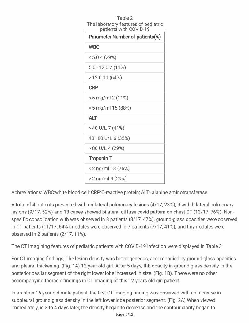
Table 2The laboratory features of pediatric
patients with COVID-19Parameter Number of patients(%)
A total of 4 patients presented with unilateral pulmonary lesions (4/17, 23%), 9 with bilateral pulmonarylesions (9/17, 52%) and 13 cases showed bilateral diffuse covid pattern on chest CT (13/17, 76%). Non-spesi�c consolidation with was observed in 8 patients (8/17, 47%), ground‐glass opacities were observedin 11 patients (11/17, 64%), nodules were observed in 7 patients (7/17, 41%), and tiny nodules wereobserved in 2 patients (2/17, 11%).
The CT imagining features of pediatric patients with COVID‐19 infection were displayed in Table 3
For CT imaging �ndings; The lesion density was heterogeneous, accompanied by ground-glass opacitiesand pleural thickening. (Fig. 1A) 12 year old girl. After 5 days, thE opacity in ground glass density in theposterior basilar segment of the right lower lobe increased in size. (Fig. 1B). There were no otheraccompanying thoracic �ndings in CT imaging of this 12 years old girl patient.
In an other 16 year old male patient, the �rst CT imaging �nding was observed with an increase insubpleural ground glass density in the left lower lobe posterior segment. (Fig. 2A) When viewedimmediately, ie 2 to 4 days later, the density began to decrease and the contour clarity began to
Page 6/13
disappear. (Fig. 2C) The lesion size and density increased signi�cantly 6 days after this diagnosis.(Fig. 2C) After 14 days (24 days after initial detection) taken for the 4th time, the lung parenchyma isobserved normally. (Fig. 2D). VRT (volumetric rendering technique) images of the same patient weresimilar in discussion. (Figs. 3A, 3B, 3C, 3D) The chest VRT image is more valuable to see ground glassconsolidation and other signs of COVID-19 pneumonia in children. This is the �rst report in the literaturediscussing VRT in this age group of COVID-19 pneumonia.
DiscussionThe outbreak of Covid-19 started in Wuhan city, Hubei province, China, where the �rstly announced casesin adults with pneumonia of unexplained etiology on December 31, 2019. A local seafood and animalmarket was de�ned to be as a potential source. Afterwards; main transmission route to cause outbreakwas de�ned through respiratory droplets or direct contact from symptomatic and asymptomatic humansinfected with Covid-19. Covid-19 has spread to other Chinese cities and internationally and caused aglobal pandemic.
COVID- 19 viral pneumonia is an acute infectious respiratory disease caused by a coronavirus subtypeSARS-CoV-2. From december 2019 to this moment, 24.355.000 total com�rmed cases, 830.155 deathsand 16.889.000 recovered cases had been con�rmed worldwide as WHO (World Health Organization) upto date records, while the actual number would be larger with nonco�med asymptmatic cases. [8] Thevirus is a highly contagious disease andcan be transmitted by an infected person or an asymptomaticcarrier through respiratory droplets. Respiratory droplets are the main route of transmission, but can alsobe transmitted by contact and digestive tract. [9] After contact to infected person; The incubation period isabout 1 to 14 days, and is supposed could be up to 24 days. Even most of the cases are mild, especiallypeople over 60 years old or those with underlying diseases are more likely to develop the severe diseaseof lower repiraturary system involvement. [10] The clinical manifestations of children patients are similarto those of adults, such as fever and cough. A few children have diarrhea and runny nose, but the overallsymptoms are relatively mild. Its think to be that the COvid-19 infection have a mild and weak clinicalprogress in childeren. Conversly to this data; in this study we presented 17 cases those all are under 17years old with 3 of them under 2 years old cases of Covid-19 infection with severe diseases.
In previous literatüre Chest CT �ndings in children were similar to those in adults, and most of them weremild cases. [11, 12] In our study; the typical manifestations were unilateral or bilateral subpleural ground-glass opacities, and consolidations with surrounding halo sign. As bilatarelly consolidations of lungssign account for up to 70% cases, they should be considered as typical signs in pediatric patients. Pleuraleffusion was seen in 4 cases. In Wei and et al study; the data for pleural effusion account zero. [13]Lesions could be still visible on chest CT when two consecutive nucleic acid tests were negative. The CTimaging of COVID‐19 infection should be differentiated with other virus pneumonias, such as respiratorysyncytial virus, in�uenza virus, parain�uenza virus, and adenovirus with its spesi�c radiological signs.[14] In addition, it should be differentiated from atypic bacterial pneumonia such as mycoplasmapneumonia and chlamydia pneumonia. However, multiple agents can overlap chest CT manifestations of
Page 7/13
pneumonia caused by COVID‐19 presenting more serious and complex imaging manifestations whichcould not be diagnostic, so epidemiological and etiological examination should be combined to make the�nal decision.
Page 8/13
Table 3The CT imagining features of pediatric patients with COVID-19 infection
Features Number of patients (%)
Pulmonary involvement
Focal 2 (11%)
Unilateral diffuse 3 (17%)
Bilateral diffuse 12 (70%)
Subpleural lesions 15 (88%)
Nonspesi�c consolidation 2(11%)
Ground-glass opaci�cation 12 (70%)
Nodules 1 (5%)
Recently; only a few studies have conducted on chest CT signs of COVID-19 in children age group.[15, 16, 17] Even normal �ndings on chest CT; some of pediatric patients manifest a severe clinicaldisease of COVID-19 pneumonia.
The CT manifestations of COVID-19 in pediatric patients are diverse and lack speci�city. Some mildpediatric patients with COVID-19 show [18, 19].
In pulmunary involvement cases; focal unilateral or bilateral diffuse, subpleural lesions, nonspesi�cconsalidation, ground-glass opaci�cation and nodules are the most presentations.
Pediatric patients with COVID-19 tend to present less lobular involvement with an increase insubpleural ground glass density in the left lower lobe posterior segments (Fig. 1a,2a).Additionally;some other �ndings like nodular ground-glass opaci�cation consolidation, consolidationwith ground-glass opaci�cation and interlobular septal thickening can also be observed in thepediatric patients [20]. All radiological �ndings have been summarized in Table 3.
Overall, rarely in pediatric cases, bilateral diffuse lung consolidation can occur and is called as “whitelung” [16]. In resolving stage, lung lesions will be completely resolved or only remain minimal linearopacities (Fig. 2d,3d). In some cases either can be a presentation of similiar to those other viralagents
With patchy opacity along the bronchial vasculer structure manisfasting as bronchopneumonia. [16]
Case differantial diagnosis should be more carefully done while pediatric patients have de�niteepidemiological history but atypical CT �ndings. Xia et al reported, underlying coinfection is verycommon in pediatric patients (9 of 20, 45%), Pleural effusion was reported in several pediatricpatients [21].
Even the gold standart is nucleic acid detection in diagnosis of COVİD-19, in suspecious cases thoseinitial RT-PCR results show negative, chest CT may be supporttive for diagnosis and managementespecially in pediatric age group. Additionally Chest Ct can suggest the healing and resolve �ndingsof lung involvement regarding the disease severness and follow up options. In this study the chest ct�ndings in 14th day of follow up have been completely recovered. (Fig. 2d,3d). Sure the nucleic acidcom�rmation test negativity can be used in follow up; unfourtanetly it will not suggest any idea inthose cases with pulmunary symptoms in childhood.
Conclusions
Page 9/13
Features Number of patients (%)In some children; COVID-19 virus pneumonia has a severe clinical and radiological course and inchest CT can present characteristic changes of subpleural ground‐glass opaciticitions and uni-bilateral consolidations which is so effective for follow up and evaluating the changes of lunglesions. In patients with positive COVID‐19 nucleic acid test from pharyngeal swab samples andespecially in course of cough and respiratuar other symptoms; the early detection of lesions by chestCT is very e�cient to decide life-saving treatment for pediatric patients. In addition; Chest CT imagingis not su�cient enough alone to determine the COVID‐19 pneumonia and early chest CT screeningand COVİD-19 PCR testing together can be more e�cent in diagnose.
DeclarationsEthics approval and consent to participate have been taken from Health Sciences University.
Ethical Committie
Consent for publication have been taken from the patients’ parents. Patient’s parents gave informedwritten consent for their personal or clinical details along with any identifying images to be publishedin this study.
Acknowledgements The authors are thankfull to all indivuduals have contributed to this study.
Funding: 'N/A'
Con�icts of interest/Competing interests: 'N/A'
Availability of data and material: 'N/A'
Authors' contributions: Author Huseyin Avni Solgun and et al. have read and approved themanuscript.
Consent for publication: 'N/A'
References1. Zhu N, Zhang D, Wang W et al (2020) A novel coronavirus from patients with pneumonia in
China, 2019. N Engl J Med 382:727–7332. Gorbalenya AE, Baker SC, Baric RS et al (2020) Severe acute respiratory syndrome-related
coronavirus: the species and its viruses – a statement of the Coronavirus Study Group. bioRxiv.https:// doi.org/10.1101/2020.02.07.937862
3. World Health Organization (2020) WHO Director-General’s remarks at the media brie�ng on 2019-nCoV on 11 February 2020. World Health Organization, Geneva. Available via https://www. who.int/dg/speeches/detail/who-director-general-s-remarks-at-themedia-brie�ng-on-2019-ncov-on-11-february-2020. Accessed 10 Apr 2020
4. National Health Commission of the People’s Republic of China (2020) Update on COVID-19 as of24:00 March 18. National Health Commission of the People’s Republic of China, China. Availablevia http://www.nhc.gov.cn/xcs/yqtb/202003/ e644c2fc18b4448db7ed4b30f68b91a6.shtml.Accessed 19 Mar 2020 (in Chinese)
5. Li Q, Guan X, Wu P et al (2020) Early transmission dynamics in Wuhan, China, of novelcoronavirus-infected pneumonia. N Engl J Med 382:1199–1207
�. Guan W, Ni Z, Hu Y et al (2020) Clinical characteristics of 2019 novel coronavirus infection inChina. N Engl J Med. https://doi.org/ 10.1056/NEJMoa2002032
7. Chan JF, Yuan S, Kok KH, et al. A familial cluster of pneumonia associated with the 2019 novelcoronavirus indicating person-to-person transmission: a study of a family cluster. Lancet.2020;395:514–523.
Page 10/13
Features Number of patients (%)�. Parry J. Wuhan: Britons to be evacuated as scientists estimate 44 000 cases of 2019‐nCOV in the
city. Brit Med J. 2020;368:m351.9. Riou J, Althaus CL. Pattern of early human‐to‐human transmission of Wuhan 2019 novel
coronavirus (2019‐nCoV), December 2019 to January 2020. Euro Surveill. 2020;25:7‐11.10. Hui DS, Azhar EI, Madani TA, et al. The continuing 2019‐nCoV epidemic threat of novel
coronaviruses to global health—the latest 2019 novel coronavirus outbreak inWuhan, China. Int JInfect Dis. 2020;91:264‐266.
11. Kanne JP. Chest CT �ndings in 2019 novel coronavirus (2019‐nCoV) infections from Wuhan,China: key points for the radiologist. Radiology. 2020.
12. Song F, Shi N, Shan F, et al. Emerging coronavirus 2019‐nCoV pneumonia. Radiology. 2020.13. Wei Xia MD1 , Jianbo Shao MD1 ,et al. Clinical and CT features in pediatric patients with COVID‐
19 infection: Different points from adults. 26 February 2020, Pediatric Pulmonology. 2020;1–6.14. Virkki R, Juven T, Rikalainen H, Svedstrom E, Mertsola J, Ruuskanen O. Differentiation of bacterial
and viral pneumonia in children. Thorax. 2002;57:438‐44115. FengK,YunYX, WangXFetal (2020) AnalysisofCTfeaturesof
15childrenwith2019novelcoronavirusinfection.ZhonghuaErKe Za Zhi 58:E0071�. Ma H, Shao J, Wang Yet al (2020) High resolution CT features of novel coronavirus pneumonia in
children.Zhonghua Fang She Xue Za Zhi 54:E00217. SunD,LiH,LuXXetal(2020)Clinicalfeaturesofseverepediatric patients with coronavirus disease
2019 in Wuhan: a single center’s observational study. World J Pediatr. https://doi.org/10.1007/s12519-020-00354-4
1�. Xia W, Shao J, Guo Y, Peng X, Li Z, Hu D (2020) Clinical and CTfeaturesinpediatricpatientswithCOVID-19infection:differentpoints from adults. Pediatr Pulmonol.
19. ZhaoR,ShenX,XuK,ShengJ(2020)One casereport of pediatric infection with COVID-19. ZhejiangMed J. https://doi.org/10. 12056/j.issn.1006-2785.2020.42.3.2020-337
20. Ma Y, Xia S, Wang M, Zhang S, Du W, Chen Q (2020) Clinical features of children with SARS-CoV-2infection:ananalysisof 115 cases. Chin J Contemp Pediatr 22:1–4
21. Zhou Y, Yang GD, Feng K et al (2020) Clinical features and chest CT �ndings of coronavirusdisease 2019 in infants and young children. Zhongguo Dang Dai Er Ke Za Zhi 22:215–220 ,
Figures
Page 11/13
Features Number of patients (%)
Figure 1
12 year old girl. It was observed that opacity in ground glass density in the posterior basilar segmentof the right lower lobe increased in size after 5 days. There were no other accompanying thoracic�ndings.
Page 12/13
Features Number of patients (%)
Figure 2
16-year-old male.The �rst imaging �nding was observed with an increase in subpleural ground glassdensity in the left lower lobe posterior segment.(2a ) When looked at once, that is, after 2 to 4 days,density began to decrease and contour clarity began to disappear. (2b) The lesion size and densityincreased signi�cantly 6 days after this diagnosis. (2c ) After 14 days (24 days after the �rstdetection) taken for the 4th time, the lung parenchyma is observed normally. (2d )
Page 13/13
Features Number of patients (%)
Figure 3
VRT (volume rendering technique) images of the same patient in Figure 2.
Supplementary Files
This is a list of supplementary �les associated with this preprint. Click to download.