2.12.) SECONDARY AMENORRHEA normal prolactin Any women with primary ovarian failure or ovarian failure before age 35 (“premature menopause”) should be karyotyped. withdrawal bleeding anovulation polycystic ovarian disease adrenal tumor ovarian tumor withdrawal bleeding high LH / FSH ovarian failure normal or low LH / FSH get head CT or MRI (see 1.13) if normal: hypothalamic amenorrhea no withdrawal bleeding outflow tract problem Asherman’s syndrome active endometritis
Transcript
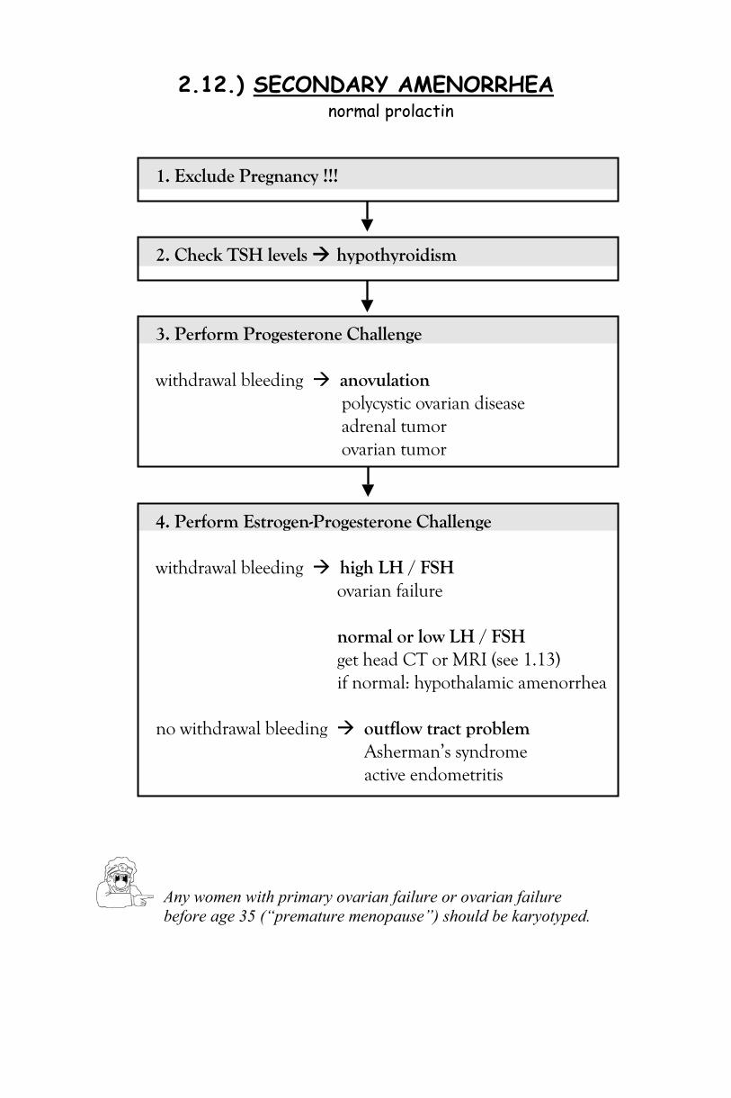
2.12.) SECONDARY AMENORRHEA normal prolactin
Any women with primary ovarian failure or ovarian failure before age 35 (“premature menopause”) should be karyotyped.
4. Perform Estrogen-Progesterone Challenge withdrawal bleeding high LH / FSH
ovarian failure
normal or low LH / FSH get head CT or MRI (see 1.13) if normal: hypothalamic amenorrhea
no withdrawal bleeding outflow tract problem
Asherman’s syndrome active endometritis
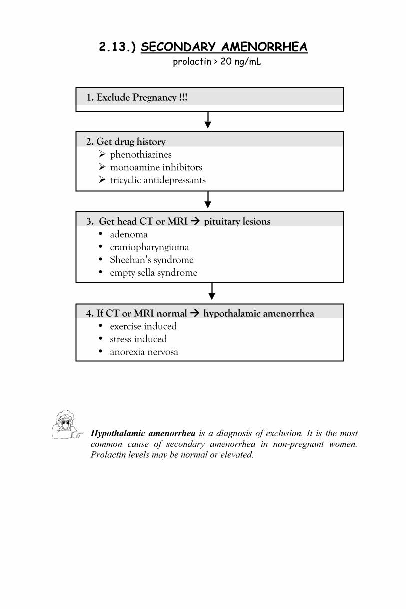
2.13.) SECONDARY AMENORRHEA prolactin > 20 ng/mL
Hypothalamic amenorrhea is a diagnosis of exclusion. It is the most common cause of secondary amenorrhea in non-pregnant women. Prolactin levels may be normal or elevated.
1. Exclude Pregnancy !!!
2. Get drug history phenothiazines monoamine inhibitors tricyclic antidepressants
3. Get head CT or MRI pituitary lesions • adenoma • craniopharyngioma • Sheehan’s syndrome • empty sella syndrome
4. If CT or MRI normal hypothalamic amenorrhea • exercise induced • stress induced • anorexia nervosa
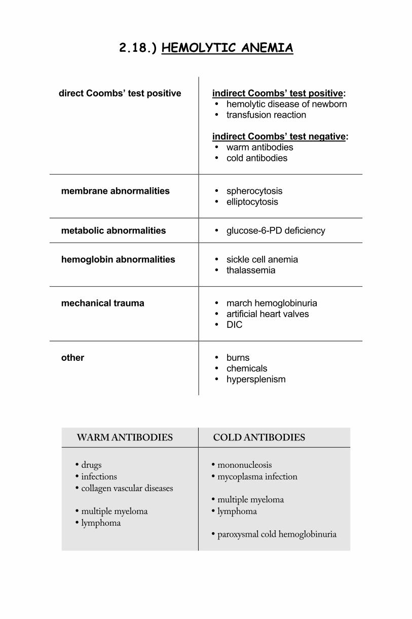
2.18.) HEMOLYTIC ANEMIA
direct Coombs’ test positive
indirect Coombs’ test positive:
• hemolytic disease of newborn • transfusion reaction
indirect Coombs’ test negative:
• warm antibodies • cold antibodies
membrane abnormalities
• spherocytosis • elliptocytosis
metabolic abnormalities • glucose-6-PD deficiency
hemoglobin abnormalities
• sickle cell anemia • thalassemia
mechanical trauma
• march hemoglobinuria • artificial heart valves • DIC
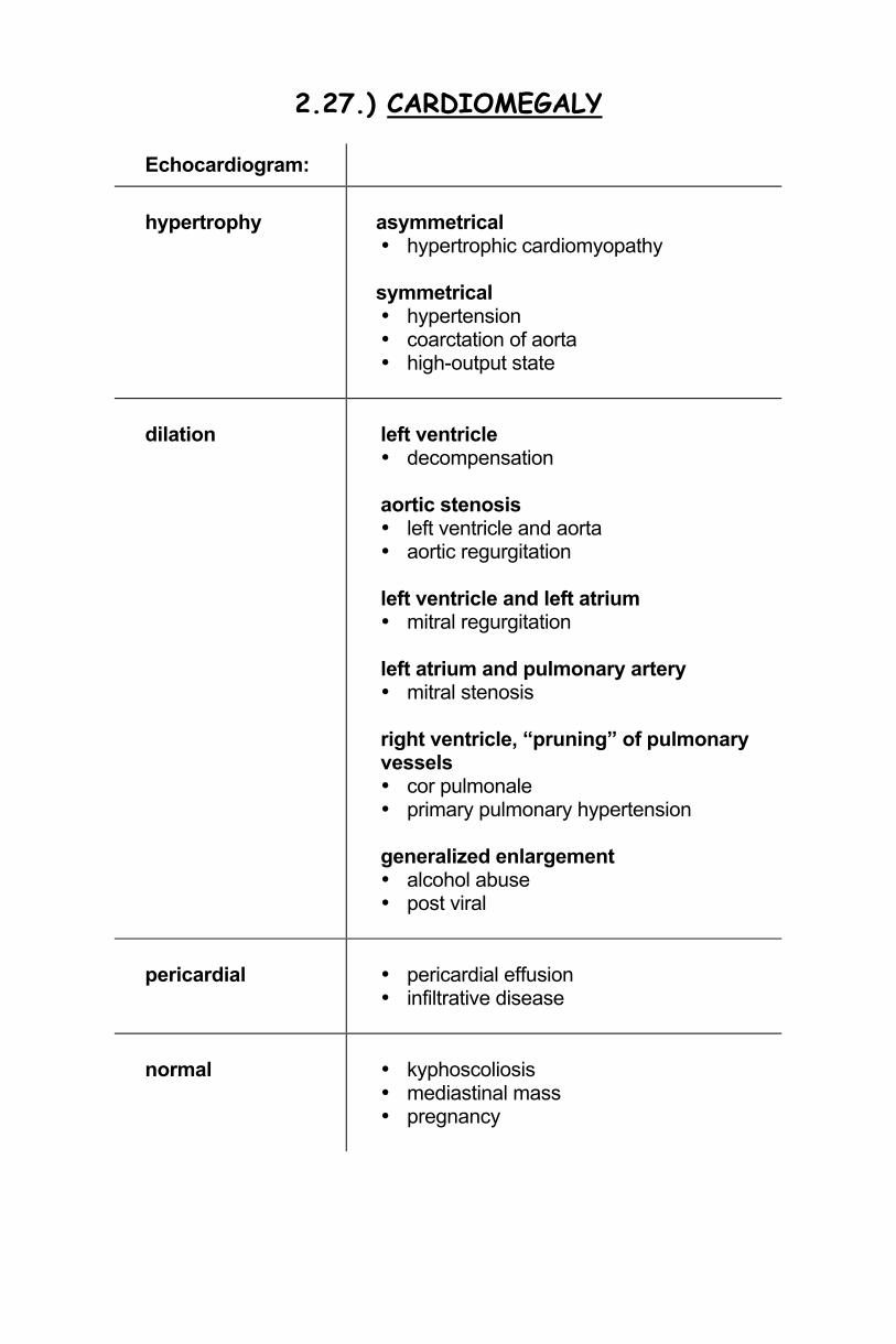
• hypertension • coarctation of aorta • high-output state
dilation
left ventricle • decompensation aortic stenosis • left ventricle and aorta • aortic regurgitation left ventricle and left atrium • mitral regurgitation left atrium and pulmonary artery • mitral stenosis right ventricle, “pruning” of pulmonary vessels • cor pulmonale • primary pulmonary hypertension
generalized enlargement • alcohol abuse • post viral
pericardial
• pericardial effusion • infiltrative disease
normal
• kyphoscoliosis • mediastinal mass • pregnancy
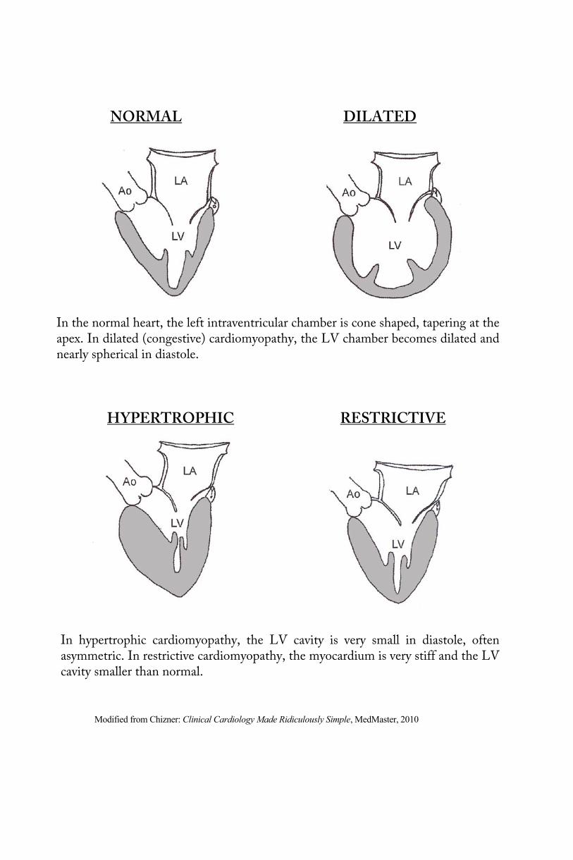
NORMAL DILATED
In the normal heart, the left intraventricular chamber is cone shaped, tapering at the apex. In dilated (congestive) cardiomyopathy, the LV chamber becomes dilated and nearly spherical in diastole.
HYPERTROPHIC RESTRICTIVE
In hypertrophic cardiomyopathy, the LV cavity is very small in diastole, often asymmetric. In restrictive cardiomyopathy, the myocardium is very stiff and the LV cavity smaller than normal.
Modified from Chizner: Clinical Cardiology Made Ridiculously Simple, MedMaster, 2010
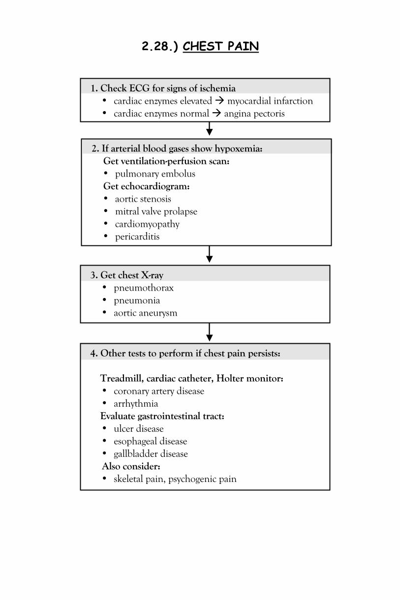
2.28.) CHEST PAIN
1. Check ECG for signs of ischemia • cardiac enzymes elevated myocardial infarction • cardiac enzymes normal angina pectoris
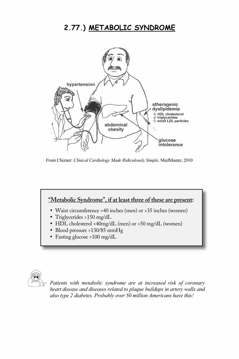
Patients with metabolic syndrome are at increased risk of coronary heart disease and diseases related to plaque buildups in artery walls and also type 2 diabetes. Probably over 50 million Americans have this!
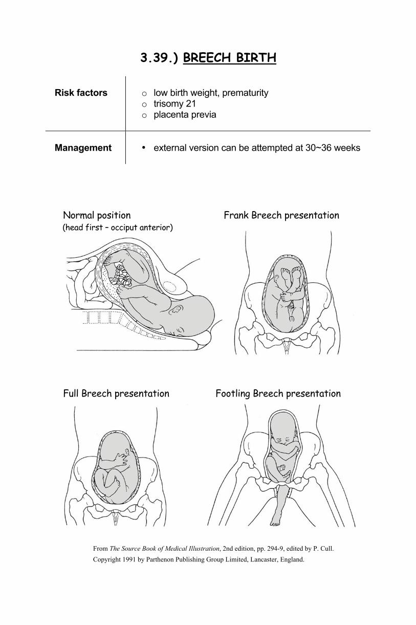
3.39.) BREECH BIRTH
Risk factors
o low birth weight, prematurity o trisomy 21 o placenta previa
Management
• external version can be attempted at 30~36 weeks
Normal position Frank Breech presentation (head first – occiput anterior)
Full Breech presentation Footling Breech presentation
From The Source Book of Medical Illustration, 2nd edition, pp. 294-9, edited by P. Cull. Copyright 1991 by Parthenon Publishing Group Limited, Lancaster, England.
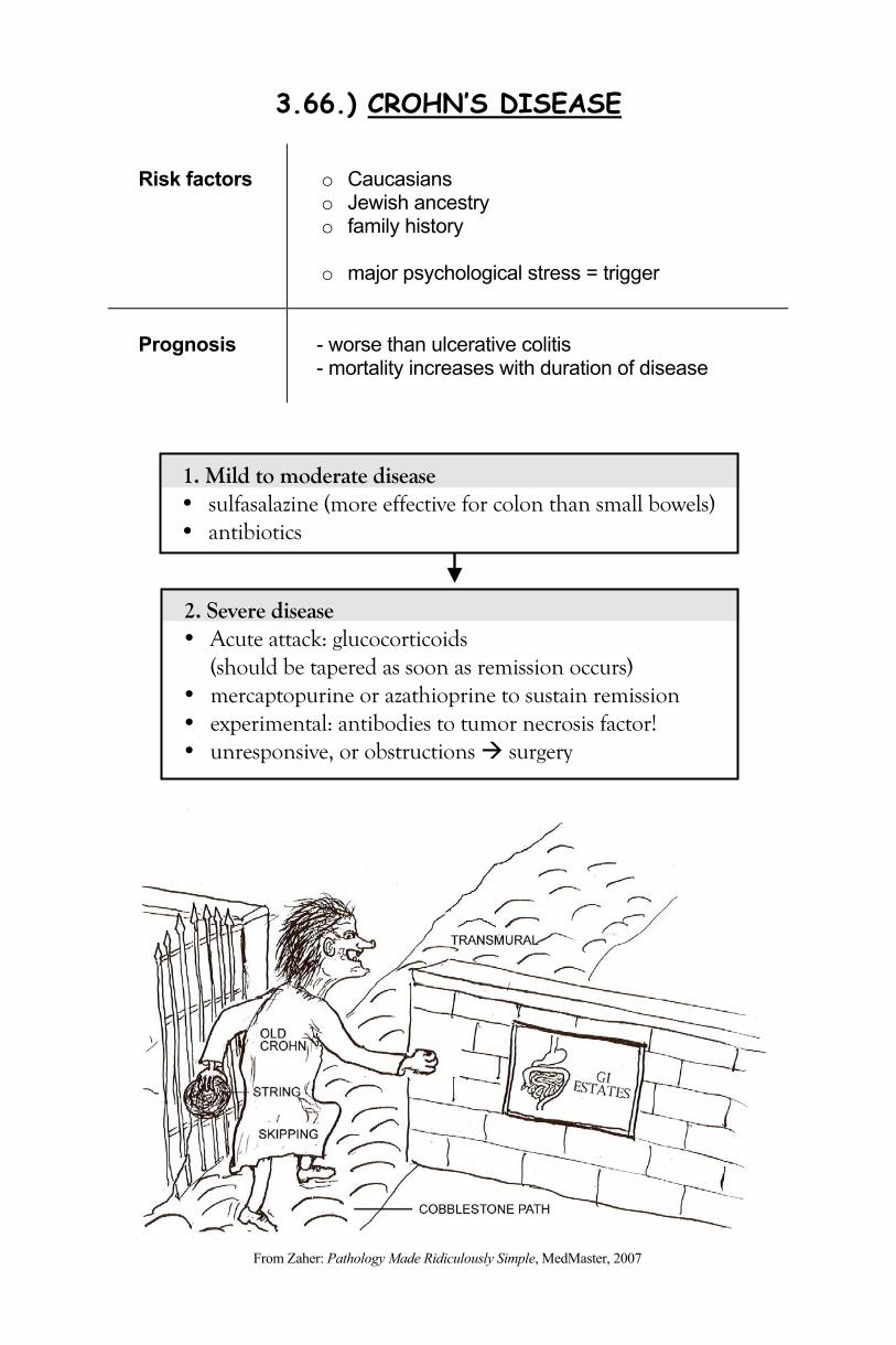
3.66.) CROHN’S DISEASE
Risk factors
o Caucasians o Jewish ancestry o family history
o major psychological stress = trigger
Prognosis
- worse than ulcerative colitis - mortality increases with duration of disease
From Zaher: Pathology Made Ridiculously Simple, MedMaster, 2007
1. Mild to moderate disease • sulfasalazine (more effective for colon than small bowels) • antibiotics
2. Severe disease • Acute attack: glucocorticoids
(should be tapered as soon as remission occurs) • mercaptopurine or azathioprine to sustain remission • experimental: antibodies to tumor necrosis factor! • unresponsive, or obstructions surgery
3.74.) DIABETES MELLITUS TYPE 1 IDDM
Risk factors
o HLA-DR3 o HLA-DR4 o monozygotic twin concordance only 50%
Honeymoon effect: Initial treatment with insulin restores some β-cell function risk of hypoglycemia due to increased endogenous insulin. Dawn phenomenon: Early morning rise in glucose due to circadian changes in GH and cortisol. Somogyi effect: Exaggerated dawn phenomenon. Nocturnal hypo- glycemia results in overshooting morning hyperglycemia. Manage by decreasing evening insulin.
3. Follow-up • quarterly physical exam, including HBA1C
1. Insulin • morning dose before breakfast • evening dose before dinner • mix intermediate (NPH) with short acting (regular) insulins
2. Family education is extremely important! • carbohydrate counting, regular meal times • physical exercise: reduce insulin or provide extra snack
3.154.) LEUKEMIA, CHRONIC
Background
CLL is the most common form of leukemia overall
Risk factors
o age, male o Philadelphia chromosome (t9:22) in CML
Prognosis
CML: often converts to AML within 2 years with poor prognosis CLL: indolent for many years
CLL • usually indolent: early treatment does NOT improve survival • chemotherapy if anemia or neutropenia or other signs of disease
• Imatinib specifically inhibits tyrosinkinase activity of the bcr/abl oncogene
• assess molecular response: bcr/abl/abl-ratio by PCR is the “gold-standard”
• consider allogenic bone marrow transplant for patients > 50y. • blast phase: treat like AML
3.227.) REYE’S SYNDROME = fatty liver plus encephalopathy
Risk factors
o Influenza B o viral infections aspirin, salicylates in children and teens
Management
1. supportive 2. mannitol to reduce cerebral edema
3.228.) RHEUMATOID ARTHRITIS
Risk factors
o female o Native Americans o HLA-DR4
Management
Anti-inflammatory
NSAIDs glucocorticoids
Disease-modifying drugs
methotrexate (low-dose) sulfasalazine rituximab
(monoclonal antibody against B cells) TNF inhibitors
Disease-modifying drugs should be given early to slow the irreversible joint destruction! Don’t expect beneficial effects until 2~6 months after initiating therapy.