39
| Date post: | 05-Jul-2015 |
| Category: |
Documents |
| Upload: | mala-propia |
| View: | 1,384 times |
| Download: | 2 times |
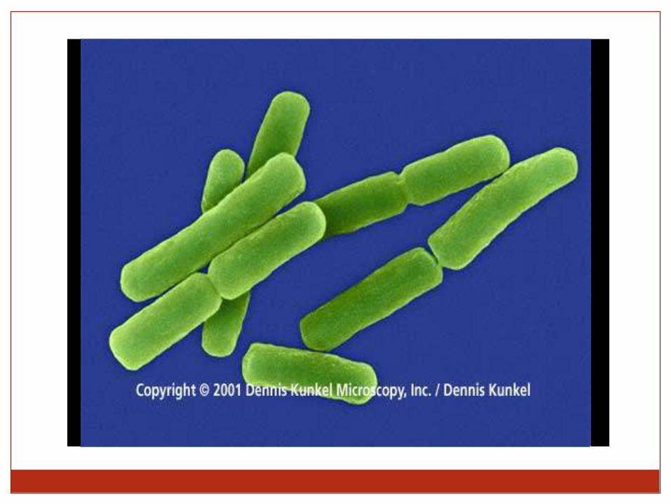

Anthrax Bacillus anthracis
Cutaneous
anthrax

M A T A K U L I A H M I K R O B I O L O G I
E V I R O V I A T I
STRUKTUR SEL BAKTERI

Prokaryotes
Domains
Bacteria &
Archaea
Simple cells –
with no nucleus
or membrane-
bound
organelles

I. Bacteria Classification: Cell shape
A. Compound Light Microscope (1000X) - stained
Cocci (Coccus) Bacilli (Bacillus) Spirilli (Spirillum)
round or oval rod-shaped helically coiled

B. Scanning Electrom Microscope (SEM) - colorized

II. Classification Bacteria: Cell arrangement
1. Diplococcus (diplo=pairs)
Neisseria gonorrhoeae - Gram-negative,
causes gonorrhea
SEMStained: Compound Microscope
1000X

Staphylococcus aureus
Staphylococcus aureus - Causes food poisoning, toxic shock syndromeand skin and wound infections such as scalded skin syndrome, scarlet fever, and impetigo.
Stained: Compound Microscope
1000X
SEM (colorized)
2. staphylococcus (staphylo- grapelike clusters)

3. streptococcus (strepto=chains)
Streptococcus pyogenes
Stained: Compound Microscope
1000XSEM (colorized)

4. Streptobacillus

Bacteria
Structure
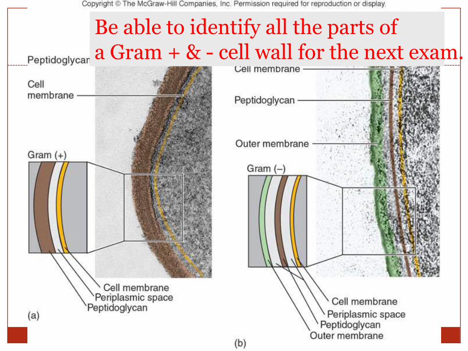
Cell wall – unique, peptidoglycan
Peptidoglycan - structural
polysaccharides
(sugars) cross-linked by
peptides (chains of amino
acids)

The Gram stain procedure
Developed in 1884 by the Danish physician
Hans Christian Gram
An important tool in bacterial taxonomy,
distinguishing so-called Gram-positive
bacteria, which remain coloured after the
staining procedure, from Gram-negative
bacteria, which do not retain dye and need to
be counter-stained.
Can be applied to pure cultures of bacteria
or to clinical specimens
Top: Pure culture of E. coli
(Gram-negative rods)
Bottom: Neisseria gonorrhoeae in a smear of urethral pus
(Gram-negative cocci, with pus cells)

Crystal violet
Gram's iodine
Decolorise with acetone
Counterstain withe.g. methyl red
Gram-positives appear purple
Gram-negatives
appear pink
The Gram Stain

Gram-positive rods
Gram-negative rods
Gram-positive cocci
Gram-negative cocci

Gram stainDistinguishes different cell wall types
Gram positive Staphylococcus aureus
Gram negative Escherichia coli
16
Be able to identify all the parts of a Gram + & - cell wall for the next exam.

Two biochemical groups of bacteria:
peptidoglycanouter
membrane

will stain will not stain
Gram positive bacteria Gram negative bacteria
Two biochemical groups of bacteria:
peptidoglycanouter
membrane

Bacteria with Chemically Unique Cell Walls
Acid-Fast Cells
Mycobacterium species
Gram + type of cell wall
Unique lipid
Mycolic acid – waxy substance
Does not decolorize

Bacterial Growth
Solid media or liquid media
Agar plates, slopes, broth culture
Atmosphere:
Aerobic, anaerobic or microaerophilic
Facultative or obligate anaerobes
Usually at 37 degrees C
Most clinically important bacteria grow overnight, or within a few days
Mycobacteria can take months
Some can not be grown

Capsules or slime layer
E.g., slime layer allows bacteria to
cling to tooth enamel or other
substrates

Pili (singular: pilus)Protein filaments that attach bacteria to other cells
& substrates
pili

Used for locomotion
Some prokaryotes have flagella(singular: flagellum)
flagella

50 nm
Base of a bacterial flagellum…
…the only known wheel in nature

Reproduction: Asexual, through binary fission
E. coli
DNA
cell wall

Binary fissionDaughter cells are identical copies
(1) (2) (3)
(4) (5) (6)
Chromosome Plasma membrane
Neither mitosis nor meiosis occurs in prokaryotes

REPRODUCTION
Asexual, through binary fission
No true sexual reproduction, since neither
mitosis nor meiosis exist in prokaryotes
Horizontal transfer of genetic material
Transformation Uptake of genetic material from the
environment
Transduction Transfer of genetic material between
prokaryotes by viruses
Conjugation Direct transfer of genetic material from one
prokaryote to another

Conjugation in E. coli
Sex pilus
Sex pilus connects cells and draws them together
Conjugation tube then forms

Bacteria
Surviving harsh conditions
Endospore – forms inside a bacterium and then persists
through inhospitable conditions
endospore


The oldest known fossils
First organisms
on Earth
Cyanobacteria
> 3 billion years
old

Distributed globally – including many
extremophiles
“Heat-loving”
Archaea
“Salt-loving”
Archaea

Methanogens
Methane-generating Archaea
Occur in oxygen-free habitats
E.g., swamp mud, guts of
ruminant animals
Cave Bacteria
Sometimes reaching
acidity of pH 0.5
Distributed globally – including many
extremophiles

Ice Bacteria & Archaea
Distributed globally – including many extremophiles

Prokaryote Nutrition – autotrophs & heterotrophs
All organisms require a source of energy & carbon
Autotrophs can
obtain all their
C from CO2

All organisms require a source of energy & carbon
Heterotrophs
require at least
one organic
nutrient, e.g.,
glucose
Prokaryote Nutrition – autotrophs & heterotrophs

All organisms require a source of energy & carbon
Phototrophs
obtain their
energy from
the sun
Prokaryote Nutrition – autotrophs & heterotrophs

All organisms require a source of energy & carbon
Chemotrophs
obtain their
energy from
chemical
compounds
Prokaryote Nutrition – autotrophs & heterotrophs

KLASIFIKASI BAKTERI


![1. Struktur Sel [Dr. Heni]](https://static.documents.pub/doc/80x56/552fab5e4a79594e368b461a/1-struktur-sel-dr-heni.jpg)


