Page 1
Abstracts
Role of interferon-; on smooth muscle cells proliferation and
migration after balloon injury by inhibiting transforming growth
factor-B signal pathway
M Yu, W Guizhao*, H Yonglin*, L Meilin, H Dayi
Cardiovascular Center of Beijing Tongren Hospital, Capital University of
Medical Sciences, Beijing, China
*Department of Cardiology of the 1st Clinical Hospital, Harbin Medical
University, Harbin, China
Background: Restenosis after balloon angioplasty results from abnormal
proliferation of phenotypically modulated vascular smooth muscle cells
(SMCs) that migrate and synthesize large amounts of extracellular matrix.
A variety of growth factors have been shown to play a role in the
development of restenotic lesions including transforming growth factor-b(TGF-b). The expression of TGF-b mRNA and protein in the arterial wall is
increased following balloon injury in rats and over expression of TGF-b by
gene transfer into normal arteries results in neointimal formation. Inter-
feron-g (IFN-g) is shown to prevent neointimal formation after vascular
injury, although the mechanism is unclear.
Objective: To understand the molecular mechanism of vascular thickening,
we examined the effects of IFN-g on SMC proliferation and migration after
balloon injury.
Methods: New Zealand white rabbit right iliac artery was injured with
balloon dilatation, arteries of both sides were collected and analyzed
morphologically 4 days after injury. SMCs derived from injured artery
were maintained in DMEM and 10% heat-inactivated FBS. Cells were
incubated at 37 �C in a humidified atmosphere of 95% O2/5% CO2. Using
this technique, SMCs exhibit typical, spindle-shaped morphology and a
multilayered hill-and-valley growth pattern. Expression of a-actin was
demonstrated by immunohistochemical staining with a smooth muscle-
specific anti-a-actin antibody. Studies were conducted on subconfluent
SMCs (passages 3–5). SMCs were divided into four groups (control, TGF-
b1, IFN-g, TGF-b1 and IFN-g). Cells from each group were treated with
medium or TGF-b1 (10 ng/ml) and/or IFN-g (500 u/ml) for 72 h separately.
At the designated times, cells were trypsinized and quantitated by using a
hemocytometer, and SMCs proliferation inhibiting rate was determined by
MTT, migration of SMCs was also detected 72 h after treatment with
cytokines. The conditioned culture medium was harvested for detection of
matrix metalloproteinase-2 by zymography. Smad7 expression was tested
by Western blot.
Results: Our results showed that, compared with group control
(2.875 ± 0.323�105 cells/ml, 279.9 ± 8.129 mm), TGF-b1 increased cell
count (4.188 ± 0.239�105 cells/ml, P < .01) and migration distance
(365.8±9.686 mm, P < .01), and cell proliferation inhibiting rate of MTT
was�19.4%; whereas IFN-g decreased cell count (1.938 ± 0.249�105 cells/
ml, P< .01) and migration distance (234.4 ± 9.722 mm, P < .01), and cell
proliferation inhibiting rate of MTT was 15.8%; TGF-b1 combined with
IFN-g increased cell count (3.125 ± 0.254�105 cells/ml, P< .01) and migra-
tion distance (323.1 ± 8.481 mm, P < .01), but the value was lower than that
in cells treated by TGF-b1, proliferation inhibiting rate was �9.1%.
Zymography showed that MMP-2 could be detected in all groups, and
pro-MMP-2 could also be detected in cells treated by TGF-b1. Compared
with group control, the relative activity of MMP-2 in cells treated with TGF-
b1, IFN-g and TGF-b1, together with IFN-g, were 143%, 95%, and 109%,
respectively. Western blot showed IFN-g can enhance Smad7 expression.
Conclusion: The results indicate that TGF-b1 can stimulate the proliferation
and migration of SMCs derived from lesions, IFN-g can inhibit SMC
proliferation and migration and reduce the stimulation of TGF-b1 on SMCs.
TGF-b1 can increase pro-MMP-2 and MMP-2 activity, IFN-g can also
decrease MMP-2 activity and lessen the stimulation of TGF-b1 on MMP-2
activity. IFN-g can enhance Smad7 expression in SMCs. IFN-g can
modulate SMCs in various aspects. IFN-g can inhibit TGF-b1 signal
pathway and its effect on SMCs.
Elective percutaneous coronary intervention in a low-volume
community hospital without on-site cardiac surgery
S-U Lee, H-J Myung, S-K Cho, Y-C Ko
Kwangju Christian Hospital, Gwangju, South Korea
Purpose: We studied the safety and efficacy of performing elective
percutaneous coronary intervention (PCI) at a low-volume center without
cardiac surgical capability, which is not recommended in ACC/AHA
guidelines for PCI (2001).
Methods: Kwangju Christian Hospital (KCH) is located 5 min from
Chonnam National University Hospital (CNUH), which is the nearest
tertiary facility with on-site cardiac surgery. Five hundred seventy-eight
cases of coronary angiography (CAG) and 138 cases of PCI were
performed at KCH by one operator from March 2002 to December 2003.
We retrospectively evaluated clinical results from 138 cases of PCI. Sixty-
four cases of stable angina, 42 cases of unstable angina, and 3 cases of acute
myocardial infarction were included.
Results: Procedural success was achieved in 108 (98%) patients with 1
(1%) in-hospital death, and 1 patient required emergent transfer due to
life-threatening access site hematoma. At mean follow-up time of 6.4±3.7
months, one patient died of noncardiac cause. Twenty-five cases of
follow-up CAG were done. Restenosis rate was 24% (six patients). Three
(2.8%) patients had recurrent angina and angiographic restenosis requiring
target vessel revascularization. Ten patients were transferred to CNUH for
bypass surgery.
Conclusion: Percutaneous coronary interventions can be performed with
safety and efficacy in low-volume community hospitals without cardiac
surgical capability in Korea.
Retroperitoneal bleeding following percutaneous coronary intervention:
incidence and a case control study of its risk factors
CL Laham, PB Berger, RJ Lennon, KN Garratt, DR Holmes, Jr
Mayo Clinic and Duke Medical Center
Background: Retroperitoneal bleeding (RPB) following percutaneous
coronary intervention (PCI) via the femoral artery has not been well
studied. We sought to determine the incidence of and risk factors associated
with RPBs.
Methods and results: We retrospectively analyzed the Mayo Clinic PCI
database and identified 55 patients (pts) with a RPB confirmed by
abdominal/pelvic computed tomography or ultrasound between 12/01/93
and 6/30/02. To identify risk factors for RPB, we performed a case control
analysis matching 4 pts without RPB (controls) for each RPB pt based on
1522-1865/04/$ – see front matter D 2004 Elsevier Inc. All rights reserved.
doi:10.1016/j.carrad.2004.03.004
Cardiovascular Radiation Medicine 4 (2003) 205–224
Page 2
gender, age within 5 years and PCI within 1 year. The incidence of RPB
was 0.44%. Maximal sheath size used during PCI in RPB patients was
7.9 ± 1.2 French versus 7.5 ± 1.1 in controls ( P= .014). Using conditional
logistic regression analysis, prior PCI (relative risk [RR] 3.65 [95%
confidence intervals 1.52, 8.77], P= .004), heparin use after sheath removal
(RR 2.22 [1.11, 4.45], P= .025), maximal sheath size (RR 1.65 [1.10, 2.49],
P= .016) and failure to achieve TIMI-3 grade flow (RR 0.10 [0.03, 0.32],
P < .001) were all associated with RPB.
Conclusions: RPB during PCI from the femoral artery is infrequent.
Administering heparin after sheath removal, larger diameter sheaths, lack
of TIMI-3 grade flow in the treated vessel(s) and prior PCI procedures are
risk factors for its occurrence.
High-risk carotid stenting can still be done safely without protection
device
WY Kadro, A Hamwi, A Othman, Y Al-Kharsa
Golden Interventional Center, Damascus, Syria
Background: Protection device has been recommended for use during
carotid stenting especially in high-risk cases, however, protection devices
are expensive and cannot be afforded in Third World countries. We report
our experience in high-risk carotid stenting without using protection device.
Methods: Fifty consecutive carotid stenting were done at our center over
the past 18 months in 46 patients. All of these patients were turned down
for carotid endartectomy by vascular surgeons because they were consid-
ered high-risk patients for such procedure. All of these cases did not meet
the entry criteria for the NASCET study. Baseline features of the patients
included: age over 80 in 15 (32.6%), concomitant severe coronary disease
in all (100%), left main disease in 20 (43.48%), bilateral significant carotid
stenoses in 7 (15.22%), nonsignificant (< 50%) contralateral carotid stenosis
in 25 (54.35%), contralateral total occlusion in 6 (13.04%), severe stenosis
( > 90%) in 27 (58.69%), subtotal occlusive lesion (string sign) in 12
(26.09%), concomitant vertebral artery disease in 10 (21.74%).
Results: Predilation was done in all cases before implanting the self-
expanding stent, one case required implanting an extra stent with overlap.
Postdilation was done if residual stenosis after stent implantation was
> 25%. Angiographic success was achieved in all cases, no death occurred
in hospital or during the first month following intervention. A neurologist
performed a neurological evaluation before and after intervention. No major
stroke happened during hospital stay or 1 month after intervention. One
transient ischemic attack (TIA) causing motor dysphasia developed in one
case and resolved completely in 2 h and one minor stroke (arm weakness)
that resolved completely within 4 days developed in another case. The TIA
and the minor stroke happened during intervention, there were no further
similar events during hospital stay or 1 month after intervention.
Conclusion: High-risk carotid stenting can still be done safely without
protection device. The cost and the time of the procedure will be less. Good
training and meticulous manipulation of the wires, balloons and stents are
required for those procedures.
Wire arterial stent: choice of material
AG Mrochek, VT Minchenya*, VA Herasevich
The Belarusian Medical Academy of Postgraduate Education, Minsk,
Belarus
*The Belarusian National Technical University, Minsk, Belarus
Background: The problem of restenosis after the implantation of the
arterial stent still remains unresolved. The initial stage of the thrombosis
in a stent gleam is adhesion of the platelets on the material surface from
which the implant is made. The aim of the study was to find out which wire
has the least adhesive properties and could be used in the manufacturing
process of the new arterial stent ‘‘BY-S-Stent.’’
Methods: To conduct the study we use the patented method, which we
recently developed (Patent #5066, Republic of Belarus). The main principle
of the method is as follows: the quantity of the platelets, detained in a
column with a sample of a wire, is determined by passing a certain volume
of blood with a standard speed through the column. Then the formula was
used to calculate the adhesive index, which allows us to compare the given
properties of various wire models. Examples of the wire models inves-
tigated were 316L and 03X18N9T-VI. Research was carried out on 10
samples of each wire model with a surface area of 60 mm2. Polychlorvinyl
tube without a wire sample was used as the control.
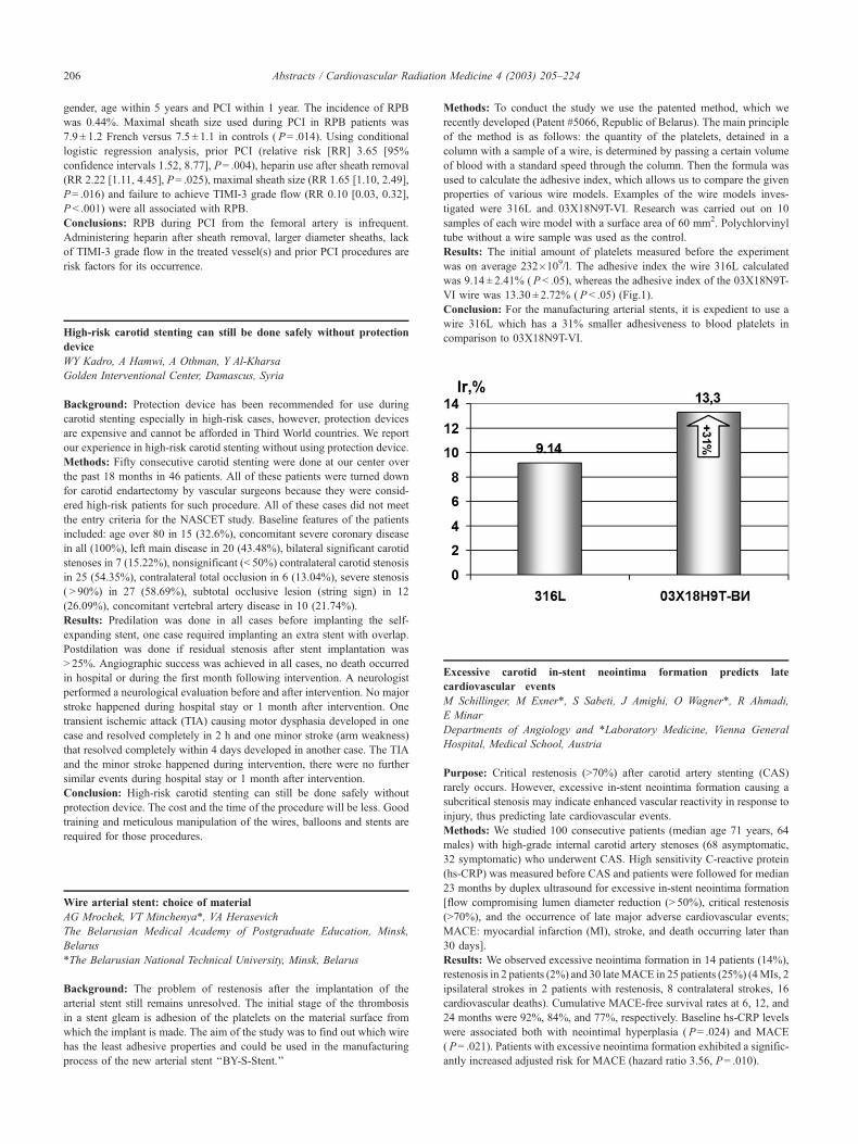
Results: The initial amount of platelets measured before the experiment
was on average 232�109/l. The adhesive index the wire 316L calculated
was 9.14 ± 2.41% ( P < .05), whereas the adhesive index of the 03X18N9T-
VI wire was 13.30 ± 2.72% ( P< .05) (Fig.1).
Conclusion: For the manufacturing arterial stents, it is expedient to use a
wire 316L which has a 31% smaller adhesiveness to blood platelets in
comparison to 03X18N9T-VI.
Excessive carotid in-stent neointima formation predicts late
cardiovascular events
M Schillinger, M Exner*, S Sabeti, J Amighi, O Wagner*, R Ahmadi,
E Minar
Departments of Angiology and *Laboratory Medicine, Vienna General
Hospital, Medical School, Austria
Purpose: Critical restenosis (>70%) after carotid artery stenting (CAS)
rarely occurs. However, excessive in-stent neointima formation causing a
subcritical stenosis may indicate enhanced vascular reactivity in response to
injury, thus predicting late cardiovascular events.
Methods: We studied 100 consecutive patients (median age 71 years, 64
males) with high-grade internal carotid artery stenoses (68 asymptomatic,
32 symptomatic) who underwent CAS. High sensitivity C-reactive protein
(hs-CRP) was measured before CAS and patients were followed for median
23 months by duplex ultrasound for excessive in-stent neointima formation
[flow compromising lumen diameter reduction (> 50%), critical restenosis
(>70%), and the occurrence of late major adverse cardiovascular events;
MACE: myocardial infarction (MI), stroke, and death occurring later than
30 days].
Results: We observed excessive neointima formation in 14 patients (14%),
restenosis in 2 patients (2%) and 30 lateMACE in 25 patients (25%) (4MIs, 2
ipsilateral strokes in 2 patients with restenosis, 8 contralateral strokes, 16
cardiovascular deaths). Cumulative MACE-free survival rates at 6, 12, and
24 months were 92%, 84%, and 77%, respectively. Baseline hs-CRP levels
were associated both with neointimal hyperplasia ( P= .024) and MACE
( P= .021). Patients with excessive neointima formation exhibited a signific-
antly increased adjusted risk for MACE (hazard ratio 3.56, P= .010).
Abstracts / Cardiovascular Radiation Medicine 4 (2003) 205–224206
Page 3
Conclusion: Excessive in-stent neointima formation after CAS indicates an
increased risk for late MACE, potentially reflecting a state of exaggerated
vascular reactivity in response to injury. Inflammation is associated both
with neointimal hyperplasia and MACE, and seems a common character-
istic of different vascular pathologies.
Radiochromic dose map and dose uniformity in the treatment region
for a catheter-based intravascular brachytherapy system
PS Wong
Department of Radiation Oncology and Biophysics, Eastern Virginia
Medical School
Background: The Novoste Beta-Cath System is a catheter-based intra-
vascular brachytherapy (IVBT) delivery device which employs an ingeni-
ous system of hydraulics to deliver the source trains to the catheter tip. The
transfer device serves to transport, hold and deliver the sources during the
procedure. In this study, a quantitative analysis was performed to invest-
igate dose distributions at vessel wall and prescription depths for a reference
lumen diameter (RLD) of 3 mm. The 3.5F 40-mm jacketed radiation source
train (JRST), which consists of a wire-jacketed series of sixteen (16) Sr90/
Y90 sources, was used for the study.
Methods and materials: Stacks of MD-55 radiochromic film strips
(80�15 mm, Fig. I) were sandwiched between b-rail delivery catheter
and solid water slabs. The setup simulates various effective depths from
Sr90/Y90 JRST, ranging from 0.8 to 2.31 mm. 18.4 Gy was delivered to the
prescription depth of 2 mm per Novoste’s protocol. Dose calibration set was
acquired using radiosurgery system with circular applicator of 3 cm
diameter. H & D curve was generated based on exposed doses ranging
from 1 to 60 Gy. Exposed films were scanned using Vidar-16 Dosimetry
Pro scanner. Quantitative analysis was performed using RIT 113 Radiation
Therapy Film Dosimetry. The net optical density (OD) measurements of
each film were converted into 2-D dose map. Longitudinal dose profiles,
which are vertically below source axis, were obtained for each film.
Results: Relative longitudinal dose uniformity within 32 mm of the
therapeutic length (Fig. II) at 0.8 mm, 1.015 mm, 1.23 mm, 1.456 mm
(vessel wall), 1.67 mm, 1.88 mm, 2.095 mm (prescription depth) and 2.31
mm effective depths are within ± 8.9%, ± 5.1%, ± 4.1%, ± 4.3%, ± 4.8%,
± 4.5%, ± 3.8%, and ± 5.5%, respectively (Table 1).
Conclusion: Higher longitudinal dose nonuniformity (± 8.9%) was
observed at 0.8 mm effective depth due to noncontinuity nature of the
JRST. Cold spots between individual sources are visible at this shadow
depth (Fig. Ia). Dose nonuniformity at vessel wall (± 4.3%) and prescription
depth (± 3.8%) verify that intended dose can be delivered to the entire
therapeutic length of 32 mm.
Stenting of significant carotid stenosis improves intracranial frame
count
WY Kadro, D Kadro, N Al-Najjar, R Shehadat
Golden Interventional Center, Damascus, Syria
Background: Carotid stenting (CS) is known to reduce the risk of stroke in
patients with significant carotid stenosis (SCS), however, its effect on
intracranial blood flow (ICBF) is not known. We therefore evaluated the
effect of CS of SCS on ICBF assessed by ICFC.
Methods: ICFC was assessed before and after CS in 17 patients who
received unilateral internal carotid artery (ICA) stenting for SCS. SCS was
defined as stenosis >70% or symptomatic stenosis >50%. All lesions had
TIMI 3 flow before CS. CS was successful in all patients (residual stenosis
<10%). Post-CS TIMI flow was III in all patients. Intracranial cerebral
angiogram before and after CS was done in the AP cranial view at a speed
of 3 frames per second. Frame 1 of ICFC was defined as the first frame that
fills the bifurcation of the ICA into the anterior cerebral (ACA) and middle
cerebral (MCA) arteries after injection. The film was then advanced frame
by frame to the end frame. The end frame of both the ACA and MCA was
assessed. The end frame was defined as the frame that fills the most distal
longitudinal branch of ACA or MCA in the AP cranial view. ICFC was
calculated by counting the frames from Frame 1 to the end frame of both
ACA and MCA.
Results: Mean ICFC for ACAwas 6.14 (range 5–7) frames before CS and
4 (range 3–6) frames after CS ( P= .003). Mean ICFC for MCA was 6.28
(range 5–8) frames before CS and 3.71 (range 3–6) frames after CS
( P= .0007).
Conclusion: CS improves ICBF as assessed by ICFC even when the flow is
excellent (TIMI 3) before stenting. This may reflect improvement in the
cerebral blood flow.
Endovascular gamma radiation therapy inhibits recurrence after
femoropopliteal artery in patients in an older population: the
Vienna experience
RM Wolfram, AC Budinsky, B Pokrajac*, R Potter*, E Minar
Department of Angiology, *Department of Radiotherapy and Radiobiology,
Medical University of Vienna, Vienna, Austria
Background: Endovascular brachytherapy (EBT) utilizing the gamma
emitter 192Ir has shown to efficiently reduce neointimal hyperplasia, the
key factor for restenosis, in patients treated with angioplasty for lesions
in the femoropopliteal arteries. The biological behavior of human cells is
known to change over time as might consequently be their response to
certain treatment modalities. This retrospective analysis was designed to
evaluate potential differences regarding the response to EBT after
femoropopliteal angioplasty according to age, and thus optimize future
treatment strategies.
Methods and results: A total of 199 patients, treated after femoropopliteal
angioplasty with either EBT (n = 100) or placebo (n = 99) within the
prospective randomized trials Vienna 2 and Vienna 3, were analyzed
according to age (median: 72 years). Patients randomized for EBT were
divided into two groups (51 patients =72 years and 49 patients >72 years).
The 12-month outcomes were compared to 99 age-matched patients treated
with angioplasty alone (44 patients =72 years, and 55 patients >72 years).
In patients younger than 72 years, recurrence occurred to a similar extent in
the EBT versus the placebo group at 12 months follow-up (37.3% vs.
56.8%; P= .08). In patients over 72, however, we observed a significant
Dose nonuniformity along the axis of the JRST
Depth0.800mm
1.015mm
1.230mm
1.456mm
1.670mm
1.880mm
2.095mm
2.310mm
Non-uniformity
± 8.9% ± 5.1% ± 4.1% ± 4.3% ± 4.8% ± 4.55% ± 3.8% ± 5.5%
Abstracts / Cardiovascular Radiation Medicine 4 (2003) 205–224 207
Page 4
reduction of restenosis with EBT as compared to angioplasty alone (32.7%
vs. 50.9%; P= .02).
Conclusion: EBT with gamma irradiation significantly reduces restenosis
in an older population after femoropopliteal angioplasty, but does not
improve outcomes in patients <72 years.
Endovascular gamma radiation therapy inhibits recurrence in
restenotic lesions of the femoropopliteal artery: the Vienna experience
R Wolfram, AC Budinsky, B Pokrajac*, R Potter*, E Minar
Department of Angiology, *Department of Radiation Therapy, Medical
University of Vienna, Vienna, Austria
Background: Intracoronary gamma radiation therapy efficiently improves
patency in patients with restenosis. This analysis was designed to
evaluate the efficacy of the gamma emitter 192Ir for the prevention of
recurrent stenosis in the femoropopliteal arteries in patients treated for
restenotic lesions.
Methods and results: A total of 199 patients, treated after femoropopliteal
angioplasty with either radiation (n= 100) or placebo therapy (n = 99)
within the prospective randomized trials Vienna 2 and Vienna 3 were
analyzed according to the stratification criterion of a de novo or recurrent
disease. A total of 66/134 patients with a de novo lesion and 34/65 patients
with a recurrent lesion had been randomized into the brachytherapy (BT)
arm. Outcomes were compared with that of 68 patients with de novo and 31
patients with recurrent lesions treated with placebo. The incidence of
recurrence at 12 months was not significantly different between the BT
and the placebo group for patients with de novo lesions (36.4% and 44.1%,
respectively; P= .32). However, the 12-month recurrence rate was signific-
antly lower in the irradiated group for patients with recurrent lesions
compared with the nonirradiated group (26.5% vs. 71.0%; P= .004).
Conclusion: Endovascular BTwith gamma irradiation significantly reduces
restenosis rate after femoropopliteal angioplasty of recurrent, but not of de
novo lesions.
Vascular brachytherapy with 192-Iridium after femoropopliteal-
stenting in high risk patients—results from the Vienna-5 Trial
RM Wolfram, AC Budinsky, B Pokrajac*, R Potter*, E Minar
Department of Angiology, *Department of Radiotherapy and Radiobiology,
Medical University of Vienna, Vienna, Austria
Objective: To evaluate the efficacy of endovascular brachytherapy (EBT)
for the prevention of restenosis after femoropopliteal stenting in high-risk
patients.
Background: Endovascular brachytherapy with the use of beta and gamma
sources has substantially decreased the restenosis rate after coronary and
peripheral interventions.
Materials and methods: A total of 88 patients with femoropopliteal lesions
(mean treatment length 16.8 ± 7.3 cm) were included into the trial. Patients
underwent PTA and stent implantation and were randomized in a double
blinded fashion to receive either gamma-EBT with a 192-Iridium source
or treatment with nonradioactive seeds. A dose of 14 Gy was prescribed at
2 mm into the arterial wall (target depth = vessel radius + 2 mm). The
primary endpoint of the study was angiographic binary restenosis >50% at
6 months. Secondary endpoint was either percutaneous or surgical TLR
after 6 months.
Results: Revascularization and EBT were successfully accomplished in all
patients. The overall 6-month recurrence rate was 34.8% in the Stent� vs.
33.3% in the Stent+EBT group ( P= .89). Nine (10.2%) patients developed
early reocclusion of the stented segment (two patients [4.3%] in the Stent�and seven [16.7%] in the Stent + EBT group), among those three patients in
the EBT group within the first 24 hours after intervention. Late thrombotic
occlusion (LTO >30 days) was observed in three patients (7.1%) in the
Stent+EBT group.
Conclusion: EBT does not improve 6-month patency after femoropopliteal
stenting in high-risk patients due to a high incidence of early and late
thrombotic occlusion.
Enhanced angiogenesis with autologous bone marrow transplantation
in a porcine non-reperfused myocardial infarction model
R Waksman, J Fournadjiev, R Baffour, R Pakala, D Hellinga, L Leborgne,
H Yazdi, E Cheneau, R Wolfram, R Seabron, K Horton, F Kolodgie*, R
Virmani*, E Rivera*
Washington Hospital Center, Washington, *Armed Forces Institute of
Pathology, Washington, DC
Background: Cell therapy is becoming a viable strategy to improve
revascularization and left ventricular function after myocardial infarction
(MI) injury. This study evaluated the effect of transepicardial bone marrow-
derived mononuclear cell (BMMNC) transplantation on infarct size, blood
vessel formation and myocardial function in a porcine model of non-
reperfused MI.
Methods: Coil implants were positioned in the coronary circulation of 13
domestic swine to produce MI. Twenty-six days later, autologous
BMMNCs were aspirated, labeled with bromdeoxyuridine (BrdU), and
cultured for 48 h. Animals underwent a left thoracotomy and 0.2 ml of BM
(�24�106 cells) were injected at eight sites (1 cm apart) within the
infarcted and border regions of eight swine; five animals injected with
saline served as controls. Animals received systemic BrdU 24 h prior to
euthanasia at 28 days. Regional contractility was assessed by transepicar-
dial echography performed at the time of BMMNC injections and 28 days
after treatment. Collateral growth, angiogenesis, and infarct size were
assessed by angiography, immunohistochemistry, and histomorphometry.
Results: Angiography revealed a trend toward increased collateral growth
in the experimental group. The size of infarct area (mm2) was smaller in the
transplanted BMMNCs group (81.83±10.65) than in the control group
(147.72 ± 23.25, P= .015). BrdU positive cells of treated and control
animals were 51.66% and 29.19%, respectively. Further, a-actin positive
cells were significantly greater in the BMMNC injected animals
(BMMNC=314.8±37.4 vs. saline = 167.1 ± 11.9/0.1 mm2, P= .02) as well
as the number of factor VIII positive endothelial cells (BMMNC=
363.3±28.2 vs. saline = 254.4 ± 28.1 cells/0.1 mm2, P= 0.03). The number
of blood vessels >50 mm was significantly increased in the BMMNC
group=317.9±54.9 vs. 149.12 ± 6.08 (P < .05). Wall motion score index
was similar in the BMMNC injected and saline groups at baseline
(1.63 ± 0.16 vs. 1.25 ± 0.25, P= .21) and at 28 days (1.83 ± 0.22 vs.
1.63 ± 0.38, respectively, P= .62).
Conclusion: Bone marrow-derived mononuclear cell engraftment of
infarcted tissue is feasible with cell viability maintained up to 28 days
and may lead to infarct size reduction. Increased angiogenesis by
BMMNCs transplantation in a model of non-reperfused MI was not
sufficient to support an improvement in left ventricular function.
Endovascular gamma radiation therapy inhibits recurrence after
femoropopliteal artery in patients with diabetes mellitus: the Vienna
experience
RM Wolfram, AC Budinsky, B Pokrajac*, R Potter*, E Minar
Department of Angiology, *Department of Radiotherapy and Radiobiology,
Medical University of Vienna, Vienna, Austria
Background: Endovascular brachytherapy (EBT) has shown to efficiently
improve patency in patients undergoing femoropopliteal angioplasty. This
analysis was designed to evaluate the efficacy of the gamma emitter 192Ir
for the prevention of recurrent stenosis in patients with diabetes mellitus
(DM) treated with percutaneous transluminal angioplasty (PTA) for lesions
in the femoropopliteal arteries.
Abstracts / Cardiovascular Radiation Medicine 4 (2003) 205–224208
Page 5
Methods and results: A total of 321 patients, treated with either EBT
(n= 176) or placebo (n= 145) after femoropopliteal PTAwithin the prospect-
ive randomized trials Pilot 48, Vienna 2, Vienna 3 and Vienna 5, were
analyzed according to the stratification criteria of DM. Patients with DM
were either randomized to receive EBT (n= 72/176) or angioplasty only
(n= 68/145). Outcomes were compared to those of patients without DM
treated with EBT (n= 104/176) or placebo (n= 77/145). In patients without
DM, recurrence rates at 12monthswere comparable between the EBTand the
placebo group (39.4% vs. 53.2%; P= .065). Twelve months recurrence rates
in patients with DM, however, were significantly lower in the EBT group
versus the group treated with angioplasty only (38.9% vs. 57.4%; P= .029).
Conclusion: EBT with gamma irradiation significantly reduces restenosis
at 12 months after femoropopliteal angioplasty in patients with DM.
Early clinical outcome of drug eluting stents compared to bare metal
stents in patients with acute myocardial infarction
J-H Sung, I-J Hwang, I-J Kim, Y-K Cho, S-W Lim, D-H Cha, D-Y Oh
Department of Cardiology, Pundang CHA Hospital, Sungnam, South Korea
Objective: This pilot study aimed to compare drug eluting stents (DES) with
bare metal stents (BMS) in patients with acute myocardial infarction (AMI).
Background: Routine stent implantation was proven to reduce the risk of
adverse events in patients with AMI. DES has been demonstrated to
virtually abolish in-stent restenosis in elective patients with relatively
simple lesions. However, the safety and clinical impacts of DES for patients
with AMI are currently unknown.
Methods: Primary angioplasty using DES was performed in seven cases and
a control group consisting of 50 patients underwent BMS implantation from
September 2003 to January 2004. In DES cohort, 10 DES were deployed (3
CYPHER and 7 TAXUS) with the average size (diameter = 3.3 ± 0.4 mm,
length = 21.5 ± 0.5 mm). Baseline characteristics were similar between two
groups with no statistically significant differences. Themajor adverse cardiac
events (MACE= death, nonfatal reinfarction, target lesion revascularization)
were observed at admission and during a postangioplasty 30-day clinical
follow-up and the short-term outcomes were evaluated.
Results: There were no in-hospital MACE in the DES group as opposed to
two cases in the BMS group (0% vs. 4%; P= .59). At 30-day MACE, there
was no significant difference between the two groups (DES 0% vs. BMS
12%; P= .33). No significant differences existed in the total MACE
between patients treated with DES and BMS (0% vs. 16%; P= .25) (Table).
Conclusion: DES tends to be safer and more effective than the BMS in
patients with AMI, although there were no statistically significant differ-
ences between the two groups. A larger study can provide better results to
prove the safety and efficiency of DES.
Bivalirudin associated intracoronary thrombosis during gamma
brachytherapy and its experimental validation in acute swine model
P Kuchulakanti, S-W Rha, D Hellinga, R Seabron, R Pakala, LF Satler, WO
Suddath, AD Pichard, KM Kent, R Waksman
Cardiovascular Research Institute, Washington Hospital Center, Washing-
ton DC
Introduction: Intracoronary brachytherapy is an approved method to treat
in-stent restenosis and the anticoagulant requirement is the same as
conventional PTCA and is associated with similar bleeding complications.
Bivalirudin is used as an anticoagulant in patients undergoing percutan-
eous transluminal coronary angioplasty (PTCA) and is replacing heparin
in most cath labs. Few cases of intracoronary thrombosis were observed
during gamma brachytherapy with bivalirudin as an anticoagulant raising
question about the mechanism of this undesirable event.
Methods: To explore the mechanism of intracoronary thrombosis, we
conducted an experiment in acute swine model simulating the clinical
situation by randomizing seven swine to sham radiation+bivalirudin
(Group A; n= 3), gamma radiation+bivalirudin (Group B; n= 2), gamma
radiation and heparin (Group C; n= 2). PTCA with balloon and stent
followed by radiation was conducted in LCX (left circumflex) and LAD
(left anterior descending) arteries with ACT monitoring exactly like in
human cases. Gamma radiation was administered with the commercially
available Checkmate system (Cordis, Miami, FL). The end points were
demonstration of thrombus by angiography and postmortem examination
of the coronaries.
Results: ACT levels were comparable without any significant difference
between the Angiomax group and the Heparin group. Clotting was seen in
entire guide catheter in one animal from Group A and at the tip of the
radiation catheter in one animal from Group B, whereas no clots were seen
in Group C (Table 1). In other animals, we observed that flushing the
guide catheter with saline periodically during the dwell time of radiation
catheter prevented clot formation. The end point of Angiographic or
postmortem demonstration of intracoronary thrombus was not observed in
any of the seven animals.
Conclusion: Thrombus formation with bivalirudin is likely due to pro-
longed dwell time of radiation catheter and stasis of blood in the catheter
which is obviated by periodic saline flushing. We conclude from our study
that intracoronary thrombus occurs as results of propagation from the guide
catheter or from migration during contrast injection and it may not be safe
to use bivalirudin in cases of gamma brachytherapy.
Protective effect of rapamycin against atherosclerosis in Apo-E
knockout mice
R Pakala, E Stabile, AA Finegold, D Hellinga, R Seabron, R Baffour, J
Fournadjiev, PK Kuchulakanti, S-W Rha, R Waksman
Division of Cardiology, Washington Hospital Center, Washington, DC
Background: Rapamycin, a macrolide antibiotic, has been shown to inhibit
in-stent restenosis when delivered locally by drug-eluting stents. In the
current study, we wanted to test the effect of oral rapamycin on athero-
sclerotic plaque progression.
Methods: Eight-week-old Apo-E knockout (ApoE) mice were fed with
normal rat chow containing 0.25% cholesterol diet (Con) or with Con
containing 100 mg/kg rapamycin (Rapa-Diet) for 8 weeks. Mice were
sacrificed, hearts dissected out, flash frozen or fixed in neutral-buffered
formalin, embedded in cryomatrix and sectioned. Formalin fixed sections
were stained with Oil-Red-O and counterstained with Harris modified
hematoxylin. Frozen sections were stained for macrophages/foam cells.
Quantitative analysis of atherosclerosis and lesion area was determined.
Plasma cholesterol, triglyceride and rapamycin levels were measured.
Results: Cholesterol and triglyceride levels were essentially the same in
both groups. No rapamycin could be detected in the Con group, whereas
Rapa-Diet animals had 117 ± 7 pg/ml. In the Con group, atherosclerotic
lesions covered 41% of the aortic arch with a plaque area of 0.96 ± 0.045
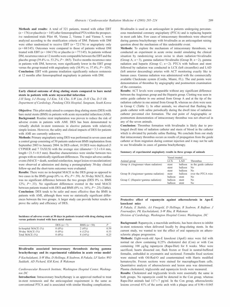
Incidence of adverse events at 30 days in patients treated with drug eluting stentsversus patients treated with bare metal stents
DES (n = 7) BMS (n = 50) P
In-hospital MACE (%) 0 (0%) 2 (4%) 0.5930-day MACE (%) 0 (0%) 6 (12%) 0.33Total MACE (%) 0 (0%) 8 (16%) 0.25
Summary of experimental angioplasty results in three groups of animals
Vessel treated
Animal group LCX LAD Thrombus
Group A (Angiomax+sham radiation) stent balloon in the guide catheterstent balloon Nostent balloon No
Group B (Angiomax+gamma radiation) stent balloon over the PTCA wireballoon stent No
Group C (Heparin+gamma radiation) balloon stent Noballoon stent No
Abstracts / Cardiovascular Radiation Medicine 4 (2003) 205–224 209
Page 6
mm2. In contrast, Rapa-Diet animals had only 22% involvement with
0.34 ± 0.024 mm2. Lesions from the control mice encroached on the vessel
lumen with multiple futures of complex atherosclerotic lesions that are
comprised of acellular cores with cholesterol clefts towards the adventitial
side and focal collection of monocytes derived macrophages towards the
luminal side. In contrast, lesions in the Rapa-Diet mice are simple, mainly
composed of monocyte-derived macrophages.
Conclusion: Oral administration of rapamycin is effective in slowing the
progression of atherosclerosis, and may be a cost-effective way for
prevention of in-stent restenosis (Fig.).
The ‘‘real world’’ clinical practice of intracoronary radiation therapy
as compared to investigational trials
S-W Rha, PK Kuchulakanti, R Pakala, AD Pichard, LF Satler, KM Kent, WO
Suddath, E Pinnow, R Torguson, RC Chan, R Deible, J Lindsay, R Waksman
Division of Cardiology, Washington Hospital Center, Washington, DC
Background: Intracoronary radiation therapy (IRT) is well established in
clinical practice as an effective treatment for in-stent restenosis (ISR). We
aimed to determine if the 6-month clinical outcome of patients (pts) treated
postapproval for marketing [commercial radiation (CR)] is equivalent to
those pts enrolled in the Washington Radiation for In-Stent restenosis Trials
[Gamma WRIST and Beta WRIST, Investigational Radiation (IR)].
Methods: The 6-month clinical outcome of 110 consecutive pts with 125
lesions who received IRT [gamma, 192Ir, 15–18 Gy (n= 6); or beta, 32P, 20
Gy (n= 20); or 90Sr/Y, 18.4–23.0 Gy (n= 99)] in CR was compared with the
6-month clinical outcome of 117 pts with 117 lesions who received IRT
[192Ir, 15 Gy (n= 65) in ‘GammaWRIST’ and 90Y, 20.6 Gy (n= 52) in ‘Beta
WRIST’] in IR. Pts in the CR were treated with wider radiation margins. The
CR received antiplatelet therapy for at least 6 months and the IR for 1 month.
Results: The baseline characteristics of both groups were similar. Use of
atheroablation devices was less in CR (15.2% vs. 32.8% in IR, P= .001).
The overall major adverse cardiac events (MACE; death, Q-wave MI and
TVR, 18.2% vs. 29.1% in IR, P= .05) were significantly lower in the CR
when compared with pts in the IR.
Conclusions: The ‘‘real world’’ clinical practice of IRT demonstrates lower
events and better clinical outcomes. This is most likely a result of
implementation of the lessons learned from the clinical trials such as
optimizing the dosimetry by using a higher dose, treating wider margins
to minimize edge effect, and administering prolonged antiplatelet therapy to
abolish late thrombosis.
Impact of major side branch on periprocedural enzyme elevation and
long-term outcome in patients undergoing PCI and brachytherapy for
in-stent restenosis
P Kuchulakanti, S-W Rha, LF Satler, WO Suddath, AD Pichard, KM Kent,
R Pakala, DA Canos, EE Pinnow, R Waksman
Division of Cardiology, Washington Hospital Center, Washington, DC
Background: Major side branch (diameter >1.5 mm, SB) involvement
within the lesion subjected for percutaneous coronary intervention (PCI) is
known to be a contributing factor for periprocedural cardiac enzyme
elevation (CE). We aimed to assess the impact of SB on CE and 6-month
outcome in-patients undergoing brachytherapy for in-stent restenosis (ISR).
Methods: Retrospective analysis of the data of 248 consecutive patients
with a single vessel ISR with SB (Gp1, n= 146) and without SB (Gp2,
n= 102) who underwent brachytherapy using both beta and gamma emitters
was conducted. The procedural complications, CE, in-hospital course and
6-month clinical outcome were compared.
Results: The baseline patient and lesion characteristics were similar among
the groups. Procedural variables were similar except that stent usage was
more in Gp1. Baseline creatine phosphokinase (CPK)-MB levels were
similar, but postprocedure CPK-MB levels were higher in Gp1. In-hospital
complications were similar between the two groups. Six months follow up
revealed higher restenosis andmajor adverse cardiac events (MACE) in Gp1.
Conclusions: Presence of SB within the restenotic segment when treated
with PCI and brachytherapy is associated with higher periprocedural CE
and MACE at 6 months. Special care should be taken when treating ISR
lesions with SB.
Three-year follow-up after intracoronary gamma radiation for in-stent
restenosis in saphenous vein grafts
S-W Rha, PK Kuchulakanti, AE Ajani, R Pakala, E Cheneau, AD Pichard,
LF Satler, KM Kent, E Pinnow, J Lindsay, R Waksman
Division of Cardiology, Washington Hospital Center, Washington, DC
Background: The Washington Radiation for In-Stent Restenosis Trial in
Saphenous Vein Grafts (SVG-WRIST) demonstrated safety and efficacy of
IRT for the treatment of ISR in saphenous vein grafts (SVG) at 12 months.
The purpose of this study was to examine whether the safety and efficacy of
intracoronary gamma radiation (IRT) for in-stent restenosis (ISR) in SVG
reported at 12 months is resilient at 36 months.
Methods: A total 126 patients (pts) with ISR in SVG underwent PTCA,
laser ablation (53% of lesions), rotational atherectomy, and/or additional
stenting (50% of lesions). Pts were randomized to either 192Ir IRT or
placebo, with a prescribed dose of 15 or 18 Gy at 2 mm from the center of
Clinical events at 6 months
Commercial IRT, N (%) Investigational IRT, N (%) P
Death 8/110 (7.3) 5/117 (4.3) .33Any MI 5/110 (4.5) 16/117 (13.7) .02Any revascularization 23/110 (20.9) 42/117 (35.9) .01TLR 9/125 (7.2) 22/117 (18.8) .01TVR 14/125 (11.2) 31/117 (26.5) .002All MACE 20/110 (18.2) 34/117 (29.1) .05Late thrombosis 1/32 (3.1%) 13/94 (13.8%) .12
Comparison of variables in patients with and without major side-branch at therestenotic lesion
With SB(Gp1, n= 146)
Without SB(Gp2, n = 102)
P
value
Usage of stents 47.3% 30.4% .008Baseline CPK-MB normal 96.6% 98.0% .49Postprocedure
CPK-MB�4 15.3% 7.0% .04CPK-MB�5 10.4% 3.0% .029
6 months follow upRestenosis 36.0% 21.5% .031All MACE 29.3% 17.8% .041
Abstracts / Cardiovascular Radiation Medicine 4 (2003) 205–224210
Page 7
the source. Pts underwent angiography at 6 months and were followed
clinically up to 36 months.
Results: The IRT group had less target lesion [TLR (17% vs. 57%,
P < .001)] and target vessel [TVR (28% vs. 62%, P< .001)] revasculariza-
tion at 12 months and continued to have lower TLR (47% vs.69%, P= .03)
and TLR-MACE rates (53% vs. 74%, P= .03) at 36 months. The Q-wave
MI, non-Q wave MI and death from cardiac causes was similar between the
two groups. TVR-MACE continued to be lower in the IRT group up to 24
months (52% vs. 71%, P= .03) but not at 36 months (69% vs. 80% in the
placebo group, P= .22).
Conclusions: In SVG WRIST, pts with ISR treated with IRT had a marked
reduction in the need for repeat revascularization at 12 months, with
sustained clinical benefit at 3 years.
Effects of contrast media on pig bone marrow derived stem cell and
calf myoblast viability and secretion of VEGF and MCP-1 by bone
marrow derived stem cells in vitro
R Baffour, R Pakala, D Hellinga, R Seabron, J Fournadjiev, R Wolfram, P
Okubagzi, SE Epstein, R Waksman
Division of Cardiology, Washington Hospital Center, Washington, DC
Background: Cell therapy for treating advanced coronary artery disease
represents a tremendous opportunity for developing new therapeutic strat-
egies. Adding contrast media to cell suspension might permit direct
injection of these cells to target area under fluoroscopy guidance. However,
the effect of contrast media on stem cells or myoblast is not well
characterized. We evaluated the effect of various contrast media on pig
bone marrow derived stem cell and calf skeletal myoblast viability and also
on the secretion of growth factors by stem cells.
Methods: Bone marrow was exposed to 10%, 20% or 33% contrast media
(diatrizoate, hypaque, hexabrix or optiray) or no contrast media (controls)
for 1 or 2 h. Bone marrow stem cells were then isolated, and viability
assessed by the trypan blue exclusion. Skeletal myoblasts from gastro-
cnemius were exposed to 10% or 33% contrast media for 15 or 30 min, and
myoblasts viability assessed. In a separate experiment, bone marrow stem
cells were isolated, and cultured in long-term culture medium for 4 weeks
after exposing them to 10% or 33% contrast media. VEGF and MCP-1
levels in the conditioned media from these cultures were assayed by ELISA.
Results: None of the contrast media tested had any effect on the viability of
bone marrow derived stem cells (99–100%). However, incubating myo-
blasts with 10% hypaque for 30 min or with 33% hypaque for 15 or 30 min
increased their viability by 8–10% over the control (control 88%)
( P= .001) (Table 1). Similarly, incubating myoblasts with 33% hexabrix
increased their viability by 8–9% ( P= .001). There were no detectable
levels of VEGF and MCP-1 in the culture medium at Day 0, but the levels
increased significantly to similar values in a time-related manner in the
control, 10% or 33% contrast media. For example, at 4 weeks, VEGF
levels were 2264±169 pg/ml; 2460±77 pg/ml; 2302±104 pg/ml; 2339±163
pg/ml; and 2194±357 pg/ml in the control, 10% diatrizoate, 10% hypaque,
10% hexabrix or 10% optiray, respectively. At 4 weeks, MCP-1 levels were
274±12 pg/ml; 290±11 pg/ml; 305±6 pg/ml; 268±5 pg/ml; 321±14 pg/ml
in the control, 10% diatrizoate, 10% hypaque, 10% hexabrix or 10%
optiray, respectively.
Conclusions: These results demonstrate that exposure of bone marrow stem
cells or skeletal myoblasts to contrast media is not deleterious. These
findings lend support to the concept that stem cell or myoblast suspensions
may be safely mixed with, at least, low concentrations of contrast media
and the mixture directly injected into ischemic myocardium under fluoro-
scopy guidance.
Intracoronary radiation therapy using a novel beta emitter for in-stent
restenosis: Tungsten WRIST
CE Dilcher, RC Chan*, S-W Rha, R Torguson, EE Pinnow, DA Canos, R
Waksman
Division of Cardiology, *Radiation Oncology, Cardiovascular Research
Institute, Washington Hospital Center, Washington, DC
Background: Intracoronary b-radiation therapy reduces in-stent restenosis
(ISR). This study, Tungsten WRIST (Washington Radiation for In-stent
Restenosis Trial), is a safety and feasibility study of intracoronary radiation
therapy (IRT) utilizing tungsten (188W), a beta emitter.
Methods: A total of 30 patients with angiographic evidence of ISR in a
previously treated native coronary artery underwent percutaneous coronary
intervention (balloon angioplasty, ablation by atherectomy, laser angio-
plasty). After the intervention, a noncentered delivery catheter with a side
guide 0.014-in. wire carrying a tungsten (188W) coil with an active length of
33 mm was inserted. Patients were randomized for either radiation with 18,
22 or 25 Gy at 2 mm from the center of the source. Aspirin and Plavix 300
mg loading dose was administered prior to intervention. Plavix 75 mg/day
was prescribed for 6 months after procedure.
Results: At 6 months follow-up, the overall binary angiographic restenosis
rate was 18.8% and the rate for target vessel revascularization (TVR) was
23% and for target lesion revascularization related major adverse cardiac
events (TLR-MACE) was 13.3% without any intergroup differences.
Comparison with the original WRIST radiation cohort utilizing a 192Iri-
dium-source (prescription dose 15 Gy at 2 mm from the source) showed a
similar TVR and TLR-MACE rate of 30% and 18%. TVR and TLR-MACE
rate of the WRIST placebo cohort was 70% and 66%.
Conclusions: Vascular brachytherapy with tungsten (188W) is feasible and
safe. The 6-month clinical outcome is similar to the original WRIST
radiation group.
Dose mapping of coronary arteries in porcine using a fiber
coupled dosimeter
CE Dilcher, RC Chan*, BL Justus**, AL Huston**, DG Hellinga, R
Seabron, R Waksman
Cardiovascular Research Institute, *Radiation Oncology, Washington Hos-
pital Center, **Naval Research Laboratory, Optical Sciences Division,
Washington, DC
Background: The purpose of this study was to measure radiation dosages
to the coronary arteries during intravascular brachytherapy (IVBT) with g-
and b-emitters utilizing, an in vivo dosimeter.
Methods: Domestic pigs were used. In each study catheters were intro-
duced into two different coronary arteries including the left circumflex
(LCX), left anterior descending (LAD), first diagonal (DI), right coronary
Myoblasts viability (% viable, mean±S.D.)
Diatrizoate Hypaque Hexabrix Optiray
Time (min) Control 10% 33% 10% 33% 10% 33% 10% 33%
0 90 ± 7 90 ± 7 90 ± 7 90 ± 7 90 ± 7 90 ± 7 90 ± 7 90 ± 7 90 ± 715 88 ± 6 92 ± 3 93 ± 3 88 ± 6 96 ± 4 88 ± 8 95 ± 4 87 ± 6 92 ± 530 88 ± 4 94 ± 5 91 ± 6 96 ± 4 97 ± 4 92 ± 3 96 ± 3 88 ± 5 87 ± 5Repeated-
measuresANOVAP value
NS .011 NS .001 .001 .048 .026 .027 .019
12- and 36-month clinical outcomes
12 months, N (%) 36 months, N (%)
EventsIr-192(N=54)
Placebo(N=59)
P
valueIr-192(N = 45)
Placebo(N=54)
P
value
Death 4 (7) 4 (7) 1.0 10 (22) 6 (11) .14Q-wave MI 1 (2) 2 (3) 1.0 2 (4) 1 (2) .45TLR 12 (20) 24 (57) <.001 21 (47) 37 (69) .03TVR 19 (32) 37 (62) <.001 29 (64) 40 (74) .30TLR- MACE 14 (23) 36 (60) <.001 24 (53) 40 (74) .03TVR-MACE 19 (32) 38 (63) <.001 31 (69) 43 (80) .22Late thrombosis 2 (4) 4 (7) .67 6 (13) 5 (9) .52
Abstracts / Cardiovascular Radiation Medicine 4 (2003) 205–224 211
Page 8
artery (RCA). A radioactive source (10 seed and 23 seed 192Iridium-/40 mm90Sr/Y-/20 mm 32P-source) and the dosimeter were loaded in each of the
catheters. Measurements were done while pulling back the dosimeter.
Results: Radiation dose is normalized to 100% dose at 2 mm radial
distance from the source. When radiating a branching artery, the dose to
the bifurcation at 5 mm from the source was 35%, 10% and 3% for the192Ir-, 90Sr/Y-, and 32P-source. Using a 23 seed 192Ir-source led to a dose of
40% at 5 mm distance. Radiating the RCA with a 23seed, 192Ir-source did
not result in dose to LAD or LCX.
Conclusions: Dose to adjacent artery segments is less with b- than with g-
emitters. Within a range of 5 mm at bifurcations significant dose exposition
is noted. Prescription doses and safety margins may be adapted in the future.
Intracoronary beta radiation for in-stent restenosis of small coronary
arteries: a dose comparison study
CE Dilcher, RC Chan*, S-W Rha, R Torguson, DA Canos, EE Pinnow,
R Waksman
Cardiovascular Research Institute, *Radiation Oncology, Washington
Hospital Center, Washington DC
Background: Intracoronary b-radiation therapy reduces in-stent restenosis
(ISR) in a dose-dependent manner. This study evaluates dose escalation of
intravascular brachytherapy (IVBT) utilizing a beta-emitter after percutan-
eous coronary intervention (PCI) of small coronary arteries.
Methods: A total of 140 patients with angiographic evidence of ISR in a
previously treated native coronary artery underwent PCI (balloon angio-
plasty, ablation by atherectomy, laser angioplasty). After successful inter-
vention, a noncentered delivery catheter was advanced carrying the active
source train (90Sr/Y) to cover the target site. A total of 90 patients received
18.4 Gy and 50 patients received 23 Gy prescribed to 2 mm radial distance
from the center of the source.
Results: The clinical 6-month follow-up showed a TVR-MACE rate (target
lesion revascularization—major cardiac adverse events) that is similar in
both groups (18.1% vs. 16.3% for 18.4 Gy and 23 Gy groups). All patients
were treated with aspirin and clopidogrel prior to the procedure. At
discharge patients were instructed to continue clopidogrel 75 mg/day for
at least 6 months.
Conclusions: Dose escalation (18.4 Gy vs. 23 Gy) utilizing a 90Sr/Y-source
is feasible, safe and effective in reducing ISR at 6-month follow-up.
IVUS-based dosimetry on patients with repeat radiated coronary art-
eries to the same site
CEDilcher, RC Chan*, P Rai, S-W Rha, R Torguson, DA Canos, RWaksman
Cardiovascular Research Institute, *Radiation Oncology, Washington Hos-
pital Center, Washington DC
Background: Intracoronary radiation reduces recurrent in-stent restenosis
(ISR). Repeat radiation may become necessary due to recurrent ISR. This
study reports outcome-related dose calculations for twice-radiated coronary
artery segments.
Methods: A total of 22 patients with angiographic evidence of ISR in a
previously treated native coronary artery were assigned for repeat percutan-
eous coronary intervention (PCI) and intravascular brachytherapy (IVBT).
IVBT was performed either with a 192Ir-, 90Sr/Y-source (prescription dose:
14–18 and 23 Gy each at 2 mm from the center of the source) or a 32P-
source (20 Gy, 1 mm deep to the vessel wall). Mean time interval between
two IVBT treatments was 394±306 days. For each patient, angiograms and
IVUS cross-sections were reviewed on the basis of anatomical landmarks
matched and the twice-radiated vessel segment was identified. IVUS-based
calculations of maximal and minimal point doses at the endothelium and
the adventitia–media border were performed. The point doses of consec-
utive slices of the twice-radiated vessel segment returned the dose range it
was exposed.
Results: Clinical follow-up at 379±146 days revealed a TVR rate of 18.2%
and a TLR rate of 13.6%. One death was reported. Maximal dose and
average dose at the endothelium was 261 and 124±72.3 Gy and maximal
dose and average dose at the adventitia –media border was 159 and
50.3±29.3 Gy. Fourteen patients had a 1.71 times longer recurrence free
interval compared with the interval between both IVBT treatments.
Conclusions: Repeat IVBT to the same ISR site is safe without any adverse
clinical events at average 12 months follow-up. A second IVBT treatment
led to a prolonged ISR-free survival for the majority of patients. The choice
of isotope did not influence outcome.
Vascular remodeling induced by endothelial cells within gas–plasma
treated scaffolds in aged nude mice
JL Polan, O Munoz, CM Agrawal, SR Bailey
University of Texas Health Science Center at San Antonio: Janey Briscoe
Center for Cardiovascular Research
Purpose: Increased blood vessel remodeling occurs from endothelial-
seeded bioresorbable gas–plasma treated polylactide acid (D,L-PLA) scaf-
folds when implanted into 2-month nude mice (N= 114) up to 72 days. Our
hypothesis is that endothelial-containing gas–plasma treated scaffolds
similarly implanted within 14-month aged nude mice will exhibit reduced
vascular remodeling due to less expression of vascular endothelial growth
factor (VEGF) and basic fibroblast growth factor (bFGF).
Methods: 14-month aged immunological-deficient nude mice were
implanted with nontreated control (n= 7), gas–plasma treated bioresorbable
scaffolds (n= 8) containing human aortic endothelial cells or were proced-
ural shams (n= 2). Scaffolds and peritoneal tissues were evaluated and
statistically compared by in situ quantification of blood vessels (scale 0–4)
at 72 days. VEGF and bFGF indirect immunostainings were prepared on
fixed scaffolds and tissue.
Results: Treated scaffolds induced blood vessel densities near the scaffolds
that were scored 1.83 ± 0.8; whereas, control scaffolds induced 1.47 ± 0.9.
Remodeling after 72 days with gas–plasma treated scaffolds in aged
nude mice.
Abstracts / Cardiovascular Radiation Medicine 4 (2003) 205–224212
Page 9
Consequently, ANOVA showed no statistical difference ( P= .55). Shams
scored an average of 0 ± 0.0. Both treated ( P= .017) and control ( P= .031)
were significantly different from the shams. Blood vessel densities above
and near the scaffolds were less for 17-month nude mice with treated
scaffolds than for 5-month nude mice ( P= 6.1E�06); whereas, control
scaffolds in the aged and young mice were not significantly different
( P= .450). VEGF and bFGF expression was surprisingly greater in aged
versus young specimens.
Conclusion: 17-month aged nude mice exhibit reduced blood vessel
remodeling despite enhanced angiogenic protein, such as VEGF and bFGF,
production when implanted with either endothelial-containing gas–plasma
and nontreated D,L-PLA scaffolds.
The results of transradial coronary intervention for chronic total
occlusion
T Shiono, S Saito, S Tanaka, Y Miyashita, S Takahashi
Shonan Kamakura General Hospital
Objective: Percutaneous coronary intervention (PCI) for chronic total
occlusion (CTO) is one of the major challenges in interventional cardiology.
The development of a tool like a tapered-tip guidewire improves the
primary success rate of TRI for CTO.
Methods: Between January 2000 and December 2002, 2926 patients
underwent PCI in Shonan Kamakura General Hospital. A total of 2034
patients (69.5%) were treated by TRI, and the others were treated by the
femoral or brachial approach. We analyzed 303 cases (10.6%) of CTO,
which included 196 patients (64.7%) treated by TRI during that. Patients
were carefully distributed to each approach while making diagnostic
coronary angiogram reference. TRI was chosen for the patients expected
to succeed even if it does not image a contralateral coronary angiogram.
We mainly used six French guidings. All procedures were enforced in
accordance with our CTO guideline: (1) successful penetration of a
guidewire <30 min, (2) total procedure time <90 min, (3) total dye
volume <300 ml.
Results: Procedure success was achieved in 145 patients (74.0%) out of
196 TRI for CTO. Among the 51 patients without successful angioplasty,
we have not passed a guidewire in a pathological change part in 35
patients (69%). We could not cross the lesion by a balloon catheter in
only 2 patients and failed to accomplish TIMI-3 flow in 14 patients. There
were no cases of death, emergency bypass surgery or major vascular
complications. In the case of transfemoral approach, four patients required
blood transfusion due to bleeding complication and procedure success rate
was 68.4% during the period. Follow-up angiography was performed in
102 patients (70.3%) and mean follow-up period was 11.2 months.
Among the case of procedure success, 40 patients underwent TLR.
Conclusions: Transradial procedure is a safe and useful method for
treatment of CTO. Various techniques such as a 5 in 6 guiding or an
anchor balloon support TRI for CTO and bring about an equivalent results
compared with conventional approach.
Chronic recovery of endothelium-dependent relaxation in pig coronary
arteries distal to sites of endovascular irradiation after three months
J Li, B Ebato, P Mulkey, NAF Chronos, KA Robinson
American Cardiovascular Research Institute, Norcross, GA
Background: We previously reported that endothelium-dependent relaxa-
tions were impaired in coronary arteries distal to sites of intracoronary
radiation (CADSIR) at 1-month postprocedure. In this study, we investigated
whether vasomotor function in CADSIR remained abnormal at 3 months.
Methods: Pig coronary artery segments 2 cm distal to sites of irradiation
were studied in an organ-chamber apparatus, 3 months after sham (S,
n= 7) or active irradiation (R, n= 8; 20 Gy). There were no fibrotic
adhesions, intramural hemorrhages or enlargement in CADSIR or S
treatment. These morphologies were present at sites of irradiation but
not in sites of sham treatment.
Results: Unlike contractions of target arteries, contractions of CADSIR
to 40 mM KCl (4.44 ± 0.62 vs. 3.85 ± 0.58, NS) and 100 mM KCl
(5.66 ± 0.81 vs. 4.49 ± 0.38, NS) were similar to sham. After NO
synthase inhibition with L-NAME, contraction to 5 mM PGF2a was
significantly higher than contraction to PGF2a before L-NAME
(5.21 ± 0.95 vs. 2.28 ± 0.51, P < .05). Maximal dose relaxation (DRmax;
3 mM) of CADSIR to calcium ionophore A23187 was not different
from S (82.8 ± 0.09% vs. 93.4 ± 0.03%, NS) and was significantly higher
than CADSIR at 1 month (35.7 ± 0.09%, P< .01). DRmax (100 pM) in
CADSIR to substance P was similar to S (41.6 ± 0.06% vs.
44.0 ± 0.07%, NS) and significantly higher than CADSIR at 1 month
(14.7 ± 0.08%, P < .01). Scanning electron microscopy showed confluent
endothelial cells with normal-appearing morphology in both groups.
Morphology of the irradiated site also showed a confluent endothelial
layer, without notable inflammatory cell adherence.
Conclusions: Endothelial function of CADSIR was similar to S at 3 months
post-irradiation and normalized compared to CADSIR at 1 month. This
temporal recovery of distal coronary vasomotor function abnormalities may
relate to angioplasty site healing and resolution of the inflammatory
response. Perfusion of distal myocardium may be affected by upstream
brachytherapy of a site of balloon catheter or stent intervention, but this
effect is expected to resolve over time.
TheraP trial dosimetric characterization of TheraSource (Pd-103)
intravascular brachytherapy source
ME Napolitano, JJ Bergman, KK Millage*, RA Hearn, JJ Rodgers
Radiation Physics, Theragenics Corporation, Buford, GA
*The Millage Group, Flemington, NJ
Purpose: The Monte Carlo Code MCNP4C was used to perform the TG-
60 based dosimetric characterization for TheraSource, a Pd-103 intra-
vascular brachytherapy source. TheraSource was used in the TheraP Trial
(a Phase I safety and feasibility study). In this trial, TheraSource was used
following PTA to deliver 20 Gy to the target tissue in the superficial
femoral and popliteal arteries. The source consists of a 40-mm active
segment of high specific activity Pd-103 coated on a nitinol wire.
Methods: Monte Carlo calculations were used to calculate the dose rate as
well as the axial and radial dose profiles. Dwell times for each step were
calculated based on vessel diameter and a prescription dose of 20 Gy to a
target 2 mm into the vessel wall. The Monte Carlo based dosimetric
characterization was combined with measured activity and compared to
measured thermoluminescent dosimeter (TLD) dose values. Source uni-
formity along the center 2/3 of the source is confirmed using Gafchromic
film. The source was manually stepped to treat interventional treatment
lengths up to 134 mm in four steps with margins of at least 10 mm. To
minimize the possibility of gaps during manual stepping of the source, a 2-
mm overlap was used. A centering balloon, 1 mm smaller than the vessel
inner diameter was used to minimize dose variation at the prescription
point due to noncentering. The dose profile was calculated at the prescrip-
tion depth along the treatment length. A 2-mm overlap was assumed, as
well as realistic permutations due to noncentering and overlap.
Results: The dose based on MCNP4C calculated dose rate combined with
measured source strength compared on average within 10% to TLD dose
measurements over the range of 0.2 to 2.0 cm from the source centerline.
Source uniformity is within ±10% throughout the center 2/3 of the source.
For lesion lengths that require stepping, localized regions in the 2-mm
overlap region receive a 20% increase in dose. In the 2-mm overlap
regions, the worst-case dose variation due to noncentering and a 2-mm
overlap ranges from 17 to 30 Gy.
Conclusions: Good agreement was found between calculated dosimetry
parameters and TLD measurements. The recommendations of TG-60 have
been followed for dosimetric characterization and source uniformity.
Abstracts / Cardiovascular Radiation Medicine 4 (2003) 205–224 213
Page 10
Variation in prescription dose due to overlap required for manual stepping
and noncentering was given careful consideration.
The use of perfusion balloon in stenting of carotid arteries with
contralateral severe stenosis or occlusion
WY Kadro, A Hamo, A Aldebs, I Eid
Golden Interventional Center, Damascus, Syria
Background: Stenting of carotid arteries with contralateral occlusion or
severe contralateral stenosis carries a high risk for seizure or loss of
consciousness during balloon inflation at predilation and postdilation.
The use of perfusion balloon for pre- and postdilation may prevent cerebral
ischemia and reduce the incidence of intolerance to balloon inflation by
preventing total obstruction to the lumen of the artery.
Methods: After having two bad experiences with seizure and loss of
consciousness while intervening on carotid artery with contralateral total
occlusion, we decided to use perfusion balloon for pre- and postdilation.
During the past 2 years, we did 29 cases of carotid artery stenting for
lesions of >70% stenosis with contralateral total occlusion or contrala-
teral severe stenosis. A 3.5-mm perfusion balloon was used for predi-
lation. Postdilation was done with 5–5.5 mm perfusion balloon. All
cases were done without the usage of protection device since it cannot
be afforded in our country due to its high cost.
Results: Procedure success was 100%, angiographic success was 100%,
intolerance to balloon inflation, i.e. seizure or loss of consciousness did not
happen in any case during pre- or postdilation. One episode of transient
facial droop happened in one case during predilation and resolved com-
pletely after balloon deflation and did not recur during postdilation. TIA
happened only in one case after postdilation and resolved completely within
2 h after intervention.
Conclusion: The usage of perfusion balloons for pre- and postdilation is
safe and prevents seizure and loss of consciousness during intervention
on carotid arteries with contralateral total occlusion or contralateral
severe stenosis.
Mobilization of bone marrow stem cells by granulocyte colony
stimulating factor for treatment of patients with severe chronic
ischaemic heart disease
J Kastrup, W Yongzhong, ****K Tagil, RS Ripa, **JC Nilsson, S Carstensen,
E Jørgensen, L Søndergaard, *B Hesse, ***HE Johnsen
Cardiac Catheterization Laboratory, The Heart Centre, *Department of
Nuclear Medicine, Rigshospitalet, Copenhagen, Denmark**Danish Research Centre for Magnetic Resonance, Hvidovre Hospital,***Department of Haematology, Herlev University Hospital, Herlev,
Denmark****Department of Clinical Physiology, Malmo, Sweden
Aim: Granulocyte colony stimulating factor (G-CSF) mobilized bone
marrow stem cells participate in neovasculogenesis in ischaemic myo-
cardium in animal studies. This is the first Phase I safety and efficacy
study with G-CSF treatment of patients with severe ischemic heart
disease (IHD).
Methods and results: Thirteen patients with severe IHD were treated with
subcutaneous injections of 5 mg G-CSF/kg body weight for 6 days. The
treatment was without any deterioration of clinical status or serious adverse
events. However, five patients were ‘‘poor mobilizers’’ to the G-CSF
treatment (max CD34+ cells <12,000/ml blood) with a maximal increase
in CD34+ cells to 8600±1600/ml blood compared to 31,000±5200/ml blood
in ‘‘mobilizers’’ (max CD34+ >12,000/ml blood). At 2 months follow-up,
patients had improved in CCS angina classification from 2.7±0.6 to 1.7±0.6
(mean±S.D., P= .01), a decline in NTG use from 1.5±2.1 to 0.5±1.2 per day
( P< .05) and number of angina pectoris attacks from 1.7±1.7 to 1.0±1.6 per
day ( P< .05). Improvement was mainly seen in the eight patients with a
maximal increase of CD34+ stem cells >12,000/ml blood. However, we
found no accompanying changes in myocardial perfusion and function on a
standard adenosine stress single photon emission computerized tomography
(SPECT), magnetic resonance images or echocardiography.
Conclusion: Treatment by G-CSF in patients with severe IHD seemed
safe without adverse side effects. Improved symptoms of myocardial
ischemia were demonstrated and the effect was related to the degree of
increase in circulating stem cells. Randomized, placebo-controlled studies
are needed to verify whether G-CSF is a new treatment option in ischemic
heart disease.
Combining angiogenesis and cell therapy for improved outcome: the
role of angiogenesis master regulator gene EPAS1
S Iacampo, R Martel, AS Fortin, G Leclerc, L-G Guy
Angiogene Inc., Montreal, Quebec, Canada
Background: Myocardial infarction caused by hypoxia results in cardiac
tissue damage. Cardiac function can be improved by cell transplantation in
the scar. Furthermore, the implanted cells can also be used as vectors for the
production of angiogenic factors to improve perfusion. This study exam-
ined the angiogenic activity of human primary cells modified with the
hypoxia inducible factor EPAS1.
Methods: Human skeletal myoblasts, mesenchymal stem cells, bone
marrow mononuclear cells and fibroblasts were transduced with an
adenovirus expressing EPAS1. VEGF was measured by ELISA and gene
induction profile was documented with microarrays. Human umbilical
vein endothelial cells (HUVECs) proliferation, migration and differenti-
ation was evaluated by colorimetric, modified Boyden chamber and
matrigel tube formation assays after supplementation with conditioned
media from vehicle, myoblasts and EPAS1 modified myoblasts. Vehicle,
myoblasts and EPAS1 modified myoblasts were subcutaneously
implanted in immunodeficient mice and angiogenesis was assessed in
the implants by histology.
Results: In all human primary cell types tested, EPAS1 was found to be
a potent VEGF inducer. Several secreted factors implicated in the
different steps of the angiogenesis process, such as MMP7, PlGF, leptin,
adrenomedullin, IL-8 and LIF, were also found to be induced by EPAS1.
The proliferation, migration and differentiation of endothelial cells were
increased by conditioned media from myoblasts (containing the secreted
factors), but EPAS1 gene transfer in the myoblasts more than doubled
this induction (see Figure). In vivo, EPAS1 transduction into myoblasts
significantly increased angiogenesis activity in subcutaneous implants
compared to myoblasts alone.
Conclusions:Myoblasts secrete several angiogenic factors that stimulate
endothelial cells. EPAS1 gene transfer enhances the production of
these factors and significantly improved the stimulation of cultured
endothelial cells by myoblasts. EPAS1 modification of myoblasts also
Abstracts / Cardiovascular Radiation Medicine 4 (2003) 205–224214
Page 11
directly improved angiogenesis in vivo. EPAS1 is thus a good
candidate for genetic modification of cells to be transplanted in the
scarred myocardium.
Cell-mediated EPAS1 gene transfer is superior to cell therapy alone for
improving perfusion and cardiac function in infarcted heart
L-G Guy, Y Morimoto*, S Iacampo, AS Fortin, BH Davis*, G Leclerc,
DA Taylor*
Angiogene Inc., Montreal, Quebec, Canada
*Division of Cardiology, Duke University Medical Center, Durham, NC
Background: Successful improvement of cardiac function after transplant-
ing skeletal myoblasts into myocardial scar has moved this treatment into
clinical trials. A major limitation of cellular cardiomyoplasty (CCM) is the
death of cells in the hypoxic infarct milieu. Thus, factors which improve
angiogenesis and cell survival might increase the efficacy of CCM. EPAS1
is a hypoxia inducible factor that regulates genes induced by low oxygen
tension, such as the angiogenic factor vascular endothelial growth factor
(VEGF). The hypothesis of this study is that EPAS1 gene transfer will
improve the angiogenic potential of myoblasts in myocardial scar and thus
the efficacy of the treatment.
Methods: Human skeletal myoblasts were transduced with an adenovirus
expressing EPAS1 and VEGF was measured by ELISA. Microarrays
were used to document the profile of gene induction. Vehicle, myoblasts
and EPAS1 modified myoblasts were implanted into cryoinjured left
ventricle of immunodeficient mice. Cardiac function was assessed at
Days 0 and 28 by magnetic resonance imaging. Fluorescent lectin was
infused in the circulation prior to sacrifice and vessel density in scar was
quantified by histology.
Results: In vitro, there was a 20-fold VEGF induction upon EPAS1 gene
transfer. Other angiogenic genes (PlGF, IL-8, and LIF) and cardioprotec-
tive genes (LIF-R, cardiotrophin 1 and adrenomedullin) were also induced
by EPAS1. Vessel density in the scar was significantly superior in the
EPAS1 modified myoblasts group and lectin staining confirmed that
vessels were functional and connected to the vasculature. LVEF change
after treatment revealed that animals in the vehicle group deteriorated
over time while those treated improved. LVEF improvement more than
doubled in the EPAS1 modified myoblast group compared to the
myoblast alone.
Conclusions: EPAS1 gene transfer increases the expression of angiogenic
and cardioprotective genes and enhances the functional impact of myo-
blasts in scar, presumably by promoting angiogenesis and cell survival.
Age and acute ischemia aremajor determinants of stem cell mobilization
efficacy of granulocyte-colony stimulating factor in patients with
myocardial infarction
H-J Kang, H-J Cho, S-Y Zhang, K-W Park, H-S Kim, B-H Oh, M-M Lee,
Y-B Park
Department of Internal Medicine, Seoul National University College of
Medicine; Cardiovascular Laboratory, Clinical Research Institute, Seoul
National University Hospital, Seoul, Korea
Background: Granulocyte-colony stimulating factor (G-CSF) is one of
the most promising stem cell mobilizer for treatment of ischemic heart
disease. Stem cell mobilizing effects of G-CSF administration were not
evaluated in patients with myocardial infarction. We evaluated stem
cell mobilizing effects of G-CSF in patients with acute and old
myocardial infarction.
Methods and results: Nine patients (60 ± 10 years old, 8 men) underwent
apheresis after 4 days administration of G-CSF with dose of 10 mg/kg/day.Five patients had acute myocardial infarction (AMI: 4 ± 2 days after AMI)
and four patients had old myocardial infarction (OMI: 94 ± 117 days after
AMI). FACS analysis was performed with apheresis products. Apheresis
products from patients with age under 65(n= 6) showed higher proportions
of AC133-positive (0.7 ± 0.6 vs. 0%, P= .04), CD34+ (13.2 ± 11.2 vs.
2.6 ± 1.5%, P= .17) and KDR+ cells (15.4 ± 13.6 vs. 2.1 ± 0.5%, P= .17).
Apheresis yield from both age group were not different (1.4 ± 0.7�109 vs.
1.3 ± 0.3�109 from 1 L of filtered blood, P= 1.0). Although limited
statistical significance, apheresis products from patients with AMI showed
higher numbers of KDR+ cell (3.4 ± 4.0�108 vs. 0.7 ± 1.3�108 from 1 L of
filtered blood, P=.11), CD34+ (2.5 ± 0.3�108 vs. 0.5 ± 0.5�108 from 1 L
of filtered blood, P = .28), and AC133+ cells (1.6 ± 2.6�107 vs.
0.1 ± 0.3�107 from 1 L of filtered blood, P= .55).
Conclusions: Although limited in the number of study population, these
results showed that stem cell mobilizing efficacy of G-CSF was influenced
by age and the presence of acute ischemia in patients with myocardial
infarction. Patients with AMI and younger age had augmented responses to
G-CSF induced stem cell mobilization. These data suggested that higher
doses and longer administration schedule of G-CSF should be considered
for patients with OMI and older age.
The essential role of p21 in radiation-induced cell cycle arrest of vas-
cular smooth muscle cell
H-J Cho, H-S Kim, H-J Cho, J-W Chung, J-S Park, K-W Park, I-H Chae,
M-M Lee
Department of Internal Medicine, Seoul National University College of
Medicine; Cardiovascular Laboratory, Clinical Research Institute, Seoul
National University Hospital, Seoul, Korea
Objective: We investigated the molecular mechanism of radiation-induced
cell cycle arrest in vascular smooth muscle cell (VSMC) and examined
whether p21 knock-out is a cause of radiation failure.
Background: Intravascular radiation therapy is an effective measure to
inhibit restenosis after angioplasty. But it has limitations such as delayed
Abstracts / Cardiovascular Radiation Medicine 4 (2003) 205–224 215
Page 12
catch-up restenosis. The biologic mechanisms for the success and failure of
radiation therapy after angioplasty have not been well studied.
Methods: Using different dosages of gamma radiation, we evaluated the
effect of radiation on VSMC apoptosis and cell cycle progression, and its
action mechanism.
Results: Irradiation significantly retarded the growth of cultured VSMC,
which was not due to induction of apoptosis but mainly due to cell cycle
arrest. Radiation showed remarkable cell cycle arrest at the G1 and G2
phases (G0/G1:S:G2/M phases = 61%:34%:5% with 0 Gy vs. 61%:9%:30%
with 16 Gy, 12 hours after radiation). In immunoblot analysis and kinase
assay, radiation increased the expression of p21 and decreased the
expression and activity of CDK2 and CDK1. In contrast, radiation did
not affect the expression and activity of CDK4 and CDK6, nor the
expression of p27 and p16. When p21 was knocked out, cell cycle of
VSMC was not arrested by radiation, leading to increased proliferation.
Conclusions: Radiation inhibits VSMC proliferation through cell cycle
arrest by enhancing p21 expression and suppressing CDK1 and 2. p21
knockout results in increased proliferation and survival after radiation
suggesting the key role of p21 in radiation-induced cell cycle arrest.
Nitric oxide synthase gene G298 allele: is it a marker for microvascular
angina in hypertensive population?
GEN Elkilany
Cardiology Department, Khalifa Hospital, Ajman, UAE
Background: Nitric oxide (NO) has an important effect on blood pressure,
arterial wall and the basal release of endothelial NO in hypertension. Until
now there are no solid data that revealed the rule of polymorphism of NOS
in patients with hypertension and microvascular angina.
Objectives: The aim of the present study is to investigate the gene of
endothelial nitric oxide synthase (eNOS).As polymorphism of this gene
may be a putative candidate for hypertension and arterial stiffening.
Patients and methods: 60 subjects were recruited for this study, 50
hypertensive patients complaining of chest pain and 10 healthy volunteers
served as control. All patients underwent stress myocardial perfusion
imaging and coronary angiography. Genotyping for all patients and controls
was performed. The linkage between hypertension, microvascular angina
and gene polymorphism was investigated.
Results: Myocardial perfusion imaging and coronary angiography revealed
that 15 patients had chest pain with true ischemia and reversible myocardial
perfusion defects (multiple and mild) but normal epicardial coronary
arteries (microvascular angina) while 15 patients had significant coronary
artery disease (CAD) and 20 hypertensive patients showed normal per-
fusion scan and coronary angiography. The prevalence of the nitric oxide
synthase gene allele was higher in the hypertensive group with micro-
vascular angina (documented by multiple reversible perfusion abnormalities
by SPECT TI-201) than it was among normotensive control subjects
( P< .005). eNOS allele was significantly higher in the hypertensive group
than normotensives, but there was no significant difference in homozygote
mutants among hypertensives, x-syndrome and patients with CAD.
Conclusion: eNOS gene polymorphism proved to be an important etiology
in microvascular angina among hypertensives. In addition, eNOS mutant
gene showed a significant increase in isolated hypertensives and in patients
with CAD.
A novel method for left anterior descending artery ligation in rats with
high success rate
SAS Shariat, MR Movahed
Department of Medicine, Center of Cardiovascular Hormone Research and
Division of Cardiology, University of California, Irvine
Background: Left anterior descending artery (LAD) ligation has been
used commonly in rats to induce left ventricular infarction for research
purposes. However, LAD ligation has been very difficult with at least 50%
mortality in rats. We developed a method of LAD ligation in rats with less
than 5% mortality.
Methods: 36 male Sprague–Dawley male rats weighting 300–350 g were
selected for LAD ligation for induction of ischemic cardiomyopathy. The
surgery was performed under full anesthesia. Left-sided thoracotomy
through cutting of the 5th and 6th ribs was performed. Pericardium was
opened and the heart was exteriorized with a holder consisting of a plastic
loop (1.5�2 cm). The LAD was localized 1–2 mm below the junction of
pulmonary conus and left atrial appendage. A 6.0 silk needle was used to
ligate LAD from the left border of the pulmonary conus to the right border
of the left atrial appendage. The heart was returned back to the chest and
chest wall was closed with presutured lopes of 4.0 silk.
Results: Mid LAD ligation was successful in all rats with less than 5%
mortality. The entire surgery was performed after initial experience in less
than 15 min. Rats survived the procedure without any complication for up
to 3 weeks follow-up.
Conclusions: Mid LAD ligation can safely be performed in rats using
ligation of LAD 1–2 mm below the junction of the pulmonary conus and
the left atrial appendage with very low mortality.
Very high rate of right ventricular infarction after ligation of mid left
anterior descending artery in rats
SAS Shariat, MN Hashemzadeh, MR Movahed
Department of Medicine, Center of Cardiovascular Hormone Research and
Division of Cardiology, University of California, Irvine
Background: The left anterior descending artery (LAD) supplies left
ventricle in human. LAD ligation has been used commonly in rats to
induce left ventricular infarction for research purposes. However, the
myocardial supply territory of LAD is not well established in rats. We
measured the infarction zone in rats after ligation of mid LAD.
Methods: 24 male Sprague–Dawley male rats weighting 300–350 g were
selected for LAD ligation for induction of ischemic cardiomyopathy. The
surgery was performed under full anesthesia. Left-sided thoracotomy was
performed through cuts in 5th and 6th ribs. After opening the pericardium,
LAD was ligated after first diagonal and septal branches. After 24 h, the
hearts were removed and stained with tetrazolium tetrachloride (TTC) for
detection of infarcted area.
Results: Mid LAD ligation induced 100% infarction of mid to apical left
anterior wall and septum involving an average of 45% of LV mass.
Surprisingly, right ventricular infarction was observed in 80% of rats
involving mid to apical right ventricular (RV) wall.
Conclusions: Mid LAD ligation after first septal and diagonal branches
causes substantial left ventricular infarct in rats. However, we find that
80% of the rats had right ventricular infarction. Therefore, hemodynamic
effect of RV infarct should be considered in researches involving LAD
ligation in rats.
Simple and safe method of endotracheal intubation in rats using
straight tip 0.035 wire used for coronary angiography
AS Shariat, PH Wang, MR Movahed*
Department of Medicine, Division of Endocrinology, *Division of Car-
diology, University of California, Irvine Medical Center
Introduction: Endotracheal intubation in rats is a challenging problem.
This is a cumbersome procedure due to narrow oral cavity and difficulty to
visualize vocal cord. It is associated with a high complication rate. We have
developed a very safe and simple method using a straight tip wire as a guide
and smallest pediatric laryngoscope for visualization before intratracheal
intubation of rats.
Method: Seventy Sprague–Dawley rats weighing between 100 and 450 g
were used for endotracheal intubation, using 0.035-in. J-guide wire,
conventional laryngoscope blade Miller Size 0 and conventional cut-down
Abstracts / Cardiovascular Radiation Medicine 4 (2003) 205–224216
Page 13
18–20 gauge catheter. Rats were anesthetized using mixture of ketamine
(50 mg/kg), zylozine (4 mg/kg) and acepromazine (1 mg/kg). After deep
anesthesia, the animals were laid down in supine position. The upper
insisura were fixed with a rubber band. Using the laryngoscope, the trachea
and vocal cord were visualized with tongue pulled back. Under direct
visualization of the vocal cord, a 0.035-in. straight tip wire was advanced
into the trachea without any difficulty and an 18–20 gauge cut-down
catheter was advanced over the wire into the trachea. The wire was removed
and the catheter was attached to the respirator.
Results: With this method it was not required to use any atropine and we
successfully intubated all 70 rats without any trauma. Successful rate was
100% with no complications or mortality related to intubation. This was
performed within 1 to 2 min after induction of anesthesia. There was no
erroneous intraesophageal or soft tissue intubation or trauma to glottis or
vocal cord.
Conclusion: Using a straight tip wire as a guide and a smallest conven-
tional laryngoscope blade Miller Size 0, we developed a very safe, quick,
and easy method for endotracheal intubation in rats without the usage of
atropine. This method allows researchers to intubate rats with no complica-
tion and makes it possible to study cardiac hemodynamic in rats without the
negative effect of atropine.
Angiogenesis in chronic refractory stable angina induced by autologous
bone marrow: Phase 1
J Vicario, C Campos, J Piva, F Faccio, L Gerardo, C Becker, HH Ortega
Sanatorio Garay, Hospital Iturraspe and U.N.L., Santa Fe, Argentina
Purpose: Based on preclinical studies with unfractionated autologous bone
marrow (UABM) via coronary sinus with transitory occlusion, a clinic
study in patients with chronic stable angina who are not candidates to
percutaneous or surgical revascularization was designed. The objectives
were to evaluate safety, tolerance and feasibility.
Method: A multicenter, prospective study with variable doses and criteria
of inclusion or exclusion defined by an independent clinic committee was
carried out. The treatment was administered to a total of 14 patients (mean
age, 64 years) with two groups: (1) (n= 10) received 60 cc of UABM and
(2) (n= 4) received 120 cc of UABM. Safety was evaluated by ophthalmo-
logic examination (basal and 30 days), laboratory parameters (basal, 24 h
and 30 days), and holter study (basal and 30 days). Tolerance was
evaluated with systemic pressure, coronary sinus pressure, symptoms, time
of hospital stay and clinical events at 180 days. Feasibility was evaluated
with Seattle Angina Questionnaire (SAQ), Canadian Cardiovascular Soci-
ety (CCS) and New York Heart Association (NYHA) angina classification
(basal, 30 days, 90 days and 180 days), myocardial perfusion imaging with
Tc sestamibi under pharmacological stress (basal, 30 days and 90 days)
with independent core lab, and coronary angiography (basal and 30 days).
The expression of CD31 was analyzed in bone marrow biopsies by
immunocytochemistry. A Wilcoxon nonparametric statistic test was used
to evaluate all parameters.
Results: No poblational differences between the doses or changes in the
safety parameters were evidenced at 30 days. No changes in the tolerance
parameters were observed except that only one patient was admitted for
angina at 60 days. Hospital stay was only 23 (± 2) h. Feasibility parameters
showed a mean improvement of 40% in SAQ, and in CCS and NYHA a
significant ( P < .05) improvement was observed. Semiquantitative myo-
cardial perfusion imaging (core lab) showed an improvement in 13/14
patients with a mean improvement of 24% at rest and 33% at stress
( P< .05). Coronary angiography showed more capillary density in 9/14
patients. The immunocytochemical study evidenced a 6.91% (±1.21) of
CD31 (endothelial precursors) positive cells in the biopsies of UABM.
Conclusions: We concluded that UABM via coronary sinus with trans-
itory occlusion is tolerable and safe. An improvement in the myocardial
perfusion at 90 days and in the quality of life at 180 days was observed.
This allows us to design further clinical studies to evaluate their
therapeutic efficacy.
Primary patency of nitinol vs. stainless steel self-expanding stents in the
femoropopliteal artery—a propensity score adjusted analysis
S Sabeti, M Schillinger, J Amighi, C Sherif, W Mlekusch, R Ahmadi, E Minar
Department of Angiology, University of Vienna Medical School,
Vienna, Austria
Purpose: To investigate intermediate-term primary patency rates of nitinol
vs. stainless steel self-expanding stents for treatment of atherosclerotic
lesions in the femoropopliteal artery in a propensity score adjusted analysis.
Methods: We studied 175 consecutive patients with peripheral artery
disease and intermittent claudication (n= 150) or critical limb ischemia
(n= 25) who underwent femoropopliteal stent implantation with either
nitinol (n= 52; Smart n= 28, Dynalink n= 18, Expander n= 6) or stainless
steel (all Wallstents, n= 123) stents in a nonrandomized setting. Stenting
was performed due to a significant residual stenosis (> 30% lumen diameter
reduction) or due to flow limiting dissection after initial balloon angioplasty
of the femoropopliteal artery. Patients were followed for median 9 months
(mean 13 months, range 6 to 66) for the occurrence of a first in-stent
restenosis defined as a >50% lumen diameter reduction indicated by color
coded duplex sonography and confirmed by angiography.
Results: Cumulative patency rates at 6, 12 and 24 months of nitinol vs.
stainless steel stents were 85%, 75% and 69% vs. 78%, 54% and 34%,
respectively ( P= .008 by Log Rank), without statistically significant
differences between the three different nitinol stents ( P= 0.72 by Log
Rank). Multivariate Cox proportional hazard analysis showed a statist-
ically significant reduced risk for restenosis of nitinol compared to the
stainless steel stents (adjusted hazard ratio 0.44, 95% confidence
interval 0.22 to 0.85, P= .014) including the propensity of receiving
a nitinol stent.
Conclusion: Nitinol stents exhibit substantially improved primary patency
rates in the femoropopliteal artery compared to stainless steel stents.
Randomized controlled trials are warranted to confirm these results.
Beta-brachytherapy with a liquid Rhenium-188 filled balloon for
treatment of in-stent restenosis
M Hoher, BJ Krause*, T Nusser, T Habig, OC Grebe, SN Reske*, V
Hombach, M Kochs, J Wohrle
Department of Cardiology, *Department of Nuclear Medicine, University
of Ulm, Ulm, Germany
Background: Coronary beta-brachytherapy with a liquid Rhenium-188
filled balloon offers a self-centering irradiation technique providing homo-
geneous dose delivery, and does not require costly additional lead shielding
in the cath lab. In de novo lesions, Rhenium-188 brachytherapy resulted in
a 6% restenosis rate at the target lesion and a 13% restenosis rate of the
total vessel, comparable with drug-eluting stents. In this study, we
evaluated the efficacy of Rhenium-188 brachytherapy for treatment of
restenotic lesions.
Methods: In 218 restenotic lesions (88% in-stent restenosis, 32% second or
third restenosis), coronary angioplasty and Rhenium-188 brachytherapy
were performed. The prescribed irradiation dose of 22.5 Gy in 0.5-mm
tissue depth was successfully delivered in all patients. The dose was the
same as previously used in our de novo trial. The diameter of the irradiation
balloon ranged between 2.0 and 4.0 mm. The length of the irradiated
segment was 74±48 mm and was chosen at least 10 mm longer than the
vessel segment traumatized by angioplasty. Combined antiplatelet therapy
was prescribed for 12 months. Patients received angiographic follow-up
after 6 and 12 months clinical control.
Results: Lesions were located in native coronary arteries (90%) or venous
bypass grafts (10%). Patients had diabetes mellitus in 32%. Pre-interven-
tional reference diameter (RD) was 2.67 ± 0.60 mm and minimal lumen
diameter (MLD) was 0.61 ± 0.36 mm. A new stent was implanted in only
8%. At follow-up, RD was 2.77 ± 0.50 mm and MLD was 2.05 ± 0.77 mm.
Late loss and late loss index at the target lesion were 0.31 ± 0.67 mm and
0.17 ± 0.39 and in the total vessel segment including edge effects 0.37± 0.71
Abstracts / Cardiovascular Radiation Medicine 4 (2003) 205–224 217
Page 14
mm and 0.21 ± 0.44, respectively. Binary restenosis rate (>50% diameter
stenosis) at the target lesion was 15% and for the total segment including
edge stenosis 21%. Target vessel revascularization rate (re-angioplasty,
CABG) was 18%.
Conclusion: In this large patient series with restenosis and in-stent
restenosis, beta-brachytherapy with a Rhenium-188 filled balloon catheter
showed a low restenosis rate and target vessel revascularization rate after 6
months. With the current technique, the occurrence of edge-stenosis is low
and does not differ between de novo or restenotic lesions.
Brachytherapy in the real world for in-stent restenosis: immediate
results, clinical and angiographic follow-up. A prospective registry in
two French centers
M Bedossa, MJ Alibelli-Chemarin, I Latorzeff, E Le Prise, D Carrie,
H Le Breton
University Hospital from Rennes and Toulouse, France
Background: Restenosis remains a major limitation of coronary angio-
plasty with bare metallic stents. The only validated treatment for diffuse in-
stent restenosis is brachytherapy. Regarding the legal difficulties to develop
this technique in France, only a few centers were allowed to perform
coronary brachytherapy in 2001.
Aim: The goal of this prospective registry was to evaluate the Novoste
system in the treatment of in-stent restenosis in French centers with a high
activity (>900 PCI each year). Two centers included patients in this
prospective registry. A systematic angiographic follow-up was scheduled
at 6 months.
Methods: Patients with a clinical and angiographic in-stent restenosis were
included. One hundred patients were included in 2 years (2, 5% of patients
during this period) with a majority of male (80%), a mean age of 66±2 years
and 26% of diabetics. The clinical status was stable angina, unstable angina
and silent ischemia in 38%, 35% and 27% of cases, respectively.
Results: 112 lesions were treated with a majority of LAD (40%). The
angiographic diameter before procedure was 2.96±0.44 mm (4.04 mm on
IVUS) and the length of lesion was19.2±9 mm. In 92% of cases, it was a
diffuse in-stent restenosis (only 8% of focal restenosis). The dose of beta
irradiation was 21±2.1 Gr. In 4% of patients, a new stent was implanted
because of vessel wall dissection. The primary success was 96% (4 patients
were treated by balloon angioplasty alone because of failure to advance the
catheter due to calcifications or tortuosity of the vessel). No in-hospital
complications occurred in successfully treated patients. At 6 months, 92%
of patients were free of symptoms. A coronary angiogram was performed in
82% of cases. The angiographic restenosis rate (>50%) was 15% and the
aspect of this restenosis was focal in six patients, diffuse in four patients and
an edge effect in three patients. The restenosis was treated by balloon
angioplasty in 10 patients, by CABG in 2 patients and medically in 1
patient. The follow-up at 1 year is ongoing.
Conclusion: When restenosis occurs after stent implantation, brachyther-
apy is safe and associated with excellent clinical and angiographic results.
This technique could keep indications despite the increasing used of drug
eluting stents. In the treatment of in-stent restenosis, randomized trials
comparing brachytherapy to drug eluting stents are necessary.
Intracoronary thermography in stable and unstable angina using a
blood flow occluding thermal system
LT Padilla, J Belardi, P Perez Balino, F Cura, O Mendiz, J Korotko,
W O’Neill
Instituto Cardiovascular de Buenos Aires, Buenos Aires, Argentina; Wm
Beaumont Hospital, Michigan, USA; Fundacion Favaloro, Buenos
Aires, Argentina
Background: Temperature measurement of coronary lesions may provide
lesion assessment beyond what is possible with conventional angiography
and ultrasound methods. It holds the potential to be a predictor of future
clinical events. However, temperature measurement studies reported thus
far have not addressed the effect that blood flow through the artery has on
a measurement. Clinical measurements have been made with a novel
thermal sensing catheter (TSC) that temporarily occludes blood flow
during the measurement.
Methods: Principal inclusion criteria were patients undergoing elective
coronary intervention for stable or unstable angina. A thermal sensing
catheter (Accumed Systems, Ann Arbor, MI) was used to make temperature
measurements in the coronary artery. The catheter temporarily occludes
flow through the artery while the temperature measurement was made. The
system takes a baseline reading of blood temperature flowing through the
artery and then surface temperature of the lesion while blood flow is
temporarily occluded. The measurement is the difference between these two
readings. We performed a 6-month follow-up.
Results: Temperature measurements were made in 15 lesions from 11
patients. Measurements were made in all three major vessels, RCA, LAD
and LCX. Five of the 11 patients had one lesion with a temperature
elevation of at least 0.3 �C over baseline, 4 of them with unstable angina
and 1 with stable angina. The system and method were found to be safe for
clinical use.
At 6 months follow-up there was one event (intrastent restenosis) in a
patient with a hot lesion and no events in patients without hot lesions.
Conclusions: Blood flow through an artery interferes with the ability to
measure lesion temperature. This occluding thermal sensing catheter was
found to be a safe and feasible method to accurately measure plaque
temperature while occluding coronary blood flow. Patients with unstable or
stable angina could have both cold and hot lesions. Larger, controlled
clinical trials would significantly increase the value of being able to assess
lesion temperature to guide decisions regarding treatment and follow-up.
NF-kB inhibition potentiates antiproliferative effects of radiation on
vascular smooth muscle cells
K-W Park , H-J Cho, S Oh, J-S Park, J-W Chung, Y-S Cho, H-S Kim, M-M
Lee, Y-B Park
Department of Internal Medicine, Seoul National University College of
Medicine; Cardiovascular Laboratory, Clinical Research Institute, Seoul
National University Hospital, Seoul, Korea
Introduction: NF-kB promotes cell survival against external stress such as
radiation. We examined whether NF-kB inhibition enhances the antiproli-
ferative effects of radiation on vascular smooth muscle cells (VSMCs).
Methods and results: To evaluate the effects of NF-kB inhibition, NF-kB
decoy oligonucleotides were transfected to VSMCs. The extent of cellular
viability and proliferation was determined by MTT assay after gamma-ray
irradiation using 137-Cs. Irradiation induced activation or nuclear trans-
location of NF-kB p65 in vascular smooth muscle cell which was
significantly blocked by NF-kB decoy transfection. RT-PCR and Western
blotting showed reduced transcription and translation of ICAM, iNOS, and
TNF-a, confirming the inactivation of NF-kB. NF-kB decoy potentiated
antiproliferative effects of radiation so that the effect of 2- and 8-Gy
radiation combined with NF-kB decoy transfection (48 h after irradiation:
1.43 ± 0.21 and 1.50 ± 0.31, respectively) was similar to that of 16-Gy
radiation without NF-kB inhibition (1.63 ± 0.35). In addition, apoptosis was
increased after NF-kB decoy transfection in irradiated VSMCs (apoptosis
fraction: 13.33 ± 2.08% vs. 26.29 ± 7.43%, for radiation only vs. radia-
tion+NF-kB decoy transfection, P < .05). However, NF-kB inhibition did
not show any additive effects on the cell cycle of irradiated VSMCs as
measured by flow cytometry analysis.
Conclusion: NF-kB inhibition reduces proliferation and survival of irradi-
ated VSMCs and this seems to be through increased apoptosis rather than
additive cell cycle arrest. Our data suggest the possibility of adjunctive gene
therapy using NF-kB decoy to improve efficacy and to decrease the adverse
effects of intracoronary radiation therapy.
Abstracts / Cardiovascular Radiation Medicine 4 (2003) 205–224218
Page 15
Combined treatment with percutaneous intramyocardial VEGF gene
therapy and cytokine-induced endothelial progenitor cell mobili-
zation: assessment of efficacy with electromechanical mapping and
echocardiography
LJ Diaz-Sandoval, S Patel, A Kawamoto, K Kusano, J-I Yamaguchi, H
Nishimura, Y-S Yoon, T Murayama, M Kearney, T Asahara, JM Isner, DW
Losordo
St. Elizabeth’s Medical Center of Boston, Tufts University School of
Medicine
Background: NOGA left ventricular electromechanical mapping (EMM)
has been previously used to assess strategies of catheter-based intramyo-
cardial gene and cell therapy. Echocardiography is well established in the
assessment of myocardial ischemia. Whether these modalities are useful to
assess a combined approach using both catheter-based intramyocardial
(IM) gene transfer of vascular endothelial growth factor-2 (VEGF-2) and
subcutaneous (SC) administration of cytokines such as stem cell factor and
granulocyte-colony stimulating factor is unknown. Therefore, we inves-
tigated the hypothesis that the use of a combination strategy with local
intramyocardial gene transfer of VEGF-2 and cytokine-induced mobiliza-
tion of endothelial progenitor cells would be more effective than the use of
either modality alone, and that the efficacy of this strategy could be better
assessed by using both EMM and echocardiography.
Methods: Myocardial ischemia was induced in 28 pigs by surgical
placement of an ameroid constrictor around the left circumflex artery. On
Week 5, animals underwent echo, coronary angiography and EMM. The
ischemic area was identified by EMM and the pigs were randomized to
receive: (A) IM empty plasmid+SC saline (n= 6); (B) IM empty plas-
mid+SC cytokines (n= 6); (C) IM VEGF-2+SC saline (n= 8); (D) IM
VEGF-2+SC cytokines (n= 8). IM injections were performed with the
NOGA catheter. Statistical data are presented as mean±standard deviation.
Groups were compared with ANOVA and the correlation between echo and
EMM was determined with the Pearson correlation coefficient (r).
Results: On Week 9, Group D showed the greatest improvement in
ischemic area versus Groups C, B and A (�30.4 vs. �12.7, 6.4, and
4.5%, P < .01) as well as in linear local shortening index (LLSI) by EMM
(6.8 vs. 3.2, 1.7 and 0.2, P < .01). Group D also had the greatest
improvement in fractional shortening (5.4 vs. 1.3, �0.3 and �1.1,
P < .01) and wall motion score by echo (�3.9 vs. �1.2, 0.2 and 1.2,
P < .005). EMM and echo findings had an excellent correlation (r = .97).
Conclusions: Our results illustrate that both EMM and echo are effective in
assessing the effects of combination therapy on myocardial ischemia. EMM
catheters have the advantage of serving both as a diagnostic and therapeutic
tool. Our findings support that a strategy using combination therapy with
EMM-guided catheter-based IM gene transfer of VEGF-2 in conjunction
with systemic administration of stem cell factor and granulocyte-colony
stimulating factor to increase the number of circulating endothelial progen-
itor cells which will home to the area of ischemia, is more effective than
either modality alone in the treatment of chronic myocardial ischemia.
Modified Mayo Clinic risk score with inclusion of elevated cardiac
biomarkers accurately predicts complications following percutaneous
coronary interventions
M Singh, CS Rihal, RJ Lennon, KN Garratt, V Mathew, DR Holmes, Jr.
Mayo Clinic, Rochester, MN
Background: Accurate risk assessment of patient-specific risk is necessary
for patient counseling and recommendation of percutaneous coronary
angioplasty (PCI). Previous studies lack post-PCI elevation of cardiac
biomarkers as a potential complication.
Objective: The goals were to identify major clinical and angiographic
risk factors associated with in-hospital cardiovascular complications
(death, Q-myocardial infarction, post-PCI CK-MB 30ULN, emergent
CABG, and/or TIA or stroke). Based on these risk factors, we con-
structed a simple risk score.
Methods and results: First PCI on 4572 patients performed between
August 2000 and October 2003 were included. Complications were
observed in 1114 (24%), 952 (85%) enzyme elevation, 37 (3%) mortality.
Bootstrap data reduction was used to choose the final model out of 27
candidate variables. Logistic regression was used to fit the final model.
The Hosmer–Lemeshow test was used to test the adequacy of the model
fit to the data. The c-index was used to assess the discriminatory ability of
the model. The integers for the scoring system were chosen to be roughly
proportional to the estimated continuous coefficients from the logistic
model. The final integer system was selected based on a high correlation
with the original parameter estimates and a reasonable range of values.
Logistic regression with the integer score of the study sample was used to
estimate the expected risk for each score. Five hundred separate bootstrap
samples were used to internally validate the simplified scoring system. The
model was applied to the samples and the c-index and observed and
expected event rates were recorded. Nine variables were selected for the
final prediction model (Fig): MI within 24 h prior to PCI, pre-procedural
shock, thrombus in any lesion, MI 1 to 7 days prior to PCI, left main
stenosis 70%, emergency PCI, ACC/AHA type-C lesion, prior bypass
surgery, and age. The model adequately fits the data (Hosmer–Lemeshow
test statistic = 8.51 on 8 df, P= .39). The model discriminated well between
event and nonevent patients (c= 0.853). In the 500 bootstrap samples for
internal validation, the mean c-index was 0.852 ± 0.007, indicating good
discriminatory ability. The average number of observed events was
1112.2 ± 28.7 compared to 1113.8 ± 18.0 expected events, for an average
of difference of 1.6 ± 23.1.
Conclusions: Nine variables were combined into a convenient risk scoring
system that accurately predicts cardiovascular complications after PCI.
Intravascular brachytherapy for renal arteries with gamma and
beta sources
N-C Yang, R Chan
Radiation Oncology, and Cardiovascular Research Institute, Washington
Hospital Center, Washington, DC
Purpose: This investigation was undertaken to study the dosimetry of the
renal arteries for the in-stent restenosis with gamma and beta sources.
Methods: The renal arteries were modeled with diameters between 4 and 6
mm. The Monte Carlo program MCNP4B was used to calculate the doses at
the bisector of the source axis at 2.0, 2.5, 3.0, 3.5 and 4.0 mm. For the
Cordis Ir-192 source, 10 seed ribbons were used. For the Novoste Sr/Y-90
system, 40 mm source was used. For the Guidant P-32 system, Galileo3 20
Abstracts / Cardiovascular Radiation Medicine 4 (2003) 205–224 219
Page 16
mm source was used. Five and one million histories were computed for the
gamma and beta sources, respectively.
Results: For a renal artery of 4 mm diameter and with prescription dose 18
Gy at 2 mm, the doses at 2.5 mm were 13.9, 12.1, and 10.5 Gy for Cordis,
Novoste, and Guidant, respectively. The doses at 3.0 mm were 11.3, 8.2,
and 6.0 Gy, respectively. The doses at 3.5 mm were 9.5, 5.7, and 3.2 Gy,
respectively. The doses at 4.0 mm were 8.2, 3.8, and 1.7 Gy, respectively.
For a renal artery of 5 mm diameter and with prescription dose 18 Gy at 2.5
mm, the doses at 3.0 mm were 14.7, 12.2, and 10.3 Gy, respectively. The
doses at 3.5 mm were 12.4, 8.5 and 5.6 Gy, respectively. The doses at 4.0
mm were 10.6, 5.6, and 2.8 Gy, respectively. For a renal artery of 6 mm
diameter and with prescription dose 18 Gy at 3.0 mm, the doses at 3.5 mm
were 15.2, 12.5, and 9.8 Gy, respectively. The doses at 4.0 mm were 13.0,
8.3, and 5.0 Gy, respectively.
Conclusions: With prescription dose 18 Gy at the intima layer. The Cordis
Ir-192 gamma source delivered enough doses to the media and adventitia
areas of the renal arteries. If a centering catheter was available, the Novoste
Sr/Y-90 system could be used, too. The depth dose of the Guidant P-32
source fell off too rapidly and could not do the job to treat renal arteries.
Heterologous transplantation of endothelial progenitor cells transfected
with human telomerase reverse transcriptase leads to myocardial
salvage and enhanced functional recovery in murine model of
myocardial infarction
LJ Diaz-Sandoval, P von Samson, C Luedemann, K Kusano, A Hanley, M
Gavin, H Ma, S Murasawa, DW Losordo
Caritas St. Elizabeth’s Medical Center of Boston, Tufts University School
of Medicine
Objectives: Transplantation of bone marrow-derived circulating endothelial
progenitor cells (EPCs) is a novel and beneficial therapeutic approach to
myocardial ischemia. However, the very low number of these cells in
peripheral blood limits the efficacy of this strategy. Human telomerase
reverse transcriptase (hTERT) extends lifespan and improves physiologic
properties of endothelial cells, increasing the availability and quality of
these cells. Therefore, we evaluated the hypothesis that a gene therapy
strategy based on transfection of EPCs with hTERT could enhance their
therapeutic potential in a murine model of acute myocardial infarction.
Methods: Total peripheral blood mononuclear cells were isolated from
five healthy human volunteers (age 34±4) by density-gradient centrifu-
gation and cultured on EBM-2 media. Day 7 cultured EPCs were trans-
fected with 500 multiples of infection of Ad/hTERT. After inducing
myocardial infarction by surgical ligation of the LAD, two groups of
male Hsd: Rhnu athymic nude rats received an intravenous injection of:
(A) 5�105 EPCs (n= 8), and (B) 5�105 transfected EPCs (hTERT group,
n= 5). LV function was assessed by echocardiography and Millar catheter
at baseline and on Day 28 when the tissue sections were taken. Nitrate
content of the cell culture media was measured after 14 days to evaluate
nitric oxide (NO) production.
Results: LV function was significantly better after 28 days in the hTERT
group (fractional shortening29.17 ± 1.18%vs. 21.75 ± 1.55%;P < .001; dP/dt
max 4344 ± 624 vs. 3150 ± 137; P< .05; dP/dt min: NS). Capillary density
was also increased (325 ± 27.5 vs. 290 ± 18.8/mm2; P < .001), and the LV-
fibrotic area was significantly reduced (4.05 ± 2.26% vs. 20.23 ± 11.09%;
P < .001). NO content in cell culture media after 14 days was higher in the
hTERT group (2.37 ± 0.41 mM/ml vs. 1.79 ± 0.17 mM/ml; P < .05).
Conclusions: Our findings suggest that telomerase transfection signific-
antly augments the therapeutic neovascularization induced by endothelial
progenitor cells, leading to enhanced capillary density, NO production, left
ventricular functional recovery (as demonstrated by improved fractional
shortening and maximal dP/dt) and myocardial salvage (as shown by the
reduction in infarct size). These data support that the use of hTERT gene
transfer enhances the therapeutic neovascularization achieved by EPC
transplantation, leading to improvements in functional recovery and myo-
cardial salvage after myocardial infarction.
Endothelial cell macromolecular barrier function but not platelet
reactivity is abnormal six months after endovascular brachytherapy
in balloon-injured pig coronary arteries
J Li, B Ebato, J Rios, J Cui, NAF Chronos, KA Robinson
American Cardiovascular Research Institute, Norcross, GA
Background: Endovascular brachytherapy limits restenosis after coronary
interventions but may have undesirable side effects such as late thrombosis,
under certain circumstances. We previously demonstrated that one month
after brachytherapy and balloon angioplasty, re-endothelialization was
inhibited by brachytherapy and platelet recruitment was augmented. The
purpose of the present study was to assess whether endothelial restitution
was complete at a later time point of 6 months postangioplasty, and to
determine whether platelet deposition was affected.
Methods: Balloon angioplasty was performed with a balloon/artery ratio of
1.3:1, in two coronary arteries of eight adult Yucatan miniature pigs. One
vessel then received intracoronary irradiation (RAD; n= 8; 20Gy b) and the
other sham treatment (S; n= 8). Six months later, platelet recruitment on the
coronary arteries was determined using autologous 111In platelet labeling.
Endothelial macromolecular barrier function was assessed using the Evans
blue dye technique and planimetric macroscopy. Histology and electron
microscopy were performed to determine endothelial and overall coronary
artery morphology.
Results: Platelet recruitment in RAD vessels was not different from sham
(429 ± 70�103 pl/vessel in RAD vs. 347 ± 67�103 pl/vessel in S, P=NS).
Coronary artery surface area with positive Evans blue staining in RAD was
significantly higher than sham (42.2 ± 7.7% vs. 9.1 ± 5.7%, P < .01). Extens-
ive perivascular fibrosis with diffuse inflammatory cell infiltration was
present in RAD but not in S; neointima in RAD but not in S occasionally
showed fibrinoid material. SEM showed confluent endothelium in both RAD
and S; however, individual endothelial cells showed circular figures sug-
gestive of cytoplasmic inclusions that were not present in S.
Conclusions: Six months after 20 Gy irradiation at the site of balloon
angioplasty, coronary artery endothelium showed abnormally high macro-
molecular permeability. However, platelet reactivity of the vessel surface
was equivalent to control. The presence of inflammatory cells and fibrosis
in the adventitia may influence these phenomena, but the mechanisms
remain to be established.
Endothelium-dependent vasomotor function of coronary arteries
remains abnormal six months after endovascular brachytherapy
J Li, B Ebato, J Cui, NAF Chronos, KA Robinson
American Cardiovascular Research Institute, Norcross, GA
Background: We previously found that endothelium-dependent relaxation
of pig coronary arteries was nearly abolished 1 month after vascular
brachytherapy. More recently, we reported partial recovery of endothe-
lium-dependent relaxation at 3 months postirradiation. The purpose of this
study was to extend the prior observations and establish the time course of
endothelium-dependent vasomotor functional recovery.
Methods: Vascular responses of isolated segments of pig coronary arteries
were studied were studied in an organ chamber apparatus. Endothelium-
dependent and -independent relaxations as well as contractile responses
were investigated 6 months after active irradiation (RAD; n= 12; 20 Gy b
radiation at 0.5 mm tissue depth from the luminal surface); or sham
treatment (S; n= 8; placement of delivery catheter with inactive wire) and
in naive controls (C; n= 8). Endothelial and overall vascular microscopic
morphology and phenotype was studied by scanning electron microscopy
(SEM), histology, and immunohistochemistry.
Results: Macroscopically, irradiated coronary arteries appeared enlarged
and were surrounded by abundant tough connective tissue. Relaxation to a
maximal dose of calcium ionophore A23187 (3 mM) was decreased in
RAD (29.8 ± 9.4% vs. 86.0 ± 7.0% in S and 90.2 ± 11.0% in C; P< .01).
Relaxation to a maximal dose of substance P (0.1 nM) was also less for
Abstracts / Cardiovascular Radiation Medicine 4 (2003) 205–224220
Page 17
RAD (29.7 ± 5.4% vs. 65.0 ± 3.9% in S and 67.4 ± 6.8% in C; P< .01).
Contractile responses to both KCl and PGF2a in the presence of nitric oxide
synthase (eNOS) blockade using N-nitro-L-arginine methyl ester (L-
NAME) were significantly decreased and endothelium-independent relaxa-
tion to SNP was potentiated in irradiated vessels. Scanning electron
microscopy showed confluency of the endothelial cell layer in irradiated
coronary arteries; some ultrastructural abnormalities including surface
microvilli and apparent cytoplasmic inclusions were present occasionally
in RAD but not as frequently in S. Immunohistochemistry showed positive
eNOS staining in the arterial endothelium of both RAD and S.
Conclusions: Endothelial-dependent relaxation responses remained abnor-
mal 6 months after endovascular brachytherapy but were partially restored
in comparison to 1 month (data not shown; from previous work). However,
a progressive increase in recovery of normal vascular function, when
considering 3-month responses (data not shown), was not demonstrated.
Since morphology and eNOS content appeared similar, as yet unidentified
factors appear to be responsible for chronically abnormal vasomotor
function in irradiated coronary vessels and determination of the source of
these mechanisms warrants further investigations.
Transplantation of autologous bone marrow mononuclear cells
does not alter arrhythmia threshold in adult swine with chronic
myocardial ischemia
F Tondato, A Yegin, J Cui, M Ebato, B Ebato, S Frohwein, J Rios, K
Robinson, N Peters, J Sanzo, T Goodchild, M Ungs, N Chronos
American Cardiovascular Research Institute, Norcross, GA
Background: Bone marrow-derived mononuclear cells (BM-MNCs) can
give rise to endothelial progenitor cells. Localized transplantation of
autologous BM-MNCs into ischemic myocardium may augment neovascu-
larization and treat heart failure. However, little is known regarding the
arrhythmogenic potential of BM-MNCs after intramyocardial transplant.
The goal of this study was to evaluate the threshold for ventricular
arrhythmia induction with conventional electrophysiologic study (EPS)
with programmed stimulation in pigs with chronic myocardial ischemia
treated with autologous BM-MNCs.
Methods: Adult Yucatan miniature pigs underwent left circumflex (LCX)
ameroid constrictor placement. At 4 weeks, animals were randomized to
receive either BM-MNCs (n= 8) or DMEM culture medium as control
(n= 8). Bone marrow (30–50 ml) was aspirated from the sternum and, if
necessary, from the iliac crest. Mononuclear cells were isolated using
density gradient centrifugation. Catheter-based (Boston Scientific Stiletto)
intramyocardial injections were performed guided by combined biplane
fluoroscopy and intracardiac echocardiography (ICE). The treatment
group received a total of 1�108 BM-MNCs at 10 sites, 5 in the
ischemic (LCX), and 5 in the nonischemic (LAD) region. Four weeks
after intramyocardial injection, global wall motion score index (GWMSI)
was evaluated by dobutamine stress echocardiography. Subsequently,
electrophysiologic study was performed with right ventricle stimulation
at apex and outflow tract, using a basic cycle length of 500 and 400 ms
and 1–3 extra stimuli.
Results: There was no between-groups difference in the total number of
cases with inducible arrhythmias between the BM-MNC and control
groups: three out of eight animals (38%) in the BM-MNC group (two
polymorphic VT and one VF) and three out of eight animals (38%) in the
control group (one monomorphic VT and two VF). There was also no
difference in global wall motion (GWMSI = 1.03 in BM-MNCs; 1.17 in
sham, P= .38) and there was no correlation between GWMSI and
ventricular arrhythmia induction (1.29 for induced pigs; 1.01 for non-
induced, P= .09).
Conclusion: Transplantation of autologous BM-MNCs into the left
ventricle of pigs with induced chronic myocardial ischemia did not affect
the threshold for ventricular arrhythmia. From an electrophysiologic
standpoint, intramyocardial injection of autologous BM-MNCs appears
to be safe.
Cost-effectiveness of the TAXUS paclitaxel-eluting coronary stent
system vs. bare stent implantation: the TAXUS IV Trial
DJ Cohen, E Mahoney, TA Lavelle, C Shi, B Justason, M Clark, M Lacey, S
Ellis, G Stone on behalf of the TAXUS IV Investigators
Harvard Clinical Research Institute, Boston, MA
Background: Previous studies have demonstrated that TAXUS paclitaxel
drug-eluting stents (DES) dramatically reduce angiographic and clinical
restenosis compared with conventional stenting. Initial hospital costs, long-
term costs, and the overall cost-effectiveness (C/E) of TAXUS compared
with bare metal stenting are unknown.
Methods: We prospectively measured medical resource use for 1314 PCI
patients randomized to either TAXUS (DES) or bare stents (BS) as part of
the TAXUS IV Trial. Costs in the cath lab were computed using the
measured resource use and unit cost estimates. Unit costs of BS and DES
were estimated at US$900 and US$2800 (current market average) per
stent, respectively. Hospital bills for all initial and subsequent hospital-
izations were collected for 460 randomly selected patients. These bills
were used to impute other inpatient and ancillary costs using a regression
model for the remaining patients. Costs were assessed from the US
societal perspective.
Results: Resource use and 1-year cost data are displayed below (see
Table). DES significantly reduced target vessel revascularizations and
rehospitalizations by 64% ( P< .001) and 31% ( P < 0.001), respectively
and follow-up costs by US$1456/pt ( P < .001). Although the initial
procedure costs per DES patient were US$2028 higher than BS, costs
were reduced to US$571 over the course of 1 year. This difference in 1-
year costs was not statistically significant ( P= .31). The incremental C/E
ratio for DES compared with BS was US$4678 per repeat revasculari-
zation avoided. The cost per quality adjusted life-year gained ($/QALY)
was US$47,798.
Conclusions: In the TAXUS IV Trial, use of paclitaxel-eluting stents led to
substantial reductions in the need for repeat revascularizations and rehospi-
talizations at net cost to the healthcare system of US$571 per patient. The
resulting C/E ratios compare favorably with other commonly accepted
medical treatments in the US which are considered cost-effective. These
findings were robust across a wide range of patient subgroups.
Autologous fibroblast transplantation into myocardial infarcts in pigs:
effects on arrhythmogenesis and arrhythmic threshold
F Tondato, K Robinson, J Cui, J Sanzo, T Goodchild, M Fowlkes, R Lee, M
Maciejevski, N Chronos, N Peters
American Cardiovascular Research Institute, Norcross, GA
Background: Myocardial infarction is often associated with lethal arrhyth-
mias, secondary to nonuniformity of myocardial electrophysiology with an
abnormal pattern of signal conduction. Autologous cell transplant used to
repair or favorably remodel infarcted myocardium has the potential to
either worsen or suppress arrhythmic tendency, dependent on effects on
electrophysiological heterogeneity. Autologous fibroblasts (Afbr) are a
realistic option for cell therapy, being readily available, easily cultured,
and resistant to hypoxia. Afbr may modify arrhythmic tendency by
TAXUS stent Bare stent P value
Procedure time (min) 51 51 NSNumber of study
stents/patient1.05 ± 0.3 1.05 ± 0.3 .81
Cath lab cost US$6324 ± 2187 US$4336 ± 2426 <.001Initial hospital cost US$11,095 ± 3195 US$9067 ± 3387 <.0011-year outcomesRepeat revascularization
(target vessel)6.9/100 patients 19.1/100 patients <.001
Rehospitalization 26.4/100 patients 38.0/100 patients <.001F/U cost US$3487 ± 7489 US$4943 ± 10,916 <.0011-year total cost US$14,582±8174 US$14,011 ± 9540 .31
Abstracts / Cardiovascular Radiation Medicine 4 (2003) 205–224 221
Page 18
creating conduction block in infarcted regions, and can readily be trans-
duced to modify phenotype and membrane channel expression. The
purpose of this study was to test whether Afbr transplant to infarcted
myocardium modifies arrhythmogenesis.
Methods: Eight pigs underwent myocardial infarction by coronary occlu-
sion with embolized microspheres. A skin sample was harvested for
dermal Afbr isolation, culture and tagging with BrdU. At 3 weeks,
animals (randomized) received epicardial injections of either tagged
dermal Afbr or medium alone via thoracotomy. A total of 2.5 ± 1 ml
(68 ± 43�106 cells/ml) of solution was injected, before which electro-
physiologic study (EPS) with programmed stimulation (two RV sites,
drive cycle length = 500, 400 and 350 ms, up to three extra stimuli) was
performed, and a subcutaneous wireless ECG-telemetry system was
implanted. Continuous ECG was recorded for 4 weeks, after which repeat
EPS was performed and animals were sacrificed for evaluation of infarcts
by histopathology and BrdU immunostaining.
Results: Immunostaining revealed engraftment of tagged cells within the
infarction scar. ECG analysis showed reduction in number of spontaneous
episodes of nonsustained VT (0.07 ± 0.37 vs. 30.6 ± 148.2 episodes/day;
P < .01) and a reduced total number of beats of nonsustained VT (0.7 ± 3.3
vs. 199.8 ± 976.8; P< .01) in the Afbr group compared with sham. At
4-week EPS, two control and none of the Afbr animals were more inducible
of VT than at baseline.
Conclusions: Dermal Afbr engraft in ischemic myocardium and are
associated with a reduction in arrhythmic episodes and arrhythmogenicity.
These initial findings indicating antiarrhythmic properties of Afbr trans-
plantation warrant further investigation.
Myocardial repair with a tissue-engineered extracellular matrix
scaffold
KA Robinson, J Li, A Redkar, M Mathison, SF Badylak, RG Matheny
American Cardiovascular Research Institute, Norcross, GA; McGowan
Institute for Regenerative Medicine, Pittsburgh, PA
Background: Synthetic or glutaraldehyde-crosslinked patch materials are
used in cardiac surgery to repair congenital defects and damaged or
diseased myocardium. However, such materials are inert and are thus
incapable of fostering functional recovery of the myocardial tissue. The
present study was undertaken to investigate the use of peracetic acid-
treated, buffer-washed, vacuum-pressed porcine urinary bladder matrix
(UBM) as an extracellular matrix scaffold for the repair of infarcted
myocardium in surgical ventriculoplasty. This and similar materials have
been used for a variety of tissue engineering applications. These include
current clinical uses in cystoplasty and hernia repair, and numerous
veterinary tissue repair procedures.
Methods: Pigs underwent myocardial infarction by catheter-based coronary
embolization. At 6 weeks, a full-thickness region of the infarcted ventricu-
lar myocardium was excised under cardiopulmonary bypass and replaced
with either 4-ply UBM or single-ply ePTFE. Samples were harvested after 1
month, examined macroscopically, and pieces were prepared for flow
cytometry, histology, and immunohistochemistry.
Results: Patched regions from both UBM and ePTFE showed pearly white
as well as brown and gray regions of tissue; however, ePTFE showed a
distinct rim of white tissue surrounding the implant, whereas in UBM, the
implant was not distinct and the extent of pearly white tissue appeared
diminished. Histology revealed an intense granulomatous and foreign-body
giant cell and fibrocellular encapsulation response to ePTFE, whereas there
were only mild and diffuse infiltrates associated with the UBM implants
with little surrounding fibrosis. Occasional cell invasion of the UBM
material was seen, whereas this was absent in ePTFE. Rare cells in UBM
were of round shape and immunoreactive for CD45 antibody staining;
numerous regions showed spindle-shaped and stellate cells that were
smooth muscle alpha-actin positive. Using flow cytometry, the cellular
populations of normal myocardium and UBM were quantified. In UBM,
19 ± 11% of cells were CD45+ (monocytes and granulocytes); 35% were
alpha-actin+ (smooth muscle cells and myofibroblasts); 11 ± 2% were
CD31+ (endothelial cells); and 49±2% were positive for cardiac myosin.
The proportions in normal myocardium were 13 ± 2% CD45+, 17% alpha-
actin+, 28 ± 6% CD31+, and 68±0% cardiac myosin+.
Conclusions: UBM implanted as a patch replacement for infarcted myo-
cardium undergoes an early phase of cellular recruitment involving a
variety of cell types found in normal myocardium, and appears favorable
in comparison to the encapsulization response to ePTFE. Ongoing studies
will investigate the long-term and functional outcomes of UBM patching as
well as investigate the mechanisms of cellular recruitment.
This study was supported in part by r21 grant #HL-072209 from the
National Heart, Lung, and Blood Institute (K.A.R.).
Oral Rapamycin to inhibit restenosis after stenting of de novo coronary
lesions: the ORBIT Study
R Waksman, AE Ajani*, AD Pichard, R Torguson, E Pinnow, D Canos, LF
Satler, KM Kent, P Kuchulakanti, C Pappas, L Gambone, N Weissman, MC
Abbott, J Lindsay
Cardiovascular Research Institute, Washington Hospital Center, Washing-
ton DC; *Royal Melbourne Hospital and NHMRC Centre of Clinical
Research Excellence in Therapeutics, Department of Epidemiology and
Preventive Medicine, Monash University, Melbourne, Australia
Objective: The aim of this pilot study was to establish safety and feasibility
of oral Rapamycin at two dosing strategies (2 and 5 mg) in achieving low
rates of repeat target vessel revascularization in de novo native coronary
artery lesions.
Background: Drug-eluting stents (utilizing antiproliferative agents such as
Rapamycin) have shown the ability to limit restenosis. Oral Rapamycin is
an alternative delivery strategy that can target multiple coronary lesions that
are targets for catheter-based revascularization with any approved metal
stent and with potentially lower cost.
Methods: Oral Rapamune to Inhibit Restenosis (ORBIT) is an open-label
study of 60 patients with de novo coronary artery stenosis treated with stent
implantation in up to two vessels. The first 30 patients received Rapamycin
2 mg/day for 30 days, and the second 30 patients received Rapamycin 5
mg/day for 30 days (loading dose being 5 mg for both groups). Patients
underwent 6-month angiographic, intravascular ultrasound (IVUS), and
clinical follow-up.
Results: Baseline clinical and procedural characteristics were similar. Side
effects including rash, mouth ulcers, diarrhea, and fatigue (with the
majority of these remitting with cessation of medication), which resulted
in Rapamycin discontinuation, were higher in the 5-mg group. At 6-month
follow-up, both cohorts had similar rates of late loss (0.6 ± 0.5 vs. 0.7 ± 0.5
mm, P=NS), in-stent binary restenosis (7.1% vs. 6.9%, P=NS), in-stent %
volume obstruction by IVUS (29% vs. 24%, P=NS), and angiographically
driven target lesion revascularization (14.3% vs. 5.4%, P=NS), (2 vs.
5 mg, respectively).
Conclusions: Oral Rapamycin as adjunct therapy to intracoronary stenting
for the prevention of restenosis is safe, feasible and associated with low
rates of repeat revascularization and may be considered for patients under-
going multivessel percutaneous coronary intervention (PCI) if proven in
larger randomized studies.
Early clinical outcomes of the first 849 patients treated with Sirolimus-
eluting stent—a single-center postmarketing surveillance study
S-W Rha, PK Kuchulakanti, R Pakala, E Cheneau, A GebreEyesus, G
Aggrey, EE Pinnow, R Torguson, AD Pichard, LF Satler, KM Kent, J
Lindsay, R Waksman
Washington Hospital Center, Washington, DC
Background: The Sirolimus-eluting stent (SES, Cypher) is currently being
used in patients (pts) undergoing percutaneous coronary intervention (PCI).
We report our initial results from a postmarketing surveillance experience
with use of SES in everyday ‘‘real world’’ clinical practice.
Abstracts / Cardiovascular Radiation Medicine 4 (2003) 205–224222
Page 19
Methods: Data of the first 849 pts (1377 lesions) treated with the SES since
its approval were collected and analyzed. All pts were discharged with
antiplatelet therapy for 3 to 12 months. Pts are followed at 30 days and 3
months from the date of SES implantation.
Results: Our cath lab utilizes SES during PCI in 45% of procedures. The
mean age of the pts was 64 ± 37 years, males 63.8% and diabetics 34.7%.
The SES was implanted in complex lesions (type B2/C lesion) 85.0%, in-
stent restenosis (ISR) 6.8%, total occlusion 3.5% and saphenous vein grafts
3.8%. The overall number of SES utilized per procedure was 1.14 ± 0.6.
The mean SES length was 21.7 ± 6.4 mm and 234 lesions (25.7%) were
implanted with long SES (> 25 mm). The mean SES diameter was
2.98 ± 0.4 mm and 225 lesions (24.4%) received a small SES (2.5 mm).
IVUS-guided PCI was performed in 76.7% of cases. The procedural
success rate was 97.2%. In-hospital, 30-day and 90-day clinical outcomes
were shown in the table.
Conclusion: In daily clinical practice of using the SES, the overall clinical
event rates up to 3 months were comparable with other previous reports.
Long-term follow-up results of this cohort are needed to prove the efficacy
and safety of SES in ‘‘real world’’ clinical practice.
Effect of ionizing radiation on the stability and performance of TAXUS
Express paclitaxel eluting stent
C Dilcher, R Chan, D Hellinga, R Seabron, R Pakala, PK Kuchulakanti,
S-W Rha, R Richard*, K Chan*, S Zhong*, JJ Barry*, R Waksman
Cardiovascular Research Institute, *Boston Scientific Corporation, Wash-
ington DC
Background: Recently, there are some reports that in some patients drug
eluting stents have failed to prevent in-stent restenosis. Vascular brachy-
therapy is an ideal option for treatment of this class of patients. However,
since ionizing radiation is known to change the chemical structure of many
polymers and drugs, we wanted to assess the effect of radiation on the
stability of the Translute polymer carrier and paclitaxel, and paclitaxel
kinetic drug release (KDR) in TAXUS Express paclitaxel eluting stents
(TAXUS stents).
Methods: Using a custom fixture designed to simulate an artery, TAXUS
stents were exposed to different doses of gamma (18 and 36 Gy) and beta
radiation (23 and 46 Gy). A control group of TAXUS stents underwent the
same procedure, but was not exposed to radiation. Before and after exposure,
Translute molecular weight was determined by gel permeation chromato-
graphy and polydispersity indices were calculated to assess Translute
stability. Stability of the paclitaxel was evaluated by determining the level
of degradants within the polymer by HPLC. Finally, paclitaxel KDR was
determined by high-performance liquid chromatography (HPLC) at various
time points as an added assessment of stability of the coating with radiation.
Results: There were no statistical differences in the molecular weights or
the polydispersity indices (ratio of weight to number average molecular
weight) of the TAXUS control stents and beta or gamma radiation-treated
TAXUS stents. Similarly, there was no difference in the amount of
paclitaxel degradants detected in the control and beta or gamma radi-
ation-treated stents. Furthermore, there was also no statistical difference in
paclitaxel KDR between the control, beta or gamma radiation treated stents.
Conclusion: Beta and gamma radiation doses typically used in vascular
brachytherapy had no significant effect on the Translute polymer carrier,
paclitaxel degradation, or paclitaxel release in this in vitro model. These
data are encouraging and support further evaluation of the use of intra-
vascular brachytherapy in the treatment of in-stent restenosis in the
presence of drug eluting stents
Angiomax versus heparin as an antithrombotic agent in patients
undergoing percutaneous coronary intervention in saphenous vein
bypass grafts
S-W Rha, PK Kuchulakanti, R Pakala, E Cheneau, G Aggrey, A GebreEye-
sus, R Torguson, M Nandalur, EE Pinnow, J Lindsay, R Waksman
Washington Hospital Center, Washington, DC
Background: Bivalirudin (Angiomax) is a thrombin-specific anticoagulant
approved for use in percutaneous coronary intervention (PCI). This study
aimed to compare the efficacy and safety of bivalirudin with heparin in
patients (pts) undergoing PCI in saphenous vein bypass grafts (SVG).
Methods: All patients were treated with bivalirudin (0.75 mg/kg bolus and
1.75 mg/kg/h infusion) or heparin as the antithrombotic agent during
standard PCI in SVGs. All pts received aspirin and clopidogrel and were
recommended to continue for at least 1 year. Periprocedural complications
and in-hospital events of both groups were compared.
Results: Overall 93 pts (131 lesions) in the Angiomax group and 1890 pts
(2718 lesions) in the heparin group were included in the study. Baseline
clinical characteristics were similar in both groups. Procedural and in-
hospital events were similar except that vascular complications were
significantly higher in the heparin group.
Conclusion: Angiomax as a single antithrombotic agent in pts undergoing
standard PCI in SVGs is clinically safe and is associated with significantly
low vascular complications as compared to heparin.
Angiomax versus heparin as an antithrombotic agent in patients
treated with sirolimus-eluting stent
S-W Rha, PK Kuchulakanti, R Pakala, E Cheneau, A GebreEyesus, G
Aggrey, EE Pinnow, R Torguson, AD Pichard, LF Satler, KM Kent, J
Lindsay, R Waksman
Washington Hospital Center, Washington, DC
In-hospital, 30-day and 90-day clinical outcomes
In-hospital 30 days 90 days
Death, % 0.2 0.7 2.0Q-wave myocardial infarction (MI), % 0.2 1.3 2.0Target lesion revascularization (TLR), % 0.1 1.3 1.6Target vessel revascularization (TVR), % 0.1 2.0 2.4TVR MACE, % 0.2 3.3 4.5Thrombosis, % 0.0 1.3 1.6
Polydispersity Index (PDI) and paclitaxel degradants in ionizing radiationtreated TAXUS Express paclitaxel eluting stent
Group PDI
Non-TaxaneDegrad(area%)
7-epi,10-DeActaxol (area%)
10-DeAcTaxol(area%)
7-Epitaxol(area%)
Totalarea%Impurity
Control 1 1.33 ± 0.01 0.23 0.31 0.28 0.42 1.24Control 2 1.33 ± 0.01 0.20 0.31 0.28 0.37 1.16Beta 23 Gy 1.34 ± 0.01 0.26 0.27 0.25 0.37 1.15Beta 46 Gy 1.34 ± 0.01 0.11 0.26 0.24 0.43 1.04Gamma
18 Gy1.35 ± 0.01 0.12 0.26 0.22 0.43 1.03
Gamma36 Gy
1.34 ± 0.01 0.10 0.25 0.27 0.44 1.06
In-hospital clinical outcomes of angiomax vs. heparin in SVG intervention
Clinical Events
Angiomax(N=93 pts/131 lesions)
Heparin(N=1890 pts/2718 lesions) P value
Bleeding complications (%)Major hematoma 0.0 1.1 .350Major bleeding 2.2 3.3 .551Hematocrit drop �15 1.2 2.5 .438Transfusion 6.5 6.1 .893
Vascular complications (%) 0.0 4.9 .028In-hospital TVR-MACE
(death, Q-MI, TVR, %)2.2 1.7 .768
Abstracts / Cardiovascular Radiation Medicine 4 (2003) 205–224 223
Page 20
Background: Angiomax (Bivalirudin) is increasingly used as a substitute
for heparin in a variety of percutaneous coronary interventions (PCI). This
study aimed to compare the clinical efficacy and safety of Angiomax, as
compared to heparin, as an antithrombotic regimen in patients (pts) treated
with sirolimus-eluting stents (SES, Cypher).
Methods: A total 881 pts with 1426 lesions underwent standard PCI with
SES for various coronary artery lesions. Patients were treated with
Angiomax (0.75 mg/kg bolus and 1.75 mg/kg/h infusion, n= 591) or
with heparin alone (80 U/kg bolus, n=290) during PCI. We compared
procedural, in-hospital and 30-day clinical outcomes in both groups.
Results: Baseline clinical and angiographic parameters were similar
between both groups. Major bleeding complications and vascular compli-
cations were similar between both groups. However, CKMB rise �5�normal, in-hospital repeat PTCA and overall in-hospital complications were
lower in the Angiomax group. At 30 days, non-Q wave MI was lower in the
Angiomax group but death, Q-wave MI, TLR, TVR and MACE were
similar between both groups.
Conclusions: Angiomax, as a single antithrombotic agent in pts undergoing
PCI with SES, is clinically safe and feasible without significant bleeding
and vascular complications as compared to heparin. Thirty-day clinical
follow-up results showed that the use of Angiomax was related to less
periprocedural and early ischemic events compared to heparin.
Clinical outcomes of angiomax vs. heparin
VariablesAngiomax(N = 591 pts)
Heparin(N = 290 pts) P value
In-hospital complications (%)CKMB 5� normal 3.9 9.3 .001Repeat PTCA 0.3 2.4 .004Overall in-hospital complications 0.5 3.1 .002
Major bleeding* 2.4 2.8 .727Vascular complications** (%) 2.7 2.8 .96830-day clinical outcomes (%)Death 0.9 1.1 .859Non Q-wave myocardial infarction 16.0 28.1 .008All MACE 1.3 1.1 1.0
* Major bleeding: major hematoma, hematocrit drop �15 or gastrointestinalbleeding.
** Vascular complication: AV fistula, pseudoaneurysm, surgical repair, retroper-itoneal bleeding or major hematoma.
Abstracts / Cardiovascular Radiation Medicine 4 (2003) 205–224224