34
Advances in Pulmonary Hypertension Workshop BPD: Challenges in Lung and Pulmonary Vascular Growth and Development Moderators: Drs. Roberta L. Keller + Nicolas Porta March 9, 2017
Advances in Pulmonary Hypertension Workshop
BPD: Challenges in Lung and Pulmonary Vascular Growth and Development
Moderators: Drs. Roberta L. Keller + Nicolas Porta
March 9, 2017

Workshop Faculty • Roberta L Keller MD
– Professor of Pediatrics, UCSF. Neonatologist, Director of ECMO, Attending Pulmonary Hypertension Service (Benioff SF)
• Hythem Nawaytou MD – Assistant Professor of Pediatrics, UCSF. Cardiologist, Attending
Echocardiography + Pulmonary Hypertension Service (Benioff SF) • Jonathan Rome MD
– Professor of Pediatrics, Penn. Cardiologist, Director of Cardiac Catheterization Laboratory + Interventional Cardiology, Director of Cardiac Procedure + Recovery Unit (CHOP)
• Peter Oishi MD – Associate Professor of Pediatrics, UCSF. Critical Care, Medical Director
PICU, Director of Pediatric ECLS (Benioff SF) • Nicolas Porta MD
– Associate Professor of Pediatrics, Northwestern. Neonatologist, Attending Pulmonary Hypertension Service (Lurie Children’s)
Workshop = Interactive!
Although we have to submit a schedule, the timing + format are meant to be open to questions + discussion throughout…
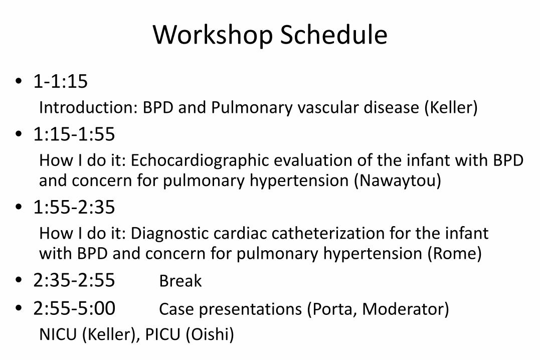
Workshop Schedule • 1-1:15
Introduction: BPD and Pulmonary vascular disease (Keller) • 1:15-1:55
How I do it: Echocardiographic evaluation of the infant with BPD and concern for pulmonary hypertension (Nawaytou)
• 1:55-2:35 How I do it: Diagnostic cardiac catheterization for the infant with BPD and concern for pulmonary hypertension (Rome)
• 2:35-2:55 Break • 2:55-5:00 Case presentations (Porta, Moderator)
NICU (Keller), PICU (Oishi)
Introduction: BPD and Pulmonary Vascular Disease
Roberta L. Keller, MD UCSF Benioff Children’s Hospital
San Francisco CA March 9, 2017

Overview
• Bronchopulmonary dysplasia (BPD) • Pulmonary hypertension in BPD • Consideration of co-morbidities in BPD
– Functional Class – Severity of lung disease, airway obstruction – Cardiovascular—structural/hemodynamic factors – Other factors—gastroesophageal reflux, feeding
difficulties, liver disease, neurologic (abnormal control of breathing due to injury or dysmaturity)

WHAT IS BRONCHOPULMONARY DYSPLASIA?
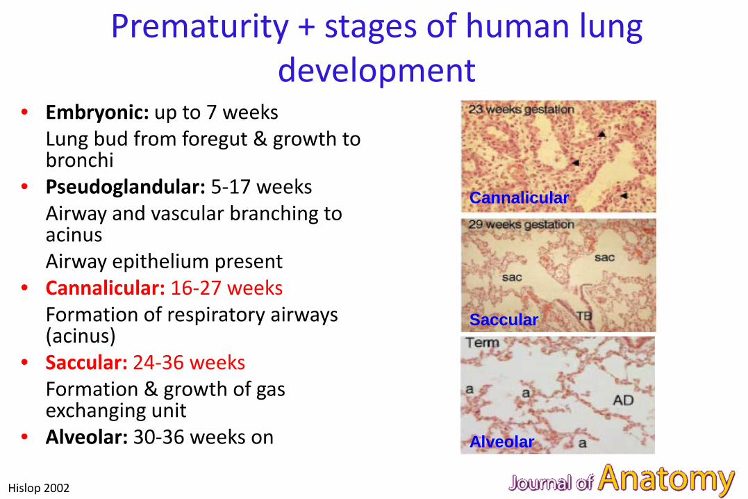
Prematurity + stages of human lung development
• Embryonic: up to 7 weeks Lung bud from foregut & growth to
bronchi • Pseudoglandular: 5-17 weeks Airway and vascular branching to
acinus Airway epithelium present • Cannalicular: 16-27 weeks Formation of respiratory airways
(acinus) • Saccular: 24-36 weeks Formation & growth of gas
exchanging unit • Alveolar: 30-36 weeks on
Hislop 2002
Cannalicular
Saccular
Alveolar

What is BPD?
• Chronic lung disease of prematurity • “Old” BPD Scarring and fibrosis of the lung, severe airway
disease in surviving preterm babies in association with high ventilator pressure + FiO2 (Northway 1967)
• “New” BPD Impaired lung and vascular development due to
extreme prematurity (< 28-30 weeks’ gestation) (Jobe 1999)

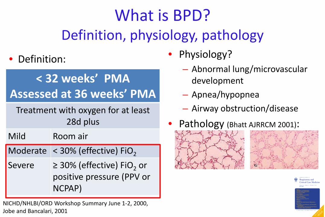
What is BPD? Definition, physiology, pathology
< 32 weeks’ PMA Assessed at 36 weeks’ PMA
Treatment with oxygen for at least 28d plus
Mild Room air Moderate < 30% (effective) FiO2
Severe ≥ 30% (effective) FiO2 or positive pressure (PPV or NCPAP)
• Physiology? – Abnormal lung/microvascular
development – Apnea/hypopnea – Airway obstruction/disease
• Pathology (Bhatt AJRRCM 2001):
NICHD/NHLBI/ORD Workshop Summary June 1-2, 2000, Jobe and Bancalari, 2001
• Definition:
WHAT IS PULMONARY HYPERTENSION IN INFANTS WITH BPD?

Defining PH in BPD
Echo measurement Criteria used for classification* Tricuspid regurgitant (TR) jet velocity**
Right ventricular systolic pressure (RVsp) > 40 mmHg [right atrial pressure (RAp) = 0] RVsp:SBP ratio ≥ 1/2 or 2/3 (RAp = 0 or 5 mmHg)
Non-restrictive cardiac shunt (PDA, VSD, atrial septum)
Right-to-left or bidirectional flow RV or PA pressure ≥ 1/2 or 2/3 systemic
Interventricular septum (IVS) position
D-shaped or convex into LV Flattened throughout the cardiac cycle Any flattening
*Assumes no RV outflow tract obstruction **By modified Bernoulli equation: RVsp = 4 x velocity2 + RAp (assumed RAp noted)
Mourani 2008, Keller 2010, Mirza 2014, Mourani 2015, Lusk 2015
Cardiac catheterization is the gold standard! Understanding contribution of pulmonary vascular disease challenging without cath
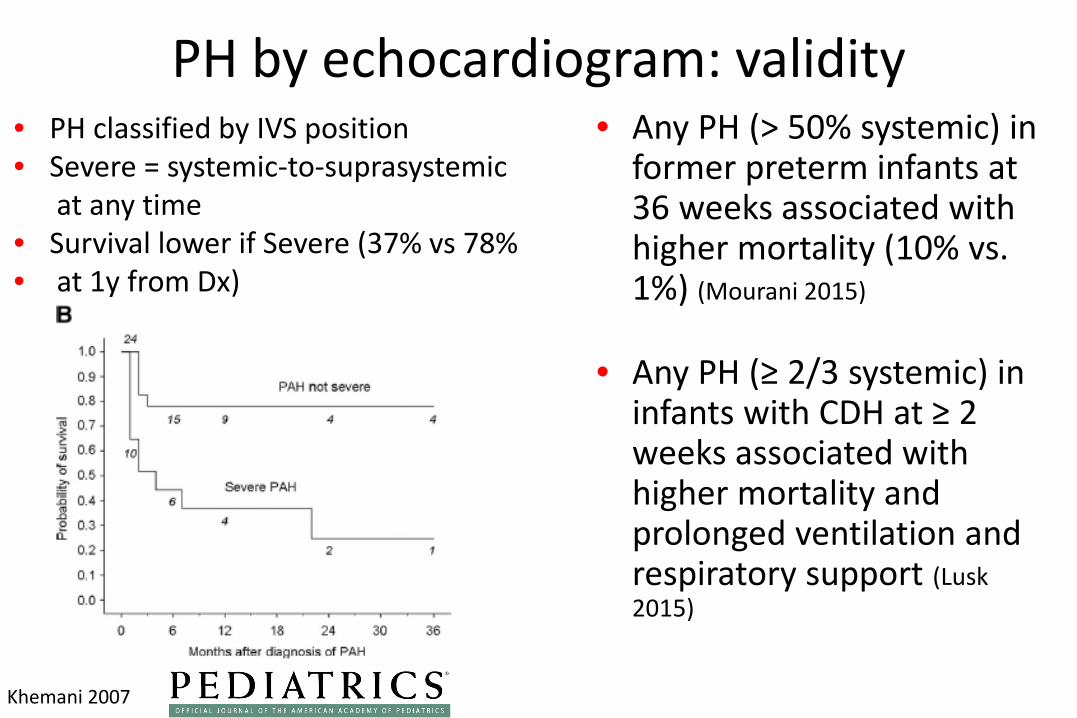
PH by echocardiogram: validity • Any PH (> 50% systemic) in
former preterm infants at 36 weeks associated with higher mortality (10% vs. 1%) (Mourani 2015)
• Any PH (≥ 2/3 systemic) in infants with CDH at ≥ 2 weeks associated with higher mortality and prolonged ventilation and respiratory support (Lusk 2015)
Khemani 2007
• PH classified by IVS position • Severe = systemic-to-suprasystemic at any time • Survival lower if Severe (37% vs 78% • at 1y from Dx)

HOW DOES PULMONARY HYPERTENSION IN BPD RELATE TO LUNG DISEASE?
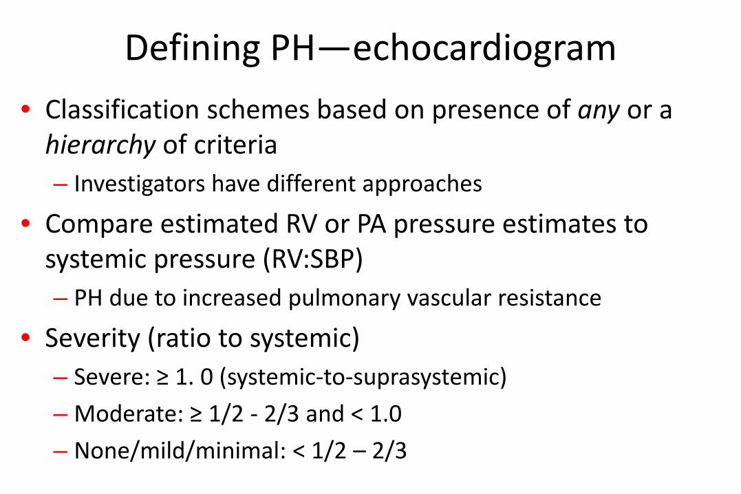
Defining PH—echocardiogram • Classification schemes based on presence of any or a
hierarchy of criteria – Investigators have different approaches
• Compare estimated RV or PA pressure estimates to systemic pressure (RV:SBP) – PH due to increased pulmonary vascular resistance
• Severity (ratio to systemic) – Severe: ≥ 1. 0 (systemic-to-suprasystemic) – Moderate: ≥ 1/2 - 2/3 and < 1.0 – None/mild/minimal: < 1/2 – 2/3
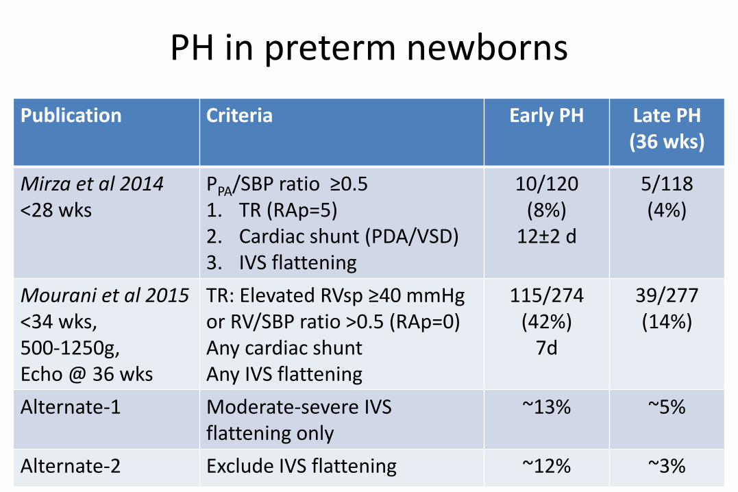
PH in preterm newborns Publication Criteria Early PH Late PH
(36 wks)
Mirza et al 2014 <28 wks
PPA/SBP ratio ≥0.5 1. TR (RAp=5) 2. Cardiac shunt (PDA/VSD) 3. IVS flattening
10/120 (8%)
12±2 d
5/118 (4%)
Mourani et al 2015 <34 wks, 500-1250g, Echo @ 36 wks
TR: Elevated RVsp ≥40 mmHg or RV/SBP ratio >0.5 (RAp=0) Any cardiac shunt Any IVS flattening
115/274 (42%)
7d
39/277 (14%)
Alternate-1 Moderate-severe IVS flattening only
~13% ~5%
Alternate-2 Exclude IVS flattening ~12% ~3%

Factors associated with Early PH (7-14d)
Clinical Respiratory Echo
Gestational age and birth weight (NS)
MV > 7d by 10d/ MV at 7d*
PDA
Any PDA treatment FiO2 > 0.3 by 10d
Duration of MV
Severity of BPD*
*Finding in both Mirza and Mourani studies

Factors associated with Late PH (36 wks) Clinical Respiratory Echo
Birth weight Duration of mechanical
ventilation (MV)
IVS flattening at 7d
Multiple gestation CPAP or MV at 36 wks RV dilation at 7d
Mortality (post-echo) Duration of O2
Severity of BPD
Discharge on O2
Mourani et al 2015
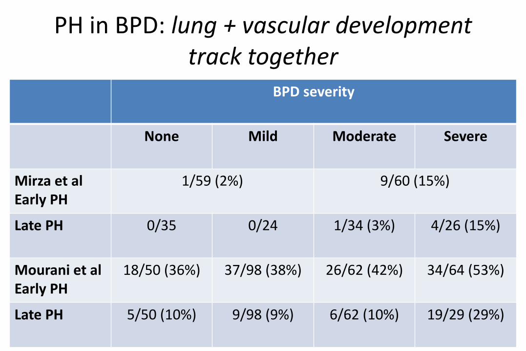
PH in BPD: lung + vascular development track together
BPD severity
None Mild Moderate Severe
Mirza et al Early PH
1/59 (2%) 9/60 (15%)
Late PH 0/35 0/24 1/34 (3%) 4/26 (15%)
Mourani et al Early PH
18/50 (36%) 37/98 (38%) 26/62 (42%) 34/64 (53%)
Late PH 5/50 (10%) 9/98 (9%) 6/62 (10%) 19/29 (29%)

CONSIDERATION OF FUNCTIONAL CLASS + COMORBIDITIES

PVRI: Functional class (0-2 y) I. Asymptomatic. Growing + developing normally, no limitation
of physical activity. II. Slight limitation of activity, dyspnea + fatigue, comfortable at
rest. Growing on centiles, behind on motor milestones. III. a—Less than ordinary activity causes undue fatigue or
syncope; frequent naps, quiet. Growth compromised, poor appetite, regression of learned motor activities. Requires excessive medical attention.
b—IIIa, plus growth severely compromised; supplemental feeding. IV. IIIb, plus syncope and/or right heart failure. Unable to carry
out any activity without dyspnea, fatigue or syncope. Not interacting with family.
Lammers 2011

Pediatric Functional Class + survival
Balkin et al, 2016
Most children with PH in this cohort improve FC during follow up/treatment Improvements in FC are associated with survival
Co-morbid cardiovascular conditions
• Occur in ~50% of infants with BPD and PH (del Cerro 2014)
• Prevalence (from highest to lowest) – pulmonary vein stenosis = PDA – aorta-pulmonary collaterals – atrial septal defect (ASD)

Additional clinical considerations • Severity of parenchymal lung disease, airway obstruction/malacia,
provision of adequate respiratory support • Gastroesophageal reflux, aspiration
– Feeding problems + reflux common in lung disease – May be associated with PH-specific therapy
• Neurologic dysfunction – Abnormal/dysmature control of breathing – Neuromuscular weakness
• Thyroid dysfunction – Hyper- and hypothyroidism associated with PH in case reports – Hyperthyroid associated with decompensation in those with known PH
• Acute respiratory infections – May be initial presentation of PH in former preterm – Increase in severity of PH by echo (del Cerro 2014)
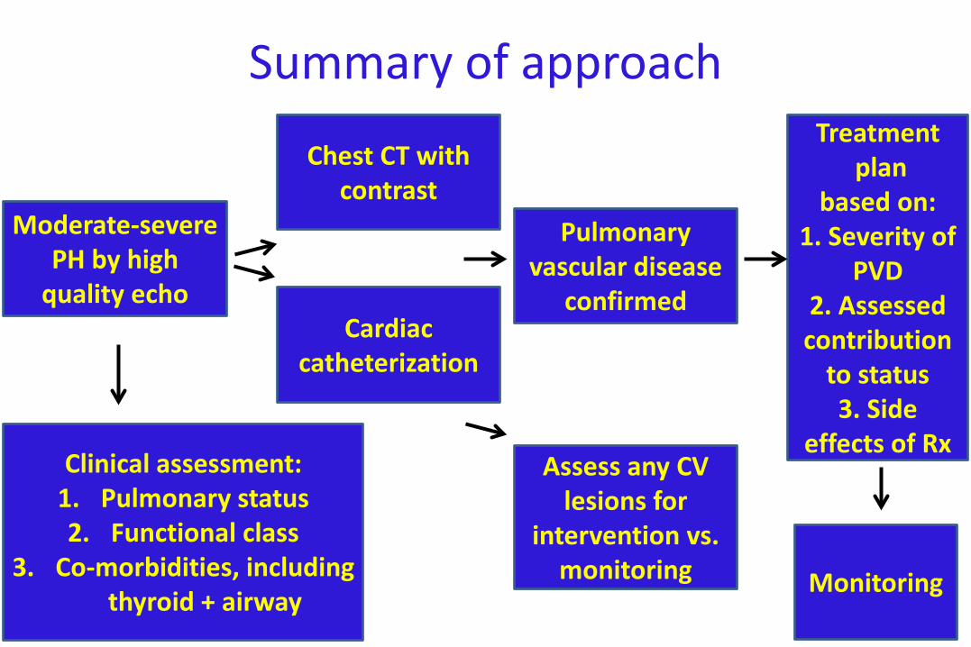
Summary of approach
Moderate-severe PH by high
quality echo
Clinical assessment: 1. Pulmonary status 2. Functional class
3. Co-morbidities, including thyroid + airway
Chest CT with contrast
Cardiac catheterization
Assess any CV lesions for
intervention vs. monitoring
Pulmonary vascular disease
confirmed
Treatment plan
based on: 1. Severity of
PVD 2. Assessed contribution
to status 3. Side
effects of Rx
Monitoring

UCSF PH Screening protocol • Who: GA < 32 weeks, moderate-to-severe BPD • When: 36 weeks PMA or prior to discharge if earlier
– If clinical concerns, echocardiogram can be done earlier (e.g., significant lung disease with elevated PCO2, increasing FiO2 requirement, poor growth)
• Response – “normal”—Follow up 4-6 mos postnatal age, if possible when off
supplemental O2
– “abnormal”—PH consult with recs for further evaluations + timing of follow up. Parental education regarding close monitoring during illness or any perioperative period.
– All infants discharged on O2 need eval/echo when it is discontinued
CASE: NICU PRESENTATION OF PULMONARY HYPERTENSION
Case: Female, 24 weeks, 620g (AGA)
• Premature ROM (12d) + unstoppable preterm labor • Intubated, surfactant, early severe lung disease with
pulmonary interstitial emphysema • Inhaled NO in 1st week for pulmonary hypertension • RLL lobectomy for pneumatocele at 25d • Extubated to nasal CPAP at 59d • 4 LPM HFNC at 81d after hydrocortisone • Increasing FiO2 at 145 d
– Echo: systemic RV pressure estimate, with dilated RA/RV, moderately depressed RV function

Transfer for evaluation of PVD • Started on sildenafil 1 mg/kg tid prior
to transfer • Transfer at 151 d: 45 5/7 weeks’
postmenstrual age • HFNC 4 LPM FiO2 1.0 • CBG 7.41/73/+20 • RR 70s, HR 130s-140s, mild retractions,
RV heave, no murmur, liver 2.5 cm • Diuretics: Furosemide tid/Aldactone bid • Growing on 10th %ile/ predominantly
NG fed – Cough/gag with oral feeding, intermittent
retching + emesis – On Ranitidine and metoclopramide
Admission CXR: Coarse bilateral opacities
consistent with CLD.
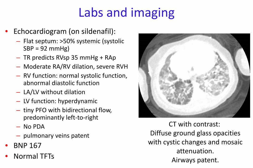
Labs and imaging • Echocardiogram (on sildenafil):
– Flat septum: >50% systemic (systolic SBP = 92 mmHg)
– TR predicts RVsp 35 mmHg + RAp – Moderate RA/RV dilation, severe RVH – RV function: normal systolic function,
abnormal diastolic function – LA/LV without dilation – LV function: hyperdynamic – tiny PFO with bidirectional flow,
predominantly left-to-right – No PDA – pulmonary veins patent
• BNP 167 • Normal TFTs
CT with contrast: Diffuse ground glass opacities
with cystic changes and mosaic attenuation.
Airways patent.
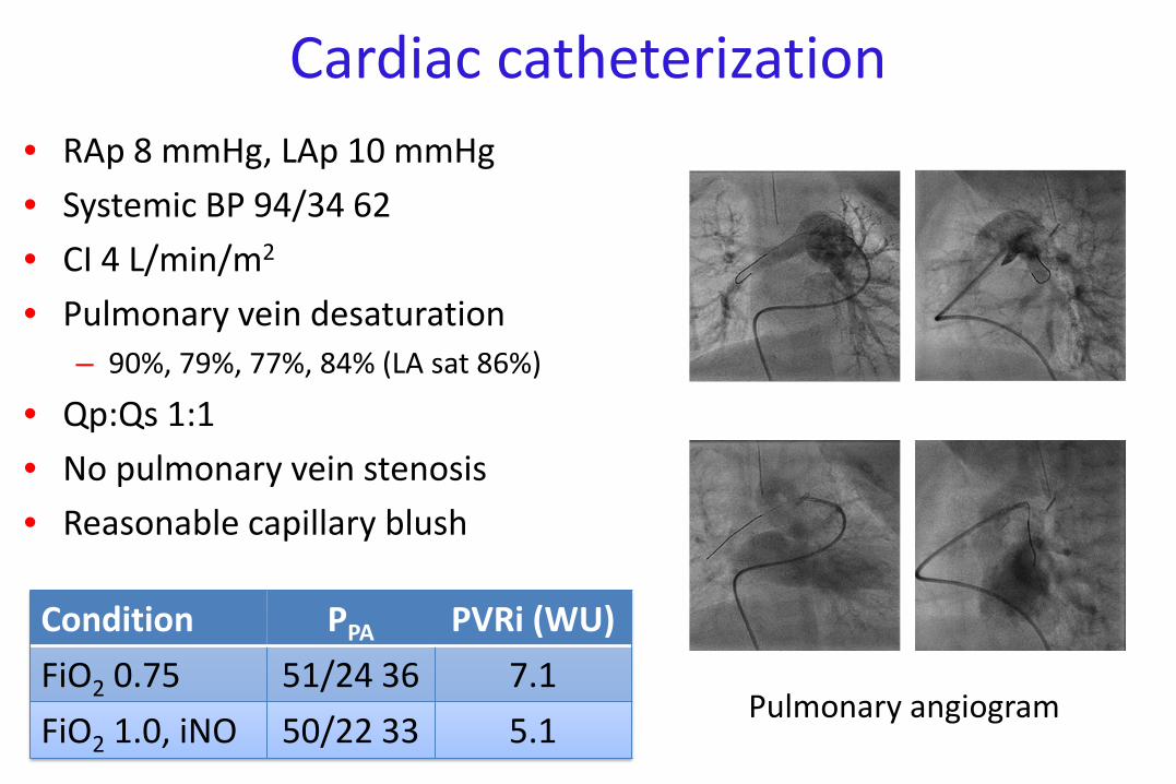
Cardiac catheterization • RAp 8 mmHg, LAp 10 mmHg • Systemic BP 94/34 62 • CI 4 L/min/m2 • Pulmonary vein desaturation
– 90%, 79%, 77%, 84% (LA sat 86%)
• Qp:Qs 1:1 • No pulmonary vein stenosis • Reasonable capillary blush
Condition PPA PVRi (WU)
FiO2 0.75 51/24 36 7.1 FiO2 1.0, iNO 50/22 33 5.1
Pulmonary angiogram

Summary: 153d
• Severe PH (systemic-to-suprasystemic) – due to pulmonary vascular disease – moderately elevated PVRi with reactivity – reasonably compensated (preserved CI)
• Severe lung disease – Requires substantial respiratory support, chronically elevated
PCO2, PV desaturation • Poor functional class
– Good weight gain but with supplemented tube feeds – Decreased endurance, emesis
• GER – Despite anti-reflux medications
Treatment plan/clinical course transfer back at 167d
• Pulmonary vascular disease: dual therapy – Bosentan initiated at 1 mg/kg bid. Advanced to 2 mg/kg bid after 1
week – Sildenafil changed to 0.75/kg mg qid – Diuretics continued – Echo: Mildly flattened IVS (improved), persistent RV dilation, mild
RA enlargement, normal RV systolic function, abnormal diastolic function, mild RVH, TR not quantifiable, pulmonary veins patent
– BNP 33 • Respiratory support weaned to 3 LPM, FiO2 45% • Feeds: taking all feeds by mouth without retching or emesis
– Good weight gain over 7d


