JOP. J Pancreas (Online) 2004; 5(5 Suppl):405-453. JOP. Journal of the Pancreas – http://www.joplink.net – Vol. 5, No. 5 Supplement – September 2004. [ISSN 1590-8577] 405 AISP - 28 th National Congress. Verona (Italy). October 28-30, 2004 (The abstracts are published as submitted by the authors) ORAL COMMUNICATIONS Somatostatin and Gabexate Are Ineffective in Preventing Post-ERCP Complications Andriulli A, Solmi L, Loperfido S, Festa V, Belmonte A, Leo P, Spirito F, Silla M, Forte G, Terruzzi V, Mescia P, Ciliberto E, Mauro M, Monica F, Perri F, on behalf of the Italian Group for Prevention of Acute Pancreatitis (GIPPA 2 study) San Giovanni Rotondo, Bologna, Treviso, Roma, Cosenza, Vasto, Caserta, Como, Campobasso, Crotone. Italy Background The issue whether it is possible to prevent pancreatic damage by the prophylactic administration of somatostatin (SS) or gabexate (GM) is still debated. Aim Aim of this study was to assess the efficacy of these drugs for prevention of post- ERCP pancreatitis. Methods A double-blind, multicenter, placebo-controlled trial was conducted in 966 patients who randomized received an intravenous infusion of SS (750 µg, n=290), GM (500 mg, n=327), or placebo (saline, n=349) that was started 30 minutes before endoscopy and continued for 6 hours afterward. Patients were evaluated clinically and serum amylase levels determined at 4 and 24 hours after endoscopy. Results No significant difference in the occurrence of pancreatitis, hyperamylasemia, or abdominal pain was observed among placebo (6.9%, 33.5%, and 4.0% respectively), SS (7.6%, 26.6%, and 2.8% respectively), and GM (6.1%, 32.7%, and 4.0% respectively). At univariate analysis of patients characteristics and endoscopic maneuvers, the Freeman’ score for difficult cannulation (P<0.001), more than 3 pancreatic injections (P<0.001), an intradiverticular papilla (P=0.01), and bile duct diameter less than 8 mm (P=0.04) were associated to post- ERCP pancreatitis. At multivariate logistic analysis only more than 3 pancreatic injections (OR=1.72, 95%CI: 1.15-2.59), and the Freeman’ score (OR=1.41, 95%CI: 1.08- 1.86) were predictive of post-ERCP pancreatitis. Conclusion Long-term (6.5 hours) administration of SS or GM is ineffective for prevention of post-ERCP pancreatitis. Pancreatic injury is related to maneuvers used to obtain biliary access rather than any patient characteristics. Cell-Mediated Immune Functions Before and After Chemotherapy in Advanced or Metastatic Pancreatic Carcinoma Patients Bellone G 1 , Carbone A 1,2 , Novarino A 3 , Addeo A 3 , Tonel E 1 , Dughera L 2 , Bertetto O 3 , Emanuelli G 1 1 Department of Clinical Physiopathology, University of Turin. 2 Department of Gastroenterology and Clinical Nutrition and 3 Department of Oncology, San Giovanni Battista Hospital. Turin, Italy Background The survival rate for patients with pancreatic carcinoma is among the poorest for all cancers. Host defense against tumor does not appear to be induced effectively, suggesting potential impairment of cell-mediated immunity. NK and T cells both play a critical role in the effector phase of immunologically mediated tumor rejection, and their development and expansion depend on the cytokines produced by T helper cells.
JOP. Journal of the Pancreas – http://www.joplink.net – Vol. 5, No. 5 Supplement – September 2004. [ISSN 1590-8577] 405
AISP - 28th National Congress. Verona (Italy). October 28-30, 2004
(The abstracts are published as submitted by the authors)ORAL COMMUNICATIONS
Somatostatin and Gabexate Are Ineffective in PreventingPost-ERCP Complications
Andriulli A, Solmi L, Loperfido S, Festa V, Belmonte A, Leo P, Spirito F, Silla M, Forte G,Terruzzi V, Mescia P, Ciliberto E, Mauro M, Monica F, Perri F, on behalf of the Italian
Group for Prevention of Acute Pancreatitis (GIPPA 2 study)San Giovanni Rotondo, Bologna, Treviso, Roma, Cosenza, Vasto, Caserta, Como, Campobasso,
Crotone. Italy
Background The issue whether it is possibleto prevent pancreatic damage by theprophylactic administration of somatostatin(SS) or gabexate (GM) is still debated.Aim Aim of this study was to assess theefficacy of these drugs for prevention of post-ERCP pancreatitis.Methods A double-blind, multicenter,placebo-controlled trial was conducted in 966patients who randomized received anintravenous infusion of SS (750 µg, n=290),GM (500 mg, n=327), or placebo (saline,n=349) that was started 30 minutes beforeendoscopy and continued for 6 hoursafterward. Patients were evaluated clinicallyand serum amylase levels determined at 4 and24 hours after endoscopy.Results No significant difference in theoccurrence of pancreatitis, hyperamylasemia,or abdominal pain was observed amongplacebo (6.9%, 33.5%, and 4.0%
respectively), SS (7.6%, 26.6%, and 2.8%respectively), and GM (6.1%, 32.7%, and4.0% respectively). At univariate analysis ofpatients characteristics and endoscopicmaneuvers, the Freeman’ score for difficultcannulation (P<0.001), more than 3 pancreaticinjections (P<0.001), an intradiverticularpapilla (P=0.01), and bile duct diameter lessthan 8 mm (P=0.04) were associated to post-ERCP pancreatitis. At multivariate logisticanalysis only more than 3 pancreaticinjections (OR=1.72, 95%CI: 1.15-2.59), andthe Freeman’ score (OR=1.41, 95%CI: 1.08-1.86) were predictive of post-ERCPpancreatitis.Conclusion Long-term (6.5 hours)administration of SS or GM is ineffective forprevention of post-ERCP pancreatitis.Pancreatic injury is related to maneuvers usedto obtain biliary access rather than any patientcharacteristics.
Cell-Mediated Immune Functions Before and After Chemotherapy inAdvanced or Metastatic Pancreatic Carcinoma Patients
1Department of Clinical Physiopathology, University of Turin. 2Department of Gastroenterologyand Clinical Nutrition and 3Department of Oncology, San Giovanni Battista Hospital. Turin, Italy
Background The survival rate for patientswith pancreatic carcinoma is among thepoorest for all cancers. Host defense againsttumor does not appear to be inducedeffectively, suggesting potential impairment
of cell-mediated immunity. NK and T cellsboth play a critical role in the effector phaseof immunologically mediated tumor rejection,and their development and expansion dependon the cytokines produced by T helper cells.
JOP. Journal of the Pancreas – http://www.joplink.net – Vol. 5, No. 5 Supplement – September 2004. [ISSN 1590-8577] 406
Aim The study investigates the cell-mediatedimmune status of advanced or metastaticpancreatic carcinoma patients before and afterchemotherapy.Methods We studied 12 patients (age range51-84); 6 underwent treatment with 5-FUcontinuous infusion for 6 weeks, cisplatinweekly and gemcitabine on days 1, 8, 28, and35. Check-ups were programmed every twomonths. Peripheral blood mononuclear cells(PBMC) were obtained from patients andnormal subjects. Lymphocyte subsets weredetermined by flow cytometry. Interleukin(IL)-12 and IL-10 was determined by ELISAin lipopolysaccharides (LPS)-stimulatedPBMC culture supernatants, while Interferon(IFN)-gamma was measured in anti-CD3-activated lymphocyte culture supernatants.NK and LAK-mediated cytotoxicities wereinvestigated against K562 and Daudi cells,respectively.Results No difference in either absolutenumber or surface phenotype of T (CD3, CD4and CD8) or NK cells (CD56) was observedin patients versus healthy subjects. LPS-stimulated PBMC from patients produced
higher levels of IL-12 total p40 (P=0.03) andIL-10 (P=0.02), and lower levels of bioactiveIL-12 p70 (P=0.006) than in normalindividuals, while IFN-gamma levelsproduced by activated T cells from patientswere found to be significantly lower (P=0.03),both NK and LAK-mediated cytotoxicitieswere also defective. From 2 to 4 months afterthe end of chemotherapy, in 75% of patientsCD4+ cell percentage had significantlyincreased (P=0.009) and there was a slightdecrease in IL-12 p40 production but nosignificant changes in IL-12 p70, IL-10 orIFN-gamma. While NK cell activity had notaltered after chemotherapy, in 50% of treatedpatients LAK activity was enhanced.Conclusions The results suggest that analtered balance between anti- (IL-10) and pro-inflammatory (IL-12, IFN-gamma) cytokinesmight be responsible for the defective cellmediated anti-tumor immunity in advanced ormetastatic pancreatic carcinoma patients.Additional larger studies are required tocorrelate changes in cellular immunefunctions and clinical response to therapy.
Three Hypervascularized Lesions of Duodenal and Jejunal Tract in aPatient with von Recklinghausen’s Disease: An Exceptional Triade?Bettini R1, Falconi M1, Cannizzaro R2, Butturini G1, Marcucci S1, Partelli S1, Pederzoli P1
1Pancreatic Unit, University of Verona. Verona, Italy.2Department of Gastroenterology and Endoscopy, CRO di Aviano. Aviano (PN), Italy
Case report DD, a 47-year-old man, wasadmitted in our hospital on April 2004 withthe diagnosis of duodenal carcinoids in apatient with von Recklinghausen’s disease.The medical history starts on September 2002with melena. Based on a gastroduodenalendoscopy with biopsy, a Barret disease wasdiagnosed and a specific medical treatmentwas started. Colonoscopy was negative forbleeding lesions. On December 2003, due tothe recurrence of melena and to transitorydiarrhea, the patient was admitted to anotherhospital. He was submitted to an enteroscopyuntil 100 cm from Treitz ligament withevidence of two sub mucosal ulcerated lesions
on Papilla major e minor; diagnosed atmicroscopic examination as carcinoids. Thediagnostic suspicious was turned toward ahypothetical duodenal gastrinoma. However,laboratory findings, even if high gastrin levelwere found, showed a negative secretin test.Aspecific endocrine tumor markers werewithin the normal values (NSE 8.5 ng/mL,normal values <12.5 ng/mL; CgA 50 ng/mL,normal values 19-98 ng/mL) as well as forlaboratory routine tests. No mutations forMEN I gene was observed. An intestinalcontrast medium X-ray and an abdominal CTraised the suspicion for another lesion on ilealsite, whilst octreoscan was negative for all the
JOP. Journal of the Pancreas – http://www.joplink.net – Vol. 5, No. 5 Supplement – September 2004. [ISSN 1590-8577] 407
aforementioned lesions. The patient was sentto our Department for a surgical approach. OnApril 2004 he underwent to laparotomy whichdemonstrated multiple liver metastases, asnew element. A frozen section of one of themwas positive for hepatic met of endocrinecarcinoma. After an intra-operative USassessment all the liver lesions were judged ascompletely resectable. Moreover, intestinalexamination confirmed at 50 cm from Treitz abrownish lesion, 6 cm in size with exophyticgrowth. The patient was operated upon ofpancreaticoduodenectomy (Whippleprocedure) with jejunal resection up to thedistal lesion, extensive lymphadenectomy,and multiple metastasectomies. Procedure
was considered with radical intent. Finalpathological assessment established thepresence of a papilla major and minor“somatostatinoma” (well differentiatedendocrine neoplasm) with strong immuno-hystochemical expression of somatostatin,ki67 <1%, 6 out 73 positive nodes, 4 solidlocalization in peripancreatic tissue, 6endocrine liver hepatic mets, a jejunal GISTwith uncertain behavior (c-kit ++; ki67 <1%).Conclusions Even if gastrointestinal tractinvolvement is not rare in vonRecklinghausen’s disease, at our knowledge,this is only the third report of synchronousassociation between the disease, jejunal GISTand ampullary endocrine tumors.
Case Report of an Intraductal Papillary Mucinous Neoplasm of theUncinate Process
1Department of Surgery and 1Department of Pathology, University of Verona. Verona, Italy
Context Intraductal papillary mucinousneoplasms (IPMNs) are usually identified byimaging findings such as the ectasia of themain pancreatic duct (central IPMNs) with apossible involvement of the side branches ofthe ductal system (branch-side IPMNs).Clinical and demographic criteria should alsobe considered: patients older than 60 yearswith pancreatitis-like pain, weight loss,diabetes, jaundice and elevated CA 19.9 levelcould be related to malignancy of theneoplasm. IPMNs may led to chronicpancreatitis (CP); moreover, in some cases, adistinction between these two diseases can bedifficult, in particular when the IPMNinvolves the entire gland.Case Report We report the case of a 68-yearCaucasian male admitted at the Department ofSurgery of the University of Verona forobstructive jaundice. His medical historyincludes a right colectomy for cancer in 1984and pulmonary lobectomy for metastasis 3years later, a cholecystectomy for gallbladderstones in 1994 and an endovascular exclusionof aortic aneurysm in February 2004. Thepatient presented with vague and intermittent
upper abdominal pain and a significant weightloss (25 kg in 4 months). At admission thebilirubin serum level was 22.63 mg/dL, CEA8.3 ng/mL and CA 19.9 level 184 U/mL.ERCP showed a regular ampulla and astenosis of the intrapancreatic bile ductassociated with a dilation of the intrahepaticbile ducts; the main pancreatic duct was seenonly 2 cm far from the papilla of Vater. CWMR showed dilation of the main pancreaticduct and of the side branches of the pancreatichead. These findings suggested the presenceof an IPMN located in the head of thepancreas.The patient underwent surgery and submittedto pylorus-preserving pancreaticoduodene-ctomy. The postoperative course wasuneventful and the patient was discharged in10th postoperative day. The pathologicfindings showed the presence of a largeintrapancreatic pseudocyst (diameter 3 cm)involving the head of the pancreas betweenthe main bile duct and the Wirsung and of CPin the pancreatic remnant.Discussion We reported a case of clinical,laboratory data including tumor markers level
JOP. Journal of the Pancreas – http://www.joplink.net – Vol. 5, No. 5 Supplement – September 2004. [ISSN 1590-8577] 408
and imaging findings suggesting thepreoperative diagnosis of a pancreatic headmalignant IPMN. Histology examination ofthe surgical specimen revealed the presenceof intrapancreatic pseudocyst associated withCP without any evidence of neoplasm.
Conclusion Even if the etiology of chronicpancreatitis is still unknown this patientrepresents in our experience the first case ofmisdiagnosis between branch-side IPMNs andCP.
May an Experimental Model of Intraductal Papillary MucinousNeoplasm of the Pancreas Have a Clinical Meaning?
1Department of Surgical Sciences and 2Department of Pathology, University of Verona.Verona, Italy
Background In the last decade intraductalpapillary mucinous neoplasms (IPMNs) haveturned from a rare entity to a continuouslyincreasing disease. While their clinicalknowledge and management have beenconsequently improved, the biology ofIPMNs and their behavior to malignancy,however, are still largely unknown. Beingsurgery the only curative approach, resectionis mandatory, but its indication in early stagesor in elderly patients and its extent inmultifocal disease is still a matter of debate.Finally no further treatment is effective inunresectable patients.Aim and Methods In our opinion, theavailability of an experimental model couldbe helpful in better understanding thebiological features of IPMNs. With thispurpose, we implanted in nude mice surgicalsamples from a 66 years-old female submittedto Longmire-Traverso procedure forpancreatic head IPMN.Results The tumor was successfullyimplanted. The xenograft was thenestablished in five following steps, allreproducing the primary pattern, and grew as
a large burden with peritoneal diffusion. Thepathological assessment of invasive IPMNconfirmed preoperative diagnosis and thepatient is still alive and free of disease 6 yearsafter surgery. So we are able to report the firstexperimental model for IPMN. No mutationsin K-ras, p53 and p16 typical of ductalcarcinoma were found neither in primary norin implanted tumor, this furthermore provingthe xenograft derived from the invasivecomponent of IPMN. We are about tocharacterize the phenotypic profile.Conclusions A real and stable experimentalmodel for IPMN appears to be an effectivetool in investigating the genetic and biologicalfeatures of this tumor and in understanding itsbehavior in the progress to malignancy. Adeeper knowledge of these aspects brings atlast on a clinical impact, since it may help usto better manage the patients suffering fromIPMN and select those undergoing surgery.Finally, the availability of such anexperimental model enable us to test inpreclinical studies chemotherapeutic drugsand adjuvant treatments for unresectablepatients.
Margins Involvement in Pancreatic Cancer ResectionBracale U, Balzano G, Zerbi A, Gavazzi F, Ortolano E, Civelli V, Reni M, Di Carlo V
Pancreas Unit and Radiochemiotherapy Unit, IRCCS S. Raffaele Hospital. Milan, Italy
Background The achievement of radicality inpancreatic cancer resection is considered oneof the main determinant of survival.
Aim The aim of this study was to define theincidence of margins involvement (stratifyingpatients in microscopic and macroscopic
JOP. Journal of the Pancreas – http://www.joplink.net – Vol. 5, No. 5 Supplement – September 2004. [ISSN 1590-8577] 409
involvement), the pattern of failure after non-radical resections and the relation betweenmargins invasion and postoperative survival.Patients and Methods From 1990 to 2002,296 patients underwent resection for non-metastatic ductal pancreatic adenocarcinoma.Patients were classified according to theUICC-R classification: R0: no marginsinvolvement; R1: microscopic involvement,R2: macroscopic residue. Statistical analysiswas performed by the chi-square test, log-rank test and Cox regression analysis(covariates stage, grading and nodal status).Results One-hundred and 75 patients (59%)underwent R0 resection, 68 patients (23%)and 53 patients (18%) underwent R1 and R2resection, respectively. The most frequentlyinvolved margin was the posterior one (69%).Follow-up was considered adequate to definethe pattern of failure in 202 out of 296patients. Local relapse (LR) was found in 52patients (40%) after R0 resection, 15 patients(39%) after R1 and 18 patients (53%) after R2(P NS). IORT was applied in 127 patients.IORT had no protective effect on LR: in R0
patients LR was found in 42% of patientsreceiving IORT and 41% of patients who didnot (P NS); in 56% of R1+2 patients receivingIORT and 45% of R1+2 patients who did not(P NS). The difference between survivalcurves of patients with different margin statuswas significant when considering R0 versusR1+2 (P<0.01). However, the survivaldifference was not significant whenconsidering R0 versus R1 resection (P=0.2).Also the multivariate analysis confirmed thatradicality was an independent prognosticfactor when considering R0 versus R1+2patients; however, the microscopic marginsinvolvement (R0 versus R1) was not asignificant determinant of survival.Conclusion Margin invasion is found inabout 40% of patients. R0 resection does notwarrant for a reduction of LR nor for asignificant prolonged survival whencompared to R1. R2 resections have a worseprognosis, but they can offer a goodpalliation. IORT did not reduce LR in nonradical resection.
Effect of Different Therapies on the Survival ofPancreatic Neuroendocrine Tumors
Department of Internal Medicine, S.Orsola-Malpighi Hospital, University of Bologna.Bologna, Italy
Aim To evaluate the efficacy of differenttherapies in patients with pancreaticneuroendocrine tumors (NET).Patients Eighty-three (37 M, 46 F)consecutive patients with pancreatic NETdiagnosed in our Department from 1978 to2003.Main outcome measures Clinical check-upand abdominal US were carried out every 3months during the first year after diagnosisand every 12 months thereafter. Survival wasestimated by means of the Kaplan-Meyer, andthe Mantel-Cox model was applied to identifyputative factors affecting the survival.
Results The median age of patients atdiagnosis was 55 years (range 19-81) and themedian age at the last observation was 60years (range 29-82). The median follow-upperiod was 36 months (range 3-264). Fifty-two patients (62.7%) had non-functioningNET, 16 (19.3%) had functioning NET and15 (18.1%) had MEN 1 disease withpancreatic involvement. The tumor waslocalized in the pancreatic head in 31 cases(37.3%), in the body in 24 cases (28.9%), inthe tail in 21 cases (25.3%) and was diffusethroughout the gland in 7 cases (8.4%). In 55patients, the median size of the tumor
JOP. Journal of the Pancreas – http://www.joplink.net – Vol. 5, No. 5 Supplement – September 2004. [ISSN 1590-8577] 410
evaluated at imaging techniques was 4.1 cm(range 1.0-11.3) and, in 41 patients, themedian size at surgery was 5.0 cm (range 1.0-13.0). Twenty-seven patients (32.5%) hadliver metastases at the time of diagnosis and43 (51.8%) developed liver metastases duringthe follow-up period. Involvement of thelymph nodes was found in 47 out of 79patients (59.5%). The median Ki67 evaluatedon 29 histological specimens was 2.9% (range1.0-84.1). Forty patients (48.2%) had radicalsurgery, 20 (24.1%) had debulking surgeryand 23 (27.7%) were treated medically. Of thelatter 23 patients, 19 (82.6%) underwent atleast one of the following treatments:somatostatin-analogs in all cases, interferon in3 (15.8%), chemotherapy in 2 (10.5%) andchemoembolization in 2 (10.5%). Forty-ninepatients (59.0%) were still alive at the time ofthe study; the median survival time was 90months (95% CI 29-151) and the 5-yearsurvival was 55.3%. Survival wassignificantly related to: age of patients at
diagnosis (OR per 10 years 1.38, 95% CI1.04-1.83; P=0.026), presence of metastases(liver metastases at diagnosis: OR 5.42, 95%CI 2.63-11.17, P<0.001; presence of lymphnode involvement: OR 4.97, 95% CI 1.91-12.90, P=0.001), type of the tumor (overallP=0.033; functioning vs. MEN: OR 7.63,95% CI 1.64-35.51, P=0.010; non-functioningvs. MEN: OR 4.65, 95% CI 1.07-20.13,P=0.040) and type of treatment (overallP<0.001; no surgery vs. radical surgery: OR5.20, 95% CI 2.12-12.72, P<0.001; debulkingvs. radical surgery: OR 4.29, 95% CI 1.76-10.48, P=0.001). Survival was notsignificantly related to: sex (P=0.151), age ofpatients (P=0.965), localization of the tumor(P=0.646), size of the tumor both at imagingtechniques (P=0.222) and at surgery(P=0.325), and Ki67 determination (P=0.341).Conclusions Radical surgery continues tohave a central role in the therapeutic approachto NET of the pancreas.
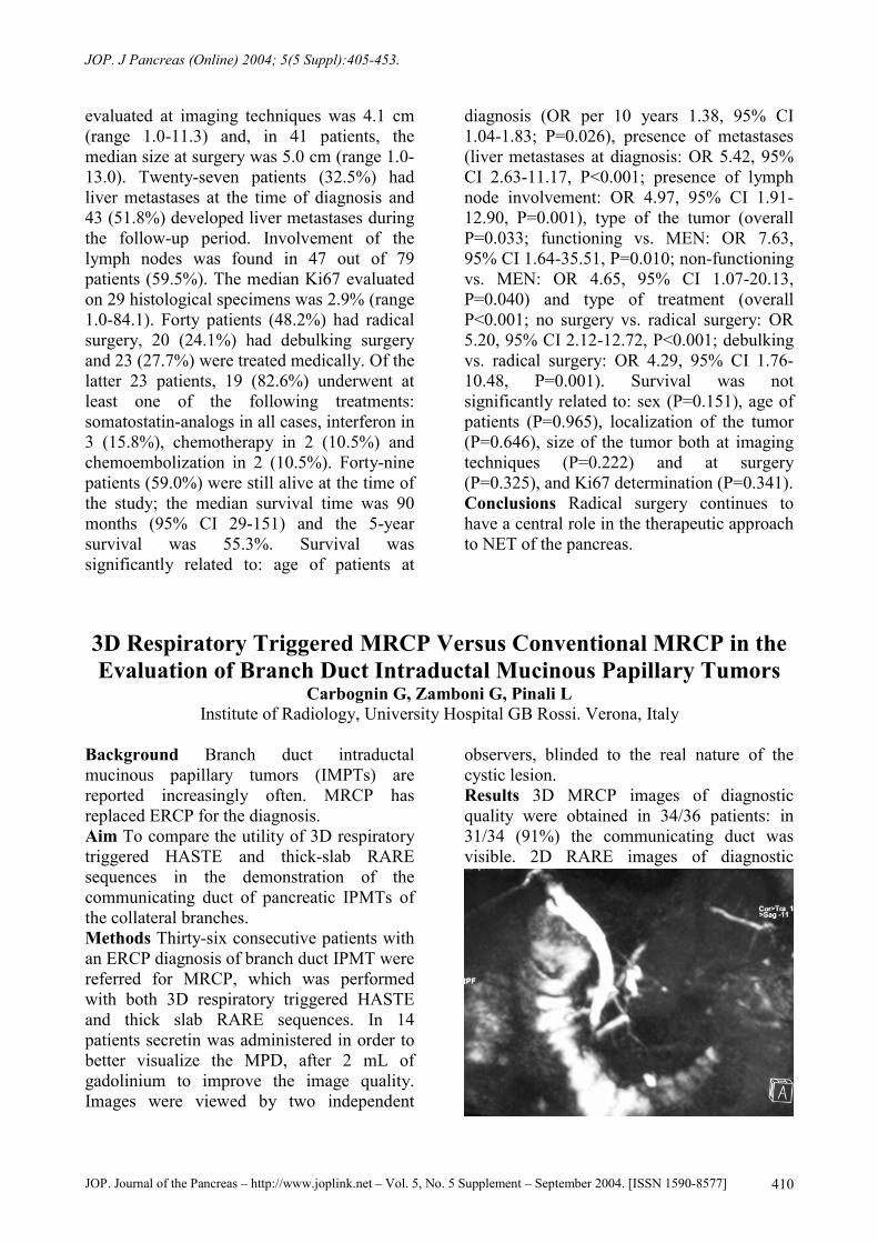
3D Respiratory Triggered MRCP Versus Conventional MRCP in theEvaluation of Branch Duct Intraductal Mucinous Papillary Tumors
Carbognin G, Zamboni G, Pinali LInstitute of Radiology, University Hospital GB Rossi. Verona, Italy
Background Branch duct intraductalmucinous papillary tumors (IMPTs) arereported increasingly often. MRCP hasreplaced ERCP for the diagnosis.Aim To compare the utility of 3D respiratorytriggered HASTE and thick-slab RAREsequences in the demonstration of thecommunicating duct of pancreatic IPMTs ofthe collateral branches.Methods Thirty-six consecutive patients withan ERCP diagnosis of branch duct IPMT werereferred for MRCP, which was performedwith both 3D respiratory triggered HASTEand thick slab RARE sequences. In 14patients secretin was administered in order tobetter visualize the MPD, after 2 mL ofgadolinium to improve the image quality.Images were viewed by two independent
observers, blinded to the real nature of thecystic lesion.Results 3D MRCP images of diagnosticquality were obtained in 34/36 patients: in31/34 (91%) the communicating duct wasvisible. 2D RARE images of diagnostic
JOP. Journal of the Pancreas – http://www.joplink.net – Vol. 5, No. 5 Supplement – September 2004. [ISSN 1590-8577] 411
quality were obtained in 36/36 patients: in25/36 (69.5%) the communicating duct wasvisible.Conclusions Thanks to its superior spatialresolution, 3D MRCP is better suitable forestablishing a diagnosis of branch duct IPMTs
than RARE sequences and almost equals thediagnostic accuracy of ERCP indemonstrating the communicating duct.RARE sequences maintain their fundamentalrole in the dynamic evaluation of pancreaticfunction.
Acute Pancreatitis Due to Simvastatin Therapy:Increased Severity After Re-ChallengeCeciliato R1, Pezzilli R1, Barakat B2, Corinaldesi R1
1Department of Internal Medicine and 2Department of Emergency Medicine,Sant’Orsola-Malpighi Hospital. Bologna, Italy
Introduction Statins are generally well-tolerated and acute pancreatitis has beenreported in only a few cases treated with thisdrugs. However, there have not been manycases reported regarding the re-challengeevidence.Case report A 64-year-old man presented atthe Emergency Room of our Hospitalcomplaining of epigastric pain of 16 hoursduration accompanied by nausea andvomiting. There was no history of alcoholingestion or previous abdominal surgery. Hehad had an acute myocardial infarction 6months before. The patient had been treatedfor the previous 6 months with simvastatin 20mg once daily for hypercholesterolemia andwith beta-blockers and aspirin for ischemicheart disease. No other medication was usedregularly. On physical examination, theabdomen was distended with hypoactivebowel sounds and diffuse tenderness whichwas maximal in the epigastrium; no reboundtenderness was present. Laboratory data onadmission showed increased serum amylaseand lipase activity (amylase: 2,884 IU/L,reference values: 0-220; lipase 3,245 IU/L,reference values 0-270). Serum values ofurea, creatinine, AST, ALT, alkalinephosphatase, triglycerides, cholesterol,calcium and bilirubin were normal.Abdominal ultrasound revealed parenchymalalterations compatible with pancreatic edema;the biliary tree was not dilated and nogallstones were seen. The patient wasconservatively treated and simvastatin was
discontinued. In December 2003, the patientreintroduced simvastatin 20 mg once daily, onhis own initiative, and seven days later, he feltepigastric pain. Laboratory examinationrevealed increased serum amylase and lipaseactivity (1,814 IU/L and 4,504 IU/L,respectively), and C-reactive proteinconcentration (27.3 mg/dL, reference values:0-0.8). Abdominal ultrasound carried out onadmission revealed an enlargement of thepancreas, there was also a dilation of the mainpancreatic duct, whereas the gallbladder andthe common bile duct were normal. Acontrast-enhanced CT scan was carried outtwo days after admission and revealed adishomogeneous enhancement of thepancreatic head with a small necrotic area andperipancreatic fluid collection. The patientwas treated conservatively, and clinical andlaboratory parameters progressivelyameliorated. Two months later, an endoscopicultrasound was carried out and no alterationsof the bile duct system were seen.Conclusion As the use of statins increases,the diagnosis of acute pancreatitis is beingmade more frequently in patients whodevelop abdominal pain of unknown etiologywhile taking these medications. When thepancreatitis is clinically confirmed, the drugshould be stopped and replaced to reduce thepossibility of further episodes of pancreatitis,especially in the form of more severe disease;the patient should also be aware of the riskinvolved in the reintroduction of the drug onhis own initiative.
1Department of Internal Medicine, S.Orsola-Malpighi Hospital, University of Bologna. Bologna,Italy. 2Department of Toxicology, Pharmacia Italia S.p.A., Pfizer Inc. Group, Nerviano (MI), Italy
Aim To evaluate the clinical value of a newimmunoassay for serum and urine TAPdetermination in assessing the diagnosis andthe severity of acute pancreatitis.Patients Thirty-four patients with acutepancreatitis (AP) (22 mild pancreatitis and 12severe disease); 12 patients with non-pancreatic acute abdomen (AA), 11 healthysubjects (HS) and 16 patients who underwenttherapeutic ERCP (ERCP).Methods Serum TAP (optical density, OD),amylase (reference range 64-92 IU/L) andlipase (reference range 46-67 IU/L) weredetermined in AP, AA, and HS at their initialobservation; AP patients were also studied forsix consecutive days from admission. InERCP patients, serum TAP, amylase andlipase, as well as urine TAP and amylase(upper reference limit 460 IU/L), weredetermined before and 6 hours after ERCP.Results Mean±SD serum TAP levels onadmission were 0.35±1.60 OD in AP patients,0.005±0.001 OD in AA patients, while HSs
had no detectable serum TAP levels. ERCPpatients had no detectable serum TAP levelsbefore and 6 hours after the execution ofERCP, whereas urine TAP concentrationsbefore the execution of the endoscopy were1.72±3.43 OD (mean±SD) and decreased 6hours after ERCP (mean±SD: 0.75±1.49 OD)(P=0.249). Using a cut off range of 0.013-0.020 OD for TAP, 138-142 IU/L foramylase, 67-98 IU/L for lipase, the sensitivityand specificity of the three markers inassessing the diagnosis of AP were 23.5% and91.7%, 94.1% and 100%, 97.1% and 100%,respectively. Using a cut off range of 0.005-0.008 OD for TAP, 409-448 IU/L, foramylase and 375-406 IU/L for lipase, thesensitivity and specificity in assessing theseverity of AP were 29.9% and 73.5% forTAP, 38.8% and 81.2% for amylase, 28.4%and 83.6% for lipase, respectively.Conclusions TAP is of limited value inassessing the diagnosis and the severity ofacute pancreatic damage.
The Orphan Receptors COUP-TFs Are Required for PancreaticStellate Cells Transdifferentiation and Modulate the Mitogenic
1Gastroenterology Unit, Department of Pathophysiology, University of Florence. Florence, Italy.2Department of Biochemistry and Molecular Biology Indiana University. Indianapolis, IN, USA
Background Pancreatic stellate cells (PSC)has been identified as the precursor cell typemainly responsible for the development ofpancreatic fibrosis. The orphan receptorschicken ovoalbumin upstream promotertranscription factors (COUP-TFI and COUP-TFII) belong to the nuclear receptorsuperfamily and play an important role indevelopment and differentiation. They bind to
cis-actin element either as homodimer or as aheterodimer with retinoid X receptor (RXR).There are evidences that COUP-TFs modulatethe activity of other nuclear receptorincluding retinoic acid receptors (RAR) andperoxisome proliferator activated receptor(PPAR).Aim In consideration of the role of nuclearreceptors in PSC activation we analyzed the
JOP. Journal of the Pancreas – http://www.joplink.net – Vol. 5, No. 5 Supplement – September 2004. [ISSN 1590-8577] 413
expression and transcriptional activity ofCOUP-TF during PSC transdifferentiation invitro.Methods Espression of COUP-TFs wasevaluated by western blot and RT-PCR.Transcriptional activity was evaluated bytransfection experiment using a luciferasereporter plasmid specific for COUP-TF (-841/-800 NHE-1 promoter). PSC proliferationwas evaluated by cell counting and H3
Tymidine incorporation.Result Both COUP-TFI and II was rapidly upregulated during PSC transdifferentiation invitro. In freshly isolated cells both receptorswas undetectable but after 24 h the II isoform
was significantly increased. In parallel,transcriptional activity was induced duringcell activation. Increased levels of thesereceptors in activated HSC by transfectioninduced cell proliferation and potentate theresponse to mitogens such as PDGF and EGF.In addition COUP-TFs antagonized PPARand RAR mediated signaling in PSC andregulate collagen and fibronectin geneexpression.Conclusion These data suggest that membersof the COUP-TF family play a role inmodulating profibrogenic response inpancreatic stellate cells.
Chromosomal Unbalance in a Cell Line fromPancreatic Ductal Carcinoma by Multicolor FISH
1Division of Surgical, Molecular and Ultrastructural Pathology, University and Hospital of Pisa.2Division of General Surgery and Transplantation, University and Hospital of Pisa and Regional
Referral Center for Pancreatic Diseases Treatment. Pisa, Italy
Introduction Genetic alterations occurring inpancreatic ductal carcinoma are not limited togene mutations of K-ras oncogene andsuppressor genes p53, p16, Smad4, but also togenomic unbalance with loss or gain of entirechromosomal arms of the same genes.Materials and methods One pancreaticadenocarcinoma cell line was derived fromprimary culture and analyzed (passages 7, 20,40, 60, 80 and 100) to evaluate the physicalstatus of chromosomes 3, 7, 9 and 17.Multicolor FISH allowed to probe thecentromeric regions of chromosomes 3, 7 and17, together with region 9p21.22 by usingfour differentially labeled probes(CEP3SpectrumRed, CEP7SpectrumGreen,CEP17SpectrumAqua, and LSI 9p21.22SpectrumGold, respectively). Cultured cellswere grown on slides for 48 h and fixed for 5min in Carnoy solution (methanol/acetic acid3:1). Probe hybridizations were carried outovernight at 39°C in a humidified chamber.Following hybridization, specimens werewashed using a solution of 0.1% NP-40 in 2X
SSC, stained with DAPI, and submitted toFISH analysis.Results Cultured adenocarcinoma cells wereanalyzed at passages 7, 20, 40, 60, 80 and100. In all cases the chromosomic profile wasfound to be altered. In particular, an increasein copy number of chromosomes 3, 7 and 17was observed. Deletion of the short arm ofchromosome 9 (9p21.22) was identifiedprecociously (passages 7 and 20).Conclusions Mutation studies in pancreaticadenocarcinoma cannot be limited to theanalysis of point mutations of K-ras oncogeneand the suppressor genes known to beinvolved in adenocarcinoma tumordevelopment. Attention must be focused alsoon the gain of chromosomal centromericregions that we found associated toextracopies of chromosomes 3, 7 and 17.Chromosome 17 is particularly relevant dueto the presence of suppressor gene p53, whichappeared to be mutated in the cell line understudy. We also identified a precociousdeletion of the short arm of chromosome 9
JOP. Journal of the Pancreas – http://www.joplink.net – Vol. 5, No. 5 Supplement – September 2004. [ISSN 1590-8577] 414
(9p21.22) that can be associated to the loss ofone copy of suppressor gene p16. Suchpattern of mutations is frequently found inpancreatic carcinoma. A more detailed
genome mapping will better define thechromosomic alterations identified in the cellline under study.
The Role of Procalcitonin in Predicting Complications and Prognosisin Severe Acute Pancreatitis: A Prospective European Trial
1Department of Surgery, Pancreatic Unit, University of Verona. Verona, Italy. 2Department ofGeneral Surgery, University of the Saarland. Homburg/Saar, Germany. 3Department of General
Surgery, University of Ulm. Ulm, Germany. 4Department of Surgery, Helsinki University CentralHospital. Helsinki, Finland. 5Department of Visceral and Transplantation Surgery, University of
Bern. Bern, Switzerland
Background Early, accurate and reliablestaging and diagnosis of infectiouscomplications in acute pancreatitis (AP) isimportant in the clinical practice.Procalcitonin (PCT), the 116 amino-acidprecursor of calcitonin, is the firstbiochemical parameter for predictinginfection and sepsis in various inflammatorydiseases; in AP the clinical value of PCTdeterminations still remains controversial.Aim To assess whether PCT play a role inpredicting complications and prognosis in AP.Methods From December 1999 untilSeptember 2003 a total of 103 patients withsevere AP were included in 5 EuropeanCentres within 96 h of disease onset. CRPwas determined routinely, PCT was assessedby a chemoluminescent immunoassay over amaximum of 21 consecutive days.Results Ninety-three (90%) patients had CT-proven intra-and/or extra-pancreatic necrosisof whom 16 (15.5%) developed infection(IN). Single organ failure was observed in 39
(38%) and MODS in 29 (28%) patients, 7patients (6.8%) died. Median PCTconcentrations revealed an early andsignificant increase in patients who developedinfected necrosis which was not observed forCRP. If IN was associated with MODS orpatients subsequently died median PCTvalues reached highest concentrations whichalready peaked at the third day after onset ofsymptoms, whereas CRP values did notdiffer.Conclusions Prediction of complications isalready possible on the 3rd day after onset ofsymptoms with high sensitivity andspecificity. Monitoring of PCT is a non-invasive and reliable method to predict IN andassociated systemic complications as well asoverall prognosis in severe acute pancreatitis.This single test parameter significantlycontributes to an improved stratification ofpatients at risk to develop majorcomplications in AP and deserves routineclinical application.
The Severity of Secretagogue-Induced Acute Pancreatitis is Reducedin Mice Lacking Phosphoinositide 3-Kinase Gamma
1Department of Clinical Physiopathology and 2Department of Genetics, Biology and Biochemistry,University of Turin. 3Pathology, S. Lazzaro Hospital. Alba (CN), Italy
Background PI3K-gamma is an intracellularsignalling molecule expressed in white blood
cells and in other tissues, including exocrinepancreas. PI3K-gamma regulates chemotaxis
JOP. Journal of the Pancreas – http://www.joplink.net – Vol. 5, No. 5 Supplement – September 2004. [ISSN 1590-8577] 415
of leukocytes and, in dispersed pancreaticacini, modulates the sustained rise in freecytosolic Ca2+ concentrations, and theactivation of trypsinogen and NF-Kappa Bafter CCK hyperstimulation.Aim Using mice lacking PI3K-gamma, westudied the function of this enzyme inpancreatic acini, and in a murine model ofacute pancreatitis (AP).Methods Amylase secretion was measured inacini after incubation with varyingconcentrations of cerulein. To elicit AP, micewere administered 6 or 13 hourly i.p.injections of a supramaximal dose (50 µg/kg)of cerulein. The severity of AP was evaluatedmeasuring the extent of acinar cellinjury/necrosis, pancreatic water content,serum amylase activity, and neutrophilinfiltration. The TUNEL method was used todetect apoptosis of pancreatic acinar cells.Results In vitro, the pattern of secretion inisolated acini was identical in PI3K-gammanull and wild-type mice. In vivo, after 6cerulein injections, PI3K-gamma deficientmice showed a significant reduction in acinar
cell necrosis, but not in pancreatic watercontent, serum amylase levels, and neutrophilinfiltration (although this was minimal in bothgroups). In agreement with a protective roleof apoptosis in AP, PI3K-gamma deficientpancreas showed an increased number ofapoptotic acinar cells. Prolongedadministration of cerulein for 13 hours furtherincreased all of the parameters of AP damage,with evident sequestration of neutrophilswithin the pancreatic tissue and theappearance of small foci of coagulativenecrosis. At 13 hours, pancreatic damage andneutrophil infiltration resulted significantlyreduced in PI3K-gamma-/- compared to wild-type mice.Conclusions Genetic ablation of PI3K-gamma significantly reduced the severity ofsecretagogue-induced AP. This protectiveeffect was associated with preserved exocrinesecretion from isolated pancreatic acini,increased apoptosis of acinar cells, anddecreased neutrophil infiltration within thepancreatic tissue.
Optical Coherence Tomography to Detect Early Stage MalignantEpithelial Lesion of the Main Pancreatic Duct
1Division of Gastroenterology and 2Department of Surgical Pathology, University Vita-SaluteIRCCS San Raffaele Hospital. Milan, Italy. 3Department of Surgical Endoscopy,
University of Brescia. Brescia, Italy
Introduction Adenocarcinoma of thepancreas is a disease often detected in anadvanced stage when most of patients arejudged unresectable. Optical coherencetomography (OCT) is a new medical deviceable to generate high-resolution real timeimaging of tissue microstructure by a micro-probe optical-fibre inserted through theendoscope operative channel. Resolution isapproximately 10 µm and penetration-depthof about 2 mm. To our knowledge, there areno studies on the utility of the OCT to detectmalignant or inflammatory pancreatic lesions.
Aim To assess the ability of OCT to detectepithelial malignant lesions of the mainpancreatic duct (MPD).Methods We have studied multiple sectionsof 10 consecutive surgical pancreaticspecimens obtained from patients (mean age61.3 years; 6 males, 4 females) affected bypancreatic head adenocarcinoma who haveundergone duodenocefalopancreasectomy(DCP). We inserted OCT probe into theMPD, within 1 hour from resection andbefore the pathological handling. A similarnumber of pancreatic specimens judged
JOP. Journal of the Pancreas – http://www.joplink.net – Vol. 5, No. 5 Supplement – September 2004. [ISSN 1590-8577] 416
tumor-free by pathologist, with normalhistological pancreatic architecture, was usedas control.Results In normal specimens, the normalOCT pattern, as confirmed by histopathology,was defined and appeared reproducible in allcases. It was possible to visualize theepithelium, the connective tissue and theacinar parenchyma below with regulararchitecture and homogeneous back-scatteredsignal. In all adenocarcinoma specimensinvolving MPD, instead, a subverted
architecture, with loss of parietal MPD layersand an heterogeneous back-scattered signalwas observed.Conclusions OCT allowed to identify thenormal and the pathological MPD layerstructure and appeared to be a reproducibletechnique. During endoscopic retrogradecholangio-pancreatography (ERCP) OCTmini-probe could be used to diagnose earlyMPD changes and to differentiate betweenmalignant ductal lesions and normal ductalappearance.
Frequency of Post-ERCP Pancreatitis in a Single Tertiary ReferralCenter Without and With Routine Prophylaxis with Gabexate:
A Six-Year SurveyMangiavillano B, Testoni PA, Mariani A, Masci E, Curioni S
Division of Gastroenterology and Gastrointestinal Endoscopy, San Raffaele Hospital,Vita-Salute San Raffaele University. Milan, Italy
Background post-ERCP pancreatitis and itsprevention have been the topic of severalinvestigations in recent years. Several drugshave been tested, administered either beforeor during the procedure, but results are stillconflicting and no data referring to the routineuse of a pharmacological prophylaxis forpost-ERCP pancreatitis have been publishedup to now.Aim The aim of the present study was toevaluate the frequency of post-ERCPpancreatitis in a series of consecutive patientsundergone ERCP procedures before and afterthe introduction of a routine prophylaxis withgabexate in all cases.Methods Data from a total of 2,461consecutive patients, 1,312 who underwentERCP procedures without gabexateprophylaxis and 1,149 with 1 g i.v. gabexate,were retrospectively evaluated during a 6-yearperiod. Patients were also subdivided in
standard- and high-risk subjects, on the basisof patient- and technique-related risk factors:984 subjects (39.9%) had one or moreconditions that placed them at high risk forpost-ERCP pancreatitis.Results Post-ERCP pancreatitis was reportedin 76 out of 2,461 patients (3.1%). In the pre-gabexate period pancreatitis was recorded in51/1,312 patients (3.9%), it was severe in 11cases (0.8%; 21.6% of all pancreatites). In thegabexate period pancreatitis was recorded in25/1,149 patients (2.2%), it was severe in 3cases (0.3%; 12.0% of all pancreatites). Theincidence of pancreatitis in the gabexateperiod appeared significantly reduced inoverall cases (P=0.019) and in high-riskpatients (P=0.019).Conclusions Routine gabexate prophylaxiscan be proposed to reduce the frequency ofpost-ERCP pancreatitis in all cases and inhigh-risk patients.
JOP. Journal of the Pancreas – http://www.joplink.net – Vol. 5, No. 5 Supplement – September 2004. [ISSN 1590-8577] 417
Smoking and Pancreatic Cancer: A Bench Exercise Aiming to aNutragenomic Intervention
Marotta F, Harada M, Pavasuthipaisit K, Minelli E, Gelosa F, Sha SH, Fesce EHepato-Gastroenterology Unit, S. Giuseppe Hospital. Milan, Italy. MHC Hospital. Tokyo, Japan.Institute of Science and Technology R&D, Mahidol University. Salaya, Thailand. WHO-Center ofBiotechnology and Natural Medicine, University of Milan. Milan, Italy; Institute of Health Care
Oriental Herbal and Medicine. Tokyo, Japan
Background Smoking is a known risk factorfor pancreatic cancer and nutrigenomic mayact as new therapeutic strategies also inpancreatic carcinogenesis.Aim The project was to test the potential of anovel phytotherapeutic compound, i.e. DTS(Denshichi-To-Shusei, Kyotsu Co., Tokyo,Japan) as a protective agent against smoke-induced DNA damage in rat pancreas.Methods Wistar rats were exposed tosidestream cigarette smoke (27±3 mg totalparticulate matter/m3) for 6 h/day for 6 weeks.Rats were allocated into 3 groups: A)supplemented with DTS 200 mg/kg/day since1 week prior smoke exposure; B) standardfood; C) healthy smoke-free as control. Aftersacrifice lungs, trachea and pancreas wereexcised and lipophilic DNA adducts wereanalyzed by 32P-postlabeling techniquefollowed by computerized visualquantification.Results Qualitative smoke-induced DNAadducts pattern was similar in lung, tracheaand pancreas. However, while lungs mainlyexpressed adduct n. 5, the major adduct in
trachea and pancreas was n. 3, the levels (1010
nucleotides) being 279±67, 88±11 and 71±14,respectively (P<0.001 vs. group C). Group Crats showed a low baseline level of similarDNA adducts. DTS-treated rats showed astatistically significant decrease of majoradducts in all tissues with an inhibitionranging from 36% to 48% (P<0.05).However, DTS did not affect the baselinelevel of DNA adducts in healthy rats.Conclusions Such in vivo data follow prior invitro findings of tobacco-specific genotoxicamines damaging also the pancreatic tissue. Anutrigenomic intervention, amenable toroutine dietary use, with DTS showed tosignificantly lower the carcinogenesis risk.Although the pathogenesis of pancreaticcancer and its interplay with environmentalfactors still remain unfolded, the presentnutraceutical, by acting via mechanisms suchas antioxidative and/or enhancement ofcarcinogen-detoxyfying activity, may beworth taking into consideration for futureresearch development with an eye on clinicalapplication.
Substance P mRNA Expression in Pancreatic Tissue Correlates withSubstance P Serum Levels in Patients with Chronic PancreatitisMascetta G1, Selvaggi F1, Di Mola FF1, Falconi M2, Bassi C2, Pederzoli P2, Friess H1,
Büchler MW1, Di Sebastiano P1
1Department of General and Transplantation Surgery, University of Heidelberg. Heidelberg,Germany. 2Department of Surgery, University of Verona. Verona, Italy
Background Pain is the leading symptom inchronic pancreatitis (CP) and often surgicalmanagement is necessary in cases withmedically intractable pain. The patho-physiology of pain in CP is still incompletelyunderstood. Recent data suggest a role for
neuropeptides, such as substance P (SP), andthe neuroimmune interaction in theinflammatory process of the pancreas. SP isinvolved in pain transmission and we alreadydemonstrated an increased expression of thisneuropeptide in CP. However no data are
JOP. Journal of the Pancreas – http://www.joplink.net – Vol. 5, No. 5 Supplement – September 2004. [ISSN 1590-8577] 418
available about the possible correlationbetween tissue and serum levels.Aim To investigate a possible correlationbetween SP mRNA expression in pancreatictissue and serum levels of SP in patientsundergoing surgical procedures because ofchronic pancreatitis to confirm the hypothesisof neuroimmune inflammation as patho-genetic factor in CP.Patients and Methods SP mRNA levels wereanalyzed by quantitative RT-PCR inpancreatic tissue specimens from 30 patientswith CP undergoing pancreatic resection and10 healthy organ donors. In addition, SPserum levels were determined before surgicalprocedure by using enzyme immunoassay(ELISA).Results Quantitative RT-PCR demonstratedincreased SP mRNA expression in CP tissues
(P<0.05). Before undergoing surgicalprocedure patients with CP exhibitedsignificantly higher SP serum levels incomparison to control group.Conclusions The present data confirm anincrease of SP expression during chronicinflammation of the pancreas. Furthermore,the SP mRNA tissue expression correlateswith increased serum levels in CP patients. SPacting essentially as a proinflammatoryneuropeptide seems to be not only localizedinto the inflamed pancreas but also influencesthe systemic environments as suggested byour findings. Based on these consideration,SP might represent a reliable marker ofneurogenic inflammation in CP patients. Thelong term postoperative control of SP serumlevels will be object of further studies.
Timing of Antibiotic Prophylaxis of Septic Complications in AcutePancreatitis: Results of a Controlled Randomized Study with
MeropenemMenchise A, Manes G, Rabitti PG, Pacelli L, Balzano A, Uomo G
Department of Internal Medicine and Department of Gastroenterology, Cardarelli Hospital.Neaples, Italy
Background Recent studies clearly show thatantibiotic prophylaxis improves the outcomeof severe acute pancreatitis (AP); the startingtime for an appropriate treatment is not wellunderstood as some authors suggest to beginas soon as possible, others only when necroticprocess is demonstrated. On the other hand,experimental studies show an early bacterialtranslocation from the gut into the pancreaswithin the first hours of AP.Aim The aim of the present study was toinvestigate this topic in a randomizedcontrolled trial using the same antibiotic witha different starting time of administration.Methods A group of 175 patients sufferingfrom AP were enrolled. Inclusion criteriawere: age greater than 18 years, diagnosis ofAP, admission within 48 hours from onset ofabdominal pain, and no intake of antibioticsover the 3 days before admission. Patients
were randomly assigned to group A (n=88),who started antibiotic therapy (meropenem500 mg i.v. tid) at admission, and group Bwho received the same schedule after thedemonstration of necrosis at contrast-enhanced CT scan. CT was performed in bothgroups at least after 48 hours ofhospitalization. The clinical outcome wascompared in the two groups.Results Twenty-six patients in group A and24 in group B showed necrosis at CT scan;these two groups resulted well matched asconcerns demographic and clinicalcharacteristics. Antibiotic treatment wasstarted after 4.7±1.4 days from hospitalizationin group B and after 1.04±0.7 days in group A(P<0.001). Pancreatic infection occurred in 3patients in group A (11.5%) and 7 in group B(29.2%; P NS); extra-pancreatic infectionsoccurred in 4 patients in group A (15.4%) and
JOP. Journal of the Pancreas – http://www.joplink.net – Vol. 5, No. 5 Supplement – September 2004. [ISSN 1590-8577] 419
in 11 patients in group B (45.8%; P<0.03).Need for surgery and length of hospitalizationwere also higher in group B (P NS vs. groupA); mortality rate was similar in the twogroups, but all 3 patients with infectednecrosis in group A and only 2 out 7 in groupB died.
Conclusions The results of the present studysuggest that very early administration ofantibiotic treatment (meropenem) isassociated with a slight improvement in theprognosis of severe AP; a study involvinglarger series of patients is clearly justified tobetter address this important topic.
Sporadic Pancreatic Ductal Carcinoma inPatients Aged Less Than 40 Years
1Division of Surgical, Molecular and Ultrastructural Pathology, University and Hospital of Pisa.2Division of General Surgery and Transplantation, University and Hospital of Pisa and Regional
Referral Center for Pancreatic Diseases Treatment. Pisa, Italy
Introduction Pancreatic ductal carcinoma isextremely rare before age 40 years.Aim The aim of this study was to evaluate thepathological and molecular features of thistumor in patients aged less than 40 years.Materials and Methods Two hundred andninety three pancreatic ductal carcinomaswere collected from 1976 to 2004 at ourInstitution. Six specimens from patients (3males and 3 females) with a mean age of 36years (range: 32-39) were included in thestudy. Familial history of pancreatic cancerwas excluded by the anamnestic analysis. Themutation analysis of BRCA2 gene on frozensamples from 2 patients is under way. Thetumor grade and stage were assessedaccording to the WHO and pTNMclassification. Molecular analysis included thestudy of K-ras and p53 mutations on lasermicrodissected tumor paraffined samples. Theexpression of p53, EGF-R, E-cadherin, beta-catenin, and microsatellite instability (MLH1,MSH2) was performed by immunohisto-chemistry.Results Pathological status was similar to that
occurring in elderly patients. K-ras mutationwas found at codon 12 only in 1/5 cases; nomutations were found at codon 61 in 6/6cases. One sample was not amplifiable due todegradation of nucleic acids. p53 mutationswere detected in 3/6 patients. p53 over-expression was evidenced in 4/6 cases. EGF-R expression on the tumor cell membrane waspresent in 3/6 cases. E-cadherin at cellmembrane was found in 3/6 cases. Abnormalaccumulation in the cytoplasm and nucleus ofbeta-catenin, was observed in 5/6 cases. Nomicrosatellite instability was found asdocumented by the MLH1 and MSH2positivity in all 6 cases.Conclusions Sporadic pancreatic ductalcarcinoma in young patients is extremely rareand shows similar pathological and biologicalaspects with respect to the older ones. Thefrequent stabilization of beta-catenin in thecytoplasm and nucleus indicates an highertransactivation activity. Furthermore, the lowincidence of K-ras mutations could suggest adifferent cancerogenic mechanism in sporadicpancreatic cancer of young patients.
JOP. Journal of the Pancreas – http://www.joplink.net – Vol. 5, No. 5 Supplement – September 2004. [ISSN 1590-8577] 420
Intraductal Papillary Mucinous Neoplasms Involving the Entire MainDuct Always Need Total Pancreasectomy?
Muselli P, Salvia R, Giardino A, Partelli S, Denitto F, Riva F, Pederzoli PDepartment of Surgery, Pancreatic Unit, University of Verona. Verona, Italy
Background Intraductal papillary mucinousneoplasms (IPMNs) secrete large quantities ofthick mucus into the lumen, partiallyobstructing it and causing both proximaldilation and attacks of obstructivepancreatitis. IPMN involving the mainpancreatic duct is nearly the most exclusiveindication for total pancreasectomy (TP), alsoconsidering the high biological impact on thepatient. We need a clear preoperativeindication for surgery.Aim To analyze patients with IPMN whounderwent TP (also in two times) in order toestimate how many times the neoplasm isassociated with obstructive chronicpancreatitis (OCP) or really involve all themain duct.Methods We analyzed all the patientsundergone TP for IPMN which involve theentire pancreas; histopathology report havebeen analyzed in order to find signs of OCP.The patients have been therefore divided inadenomas, borderline, carcinomas in situ andinfiltrating carcinomas.Results From 1994 to 2003 we underwent 33patients to TP, 20 for IPMN; in 5 cases TP
was performed in 2 times. Pathology reports 7cases of moderate dysplasia (borderline), 4 forcarcinoma in situ and 9 for invasivecarcinoma. The presence of OCP in theresidual parenchyma was present in 15patients (75%): in all patients undergone TPd’emblée, in 2 patients who underwent TPwith increasing resections and other 2submitted to TP in 2 times. The total mediansurvival was 110 months (US 95%: 57;104)from surgery. Five patients dead during thefollow up: 4 IPMIC and one IPMB 9 monthsafter for AMI. The greater survival of patientswith associated PCO is statistically significant(P<0.05) than the real IPMN.Conclusions IPMNs involving the entiremain duct are only the 25% of the analyzedpopulation; in the other 75% the ductaldilation is due to an associated OCP. Anotherevidence is a better overall survival in patientswith histological diagnosis of OCP.Concerning diffuse main duct ectasia, the realextension of the neoplasm should be carefullyevaluated and so it is mandatory to verify allthe malignancy clinical, laboratory andradiological findings before performing TP.
X-Ray Cross-Complementing 1 Gene Polymorphisms, not X-RayCross-Complementing 3 or Cytochrome p450, Might Predispose to
1Department of Laboratory Medicine and 2Department of Medical and Surgical Sciences,University of Padua. Padua, Italy
Background An individual predisposition tocancer might be searched in polymorphismsof genes involved in DNA repair, as X-raycross-complementing (XRCCs), or involvedin carcinogens activation, as cytochrome p450(CYP1A1).
Aims 1. to ascertain whether there was anyassociation between the XRCC1+22163 C/T(Arg194Trp), XRCC1+24011 G/A (Arg399Gln),XRCC3+16064 C/T (Thr241Met) orCYP1A1+4889 A/G (Ile462Val) polymorphismsand pancreatic cancer (PC); 2. to verify any
JOP. Journal of the Pancreas – http://www.joplink.net – Vol. 5, No. 5 Supplement – September 2004. [ISSN 1590-8577] 421
correlation between the serum levels ofvitamins A and E and PC cancer diagnosis,staging, grading and survival.Methods We studied 91 PC patients and 29chronic pancreatitis (CP). Survival wasavailable for 44 PC. The geneticpolymorphisms were analyzed by RFLP.Serum vitamins A and E were measured by anHPLC procedure.Results XRCC1+22163 C/T, XRCC3+16064 C/Tand CYP1A1+4889 A/G were not correlatedwith diagnosis. None polymorphism wascorrelated with tumor stage, tumor grade, orthe onset of metastases after surgery. Survivalwas influenced only by stage (Log rank=12.4,P<0.01). In patients with less than 60 years,not in those with more than 60 years,XRCC1+22163 CT was significantlycorrelated with PC (Fisher’s exact test:
P<0.05). Vitamins A and E levels did notsignificantly differ between PC and CP.Vitamin A was significantly lower in stageIII-IV (556±53 nmol/L, mean±SE) than stageI-II PC (896±112) (t=3.08, P<0.01). Thelowest serum levels of vitamin E were in PCpatients who developed liver or localmetastases after surgery (10.7±1.4 µmol/L,mean±SE), with respect to those whodeveloped lung metastases (22.2±2.9)(F=8.41, P<0.01).Conclusions XRCC1+22163 CT genotypeseems involved in favoring PC in subjectswith less than 60 years; these effects might beconsequent to an altered protein efficiency inDNA repair; the antioxidant vitamins A and Emight partly counteract tumor growth andspread.
Artificial Neural Networks for the Prediction of Diabetes MellitusOccurrence in Patients Affected by Chronic Pancreatitis
1Gastroenterology Unit S. Luigi Hospital Orbassano (Turin), Italy. 2Semeion Research Center.Rome, Italy, 3Bracco Imaging. Milan, Italy
Background A number of clinical variableshave been related to the occurrence ofdiabetes mellitus in patients affected bychronic pancreatitis, but at present, in thesingle patients we are not able to predict theoccurrence of diabetes mellitus. Artificialneural networks (ANNs) allow to discoverhide and complex relations between variables,and to solve complexes problems.Aim Artificial neural networks have beenused to identify the variables predictive ofdiabetes in patients suffering from chronicpancreatitis (CP), and to predict the presenceof diabetes in these patients.Methods The analyzed data base consisted of92 patients, 36 of which were female, in agebetween 20 and 83 years. In all patients,chronic pancreatitis was diagnosed by clinicalhistory, imaging and functional pattern. Thevariables used as neural networks input were:sex, age, family history, illness onset, alcoholconsumption, smoking, gallstone disease,
dyslipidemia, dyspepsia, pain, serum enzymesrise, calcifications, exocrine insufficiency,other pathologies, genetic mutations (CFTR,SPINK-1, POLY-T). Diabetes mellitusrepresented the target to be predicted. Threeresearch protocols were used, all based onsupervised neural networks: 1) randomresearch protocol implemented by utilizationof back propagation neural networks andlinear statistical model; 2) optimized protocolwith “artificial organisms” Training &Testing (T&T) and Input Selection (I.S.)(Semeion®); 3) heuristic protocol based onthe variables most frequently selected by I.S.Results The best classification was achievedby the heuristic protocol with the 92.6% ofaccuracy. The variables that resultedpredictive of diabetes were: age, familyhistory, alcohol, other pathologies, geneticmutations.Conclusions Artificial neural networksprovide an important help in clinical practice.
JOP. Journal of the Pancreas – http://www.joplink.net – Vol. 5, No. 5 Supplement – September 2004. [ISSN 1590-8577] 422
Their use has permitted to identify thevariables related to diabetes and predicted thepresence of diabetes with an accuracy higher
than 92% in single patients affected bychronic pancreatitis.
Causes of Asymptomatic HyperamylasemiaPatrizi F, Frulloni L, Bovo P, Vaona B, Katsotourchi A, Ferri B, Bernardoni L, Coato E,
Faitini K, Bonzanini O, Cavallini GDepartment of Surgical and Gastroenterological Sciences, University of Verona. Verona, Italy
Background Elevated levels of serumpancreatic amylase have been observed inasymptomatic patients. Hyperamylasemia(HA) may be related to many clinicalconditions, such as celiac disease,dyslipidemia, macroamylasemia, hepatitis C,renal failure.Aim The aim of the study was to identify thecauses of HA and to find out any pancreaticdisease during the follow-up.Patients and Methods We enrolledasymptomatic patients referring to our Centerover the period 1994-2004, with at least 2documented abnormally elevated serum levelsof pancreatic amylase. We retrospectivelyevaluated clinical history, blood analysis andinstrumental findings of these patients at thefirst observation of HA and during the follow-up.Results We studied 49 patients (33 males, 16females; mean age 46.3±13 years) with amean follow-up time of 4.4±3.5 years. Fourout of 42 patients (9.5%) had at least onefamily member with a documented HA.Seventy-eight percent of patients wereteetotalers and 22% drank less than 40 g ofalcohol/day; 26% of patients were smokers(13.4±5.9 cigarettes/day). At first observationof HA, the increase over the upper normal
serum level was 2.7±1.6 fold for amylase, and2.1±1.6 fold for lipase. During follow-up,enzyme concentrations remained elevated,although wide fluctuations were observed and10 patients (20.4%) had transientnormalization. Twenty-seven out of 44patients (61.4%) had cholesterol levels higherthan 200 mg/dL and 8/35 (22.9%)triglyceridemia higher than 160 mg/dL. Atabdominal US, we found hepatic andpancreatic steatosis in 28.6% and 37% ofpatients, respectively. Macroamylasemia wasfound in 12 patients (25%). Celiac diseasewas diagnosed in 5/15 patients (33.3%) andall had macroamylasemia. Biliary lithiasiswas detected in 12/35 patients (34.3%), aslight increase of CA 19-9 in 3/16 (18.7%)and one patients was HCV positive. Pancreasdivisum was diagnosed in 2 patients and renaldiseases in 2 patients. No possible cause ofHA was found in only 6 patients (12%). Nopancreatic diseases were observed during thefollow-up.Conclusions We may recognize a possiblecause of asymptomatic HA in the majority ofpatients. Dyslipidemia, in particularhypercholesterolemia, is frequently associatedwith HA.
JOP. Journal of the Pancreas – http://www.joplink.net – Vol. 5, No. 5 Supplement – September 2004. [ISSN 1590-8577] 423
Quality of Life in Patients with Chronic PancreatitisPezzilli R1, Morselli-Labate AM1, Ceciliato R1, Frulloni L2, Cavestro GM3, Ferri B2, Gullo L1,
Corinaldesi R1
1Department of Internal Medicine and Gastroenterology, Sant’Orsola-Malpighi Hospital,University of Bologna. Bologna, Italy. 2Department of Surgical and Gastroenterological Sciences,University of Verona. Verona, Italy. 3Department of Clinical Science, Chair of Gastroenterology,
University of Parma. Parma, Italy
Introduction Health-related quality of life isbecoming a major issue in the evaluation ofany therapeutic intervention in patients withchronic or hard to cure diseases.Aims To assess the quality of life in a largegroup of patients with chronic pancreatitis,the majority of whom have had the disease fora long time, and to evaluate which factorslinked to the disease are able to influence thequality of life.Subjects and methods A total of 190consecutive patients (157 M, 33 F; mean age58.6 years, range 18-92) with proven chronicpancreatitis were enrolled in the study fromJanuary 2003 to June 2003. The mean age ofonset of the pancreatitis was 42.3±14.8 yearsand the mean time interval between diagnosisand admission to the study was 201±141months (range 0-629 months). The etiologywas alcohol abuse in 147 patients (77.4%),due to other causes in 11 (5.8%) (hereditarypancreatitis in 5, associated with pancreasdivisum in 2, cystic dystrophy of the duodenalwall in 2, CFTR gene mutation in 1,autoimmune pancreatitis in 1); in theremaining 32 patients (16.8%), thepancreatitis was idiopathic. Fifty-two patientsof the 147 drinkers (35.4%) continued todrink alcohol after the diagnosis of chronicpancreatitis. One hundred and forty-sevenpatients (77.4%) were smokers and 89 ofthem (60.5%) continued to smoke at the timeof the study. One hundred and twenty-fourpatients (65.3%) had pancreatic calcification,
75 (39.5%) had pseudocysts, and 133 (70.0%)had a dilatation of the Wirsung duct. Fecalelastase and/or fecal chymotrypsin wereevaluated in 94 patients; 80 of them (85.1%)had pancreatic insufficiency. One-hundredpatients (52.6%) had diabetes secondary topancreatitis. Eighty patients (42.1%) had hadpancreatic surgery for chronic pancreatitis and16 (8.4%) underwent endotherapy. Ahistological diagnosis of chronic pancreatitiswas available in 79 patients (41.6%). Sixty-five patients (34.2%) had pancreatic pain inthe month before the study enrollment. TheSF-36 questionnaire was used for assessingthe health-related quality of life.Results The z-scores of the 8 domains of thepatients with chronic pancreatitis weresignificantly negative indicating an overallimpairment of the quality of life whencompared to the Italian normative sample.Pancreatic pain was the unique clinicalvariable able to significantly impair all 8domains of the SF-36, while Wirsung dilationand diabetes were negatively related to somephysical and mental domains. Body massindex was the unique variable positivelyrelated with some SF-36 domains.Conclusions Pain may be considered themost important factor affecting the quality oflife of chronic pancreatitis patients; moreover,alimentary and metabolic factors deservemore attention in improving the quality of lifeof these subjects.
JOP. Journal of the Pancreas – http://www.joplink.net – Vol. 5, No. 5 Supplement – September 2004. [ISSN 1590-8577] 424
Distal Pancreatic Neoplasms: Is There a Role for Minimally-InvasiveSurgical Procedures? Indications, Technique and Results on 32
Consecutive Patients Treated by the Same Surgical TeamPiccoli M1, Bassi C2, Butturini G2, Salvia R2, Falconi M2, Casetti L2, Pederzoli P2, Melotti GL1
1Department of General Surgical, S. Agostino Hospital. Modena, Italy. 2Department ofGastroenterological and Surgical Sciences, University of Verona. Verona, Italy
Background Distal pancreatic laparoscopicresection procedure is feasible and safe withresults comparable with open resections eventhough post-operative fistula complicationremains the most challenging problem.Aim The aim of the study is to review ourexperience in order to confirm the feasibilityand the safety of the procedure and tohighlight the technique.Methods Between May 1999 to May 2004 weperformed 32 distal pancreatectomies forbenign or border line cystic or solid tumors,22 (69%) “spleen preserving” and 10 (31%)“spleen including”. The technique includessupine decubitus, infragastric access,retrograde pancreatectomy in order to sparepancreatic healthy tissue and to preserve thesplenic vessels, pancreatic section withendoGIA, drainage of the stump by a softdrain.Results The mean operative time was 148minutes (range 75-200) with no conversion
rate and no blood transfusion. Eighteen out of22 spleen preserving procedures were withsplenic vessels preservation. There were 6minor complications (18.7%): one trocarbleeding and 5 pancreatic fistulas resolvedwithin 30 days without invasive procedures; 5major complications (15.5%): one pancreaticfistula requiring CT guided drainage, onesplenic infarction requiring splenectomy, 3abdominal abscesses requiring re-operations.The overall reoperation rate was 12.5% withthree open procedures and one laparoscopy.The mean hospital stay was 9 days (range 7-20) with no mortality.Conclusion This experience of the samesurgical team, the largest in the world to ourknowledge, confirms that distal laparoscopicpancreatectomy is feasible and safe. The issueof the pancreatic stump management remainsthe most strong challenge for the surgeoneither laparoscopic or open.
Results of Treatment of Distal Pancreatic CarcinomaRocchetti S1, Balzano G1, Zerbi A1, Reni M2, Beneduce AA1, Ortolano E1, Cristallo M1,
Di Carlo V1
1Pancreas Unit and 2Radio-Chemiotherapy Unit, IRCCS S. Raffaele. Milan, Italy
Background Cancer arising in the leftpancreas are thought to have a worseprognosis than those found in the head of thepancreas for their later diagnosis.Aim To define tumor features, surgicaloutcome and long-term survival in tumors ofbody and tail of the pancreas in comparisonwith cancer arising in the head.Methods Data were prospectively collected inour pancreatic surgery data-base. Fifty-twopatients with ductal adenocarcinomas of thedistal pancreas and 248 with ductal
adenocarcinomas of the head of the pancreasunderwent surgical resection from 1990 to2002. Chi-square test, long-rank test and Coxregression analysis have been used forstatistical evaluation.Results Tumors of the left body and tail ofthe pancreas were larger than tumors of thehead (4.1 vs. 2.8 cm; P<0.001). Radicalityand grading did not show any statisticaldifference. Number of removed nodes washigher in pancreaticoduodenectomy (PD) thanin distal pancreatectomy (DP) (20.8 vs. 10.8
JOP. Journal of the Pancreas – http://www.joplink.net – Vol. 5, No. 5 Supplement – September 2004. [ISSN 1590-8577] 425
nodes; P<0.001), but N1 rate were similar inthe two groups. Mortality and relaparotomywere 0% and 1.9% after left pancreatectomyand 2.8% and 7.2% afterpancreaticoduodenectomy, respectively (PNS). Incidence of pancreatic fistulas washigher in distal pancreatectomy (33% vs.16%; P<0.01), whereas postoperative hospitalstay was significantly lower in pancreatico-duodenectomy (12.6±4.5 days vs. 19.5±9.7;P<0.001). Also survival did not show anydifference between the two groups of patients(median: 17 months after distal
pancreatectomy and 19 months afterpancreaticoduodenectomy). Multivariateanalysis confirmed that site of the tumor didnot influence prognosis, while diameter,grading and radicality were independentprognostic factors.Conclusions Tumor arising in the body andtail of the pancreas have greater diameter thantumors of the head, even if this finding doesnot worse prognosis. Pancreatic fistulas aremore frequent after DP but they are less (nomortality and less length of hospital stay).
Diffuse Carcinoma with “Jump” Lesion and Neuroendocrine Tumorof the Pancreas: Intraoperative Trap
Russello D, La Greca G, Randazzo V, Barbagallo F, Fasone A, Latteri S, , Galia A,Scuderi M, Di Stefano A
Unit of Hepato-Biliary and Mini-Invasive Surgery, Cannizzaro Hospital, University of Catania.Catania, Italy
Case report We report a case of a diabeticpatient submitted to surgery because ofpancreatic cancer. A preoperative CT scanshowed a 35 mm tumor limited to the head ofthe pancreas. A Whipple procedure wasplaned. During surgery a 4 mm suspectnodule of the liver was diagnosed byintraoperative ultrasonography. Theintraoperative frozen sections excluded ametastasis. After duodeno-pancreatectomy theresected pancreas was controlled by thepathologist showing that free margins wereonly 3 mm but otherwise the pancreasremnant appeared macroscopically normal.Other 20-mm pancreas were anyway resectedto increase the negative margin. The distalmargin was controlled again by thepathologist but resulted surprisingly invadedby adenocarcinoma. A total pancreatectomywas then performed. The definitive pathologyshowed a microcystic mucinous carcinomainvolving the whole pancreas (pT3N1M0),characterized by some “jump” lesionalternating normal pancreas and carcinoma,showing also diffuse neuroendocrine
proliferation and a 4-mm neuroendocrinetumor.Conclusions This rare association ofneuroendocrine tumors with carcinoma of thepancreas is exclusively described for theserous type of adenoma/adenocarcinoma. Toour knowledge this is the first reportconcerning the association of aneuroendocrine tumor with a microcysticadenocarcinoma of the mucinous type.Retrospectively analyzing this case about thesuspect liver nodule together with the rareassociation of two different cancers withdifferent prognostic significance we wouldlike to stress out the importance ofintraoperative pathological examinationspecimen to avoid strategical mistakes. Thereis also the risk to leave cancer in thepancreatic remnant estimating R0 a resectionthat is unfortunately R1. The possibility of the“jump” of the cancer, and of the associationof different cancers underlines also thepossible multifocal and multiclonal originand/or development of pancreatic cancer thatwe believe should be better investigated.
JOP. Journal of the Pancreas – http://www.joplink.net – Vol. 5, No. 5 Supplement – September 2004. [ISSN 1590-8577] 426
Acute Pancreatitis in Pediatric Age: A Case ReportSabbi T1, Ciminera AE1, Pane A2, Atzori P2, Dall’Oglio L2, Palumbo M1
1Pediatric Operative Unit, 'Belcolle' Hospital. Viterbo, Italy. 2Digestive Endoscoy and Surgery,'Bambino Gesù' Hospital. Rome, Italy
Context Acute and chronic pancreatitis areuncommon in pediatric age. North Americanepidemiological studies showed that acutepancreatitis ad cystic fibrosis are common inchildren. The most important factors for thedevelopment of pancreatitis are: abdominaltrauma, infectious diseases, and particularlysystemic diseases, malformations of biliarytree, some drugs. Diagnosis in pediatric agecould be difficult and there are notstandardized protocols about follow-up inchildren with acute, recurrent or chronicpancreatitis.Case report A 4-year old girl was admitted tohospital because she presented incoerciblevomit, diarrhea and important abdominalpain. She presented pathologic values oflipase, amylase and C reactive protein,associated to cholestasis. The ultrasonographyof pancreatic region showed peripancreaticnecrosis The patient performed a therapy with
antibiotics and somatostatin. After some daysthere is the resolution of clinical symptoms,the reduction of pancreatic enzymes and theimprovement of the ultrasonography finding.After two months she underwent toendoscopic retrograde cholangiopancreato-graphy (ERCP), that showed the fusiformdilatation of choledochus with long biliary-pancreatic duct. The ERCP permitted to drainand to disinfect the biliary tract. After onemonth, the patient underwent tocholecystectomy, choledochectomy with,hepatic-jejunostomy. In the last five monthsafter the surgery the girl was alwaysasymptomatic.Conclusions The cyst of choledochus, and the“pancreas divisum” are the most frequentanatomic malformations in children withacute pancreatitis, however capable ofsurgical correction.
Prevalence of CFTR and SPINK1 Gene Mutations in ChronicPancreatitis Patients
Salacone P, Arduino C, Salmin P, Brusco A, Bacillo E, Pagano N, Gaia EGastroenterology Unit, San Luigi Gonzaga Hospital. Orbassano (TO), Italy. Unit of MedicalGenetics, San Giovanni Battista Hospital. Turin, Italy. Department of Genetics, Biology and
Biochemistry, University of Turin. Turin, Italy
Background Three genes are involved inchronic pancreatitis: CFTR (cystic fibrosistransmembrane conductance regulator),PRSS1 (cationic trypsinogen) and SPINK1(trypsin inhibitor) but the prevalence ofmutations varies in different populations.Aims To determine the prevalence rate ofCFTR and SPINK1 gene mutations in patientswith chronic pancreatitis from North-WestItaly.Methods We evaluated 128 patients withchronic pancreatitis (age between 19 and 83
years, 65 males). The diagnosis wasestablished by clinical symptoms, imagingand laboratory tests. Patients with a diagnosisof cystic fibrosis were excluded. All patientswere analyzed for the most frequent CFTRmutations by OLA (Cystic Fibrosis Assay,Perkin Elmer) and screening for all 27 exonswas performed with denaturant gradient gelelettrophoresis (DGGE) analysis. The N34Smutation in the SPINK1 gene was analyzedby amplification of the exon 2 of the genewith specific oligonucleotides and HindII
JOP. Journal of the Pancreas – http://www.joplink.net – Vol. 5, No. 5 Supplement – September 2004. [ISSN 1590-8577] 427
Restriction Enzyme digestion.Results We found 14 out of 128 patients tocarry mutations in the CFTR gene. Two newmutations (N187K and I497V) wereidentified. Two patients were compoundheterozygotes (R1162X/F1052V andR334W/2183AA>G respectively) while theother 12 patients were heterozygote for onemutation. The 5T allele was identified in 10of 128 patients (7.8%), all heterozigous forthis gene variant. A SPINK1 N34S mutationwas present in 5 patients (3.9%), in a patientin combination with a 5T allele.
Conclusion These results confirm previousreports of a low frequency of the N34Smutation in Italian patients, while about 2%of patients with chronic pancreatitis carry twomutations in the CFTR gene. These patientscarrying mild mutations of CFTR gene, coulddevelop later in their life multiorganicmanifestations of cystic fibrosis. A screeningfor CFTR mutations in chronic pancreatitispatients can identify these CF patients, andchange our clinical approach.
High MUC1 Concentrations Predict Adverse Outcome: Study of 155Patients with Histologically Confirmed Pancreatic Cancer
Scaltrini F, Zerbi A, Balzano G, Postillo M, Sordi V, Percalli A, Di Carlo V, Piemonti LLaboratory of Experimental Surgery, IRCCS San Raffaele. Milan, Italy
Background MUC1 is a polymorphic, highlyglycosylated, type I transmembrane proteinexpressed by ductal epithelial cells of manyorgans including pancreas. MUC1 isoverexpressed and differentially glycosylatedby pancreatic ductal adenocarcinomas andthere are evidence in vitro that it couldcontribute to invasive and metastatic potentialby cell surface adhesion propertiesmodification and by dendritic cell functionmodulation.Aim The aim of this investigation was toinvestigated the prognostic impact in vivo ofMUC1 in patients with pancreatic cancer.Methods MUC1 concentrations weremeasured prospectively in 155 patients withhistologically confirmed pancreatic cancer atthe diagnosis. MUC1 concentrations wererelated to patient outcome by both univariateand multivariate analysis.Results Patients with high concentrations of
MUC1 (H-MUC1 >25 units/L; 31.6% ofpatients) had a significantly shorter overallsurvival pattern than those with lowconcentrations (L-MUC1) (median survival:175±32 vs. 350±44; P=0.0017). As aprognostic factor, MUC1 was independent oftumor size, presence of metastasis, surgerytreatment, and patient age. MUC1 was alsopredictive of outcome irrespective of the typeof therapy administered, i.e., whetherchemotherapy, or radiotherapy wasadministered. As well as being prognostic inthe total population of patients, MUC1 alsopredicted outcome in different subgroups ofpatients, including those submitted toresective surgery, node-negative and node-positive disease, high and low grading.Conclusion Assay of MUC1 is a relativelyinexpensive, convenient, and noninvasivemethod for evaluating prognosis in newlydiagnosed pancreatic cancer patients.
1Department of General and Transplantation Surgery, University of Heidelberg. Heidelberg,Germany. 2Department of General Surgery, University of Chieti. Chieti, Italy. 3Department of
Surgery, University of Verona. Verona, Italy
Background Pituitary adenilate cyclase-activating polypeptide (PACAP) is amultifunctional neuropeptide released byenteric nervous system and synthesized byimmune cells during chronic inflammatoryconditions. Recent reports have shown thatPACAP acts essentially as a potent anti-inflammatory mediator stimulating the releaseof interleukin 10 (IL-10) from macrophages.IL-10 is a cytokine that reduces the magnitudeof inflammatory response. Up to day no dataare available about the protective role ofPACAP in human chronic pancreatitis (CP)and its relationship with IL-10 production.Aim In the present study the geneticexpression of PACAP, PACAP specificreceptor type 1 (PAC-1) and the geneencoding IL-10 were evaluated in patientswith CP.Patients and Methods Pancreatic tissuespecimens obtained from 14 patients withchronic pancreatitis and 8 healthy organdonors were analyzed. PACAP, PAC-1 andIL-10 mRNA expression levels were studiedby quantitative RT-PCR. In addition, PAC-1receptor and IL-10 were analyzed withWestern Blot. Respective proteins werelocalized with immunohistochemistry.Results RT-PCR analysis showed that
PACAP and IL-10 mRNA expression levelswere increased in CP tissue samplescompared with normal controls (P<0.05). Incontrast, PAC-1 mRNA levels were expressedat low levels in chronic pancreatitis tissuesamples. Using immunohistochemistry,PACAP protein was localized mainly inpancreatic enlarged nerves, immune cells,vessels and in atrophic pancreaticparenchyma. Faint PAC-1 receptorimmunoreactivity was present in CP samplesshowing similar tissue distribution ofPACAP. Increased IL-10 immunoreactivitywas seen mainly in atrophic pancreaticparenchyma and immune cells in CP. WesternBlot analysis demonstrated an increasedexpression of PAC-1 and IL-10 proteins in CPtissues compared with controls.Conclusion In the present study we show forthe first time quantitative tissue changes ofPACAP protein, PAC-1 receptor and IL-10expression in human CP. Taken together,these findings indicate an activation ofPACAP pathway: on the other hand, thereduction in PAC-1 mRNA expressionsuggests an impairment of PACAP/IL-10anti-inflammatory action. This might in partexplain the perpetuation of chronic painfulinflammation in CP.
18-FDG PET in Differentiating Malignant From Benign PancreaticCystic Lesions: A Prospective Study
1Surgical Clinic IV, Department of Medical and Surgical Sciences, University of Padua. Padua,Italy. 2PET Center, Nuclear Medicine and 3Department of Radiology, Castelfranco Veneto
Hospital. Treviso, Italy
Background Differential diagnosis betweenbenign and malignant pancreatic cystic
lesions remains difficult. After reporting that18-FDG PET was useful to discriminate
JOP. Journal of the Pancreas – http://www.joplink.net – Vol. 5, No. 5 Supplement – September 2004. [ISSN 1590-8577] 429
malignant from benign cystic lesions in 56patients (Ann Surg 2001; 134:675-80), wetried to confirm the reliability of 18-FDG PETin a further large cohort of patients.Materials and Methods From February 2000to June 2003, 50 patients with suspectedcystic neoplasms (n=33) or intra ductalpapillary mucinous tumors (IPMN, n=17)were prospectively investigated with 18-FDGPET, abdominal computed tomography (CT),serum CA 19-9 assay and, in some instances,with MRI. The validation of diagnosis wasbased on pathologic findings after surgery(n=33), percutaneous biopsy (n=3), andaccording to follow-up in 14 patients (follow-up range: from 1 to 3 years). The 18-FDGPET was analyzed visually and semiquantitatively using the standard uptake value(SUV). It was considered positive when afocal uptake occurred with a SUV of at least2.5. The accuracy of 18-FDG PET and CTwas determined for differential diagnosisbetween benign and malignant cystic lesions.
Results Seventeen patients had malignanttumors; 16 patients (94%) showed 18-FDGuptake with a standard uptake value rangingfrom 2.5 to 7.0, including two patients withcarcinoma in situ. Eleven patients (65%) werecorrectly identified as having malignancy bycomputed tomography. Thirty patients hadbenign tumors: two patients showed increased18-FDG uptake of 2.6 and 3.0 (a partiallyintra splenic pseudocyst and a pancreaticlocalization of Tangier’s disease,respectively). Four patients with benign cystictumors showed CT findings of malignancy.Sensitivity, specificity, positive and negativepredictive values, and efficiency for 18-FDGPET and CT scanning in detecting malignanttumors were 94%, 94%, 89%, 97%, 94%, and65%, 88%, 73%, 83%, and 80%, respectively.Conclusions 18-FDG PET is accurate inidentifying malignant pancreatic cysticlesions and should be routinely used incombination with CT in the preoperativeevaluation of patients with cystic lesions ofthe pancreas.
Laparoscopic Pancreatic Resection: Preliminary ExperienceStaudacher C, Orsenigo E, Carlucci M, Crippa S, Tamburini A, Casiraghi T, Baccari P,
Di Palo SDepartment of Surgery, Vita-Salute University, San Raffaele Hospital. Milan, Italy
Aim To evaluate the results of laparoscopicpancreatectomy for pancreatic tumors.Methods Four women and three menunderwent laparoscopic pancreatectomy andwere collected retrospectively from June 2002to February 2004.Results Pancreaticoduodenectomy (n=4),intermediate pancreatectomy (n=1) and distalpancreatic resection with splenectomy (n=2)were successfully performed. Operativemortality was nil. The postoperativemorbidity included two low-output pancreaticleaks. The mean operating time, blood loss
and hospital stay was 342 minutes, 289 mLand 14 days, respectively. Pathologicaldiagnosis were ductal adenocarcinoma in one,neuroendocrine tumor in five and metastaticmelanoma in one. All patients remain well ata median follow-up of 7 months (range 1-20).Conclusions Patients appear to benefit fromlaparoscopic pancreatectomy for pancreatictumors. Minimally invasive approach ensuresan adequate treatment despite it requires theexpertise of highly skilled laparoscopicsurgeons.
JOP. Journal of the Pancreas – http://www.joplink.net – Vol. 5, No. 5 Supplement – September 2004. [ISSN 1590-8577] 430
Pancreatic Cancer Resection in Elderly PatientsZanini N, Casadei R, Piccoli L, Greco VM, Marrano N, Minni F
'Minni' General Surgery Unit, Department of Surgical and Anesthesiological Sciences,S.Orsola-Malpighi Hospital, University of Bologna. Bologna, Italy
Background Pancreatic resections continueto be associated with significance post-operative morbidity and occasionallymortality. In patients aged 70 years or olderthese procedures are considered high riskoperations.Aim To establish the feasibility of pancreaticcancer resection in elderly patients.Methods This study retrospectively comparestwo groups of patients observed in ourInstitute between 1998 and 2003: group Aincludes 25 pancreatectomies performed inpatients aged 70 years or older; group Bincludes 25 pancreatectomies in patientsyounger than 70 years old. The two groupswere similar regarding sex, pathology, type ofresection and stage.Results Mean age was 75 years (range 70-88)in group A and 59.8 (range 30-68) in group B.Post-operative mortality rate was 8% (2patients, one of which over 80 years) in groupA, 4% (1 patient) in group B (P NS). Post-operative morbidity rate was 40% in group Aand 36% in group B (P NS). In particular,incidence of pancreatic fistula was similar:7/25 (28%) in group A and 6/25 (24%) in
group B. Mean hospital stay was 18 days(range 8-38) in group A and 20 days (range 8-46) in group B (P NS). In group A, 5 patients(20%) were alive free of disease (meanfollow-up: 10 months, range 2-20 months); 1(4%) is alive not free of disease (follow-up:11 months) and 19 (76%) are dead of disease(mean follow-up: 15 months, range 1-48months). In group B, 4 patients (16%) werealive free of disease (mean follow-up: 25months, range 2-58 months); 2 (8%) are alivenot free of disease (mean follow-up: 20months, range 16-23 months), and 19 (76%)are dead of disease (mean follow-up: 13months, range 1-37 months). Survival was notstatistically different between the two groups(P NS).Conclusions Our experience suggests that ageis not a contraindication for pancreatic cancerresection. It is very important, however, toestablish the upper age limit above which apancreatic resection should not be performed.Thus, exhaustive pre-operative work-up andcarefully patient selection are bothfundamental.
POSTER SESSION
Neuroendocrine Pancreatic Cancer: Diagnoses and Characterizationwith Spiral Computed Tomography and Nuclear Magnetic Resonance
1'G.Gavelli' Unit, Department of Radiological Area and 2'F.Minni' Unit, Department of Surgery,Sant’Orsola Hospital, University of Bologna. Bologna, Italy
Background Neuroendocrine pancreaticcancer are neoplasms not so frequent and theyare very difficult to diagnose and todifferentiate from other solid pancreaticmasses.Aim To evaluate the computed tomography(CT) and nuclear magnetic resonance (NRM)
accuracy, using specific protocols, lookingfeatures of functioning and non-functioningneuroendocrine tumors of pancreas.Methods From 1990 till today, 72consecutive patients affected byneuroendocrine pancreatic cancer wereexamined. The first patients analyzed were
JOP. Journal of the Pancreas – http://www.joplink.net – Vol. 5, No. 5 Supplement – September 2004. [ISSN 1590-8577] 431
studied with angiography, the recent oneswith spiral CT and NMR.Results The study of the pancreas withdedicate CT protocols and dynamic sequencesis able to characterize neuroendocrine tumorsand to differentiate them from otherpancreatic cancer, based on the differentcontrast enhancement of the masses afterinfusion of iodate contrast agents.Neuroendocrine tumor are solid mass, wellmarginated, with expansive and notinfiltrative growth and dilated vascularthromboses; sometimes they present signs ofmore aggressivity: necrotic or hemorrhagicdegeneration and intralesional calcifications.
The three-phasic study with NMR afterinfusion of paramagnetic contrast agents canhelp to recognize pancreatic neuroendocrinetumor and sottoglissonian hepaticmicrometastases that are not visible with CTsequences.Conclusions The gold standard for diagnosisof pancreatic neuroendocrine tumors is 111Inscintigraphy with octreotide. The abovementioned imaging techniques are useful forstaging and sometimes for characterizingneuroendocrine tumors, even if clinicalinformation is in any case very important. Thepathologists, using immunohistochemistry arethe only ones that can distinguish all thepancreatic masses.
Has Distal Bile Duct Cancer a Better Prognosis thanPancreatic Cancer?
Beneduce AA, Zerbi A, Balzano G, Civelli V, Rocchetti S, Ortolano E, Di Carlo VDepartment of Surgery, IRCCS San Raffaele Hospital. Milan, Italy
Background Pancreatoduodenectomy is thestandard treatment for all the malignancies ofperiampullary region. In our experience, themost frequent malignancy of this region isrepresented by pancreatic adenocarcinoma(65%), followed by ampullary carcinoma(13%), distal bile duct cancer (10%) andduodenal cancer (3%).Aim To compare outcome of patientsundergoing to pancreatoduodenectomy fordistal bile duct cancer or for pancreaticcancer.Methods Since 1990 to 2002, 464pancreatoduodenectomies for malignancieswere performed in our Department. Amongthese, 303 were performed for pancreaticcancer (group 1) and 46 for distal bile ductcancer (group 2). These two groups werecompared, with a post-hoc analysis, bypostoperative complications and overallsurvival.
Results We observed a mortality rate of 2.6%in group 1 and 6.8% in group 2 (P NS).Reoperation rate was 7.9% in group 1 and8.7% in group 2 (P NS). The incidence ofpancreatic fistula was 16.1% in group 1 and30.4% in group 2 (P<0.05). Median overallsurvival was 18 months in group 1 and 17months in group 2. Patients affected by smalldistal bile duct (without invasion ofpancreatic parenchyma; T1-T2 stage) showeda median survival of 60 months versus amedian survival of 12 months (P<0.01) inpatients with more advanced cancer (T3 andT4).Conclusions Morbidity of pancreato-duodenectomy for distal bile duct cancer ishigher than for pancreatic cancer. Distal bileduct cancer has the same dismal prognosis ofpancreatic cancer. Only early stage bile ductcancers have a better prognosis.
JOP. Journal of the Pancreas – http://www.joplink.net – Vol. 5, No. 5 Supplement – September 2004. [ISSN 1590-8577] 432
Early and Late Results of Surgical Treatment of InsulinomasCapitanio V, Zerbi A, Balzano G, Rocchetti S, Beneduce AA, Scaltrini F, Di Carlo V
Department of Surgery, IRCCS San Raffaele Hospital. Milan, Italy
Background Wide series on early and lateresults of surgery in the treatment ofpancreatic insulinoma are lacking.Aim To assess post-operative and long termresults after surgical treatment of insulinomas.Results In the last 23 years we observed 91patients with pancreatic insulinoma (51females and 42 males); mean age was 42years. According to WHO histopathologicalclassification, 62 tumors were classified ashaving a benign behavior (66.6%), 19(20.4%) as uncertain behavior, 4 as welldifferentiated carcinoma. We identified 5individuals affected by MEN-1. Preoperativeimaging techniques correctly identified 82/86insulinomas. Endoscopic ultrasonography andMR showed the higher sensitivity (87.5% and80.0%), followed by ultrasounds (72.2%),angiography (63.5%) and CT scan (53.4%).Intraoperative handling and ultrasoundsallowed to identify 2/4 occult insulinomas.Surgical procedures were: 54 enucleations (1laparoscopic), 22 distal pancreatectomies (2laparoscopic, 2 with splenectomy), 7 median
pancreatectomies, 6 pancreatoduoden-ectomies. No mortality was observed.Morbidity was 36.3%, with a relaparotomyrate of 3.2%. The most frequent complicationwas pancreatic fistula (22.1%): all of them,except one, underwent spontaneous closureafter 1-3 months. We observed 8 deaths(mean follow-up of 93 months): 2 of themrelated to disease progression in patients withmalignant tumors, whereas the other 2patients with malignant insulinoma are stillalive with metastatic stable disease. Sixpatients died of unrelated diseases. Norecurrence of symptoms was observed in anypatient. Overall 5- and 10-year actuarialsurvival rates were 93.7% and 86.8%respectively.Conclusions Careful preoperative andintraoperative evaluation are able to correctlylocalize the most of insulinomas. Pancreaticfistula is a frequent complication of this typeof surgery, but it can be easily managed.Surgery allows satisfactory late results, bothfor survival and symptoms resolution.
Role of Fecal Elastase 1 in Pancreatic Cancer: A Pilot StudyCavestro GM, Nouvenne A, Merli R, Comparato G, Leandro G, Corrente V, Dalla Valle R,
Soliani P, Sianesi M, Franzè A, Di Mario FChair of Gastroenterology, Department of Clinical Sciences, University of Parma. Parma, Italy.Unit of Gastroenterology. Castellana Grotte (BA), Italy. Unit of Gastroenterology and DigestiveEndoscopy. Parma, Italy. Surgical and Transplantation Clinic, Department of Surgical Sciences,
University of Parma. Parma, Italy
Background Fecal elastase 1 (E1) is anindirect test of pancreatic function. Few studyinvestigated the role of E1 in pancreaticcancer.Aims 1) To evaluate fecal elastase levels innormal healthy subject in comparison topatients with pancreatic cancer; 2) to assessthe relationship between fecal elastase 1 andtumor topography in patients with pancreaticcancer.
Methods Twenty-six consecutive patients (15male, 11 female, mean age 72±21 years)admitted in our GI unit between January 2003and February 2004 with first diagnosis ofpancreatic cancer were enrolled in the study.Clinical, laboratory, histologic and imagingdata were prospectively collected. Cancertopography was as follow: 14 patients hadhead cancer; 5 patients had body-tail cancer; 7patients had head-body cancer. The head-
JOP. Journal of the Pancreas – http://www.joplink.net – Vol. 5, No. 5 Supplement – September 2004. [ISSN 1590-8577] 433
body group of patients was removed from thestudy when we analyzed the relationshipbetween fecal elastase levels and pancreaticcancer topography. Control group wascomposed of 165 normal healthy subject (70male, 95 female, mean age 68±27 years). Allpatients and controls gave their informedconsent. Stool elastase level was measured byan immunoenzymatic method (MeridianBioscience Europe). According to our normalcontrol group, we considered abnormal E1values lower than 200 µg/g. Mann-Whitneyrank-sum test was used fot data analysis.Results Fecal elastase 1 levels weresignificantly decreased in pancreatic cancerpatients (276.2±234.4) in comparison to
normal subjects (504.5±145.3) (P<0.0001). Acorrelation between E1 levels and cancertopography was found: patients withpancreatic head cancer showed lower E1levels (189.4±180.7) in comparison topatients with pancreatic body-tail cancer(421.2±229.1) but did not achieve significantlevel (P=0.078) because of low power of thetest.Conclusions Fecal elastase 1 is significantlydecreased in patients with pancreatic cancerin comparison to normal healthy subject.Moreover, fecal elastase 1 could have asignificant correlation with pancreatic cancerlocalization.
Preoperative Administration of Interleukin-2 Increases Natural KillerCells Count in Operable Pancreatic Cancer PatientsDegrate L, Perego E, Nobili C, Franciosi C, Romano F, Uggeri Franco
Department of Surgery, San Gerardo Hospital, University of Milano-Bicocca. Monza (MI), Italy
Background Natural killer (NK) cells arelarge granular lymphocytes, which exhibitnatural cytotoxicity against a broad range ofhuman tumors, playing an important role inhost anticancer defense mechanism. Highlevels of NK cells count and/or NK cellactivity in peripheral blood of cancer patientsare associated with a lower incidence ofmetastases and a better prognosis.Furthermore, major surgery impairs naturalcitotoxicity and this may facilitate thedissemination of tumor cells in the peri-operative period. Interleukin-2 (IL-2) is acytokine with pleiotropic immunobiologicalactivities, which include stimulation of theinnate immunity against cancer cells, byexpanding the number of circulating NK cellsand activating their cytotoxic activity.Aim The aim of this study is to investigate theinfluence of preoperative administration ofhigh doses of recombinant IL-2 (rIL-2) on NKcells blood count in the peri-operative periodin patients with pancreatic adenocarcinoma.Methods Nine consecutive patients withresectable pancreatic adenocarcinoma (6males, 3 females; mean age: 67 years)received 12 millions UI/day of rIL-2, divided
in 2 daily subcutaneous injections of 6millions UI, for 3 consecutive days prior tosurgical radical resection (R0). NK cells(CD16+/CD56) count in peripheral blood wasmeasured by flow cytometry beforeadministration of rIL-2, and in 7th and 14th
post-operative day. Seven patients underwentpancreaticoduodenectomy, 1 splenopancreat-ectomy and 1 total pancreatectomy.Results Toxicity related to rIL-2 treatmentwas mild (grade 1-2 of WHO). The basalcount of NK cells was 208±97 mm-3. In thepost-operative period, we observed asignificant increase of NK cells count both in7th (318±129 mm-3, P<0,01) and in 14th day(299±60 mm-3, P<0.03).Conclusions This preliminary study showsthat preoperative subcutaneously rIL-2administration at high doses (12 millionsUI/day) abrogates surgery-induced decline ofNK cells in patients affected by pancreaticcancer. The high NK cells count obtainedwith rIL-2 immunotherapy could have aprognostic impact on the course of thedisease, because of the important role of NKcells in anticancer defense.
JOP. Journal of the Pancreas – http://www.joplink.net – Vol. 5, No. 5 Supplement – September 2004. [ISSN 1590-8577] 434
Five-Year Results of Extended Vs. Standard Lymphadenectomy inPancreatic Cancer
Del Chiaro M, Boggi U, Croce C, Sgambelluri FA, Balzano E, Gremmo F, Massa M,Signori S, Vistoli F, Mosca F
Regional Referral Center for Pancreatic Disease Treatment, University of Pisa. Pisa, Italy
Background The role of lymphadenectomyin pancreatic cancer (PC) is still matter ofdebate.Aim To analyze short and long term results ofpancreatic resections associated to standard(SL) vs. extended lymphadenectomy (EL) inPC.Methods We retrospectively analyzed thedata of 87 patients diagnosed with locally nonadvanced PC undergoing pancreatectomy atour Center between November 1987 andJanuary 1998. Overall, 44 patients (50.6%)received a SL and 43 (49.4%) EL. There werenot significant differences between the twogroups regarding demographics and otherbaseline characteristics. Actual survival wasestimated after a minimum follow-up of 5years.Results The mean number of lymph nodesretrieved were 9.8 in SL group as compared to25.3 in EL group. Mortality (SL: 4.5% vs.EL: 2.3%; P NS) and morbidity rates (SL:31.8% vs. EL: 44.2%; P NS) were similarirrespective of the type of lymphadenectomy.Overall, the incidence of diarrhea wassignificantly higher in EL vs. SL (78.9% vs.
22.7%; P<0.001). One, 3 and 5-year survivalrates were similar in both groups (SL: 56.8%,14.6% and 7.8%; EL: 70.7%; 25.2% and14.0%; P NS), even considering thesubgroups of patients with metastatic lymphnodes (SL: 52.2%; 10.4% and 5.2%;. EL:60.9%; 23.6% and 14.0%: P NS). However, 1,3, and 5-year survival rates of node positivepatients undergoing EL (60.9%; 23.6% and14.0%) equaled that of node negative patientsundergoing either SL (65.2%; 20.7% and13.8%) or EL (83.3%; 26.9% and 14.0%).Node positive patients treated by SL achievedclearly lower survival rates at the same timepoints (52.2%; 10.4% and 5.2%), although thedifference was not statistically relevant.Conclusions EL does not increase morbidityand mortality of pancreatoduodenectomy, butit seems to negatively affect the quality of lifesince this treatment is plagued by a highincidence of severe diarrhea. According tothis study EL does not offer a statisticallysignificant survival advantage, even if itseems to eliminate the negative prognosticimplications of lymph node metastasis onlong-term survival.
Surgical Approaches to the Treatment of Pancreatic NeuroendocrineTumors with Synchronous Hepatic Metastases
Del Chiaro M, Boggi U, Balzano E, Croce C, Sgambelluri FA, Gremmo F, Signori S, Vistoli F,Mosca F
Regional Referral Center for Pancreatic Disease Treatment, University of Pisa. Pisa, Italy
Background Pancreatic tumors with livermetastases are traditionally considered non-surgical diseases. Nevertheless somehistological types of tumors - such asneuroendocrine tumors - because of their lessaggressive biological behavior as compared toductal adenocarcinoma, can be considered
surgical diseases even when locally advancedor metastatic.Aim To analyze short- and long-term resultsin a series of patients undergoing concurrentresection of primary pancreaticneuroendocrine tumor (PPNT) and multipleliver metastases (MLM).
JOP. Journal of the Pancreas – http://www.joplink.net – Vol. 5, No. 5 Supplement – September 2004. [ISSN 1590-8577] 435
Materials and Methods Between November1987 and January 2004, 14 out of 48 patientsdiagnosed with PPNT were also found toharbor concurrent MLM (32.6%). There were7 males and 7 females; ranging in agebetween 27 and 74 years (mean age 59.6years). Prior to referral to us, 8 patients(57.1%) had already received palliativeprocedures for either obstructive jaundice orupper gastrointestinal obstruction in theabsence of histological demonstration oftumor type. The mean time interval betweenpalliation and second-look resection was8±2.2 months.Results Resection of PPNTs requiredpancreaticoduodenectomy in 6 patients(42.8%) and distal pancreatectomy with en-bloc splenectomy in the remaining 8 patients(57.2%). Synchronous MLM metastases weretreated by enucleation in 10 patients (42.9%),
segmentectomy in 2 patients (14.4%), lefthepatectomy combined with right lobeenucleations in 1 patient (7.1%) and wedgeresection in 1 patient (7.1%). Mean post-operative stay was 16.3±2.8 days. No patientdied and post-operative complicationsoccurred in 3 patients (21.4%), without needto repeat surgery. Overall, 1-, 3- and 5-yearsurvival rates were 100%, 80% and 60%,respectively. All the 7 patients withoutresidual tumor (R0) survived the 5-yearperiod while 1-, 3-, and 5-year survival ofthose with residual tumor (R1 or R2) was100%, 80% and 40%, respectively (P NS).Conclusions In this series simultaneousextirpation of PPNT and MLM resulted inlow morbidity and valuable long-term results.Radical tumor clearance (R0) shoulddefinitely be pursued since it is associatedwith excellent long-term survival.
Cooperative Inhibition of Pancreatic Carcinoma Cell Lines Growth inNude Mice by Gemcitabine and Trichostatin A
1Biochemistry Unit, Department of Neurological Sciences. 2Medical Research Laboratories,Department of Surgical Sciences. 3Department of Pathology. University of Verona. Verona, Italy
Background The availability of moreeffective adjuvant treatments is a goalstandard in pancreatic carcinoma, butcurrently even the most activechemotherapeutic agents, like gemcitabine,show a response rate less than 20% in clinicalstudies. Therefore the research has beenfocused, rather on the detection of new drugs,on substances able to enhance the efficacy ofthe previously tested chemotherapeutics. Thehistone deacetylase inhibitors, like trichostatinA (TSA), promote histone hyperacetylationand induce a high degree of apoptosis both inp53 positive and p53 negative tumor cells.Aim Starting from the previous in vitroresults on several cell lines (data in press), weevaluated the effectiveness of a combinedtreatment with TSA and gemcitabine on invivo pancreatic cancer cells growth.
Methods A 5x10⁶ suspension of T3M4pancreatic cancer cells were subcutaneouslyimplanted in four groups of five nude mice,respectively. Seven days after each group wasrandomly treated with a twice weeklyinjection of DMSO (control), TSA (0.25mg/kg), gemcitabine (2.5 mg/kg) or TSA plusgemcitabine. Tumor growth was dailymonitored and the animals were sacrificedafter a 4 week treatment.Results We reported neither drug relatedtoxicity nor mortality in any of the treatedgroups. TSA or gemcitabine alone did notsignificantly reduce the tumor burden,compared to control group, as shown by themean tumor weight at the end of treatment.Instead, combined treatment reached an about50% reduction in mean tumor weight, ifcompared both to control and single treatmentgroups. Higher doses treatments (i.e. TSA 1
JOP. Journal of the Pancreas – http://www.joplink.net – Vol. 5, No. 5 Supplement – September 2004. [ISSN 1590-8577] 436
mg/kg and/or gemcitabine 25 mg/kg) did notincrease either growth inhibition or toxicity.Conclusions These in vivo results seem toconfirm a significant effectiveness in reducingthe pancreatic cancer cell growth by a
combined treatment with TSA andgemcitabine. The advantage in scheduling aphase 2 clinical trial from these data has to beevaluated.
Contrast Enhanced Ultrasonography vs. Spiral CT in the Detection ofthe Vascularization of Solid Pancreatic Lesions
D'Onofrio M, Zamboni G, Malagò R, Morana GDepartment of Radiology, University of Verona. Verona, Italy
Aim To compare contrast enhancedultrasonography (CEUS) vs. spiral CT in thedetection of the vascularization of solidpancreatic lesions.Methods 43 resected lesions of the pancreaswere studied with CEUS, by using i.v.injection of Sonovue (Bracco, Milan, Italy)and microbubble-specific harmonic mode(Coherent Contrast Imaging) with lowmechanical index (MI<0.2) on a Sequoia 512(Acuson, Mountain View, CA, USA), andbiphasic Spiral CT. The lesion enhancementwas scored in comparison to the normalparenchyma and to the precontrastographiccharacteristics of the lesion as: 1=less,2=equal, 3=little more, 4=more. All the
lesions underwent pathological examinationusing hematoxylin-eosin and CD34 markersstains with an evaluation of the microvesseldensity (MVD) scored in comparison to thenormal parenchyma as: 1=less, 2=equal,3=little more, 4=more. The correlation ofCEUS and spiral CT with the MVD of thelesions was established with the Spearman’stest.Results The correlation of CEUS with theMVD of the lesions was significantly superior(rs=0.915; P<0.001) to that of Spiral CT(rs=0.675; P<0.001).Conclusions CEUS is better than Spiral CT inthe detection of the vascularization of solidpancreatic lesions.
Clinical Finding in Patients Suffering from Pancreatitis Associatedwith Gene Mutations (CFTR, SPINK1, K8)
1Department of Surgical and Gastroenterological Sciences, University of Verona. Verona, Italy.2Chair of Gastroenterology, University of Parma. Parma, Italy
Background In the last decade, CFTR,SPINK1 and K8 gene mutations have beendescribed in patients suffering frompancreatitis. Little data are available onclinical and instrumental findings of thesepatients.Aim To evaluate the clinical aspects andoutcome of patients suffering frompancreatitis associated with gene mutation.
Patients and Methods We studied patientswith one or more mutations on CFTR,SPINK1 and K8 gene referring to our Centerbetween 1998 and 2003.Results We studied 46 patients (27 males, 19females; mean age 46.3±16.9 years), 33patients with CFTR gene mutations (18males, 15 females; mean age at onset ofpancreatitis 35.5±17.9), 9 patients with
JOP. Journal of the Pancreas – http://www.joplink.net – Vol. 5, No. 5 Supplement – September 2004. [ISSN 1590-8577] 437
SPINK1 gene mutations (6 males, 3 females;41.4±21 years) and 6 with K8 gene mutations(5 males, 1 female; 45.3±18.3 years). In onepatient we documented mutations in all threegenes investigated (deltaF508 on CFTR,N34S on SPINK1 and G61C on K8). Themean follow-up time from the clinical onsetof pancreatitis was 10.2±8.1 years. Familyhistory of pancreatitis was found in 4 patients(8%). Only 3 patients (6%) drank more than80 g/day of alcohol and 24 (48%) smoked18.2±10.0 cigarettes/day. Two patients (4%)were asymptomatic, whereas 44 patients had5.8±3.6 recurrences of pancreatitis during thefollow-up. We observed 8 episodes of severeacute pancreatitis in 7 patients (15%). Thirty-one patients (67%) had a definitive diagnosis
of chronic pancreatitis (CP) after 4.3±5.4years. Calcifications were observed in 22 CPpatients (71%), diabetes in 11 (37%) andsteatorrhea in 12 (39%). Eighteen out of 31CP patients (58%) underwent surgery, (12derivations and 6 resections). Endoscopicsphincterotomy was performed in 18 patients,8 of whom underwent more than 1 procedure.Three patients died, one of whom forpancreatic adenocarcinoma.Conclusions Pancreatitis associated withgene mutations is characterized by numerousrecurrences and by evolution into CP.Exocrine and endocrine insufficiencies arefrequent and appear after few years from thediagnosis of CP.
Multislice CT in the Staging of Pancreatic Adenocarcinoma:State of the Art
1'G. Gavelli' Unit, Department of Radiologic Area and 2'F. Minni' 1st Surgical Clinic, Department ofSurgery. Sant’Orsola Hospital, University of Bologna. Bologna, Italy
Background New therapeutic horizons ofpancreatic cancer give the possibility to studypancreatic masses with innovate imagingtechniques.Aim To demonstrate that multislice computedtomography (MSCT) is the gold standard tostudy the tumor dimensions (T), theinfiltration of the major peripancreatic vesselsusing post-processing multiplanarreconstruction, the identification ofmetastases and the lymph node dimensionsand characteristics.Methods One-hundred and 7 consecutivepatients with pancreatic tumors underwentassessment with MSCT. The T parameter, theinfiltration of peripancreatic vascularstructure, the metastases, the localization andthe dimensions of lymph nodes (N) wereevaluated.
Results Our study demonstrated that MSCTin the staging of pancreatic cancer, has anhigh diagnostic accuracy: sensitivity 98% andspecificity 80%.Conclusion The metastases, an importantinvolvement and longitudinal extension ofvascular infiltration, are basic parameters indecision of therapeutic strategy. We re-evaluated the N parameter comparing CTresults with pathology that considered eitherthe dimensions than the existence ofmetastases. The dimensions of lymph nodes(less or greater than 1 cm) is not predictive ofmetastatic ones: nodes under 1 cm can bepositive N+ (45.5%) or negative (N-). Inconclusion, MSCT has an high accuracy instaging of pancreatic adenocarcinoma, givingto surgeons a specific lymph node map forpartial lymphoadenectomy.
1Department of Medical and Surgical Sciences and 2Department of Laboratory Medicine,University of Padua. Padua, Italy
Background An impaired host immunitymight concur in determining the tremendousprognosis of pancreatic cancer (PC).Aim To ascertain whether the pattern of bloodlymphocyte immunophenotype in PCcorrelates with: 1) tumor stage; 2) tumorgrade; 3) the development of metastases aftersurgery; 4) survival.Methods We studied 46 PC patients. Stagingwas: stage I=2%, stage II=11%, stageIII=48%, stage IV=39%. Grading was:G1=19%, G2=40%, G3=41%. Survival wasavailable for 32 patients (min=1, max=24,median=19 months). Metastases were foundafter surgery in 76% of these patients.Lymphocyte immunophenotype wasdetermined by FACS analysis. The followingwere considered: CD16/CD56+ (naturalkiller); CD19+ (B lymphocytes); CD3+ (Tlymphocytes), CD4+ (T helper), CD8+ (Tcytotoxic).Results Tumor stage did not correlate withlymphocyte immunophenotype or totallymphocyte count. CD16/56+ were lower inpatients with (14.7±1.14%, mean±SE) than inthose without lymphnode metastases(25.4±4.9%)(t=9.83, P<0.05). CD4+ werelower in patients with undifferentiated
(47.6±2.1%) than in those with welldifferentiated PC (55.6±2.1%) (MannWhitney U=56.5, P<0.05). Tumor stage, notgrade (squared chi=3.55, P NS), correlatedwith the development of metastases aftersurgery (squared chi=12.75, P<0.01). Totallymphocyte count discriminated patients whodeveloped from patients who did not developdistant metastases after surgery with asensitivity of 83% and a specificity of 80%(cut-off=1.5x10⁹/L). The overall survival ofpatients correlated with tumor stage (Logrank=12.4, P<0.01), but also with totallymphocyte count (Log rank=13.7, P<0.001).The association between survival and totallymphocyte count was confirmed when stageIII (Log rank=10.2, P<0.005) or stage IVpatients (Log rank=3.0, P=0.08) wereconsidered singly.Conclusions The presence of lymphnodemetastases at diagnosis or the development ofmetastases after surgery are significantlyassociated with a reduction of natural killercells and total lymphocyte count. A reductionin the latter at diagnosis could predictpatients’ survival, independently from tumorstage.
Laser Microdissection on Primary Cell Cultures ofPancreatic Adenocarcinoma
1Division of Surgical, Molecular and Ultrastructural Pathology, University and Hospital of Pisa.2Division of General Surgery and Transplantation, University and Hospital of Pisa and Regional
Referral Center for Pancreatic Diseases Treatment. Pisa, Italy
Background Setting up of primary culturesfrom pancreatic adenocarcinoma is a delicateand complex procedure with very low
chances of succeeding. One of the majorissues is represented by the difficulty toseparate epithelial cells from fibroblasts,
JOP. Journal of the Pancreas – http://www.joplink.net – Vol. 5, No. 5 Supplement – September 2004. [ISSN 1590-8577] 439
which are very abundant due to the markeddesmoplastic reaction associated to pancreaticadenocarcinoma.Aim Aim of the present study was to expandthe epithelial cell population by using lasermicrodissection on primary cultures derivedfrom pancreatic adenocarcinoma.Materials and methods Forty-six samplesfrom pancreatic adenocarcinoma (collectedbetween December 2001 and June 2004) wereselected to set up primary cultures. Cells weregrown in RPMI 1640 (10% FCS, 1% L-Glu,1% antibiotics) at 37°C in 5% CO2humidified atmosphere. Four primary cultures(PP109, PP117, PP147, and PP161) and 1 cellline (PP78) were obtained. Line PP78 andprimary culture PP117 were sown (passage105 and 45, respectively) on poly-ethylenetereftalate (PET) coated Petri dishes,specifically designed for laser microdissection(Leica ASLMD), and 4.000 cells, wheremicrodissected from both cultures, transferredon chamber slides and incubated as above.
We also cultured with same modalitiesepithelial cells from PP219, a culturecomposed by epithelial and fibroblast cells.Results Microdissected cells from PP78 andPP117 culture grew to form a single layer.Cells were then transferred to a fresh flaskand passaged 10 additional times, up topassage 115 and 55 respectively. Epithelialcells from PP219 culture are still under study.Conclusions Cell growth and migration onPET coating were comparable to those onstandard solid supports used for cell cultures.Laser microdissection allowed successfulisolation and expansion of cell cultures. If theepithelial cells microdissected from PP219will show the same behaviour of PP78 andPP117, this new method could offer severaladvantages: 1) reduced primary culture timesetting, by isolating a target cell type from theoriginal mixed cell population; 2) isolation ofdifferent cell populations within a singleprimary culture.
Mutational Study of K-ras Oncogene in Pancreatic Ductal CarcinomaFunel N1, Esposito I1, Menicagli M1, Campani D1, Pollina LE1, Decarli N1, Chifenti B1,
Morelli M1, Bertacca G1, Di Cristofano C1, Cavazzana AO1, Santucci A3, Del Chiaro M2,Boggi U2, Mosca F2, Bevilacqua G1
1Division of Surgical, Molecular and Ultrastructural Pathology, University and Hospital of Pisa.Pisa, Italy. 2Division of General Surgery and Transplantation, University and Hospital of Pisa andRegional Referral Center for Pancreatic Diseases Treatment. Pisa, Italy. 3Department of Molecular
Biology, University of Siena. Siena, Italy
Background K-ras mutations are a frequentevent in infiltrating pancreatic carcinoma.They are to be considered a precocious eventin tumour development, given their presencein pre-neoplastic lesions (PanIN). K-rasmutations found in pancreatic carcinoma aremost frequently located at codons 12 and 13,with reported frequencies ranging between70% and 80%. Rare mutations at codon 61have also been reported.Materials and methods Eighty-nine cases ofpancreatic ductal carcinoma (samplescollected between December 2001 and June2004) were collected. Fresh sections from 52selected tumors were laser micro-dissected
(Leica ASLMD) and DNA extractionsperformed. Cell cultures from 46 tumorsamples were attempted. Four primarycultures (PP109, PP117, PP147, PP161) andone cell line (PP78) were established. K-rasmutational analysis was carried out onprimitive tumor samples as well as oncorresponding cell cultures with reference tocodons 12, 13 (exon 1), and 61 (exon 2) byPCR and automated sequencing.Results Mutational analysis of codon 12 and13 showed mutations in 43/52 tumors(82.7%). Primary cultures all showed mutatedat codon 12 (PP109 GGT -> GTT, PP117GGT -> GAT, PP147 GGT -> CGT, PP161
JOP. Journal of the Pancreas – http://www.joplink.net – Vol. 5, No. 5 Supplement – September 2004. [ISSN 1590-8577] 440
GGT -> GAT). Line PP78 was mutated atcodon 61 (CAA -> CAC), while codons 12and 13 were unaltered. Three out of 9primitive tumors unaltered at codons 12 and13 were mutated at codon 61 (2 CAA -> CACand 1 CAA -> CTA) No histologicaldifferences were observed between these 3tumors and primitive tumors mutated atcodons 12 and 13. In the set of samplesexamined K-ras mutation frequency was88.5%. The cell culture mutational profilewas identical to the one evidenced in theoriginal primitive tumor. Preliminary post-genomic analysis, bi-dimensional protein
electrophoresis and mass spectrometry,carried out on primary cultures mutated atcodons 12 (PP117) and 61 (PP78), revealeddifferent protein maps.Conclusions In our study mutation analysisof K-ras gene gave a total mutation frequencyof 88.5%, a proportion of which (5.7%) wasattributable to codon 61. Alterations at codon61 are infrequently reported in pancreaticcarcinoma, with only one study on a culturedcell line (T3M4). The two protein mapscorresponding to two different mutationscould reveal a different functional status of K-ras oncogene.
Development of a Novel Anti-TAP Cross-Species ReactiveImmunoassay for the Detection of Acute Pancreatitis
1Toxicology Department, Pharmacia Italia S.p.A., Nerviano (MI), Italy. 2Department of InternalMedicine, S.Orsola-Malpighi Hospital. Bologna, Italy
Background The pathological activation oftrypsin activating peptide (TAP) within thepancreas constitutes a critical step in theinitiation of acute pancreatitis.Aim We describe the generation of a newanti-TAP antibody which is reactive acrossanimal species (rat, mouse, dog, monkey,man) and has a very high affinity for itsantigen.Methods The 8-amino-acids TAP antigenFPLEDDDK was synthesized and used forcovalent coupling to keyhole limpethemocyanin (KLH) as a carrier protein via anadded C-terminal cysteine residue appendedafter a small glycine spacer(FPLEDDDKGGC). The molecular weight ofthe derived immunogen is equal to 1,195 Da.Three rabbits were immunized following theprocedure for the generation of anti-idiotypicantibodies to enhance immunogenicity of thepeptide. The rabbits generated a potenthumoral response which proved specific tothe peptide in a quantitative ELISA. Thedeveloped antibodies were purified using anaffinity column where the TAP peptide had
been immobilized. Yields of the purifiedantibodies varied between 23 and 88% ofbound and recoverable antibodies. Theantibodies were further characterized for theirpotency, specificity and functional dynamicrange of TAP detection using diverseimmunoassays such as Western or Dot-blotting and direct or competitive ELISAs.Results When comparing the performance ofour best antibody (563) to a commerciallyavailable one (Biotrin) in a competitiveimmunoassay format, we found that our assayprovides a similar dynamic range but bettersensitivity at lower TAP concentrations.Developing reverse phase protein arrays fromthe plasma of treated animals, we show thatthe assay can be easily miniaturized andrequires as little as few microliters volume ofsamples.Conclusion The antibody can be efficientlyused to identify and quantify the levels ofTAP in body fluids, both in experimentalanimals as well as in clinical samples fromhuman donors.
JOP. Journal of the Pancreas – http://www.joplink.net – Vol. 5, No. 5 Supplement – September 2004. [ISSN 1590-8577] 441
Quality of Life After Total Pancreatectomy. Ten-Year ExperienceLandoni L, Salvia R, Festa L, Muselli P, Giardino A, Butturini G, Falconi M, Bassi C,
Pederzoli PEndocrinological Surgery, Department of Surgery, University of Verona. Verona, Italy
Background Though extremely rare, surgicalindication for total pancreatectomy (TP) mayplay a role both in case of multifocal and low-grade malignant diseases and for intra-operative complications or difficultmanagement of pancreatic stump. A carefulpreoperative assessment has always to beperformed, but little is still known aboutquality of life (QoL) in patients operated on.Aim To evaluate QoL and health status scorein patients submitted to TP in our series.Methods All the patients operated on TP inour Department from 1994 to 2003 wereevaluated by means of the EORTC QLQ-C30.Health status and QoL, with a score betweenone and seven, and further queries about theendocrine/exocrine balance were assessed.Results We submitted to TP 33 patients, 18males and 15 females. Over a median followof 34 months (range 1.5-112) eight patientsdied (25%) for progression of the underlyingdisease, 3 patients are dropped out (9.4%) and22 (65.6%) are still alive. The latter group ofpatients was submitted to the questionnaire. Amedian of 30.5 IU/day insulin (range 18-53)
is required in order to control glycemia; onepatient requires subcutaneous insulin infusion.The latest HbA1 level was normal (less than7%) in 18 patients (85.7%). Fifteen (72%)patients claim hypoglycemic episodes with adaily and weekly appearance in 30% and 70%of cases, respectively. Neither one of thepatients followed up died for endocrinefailure nor developed signs of diabeticperipheral neuropathy. The exocrine functionis replaced by a mean of 90,000 IU/day oflipases (50,000-130,000); even though, 42%patients still complain a certain degree ofsteatorrhea. The median QoL score was 5.5(range 3-7) and the median health status scorewas 5 (range 3-7).Conclusions In most of the patients whounderwent a TP, QoL and the health statusappear to be acceptable. Instead of thereplacement therapy, the most frequentcomplication seems to be hypoglycemicepisodes and steatorrhea. Nevertheless, TP,whenever indicated, appears a safe procedurewith acceptable QoL in long-term survivors.
A “Shotgun” Proteomic Approach Identify Versican andMAC25/Angiomodulin as Novel Molecules Released by Pancreatic
Cancer Cells: A Preliminary CharacterizationMafficini A1, Scarpa A1, Mauri P2, Bassi C3, Mina M1, Bonora A3, Pederzoli P3, Sorio C1
1Department of Pathology, University of Verona. Verona, Italy. 2Institue of BiomedicalTechnology (ITB-CNR). Segrate (MI), Italy. 3Department of Surgical and Gastroenterological
Sciences, University of Verona. Verona, Italy
Background With the development ofshotgun approaches such as multidimensionalprotein identification technology (MudPIT),proteomics is gaining efficiency as a tool fordetection and analysis of proteins fromcomplex mixtures, allowing the identificationof new biomarkers which are critical for
better understanding the biology of cancerand to improve its early and non invasivedetection.Aim We concentrated our investigation onproteins secreted by SUIT-2, a pancreaticcancer cell line, in order to get a firstindication of proteins produced and released
JOP. Journal of the Pancreas – http://www.joplink.net – Vol. 5, No. 5 Supplement – September 2004. [ISSN 1590-8577] 442
by pancreatic cancer cells. The presence ofthe most relevant proteins identified should beassayed on a broader spectrum of samples.Methods We analyzed small amounts ofserum-free supernatant, produced by culturedSUIT-2 cells, with MudPIT; the analysis wasbroadened using RT-PCR in a series ofadenocarcinoma cell lines and xenografts. Inaddition, immunohistochemistry in bothprimary pancreatic cancers and in SUIT-2cells embedded in MATRIGEL® andimplanted in nude mice was used to confirmthe production and release of the proteins inthe extracellular matrix.Results We identified 47 major secretedproteins, some of which had never beendescribed as released by pancreaticadenocarcinoma cells. This is the case forchondroitin sulfate proteoglycan 2(CSPG2/versican), which can be produced infour isoforms due to alternative splicing, and
angiomodulin (MAC25/IGFBP-rP1/IGFBP7).Nine of 19 (47%) cell lines were positive forCSPG2 and seven (37%) for MAC25 mRNA.Fourteen of 15 (93%) xenografts werepositive for CSPG2 mRNAs, and 12 (80%)were positive for MAC25. Immunohisto-chemistry in both primary pancreatic cancersand in MATRIGEL®-embedded SUIT-2 cellsconfirmed the production and release of theproteins in the extracellular matrix in vivo.Conclusions We show that CSPG2/versicanand MAC25 are synthesized and secreted bypancreatic adenocarcinoma cells, both in vitroand in vivo. Therefore, CSPG2/versican is notonly a product of the stromal reaction that is atypical feature of pancreatic adenocarcinomabut is actively produced by cancer cells whichcontribute to its accumulation in theextracellular milieu. The role of MAC25 inpancreatic adenocarcinoma is still unknownand requires further investigations.
Ten-Year Experience of Intra-Arterial Chemotherapy in PancreaticAdenocarcinoma
1Department of Oncology, USL 1. Massa Carrara, Italy. 2'C' Unit of General Surgery, University ofVerona. Verona, Italy. 3Endocrinological Surgery, University of Verona. Verona, Italy
Background Before introduction ofgemcitabine (GEM), standard treatment forunresectable pancreatic carcinoma was “bestsupportive care”. Now GEM is the treatmentof first choice and is generally included inevery kind of chemotherapic regimen.However overall survival has not significantlychanged. Another way of investigation isintra-arterial chemotherapy (IAC) based on astrong pharmacological and biologicalrationale.Aim To identify the significance, the resultsand the influence of IAC on natural history ofpancreatic adenocarcinoma (PA).Methods From January 1994 to January 2004we have treated 197 patients with FLECregimen (5FU 1,000 mg/m2, leucovorin 100mg/m2, epirubicin 60 mg/m2, carboplatin 300mg/m2) administered into celiac axis by bolusinfusion through an angiographic catheter. In
adjuvant setting the doses were decreased of25%.Results Patient characteristics: M/F = 119/78;median age 62 years (range 38-78); stageIII/IV 80/82 and radically resected 35;location head/body/tail 130/52/15; histologyadenocarcinoma/mucinoustype/undifferentiated 185/8/4. A total of 696cycles were administered. Grade III/IVtoxicities were: alopecia 24%, hematological16%, nausea-vomiting 8%, diarrhea 2%, 1sudden death, 1 preinfarction angina, 2transitory ischemic attacks, 1 iliac intimaldissection. Clinical benefit was observed in36/162 (22%); CT-scan response: PR 22/162(14%); CA 19-9 PR 42/162 (26%); medianoverall survivals: III/IV/resected 9.2/5.6/32.5months. In univariate analysis survival wasfound to be affected by radical surgery, stage,basal value of CA 19-9, clinical benefit,
JOP. Journal of the Pancreas – http://www.joplink.net – Vol. 5, No. 5 Supplement – September 2004. [ISSN 1590-8577] 443
decrease of CA 19-9 more than 50% andnumber of administered cycles (more than 3).Significant prognostic factors in multivariateanalysis were: radical surgery and number ofadministered cycles.Conclusions PA needs an integrated approachand up to now only surgery is potentiallycurative. A three-departments disease
(pancreatic, peritoneal and hepatic) whereIAC might be considered, but only in a globalstrategy of cure. FLEC regimen is active andwell tolerated and after radical surgeryshowed a very interesting median survival of32.5 months with 2 and 3-year survival of54% and 48%, respectively.
Main Pancreatic Duct Structure: Comparison Between OpticalCoherence Tomography Images and Histological Specimen
1Division of Gastroenterology, University Vita-Salute IRCCS San Raffaele Hospital. Milan, Italy.2Department of Surgical Pathology and 3Department of Gastroenterology, University of Messina.
Messina, Italy
Background Optical coherence tomography(OCT) is a new medical device able togenerate high-resolution real time imaging oftissue microstructure by a micro-probeoptical-fibre, inserted through the endoscopeoperative channel. Resolution isapproximately 10 µm and penetration-depthof about 2 mm. To our knowledge, there areno studies on the utility of the OCT to assesspancreatic duct anatomical pattern.Aim To assess the ability of OCT to definethe normal pattern of the main pancreatic duct(MPD).Methods We have studied a number ofmultiple sections of pancreatic specimensjudged tumor-free by pathologist, with anormal histological pancreatic architecture,obtained by 10 consecutive surgicalpancreatic specimens of patients (mean age61.3 years; 6M, 4F) affected by pancreatichead adenocarcinoma who have undergoneduodenocefalopancreasectomy (DCP). The
OCT probe has been inserted into the MPDwithin 1 hour from resection and before thepathological handling.Results In all the specimens judged to benormal by histology OCT has shown asuperimposed layers architecture in whichfrom the surface of the duct we can recognizethe epithelial cells layer (reflective), the fibro-muscle layer (hyper-reflective) and the acinarstructure (reflective). The thickness of theductal wall measured by OCT was similar(0.5 mm) to the correspondent histologicalspecimen.Conclusions In this preliminary experienceOCT has shown to be able to characterize thehistological appearance of the main pancreaticduct wall. This ability could be used to studyin vivo, during endoscopic retrogradecholangiopancreatography (ERCP) the MPDto recognize early pathologic changes in ductwall.
JOP. Journal of the Pancreas – http://www.joplink.net – Vol. 5, No. 5 Supplement – September 2004. [ISSN 1590-8577] 444
A Comprehensive in Vitro Characterization of Pancreatic DuctalCarcinoma Cell Line Biologic Behavior and Its Correlation with the
Structural and Genetic ProfileMonti P, Marchesi F, Reni M, Percalli A, Sordi V, Zerbi A, Balzano G, Di Carlo V,
Allavena P, Piemonti LLaboratory of Experimental Surgery, IRCCS San Raffaele. Milan, Italy
Background There are a large number ofstable pancreatic ductal carcinoma cell lines(PDCL) that are used by researchersworldwide. Detailed data about theirdifferentiation status and genetic alterationsare present in literature but a systematiccorrelation with cell biologic behavior areoften lacking.Methods Twelve PDCL were clustered bysource of tumor cell (ascites, primary tumor,metastasis) and the data of functional cellbiology were correlated with the reportedstructural and genetic profiles.Results MHCI expression, chemosensitivityand aneuploidy, appeared related to the sourceof PDCL and proliferative capacity appearedrelated to the grade of differentiation. Nocorrelation between genetic/structural featuresof PDCL and biologic behavior was found.All the cell lines appeared generallyinsensitive to in vitro treatment with 5-
fluouracil and showed variable degree ofsusceptibility to gemcitabine, raltitrexed andoxaliplatin. All the PDCL showed resistanceto Fas-mediated apoptosis but weresignificantly sensitive to the pro-apoptoticeffect of inflammatory cytokines (IL-1beta,TNFalpha and IFNgamma). PDCL werecharacterized for the secretion of severalfactors relevant to the tumor-immune cross-talk. VEGF, CCL2, CCL5 and TGFbeta werethe factors most frequently released; lessfrequent was the secretion of CXCL8,CCL22, IL-6 and sporadically CXCL12, IL-10 and HGF. The cytokines IL-1beta andTNFalpha were always undetectable.Conclusion A clear correlation betweenstructural/genetic features and function couldnot be detected suggesting the weakness of a“morphologic” classification for the in vitrostudies of pancreatic cancer.
Palliative Treatment of the Pancreatic Carcinoma:What Surgical By-Pass?
Neri V, Ambrosi A, Fersini A, Tartaglia N, Petito L, Santacroce C, Valentino TPDivision of General Surgery, Department of Surgical Sciences, University of Foggia,
Polyclinic of Foggia. Foggia, Italy
Background The palliative treatment of theneoplastic obstructive jaundice foresees twopossibilities: the position of endoscopicbiliary stents or the surgical biliary by-pass.The digestive-biliary by-pass aredifferentiated in cholecystojejunostomy andhepatic-jejunostomy.Aim The aim of the study is to evaluate theresults after the cholecystojejunostomy.Methods In the period from 1998 to June2003, we have retrospectively evaluated 31patients with jaundice from inoperable
pancreatic neoplasia: 18 patients were treatedwith position of endoscopic stent; 13 patientsunderwent the palliative intervention ofcholecystojejunostomy. The preoperativeevaluation was made evaluating the followingparameters: instrumental evaluation (US/CT)of the increase of the gallbladder volume;notice of the neoplastic lesion; intraoperatoryevaluation: patency of the cystic duct;cholecystojejunal anastomosis on a Roux enY jejunal loop (by means of stapler ormanual).
JOP. Journal of the Pancreas – http://www.joplink.net – Vol. 5, No. 5 Supplement – September 2004. [ISSN 1590-8577] 445
Results We have evaluated: operative timelength (mean: 60 minutes), reduction of thejaundice in all patients; specific morbidity: noanastomotic dehiscences, mean patientmobilization equal to 2 days, meanresumption of the alimentation in the fourthday in, mean hospital dismissal in the seventhday, one bronchopneumonic infiltrate. The
distant results confirm the reduction of thejaundice at a follow-up of 6 and 8 months.Conclusions The cholecystojejunostomy wassafe, because of the very low postoperativemorbidity and effective, to control thejaundice even after distant time from theintervention, in the palliative treatment of thepancreatic carcinoma.
Mangafodipir Trisodium Enhanced MRI and PancreaticNeuroendocrine Tumors: Is There a Role?
Nicoletti R, Zanello A, Cappio S, Zerbi A, Di Carlo V, Del Maschio AChair and Unit of Radiology, University Vita-Salute, IRCCS H San Raffaele. Milan, Italy
Background Mangafodipir trisodium (Mn-DPDP) is an intracellular paramagneticcontrast medium uptaken by the normal liver,pancreatic, cardiac and adrenal tissue. Theenhanced normal pancreatic tissue increasesdetectability of focal solid lesions, that arerelatively hypointense.Aim To report our experience regarding theMn-DPDP enhancement of primaryneuroendocrine pancreatic tumors.Methods Among 51 patients with focalpancreatic lesion who had had a Mn-DPDP-enhanced MR examination (1.5T magnet), weretrospectively reviewed 7 cases with apathological diagnosis of neuroendocrinetumor. For each lesion we calculated contrast
to noise ratio (C/N) and contrast index (CI)before and after Mn-DPDP infusion.Results Among the 7 tumors 5 wereinsulinomas; 1 was a somatostatinoma and 1was indeterminate. All these tumors werevisible on plain T1w GRE images ashypointense nodules within the pancreaticgland. After Mn infusion 3 lesions increasedC/N and CI; 4 lesions (57%) enhanced asmuch as to decrease C/N and CI determininga reduction of lesion detectability.Conclusions Neuroendocrine tumors enhanceafter Mn-DPDP infusion and the enhancementcan decrease lesion detectability. ThereforeMn-DPDP should be used carefully or shouldnot be used when a pancreatic neuroendocrinetumor is suspected.
Role of Surgery in Complications of Severe Acute PancreatitisOrtolano E, Zerbi A, Balzano G, Rocchetti S, Gavazzi F, Capitanio V, Beneduce AA, Di Carlo V
Department of Surgery, IRCCS San Raffaele. Milan, Italy
Background Severe acute pancreatitis is stillassociated with significant morbidity andmortality, related to early and latecomplications of the disease.Aim To evaluate the role and timing ofsurgery in early and late complications ofsevere acute pancreatitis.Methods A retrospective study throughrecords of patients admitted to our Institutionfrom 1994 to 2004. Search criteria included
either a diagnosis of severe acute pancreatitisor of late complications of a previous severeacute attack.Results Sixty-five consecutive patients metour criteria. Forty-four patients were admittedfor severe acute pancreatitis (group A);twenty-one for a late sequela (group B). Ingroup A the mean hospital stay was 47 days;18 days in group B. The overall hospitalmortality was 4.5% (3 deaths in group A).
JOP. Journal of the Pancreas – http://www.joplink.net – Vol. 5, No. 5 Supplement – September 2004. [ISSN 1590-8577] 446
Twenty-three (52%) patients of group Adeveloped an early systemic complication;only one in group B. All the patients in groupA developed early or late local complicationsduring hospitalization; 25 patients (57%)underwent a surgical procedure; 19 patients(43%) were treated by a conservativeapproach (two underwent a late surgicalprocedure). The surgical proceduresperformed were as follows: necrosectomyand/or drainage of infected necrosis 23;cholecystectomy 16; cysto-jejunostomy 7 andcysto-gastrostomy 1; external biliary drainage
(T-tube) 6; nutritional jejunostomy 4;cholecystostomy 3; colonic and/or ilealresections with ileostomy 4; gastro-enteralanastomosis 2. In group B 4 patients (19%)were treated by a conservative approach, 17(81%) by surgery.Conclusions Whereas surgery for early localcomplications can only be recommended inselected cases, the surgical treatment is thefirst choice when late local complicationsoccur. A multi-disciplinary and single patient-tailored approach are important in order toreach low mortality rate and better prognosis.
Scoring-System Utility in Management of Severe Acute PancreatitisPadalino P, Chiara O, Ravizzini C, Gattinoni P, Paolelli L, Milani M, Cimbanassi S,
Traversone A, Salvini P, Andreoni BDepartment of Emergency Surgery, 'Maggiore' Hospital IRCCS. Milan, Italy. School of General
Surgery and Emergency Surgery, University of Milan. Milan, Italy
Background There is considerablecontroversy about timing for surgery in acutepancreatitis. It is generally accepted thatpatients with infected pancreatic necrosisshould receive surgical management. Scoringsystems are widely used in intensive careunits but they are usually used in order toassess the severity of diseases but not fordecision-making.Aim In this study, modifications of clinicalconditions, identification of pancreaticinfection, scores and time for surgery areanalyzed.Methods Twenty-one patients with infectedpancreatic necrosis are studied in aretrospective study with prospective collecteddata. The Atlanta’s criteria were used toevaluate severity of pancreatitis. Necrosis wasidentified with dynamic contrast-enhancedCT scan. Ultrasound or CT guided fine needleaspiration was used to recognize infectednecrosis. All patients need intensive care andall were submitted to open-packing. Ninepatients were referred from other Hospitalsbecause of previous unsuccessful surgicaltreatments. APACHE II, SAPS II and SOFAwere computed, daily or every other day,before and after surgery respectively. Two
particular phases of disease were observed:the day of ICU admission and the day ofsurgical treatment.Results Mean age was 60.8 years. They allsuffered from severe acute pancreatitis: meanRanson score was 6.2 (range 3-8), meanAPACHE II was 14.9 (range 4-28). In allcases the extent of necrosis exceed 50%.Infection was documented before any surgicaltreatment. Open-packing was performed in allpatients and was done after two weeks fromthe beginning of disease. Multiple organdysfunction was present in nine (43%) andseventeen patients (81%) on day of ICUadmission and day of surgical treatment,respectively. Six patients died, three becauseof persistence of sepsis (mortality rate28.5%). Patients who died had higherAPACHE II, SAPS II and SOFA scores. Inpatients who died at ICU admission day themean SOFA score was 5.0±3.0 while at dayof open-packing the mean SOFA score was8.5±3.0. The difference between SOFAscores, in these particular moments of disease,is significant.Conclusions In patients who died aprogressive deterioration was noticed duringpre-operative period despite intensive medical
JOP. Journal of the Pancreas – http://www.joplink.net – Vol. 5, No. 5 Supplement – September 2004. [ISSN 1590-8577] 447
management. Clinical derangement wasassociated with scores increasing. Thiscondition could indicate “a not controlledinflammation” or an “unrecognized sepsis”.In our opinion, score systems are particularlyuseful when cultures are negative and clinical
condition are critical. At the second or thirdweeks, when a very sick clinical condition arepresent, high SOFA score could contribute toidentify the “best” surgical timing evenwithout identification of pancreatic infection.
Intraductal Papillary Mucinous Neoplasms. Personal Experience and10-Year Literature Review
1Surgical Clinic IV, Department of Medical and Surgical Sciences, University of Padua. Padua,Italy. 2PET Center, Nuclear Medicine, and 3Department of Radiology, Castelfranco Veneto
Hospital. Treviso, Italy
Background Preoperative differentialdiagnosis among intraductal papillarymucinous neoplasms (IPMNs: benign,borderline, in situ and invasive carcinoma) isstill very difficult in several cases.Aim We reviewed 31 our patients andpatients reported in the last 10 years.Materials and Methods Thrity-one patientswith IPMN were treated between 1991 toOctober 2003 in our Surgical Department; 21were prospectively investigated also with 18-FDG PET. It was considered positive when afocal uptake occurred with a standard uptakevalue (SUV) of at least 2.5. A literaturereview of the last 10 years allowed us tocollect 1,037 patients.Results Among our patients there were 20males and 11 females with a mean age of 62years (range 46-78). An adenoma wasdiagnosed in 7 patients, a borderline tumor in5, a carcinoma in situ in 3 and an invasivecancer in 9 patients. Twenty patientsunderwent resection, 1 patient underwentpalliative surgery, and 10 patients did notundergo surgery. 18-FDG PET resultednegative in 5 adenomas (A) and 5 borderline(B) IPMNs, while resulted positive in 3carcinomas in situ (CiS) and in 8 invasive
carcinomas (IC). Among the 1,037 IPMNsthere were 326 A, 67 B, 162 CiS, 317 IC, 54A/B, 48 CiS/IC, 22 B/CiS/IC, and 41 wereundefined. Malignancy was reported in 77%of 223 main duct IPMNs and in 38% of 225branch duct IPMNs. Three-hundreds and 93patients underwent pancreatoduodenectomy,126 left pancreatectomy, 112 totalpancreatectomy, 49 limited resection and 15did not undergo surgery. Data were missingfor 342 patients. Two out of 159 A relapsedand both DOD. Two out of 28 B relapsed and1 DOD. Four out of 82 CiS relapsed and 3DOD. One-hundred and 8 out of 227 ICrelapsed and 89 DOD. Four out of 62 A/Brelapsed and all DOD. Nineteen out of 109CiS/IC relapsed and 16 DOD. Seven out of114 A/B/CiS relapsed and 1 DOD. Survivaldata of 277 patients were not reported.Conclusions 18-FDG PET is very accurate indistinguishing benign from malignant IPMNs.Adenomas, borderline tumours and in situcarcinomas have a quite similar survival rate.Invasive carcinomas have a high relapse rateand a poor prognosis. Finally a thoroughexamination of the surgical specimen isneeded to avoid an under evaluation of thedisease.
1Department of Surgery and 2Laboratory of Radioimmunology, San Gerardo Hospital, Universityof Milano-Bicocca. Monza (MI), Italy
Background Insulin-like growth factor-I(IGF-I) is the major endocrine and paracrinemediator of the effects of the growthhormone; it thus has a strong influence on cellproliferation and differentiation. Due to itsmitogenic and proangiogenic action, it plays acritical role in the development andprogression of human cancer. High serumconcentrations of IGF-I are indeed associatedwith an increased risk of breast, prostate,colorectal and lung cancers. Particularly, IGF-I has been implicated in the pathogenesis ofpancreatic cancer.Aim The aim of this study is to assesspossible variations in the serum levels of IGF-I in patients with pancreatic cancer ascompared to a healthy control group.Methods Serum levels of IGF-I weremeasured by RIA in 11 patients (6 males, 5females; mean age: 69 years) withhistological diagnosis of pancreaticadenocarcinoma and compared to those of 20age-matched healthy control subjects (11
males, 9 females), from September 2002 toJanuary 2004. In 8 of the 11 patients thecancer was in the head of the pancreas, in 2patients in the tail and in 1 in the body. Thedisease’s stage was the same for all thepatients (T3N1M0), with histopathologicgrading ranging from G1 to G3.Results IGF-I levels obtained from the serumof the patients with pancreatic adeno-carcinoma were not significantly differentfrom the levels of the control group(124.3±83.0 ng/mL vs. 136.8±61.5 ng/mL,P>0.6).Conclusions Despite the high levels observedin patients with different solid neoplasms,IGF-I serum values in pancreatic cancerpatients are not higher than the levelsobtained in healthy subjects, probablybecause of its predominant paracrine action,through high peritumoral concentrations.Therefore IGF-I in pancreatic cancer is still tobe investigated, particularly its local andendocrine role.
The ELISA Fecal Elastase-1 Polyclonal Assay Reacts with DifferentAntigens than Those of the Monoclonal Assay
Department of Internal Medicine and Gastroenterology, S.Orsola-Malpighi Hospital. Bologna, Italy
Background The measurement of fecalelastase-1 concentrations by means of anELISA based on monoclonal antibodies(Elastase-1 Schebo Diagnostic, Giessen,Germany) is highly specific for humanelastase-1 and it has become an acceptedindirect test of exocrine pancreatic function.Its usefulness has been demonstrated invarious clinical studies including comparisonwith direct function tests and ERCP
morphology. Recently, a new ELISA kit forelastase-1 determination (Elastase-1 ELISA,Bioserv Diagnostics, Rostock, Germany)based on polyclonal antibodies iscommercially available.Aim To compare the results of these twodifferent measurements of fecal elastase-1.Patients and methods Six subjects (2pancreatic endocrine non-functioning tumors,1 pancreatic adenocarcinoma, 1 chronic
JOP. Journal of the Pancreas – http://www.joplink.net – Vol. 5, No. 5 Supplement – September 2004. [ISSN 1590-8577] 449
pancreatitis, 1 liver cirrhosis, 1 healthysubject; 2 males and 4 females; median age58 years, range 38-76 years) were included inthe study. Six replicates for each sample weremeasured. The stools of these patients werecollected and stored at -20°C until analysis.The stool samples were then processedaccording to the manufacturers instructionsand were determined in duplicate using thetwo assays. Furthermore, 3 standards of themonoclonal ELISA assay (St50: 50 µg; St150:150 µg; St500: 500 µg) were processed induplicate by using the polyclonal ELISA kit.Finally, chymotrypsin using a colorimetricmethods (Chymo, Roche Diagnostics GmbH,Mannhein, Germany; low reference value 6.0U/g) was also determined in the feces of thesix patients (median 14.0 U/g, range 2.1-19.9U/g). The Spearman rank correlation test wasapplied.Results The median polyclonal elastase-1determination was 293 µg/g (range 82-564µg/g) and the median monoclonal elastase-1determination was 242 µg/g (range 33-482µg/g) in the 6 subjects. Both kits showed a
good intra-subject coefficient of variation(polyclonal: median 6.9%, range 2.0-8.6%;monoclonal: median 2.0%, range 0.6-9.0%),whereas no significant relationship was foundbetween the two ELISA kits (rs=0.771,P=0.072); the differences of theconcentrations evaluated by using thepolyclonal assay vs. the monoclonal assayranged from -31% to +351% (median +27%).Polyclonal elastase-1 assay alone wassignificantly related to fecal chymotrypsindetermination (polyclonal: rs=0.829, P=0.042;monoclonal: rs=0.543, P=0.266). The resultsof the measurement of the standards of themonoclonal assay with the polyclonal kitwere highly different than those expected(St50: 3 µg, St150: no detectable; St500: 6µg).Conclusion The ELISA polyclonaldetermination seems to react with differentantigens than elastase-1. Therefore, accuratecross-reactivity studies comparing the twoelastase-1 assays are required and thereference value of polyclonal ELISA kitshould be specifically re-evaluated.
Pancreatic Elastase-1 in Stools - a Marker of Exocrine PancreasFunction - Correlates with Both Residual Beta-Cell Secretion and
Metabolic Control in Type 1 Diabetic SubjectsSalacone P1, Cavalot F2, Bonomo K2, Perna P2, Bacillo E1, Pagano N1, Gallo M1, Mattiello L2,
Trovati M2, Gaia E1
1Gastroenterology Unit, Department of Internal Medicine, San Luigi Gonzaga Hospital. 2Unit ofDiabetology, Department of Biological and Clinical Sciences, University of Turin,
San Luigi Gonzaga Hospital. Orbassano (TO), Italy
Background A strict correlation betweenendocrine function, in particular for whatconcerns insulin, and endocrine secretion hasbeen demonstrated. In patients affected byType 1 diabetes mellitus, low concentration ofpancreatic elastase in stools, are related to apoor metabolic control, but a relation betweenthis protein and residual beta cell function hasnot been shown.Aim We evaluated the correlation betweenstool elastase, the residual beta cell activity
and glycemic control in patients affected bytype 1 diabetes mellitus.Methods We studied 37 consecutive patients,affected by diabetes mellitus type 1 (16males, mean age 34±2 years) and 20 healthycontrols. In the patients we excluded causesof malabsorption or gastroenterologicaldisease. We dosed stool elastase, C-peptide,HbA1c. In patients with stool elastase lessthan 200 µg/g of stools, 24 hours fecal fatwere obtained.
JOP. Journal of the Pancreas – http://www.joplink.net – Vol. 5, No. 5 Supplement – September 2004. [ISSN 1590-8577] 450
Results The fecal elastase level in subjectsaffected by type 1 diabetes mellitus resultedsignificantly lower than controls (263±36 vs.438±38 µg/g stools; P=0.0004). 21 patientsout of 37 had fecal elastase values less thanthe normal, and among these, 4 presentedsteatorrhea (24 hours fecal fats greater than 6g/24 hours). Of the 21 patients with abnormalvalues of fecal elastase, 20 presented C-peptide concentrations less than or equal to0.5 ng/mL (r=0.565, P=0.0003). The values offecal elastase resulted inversely related to that
of HbA1c (r=0.519; P=0.0001): in 19 subjectswith HbA1c greater than 8%, 14 presentedvalues of fecal elastase less than 200 µg/gstools.Conclusions This study shows that theresidual pancreatic endocrine activity andglycemic control in type 1 diabetes mellitusare related to the pancreatic exocrine function.Moreover, our results confirm the frequentfinding of a sub-clinical pancreatic exocrinedeficit in these subjects.
Usefulness of 18-FDG PET in the Diagnosis and Management ofNonpancreatic Periampullary Neoplasms
1Surgical Clinic IV, Department of Medical and Surgical Sciences, University of Padua. Padua,Italy. 2PET Center, Nuclear Medicine, and 3Department of Radiology, Castelfranco Veneto
Hospital. Treviso, Italy
Background The role of 18-fluorodeoxyglucose positron emissiontomography (18-FDG PET) in the clinicalmanagement of non pancreatic periampullarytumors has not been studied so far.Aim Aim of this study was to examine theclinical impact of 18-FDG PET in thediagnosis and follow-up of patients withperiampullary neoplasms.Materials and Methods Twenty-threepatients (14 males and 9 females, mean age65.1 years, range 41-83) underwent whole-body 18-FDG PET and abdominal computertomography (helical CT). In all patientsmalignant or benign disease was confirmedpathologically after surgery. The 18-FDGPET was analyzed visually and semiquantitatively using the standard uptake value(SUV). It was considered positive when afocal uptake occurred with a SUV of at least2.5.Results There were 14 ampullary tumors (9adenocarcinomas and 5 adenomas), 7 bileduct neoplasms (6 adenocarcinoma and 1carcinoma in situ), and 2 duodenal tumors (1adenocarcinoma and 1 leiomyoma). Eighteenpatients underwent pancreaticoduodenectomy,
2 local excision, and 3 bypass surgery. 18-FDG PET showed increased focal uptake in20 patients (87%): 11/14 (79%) of ampullarytumors, and 100% of bile duct and duodenaltumors. 18-FDG PET showed focal uptake in11/12 patients without detectable mass at CTscan. 18-FDG PET showed also lymph nodemetastases in 5 patients. SUV value of 2.7discriminated adenomas or non invasivecancers (n=6) from invasive malignancies(n=14). Follow-up including CT and 18-FDGPET was performed in 8 patients: 18-FDGPET showed recurrent disease not seen by CTin 4 patients, confirmed CT findings in 3. Onepatient underwent resection of the recurrence,two patients underwent palliative surgery, 4patients did not undergo surgery. Finally aprimary lung cancer was detected by 18-FDGPET and resected.Conclusions 18-FDG PET showed highsensitivity for detecting periampullaryneoplasms. The 18-FDG PET may be usefulin clinical practice when no mass has beenidentified by traditional imaging, todifferentiate benign or border-line lesionsfrom invasive tumors, and in the follow-up toidentify recurrent disease.
JOP. Journal of the Pancreas – http://www.joplink.net – Vol. 5, No. 5 Supplement – September 2004. [ISSN 1590-8577] 451
New Methods of Treating the Pancreatic Fistulas:The Use of Sclerosing Substances
Zanini N1, Piccoli L1, Casadei R1, Albini Riccioli L2, Calculli L2, Bassi F2, Minni F1
1Department of Surgical Science and Anesthesiology General Surgery and 2Radiological ImageSection of the Department of Radiological and Histopathological Sciences.
University of Bologna and S.Orsola-Malpighi Hospital. Bologna, Italy
Background Treatment of pancreatic fistulasis usually medical, radiological, rarelysurgical.Aim To evaluate the efficacy of new methodusing sclerosing substance injected inpancreatic duct through CT-guidedpercutaneous catheter drainage before.Methods Embolization of pancreatic fistulawas carried out in three patients (2 males, 1female; median age 41 years, range 26-49years). In 2 cases the fistula arose after a PD,in the third case it arose after a resection of acyst of the pancreatic body. All patientspresented postoperative pancreatic purefistula, middle daily output(15-20 cm3/day)persisted for middle time of 8 months (range3-11months). All cases show pancreaticduct’s fistula that was demonstrated byinjection of soluble contrast-material throughCT guided percutaneous catheter drainage. Atlast, through the same catheter a total volume
of Ethibloc (approximately 8 mL) wasinjected in pancreatic duct in one singlesitting. CT scan examination was immediatelyperformed and after 48-72 h.Results In consequence of this treatment theoutput of this fistula considerably decreasedin all cases respectively from 15, 20, 20cm3/day , to 1, 2, 4 cm3/day after 4 days. Twocases demonstrated by means of US completeregression of the fistula at 26 and 16 months.But after 7 days by treatment, the last patient,showed the recurrence of the fistula withoutput of 10 cm3/day, and after 6 months CTdemonstrated the persistence of a smallpancreatic pseudocyst.Conclusion This method to treat pancreaticfistulas after pancreatic resections allowsexcellent results, without majorcomplications. It may be perform in persistentpancreatic fistula of middle daily output, and,if necessary, may be repeat again.
THE FOLLOWING ABSTRACTS HAVE BEEN ACCEPTED FOR PRESENTATION AT THEPOSTER SESSION AFTER THE DATE OF PUBLICATION.
Significance and Rationale for Pancreatoduodenectomy withPortal/Mesenteric Vein Resection
1Division of Surgery and 2Division of Gastroenterology, 'Maggiore' Hospital. Bologna, Italy
Background The involvement of the porto-mesenteric trunk by pancreaticadenocarcinoma is not associated withhistologic parameters suggesting a poorprognosis but it seems to be function of tumorlocation and it is often impossible todetermine if venous invasion is desmoplasticor cancerous at preoperative imaging.Pancreatectomy with en bloc vein resectioncould be useful in order to obtain an R0resection.
Aim The authors examine the rationale ofpancreato-duodenectomny with portal/mesenteric vein resection by reviewing theirexperience.Methods During the last year, we performed30 pancreatic resection for pancreaticadenocarcinoma: 15 total pancreatectomy(TP), 10 duodenopancreatectomy (DCP), 1small bowel autotransplantation (a patientswith involvement of the superior mesentericartery) and 4 left pancreatectomy (PS). In 7
JOP. Journal of the Pancreas – http://www.joplink.net – Vol. 5, No. 5 Supplement – September 2004. [ISSN 1590-8577] 452
cases (4 TP, 2 DCP and 1 PS ) a portal veinresection was performed, due to the presenceof suspicious infiltration of portal/mesentericvein.Results The mortality and morbidity of the 30pancreatic resections was 6% and 23%,respectively. The mortality of the patientswith venous was 14% (1/7, CID) and themorbidity rate was 42% (1 subfreniccollection, 1 delayed gastrointestinal functionrecovery and 1 gastro-intestinal hemorrhage):no complication was correlated with thevenous resection. The median hospital staywas 14 day. At the histological examination,only in one case the resected vein was
infiltrated by pancreatic tumor; in the othercases the vein was not dissectable from theparenchyma because of lymph nodemetastasis (3 cases) or perineural metastasis(3 cases; 1 of these with node metastasis). Inall cases except one (R1) the procedure wasclassified as R0 resection at the histologicalexamination.Conclusions In our experience,pancreatoduodenectomy combined withportal/mesenteric vein resection should beperformed every time the vein was notdissectable from the pancreas, but it has to berecommended only when a margin-negativeresection is expected to be achieved.
Small Bowel Autotransplantation for Pancreatic AdenocarcinomaInvolving the Superior Mesenteric Artery
1Division of Surgery and 2Division of Gastroenterology, 'Maggiore' Hospital. Bologna, Italy
Background The presence of vascularinvolvement due to adenocarcinoma of thehead of the pancreas is still considered bysome authors a contraindication for surgery.Recently an improved survival afterpancreatectomy with associated resection ofthe superior mesenteric vein or the portal veinwas demonstrated, while the involvement ofthe superior mesenteric artery still remains anabsolute contraindication for surgery.Aim The authors described a case of smallbowel autotransplantation in a 60-year oldwoman affected by an adenocarcinoma of thehead of the pancreas with infiltration of thesuperior mesenteric artery.Methods Intraoperatively, a neoplasticinvolvement of the common hepatic arterywas detected, thus, a revascularization of theliver by an anastomosis of the commonhepatic artery with the left gastric artery, wasnecessary. Then, the stomach, duodenum,pancreas, spleen, small bowel, right colon,extrahepatic bile duct, cephalad superiormesenteric vein and proximal superiormesenteric artery were resected, with en-blocdissection of the paraaortic lymph nodes. The
resected organs were flushed with chilledCelsior solution, placed in an ice-cold bathand, then, the total pancreatectomy wasperformed. Only the small bowel wasautotransplanted by anastomozing the portalvein to the superior mesenteric vein and byanastomozing the superior mesenteric arteryof the small bowel with the proximal superiormesenteric artery with the interposition of acadaveric graft. Then, the gastrointestinalcontinuity was completed.Results The intervention was 12 hours long,while the cold ischemia time was about 2hours. The patient started normal oral feedingafter 4 days. In the 6th postoperative day, thepatient developed a massive hemoperitoneumdue to the dissection of the common hepaticartery and reintervention was necessary.Unfortunately, the patient died 10 days afterthe first intervention for a multi organ failure.Conclusions Small bowel autotransplantationcould represent the only therapy in patientsaffected by pancreatic carcinoma withinvolvement of the superior mesenteric artery,at least in selected cases.
JOP. Journal of the Pancreas – http://www.joplink.net – Vol. 5, No. 5 Supplement – September 2004. [ISSN 1590-8577] 453
Diagnostic Pitfall: Portal Cavernoma Mimicking Pancreatic TumorBiolchini F1, Mastrangelo L1, Landolfo G1, Billi P2, Lerro MF1, Martuzzi F1, Talarico F1,
Selleri S1, Lazzari A1, D’Imperio N2, Jovine E1
1Division of Surgery and 2Division of Gastroenterology, 'Maggiore' Hospital. Bologna, Italy
Background Pancreatoduodenectomy is theonly possibility for cure patients withpancreatic tumor. The diagnosis remains insome cases difficult although the ameliorationof diagnostic imaging.Aim The authors described a cases of portalcavernoma that was misdiagnosed as apancreatic cystic neoplasm.Methods A male aged 59 was admitted to ourdivision with a diagnosis of paraduodenalneoformation. This lesion, compressing theinferior vena cava and the portal vein, seemedto be of pancreatic origin at CT scan. Thepatients was submitted to an echoendoscopythat described the lesion as non-homogeneous, hypoechoic, with internalsepimentation, posteriorly located to the 2nd
duodenal portion, with some signs of portalhypertension, suspected splenic veintrombosis and a non-homogeneous pancreatichead, in absence of clear focal lesions.However, the histological examination of thefine needle biopsy suggested a pancreaticcystic lesion.Results The patient underwent surgery andonly after pancreatic resection was performed,the surgeon should identify the lesion as a
thrombosed portal cavernoma. Theintervention has consisted of a totalpancreatectomy with en-bloc portalcavernoma resection. Cephalad portal veinand proximal mesenteric vein thrombectomyand a end-to-end porto-mesentericanastomosis was performed. The operationwas 7 hours long. The patients startimmediately an insulin i.v. infusion that wasconverted to s.c. infusion on post-operationday 4th when the patient started oral feeding.The post-operation course was complicatedwith a subfrenic collection due to gastricperforation that necessitated reintervention.The patients was then dismissed 36 days afterintervention.Conclusions The diagnosis of pancreaticlesion could be sometimes very difficultdespite the recent innovations in imaging andendoscopy. However, the patients withneoformation of the duodenum-pancreaticbloc could be examined by a team ofradiologists, gastroenterologists and surgeonsskilled in biliary and pancreatic pathology inorder to prevent some unnecessary andsomewhat dangerous procedures.