TECHNICAL INNOVATION Application of 3-D printing (rapid prototyping) for creating physical models of pediatric orthopedic disorders Zbigniew A. Starosolski & J. Herman Kan & Scott D. Rosenfeld & Rajesh Krishnamurthy & Ananth Annapragada Received: 10 May 2013 /Revised: 24 July 2013 /Accepted: 23 August 2013 /Published online: 8 November 2013 # Springer-Verlag Berlin Heidelberg 2013 Abstract Three-dimensional printing called rapid prototyping, a technology that is used to create physical models based on a 3-D computer representation, is now commercially available and can be created from CT or MRI datasets. This technical innovation paper reviews the specific require- ments and steps necessary to apply biomedical 3-D printing of pediatric musculoskeletal disorders. We discuss its role for the radiologist, orthopedist and patient. Keywords Biomedical three-dimensional printing . Rapid prototyping . Computed tomography . Magnetic resonance imaging . Orthopedic disorders . Physical models . Pediatric Introduction Three-dimensional printing, also known as “additive manufacturing” and “rapid prototyping,” a technology that uses a 3-D computer representation to create solid objects from a feedstock material, has been available for nearly 30 years [1]. Three-dimensional print models for orthopedic conditions can improve understanding of anatomy and pathology by way of tactile and visual experience for both the surgeon and patient to complement images displayed on a computer monitor. The accuracy of the 3-D print is, of course, dependent on the contrast in the parent image, and also on the technical capabilities of the printer. A high contrast-to-noise ratio en- ables clear segmentation of the anatomy to be reconstructed. Musculoskeletal radiologic images lend themselves well to this technique because of the high conspicuity of ossified structures within the surrounding soft tissue. The purpose of this technical innovation paper is to describe the computer hardware and 3-D printer technical requirements as well as discuss clinical applications of 3-D print technology for pedi- atric musculoskeletal disorders. Description The 3-D printing field uses a vocabulary wherein words and acronyms that have specific meanings within the 3-D printing field have other meanings in other fields. For readers wishing to stay abreast of the most recent jargon of the field, a live glossary of 3-D printing terms is available at http://reprap.org/ wiki/Glossary . Printer selection Current 3-D printing devices use a range of technologies, depending on the throughput requirements and spatial dimen- sions of the output. Consumer devices have a spatial limit of about 20 cm×20 cm×20 cm. Low-throughput devices take minutes to hours for a print and use a thermoplastic wire feedstock and an extrusion head that lays down a 0.1- to 0.5-mm thread in a raster pattern, layer by layer, to compose the desired 3-D shape. The melted thread in a given layer adheres to the previously deposited layer. As the print cools, the thermoplastic material hardens and sets the shape. Higher-throughput devices use a powder sintering tech- nique, applying confocal lasers to locally heat the powder bed [1]. They can typically print a complex shape at least an Z. A. Starosolski : J. H. Kan (*) : R. Krishnamurthy : A. Annapragada The Singleton Department of Pediatric Radiology, Texas Children’ s Hospital, 6701 Fannin St., Ste. 470, Houston, TX 77030, USA e-mail: [email protected]S. D. Rosenfeld Department of Orthopedic Surgery, Texas Children’ s Hospital, Houston, TX, USA Z. A. Starosolski Faculty of Automatic Control, Electronics and Computer Science, Silesian University of Technology, Gliwice, Poland Pediatr Radiol (2014) 44:216–221 DOI 10.1007/s00247-013-2788-9
Transcript
TECHNICAL INNOVATION
Application of 3-D printing (rapid prototyping) for creatingphysical models of pediatric orthopedic disorders
Zbigniew A. Starosolski & J. Herman Kan &
Scott D. Rosenfeld & Rajesh Krishnamurthy &
Ananth Annapragada
Received: 10 May 2013 /Revised: 24 July 2013 /Accepted: 23 August 2013 /Published online: 8 November 2013# Springer-Verlag Berlin Heidelberg 2013
Abstract Three-dimensional printing called rapid prototyping,a technology that is used to create physical models basedon a 3-D computer representation, is now commerciallyavailable and can be created from CT or MRI datasets.This technical innovation paper reviews the specific require-ments and steps necessary to apply biomedical 3-D printingof pediatric musculoskeletal disorders. We discuss its rolefor the radiologist, orthopedist and patient.
Three-dimensional printing, also known as “additivemanufacturing” and “rapid prototyping,” a technology that usesa 3-D computer representation to create solid objects from afeedstock material, has been available for nearly 30 years [1].Three-dimensional print models for orthopedic conditions canimprove understanding of anatomy and pathology by way oftactile and visual experience for both the surgeon and patient tocomplement images displayed on a computer monitor.
The accuracy of the 3-D print is, of course, dependent onthe contrast in the parent image, and also on the technicalcapabilities of the printer. A high contrast-to-noise ratio en-ables clear segmentation of the anatomy to be reconstructed.Musculoskeletal radiologic images lend themselves well tothis technique because of the high conspicuity of ossifiedstructures within the surrounding soft tissue. The purpose ofthis technical innovation paper is to describe the computerhardware and 3-D printer technical requirements as well asdiscuss clinical applications of 3-D print technology for pedi-atric musculoskeletal disorders.
Description
The 3-D printing field uses a vocabulary wherein words andacronyms that have specific meanings within the 3-D printingfield have other meanings in other fields. For readers wishingto stay abreast of the most recent jargon of the field, a liveglossary of 3-D printing terms is available at http://reprap.org/wiki/Glossary.
Printer selection
Current 3-D printing devices use a range of technologies,depending on the throughput requirements and spatial dimen-sions of the output. Consumer devices have a spatial limit ofabout 20 cm×20 cm×20 cm. Low-throughput devices takeminutes to hours for a print and use a thermoplastic wirefeedstock and an extrusion head that lays down a 0.1− to0.5-mm thread in a raster pattern, layer by layer, to composethe desired 3-D shape. The melted thread in a given layeradheres to the previously deposited layer. As the print cools,the thermoplastic material hardens and sets the shape.
Higher-throughput devices use a powder sintering tech-nique, applying confocal lasers to locally heat the powderbed [1]. They can typically print a complex shape at least an
Z. A. Starosolski : J. H. Kan (*) :R. Krishnamurthy :A. AnnapragadaThe Singleton Department of Pediatric Radiology, Texas Children’sHospital, 6701 Fannin St., Ste. 470, Houston, TX 77030, USAe-mail: [email protected]
S. D. RosenfeldDepartment of Orthopedic Surgery, Texas Children’s Hospital,Houston, TX, USA
Z. A. StarosolskiFaculty of Automatic Control, Electronics and Computer Science,Silesian University of Technology, Gliwice, Poland
order of magnitude faster than the extrusion technique. Thesedevices are capable of using higher strength and melting-pointfeedstocks, including metals. The technique is particularlyefficient for high-value materials because waste is eliminated;the technique is of great utility in making titanium parts, ahigh-value material that is notoriously difficult to machine byconventional subtractive methods.
In this technical innovation paper, we restrict our atten-tion to low-throughput print methods that create a smallnumber (usually 1 or 2, rarely higher) of copies of theselected anatomy. Such models are useful for visualizingthe anatomy of interest as a physical object for use insurgical templates and simulation as well as patient educa-tion. Larger numbers of models are rarely necessary forthese purposes. We therefore focus our attention on thethermoplastic wire extrusion device class. Within this class,a wide range of printer options are available. The parame-ters that differentiate the devices in this class include:
(1) Printable volume. This defines the maximum dimensionof the print in each of the three principal directions.
(2) Number of heads. Most printers can accommodate onefeedstock, and an increasing number of devices can hold2, 3 or more feedstocks. The “heads” or extrusion noz-zles can each be individually optimized for the feedstockthey deliver. They can be used for different colors orcompletely different materials.
(3) Spatial resolution. Contemporary 3-D printers have afilament thickness of about 0.1 mm. In practice, becauseof the vibrations originating in the motion of the headsthemselves, the spatial resolution achievable rarelyreaches this limit and is usually about 0.5 mm. Instru-ments with careful vibration suppression can achievehigher resolutions.
Among these low-throughput printers, costs vary quitesignificantly. High-end systems such as those marketed byStratasys (Eden Prairie, MN) and 3D Systems (Rock Hill, SC)can easily reach $50,000 to $100,000. These systems arecharacterized by multiple print heads, vibration isolation(and consequently improved spatial resolution), more precisetemperature control (and therefore less distortion uponcooling) and a wider range of feedstock possibilities. Low-end printers, such as those marketed by MakerBot Industries(Brooklyn, NY), are also available.
In our laboratory we use a Replicator model printer, alow-end consumer device manufactured by MakerBot thatsells for less than $2,000 (Fig. 1). The printer has twoheads and comes with ReplicatorG v.0037 software forprinter control. Communication with the printer is doneusing the USB interface, or through the network. Thesoftware is also capable of generating an output file thatcan be transferred to the printer on a standalone devicesuch as a SanDisk memory card (Milpitas, CA).
Material parameters
Numerous plastics have been used as feedstocks in 3-D print-ing, based on the desired end application. Poly-lactic acid(PLA) and starch-derived polymers are typically used forbiocompatible applications. For most solid printing applica-tions, the material of choice is acrylonitrile butadiene styrene.In our laboratory we use clear, natural (off-white) or blackacrylonitrile butadiene styrene filament feedstock, 1.75 mm indiameter (Fig. 1).
Software and pre-processing
Before being loaded into the ReplicatorG software, a radiolog-ic image needs to be converted into the standard surfacedescription language (STL) format. In our laboratory, data foreach case is transferred from the hospital PACS using theDICOM file format. The bones in the anatomical region ofinterest are segmented with 3D Slicer 4.1.1 (free software thatcan be downloaded from http://www.slicer.org) using themodule “EMsegmenter without atlas.” Any obvious errors inautomated segmentation are manually corrected. Thissegmented volume is then converted to STL using themodule “ModelMaker” with default settings. Inspection andcorrection of themodeled 3-D surface is donewithMeshLab v.1.2.3-64bit (downloadable at http://meshlab.sourceforge.net).Generally, three types of corrections are required in all models:(1) unifying duplicated verticals, deleting edges that are repre-sented more than once in the model, (2) unifying duplicatedfaces, deleting faces that are represented more than once in themodel and (3) removing isolated pieces that are obviouslyartifactual. An example of this process in action is shown inFig. 2, from a patient with deformity related to coxa vara and
Fig. 1 A low-end 3-D printer made by MakerBot (Brooklyn, NY).Acrylonitrile butadiene styrene filament feedstock that is 1.75 mm indiameter (arrow) is the material used for 3-D print generation in thismodel printer
slipped capital femoral epiphyses. The software is straightfor-ward to use, and any technologist with a grasp of 3-D imageprocessing can easily be trained to run the software.
Model orientation and external support
The 3-D printing process starts at the base of the shape to beprinted, laying down the base on the printer plate, and buildingupward from the base. For simple models (e.g., a pyramid),the base is the largest cross-sectional area of the model, and allsubsequent layers, being smaller and axially aligned, are sup-ported completely by the lower layers. Such shapes are gen-erally described as concave to the base. For more complexshapes, usually convex to the base, such as a sphere, the baseis smaller than subsequent layers, which consequently have nounderlying support, causing distortions in the shape uponprinting. It is therefore important to orient the model forprinting such that the incidence of overhangs or cantileveredportions is minimized. It is generally not possible to eliminatethem altogether, and this is remedied by introducing a “raftsupport” below such cantilevered elements. Upon completionof the print, this support is then manually trimmed, freeing theparent shape. This raft support is usually made with a sparseparallel pattern so it is easy to distinguish from the parentshape while trimming. The ReplicatorG software (MakerBot,Brooklyn, NY) has an automated feature to add these sup-ports, and in our laboratory we use it in its default setting(external supporting material). Alternatively, the support raft
can be printed using the second head and in a differentmaterial from the primary printed object.
To conserve material, increase printing speed, and decreasecooling time, it is important to minimize the amount ofcompletely solid material in the print. Most objects are ade-quately represented by a hollow shell, although somehoneycombed filling within the shape is useful for structuralintegrity and overall rigidity. In cases where a surgeon mightcut the print to simulate a surgery, filling of the object is alsonecessary to preserve the shape after the cut is completed. TheReplicatorG software provides a single, simple parameter tocontrol the filling: the percentage filled. At 100%, it prints acompletely solid object, and at 0%, it prints the shell alone. Inour laboratory, we use 5% as the default. This provides ahoneycomb interior that provides excellent strength and struc-tural integrity even after a physical cut is made. Figure 3 showsthe printing of the tarsal bones, with external supporting ma-terial for cantilevered elements.
Printer parameters
For a given feedstock the linear velocity and temperature of theextrusion control the majority of the performance characteris-tics. At high head temperatures, the fluidity of the feedstockplastic is higher, potentially allowing better defect filling in theprint. However, the higher temperature requires longer cool-down times after completion of the print (about 1 h). At thesehigh temperatures, the model may be exposed to unbalancedgravitational forces before it has totally hardened, and has the
Fig. 2 Model of coxa vara in a16-year-old boy. a Lower panels:segmentation of the DICOMimages in Slicer. Upper panel:volume-rendered segmentedgeometry after conversion of CTDICOM data to standard surfacedescription language (STL)format. b Three-dimensionalprint of the coxa vara deformity
Fig. 3 Printing support network.a Sagittal volume-rendered CTimage of the ankle. bCorresponding 3-D print beingprocessed by the 3-D printer. Notethe parallel under the printedobject, necessary to supportcantilevered parts during printing.The parallel or longs strips istrimmed off after printing tocomplete the process
218 Pediatr Radiol (2014) 44:216–221
potential to be distorted by gravity or handling before finalfinishing. Similarly, extrusion-head linear velocity has an effecton deposited material: the higher the velocity, the more likelythe filament will be interrupted while printing. Such defects areoften covered up by subsequent layers, which results in anoverall weakening of the printed object. However, when presenton superficial layers, they are visible and represent an inaccu-racy in the printed shape. Similarly, too slow a head velocityleads to material pooling, causing false “plugs” to appear in theprint. Another parameter that is important for larger complexshapes is the temperature of the platen on which the print restswhile in process. If it is too hot, it prevents quick hardening ofthe print. If it is too cold, it causes thermal stresses. In ourlaboratory, we use a feed rate of 40 mm/s matched by a travelrate of 40 mm/s, a layer thickness of 0.29 mm, a print temper-ature of 235°C and a platen temperature of 110°C as our defaultparameters and adjust these values on a case-by-case basis.
Discussion
Three-dimensional printers used to be rather expensive andwere only accessible to a few sophisticated machine shops asrecently as 10 years ago, primarily because the computerpower required to process these sophisticated datasets wasonly available on high-end workstations.
The technology has attracted much attention in the recentpast, fueled by a combination of circumstances: (1) Rapidlyincreasing computing power has made the production of 3-Dcomputer representations of complex objects feasible even onentry-level personal computers. (2) Rapidly falling costs of theembedded systems within the printers themselves have re-duced the costs of 3-D printers to consumer-accessible levels.(3) A small number of extreme projects have caught theattention of the popular media. For example in 2013 Dutcharchitect Janjaap Ruijssenaars announced his intention to 3-Dprint an entire house. A student entrepreneur at the Universityof Texas published plans for a completely 3-D printed gun. Itwas downloaded more than 100,000 times before being re-moved at the request of the U.S. Department of HomelandSecurity. (4) The U.S. president mentioned in a recent State ofthe Union speech the establishment of a 3-D printing researchhub, the National AdditiveManufacturing Innovation Institutein Youngstown, OH.
This combination of notoriety and falling costs has driven amarked interest in the technology at the consumer level, in turnfurther contributing to reducing costs. A consumer printer canbe acquired for as little as $2,000, while professional-gradeinstruments can be acquired for about $10,000. In fact, a widelycited Gartner Group report [2] predicted that enterprise class 3-D printers would be selling for less than $2,000 by 2016.
Three-dimensional printing offers an alternative methodol-ogy to view complex 3-D shapes from a 2-D CT or MRI
dataset. This technique has been applied for defining cranio-facial deformities and rheumatologic cervical spine deformi-ties, for orthopedic biomechanical testing of the knee, forcardiac applications and for forensic imaging [3–8].
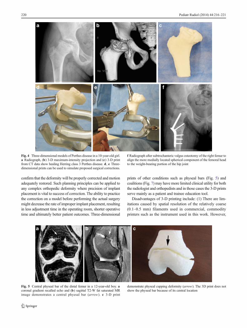
A 3-D print at a 1:1 scale provides a tactile and visualexperience. Cooke et al. [9] studied the way humans usevisuo-haptic inputs to understand solid objects and concludedthat using both modes of input leads to less ambiguity in theunderstanding the shape of the object. A natural extension istherefore to use radiologic images coupled with 3-D printingto reconstruct three-dimensional models of anatomy. Suchmodels have advantages over conventional volume renderingon a 3-D workstation for surgical planning and patient educa-tion. Theymake it easy for non-radiologists (surgeons, patients)to physically hold in their hands a model of the anatomy ofinterest and use visuo-haptic inputs to better understand thecondition to be treated. The 3-D prints could be used during thediscussion and surgical consenting process so that the patient andfamily understand the gravity of the medical or surgical condi-tion. Surgeons might refine or even experiment with differenttechniques on a 1:1 scale print model of the actual orthopedicdeformity in a no-stress environment prior to definitive surgicalcorrection (Fig. 4).
Three-dimensional printing has a host of pediatric ortho-pedic applications for congenital and acquired disorders in-cluding but not limited to the evaluation of pediatric develop-mental hip deformities (Fig. 4), post-traumatic physeal bars(Fig. 5), 3-D nature of Blount disease (Fig. 6) and subtalarcoalitions (Fig. 7). Three-dimensional printing does not sup-plant the diagnostic interpretation of 2-D CTandMRI datasetsbut should be used to complement imaging interpretation. Weanticipate that the utility of 3-D printing will be further en-hanced when non-experienced observers are involved, forexample patients and professionals without formal radiologytraining and experience.
More experience with 3-D print datasets will be necessary tohelp refine which orthopedic pathologies might benefit from theaddition of a 3-D print. Anecdotally, 3-D prints of osteo-articularalignment disorders have most often been requested by ourorthopedists. The orthopedists have noted that these modelsadd clinical value because they can help them understand howto correct the alignment disorder from a 3-D perspective thatusually cannot be fully appreciated on a computer monitor. Apatient with a complex proximal femoral deformity fromhealingPerthes disease serves as an example (Fig. 4). Reconstruction insuch a patient requires an understanding of the precise locationand size of the deformity of the femoral head and neck and of thedegree of hip flexion when it comes into contact with theacetabular rim. Additionally the femoral neck-shaft angle, rela-tive neck length, trochanteric height, and femoral version mightall be deranged. A 3-D print allows for the ability to study thedeformity and not only plan the surgery including the exactplacement of implants, but also simulate the procedure to
Pediatr Radiol (2014) 44:216–221 219
confirm that the deformity will be properly corrected andmotionadequately restored. Such planning principles can be applied toany complex orthopedic deformity where precision of implantplacement is vital to success of correction. The ability to practicethe correction on a model before performing the actual surgerymight decrease the rate of improper implant placement, resultingin less adjustment time in the operating room, shorter operativetime and ultimately better patient outcomes. Three-dimensional
prints of other conditions such as physeal bars (Fig. 5) andcoalitions (Fig. 7) may have more limited clinical utility for boththe radiologist and orthopedists and in those cases the 3-D printsserve mainly as a patient and trainee education tool.
Disadvantages of 3-D printing include: (1) There are lim-itations caused by spatial resolution of the relatively coarse(0.1–0.5 mm) filaments used in commercial, commodityprinters such as the instrument used in this work. However,
Fig. 5 Central physeal bar of the distal femur in a 12-year-old boy. acoronal gradient recalled echo and (b) sagittal T2-W fat saturated MRimage demonstrates a central phyesal bar (arrow ). c 3-D print
demonstrate physeal cupping deformity (arrow). The 3D print does notshow the physeal bar because of its central location
Fig. 4 Three-dimensional models of Perthes disease in a 10-year-old girl.a Radiograph, (b) 3-D maximum-intensity projection and (c) 3-D printfrom CT data show healing Herring class 3 Perthes disease. d , e Three-dimensional prints can be used to simulate proposed surgical corrections.
f Radiograph after subtrochanteric valgus osteotomy of the right femur toalign the more medially located spherical component of the femoral headto the weight-bearing portion of the hip joint
220 Pediatr Radiol (2014) 44:216–221
this resolution is comparable to that of the images them-selves (usually 0.5–1 mm). (2) Difficulty in observinginternal architecture with a 3-D model, such as a centralphyseal bar hidden by the metaphysis and epiphysis(Fig. 5). (3) The cartilage and soft-tissue support structuresare not typically included with 3-D prints because of theircomplexity. Consequently, 3-D prints do not comprehen-sively illustrate all biomechanical elements of a joint andshould not be interpreted in isolation without the originalorthogonal CT or MRI datasets.
In summary, 3-D printing is a commercially availableand inexpensive tool that has significant application inpediatric musculoskeletal disorders. Three-dimensionalprinting supplements but does not replace interpretationof 2-D CT and MRI datasets with conventional post-processing. The value of 3-D printing is the additionaltactile and visual experience it affords to both the ortho-pedist and patient in understanding congenital and ac-quired pediatric musculoskeletal disorders and its potentialto aid in treatment planning and simulation.
Conflicts of interest None
References
1. Beaman JJ, Barlow JW, Bourell DL et al (eds) (1997) Solid freeformfabrication: a new direction in manufacturing: with research andapplications in thermal laser processing. Springer, New York
2. Rivera J, Goasduff L (2013) Gartner says early adopters of 3D printingtechnology could gain an innovation advantage over rivals. Accessedvia http://www.gartner.com/newsroom/id/2388415
3. Sailer HF, Haers PE, Zollikofer CP et al (1998) The value ofstereolithographic models for preoperative diagnosis of craniofacialdeformities and planning of surgical corrections. Int J Oral MaxillofacSurg 27:327–333
4. Mizutani J, Matsubara T, Fukuoka M et al (2008) Application of full-scale three-dimensional models in patients with rheumatoid cervicalspine. Eur Spine J 17:644–649
6. Markert M, Weber S, Lueth TC (2007) A beating heart model 3Dprinted from specific patient data. Conf Proc IEEE Eng Med Biol Soc2007:4472–4475
7. Ebert LC, Thali MJ, Ross S (2011) Getting in touch – 3D printing inforensic imaging. Forensic Sci Int 211:e1–e6
8. Esses SJ, Berman P, Bloom AI, Sosna J (2011) Clinical applications ofphysical 3D models derived from MDCT data and created by rapidprototyping. AJR 196:W683–8
9. Cooke T, Jäkel F, Wallraven C et al (2007) Multimodal similarity andcategorization of novel, three-dimensional objects. Neuropsychologia45:484–495
Fig. 7 Fibrous subtalar coalition in a 17-year-old girl. a Harris view, (b) coronal reformatted CTand (c) 3-D print demonstrate a fibrous coalition of themiddle subtalar joint (arrows)
Fig. 6 Model of Blount disease in an 8-year-old girl. a Radiograph, (b) CT coronal reconstruction and (c) 3-D print demonstrate metaphyseal beaking(arrow), medial physeal irregularity and diminished epiphyseal height