Orthopedic Basic Science Orthopedic Basic Science for General Surgeon for General Surgeon for General Surgeon for General Surgeon Pongsak Pongsak Yuktanandana Yuktanandana MD., M.Sc. MD., M.Sc. Associate professor of Orthopedics Associate professor of Orthopedics Associate professor of Orthopedics Associate professor of Orthopedics Basic Science Section, RCOST Basic Science Section, RCOST
Transcript
Orthopedic Basic Science Orthopedic Basic Science for General Surgeonfor General Surgeonfor General Surgeonfor General Surgeon
�� Central Central Haversian Haversian CanalCanal for blood vessels for blood vessels and nerves and nerves and nerves and nerves
�� Concentric layersConcentric layers (rings) (rings) of matrix in lamellaof matrix in lamella
�� Osteocytes Osteocytes in spaces in spaces called called lacunalacuna
�� Connected to each Connected to each other and the Haversian other and the Haversian Canal by Canal by canniculi canniculi (little (little canals)canals)canals)canals)
�� CorticalCortical--compactcompact
�� TrabecularTrabecular--porousporous
�� Cells in remodeling: Cells in remodeling: osteoblasts (build),osteoblasts (build),
�� osteoclasts (remove)osteoclasts (remove)
Bone:Cortical(compact) and Bone:Cortical(compact) and
Trabecular (spongy)Trabecular (spongy)
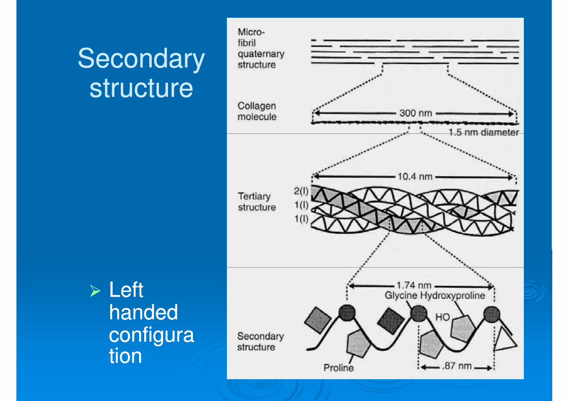
Hierarchy of Structure of TendonHierarchy of Structure of Tendon
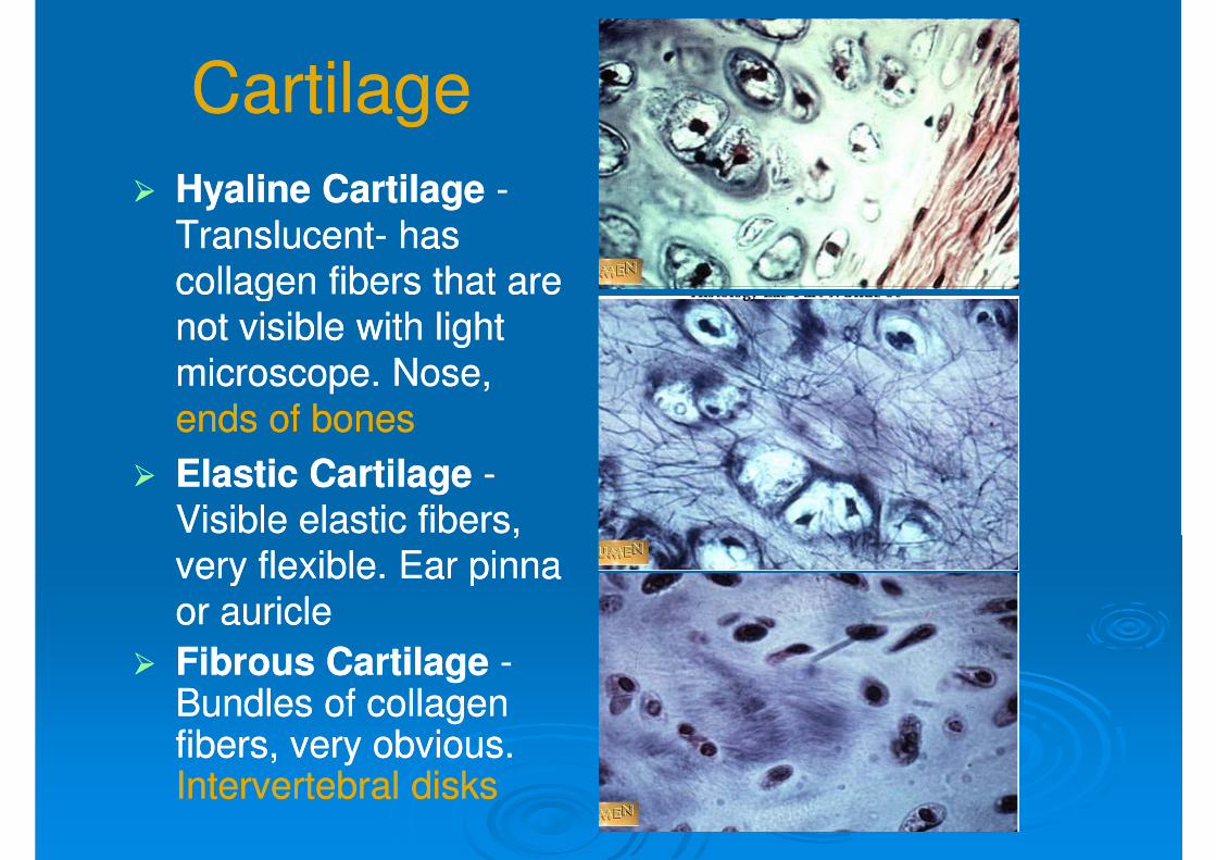
CartilageCartilage
�� Hyaline CartilageHyaline Cartilage --
TranslucentTranslucent-- has has
collagen fibers that are collagen fibers that are collagen fibers that are collagen fibers that are
not visible with light not visible with light
microscope. Nose, microscope. Nose,
ends of bonesends of bones
�� Elastic CartilageElastic Cartilage --
Visible elastic fibers, Visible elastic fibers,
very flexible. Ear pinna very flexible. Ear pinna very flexible. Ear pinna very flexible. Ear pinna
or auricleor auricle
�� Fibrous CartilageFibrous Cartilage --Bundles of collagen Bundles of collagen fibers, very obvious. fibers, very obvious. Intervertebral disksIntervertebral disks
�� Hydroxyproline and hydroxylysine are Hydroxyproline and hydroxylysine are unique to collagen and moleculeunique to collagen and moleculeunique to collagen and moleculeunique to collagen and molecule
Secondary Secondary structurestructure
�� Left Left handed handed configuraconfigurationtion
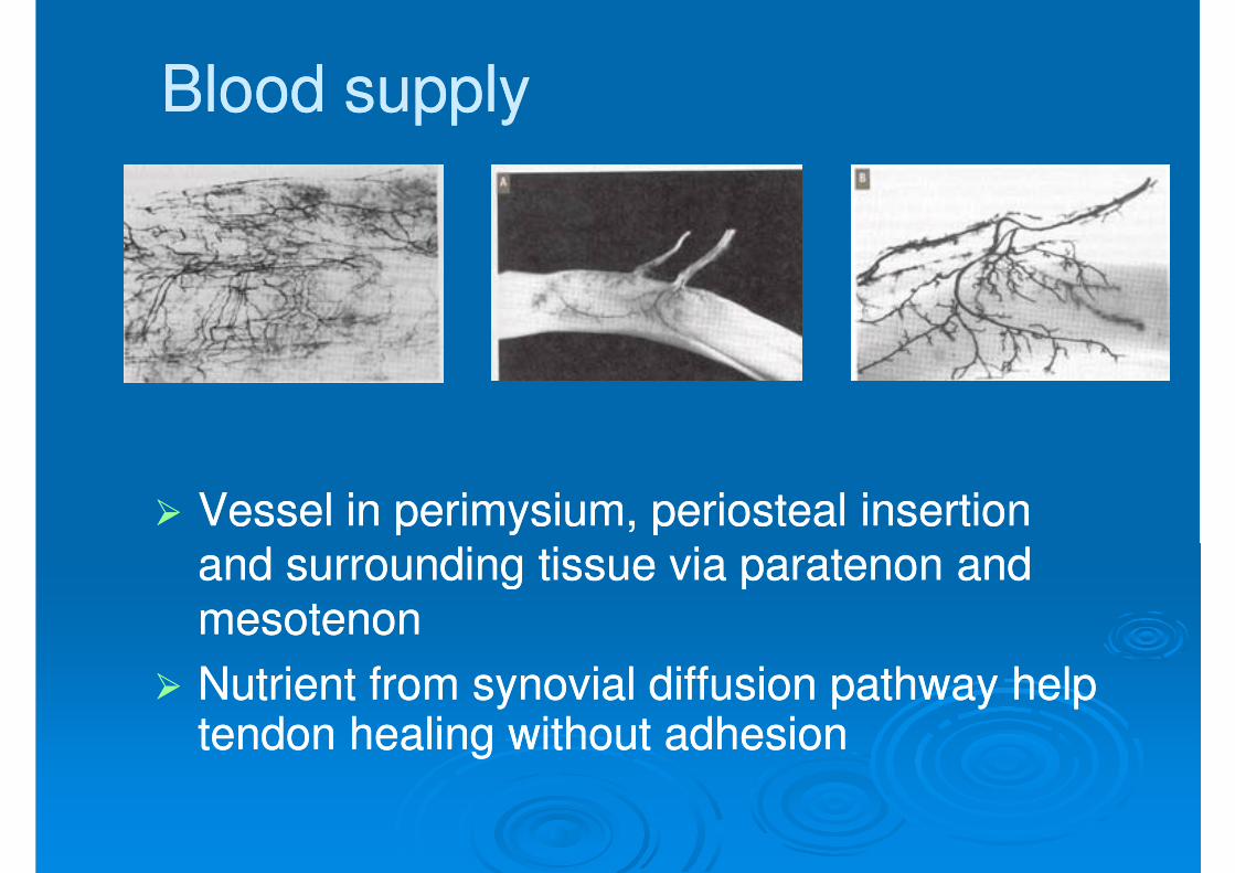
Blood supplyBlood supply
�� Vessel in perimysium, periosteal insertion Vessel in perimysium, periosteal insertion and surrounding tissue via paratenon and and surrounding tissue via paratenon and and surrounding tissue via paratenon and and surrounding tissue via paratenon and
mesotenonmesotenon
�� Nutrient from synovial diffusion pathway help Nutrient from synovial diffusion pathway help tendon healing without adhesiontendon healing without adhesion
BiomechanicsBiomechanics
�� Tensile propertiesTensile properties
TimeTime-- and historyand history--dependent behavior of dependent behavior of �� TimeTime-- and historyand history--dependent behavior of dependent behavior of tendonstendons
�� Factor affecting the mechanical properties Factor affecting the mechanical properties
Collagen is the strongest fibrous proteinCollagen is the strongest fibrous protein�� Collagen is the strongest fibrous proteinCollagen is the strongest fibrous protein
�� Collagen fiber arrange parallel to direction of Collagen fiber arrange parallel to direction of
tensile forcetensile force
�� Tensile properties is characterized by:Tensile properties is characterized by:
�� Mechanical(material) properties of tendon Mechanical(material) properties of tendon �� Mechanical(material) properties of tendon Mechanical(material) properties of tendon
�� Strain energy density (Strain energy density (ωω in MPa)in MPa)
Structural propertiesStructural properties
�� Structural properties(loadStructural properties(load--elongation elongation relationship) of bonerelationship) of bone--tendontendon--muscle muscle relationship) of bonerelationship) of bone--tendontendon--muscle muscle
depend on material properties of:depend on material properties of:
�� Straightening of crimped fiberStraightening of crimped fiber
�� Orienting fiber in direction of loadingOrienting fiber in direction of loading
�� Toe region is smaller in tendon than in ligamentToe region is smaller in tendon than in ligament
�� Linear region: Linear region: �� stiffness(slope N/mm)stiffness(slope N/mm)
�� Failure region:Failure region:�� Ultimate load (load at failure in N)Ultimate load (load at failure in N)
�� energy absorbed to failure (area under the curve)energy absorbed to failure (area under the curve)
TimeTime--history dependent history dependent viscoelastic properties of tendonsviscoelastic properties of tendons
�� Elongation depend on time and history of force Elongation depend on time and history of force applicationapplicationapplicationapplication
�� Up to interaction between collagen and ground Up to interaction between collagen and ground substancesubstance
�� Time dependentTime dependent�� Creep and stress relaxationCreep and stress relaxation
�� History dependentHistory dependent�� History dependentHistory dependent�� Shape of load elongation depend on previous loadingShape of load elongation depend on previous loading
Time dependent Time dependent
�� Creep is the time dependent elongation of tissue Creep is the time dependent elongation of tissue
when subjected to a constant load.when subjected to a constant load.
Time dependentTime dependent
�� StressStress--relaxation is the timerelaxation is the time--dependent decrease in dependent decrease in load when subjected to constant elongationload when subjected to constant elongation
History dependentHistory dependent
�� Shape of loadShape of load--elongation curve vary depend on elongation curve vary depend on
previous loading.previous loading.
�� Load and unloading curve follow different paths Load and unloading curve follow different paths
during single cycle, forming Hysteresis loop.during single cycle, forming Hysteresis loop.
�� Peak force decrease after many cycle.Peak force decrease after many cycle.
After many cycle load and unloading curve After many cycle load and unloading curve
History dependentHistory dependent
�� After many cycle load and unloading curve After many cycle load and unloading curve become similar to previous cycle.become similar to previous cycle.
�� In isometric contraction, tendon creep In isometric contraction, tendon creep shorten muscle and reduce fatigue stress.shorten muscle and reduce fatigue stress.
PreconditioningPreconditioning
�� First few cycles of elongation following inactivity First few cycles of elongation following inactivity reveal larger area of hysteresis(energy loss)reveal larger area of hysteresis(energy loss)reveal larger area of hysteresis(energy loss)reveal larger area of hysteresis(energy loss)
�� After conditioning(warm up), cycle become more After conditioning(warm up), cycle become more repeatablerepeatable
�� Preconditioning is important to avoid Preconditioning is important to avoid experimental errorexperimental error
�� After preconditioning, elastic strain energy is After preconditioning, elastic strain energy is 9090%%--9696% per cycle(save energy)% per cycle(save energy)After preconditioning, elastic strain energy is After preconditioning, elastic strain energy is 9090%%--9696% per cycle(save energy)% per cycle(save energy)
Factor affecting mechanical Factor affecting mechanical properties of tendonsproperties of tendons
�� Anatomical location Anatomical location
�� Exercise and immobilizationExercise and immobilization
�� AgeAge
�� Laser/heat treatmentLaser/heat treatment
LigamentLigament
�� Shorter and widerShorter and wider
Lower % of collagenLower % of collagen
�� Longer and narrowerLonger and narrower
�� Higher % of collagenHigher % of collagen
TendonTendon
�� Lower % of collagenLower % of collagen
�� Larger % of ground Larger % of ground
substancesubstance
�� Broader distribution Broader distribution of fiber direction of fiber direction
�� Higher % of collagenHigher % of collagen
�� Lower % of ground Lower % of ground
substancesubstance
�� More longitudinally More longitudinally organized fiber directionorganized fiber direction
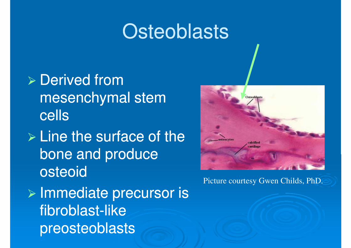
�� Derived from Derived from �� Derived from Derived from mesenchymal stem mesenchymal stem
cellscells
�� Line the surface of the Line the surface of the bone and produce bone and produce
osteoidosteoidosteoidosteoid
�� Immediate precursor is Immediate precursor is fibroblastfibroblast--like like
preosteoblasts preosteoblasts
Picture courtesy Gwen Childs, PhD.
OsteocytesOsteocytes
�� Osteoblasts surrounded Osteoblasts surrounded by bone matrix by bone matrix by bone matrix by bone matrix
�� trapped in lacunaetrapped in lacunae
�� Function poorly Function poorly
understood understood
�� regulating bone regulating bone �� regulating bone regulating bone
metabolism in response metabolism in response to stress and strainto stress and strain
Picture courtesy Gwen Childs, PhD.
Osteocyte NetworkOsteocyte Network
�� Osteocyte lacunae are connected by Osteocyte lacunae are connected by canaliculicanaliculicanaliculicanaliculi
�� Osteocytes are interconnected by long cell Osteocytes are interconnected by long cell processes that project through the processes that project through the canaliculicanaliculi
�� Preosteoblasts also have connections via Preosteoblasts also have connections via canaliculi with the osteocytescanaliculi with the osteocytescanaliculi with the osteocytescanaliculi with the osteocytes
�� Network probably facilitates response of Network probably facilitates response of bone to mechanical and chemical factorsbone to mechanical and chemical factors
OsteoclastsOsteoclasts�� Derived from Derived from
�� Multinucleated cells Multinucleated cells whose function is bone whose function is bone resorptionresorption
�� Reside in bone Reside in bone resorption pits resorption pits (Howship’s lacunae)(Howship’s lacunae)(Howship’s lacunae)(Howship’s lacunae)
�� Parathyroid hormone Parathyroid hormone stimulates stimulates receptors on receptors on osteoblastsosteoblasts that activate that activate osteoclastic bone osteoclastic bone resorptionresorption
Picture courtesy Gwen Childs, PhD.
Components of Bone FormationComponents of Bone Formation
�� CortexCortex
�� PeriosteumPeriosteum
�� Bone marrowBone marrow
�� Soft tissueSoft tissue
Prerequisites for Bone HealingPrerequisites for Bone Healing
�� Mechanism by which a long bone grows in Mechanism by which a long bone grows in �� Mechanism by which a long bone grows in Mechanism by which a long bone grows in widthwidth
�� Osteoblasts differentiate directly from Osteoblasts differentiate directly from
preosteoblasts and lay down seams of preosteoblasts and lay down seams of osteoid osteoid osteoid osteoid
�� Does NOT involve cartilage anlageDoes NOT involve cartilage anlage
Intramembranous Bone Intramembranous Bone
FormationFormation
Picture courtesy Gwen Childs, PhD.
Endochondral Bone FormationEndochondral Bone Formation
�� Mechanism by which a long bone grows in Mechanism by which a long bone grows in lengthlengthlengthlength
�� Osteoblasts line a cartilage precursorOsteoblasts line a cartilage precursor
�� The chondrocytes hypertrophy, The chondrocytes hypertrophy, degenerate and calcify (area of low degenerate and calcify (area of low oxygen tension)oxygen tension)
Vascular invasion of the cartilage occurs Vascular invasion of the cartilage occurs �� Vascular invasion of the cartilage occurs Vascular invasion of the cartilage occurs followed by ossification (increasing oxygen followed by ossification (increasing oxygen tension)tension)
Endochondral Bone FormationEndochondral Bone Formation
Picture courtesy Gwen Childs, PhD.
Blood SupplyBlood Supply
�� Long bones have Long bones have three blood suppliesthree blood suppliesthree blood suppliesthree blood supplies
�� Nutrient artery Nutrient artery
(intramedullary)(intramedullary)
�� Periosteal vesselsPeriosteal vessels
�� Metaphyseal vesselsMetaphyseal vessels
Periosteal
vessels
Nutrient
artery
Metaphyseal
vessels
Figure adapted from Rockwood and Green, 5th Ed
Nutrient ArteryNutrient Artery
�� Normally the major blood supply for the Normally the major blood supply for the �� Normally the major blood supply for the Normally the major blood supply for the diaphyseal cortex (diaphyseal cortex (80 80 to to 8585%)%)
�� Enters the long bone via a nutrient Enters the long bone via a nutrient
foramen foramen
�� Forms medullary arteries up and down the Forms medullary arteries up and down the �� Forms medullary arteries up and down the Forms medullary arteries up and down the bonebone
Periosteal VesselsPeriosteal Vessels
�� Arise from the capillaryArise from the capillary--rich periosteumrich periosteum
�� Supply outer Supply outer 15 15 to to 2020% of cortex normally% of cortex normally
�� Capable of supplying a much greater Capable of supplying a much greater proportion of the cortex in the event of injury proportion of the cortex in the event of injury
to the medullary blood supplyto the medullary blood supply
Metaphyseal VesselsMetaphyseal Vessels
�� Arise from periarticular vesselsArise from periarticular vessels�� Arise from periarticular vesselsArise from periarticular vessels
�� Penetrate the thin cortex in the Penetrate the thin cortex in the metaphyseal region and anastomose with metaphyseal region and anastomose with
the medullary blood supplythe medullary blood supply
Vascular Response in Fracture Vascular Response in Fracture
RepairRepair
�� Fracture stimulates the release of growth Fracture stimulates the release of growth �� Fracture stimulates the release of growth Fracture stimulates the release of growth factors that promote angiogenesis and factors that promote angiogenesis and
vasodilationvasodilation
�� Blood flow is increased substantially to the Blood flow is increased substantially to the fracture sitefracture sitefracture sitefracture site
�� Peaks at two weeks after fracturePeaks at two weeks after fracture
Mechanical StabilityMechanical Stability
�� Early stability promotes Early stability promotes revascularizationrevascularizationrevascularizationrevascularization
�� After first month, After first month, loading and loading and
�� Mechanical load and small displacements Mechanical load and small displacements at the fracture site stimulate healingat the fracture site stimulate healingat the fracture site stimulate healingat the fracture site stimulate healing
�� Inadequate stabilization may result in Inadequate stabilization may result in excessive deformation at the fracture site excessive deformation at the fracture site interrupting tissue differentiation to bone interrupting tissue differentiation to bone (soft callus)(soft callus)
�� OverOver--stabilization, however, reduces stabilization, however, reduces �� OverOver--stabilization, however, reduces stabilization, however, reduces periosteal bone formation (hard callus)periosteal bone formation (hard callus)
Stages of Fracture HealingStages of Fracture Healing
Mechanisms for Bone HealingMechanisms for Bone Healing
��Direct (primary) bone healingDirect (primary) bone healing
�� Indirect (secondary) bone healingIndirect (secondary) bone healing
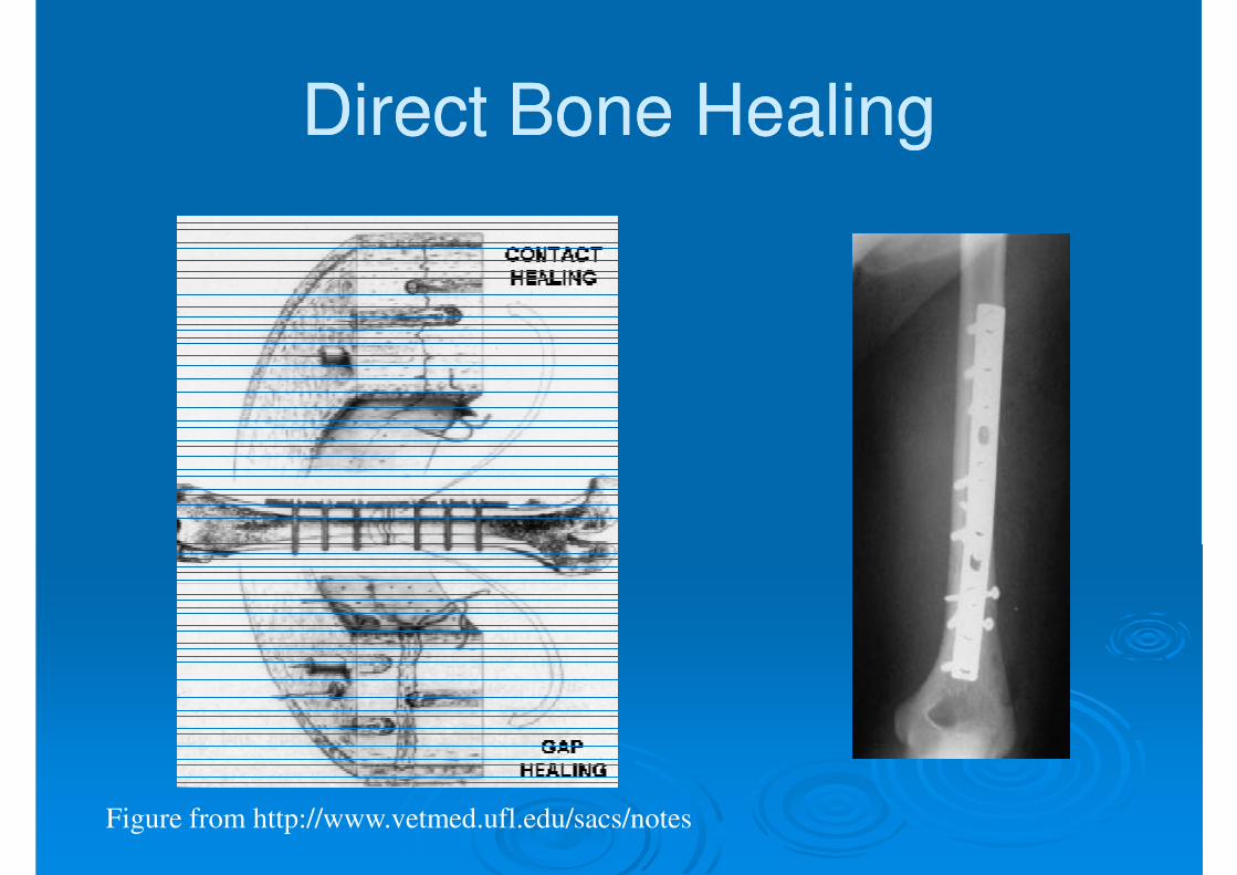
Direct Bone HealingDirect Bone Healing
�� Mechanism of bone healing seen when there Mechanism of bone healing seen when there is no motion at the fracture site (i.e. rigid is no motion at the fracture site (i.e. rigid
internal fixation)internal fixation)
�� Does not involve formation of fracture callusDoes not involve formation of fracture callus
�� Osteoblasts originate from endothelial and Osteoblasts originate from endothelial and �� Osteoblasts originate from endothelial and Osteoblasts originate from endothelial and
perivascular cellsperivascular cells
Direct Bone HealingDirect Bone Healing
�� A cutting cone is formed that crosses the A cutting cone is formed that crosses the fracture site fracture site fracture site fracture site
�� Osteoblasts lay down lamellar bone Osteoblasts lay down lamellar bone
behind the osteoclasts forming a behind the osteoclasts forming a secondary osteonsecondary osteon
�� Gradually the fracture is healed by the Gradually the fracture is healed by the �� Gradually the fracture is healed by the Gradually the fracture is healed by the
formation of numerous secondary osteonsformation of numerous secondary osteons
�� A slow process A slow process –– months to yearsmonths to years
Direct Bone HealingDirect Bone Healing
Figure from http://www.vetmed.ufl.edu/sacs/notes
Indirect Bone HealingIndirect Bone Healing
�� Mechanism for healing in Mechanism for healing in fractures that are not rigidly fractures that are not rigidly fractures that are not rigidly fractures that are not rigidly fixed. fixed.
�� Competes with BMPCompetes with BMP--2 2 for receptorsfor receptors
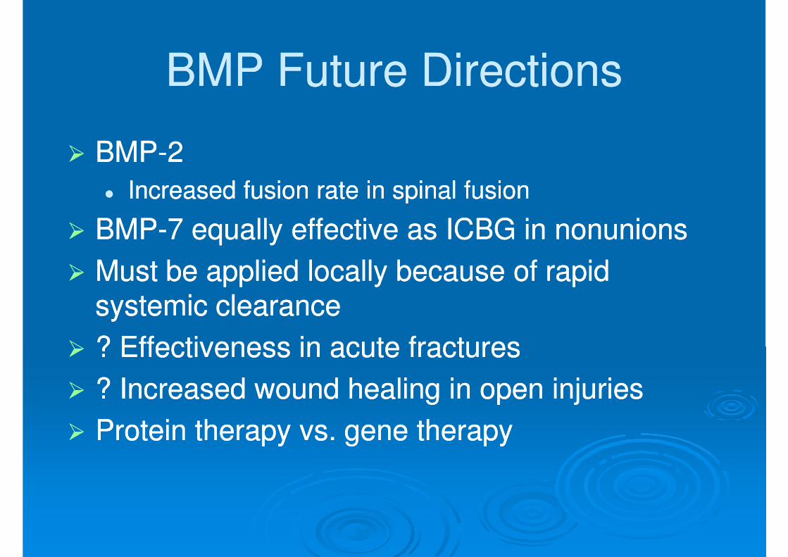
BMP Future DirectionsBMP Future Directions
�� BMPBMP--2 2
Increased fusion rate in spinal fusion Increased fusion rate in spinal fusion �� Increased fusion rate in spinal fusion Increased fusion rate in spinal fusion
�� BMPBMP--7 7 equally effective as ICBG in nonunionsequally effective as ICBG in nonunions
�� Must be applied locally because of rapid Must be applied locally because of rapid systemic clearance systemic clearance
�� ? Effectiveness in acute fractures? Effectiveness in acute fractures
�� ? Increased wound healing in open injuries? Increased wound healing in open injuries
�� Protein therapy vs. gene therapyProtein therapy vs. gene therapy
Cytokines Cytokines �� InterleukinInterleukin--11,,--44,,--66,,--1111, macrophage and , macrophage and
granulocyte/macrophage (GM) colonygranulocyte/macrophage (GM) colony--stimulating factors (CSFs) and Tumor stimulating factors (CSFs) and Tumor stimulating factors (CSFs) and Tumor stimulating factors (CSFs) and Tumor Necrosis FactorNecrosis Factor
�� Stimulate bone resorptionStimulate bone resorption�� ILIL--1 1 is the most potentis the most potent
�� ILIL--1 1 and ILand IL--6 6 synthesis is decreased by synthesis is decreased by estrogenestrogenestrogenestrogen�� May be mechanism for postMay be mechanism for post--menopausal bone menopausal bone
resorptionresorption
�� Peak during Peak during 11stst 24 24 hours then again during hours then again during remodelingremodeling
�� Regulate endochondral bone formationRegulate endochondral bone formation
Specific Factor Stimulation of Specific Factor Stimulation of
Osteoblasts and OsteoclastsOsteoblasts and Osteoclasts
CytokineCytokine Bone FormationBone Formation Bone ResorptionBone ResorptionCytokineCytokine Bone FormationBone Formation Bone ResorptionBone Resorption
�� Effect on bone resorption is species dependent Effect on bone resorption is species dependent and their overall effects in humans unknownand their overall effects in humans unknownand their overall effects in humans unknownand their overall effects in humans unknown
�� Prostaglandins of the E seriesProstaglandins of the E series�� Stimulate osteoblastic bone formationStimulate osteoblastic bone formation
�� Inhibit activity of isolated osteoclastsInhibit activity of isolated osteoclasts
�� LeukotrienesLeukotrienes�� Stimulate osteoblastic bone formationStimulate osteoblastic bone formation
�� Enhance the capacity of isolated osteoclasts to form Enhance the capacity of isolated osteoclasts to form resorption pitsresorption pits
HormonesHormones
�� EstrogenEstrogen�� Stimulates fracture healing through receptor mediated Stimulates fracture healing through receptor mediated �� Stimulates fracture healing through receptor mediated Stimulates fracture healing through receptor mediated
mechanismmechanism
�� Modulates release of a specific inhibitor of ILModulates release of a specific inhibitor of IL--11
�� Thyroid hormonesThyroid hormones�� Thyroxine and triiodothyronine stimulate osteoclastic Thyroxine and triiodothyronine stimulate osteoclastic
bone resorptionbone resorption
�� GlucocorticoidsGlucocorticoids�� GlucocorticoidsGlucocorticoids�� Inhibit calcium absorption from the gut causing Inhibit calcium absorption from the gut causing
increased PTH and therefore increased osteoclastic increased PTH and therefore increased osteoclastic bone resorptionbone resorption
•• Increased bone formationIncreased bone formation
�� Growth HormoneGrowth Hormone
�� Mediated through IGFMediated through IGF--1 1 (Somatomedin(Somatomedin--C)C)�� Mediated through IGFMediated through IGF--1 1 (Somatomedin(Somatomedin--C)C)
�� Increases callus formation and fracture Increases callus formation and fracture
strengthstrength
Vascular FactorsVascular Factors
�� MetalloproteinasesMetalloproteinasesDegrade cartilage and bones to allow invasion Degrade cartilage and bones to allow invasion �� Degrade cartilage and bones to allow invasion Degrade cartilage and bones to allow invasion of vesselsof vessels
�� Causes reduced activity and proliferation of Causes reduced activity and proliferation of �� Causes reduced activity and proliferation of Causes reduced activity and proliferation of osteochondral cellsosteochondral cells
�� Nicotine causes vasoconstriction diminishing blood Nicotine causes vasoconstriction diminishing blood flow at fracture siteflow at fracture siteflow at fracture siteflow at fracture site
�� Diabetes MellitusDiabetes Mellitus�� Associated with collagen defects including decreased Associated with collagen defects including decreased
collagen content, defective crosscollagen content, defective cross--linking and linking and alterations in collagen subalterations in collagen sub--type ratiostype ratios
Electromagnetic FieldElectromagnetic Field
�� In vitro bone deformation produces piezoelectric In vitro bone deformation produces piezoelectric currents and streaming potentialscurrents and streaming potentialscurrents and streaming potentialscurrents and streaming potentials
�� Electromagnetic (EM) devices are based on Electromagnetic (EM) devices are based on Wolff’s Law that bone responds to mechanical Wolff’s Law that bone responds to mechanical
stress: Exogenous EM fields may simulate stress: Exogenous EM fields may simulate mechanical loading and stimulate bone growth mechanical loading and stimulate bone growth and repairand repairand repairand repair
�� Clinical efficacy very controversialClinical efficacy very controversial
�� Approved by the FDA for the treatment of nonApproved by the FDA for the treatment of non--unionsunionsunionsunions
�� Efficacy of bone stimulation appears to be Efficacy of bone stimulation appears to be frequency dependantfrequency dependant�� Extremely low frequency (ELF) sinusoidal electric Extremely low frequency (ELF) sinusoidal electric
fields in the physiologic range are most effective (fields in the physiologic range are most effective (15 15 to to 30 30 Hz range)Hz range)
�� Specifically, PEMF signals in the Specifically, PEMF signals in the 20 20 to to 30 30 Hz range Hz range �� Specifically, PEMF signals in the Specifically, PEMF signals in the 20 20 to to 30 30 Hz range Hz range (postural muscle activity) appear more effective than (postural muscle activity) appear more effective than those below those below 10 10 Hz (walking)Hz (walking)
UltrasoundUltrasound
�� LowLow--intensity ultrasound is approved by intensity ultrasound is approved by the FDA for stimulating healing of fresh the FDA for stimulating healing of fresh the FDA for stimulating healing of fresh the FDA for stimulating healing of fresh
fracturesfractures
�� Modulates signal transduction, increases Modulates signal transduction, increases gene expression, increases blood flow, gene expression, increases blood flow,
enhances bone remodeling and increases enhances bone remodeling and increases enhances bone remodeling and increases enhances bone remodeling and increases callus torsional strength in animal modelscallus torsional strength in animal models
UltrasoundUltrasound
�� Human clinical trials show a decreased Human clinical trials show a decreased �� Human clinical trials show a decreased Human clinical trials show a decreased time of healing in fresh fracturestime of healing in fresh fractures
�� Has also been shown to decrease the Has also been shown to decrease the
healing time in smokers potentially healing time in smokers potentially reversing the ill effects of smokingreversing the ill effects of smokingreversing the ill effects of smokingreversing the ill effects of smoking
Fracture Healing SummaryFracture Healing Summary
�� Fracture healing is influenced by many Fracture healing is influenced by many �� Fracture healing is influenced by many Fracture healing is influenced by many variables including mechanical stability, variables including mechanical stability,
electrical environment, biochemical electrical environment, biochemical factors and blood flowfactors and blood flow
�� Our ability to enhance fracture healing Our ability to enhance fracture healing
will increase as we better understand will increase as we better understand will increase as we better understand will increase as we better understand the interaction between these variables the interaction between these variables