About Science Prof Online PowerPoint Resources • Science Prof Online (SPO) is a free science education website that provides fully-developed Virtual Science Classrooms, science-related PowerPoints, articles and images. The site is designed to be a helpful resource for students, educators, and anyone interested in learning about science. • The SPO Virtual Classrooms offer many educational resources, including practice test questions, review questions, lecture PowerPoints, video tutorials, sample assignments and course syllabi. New materials are continually being developed, so check back frequently, or follow us on Facebook (Science Prof Online) or Twitter (ScienceProfSPO) for updates. • Many SPO PowerPoints are available in a variety of formats, such as fully editable PowerPoint files, as well as uneditable versions in smaller file sizes, such as PowerPoint Shows and Portable Document Format (.pdf), for ease of printing. • Images used on this resource, and on the SPO website are, wherever possible, credited and linked to their source. Any words underlined and appearing in blue are links that can be clicked on for more information. PowerPoints must be viewed in slide show mode to use the hyperlinks directly. • Several helpful links to fun and interactive learning tools are included throughout the PPT and on the Smart Links slide, near the end of each presentation. You must be in slide show mode to utilize hyperlinks and animations. • This digital resource is licensed under Creative Commons Attribution-ShareAlike 3.0: http://creativecommons.org/licenses/by-sa/3.0/ Alicia Cepaitis, MS Chief Creative Nerd Science Prof Online Online Education Resources, LLC [email protected]From the Virtual Microbiology Classroom on ScienceProfOnline.com Image: Compound microscope objectives, T. Port Tami Port, MS Creator of Science Prof Online Chief Executive Nerd Science Prof Online Online Education Resources, LLC [email protected]
Transcript
About Science Prof Online PowerPoint Resources
• Science Prof Online (SPO) is a free science education website that provides fully-developed Virtual Science Classrooms, science-related PowerPoints, articles and images. The site is designed to be a helpful resource for students, educators, and anyone interested in learning about science.
• The SPO Virtual Classrooms offer many educational resources, including practice test questions, review questions, lecture PowerPoints, video tutorials, sample assignments and course syllabi. New materials are continually being developed, so check back frequently, or follow us on Facebook (Science Prof Online) or Twitter (ScienceProfSPO) for updates.
• Many SPO PowerPoints are available in a variety of formats, such as fully editable PowerPoint files, as well as uneditable versions in smaller file sizes, such as PowerPoint Shows and Portable Document Format (.pdf), for ease of printing.
• Images used on this resource, and on the SPO website are, wherever possible, credited and linked to their source. Any words underlined and appearing in blue are links that can be clicked on for more information. PowerPoints must be viewed in slide show mode to use the hyperlinks directly.
• Several helpful links to fun and interactive learning tools are included throughout the PPT and on the Smart Links slide, near the end of each presentation. You must be in slide show mode to utilize hyperlinks and animations.
• This digital resource is licensed under Creative Commons Attribution-ShareAlike 3.0: http://creativecommons.org/licenses/by-sa/3.0/
Cells that Gram stain- Gram positive and Gram negative
• Cells that resist Gram stain- Genus Mycobacterium and Norcardia- Stained using Acid-fast staining techniques
• Cells that lack cell walls– Will retain counterstain (second color
applied during differential staining).
Images: Gram positive bacteria , Gram-negative bacteria & Acid fast bacteria, all under oil immersion @1000XTM, T. PortFrom the Virtual Microbiology Classroom on ScienceProfOnline.com
• Dead Gram-negative bacteria release lipid-A when this outer membrane disintegrates.
• In animals with a Gram-negative bacterial infection, free lipid-A may trigger fever, vasodilation, inflammation, shock and blood clotting.
• Killing large numbers of Gram-negative bacteria with antimicrobial drugs releases lots of lipid-A, which can threaten the patient more than the presence of live Gram-negative bacteria.
From the Virtual Microbiology Classroom on ScienceProfOnline.com
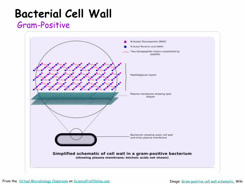
Q: Why are these differences in cell wall structure so important?
Beta-lactam Antibiotic Resistance Beta-lactam antibiotics (β-Lactam) are a broad class of antibiotics that all contain a β-lactam ring in their molecular structures.
Beta-lactam drugs include penicillin derivatives (penams), cephalosporins (cephems), monobactams, and carbapenems.
These antibiotics work by inhibiting cell wall synthesis in bacteria and are the most widely used group of antibiotics.
Some bacteria have developed resistance to β-lactam antibiotics and are able to synthesize an enzyme called β-lactamase, that attacks the β-lactam ring, inactivating the antibiotic.
Penicillin
Cephalosporin
Images: B-lactam Antibiotics, Action of B-lactamase, Wiki; From the Virtual Microbiology Classroom on ScienceProfOnline.com
Coccus-shaped bacteria, which divides in a way that results in grape-like clusters.
- Staphylococcus aureus (golden staph), most common cause of staph infections.
- Approximately 20–30% of general population “Staph carriers."
- S. aureus can cause illnesses ranging from minor skin infections to life-threatening diseases, such as meningitis, Toxic shock syndrome (TSS) & septicemia.
Some strains of E. coli inhabit gastrointestinal tracts of warm-blooded animals as normal flora and provide a portion of the microbially-derived vitamin K for their host.
While many strains of E. coli are harmless commensals, of some are human pathogens.
Common cause of bacterial food poisoning and urinary tract infections.
Bacteria must be able to “stick” to cause infection (otherwise, in case of UTI, bacteria would just get peed out).
Bladder lined with proteins, to prevent this. E. coli has fimbriae to help it stick.
From the Virtual Microbiology Classroom on ScienceProfOnline.com
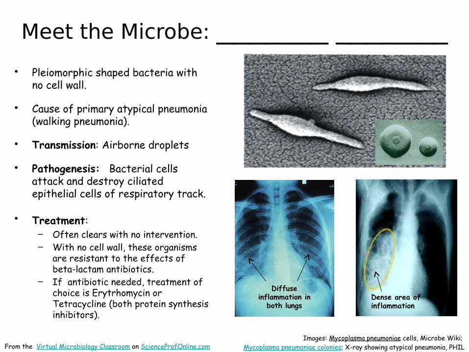
Meet the Microbe: ______ ___
Our lab friend E. coli.
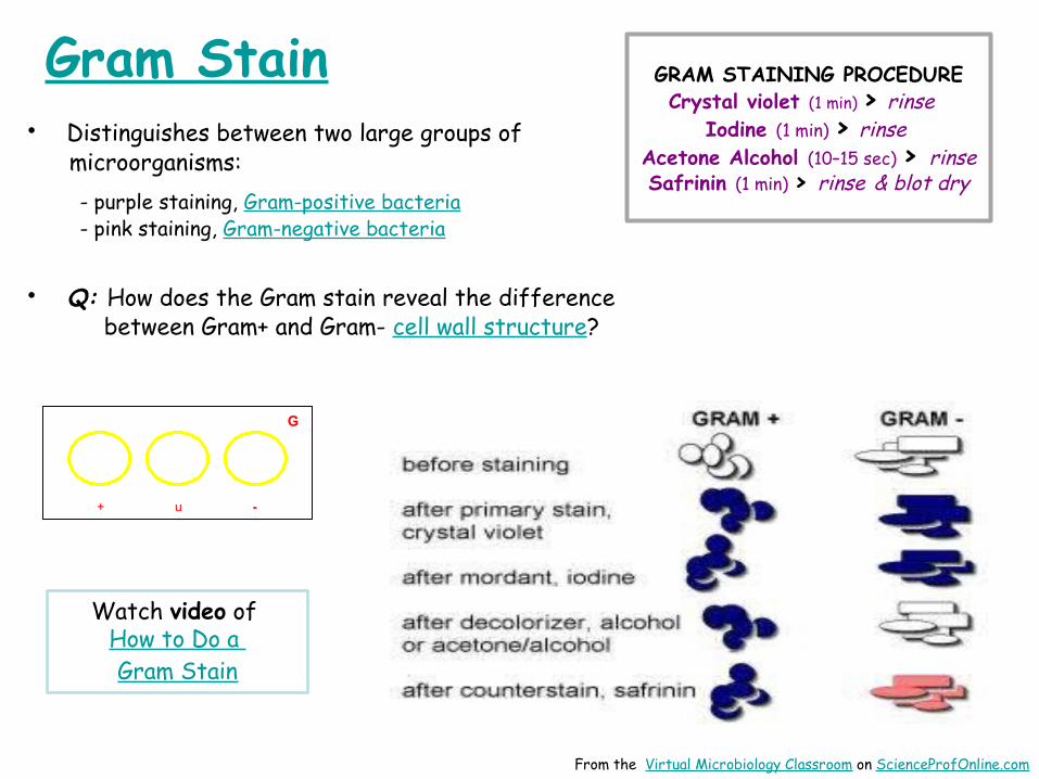
Gram Stain
Images: MacConkey’s, T. Port; E.coli with fimbria, National Library of Science; : E. coli @10,000xTM; Gram stain E. coli, T. Port;
• Cells that Gram stain- Gram positive and Gram negative
Cells that resist Gram stain- Genus Mycobacterium and Norcardia- Stained using Acid-fast staining techniques
• Cells that lack cell walls– Will retain counterstain (second color
applied during differential staining).
Images: Gram positive bacteria , Gram-negative bacteria & Acid fast bacteria, all under oil immersion @1000XTM, T. PortFrom the Virtual Microbiology Classroom on ScienceProfOnline.com
Q: Why Gram variable?• Both __________ and ______________, caused by
M. leprae and M. tuberculosis respectively, have plagued mankind for centuries.
• Thought that M. tuberculosis and M. leprae evolved from a soil bacterium that infected cows, then made jump to humans about the time of animal domestication, 10,000 years ago.
• M. tuberculosis doubles population every 18-24 hours,
• M. leprae doubles population about every 14 days.
• Q: What might be the impact of generation time on the course of the infectious diseases these microbes cause?
Images: TB Culture, Public Health Image Library (PHIL) #4428, Dr. George Kubica; 24 yo man from Norway, suffering from leprosy; Pierre Arents; Acid fast stain of Mycobacteria smegmatis & Staph, T. Port
Mycobacteria colonies Eewwww, looks like ear wax.
Man with Leprosy
Acid-fast stain
The pink is our lab friend Mycobacteriumsmegmatis
From the Virtual Microbiology Classroom on ScienceProfOnline.com
• Cells that Gram stain- Gram positive and Gram negative
• Cells that resist Gram stain- Genus Mycobacterium and Norcardia- Stained using Acid-fast staining techniques
Cells that lack cell walls– Will retain counterstain (second color
applied during differential staining).
Images: Gram positive bacteria , Gram-negative bacteria & Acid fast bacteria, all under oil immersion @1000XTM, T. PortFrom the Virtual Microbiology Classroom on ScienceProfOnline.com
• Differential Stain Laboratory Main Page on the Virtual Microbiology Classroom of Science Prof Online.
• Gram Stain Interactive Tutorial. This is an extremely useful tutorial that shows, step-by-step, what happens in Gram-positive and Gram-negative cells during Gram staining.
• Acid-fast Stain Animated Tutorial. The staining procedure depicted in this tutorial differs a bit from how we do it in lab, but this tutorial is still very useful. Shows the steps of the staining procedure and the resulting color of Acid-fast and Nonacid-fast cells.
• Videos of differential staining procedures: Gram, Acid-fast, Endospore
• Drug Resistant TB: Past, Present & Future, Chang et al (2010) Official Journal of the Asian Pacific Society of Respirology, DOI: 10.1111/j.1440-1843.2010.01738.x
Smart Links
(You must be in PPT slideshow view to click on links.) From the Virtual Microbiology Classroom on ScienceProfOnline.com