Page 1/19 Outcomes of Proton Therapy for Non-small Cell Lung Cancer in Patients with Interstitial Pneumonia Shingo Hashimoto ( [email protected]) Nagoya Proton Therapy Center https://orcid.org/0000-0002-7525-0288 Hiromitsu Iwata Nagoya Proton Therapy Center Yukiko Hattori Nagoya Proton Therapy Center Koichiro Nakajima Nagoya Proton Therapy Center Kento Nomura Nagoya Proton Therapy Center Kensuke Hayashi Nagoya Proton Therapy Center Toshiyuki Toshito Nagoya Proton Therapy Center Eiko Yamamori Tohoku University Hospital: Tohoku Daigaku Byoin Kenji Akita Nagoya City University West Medical Center: Nagoya Shiritsu Daigaku Igakubu Fuzoku Seibu Iryo Center Jun-etsu Mizoe Hokkaido Ohno Kinen Byoin: Hokkaido Ono Kinen Byoin Hiroyuki Ogino Nagoya Proton Therapy Center Yuta Shibamoto Nagoya City University Graduate School of Medical Sciences and Medical School: Nagoya Shiritsu Daigaku Daigakuin Igaku Kenkyuka Igakubu Research Keywords: Proton therapy, Interstitial pneumonia, Idiopathic pulmonary brosis, Lung cancer, Radiation pneumonitis, Quality of life Posted Date: September 13th, 2021 DOI: https://doi.org/10.21203/rs.3.rs-861118/v1 License: This work is licensed under a Creative Commons Attribution 4.0 International License. Read Full License
Transcript
Page 1/19
Outcomes of Proton Therapy for Non-small Cell LungCancer in Patients with Interstitial PneumoniaShingo Hashimoto ( [email protected] )
Nagoya Proton Therapy Center https://orcid.org/0000-0002-7525-0288Hiromitsu Iwata
Nagoya Proton Therapy CenterYukiko Hattori
Nagoya Proton Therapy CenterKoichiro Nakajima
Nagoya Proton Therapy CenterKento Nomura
Nagoya Proton Therapy CenterKensuke Hayashi
Nagoya Proton Therapy CenterToshiyuki Toshito
Nagoya Proton Therapy CenterEiko Yamamori
Tohoku University Hospital: Tohoku Daigaku ByoinKenji Akita
Nagoya City University West Medical Center: Nagoya Shiritsu Daigaku Igakubu Fuzoku Seibu Iryo CenterJun-etsu Mizoe
Hokkaido Ohno Kinen Byoin: Hokkaido Ono Kinen ByoinHiroyuki Ogino
Nagoya Proton Therapy CenterYuta Shibamoto
Nagoya City University Graduate School of Medical Sciences and Medical School: Nagoya Shiritsu Daigaku DaigakuinIgaku Kenkyuka Igakubu
Research
Keywords: Proton therapy, Interstitial pneumonia, Idiopathic pulmonary �brosis, Lung cancer, Radiation pneumonitis,Quality of life
Posted Date: September 13th, 2021
DOI: https://doi.org/10.21203/rs.3.rs-861118/v1
License: This work is licensed under a Creative Commons Attribution 4.0 International License. Read Full License
Interstitial pneumonia (IP) is a disease with a poor prognosis. In addition, IP patients are more likely to develop lungcancer. Since IP patients frequently develop toxicities during cancer treatment, minimally invasive cancer treatment iswarranted for such patients to maintain their quality of life. This study retrospectively investigated the e�cacy andsafety of proton therapy (PT) for non-small cell lung cancer (NSCLC) in patients with IP.
Methods:
Twenty-nine NSCLC patients with IP were treated with PT between September 2013 and December 2019. The patientshad stage IA to IIIB primary NSCLC. Ten of the 29 patients exhibited the usual interstitial pneumonia pattern. Theprescribed dose was 66-74 Grays (relative biological effectiveness) in 10-37 fractions.
Results:
The median follow-up period was 17.4 months (interquartile range (IQR), 9.5–32.7). The median patient age was 77years (IQR, 71–81). The median planning target volume was 112.0 ml (IQR, 56.1–246.3). The 2-year local control,progression-free survival, and overall survival rates were 77% (95% con�dence interval: 34 to 94), 31% (13–50), and 50%(26–70), respectively. According to the Common Terminology Criteria for Adverse Events (version 4.0), grade 3 acuteradiation pneumonitis (RP) was observed in 1 patient. Two patients developed grade 3 late RP, but no other patientsexperienced serious toxicities. The patients’ quality of life (European Organization for Research and Treatment of CancerQLQ-C30 and QLQ-LC13 and SF-36) scores had not changed after 3 months.
Conclusions:
PT may safely control NSCLC without adversely affecting the daily lives of IP patients.
BackgroundInterstitial pneumonia (IP) is a group of diffuse parenchymal lung disorders that can affect mortality [1]. Theclassi�cation of IP is based on pathological and imaging �ndings. Among the various types of IP, idiopathic pulmonary�brosis (IPF) is associated with the worst prognosis [2]. With an estimated incidence of 4.6 to 16.3 per 100,000, IPF is themost common form of idiopathic IP [3]. The disease occurs as often as stomach, brain, and testicular cancer [4].Although the course of the disease is variable and unpredictable, the median survival time from diagnosis is 2–4 years[5]. IP, especially IPF, is often accompanied by lung cancer (frequency: 10–20% of cases) [6].
Systemic therapy for non-small cell lung cancer (NSCLC) has changed markedly over the last 15 years. However, mostclinical trials exclude lung cancer patients with IP; and hence, their treatment has not improved. This is because surgery,drug therapy, and radiotherapy can occasionally lead to the fatal exacerbation of IP [7–9]. Lung cancer treatment inpatients with IP requires the prognoses of both the lung cancer and IP to be estimated and compared. If the prognosis ofthe lung cancer is considered to be worse than that of the IP, the safest treatment from among surgery, drug therapy, andradiotherapy is selected, taking the patient’s condition into account.
Radiotherapy using photon beams, including conventional radiotherapy and stereotactic body radiotherapy (SBRT), hasbeen reported to be di�cult in IPF patients due to the high incidence of life-threatening pneumonia seen after treatment[9, 10]. On the other hand, proton therapy (PT) is gaining attention as a new and effective treatment option. The greatestadvantage of PT is that the physical properties of proton beams, especially with respect to the Bragg peak, improve the
Page 3/19
dose distribution; i.e., PT reduces unnecessary doses to multiple sensitive organs at risk (OAR) and enables high-dose,uniform irradiation of tumors [11].
In recent years, many PT facilities have been built, and the number of lung cancer patients receiving PT is increasing. InJapan, medical insurance coverage of PT for NSCLC is currently under active debate, and the government is requestingfurther evidence. Although the outcomes of PT are gradually being revealed by numerous investigations, there are stillfew reports about PT for lung cancer patients with IP. The purpose of this study was to evaluate the incidence of post-PTadverse events, especially in the lungs, in NSCLC patients with IP. We also evaluated health-related quality of life(HRQOL), an important outcome measure used in clinical trials, before and after PT.
Methods
Study designWe retrospectively analyzed the outcomes and safety of PT for NSCLC patients with IP treated in previous and ongoingprospective clinical studies of PT. This study was approved by the institutional review board of Nagoya City Hospital(numbers 20-04-327-07). Written informed consent was obtained from all subjects. Between September 2013 andDecember 2019, 325 patients were enrolled in prospective studies at Nagoya Proton Therapy Center. Two diagnosticradiologists diagnosed IP based on high-resolution computed tomography (CT) images obtained before the PT. Twenty-nine patients with IP were extracted from among the 325 patients and evaluated in this study. The patients’ CT imageswere also examined in detail to determine the presence/absence of the usual interstitial pneumonia (UIP) pattern, whichis a clinical indicator of IPF. The radiographic diagnosis of the UIP pattern was based on bilateral, predominantly basal,predominantly subpleural, reticular abnormalities and honeycombing with or without traction bronchiectasis [12].
Patient eligibility and disease stagingThe inclusion criteria were as follows: 1) histologically con�rmed NSCLC; 2) clinical stage IA to IIIC disease (8th edition ofthe TNM staging classi�cation of the Union for International Cancer Control, UICC); 3) IP that was diagnosed based onhigh-resolution CT imaging before the PT; 4) an Eastern Cooperative Oncology Group performance status of 0–2; 5) noneof the OAR dose constraints being exceeded; 6) no previous irradiation of the target region for the PT; 7) no history ofchemotherapy; 8) an age of ≥ 20 years; and 9) written informed consent provided.
The exclusion criteria were as follows: 1) pregnancy; 2) synchronous or metachronous cancer within the past 5 years; 3)active infectious disease; 4) other severe comorbidities, e.g., hypertension or diabetes mellitus; and 5) a severepsychological disorder. Medical inoperability and the suitability of the patients for chemotherapy were determined bymultidisciplinary thoracic specialists, including thoracic surgeons and pulmonologists. Staging was performed based onmagnetic resonance imaging (MRI) of the brain, CT of the chest and upper abdomen, and 18F-deoxyglucose-positronemission tomography-CT (PET-CT) within 1 month before the start of the PT.
PT and treatment planningOur PT procedures were described in detail previously [13, 14]. PT was planned using the VQA planning system (version3.0.5, Hitachi, Ltd., Tokyo, Japan) with the pencil-beam algorithm and was performed using the PROBEAT-III system(Hitachi, Ltd.) [15–17]. In patients that did not have lymph node metastasis, the prescribed isocenter dose was 66 Gy(relative biological effectiveness, RBE) in 10 fractions for peripherally located tumors and 72.6 GyRBE in 22 fractions forcentrally located tumors. In cases involving lymph node metastasis, the isocenter dose was 70.2 GyRBE in 26 fractionsfor patients that did not receive chemotherapy and 70–74 GyRBE in 35–37 fractions to the primary site and 66 GyRBE in33 fractions to the lymph nodes in patients that received chemotherapy. This resulted in biologically effective doses(calculated with an α/β ratio of 10 Gy) of 110, 97, 89, 84–89, and 79 GyRBE, respectively. All PT was performed once a
Page 4/19
day, 5 days a week. Two to four beam portals were used for each treatment. An RBE value of 1.1 was used based onInternational Commission on Radiation Units and Measurements (ICRU) Report 78 [18] and our previous investigation[19].
Patients with highly movable tumors underwent �ducial marker placement. When the tumor was located near abronchus, three 1.5-mm gold markers were implanted using bronchoscopy. For tumors located away from the bronchi,0.28- or 0.5-mm markers were percutaneously implanted according to the procedure reported for liver tumors [20]. Markerinsertion was performed without serious pneumothorax occurring. Patients were immobilized in the supine position withour own device-free compressed shell �xation method to reduce the respiratory movement of the tumors [21]. CTsimulations based on 4-dimensional CT (slice thickness: 2 mm), which was performed using a 16-row multi-detector CTscanner, were conducted for all patients. The planning target volume (PTV), dose constraints for normal tissues, andrespiratory gating irradiation were described in detail previously [13, 14].
Evaluation and follow-upThe patients were followed up at 6-week intervals until 6 months after the PT and at 3-month intervals thereafter. Theroutine follow-up studies included chest and upper abdominal CT scans and tumor marker examinations. MRI and PET-CT were usually performed annually or whenever necessary. Acute and late treatment-related toxicities were assessedusing the National Cancer Institute Common Toxicity Criteria for Adverse Events (version 4.0). Local recurrence wasdiagnosed based on the expansion of a consolidated �brotic mass within the irradiated area on CT images and PET-CT. Ifrecurrence was strongly suspected, a biopsy was performed, depending on the condition of the patient’s lungs. Theresponse after PT was evaluated using the Response Evaluation Criteria in Solid Tumors (RECIST) [22]. HRQOL scoreswere calculated using the European Organization for Research and Treatment of Cancer (EORTC) core quality of lifequestionnaire (QLQ-C30, version 3.0), the EORTC quality of life questionnaire - lung cancer module (QLQ-LC13), and theShort-Form Health Survey (SF-36) before and 3 months after the PT [23–25].
Statistical analysisLocal control (LC), progression-free survival (PFS), and overall survival (OS) rates were calculated using the Kaplan-Meiermethod from the date of the �rst round of the PT. The following dosimetric factors were examined with the use of dose-volume histograms of the lungs without the gross tumor volume (GTV): the mean lung dose (MLD), the lung volumesreceiving doses of ≥ 5/10/20/40 GyRBE (the lung V5GyRBE, lung V10GyRBE, lung V20GyRBE, and lung V40GyRBE, respectively).In addition, the conformity index (CI) was de�ned as the ratio of the volume receiving at least 95% of the prescribed dose
to the PTV. A CI approaching 1 indicates better dose convergence. The homogeneity index (HI) (D2%−D98%/ D50%) ofeach plan was determined. An HI approaching 0 indicates better dose uniformity. These parameters were de�ned asoutlined in ICRU Report 83 [26]. Dosimetric parameters and HRQOL scores were analyzed using the Mann-Whitney U test.P-values of < 0.05 were considered to be signi�cant. All statistical analyses were performed with EZR (version 1.51) [27].
Results
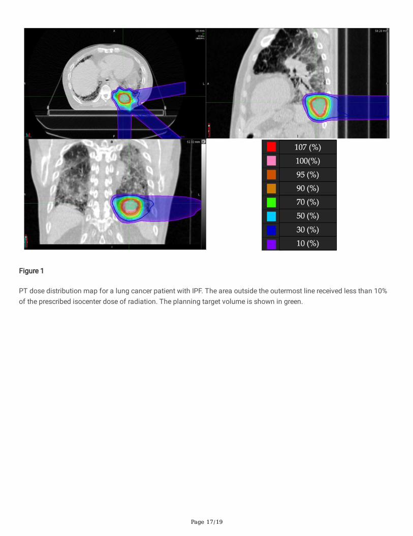
Representative caseThe PT plan for a representative patient is shown in Fig. 1. This 73-year-old male patient was diagnosed with IPF andreceived home oxygen therapy before lung cancer was found. A lung nodule had grown in the left lower lobe over time,but performing a biopsy was di�cult because of the presence of IPF. After consulting our cancer board, the nodule wastreated with PT under a diagnosis of cT2bN0M0 stage IIA lung cancer. The prescribed dose was 66 GyRBE in 6.6-GyRBEdaily fractions. No serious toxicities developed during or after the treatment.
Patients
Page 5/19
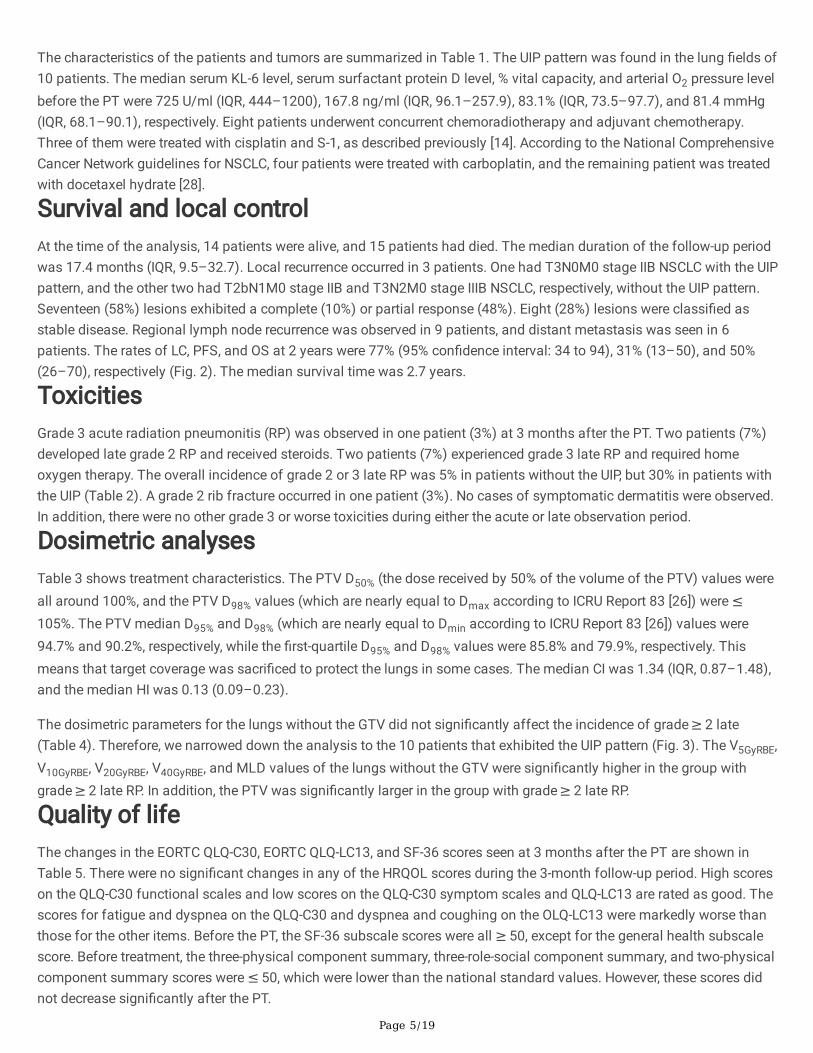
The characteristics of the patients and tumors are summarized in Table 1. The UIP pattern was found in the lung �elds of10 patients. The median serum KL-6 level, serum surfactant protein D level, % vital capacity, and arterial O2 pressure levelbefore the PT were 725 U/ml (IQR, 444–1200), 167.8 ng/ml (IQR, 96.1–257.9), 83.1% (IQR, 73.5–97.7), and 81.4 mmHg(IQR, 68.1–90.1), respectively. Eight patients underwent concurrent chemoradiotherapy and adjuvant chemotherapy.Three of them were treated with cisplatin and S-1, as described previously [14]. According to the National ComprehensiveCancer Network guidelines for NSCLC, four patients were treated with carboplatin, and the remaining patient was treatedwith docetaxel hydrate [28].
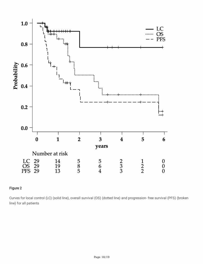
Survival and local controlAt the time of the analysis, 14 patients were alive, and 15 patients had died. The median duration of the follow-up periodwas 17.4 months (IQR, 9.5–32.7). Local recurrence occurred in 3 patients. One had T3N0M0 stage IIB NSCLC with the UIPpattern, and the other two had T2bN1M0 stage IIB and T3N2M0 stage IIIB NSCLC, respectively, without the UIP pattern.Seventeen (58%) lesions exhibited a complete (10%) or partial response (48%). Eight (28%) lesions were classi�ed asstable disease. Regional lymph node recurrence was observed in 9 patients, and distant metastasis was seen in 6patients. The rates of LC, PFS, and OS at 2 years were 77% (95% con�dence interval: 34 to 94), 31% (13–50), and 50%(26–70), respectively (Fig. 2). The median survival time was 2.7 years.
ToxicitiesGrade 3 acute radiation pneumonitis (RP) was observed in one patient (3%) at 3 months after the PT. Two patients (7%)developed late grade 2 RP and received steroids. Two patients (7%) experienced grade 3 late RP and required homeoxygen therapy. The overall incidence of grade 2 or 3 late RP was 5% in patients without the UIP, but 30% in patients withthe UIP (Table 2). A grade 2 rib fracture occurred in one patient (3%). No cases of symptomatic dermatitis were observed.In addition, there were no other grade 3 or worse toxicities during either the acute or late observation period.
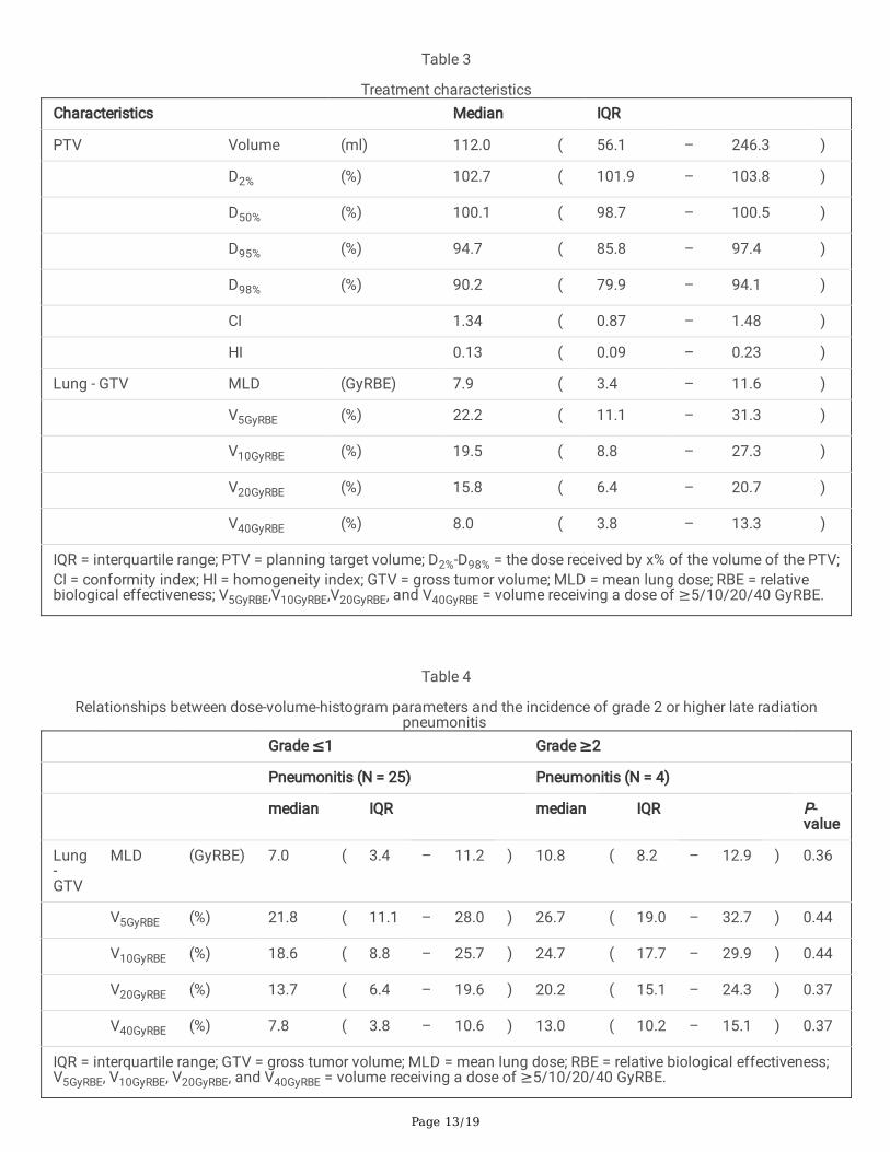
Dosimetric analysesTable 3 shows treatment characteristics. The PTV D50% (the dose received by 50% of the volume of the PTV) values wereall around 100%, and the PTV D98% values (which are nearly equal to Dmax according to ICRU Report 83 [26]) were ≤ 105%. The PTV median D95% and D98% (which are nearly equal to Dmin according to ICRU Report 83 [26]) values were94.7% and 90.2%, respectively, while the �rst-quartile D95% and D98% values were 85.8% and 79.9%, respectively. Thismeans that target coverage was sacri�ced to protect the lungs in some cases. The median CI was 1.34 (IQR, 0.87–1.48),and the median HI was 0.13 (0.09–0.23).
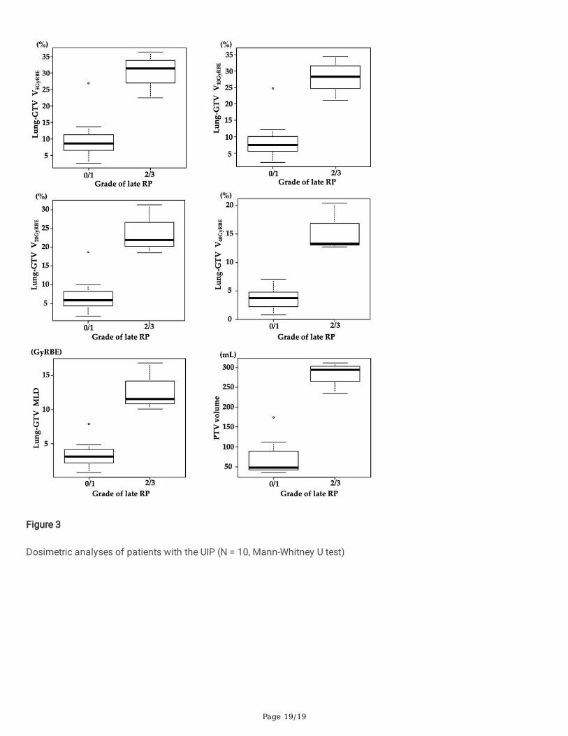
The dosimetric parameters for the lungs without the GTV did not signi�cantly affect the incidence of grade ≥ 2 late(Table 4). Therefore, we narrowed down the analysis to the 10 patients that exhibited the UIP pattern (Fig. 3). The V5GyRBE,V10GyRBE, V20GyRBE, V40GyRBE, and MLD values of the lungs without the GTV were signi�cantly higher in the group withgrade ≥ 2 late RP. In addition, the PTV was signi�cantly larger in the group with grade ≥ 2 late RP.
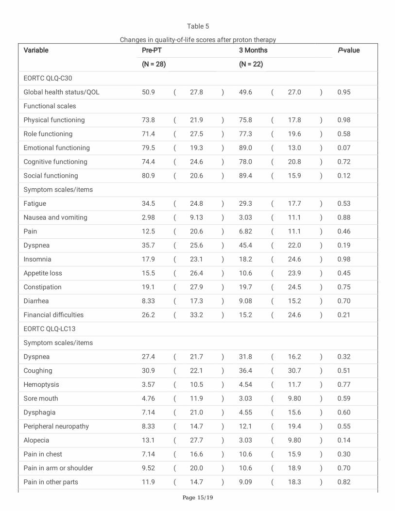
Quality of lifeThe changes in the EORTC QLQ-C30, EORTC QLQ-LC13, and SF-36 scores seen at 3 months after the PT are shown inTable 5. There were no signi�cant changes in any of the HRQOL scores during the 3-month follow-up period. High scoreson the QLQ-C30 functional scales and low scores on the QLQ-C30 symptom scales and QLQ-LC13 are rated as good. Thescores for fatigue and dyspnea on the QLQ-C30 and dyspnea and coughing on the OLQ-LC13 were markedly worse thanthose for the other items. Before the PT, the SF-36 subscale scores were all ≥ 50, except for the general health subscalescore. Before treatment, the three-physical component summary, three-role-social component summary, and two-physicalcomponent summary scores were ≤ 50, which were lower than the national standard values. However, these scores didnot decrease signi�cantly after the PT.
Page 6/19
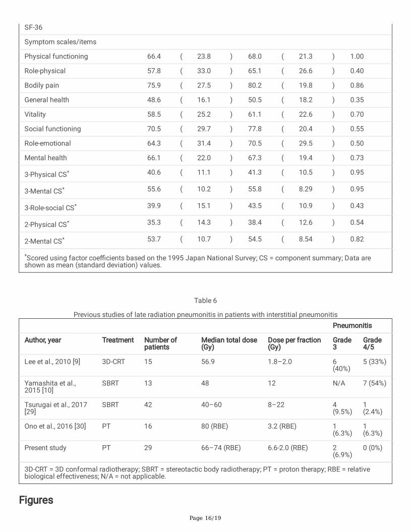
DiscussionIn the present study, grade 3 acute RP only occurred in one patient (3%). The incidence of grade 2 or 3 late RP after PTwas 35%, while there were no cases of grade 4 or 5 late RP. Table 6 summarizes the frequencies of grade 3, 4, or 5 late RPin IP patients in previous studies [9, 10, 29, 30]. It was reported that RP occurred frequently in patients with IP, especiallythose with IPF. These studies suggest that X-ray irradiation may cause fatal pneumonia in IP patients. In the presentstudy of PT, there were only two cases (6.9%) of grade 3 late RP, suggesting that PT is associated with a lower risk offatal pneumonia among lung cancer patients with IP than X-ray therapy. This may be due to the physical characteristicsof PT, as it reduces the doses delivered to the surrounding normal organs [11, 31].
Previous studies have shown that IPF patients with lung cancer have shorter survival times than patients with IPF alone[32, 33]. However, many treatment-related deaths have been reported in lung cancer patients with IPF. Surgery, such aslobectomy and biopsies, also worsens IPF. The reported postoperative IPF exacerbation rates range from 9.3–30% [7, 34,35]. The risk of pulmonary toxicity from drug therapy, such as pemetrexed, has been reported to be approximately 3.5% inpatients without IP, 12.0% in patients with IP, and up to 16.7% in patients with IPF [36]. In our study, 10 patients thatexhibited the UIP pattern, which is suggestive of IPF, were treated with PT, and only one of them developed late grade 3RP. There were no deaths associated with PT. Therefore, PT can be considered to be relatively safe. However, evennarrowly localized radiotherapy for patients with IPF was reported to lead to marked variation in the frequency of RP [37].Therefore, the necessity of interventions, including PT, should be carefully assessed in lung cancer patients with IPF.
Conventional radiotherapy for lung cancer patients with IP may be associated with a high risk of life-threateningpneumonia. SBRT may be safer if patients that were at high risk were excluded based on pretreatment CT evaluations orthe measurement of biomarker levels [10, 29]. However, SBRT is generally used as a treatment option for early stage lungcancer, and treating large targets with SBRT is technically di�cult [38, 39]. In our study, the median PTV of the patientstreated with PT was large due to the inclusion of stage I to III patients, while the doses delivered to the lungs were keptlow (Table 3). Our study suggests that the physical properties of PT are advantageous. The treatment of stage III lungcancer in IP patients carries a risk of life-threatening pneumonia, and PT is a safer treatment option for these patients.
QOL evaluations are important for comparing treatment modalities. Surgery is highly invasive and often leads to poorQOL. In a previous study, it was reported that patients’ QLQ-C30 scores had not returned to their preoperative levels at 6months after lung cancer surgery [40]. Postoperative patients tend to experience persistent physical function problems,such as shortness of breath and pain in the arms and chest [41]. Reductions of 10% in the physical and mentalcomponent summary scores of the SF-36 from the baseline after lung cancer surgery have been reported to beassociated with a high risk of death [42]. Although there is no consensus on what constitutes a signi�cant difference inQOL data, a 10% difference in the SF-36 summary score is generally considered to be a clinically relevant difference. Inour study, no signi�cant reductions in HRQOL scores were seen after PT. As this study focused on lung cancer patientswith IP, PT can be considered to be a less invasive treatment. However, the changes in QLQ-LC13 dyspnea scores seen at3 months after radiotherapy have been shown to be correlated with lung V30Gy, V40Gy, V50Gy, and MLD values [43].Previous studies have suggested that a lung V40Gy cut-off value of 11% exhibits good sensitivity and speci�city as apredictor of dyspnea. Our results showed that grade 2 or 3 late pneumonia developed in patients with lung V40GyRBE
values of > 11% (Table 4). The indications for PT for large PTV that require wide-�eld irradiation must be carefully judgedin consideration of the risks and bene�ts.
This study had several limitations. Dosimetric analyses of the PTV showed that the D50% tended to be relatively wellpreserved, but D95% was sacri�ced in some cases to ensure lung safety (Table 3). Sacri�cing the PTV D95% in this mannermay be clinically acceptable, but it may negatively affect long-term prognosis. We try to achieve both high PTV coverageand low lung exposure using respiratory-gated irradiation with gold marker implantation [6]. As another limitation, the
Page 7/19
HRQOL survey period was only 3 months. This was because in our prospective clinical studies the HRQOL surveys werescheduled to be conducted at 3 and 24 months after the PT. However, at 24 months su�cient data were not available forsome patients due to the length of the follow-up period being too short or an HRQOL survey not being performed. Furthercase accumulation and multicenter trials will be needed to assess late toxicities. Finally, patient selection bias must alsobe considered. Only patients who were judged to be suitable for this costly treatment by a pulmonologist were referred toour facility. In Japan, PT for lung cancer is not covered by medical insurance, and only wealthy people can receive thistreatment. Thus, the prognosis of the patients in our study may have been abnormally good, as the patients probably hadaccess to adequate standard medical support in addition to PT.
Immune checkpoint inhibitors have been developed in recent years, and many patients will continue to be treated withthem. In a prospective study in which nivolumab was administered to 6 NSCLC patients with mild IP, no life-threateningpneumonia occurred [44]. Even when they are used in combination with radiotherapy, there are many uncertaintiesregarding the risk of immune checkpoint inhibitors in patients with IP. There are also reports suggesting that a history ofthoracic radiation is a risk factor for pneumonia during treatment with immune checkpoint inhibitors [45]. Our studyshowed that PT could reduce the radiation dose delivered to normal lung tissue, and the incidence of clinicallyproblematic pneumonia was low. When immune checkpoint inhibitors need to be given to lung cancer patients with IP, PTcould be useful for reducing the risk of adverse events. Therefore, at our facility, several IP patients with stage III NSCLChave been treated with durvalumab as maintenance therapy after chemotherapy combined with PT after approval wasgranted by the cancer board. The results of a prospective trial of this approach will also be reported in the future. Wehope that PT can contribute to safer treatment in many lung cancer patients with IP.
ConclusionsPT is a safer treatment for NSCLC in patients with IP than conventional radiotherapy and SBRT. When patients thatexhibit the UIP pattern require clinical treatment, PT may be considered as a treatment option.
AbbreviationsIP: interstitial pneumonia; PT: proton therapy; NSCLC: non-small cell lung cancer; IQR: interquartile range; RP: radiationpneumonitis; IPF: idiopathic pulmonary �brosis; SBRT: stereotactic body radiotherapy; OAR: organ at risk; HRQOL: health-related quality of life; CT: computed tomography; UIP: usual interstitial pneumonia; UICC: Union for International CancerControl; MRI: magnetic resonance imaging; PET-CT: positron emission tomography-CT; RBE: relative biologicaleffectiveness; ICRU: International Commission on Radiation Units and Measurements; PTV: planning target volume;RECIST: Response Evaluation Criteria in Solid Tumors; EORTC; European Organization for Research and Treatment ofCancer; QLQ-C30: the EORTC core quality of life questionnaire; QLQ-LC13: the EORTC quality of life questionnaire - lungcancer module; SF-36: the Short-Form Health Survey; LC: local control; PFS: progression-free survival; OS: overall survival;GTV: gross tumor volume; MLD: mean lung dose; CI: conformity index; HI: homogeneity index.
DeclarationsAcknowledgements
The authors thank Drs. Katsumi Nakamae, Masaki Hara, Fumiya Baba, Shigeru Sasaki, Masanosuke Oguri, the membersof protocol committee, and all of the staff at Nagoya Proton Therapy Center for their valuable help with this research.
Author Contributions
Page 8/19
Conceptualization, S.H. and H.I.; Methodology, S.H. and H.I.; Formal Analysis, S.H.; Investigation, S.H., H.I., Y.H., K.N.(Koichiro Nakajima), K.N. (Kento Nomura), K.A., and H.O.; Resources, K.H. and T.T.; Data Curation, S.H. and E.Y.; Writing—Original Draft Preparation, S.H.; Writing—Review and Editing, H.I. and Y.S.; Supervision, J.M.; Project Administration, Y.S.;All of the authors have read and agreed to the published version of the manuscript.
Funding
Supported by JSPS KAKENHI Grant Number 19K17175.
Availability of data materials
The data presented in this study are available on request from the corresponding author. The data are not publiclyavailable due to institutional guidelines.
Ethics approval and consent to participate
The study was conducted according to the guidelines of the Declaration of Helsinki and was approved by theinstitutional review board of Nagoya City Hospital (20-04-327-07, August 5th, 2020).
Consent for publication
Not applicable.
Competing interests
The authors declare that they have no con�icts of interest.
References1. Antoniou KM, Margaritopoulos GA, Tomassetti S, Bonella F, Costabel U, Poletti V. Interstitial lung disease. European
Respiratory Review. European Respiratory Society; 2014;23:40–54.
2. Richeldi L, Collard HR, Jones MG. Idiopathic pulmonary �brosis. The Lancet. Lancet Publishing Group;2017;389:1941–52.
3. King TE, Pardo A, Selman M. Idiopathic pulmonary �brosis. The Lancet. Elsevier; 2011;378:1949–61.
4. Hutchinson J, Fogarty A, Hubbard R, McKeever T. Global incidence and mortality of idiopathic pulmonary �brosis: Asystematic review. European Respiratory Journal. European Respiratory Society; 2015;46:795–806.
5. Ley B, Collard HR, King TE. Clinical course and prediction of survival in idiopathic pulmonary �brosis. AmericanJournal of Respiratory and Critical Care Medicine. American Thoracic Society; 2011;183:431–40.
�. Ogura T, Takigawa N, Tomii K, Kishi K, Inoue Y, Ichihara E, et al. Summary of the Japanese Respiratory Societystatement for the treatment of lung cancer with comorbid interstitial pneumonia. Respiratory Investigation. ElsevierB.V.; 2019;57:512–33.
7. Sato T, Teramukai S, Kondo H, Watanabe A, Ebina M, Kishi K, et al. Impact and predictors of acute exacerbation ofinterstitial lung diseases after pulmonary resection for lung cancer. Journal of Thoracic and Cardiovascular Surgery.Mosby Inc.; 2014;147.
�. Ichihara E, Miyahara N, Maeda Y, Kiura K. Managing lung cancer with comorbid interstitial pneumonia. InternalMedicine. Japanese Society of Internal Medicine; 2020;59:163–7.
9. Lee YH, Kim YS, Lee SN, Lee HC, Oh SJ, Kim SJ, et al. Interstitial lung change in pre-radiation therapy computedtomography is a risk factor for severe radiation pneumonitis. Cancer research and treatment : o�cial journal of
Page 9/19
Korean Cancer Association. Korean Cancer Association; 2015;47:676–86.
10. Yamashita H, Kobayashi-Shibata S, Terahara A, Okuma K, Haga A, Wakui R, et al. Prescreening based on thepresence of CT-scan abnormalities and biomarkers (KL-6 and SP-D) may reduce severe radiation pneumonitis afterstereotactic radiotherapy. Radiation oncology (London, England). BioMed Central; 2010;5:32.
11. Ohno T, Oshiro Y, Mizumoto M, Numajiri H, Ishikawa H, Okumura T, et al. Comparison of dose-volume histogramsbetween proton beam and X-ray conformal radiotherapy for locally advanced non-small-cell lung cancer. Journal ofRadiation Research. Oxford University Press; 2014;56:128–33.
12. Travis WD, King TE, Bateman ED, Lynch DA, Capron F, Center D, et al. American Thoracic Society/EuropeanRespiratory Society international multidisciplinary consensus classi�cation of the idiopathic interstitial pneumonias.American Journal of Respiratory and Critical Care Medicine. American Lung Association; 2002;165:277–304.
13. Nakajima K, Iwata H, Ogino H, Hattori Y, Hashimoto S, Toshito T, et al. Clinical outcomes of image-guided protontherapy for histologically con�rmed stage I non-small cell lung cancer. Radiation oncology (London, England).2018;13:199.
14. Iwata H, Akita K, Yamaba Y, Kunii E, Takakuwa O, Yoshihara M, et al. Concurrent chemo-proton therapy usingadaptive planning for unresectable stage 3 non-small cell lung cancer: a phase 2 study. International Journal ofRadiation Oncology Biology Physics [Internet]. Elsevier Inc.; 2020 [cited 2021 Feb 24]; Available from:https://pubmed.ncbi.nlm.nih.gov/33227444/
15. Toshito T, Omachi C, Kibe Y, Sugai H, Hayashi K, Shibata H, et al. A proton therapy system in Nagoya Proton TherapyCenter. Australasian physical & engineering sciences in medicine / supported by the Australasian College of PhysicalScientists in Medicine and the Australasian Association of Physical Sciences in Medicine. 2016;
1�. Nakajima K, Iwata H, Ogino H, Hattori Y, Hashimoto S, Nakanishi M, et al. Acute toxicity of image-guidedhypofractionated proton therapy for localized prostate cancer. International Journal of Clinical Oncology.2018;23:353–60.
17. Hashimoto S, Sugie C, Iwata H, Ogino H, Omachi C, Yasui K, et al. Recovery from sublethal damage and potentiallylethal damage. Strahlentherapie und Onkologie. 2018;194:343–51.
1�. Paganetti H, Niemierko A, Ancukiewicz M, Gerweck LE, Goitein M, Loe�er JS, et al. Relative biological effectiveness(RBE) values for proton beam therapy. International journal of radiation oncology, biology, physics. 2002;53:407–21.
19. Iwata H, Ogino H, Hashimoto S, Yamada M, Shibata H, Yasui K, et al. Spot scanning and passive scattering protontherapy: relative biological effectiveness and oxygen enhancement ratio in cultured cells. International journal ofradiation oncology, biology, physics. 2016;95:95–102.
20. Ohta K, Shimohira M, Murai T, Nishimura J, Iwata H, Ogino H, et al. Percutaneous �ducial marker placement prior tostereotactic body radiotherapy for malignant liver tumors: an initial experience. Journal of radiation research. OxfordUniversity Press; 2016;57:174–7.
21. Hashimoto S, Katsurada M, Muramatsu R, Asai K, Tanaka K, Hayashi K, et al. Effect of a device-free compressedshell �xation method on hepatic respiratory movement: analysis for respiratory amplitude of the liver and internalmotions of a �ducial marker. Practical Radiation Oncology. 2019;9:e149–55.
22. Therasse P, Arbuck SG, Eisenhauer EA, Wanders J, Kaplan RS, Rubinstein L, et al. New guidelines to evaluate theresponse to treatment in solid tumors. Journal of the National Cancer Institute. Oxford University Press;2000;92:205–16.
23. Kobayashi K, Takeda F, Teramukai S, Gotoh I, Sakai H, Yoneda S, et al. A cross-validation of the EuropeanOrganization for Research and Treatment of Cancer QLQ-C30 (EORTC QLQ-C30) for Japanese with lung cancer.European Journal of Cancer. Eur J Cancer; 1998;34:810–5.
Page 10/19
24. Bergman B, Aaronson NK, Ahmedzai S, Kaasa S, Sullivan M. The EORTC QLQ-LC13: a modular supplement to theEORTC core quality of life questionnaire (QLQ-C30) for use in lung cancer clinical trials. European Journal of Cancer.Eur J Cancer; 1994;30:635–42.
25. Fukuhara S, Bito S, Green J, Hsiao A, Kurokawa K. Translation, adaptation, and validation of the SF-36 health surveyfor use in Japan. Journal of Clinical Epidemiology. J Clin Epidemiol; 1998;51:1037–44.
2�. Grégoire V, Mackie TR. State of the art on dose prescription, reporting and recording in intensity-modulated radiationtherapy (ICRU report No. 83). Cancer radiothérapie : journal de la Société française de radiothérapie oncologique.2011;15:555–9.
27. Kanda Y. Investigation of the freely available easy-to-use software “EZR” for medical statistics. Bone marrowtransplantation. 2013;48:452–8.
2�. Ettinger DS, Wood DE, Aggarwal C, Aisner DL, Akerley W, Bauman JR, et al. Non-small cell lung cancer, version1.2020: Featured updates to the NCCN guidelines. JNCCN Journal of the National Comprehensive Cancer Network[Internet]. Harborside Press; 2019 [cited 2021 Mar 4];17:1464–72. Available from:https://pubmed.ncbi.nlm.nih.gov/31805526/
29. Tsurugai Y, Takeda A, Sanuki N, Enomoto T, Kaneko T, Hara Y, et al. Stereotactic body radiotherapy for lung cancerpatients with idiopathic interstitial pneumonias. Radiotherapy and Oncology. Elsevier Ireland Ltd; 2017;125:310–6.
30. Ono T, Hareyama M, Nakamura T, Kimura K, Hayashi Y, Azami Y, et al. The clinical results of proton beam therapy inpatients with idiopathic pulmonary �brosis: a single center experience. Radiation oncology (London, England).BioMed Central; 2016;11:56.
31. Kadoya N, Obata Y, Kato T, Kagiya M, Nakamura T, Tomoda T, et al. Dose-volume comparison of proton radiotherapyand stereotactic body radiotherapy for non-small-cell lung cancer. International Journal of Radiation OncologyBiology Physics. Int J Radiat Oncol Biol Phys; 2011;79:1225–31.
32. Lee T, Park JY, Lee HY, Cho YJ, Yoon H Il, Lee JH, et al. Lung cancer in patients with idiopathic pulmonary �brosis:Clinical characteristics and impact on survival. Respiratory Medicine. W.B. Saunders Ltd; 2014;108:1549–55.
33. Tomassetti S, Gurioli C, Ryu JH, Decker PA, Ravaglia C, Tantalocco P, et al. The impact of lung cancer on survival ofidiopathic pulmonary �brosis. Chest. American College of Chest Physicians; 2015;147:157–64.
34. Kanzaki M, Kikkawa T, Maeda H, Kondo M, Isaka T, Shimizu T, et al. Acute exacerbation of idiopathic interstitialpneumonias after surgical resection of lung cancer. Interactive Cardiovascular and Thoracic Surgery. InteractCardiovasc Thorac Surg; 2011;13:16–20.
35. Koizumi K, Hirata T, Hirai K, Mikami I, Okada D, Yamagishi S, et al. Surgical treatment of lung cancer combined withinterstitial pneumonia: the effect of surgical approach on postoperative acute exacerbation. Annals of thoracic andcardiovascular surgery : o�cial journal of the Association of Thoracic and Cardiovascular Surgeons of Asia. AnnThorac Cardiovasc Surg; 2004;10:340–6.
3�. Kato M, Shukuya T, Takahashi F, Mori K, Suina K, Asao T, et al. Pemetrexed for advanced non-small cell lung cancerpatients with interstitial lung disease. BMC Cancer. BioMed Central Ltd.; 2014;14.
37. Yamaguchi S, Ohguri T, Ide S, Aoki T, Imada H, Yahara K, et al. Stereotactic body radiotherapy for lung tumors inpatients with subclinical interstitial lung disease: the potential risk of extensive radiation pneumonitis. Lung cancer(Amsterdam, Netherlands). 2013;82:260–5.
3�. Shibamoto Y, Hashizume C, Baba F, Ayakawa S, Miyakawa A, Murai T, et al. Stereotactic body radiotherapy using aradiobiology-based regimen for Stage I non-small-cell lung cancer: Five-year mature results. Journal of ThoracicOncology [Internet]. Lippincott Williams and Wilkins; 2015 [cited 2021 Mar 2];10:960–4. Available from:https://pubmed.ncbi.nlm.nih.gov/26001145/
Page 11/19
39. Nagata Y, Hiraoka M, Shibata T, Onishi H, Kokubo M, Karasawa K, et al. Prospective trial of stereotactic bodyradiation therapy for both operable and inoperable T1N0M0 non-small cell lung cancer: Japan Clinical OncologyGroup Study JCOG0403. International Journal of Radiation Oncology Biology Physics. Elsevier Inc.; 2015;93:989–96.
40. Szeliga E, Czenczek-Lewandowska E, Kontek A, Wolan-Nieroda A, Guzik A, Walicka-Cupryś K. Evaluation of thequality of life after surgical removal of lung cancer. Advances in Respiratory Medicine. Via Medica; 2019;87:14–9.
41. Koller M, Warncke S, Hjermstad MJ, Arraras J, Pompili C, Harle A, et al. Use of the lung cancer-speci�c quality of lifequestionnaire EORTC QLQ-LC13 in clinical trials: a systematic review of the literature 20 years after its development.Cancer. John Wiley and Sons Inc.; 2015;121:4300–23.
42. Möller A, Sartipy U. Associations between changes in quality of life and survival after lung cancer surgery. Journal ofThoracic Oncology. Lippincott Williams and Wilkins; 2012;7:183–7.
43. Sardaro A, McDonald F, Bardoscia L, Lavrenkov K, Singh S, Ashley S, et al. Dyspnea in patients receiving radicalradiotherapy for non-small cell lung cancer: a prospective study. Frontiers in Oncology. Frontiers Media S.A.; 2020;10.
44. Fujimoto D, Morimoto T, Ito J, Sato Y, Ito M, Teraoka S, et al. A pilot trial of nivolumab treatment for advanced non-small cell lung cancer patients with mild idiopathic interstitial pneumonia. Lung Cancer. Elsevier Ireland Ltd;2017;111:1–5.
45. Sul J, Blumenthal GM, Jiang X, He K, Keegan P, Pazdur R. FDA approval summary: pembrolizumab for the treatmentof patients with metastatic non‐small cell lung cancer whose tumors express programmed death‐ligand 1. TheOncologist. Wiley; 2016;21:643–50.
Tables
Page 12/19
Table 1
Patient and tumor characteristicsCharacteristics Patients
Age (years)
Median (IQR) 77 (71–81)
Sex
Male/Female 25/4
Performance status
0/1/2 21/6/2
UICC (8th ed.) stage
IA2/IA3/IB/IIA/IIB/IIIA/IIIB 5/2/3/1/5/5/8
UIP pattern
Yes/No 10/19
Smoking
Yes/No 24/5
Histopathology
Squamous cell carcinoma 13
Adenocarcinoma 9
Large cell carcinoma 1
NSCLC 2
Clinical malignancy 4
IQR = interquartile range; UICC (8th ed.) = Union for International Cancer Control 8th edition; UIP = usual interstitialpneumonia; NSCLC = unclassi�ed non-small cell lung cancer.
Table 2
Incidence of late radiation pneumonitis Grade 0/1 Grade 2 Grade 3
UIP (+) 7 2 1
UIP (-) 18 0 1
UIP = usual interstitial pneumonia.
Page 13/19
Table 3
Treatment characteristicsCharacteristics Median IQR
PTV Volume (ml) 112.0 ( 56.1 – 246.3 )
D2% (%) 102.7 ( 101.9 – 103.8 )
D50% (%) 100.1 ( 98.7 – 100.5 )
D95% (%) 94.7 ( 85.8 – 97.4 )
D98% (%) 90.2 ( 79.9 – 94.1 )
CI 1.34 ( 0.87 – 1.48 )
HI 0.13 ( 0.09 – 0.23 )
Lung - GTV MLD (GyRBE) 7.9 ( 3.4 – 11.6 )
V5GyRBE (%) 22.2 ( 11.1 – 31.3 )
V10GyRBE (%) 19.5 ( 8.8 – 27.3 )
V20GyRBE (%) 15.8 ( 6.4 – 20.7 )
V40GyRBE (%) 8.0 ( 3.8 – 13.3 )
IQR = interquartile range; PTV = planning target volume; D2%-D98% = the dose received by x% of the volume of the PTV;CI = conformity index; HI = homogeneity index; GTV = gross tumor volume; MLD = mean lung dose; RBE = relativebiological effectiveness; V5GyRBE,V10GyRBE,V20GyRBE, and V40GyRBE = volume receiving a dose of ≥5/10/20/40 GyRBE.
Table 4
Relationships between dose-volume-histogram parameters and the incidence of grade 2 or higher late radiationpneumonitis
*Scored using factor coe�cients based on the 1995 Japan National Survey; CS = component summary; Data areshown as mean (standard deviation) values.
Table 6
Previous studies of late radiation pneumonitis in patients with interstitial pneumonitis Pneumonitis
Author, year Treatment Number ofpatients
Median total dose(Gy)
Dose per fraction(Gy)
Grade3
Grade4/5
Lee et al., 2010 [9] 3D-CRT 15 56.9 1.8–2.0 6(40%)
5 (33%)
Yamashita et al.,2015 [10]
SBRT 13 48 12 N/A 7 (54%)
Tsurugai et al., 2017[29]
SBRT 42 40–60 8–22 4(9.5%)
1(2.4%)
Ono et al., 2016 [30] PT 16 80 (RBE) 3.2 (RBE) 1(6.3%)
1(6.3%)
Present study PT 29 66–74 (RBE) 6.6-2.0 (RBE) 2(6.9%)
0 (0%)
3D-CRT = 3D conformal radiotherapy; SBRT = stereotactic body radiotherapy; PT = proton therapy; RBE = relativebiological effectiveness; N/A = not applicable.
Figures
Page 17/19
Figure 1
PT dose distribution map for a lung cancer patient with IPF. The area outside the outermost line received less than 10%of the prescribed isocenter dose of radiation. The planning target volume is shown in green.
Page 18/19
Figure 2
Curves for local control (LC) (solid line), overall survival (OS) (dotted line) and progression- free survival (PFS) (brokenline) for all patients
Page 19/19
Figure 3
Dosimetric analyses of patients with the UIP (N = 10, Mann-Whitney U test)