Page 1
9/11/2012
1
1
Chapter 33
Nontraumatic
Musculoskeletal Disorders
2
Learning Objectives
• Outline musculoskeletal structure and function.
• Describe how to perform a detailed assessment of the extremities and spine.
• Specify questions in the patient history that help identify musculoskeletal problems.
• Describe assessment and management of specific nontraumatic musculoskeletal disorders based on an understanding of the pathophysiology.
3
Copyright © 2013 by Jones & Bartlett Learning, LLC, an Ascend Learning Company
Page 2
9/11/2012
2
Anatomy and Physiology Review
• Musculoskeletal system
– Bones
– Muscles
– Tendons and ligaments
– Articulating surfaces
• Joints
• Bursa
• Discs
4
Skeletal System
• Contains bony structures that provide support and protection for body
• Provides system of levers on which muscles act to produce body movement
5
Skeletal System
• Contains 206 individual bones, divided into two categories– Axial skeleton
• Skull
• Hyoid bone
• Vertebral column
• Thoracic cage
– Appendicular skeleton• Bones of upper and lower extremities
• Girdles, by which attached to body
6
Copyright © 2013 by Jones & Bartlett Learning, LLC, an Ascend Learning Company
Page 3
9/11/2012
3
7
Skeletal System
• Body movement made possible by bones connected to other bones
– With exception of hyoid bone, every bone in body connects to at least one other bone by way of joints
– Three major classes of joints
• Fibrous
• Cartilaginous
• Synovial
8
Skeletal System
• Fibrous joints
– Two bones united by fibrous tissue that have little or no movement
– Example: suture in skull bones
• Cartilaginous joints
– Unite two bones by means of hyaline cartilage and fibrocartilage
– Slightly movable
– Examples: epiphyseal plate of a growing bone, junctions of intervertebral discs
9
Copyright © 2013 by Jones & Bartlett Learning, LLC, an Ascend Learning Company
Page 4
9/11/2012
4
Skeletal System
• Synovial joints
– Contain synovial fluid that allows for considerable movement
– Most joints that unite bones of appendicularskeleton
– Example: hinge joint of elbow and knee, ball‐and‐socket joints of shoulder and hip
10
11
Muscular System
• Responsible for execution of movement and postural maintenance
• Major types of muscles
– Skeletal
– Cardiac
– Smooth muscle
12
Copyright © 2013 by Jones & Bartlett Learning, LLC, an Ascend Learning Company
Page 5
9/11/2012
5
Muscular System
• Skeletal muscle
– Most common
– Most involved in musculoskeletal disorders
– Attached to bones by tendons
– Ligaments connect bone or cartilage, helps strengthen and support joints
13
Muscular System
• Skeletal muscle– Has specialized contractile cells known as muscle fibers
– When nervous impulse passes through muscle fiber, specialized chemicals cause muscle to contract
– Most extend from one bone to another and cross at least one joint
– Contraction of some muscles and simultaneous relaxation of other muscles produce body movement
• Made possible by pulling one bone toward other across movable joint
14
Muscular System
• Muscle tone
– Postural maintenance is result of muscle tone
– Constant tension produced by muscles of body for long periods of time
– Responsible for keeping back and legs straight, head in upright position, abdomen from bulging
– Postural maintenance balances distribution of body weight
• Puts less strain on muscles, tendons, ligaments, bones
15
Copyright © 2013 by Jones & Bartlett Learning, LLC, an Ascend Learning Company
Page 6
9/11/2012
6
General Assessment Strategies
• General assessment of patient’s musculoskeletal system includes examination of
– Extremities
– Spine
– Vascular system
– Motor system
16
General Assessment Strategies
• Purpose of assessment
– Pain or tenderness
– Swelling
– Abnormal or loss of movement
– Decreased sensation
– Circulatory changes
– Deformity
17
Extremity Exam
• Examine for function and structure
– General appearance
– Body proportions
– Ease of movement
– Limitation in range of motion
– Unusual increase in mobility of joint
18
Copyright © 2013 by Jones & Bartlett Learning, LLC, an Ascend Learning Company
Page 7
9/11/2012
7
Extremity Exam
• Abnormal findings– Signs of inflammation
• Swelling• Tenderness• Increased heat• Redness• Decreased function
– Asymmetry– Crepitus– Deformities– Decreased muscular strength– Atrophy
19
Extremity Exam
• Assessment should include
– Evaluation of skin and tissue overlying muscles, cartilage, bones
– Joints for soft tissue injury, discoloration, swelling
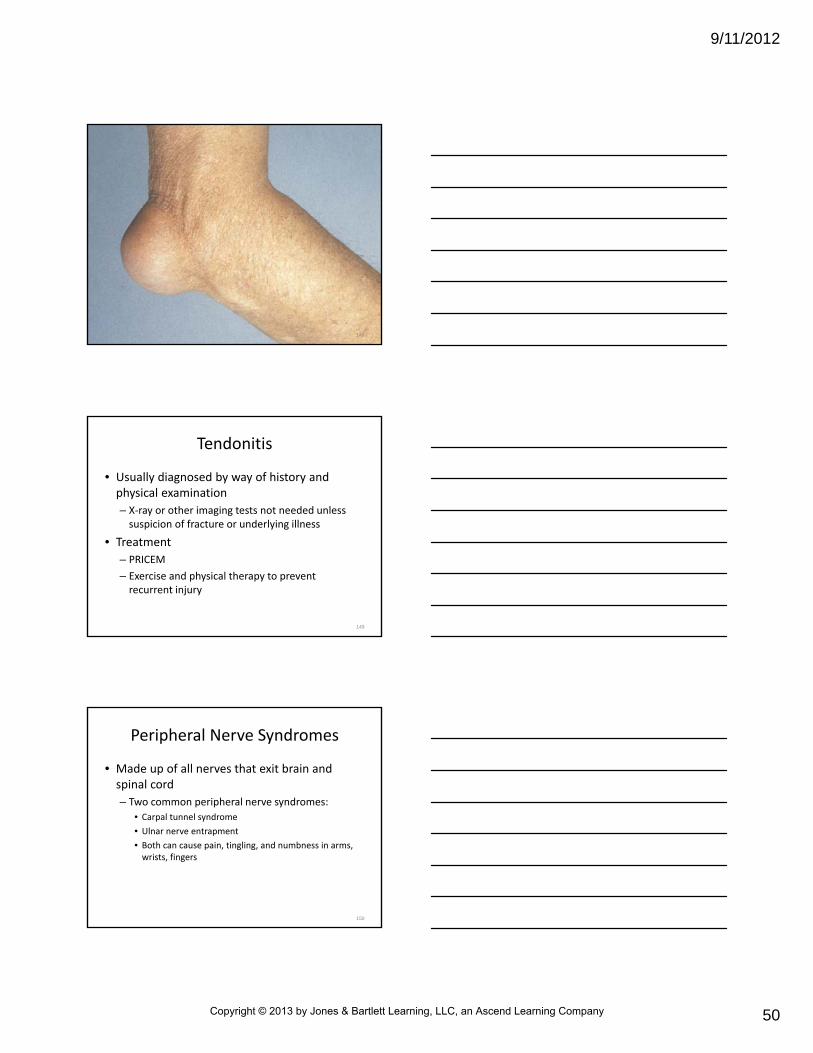
– Reasonably symmetrical in structure and muscularity
20
Extremity Exam
• Circulatory status of each extremity
– Skin color
– Temperature
– Sensation
– Presence of distal pulses
21
Copyright © 2013 by Jones & Bartlett Learning, LLC, an Ascend Learning Company
Page 8
9/11/2012
8
Extremity Exam
• Joints
– Full range of motion
– All movement should be made without
• Pain
• Deformity
• Limitation
• Instability
22
Extremity Exam
• Hands and wrists– Inspect for contour and positional alignment
– Hands, wrists, and joints of each finger for• Tenderness
• Swelling
• Deformity
• Elbows – Assess and palpate in flexed and extended positions
– All movement should be made without pain or discomfort
23
Extremity Exam
• Shoulders
– Both shoulders should be symmetrical and palpated for integrity of
• Clavicles
• Scapulae
• Humeri
– Should be able to shrug shoulders, raise and extend both arms without pain or discomfort
24
Copyright © 2013 by Jones & Bartlett Learning, LLC, an Ascend Learning Company
Page 9
9/11/2012
9
Extremity Exam
• Pelvis and hips
– Structural integrity of patient’s iliac crest and symphysispubis to determine stability
– Should be no deformity or point tenderness during palpation
• Knees
– Inspect and palpate for swelling and tenderness
– Patella should be nontender and in midline position
– Patient should be able to bend and straighten each knee without pain
25
Extremity Exam
• Ankles and feet
– Inspect for contour, position, size
– Abnormal findings: tenderness, swelling, deformity
• Toes should be straight and aligned with each other
• Surface of ankles and feet should be free of deformities, nodules, swelling, calluses
26
Spine Exam
• Visual assessment of cervical, thoracic, lumbar curves
• Abnormal findings
– Curvature of spine from abnormal lordosis, kyphosis, scoliosis
– Differences in height of shoulders or iliac crest that might result from abnormal spinal curvature
27
Copyright © 2013 by Jones & Bartlett Learning, LLC, an Ascend Learning Company
Page 10
9/11/2012
10
Spine Exam
• Neck
– In midline position
– Posterior neck should be free of point tenderness and swelling
– Patient should be able to bend head forward, backward, from side to side without pain or discomfort
28
Spine Exam
• Thoracic lumbar spine
– Inspect for signs of
• Injury
• Swelling
• Discoloration
– In normal exam, spine is nontender to palpation
29
Vascular Exam
• Peripheral vascular system
– Components
• Arteries
• Veins
• Lymphatic system
• Fluids exchanged in capillary beds
– Should be part of general assessment strategy
– Upper and lower extremities assessed for color, texture, arterial insufficiency
– Lymph nodes should be nonswollen and nontender
30
Copyright © 2013 by Jones & Bartlett Learning, LLC, an Ascend Learning Company
Page 11
9/11/2012
11
Vascular Exam
• Abnormal findings
– Pale or cyanotic skin
– Weak or diminished pulses
– Skin that is cold to the touch
– Absence of hair growth
– Pitting edema
31
Motor Exam
• Observe patient while moving and at rest
– Note
• Abnormal or involuntary movements
• Posture
• Level of activity
• Fatigue
32
Motor Exam
• Observe patient while moving and at rest
– Muscle strength should be equal on both sides of body
– Assess for agility
– Test for
• Flexion
• Extension
• Abduction of upper and lower extremities
33
Copyright © 2013 by Jones & Bartlett Learning, LLC, an Ascend Learning Company
Page 12
9/11/2012
12
Motor Exam
• Evaluate coordination for
– Point‐to‐point movements
• Touch finger to nose
• Touch each heel to opposite shin
34
Motor Exam
• Evaluate coordination for
– Gait
• Walk toe to toe
• Walk on toes
• Walk on heels
– Stance
• Romberg test
• Pronator Drift test
35
Motor Exam
• Should be responsive to
– Sensations of pain
– Temperature
– Position
– Vibration
– Touch
36
Copyright © 2013 by Jones & Bartlett Learning, LLC, an Ascend Learning Company
Page 13
9/11/2012
13
Motor Exam
• Conducted by sensory pathways of nervous system
– Sensory exams can be performed on conscious patients by using light touch on each hand and foot
– Proceed from head to toe, symmetrical on both sides of body
37
• General management for musculoskeletal disorder are same as for most other patient‐care encounters
– Prehospital care guided by patient’s chief complaint and severity of patient’s condition
General Management Strategies
38
• General management strategies
– Scene size‐up to ensure personal safety
– Primary survey to ensure airway, ventilation, circulation
– Secondary assessment, reassessment
– Pharmacological and nonpharmacologicalmeasures to ensure comfort
– Transport considerations
– Effective therapeutic communications
General Management Strategies
39
Copyright © 2013 by Jones & Bartlett Learning, LLC, an Ascend Learning Company
Page 14
9/11/2012
14
Patient History
• Focus on patient’s chief complaint, ask particular questions
– Complaint: joint pain
• Where is the specific site of the pain?
• Does the pain change during the course of the day?
• Have you had a recent injury?
• How long has there been pain in the joint?
• Does the pain get better or worse with movement?
40
Patient History
• Focus on patient’s chief complaint, ask particular questions
– Complaint: back pain
• Is the pain confined to the back, or does it radiate to the upper or lower limbs?
• Is the pain made worse by coughing or sneezing?
• Was the pain sudden or gradual in onset?
41
Patient History
• Focus on patient’s chief complaint, ask particular questions
– Complaint: gait or balance Issues
• Do you trip when walking?
• Do you stagger to one particular side?
• Do you ever injure yourself when walking?
42
Copyright © 2013 by Jones & Bartlett Learning, LLC, an Ascend Learning Company
Page 15
9/11/2012
15
Management Guidelines
• Prehospital care
– Primarily supportive
– Limited to immobilization of affected area or body part
– Application of ice and/or elevation of extremity to reduce pain, swelling
– Analgesics to relieve pain
– Gentle transport
43
Osteomyelitis
• Acute or chronic bone infection
– Affects 2 out of every 10,000 people
– Can be caused by number of microbial agents, most commonly staphylococcus aureus
– Can result from
• Open fracture or minor wound infection
• Systemic infection that allows bacteria to spread in bloodstream and enter bone
44
Osteomyelitis
• If left untreated
– Can become chronic, resulting in
• Decreased blood supply to bone
• Eventual death of bone tissue
• Affects both children and adults, can affect any bone
– In adults, vertebra and pelvis most often affected
– In children, long bones most affected
45
Copyright © 2013 by Jones & Bartlett Learning, LLC, an Ascend Learning Company
Page 16
9/11/2012
16
Osteomyelitis
• Those at increased risk
– Recent orthopedic surgery
– Elderly
– IV drug abusers
– Sickle cell disease
– Hemodialysis
– Compromised immune systems
46
Osteomyelitis
• Signs and symptoms– Pain and/or tenderness in infected area
– Swelling and warmth in infected area
– Fever, chills
– General malaise
– Drainage of pus through skin
– Excessive sweating
– Back or neck pain (if spine involved)
– Swelling of ankles, feet, legs
– Walking that is painful or with limp
47
Osteomyelitis
• Diagnostic tools
– Blood tests to confirm infection
– Blood cultures to identify bacteria
– Needle aspiration
– Biopsy
– Bone scans
48
Copyright © 2013 by Jones & Bartlett Learning, LLC, an Ascend Learning Company
Page 17
9/11/2012
17
Osteomyelitis
• Treatment
– Oral or IV antibiotics to manage infection, prevent reinfection
– Surgical drainage of wound or abscess
– Immobilization of affected bone or surrounding joints
– Surgery to scrape infection from affected bone
– Rarely, amputation of affected limb may be required
49
Bone Tumors
• Abnormal growth of cells within bone– Can be malignant (cancerous) or benign (noncancerous)
– Most are benign and not life threatening
– Common benign bone tumors• Nonossifying fibroma unicameral bone cyst
• Osteochondroma
• Giant cell tumor
• Enchondroma
• Fibrous dysplasia
50
Bone Tumors
• Malignant tumors
– Can spread cancer cells throughout body (metastasize) via blood or lymphatic system
– Primary tumor
• In original site where it first arose
– Secondary
• Originates in another area of body and spreads to bone
51
Copyright © 2013 by Jones & Bartlett Learning, LLC, an Ascend Learning Company
Page 18
9/11/2012
18
Bone Tumors
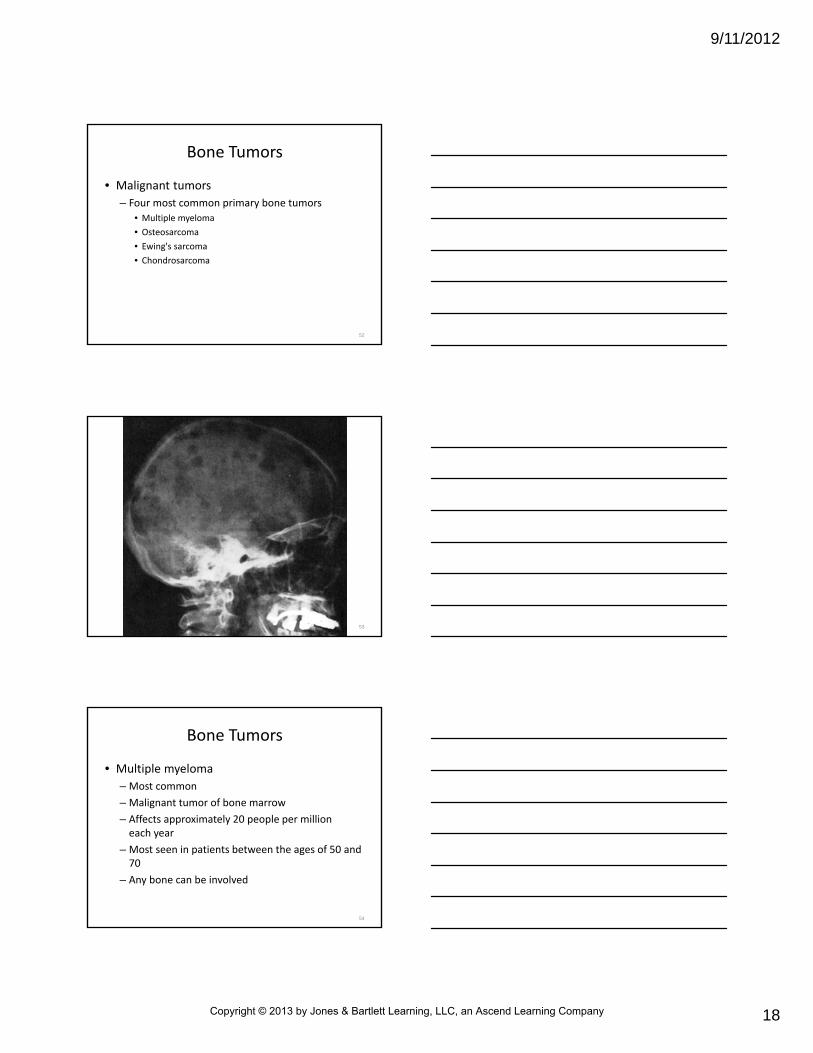
• Malignant tumors
– Four most common primary bone tumors
• Multiple myeloma
• Osteosarcoma
• Ewing's sarcoma
• Chondrosarcoma
52
53
Bone Tumors
• Multiple myeloma
– Most common
– Malignant tumor of bone marrow
– Affects approximately 20 people per million each year
– Most seen in patients between the ages of 50 and 70
– Any bone can be involved
54
Copyright © 2013 by Jones & Bartlett Learning, LLC, an Ascend Learning Company
Page 19
9/11/2012
19
Bone Tumors
• Osteosarcoma
– Second most common bone cancer
– Occurs in 2 or 3 new people per million each year
– Most occur in teenagers
– Most tumors occur around knee
– Other common locations include hip and shoulder
55
Bone Tumors
• Ewing's sarcoma
– Most commonly occurs between 5 and 20 years of age
– Most common locations
• Upper and lower leg
• Pelvis
• Upper arm
• Ribs
56
Bone Tumors
• Chondrosarcoma
– Most common in patients between 40 and 70 years of age
– Most cases occur around hip, pelvis, or shoulder
57
Copyright © 2013 by Jones & Bartlett Learning, LLC, an Ascend Learning Company
Page 20
9/11/2012
20
Bone Tumors
• Signs and symptoms
– Most experience dull or aching pain in area of tumor
• Sometimes made worse with physical activity
• Often awakes patient at night
– Other patients will not complain of pain, discovered painless mass on self examination
58
Bone Tumors
• Pathologic fractures not common in these patients
– Fractures result from trauma or a metabolic disease, such as osteoporosis, where bone weakened by tumor breaks
59
Bone Tumors
• Benign tumors may or may not require treatment (depending on specific tumor)– Some benign tumors can be aggressive and quickly destroy bone
• Malignant tumors may require medication therapy or surgical removal– Others resolve on their own (especially some bone tumors in children)
– Most malignant tumors are surgically removed and treated with radiation
60
Copyright © 2013 by Jones & Bartlett Learning, LLC, an Ascend Learning Company
Page 21
9/11/2012
21
Bone Tumors
• If the cancer has metastasized, other care may include additional
– Radiation
– Chemotherapy
– Cryosurgery
• Freezing and killing cancer cells with liquid nitrogen
61
Bone Tumors
• In some patients, bone implant or amputation of affected limb will be needed
• Patient follow‐up with regular blood tests and x‐ray required because bone cancer can recur
• People who have had bone cancer, particularly children and adolescents, have increased likelihood of developing another type of cancer, such as leukemia, later in life
62
Lower Back Pain
• Americans spend at least $50 billion each year on lower back pain
– Most common cause of job‐related disability
– Leading contributor to missed work
– Back pain is second most common neurological ailment in U.S.
63
Copyright © 2013 by Jones & Bartlett Learning, LLC, an Ascend Learning Company
Page 22
9/11/2012
22
Lower Back Pain
• Vertebral C column consists of
– 26 bones divided into 5 regions
• 7 cervical vertebrae
• 12 thoracic vertebrae
• 5 lumbar vertebrae
• 1 sacral bone
• 1 coccygeal bone
64
Lower Back Pain
• Vertebral C column consists of
– Together, vertebrae protect
• Spinal cord
• Rootlets
• 31 pairs of spinal nerves that convey sensation
65
Lower Back Pain
• Weight‐bearing portion of vertebrae is bony vertebral body
– Intervertebral discs located between bodies of adjacent vertebrae
• Serve as shock absorbers
• Allow for flexibility of back
• Prevent vertebral bodies from rubbing against each other
66
Copyright © 2013 by Jones & Bartlett Learning, LLC, an Ascend Learning Company
Page 23
9/11/2012
23
67
Lower Back Pain
• Acute back pain
– Usually of short duration, lasting only few days to few weeks
– Most caused by trauma to lower back
– Can be caused by
• Arthritis
• Degenerative joint disease of spine
• Viral infections
• Congenital abnormalities
68
Lower Back Pain
• Chronic back pain
– Persists more than 3 months
– Can be progressive and debilitating
69
Copyright © 2013 by Jones & Bartlett Learning, LLC, an Ascend Learning Company
Page 24
9/11/2012
24
Intervertebral Disc Disorders
• Intervertebral discs– Found between bodies of vertebrae– Act as shock absorbers– Prevent bones of spine from grinding against each other
– Allow for flexibility of back– Each disc has a central area composed of jelly‐like substance, called nucleus pulposus
• Surrounded by concentric rings of fibrous tissue (annulus fibrosis)
• Undue stress on disc can force gel against inner ring and crack it
70
Intervertebral Disc Disorders
• From there, gel pushes outward, cracking successive rings in its path
• If stress on back is severe enough, vertebral discs and fibrous tissues can be damaged, causing disc to bulge or protrude
• If gel eventually breaks through outer ring, it can pinch nerve root leading from vertebra
– Result in “slipped” or herniated disc
71
72
Copyright © 2013 by Jones & Bartlett Learning, LLC, an Ascend Learning Company
Page 25
9/11/2012
25
Intervertebral Disc Disorders
• Disc injury most often affects lumbar spine in patients 25 to 45 years of age
• Most common risk factor for developing lumbar disc disease is lack of exercise that allows muscles of back to weaken
• Symptoms of herniated disc vary greatly depending on position of herniated disc and size of herniation
73
Intervertebral Disc Disorders
• Common signs and symptoms of herniation
– Lower back ache
– Numbness or weakness in lower extremities
– Deep muscle pain and muscle spasms
– Acute or gradual leg pain (usually in only one leg)
74
Intervertebral Disc Disorders
• Common signs and symptoms of herniation
– “Shooting” pain in leg when sneezing, coughing, or straining
• May be aggravated by sitting, prolonged standing, bending, twisting
– Nerve‐related symptoms
• Muscle weakness in one or both legs
• Pain in front of thigh
• Sciatica
75
Copyright © 2013 by Jones & Bartlett Learning, LLC, an Ascend Learning Company
Page 26
9/11/2012
26
Intervertebral Disc Disorders
• Goals of treatment for herniated disc
– Relieve pain
– Weakness
– Numbness in leg caused by pressure on spinal nerve root or spinal cord
76
Intervertebral Disc Disorders
• Treatment– Bed rest
– Analgesics
– Anti‐inflammatories
– Muscle relaxants
– Corticosteroids
– Physical therapy and exercise programs can help strengthen back and prevent recurrent injury
– Most heal without surgery to remove herniated disc (discectomy)
77
Cauda Equina Syndrome
• Rare disorder of lumbar spine that affects bundle of nerve roots at lower end of spinal cord
– Surgical emergency that occurs when nerve roots are compressed and paralyzed, cutting off sensation and movement
– If not treated, syndrome can result in
• Permanent paralysis
• Impaired bladder and bowel function
• Loss of sexual sensation
– Even with surgery, nerve damage may be irreversible
78
Copyright © 2013 by Jones & Bartlett Learning, LLC, an Ascend Learning Company
Page 27
9/11/2012
27
Cauda Equina Syndrome
• Can be caused by– Herniated disc
– Spinal tumor
– Infection
– Spinal stenosis (narrowing of spinal canal)
– Spinal trauma• Direct trauma
• Falls
• Gunshot wounds
• Stabbings
79
Cauda Equina Syndrome
• Signs and symptoms– Bladder and/or bowel dysfunction
• Loss of control
• Inability to urinate or defecate
– Severe or progressive weakness in lower extremities
– Loss of sensation or altered sensation between legs, over buttocks, inner thighs, and back of legs (saddle area), and feet and heels
– Pain, numbness, or weakness that spreads to one or both legs that may cause stumbling gait or difficulty rising from sitting position
80
Lower Back Strains and Sprains
• Lower back carries much of body’s weight during walking, running, lifting, other activities
• Because muscles, ligaments, bones of spine provide control and strength for many movements, sprains and strains of the lower back (especially lumbar spine) are common injuries
81
Copyright © 2013 by Jones & Bartlett Learning, LLC, an Ascend Learning Company
Page 28
9/11/2012
28
Lower Back Strains and Sprains
• Often caused by
– Twisting or pulling
– Improper lifting that puts back at risk for injury
• Estimated that 50 percent of all EMS personnel develop back pain every year
• 1 in 4 will have career‐ending back injury within first 4 years of service
82
Lower Back Strains and Sprains
• Strain
– Injury to muscle or tendon
• Sprain
– Stretching or tearing of ligament beyond its normal range of movement
• Differentiating sprain from strain is difficult and unnecessary in prehospital setting
– Signs, symptoms, treatment, and prognosis for both conditions are same
83
Lower Back Strains and Sprains
• Signs and symptoms
– Pain
– Warmth
– Muscle spasms
– Swelling of affected area
84
Copyright © 2013 by Jones & Bartlett Learning, LLC, an Ascend Learning Company
Page 29
9/11/2012
29
Lower Back Strains and Sprains
• Treatment– Most are successfully treated with bed rest for 24 to 48 hours to allow back to heal
– Analgesics
– Muscle relaxants
– Anti‐inflammatories
– Physical therapy and exercise programs can help to strengthen back muscles and prevent future injury
– Back pain that does not resolve with these measures requires further evaluation
85
Joint Disorders
• Any disease or injury that affects human joints
– May be short lived or chronic
– Produce inflammation as result of disease
• Various forms of arthritis
86
Arthritis
• Inflammatory condition of joint, characterized by pain and swelling
– Limits activity of nearly 19 million adults
– Is most common cause of disability in U.S.
– Grouped into three general categories
• Osteoarthritis
• Rheumatoid arthritis
• Gout
87
Copyright © 2013 by Jones & Bartlett Learning, LLC, an Ascend Learning Company
Page 30
9/11/2012
30
Osteoarthritis
• Chronic, degenerative joint disease, most often seen in people over 40 years of age
• Onset of disease is gradual and affects women more often than men
• Mechanical in nature, resulting from normal wear‐and‐tear on joints over course of person’s life
88
Osteoarthritis
• Marked by
– Breakdown of cartilage that covers surfaces of joints
– Formation of bone spurs (bony growths formed on normal bone)
89
Osteoarthritis
• Wearing away of cartilage and overgrowth of bone lead to pain and stiffness
– As disease progresses, bone rubs against bone, causing severe pain and reduced mobility
– Joints most commonly affected
• Knees
• Hips
• Hands
• Cervical and lumbar spine
90
Copyright © 2013 by Jones & Bartlett Learning, LLC, an Ascend Learning Company
Page 31
9/11/2012
31
Rheumatoid Arthritis
• Inflammatory disease of joints that causes
– Pain
– Swelling
– Stiffness
– Loss of function
• Estimated that about 1.3 million people in U.S. have disease
91
Rheumatoid Arthritis
• Occurs in all races and ethnic groups
• Symptoms usually become apparent in middle and later life
– Can develop in young adults and children
92
Rheumatoid Arthritis
• Develops when lymphocytes travel to synovium in joints, causing inflammation (synovitis)
– During process, normally thin synovium becomes thick and makes joint swollen and puffy to touch
– As disease progresses, inflamed synovium invades and damages cartilage and bone of joint
• Surrounding muscles, ligaments, tendons become weakened
93
Copyright © 2013 by Jones & Bartlett Learning, LLC, an Ascend Learning Company
Page 32
9/11/2012
32
Rheumatoid Arthritis
• Can cause more generalized bone loss that may lead to osteoporosis
• Generally occurs in symmetrical pattern (e.g., both hands, both knees)
94
Rheumatoid Arthritis
• Hallmark of disease
– Visible swelling and inflammation of finger joints closest to affected hand
– May also affect other areas of body
– Often associated with
• Fatigue
• Occasional fever
• General malaise
95
96
Copyright © 2013 by Jones & Bartlett Learning, LLC, an Ascend Learning Company
Page 33
9/11/2012
33
Rheumatoid Arthritis
• Patients have varying degrees of disease
– Some have only limited bouts, followed by remission and little damage
– Others disease is regularly active, lasting many years
• Often leads to severe joint damage and disability
97
Rheumatoid Arthritis
• Physician care
– Anti‐inflammatories to reduce pain and inflammation
– Disease‐modifying antirheumatic drugs (DMARDs) to slow course of disease
– Analgesics
– Physical therapy
– Joint replacement
– Tendon reconstruction
– Synovectomy
98
When caring for a patient with severe rheumatoid arthritis or
anklyosing spondylitis, how might you have to modify your care if you
suspect spine injury?
99
Copyright © 2013 by Jones & Bartlett Learning, LLC, an Ascend Learning Company
Page 34
9/11/2012
34
Septic Arthritis
• Also known as infectious arthritis
– Results from direct invasion of joint space by various microorganisms
• Bacteria (most common)
• Viruses
• Mycobacteria
• Fungi
– Affects about 20,000 people in U.S. each year
– Can occur in both children and adults
100
Septic Arthritis
• Disease process begins when infectious agent (most commonly Staphylococcus aureus) enters joint
– Usually occurs from active infection elsewhere in body
• Respiratory tract infection
• Urinary tract infection
– Can occur from direct invasion (e.g., open wound near joint, joint surgery)
101
Septic Arthritis
• When bacterium reaches synovium in joint, immune system is activated and cartilage begins to be destroyed– Results in inflammation and reduced blood flow to joint and surrounding structures
– Previously damaged joints, especially from rheumatoid arthritis, are most susceptible to infection
– Most commonly involved joint is knee, followed by hip, shoulder, ankle, wrist
102
Copyright © 2013 by Jones & Bartlett Learning, LLC, an Ascend Learning Company
Page 35
9/11/2012
35
Septic Arthritis
• Signs and symptoms
– Fever
– Shaking chills
– Severe pain
– Warmth
– Swelling in affected joint
103
Septic Arthritis
• Treatment
– Antibiotics to resolve infection
– In severe cases, joint may need surgical reconstruction or replacement
104
Gout
• Form of arthritis marked by deposit of uric acid crystals (monosodium urate) in and around joint
– Affects mostly men
– Thought to be hereditary
– Affects about 2.7 of every 1,000 adults
• For unknown reasons, gout surfaces most often in metatarsophalangeal joint of big toe
– May also present anywhere in lower or upper extremities
105
Copyright © 2013 by Jones & Bartlett Learning, LLC, an Ascend Learning Company
Page 36
9/11/2012
36
Gout
• In elderly, many joints may be affected
– May also develop tophi (masses of urate crystals deposited in soft tissue)
– Usually affects cooler areas of body such as elbows, ears, distal finger joints
– Also associated with increased risk for developing kidney stones
106
Gout
• In acute gout, affected joint and surrounding tissues appear hot, red, swollen
– Pain is usually intense and made worse by stimulation or light touch
– May remit for long periods, followed by flares that last days to weeks
– Chronic gout can lead to degenerative form of arthritis called gouty arthritis
107
Gout
• Risk factors
– Joint injury
– Obesity
– Hypertension
– Alcohol use
– Diuretics that lead to hyperuricemia
– Diets rich in meat and seafood
108
Copyright © 2013 by Jones & Bartlett Learning, LLC, an Ascend Learning Company
Page 37
9/11/2012
37
Gout
• Most treatable form of arthritis
– Pain medicine
– Anti‐inflammatories
– IM and oral corticosteroids
– Acute episode usually subsides within 24 hours after treatment begins
109
Muscle Disorders
• Muscles purposes– Movement
– Postural maintenance
– Heat production
• Inflammation of skeletal muscle can result from– Injury
– Infection
– Autoimmune disease
110
Myalgia
• Means muscle pain or pain in multiple muscles
– Many causes and various types
– Can be acute and temporary, or can be chronic
• Most often results from
– Overuse
– Muscle injury
– Stress
– Virus
– Infection
– Autoimmune disorders
111
Copyright © 2013 by Jones & Bartlett Learning, LLC, an Ascend Learning Company
Page 38
9/11/2012
38
Myalgia
• Can be indication of serious illness
– Inflammatory myopathies
– Chronic fatigue syndrome
112
Inflammatory Myopathies
• Group of diseases that involve chronic muscle inflammation accompanied by muscle weakness– Causes
• Injury
• Infection
• Autoimmune disease
• Alcohol
• Illicit drug use
• Prescribed medications
113
Inflammatory Myopathies
• Three main types
– Polymyositis
– Dermatomyositis
– Myositis
114
Copyright © 2013 by Jones & Bartlett Learning, LLC, an Ascend Learning Company
Page 39
9/11/2012
39
Inflammatory Myopathies
• General symptoms common to these disorders
– Slow and progressive muscle weakness that begins in muscles closest to trunk of body
– Fatigue after walking or standing
– Frequent trips and falls
– Difficulty swallowing or breathing
115
Inflammatory Myopathies
• Dermatomyositis
– Characterized by skin rash that precedes or accompanies progressive muscle weakness
• Rash looks patchy, with bluish‐purple or red discolorations
• Develops on eyelids and on muscles used to extend or straighten joints, including knuckles, elbows, heels, toes
116
Inflammatory Myopathies
• Dermatomyositis
– Red rashes and swelling may also occur on face, neck, shoulders, upper chest, back, other locations
– Rash sometimes occurs without obvious muscle involvement
– May be associated with collagen‐vascular or autoimmune diseases, such as lupus
117
Copyright © 2013 by Jones & Bartlett Learning, LLC, an Ascend Learning Company
Page 40
9/11/2012
40
Inflammatory Myopathies
• Polymyositis
– Affects skeletal muscle on both sides of body
– Rarely seen in persons under age 18
• Most cases are in adults between ages of 31 and 60
• Slow, progressive muscle weakness leads to difficulties climbing stairs, rising from sitting position, lifting objects, or reaching overhead
118
Inflammatory Myopathies
• Polymyositis
– May also experience
• Arthritis
• Shortness of breath
• Difficulty swallowing and speaking
• Cardiac dysrhythmias
119
Inflammatory Myopathies
• Polymyositis
– Muscles farther away from trunk of body may be affected as disease progresses
• Forearms
• Around ankles and wrists
– May be associated with collagen‐vascular or autoimmune diseases (e.g., lupus) and with infectious disorders, such as HIV‐AIDS
120
Copyright © 2013 by Jones & Bartlett Learning, LLC, an Ascend Learning Company
Page 41
9/11/2012
41
Inflammatory Myopathies
• Myositis
– Also known as inclusive body myositis (IBM)
– Characterized by progressive muscle weakness and wasting
– Often begins with weakness in wrists and fingers that causes difficulty with pinching, buttoning, gripping objects
121
Inflammatory Myopathies
• Myositis
– May be weakness of wrist and finger muscles and atrophy of muscles in forearms and legs
– Difficulty swallowing occurs in about half of patients
– Symptoms usually begin after age 50, although disease can occur much earlier
122
Inflammatory Myopathy Management
• No cure for inflammatory myopathies
– Options for dermatomyositis and polymyositis
• Medication to reduce inflammation
• Physical therapy
• Exercise
• Heat therapy
• Orthotics
• Assistive devices
• Rest
123
Copyright © 2013 by Jones & Bartlett Learning, LLC, an Ascend Learning Company
Page 42
9/11/2012
42
• No cure for inflammatory myopathies
– Standard treatment
• Oral or IV corticosteroid drugs and immunosuppressant drugs
• Periodic treatment using IV immunoglobulin may also improve recovery
Inflammatory Myopathy Management
124
• No standard course of treatment for myositis
– Generally unresponsive to corticosteroids and immunosuppressive drugs
– Physical therapy may be helpful in maintaining mobility
– Other therapy is symptomatic and supportive
Inflammatory Myopathy Management
125
Chronic Fatigue Syndrome
• Debilitating and complex disorder
– Characterized by profound fatigue not improved by bed rest
• May be worsened by physical or mental activity
– Affects between and 1 and 4 million Americans
• 25 percent of whom are unemployed or on disability because of illness
126
Copyright © 2013 by Jones & Bartlett Learning, LLC, an Ascend Learning Company
Page 43
9/11/2012
43
Chronic Fatigue Syndrome
• Debilitating and complex disorder
– According to CDC, about 40 percent of people in general population who report symptoms of CFS have serious, treatable, previously unrecognized medical or psychiatric condition
• Diabetes
• Thyroid disease
• Substance abuse
127
Chronic Fatigue Syndrome
• Primary signs and symptoms– Difficulties with memory and concentration
– Problems with sleep
– Persistent muscle pain that lasts 6 months or more
– Joint pain (without redness and swelling)
– Headache
– Tender lymph nodes
– Sore throat
– Malaise following exertion
128
Chronic Fatigue Syndrome
• Other symptoms may include
– Irritable bowel
– Depression, irritability, mood swings, anxiety, panic attacks
– Chills and night sweats
– Visual disturbances (blurring, sensitivity to light, eye pain)
– Allergies or sensitivities to foods, odors, chemicals, medications, or noise
– Brain fog (feeling like you're in mental fog)
– Difficulty maintaining upright position, dizziness, balance problems, or fainting
129
Copyright © 2013 by Jones & Bartlett Learning, LLC, an Ascend Learning Company
Page 44
9/11/2012
44
Chronic Fatigue Syndrome
• Often follows cyclical course, alternating between periods of illness and relative well‐being
• Some patients experience partial or complete remission of symptoms during course of illness, but symptoms often reoccur
130
Chronic Fatigue Syndrome Management
• Prehospital care is primarily supportive
• Diagnosis is based on history and clinical signs and symptoms
131
Chronic Fatigue Syndrome Management
• Management
– Combination of therapies tailored to severity of illness
• Counseling and behavioral therapy
• Drug therapy to relieve symptoms
• Relaxation therapy to reduce anxiety
• Support groups with others who have illness
– No cure
132
Copyright © 2013 by Jones & Bartlett Learning, LLC, an Ascend Learning Company
Page 45
9/11/2012
45
Overuse Syndromes
• Overuse of muscles, tendons, ligaments, supporting structures can result in numerous injuries and ailments (overuse syndromes)
– Bursitis
– Muscle strain
– Tendonitis
133
Bursitis
• Inflammation of one or more bursae often caused by excessive use of joint
– Small sac containing synovial fluid that helps ease friction between tendon and skin or between a tendon and bone
– Causes
• Inflammation from injury, compression, overuse
• Infection
134
135
Copyright © 2013 by Jones & Bartlett Learning, LLC, an Ascend Learning Company
Page 46
9/11/2012
46
Bursitis
• Other causes
– Crystal deposits (uric acids) associated with some diseases such as
• Gout
• Rheumatoid arthritis
• Scleroderma
136
Bursitis
• Areas most commonly affected
– Elbow
– Shoulder
– Hip
– Knee
– Achilles tendon
137
Bursitis
• Risk factors– Overuse or repetitive use
• Running
• Stair climbing
• Bicycling
• Standing for prolonged periods
– Disease• Arthritis
• Thyroid disease
• Diabetes
138
Copyright © 2013 by Jones & Bartlett Learning, LLC, an Ascend Learning Company
Page 47
9/11/2012
47
Bursitis
• Risk factors
– Leg‐length inequality
• When leg is shorter than other by inch or more, it affects walking and could lead to irritation of hip bursa
– Previous surgery
• Around hip or prosthetic implants in hip can irritate bursae
139
Bursitis
• Risk factors
– Leg bone spurs or calcium deposits:
• Can develop within tendons that attach to trochanter, causing irritation and inflammation to bursa
– Crystal deposits
• Uric acid may be deposited as crystals in joints, causing bursitis
140
Bursitis
• Most commonly affects people over 40 years of age
– Primary symptom
• Pain, which may be sudden and severe
• Loss of motion in joint caused by crystal deposits
141
Copyright © 2013 by Jones & Bartlett Learning, LLC, an Ascend Learning Company
Page 48
9/11/2012
48
Bursitis
• Treatment
– Corticosteroids
– Antibiotics
– Physical therapy
– Needle aspiration of bursal fluid
– Surgical removal or drainage of infected bursal sac
142
Muscle Strains
• “Pulled muscles” are slight tears in muscles or tendons– Usually result from excessive stretching or use
– Tiny tears in damaged muscle cause muscle fibers to spasm
• Results in pain that can last for days to weeks
– When strained muscles heal, scar tissue replaces injured muscle fibers
• May cause some weakening of muscle and may allow muscle injury to recur
143
Muscle Strains
• Two commonly injured muscles in athletes
– Hamstring
– Quadriceps
• Both cross hip and knee joints
– Another common site for muscle strains is lower back
144
Copyright © 2013 by Jones & Bartlett Learning, LLC, an Ascend Learning Company
Page 49
9/11/2012
49
Muscle Strains
• Graded according to their severity
– Grade 1 strain is mild and usually heals readily
– Grade 3 strain is severe tear of muscle that may take months to heal
• Treatment
– PRICEM formula
– Therapeutic ultrasound• Torn muscles are broken down to allow them to heal properly
– Surgical repair
145
Tendonitis
• Inflammation of tendon
• Tendon
– Tough and flexible band of fibrous tissue that connects muscles to bones
– Most often becomes inflamed from overuse
• Results in tendon and surrounding tissues being swollen and tender
• Movement may be painful or limited
• Most any tendon can become inflamed
146
Tendonitis
• Most common areas affected are wrist, ankle and heel, knee, and rotator cuff of shoulder
• Risk factors– Advancing age
– Occupations that involve repetitive motions
– Forceful exertion
– Awkward positions
– Certain sports, such as bowling, swimming, tennis, baseball, and basketball
147
Copyright © 2013 by Jones & Bartlett Learning, LLC, an Ascend Learning Company
Page 50
9/11/2012
50
148
Tendonitis
• Usually diagnosed by way of history and physical examination
– X‐ray or other imaging tests not needed unless suspicion of fracture or underlying illness
• Treatment
– PRICEM
– Exercise and physical therapy to prevent recurrent injury
149
Peripheral Nerve Syndromes
• Made up of all nerves that exit brain and spinal cord
– Two common peripheral nerve syndromes:
• Carpal tunnel syndrome
• Ulnar nerve entrapment
• Both can cause pain, tingling, and numbness in arms, wrists, fingers
150
Copyright © 2013 by Jones & Bartlett Learning, LLC, an Ascend Learning Company
Page 51
9/11/2012
51
Carpal Tunnel Syndrome
• Entrapment neuropathy that occurs when median nerve becomes pressed or squeezed at wrist in carpal tunnel
– Carpal tunnel is narrow, rigid passageway of ligament and bones at base of hand
• Houses median nerve and tendons
• Median nerve controls sensations to palm side of thumb and fingers
151
152
Carpal Tunnel Syndrome
• Median nerve provides impulses to small muscles in hand that allow fingers and thumb to move
• Compression of median nerve can cause– Pain
– Weakness
– Numbness in hand and wrist, radiating up arm
• Compression can be caused by any condition that decreases space in carpal
153
Copyright © 2013 by Jones & Bartlett Learning, LLC, an Ascend Learning Company
Page 52
9/11/2012
52
Carpal Tunnel Syndrome
• Condition most likely due to a congenital predisposition
– Carpal tunnel is simply smaller in some people than in others
– Women are three times more likely than men to develop carpal tunnel syndrome
• Carpal tunnel itself may be smaller in women than in men
154
Carpal Tunnel Syndrome
• Other contributing factors – Trauma or injury to wrist that causes swelling (e.g., sprain or fracture)
– Overactivity of pituitary gland
– Hypothyroidism
– Rheumatoid arthritis
– Mechanical problems in wrist joint
– Repeated use of vibrating hand tools
– Fluid retention during pregnancy or menopause
– Cyst or tumor in canal
155
Carpal Tunnel Syndrome
• Symptoms usually begin gradually, often during sleep– Frequent burning, tingling, or itching numbness in palm of hand and fingers
– Sleep is often interrupted with need to “shake out” wrist or hand
• As symptoms worsen, tingling may occur during day
– Patient may have decreased grip strength• Difficult to form fist, grasp small objects, perform other manual tasks
156
Copyright © 2013 by Jones & Bartlett Learning, LLC, an Ascend Learning Company
Page 53
9/11/2012
53
Carpal Tunnel Syndrome
• Early diagnosis and treatment are important in preventing permanent damage to median nerve
– Diagnosed through various tests
• Percussion of the median nerve (Tinel test)
• Wrist‐flexion tests (Phalen’s test)
• Compression tests
• Nerve conduction studies
157
Carpal Tunnel Syndrome
• Treatment
– Drug therapy to control pain, decrease swelling, and reduce inflammation
– Wrist splinting to maintain correct wrist position
– Exercise and physical therapy to restore wrist strength
– Surgery to release pressure in carpal tunnel
158
Ulnar Nerve Entrapment
• Occurs when ulnar nerve in arm becomes compressed
– Often called “funny bone” travels from under clavicle and along inside of upper arm
– Passes through cubital tunnel, behind inside of elbow, where it can be palpated
– Beyond elbow, nerve travels under muscles on inside of arm and into hand on side of palm with little finger
159
Copyright © 2013 by Jones & Bartlett Learning, LLC, an Ascend Learning Company
Page 54
9/11/2012
54
160
Ulnar Nerve Entrapment
• Ulnar nerve
– Provides sensation to little finger and half of ring finger
– Controls most of small muscles in hand that help with fine movements, and some larger muscles in forearm that help make strong grips
– Entrapment most commonly occurs behind elbow
161
Ulnar Nerve Entrapment
• Causes
– Previous injury to elbow
– Bone spurs
– Swelling
– Cysts
162
Copyright © 2013 by Jones & Bartlett Learning, LLC, an Ascend Learning Company
Page 55
9/11/2012
55
Ulnar Nerve Entrapment
• Signs and symptoms– Similar to those caused by compression of medial nerve
– Numbness
– Pain
– Tingling in elbow, forearm, wrist, fingers
– Hand has “fallen asleep”
– Symptoms frequently occur during sleep when elbows are commonly flexed, during daytime activities that involve elbow bending
163
Ulnar Nerve Entrapment
• Diagnosed and treated with tests and therapies similar to those for carpal tunnel syndrome
– Surgery is sometimes used to reposition ulnarnerve to avoid compression and entrapment
164
Soft Tissue Infections
• Can destroy muscles, skin, underlying tissue
– Most are rare and are caused by bacterial infection
– Soft tissue infections specific to nontraumatic musculoskeletal disorders
• Gangrene
• Paronychia
• Flexor tenosynovitis of hand
165
Copyright © 2013 by Jones & Bartlett Learning, LLC, an Ascend Learning Company
Page 56
9/11/2012
56
Fasciitis
• Inflammation of the fascia
– Fascia is strong connective tissue that forms under skin
• Envelops and isolates muscles/groups of muscles in body
• Provides support and protection for body organs and structures
166
Fasciitis
• Necrotizing fasciitis
– “Flesh‐eating bacteria”
– Rare infection of deep layers of skin and subcutaneous tissues
– Rapidly spreads in deep fascial plane with secondary necrosis (tissue death) in subcutaneous tissue
– Most likely to occur in people with compromised immune systems
– Many bacteria can cause disease
• Some resistant to antibiotics
167
Fasciitis
• The infection begins slowly, usually at site of broken skin (minor or major trauma or surgery)
– Often patient will complain of intense pain out of proportion to appearance of injury
– As disease progresses, affected area quickly becomes red, hot, swollen
• Skin color may become violet‐purple, blisters may form as necrosis develops in subcutaneous tissues
• Fever, diarrhea, vomiting are common
• If left untreated, infection may become systemic, leading to death
168
Copyright © 2013 by Jones & Bartlett Learning, LLC, an Ascend Learning Company
Page 57
9/11/2012
57
Fasciitis
• Necrotizing fasciitis management
– Surgical debridement of affected area
– IV antibiotics
– Support of vital functions
– Aggressive surgical removal of infected tissue is usually necessary
– Need for skin grafts common
– Most patients require intensive care monitoring
169
Fasciitis
• Hyperbaric oxygen therapy may be option
– Can increase O2 within body's tissues
– Force O2 into hypoxic tissue
– Decrease edema
– Destroy anaerobic bacteria
– Promote growth of new blood vessels within soft tissue
170
Gangrene
• Complication of tissue necrosis– Characterized by decay and death of body tissue, which becomes black (and/or green) and malodorous (foul smelling)
– Causes• Decreased blood supply to body part or organ, most commonly toes, fingers, feet, hands
• Infection
• Disease
• Frostbite
• Other soft tissue injury
171
Copyright © 2013 by Jones & Bartlett Learning, LLC, an Ascend Learning Company
Page 58
9/11/2012
58
172
Gangrene
• Dry gangrene– Caused by reduction of blood flow through arteries (not
infection)
– Appears gradually and progresses slowly
– Associated with• Arteriosclerosis
• Diabetes
• Cigarette smoking
• Genetics
• Other factors
– Tissues appear dry and discolored and will eventually slough away
173
Gangrene
• Wet gangrene (moist gangrene)
– Develops as complication of untreated, infected wound
• Swelling results from bacterial infection that causes sudden decrease in blood flow
• Gas gangrene is type of wet gangrene caused by bacteria Clostridia that produces poisonous toxins and gas
– Tissues appear moist and produce oozing fluid or pus
174
Copyright © 2013 by Jones & Bartlett Learning, LLC, an Ascend Learning Company
Page 59
9/11/2012
59
Gangrene Management
• Treatment
– Depends on type of gangrene (dry vs. wet) and how much tissue is compromised
– Immediate treatment is needed in all cases of wet gangrene, in some cases of dry gangrene
175
Gangrene
• Treatment– Treatment for both usually involves:
• Surgery
• Antibiotic therapy
• Anticoagulant therapy
• Pain management
• Supportive care
• Rehabilitation (especially with surgical or autoamputation)
• HOBT
176
Paronychia
• Common skin infection that occurs around nails
– Usually caused by injury (e.g., nail biting, pulling hangnail, trimming cuticle) that allows for invasion of bacteria, yeast, fungus
– Common in persons with diabetes and those who have their hands submersed in water for long periods of time
177
Copyright © 2013 by Jones & Bartlett Learning, LLC, an Ascend Learning Company
Page 60
9/11/2012
60
178
Paronychia
• Symptoms
– Pain and redness around nail
– Pus‐filled blisters (especially with bacterial infection)
– Nails that are abnormally shaped or have unusual color
179
Paronychia
• Physician care
– Incision and drainage of infection
– Nail removal
– Antibiotic therapy
– Warm hand soaks may relieve discomfort
• Usually responds well to treatment
180
Copyright © 2013 by Jones & Bartlett Learning, LLC, an Ascend Learning Company
Page 61
9/11/2012
61
Paronychia
• Rarely, some infections can be prolonged
– Signs of systemic infection
• Chills
• Red streaks proximal to infection
• Fever
• Malaise
• Joint pain
• Muscle spasm
181
Flexor Tenosynovitis
• Pathologic state that causes disruption of tendon function in hand
– Most cases are result of infection
– Can be secondary to acute or chronic inflammation as result of overuse or disease (e.g., diabetes, arthritis)
– When infectious agents enter closed space of tendon sheath, immune response causes swelling
• Interferes with gliding mechanism of wrist, hand, fingers
• Can result in disruption of tendon sheath
• May lead to tendon necrosis
182
Flexor Tenosynovitis
• Considered orthopedic emergency
– If left untreated, infection may become systemic, spreading to fascia, synovial joint spaces, skin
– Subsequent osteomyelitis may result
183
Copyright © 2013 by Jones & Bartlett Learning, LLC, an Ascend Learning Company
Page 62
9/11/2012
62
Flexor Tenosynovitis
• Primary cause of infectious flexor tenosynovitis is penetrating trauma that allows native skin flora (both Staphylococcusand Streptococcus) to invade tendon sheath
• May present with fever and chills
184
Flexor Tenosynovitis
• Other signs and symptoms include (Kanavel’ssign)
– Severe pain on passive range of motion
– Swollen digits (“sausage links”)
– Fingers that are held slightly flexed
– Swelling and tenderness along flexor sheath
185
Flexor Tenosynovitis
• In most cases, surgical drainage is required
– Other treatments
• Antibiotics
• Splinting
• Elevation of hand
– Require physician care and follow‐up
– Prehospital care is primarily supportive
186
Copyright © 2013 by Jones & Bartlett Learning, LLC, an Ascend Learning Company
Page 63
9/11/2012
63
Summary
• While more common in advanced age, nontraumatic musculoskeletal disorders affect patients of all ages
• Musculoskeletal system is composed of bones muscles, tendons, ligaments, and articulating surfaces– Three types of joints are fibrous, cartilaginous, and synovial
– Muscles are responsible for movement, posture, and heat production
187
Summary
• Extremities and spine should be examined to determine structure and function
– Specific assessments include range of motion, vascular evaluation, and a motor and sensory exam
• Prehospital management of nontraumatic musculoskeletal disorders includes routine care and pain management
188
Summary
• Important historical data to gather on a patient with this type of disorder should relate to onset of signs and symptoms; nature and location of pain; presence of weakness or other alteration in motor function; and presence of sensory abnormalities
189
Copyright © 2013 by Jones & Bartlett Learning, LLC, an Ascend Learning Company
Page 64
9/11/2012
64
Summary
• Osteomyelitis is a bone infection
– May result from an open fracture, wound infection, or systemic infection
– Signs and symptoms include pain, signs of inflammation, fever, pus, and other functional alterations
190
Summary
• Bone tumors are benign or malignant abnormal cell growths within bone
– Multiple myeloma is most common type of primary bone cancer and is characterized by pain and fractures
• Acute or chronic lower back pain is common, and may be caused by trauma, arthritis, infections, or congenital abnormalities
191
Summary
• Intervertebral discs can herniated and compress adjacent nerves, causing severe pain and weakness
• Cauda equina syndrome is caused by compression of nerve roots at the distal end of the spine
– If compression is not relieved promptly, permanent paralysis and incontinence can occur
192
Copyright © 2013 by Jones & Bartlett Learning, LLC, an Ascend Learning Company
Page 65
9/11/2012
65
Summary
• Strain is an injury to a muscle or tendon
– Sprain is stretching or tearing of a ligament
– Both conditions cause pain
193
Summary
• Arthritis is an inflammatory condition of a joint characterized by pain and swelling
– Osteoarthritis is a chronic degenerative joint disease that has a gradual onset
– Rheumatoid arthritis is an autoimmune disease that affects synovial joints and causes severe pain, disability, and deformity
194
Summary
• Arthritis is an inflammatory condition of a joint characterized by pain and swelling
– Ankylosing spondylitis is a form of arthritis that primarily affects the spine and requires modifications in prehospital care
– Septic arthritis is infection of a joint
– Gout is a type of arthritis caused by deposits of uric acid in joint space
195
Copyright © 2013 by Jones & Bartlett Learning, LLC, an Ascend Learning Company
Page 66
9/11/2012
66
Summary
• Myalgia is pain in one or more muscles and can be caused by an autoimmune disorder, overuse, or infection
• Inflammatory myopathies are a group of diseases characterized by muscle inflammation and weakness
– Can be caused by autoimmune disease, injury, infection, or drugs
196
Summary
• Chronic fatigue syndrome is characterized by severe fatigue not improved by rest
• Fibromyalgia causes fatigue and “tender points” on neck, shoulders, back, hips, arms, and legs
• Bursitis is inflammation of one or more bursae
– Often caused by overuse
197
Summary
• Muscle strains are slight tears in muscle caused by overuse or by injury
• Tendonitis is inflammation of a tendon
• Carpal tunnel syndrome is a type of neuropathy caused by entrapment of median nerve after repetitive movement
– Causes pain, numbness, and weakness
198
Copyright © 2013 by Jones & Bartlett Learning, LLC, an Ascend Learning Company
Page 67
9/11/2012
67
Summary
• Ulnar nerve entrapment occurs when ulnar nerve (funny bone) is compressed
• Fasciitis is inflammation of the connective tissue that lies under the skin
– Necrotizing fasciitis (flesh‐eating bacteria) is a rare infection that begins in the fascia and can become systemic
199
Summary
• Gangrene is a complication of tissue necrosis
– Occurs when tissue decays
• Paronychia is a skin infection around the nails
• Flexor tenosynovitis is often caused by infection
– Can lead to dysfunction, necrosis, and systemic infection
200
Questions?
201
Copyright © 2013 by Jones & Bartlett Learning, LLC, an Ascend Learning Company