Instituto de Neurociencias de Alicante Universidad Miguel Hernández Consejo Superior de Investigaciones Científicas Tesis doctoral Characterization and fate mapping of the thalamic eminence and the caudoventral pallium in mice Nuria Ruiz Reig Directores: Alfonso Fairén Eloisa Herrera 2016
Transcript
Instituto de Neurociencias de Alicante
Universidad Miguel Hernández
Consejo Superior de Investigaciones Científicas
Tesis doctoral
Characterization and fate mapping of the thalamic
eminence and the caudoventral pallium in mice
Nuria Ruiz Reig
Directores:
Alfonso Fairén
Eloisa Herrera
2016
ACKNOWLEDGMENT
நான் இந்த ஆய்வறிக்ைகைய இைறவனுக்கு அர்பணிக்கிேறன்
ACKNOWLEDGMENT
When I started my career in Biology, I able to grasp the importance of life science and its
contribution to the major growth of human evolution. I realized this growth is not just a
miracle, very few people spent their whole life time and did many breakthroughs in life
science and unwind the mystery of mankind. This impact leads me to take a foot step in
PhD. Moreover, it took me six years to complete my PhD in Neuroscience and I am
happy for contributed to science by presented this thesis.
First, I would like to thank my parents who gave me the freedom for my career
decisions. My mother always advised me to study and work that makes me happy. I also
would like to thanks my sister Aitana and my aunts Mamen, Pepa, Maru and Emilita.
My heartfelt thanks to Alfonso, he gave me the first opportunity to work in his
Laboratory and for believing in me. He always shares his knowledge with me, I grow
up as a researcher in his laboratory. There are no words to express my deepest gratitude
to Eloisa. Her guidance, advice and care in my difficult times encouraged me to complete
the thesis.
My special thanks to Michele Studer and Thomas Theil.
Michele always treat me as her lab member from the beginning. She helped me
quite a lot in the last two years and it was very comfortable to work with her. I would
like to take this opportunity to thank her lab members for created a friendly atmosphere
1. 6. 2. Transcription factors responsible for the correct patterning of the telencephalon ........................................................................................................... 28
SECTION 1. The lateral thalamic eminence is the main origin of lot cells .................... 73
1. 1. Comparative expression of lot cells markers ........................................................ 73
1. 2. mGluR1/lot cells do not originate in the dorsal pallium ..................................... 76
1. 3. mGluR1/lot cells do not originate from Wnt3a progenitors in the hem, nor from Dbx1 progenitors in the ventral pallium or the septum ......................................... 78
1. 4. A small percentage of mGluR1/lot cells express p73 ........................................... 80
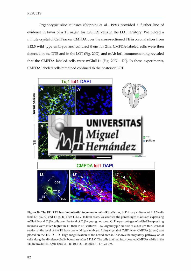
1. 5. The TE can produce lot cells ................................................................................... 81
1. 6. The thalamic eminence expresses genes associated with glutamatergic neurogenesis ........................................................................................................................... 83
1. 7. mGluR1 cells are generated in the lateral TE........................................................ 84
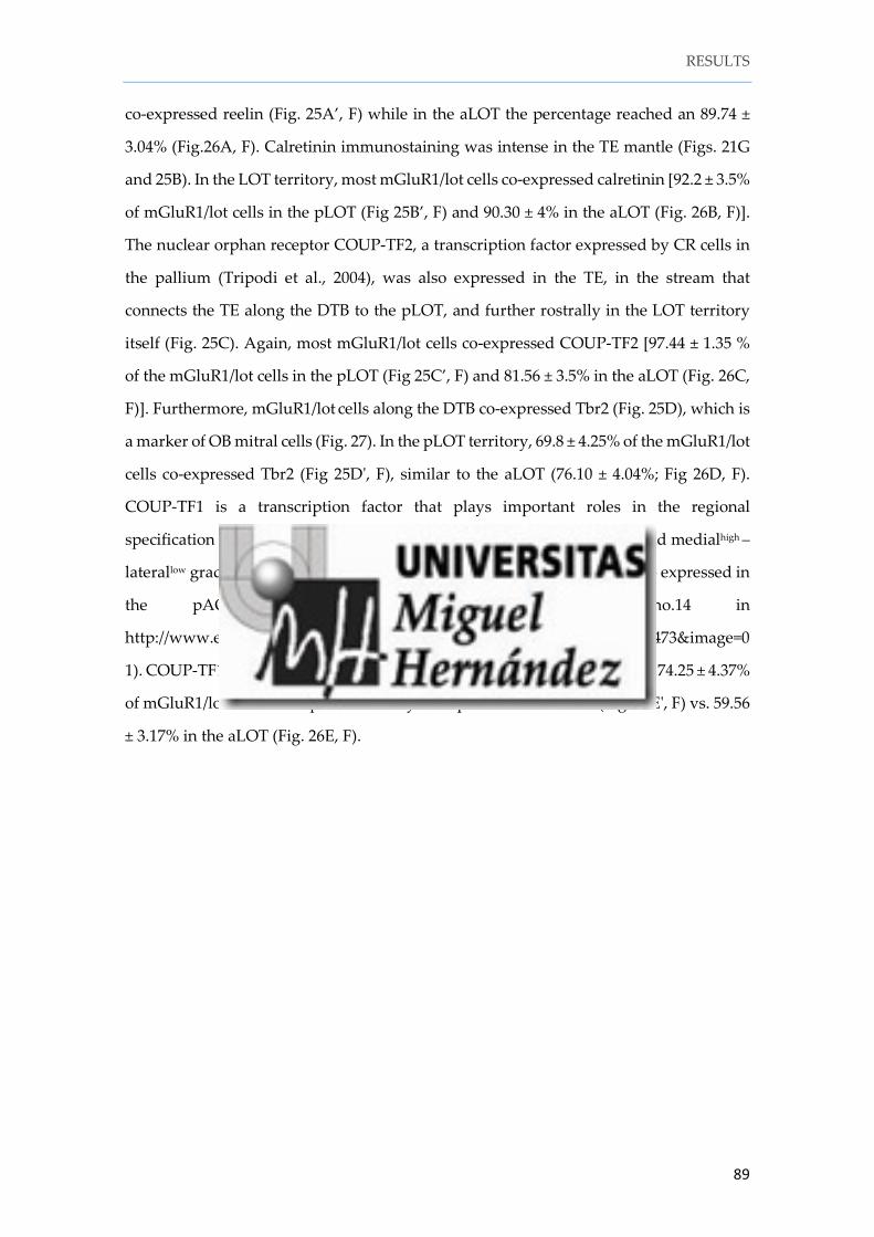
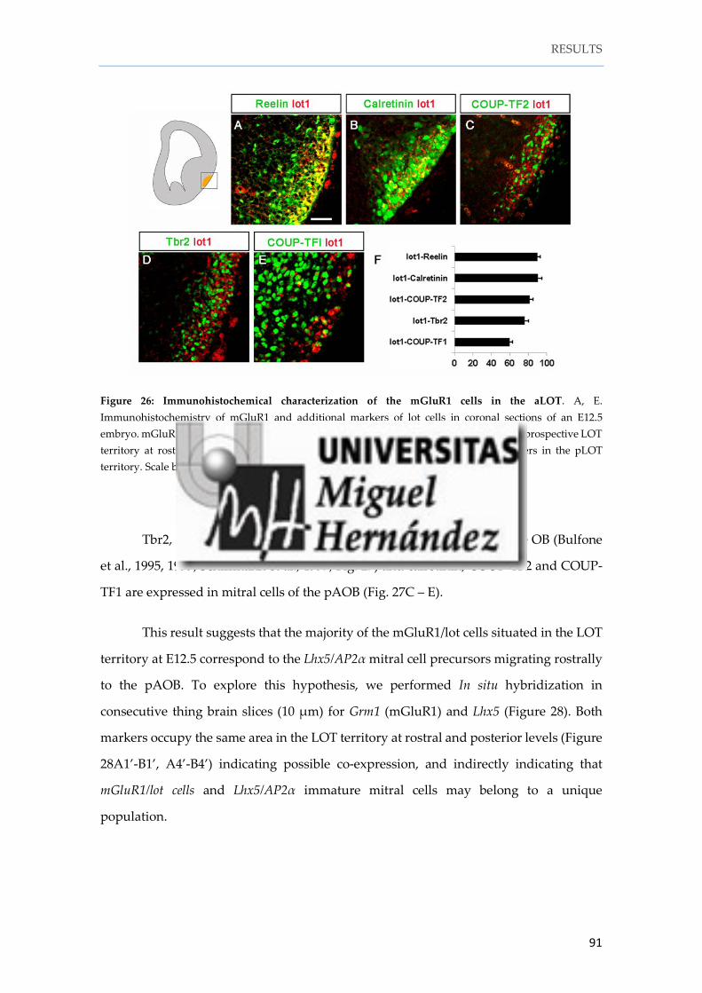
1. 8. mGluR1/lot cells share molecular features with pAOB mitral cells .................. 88
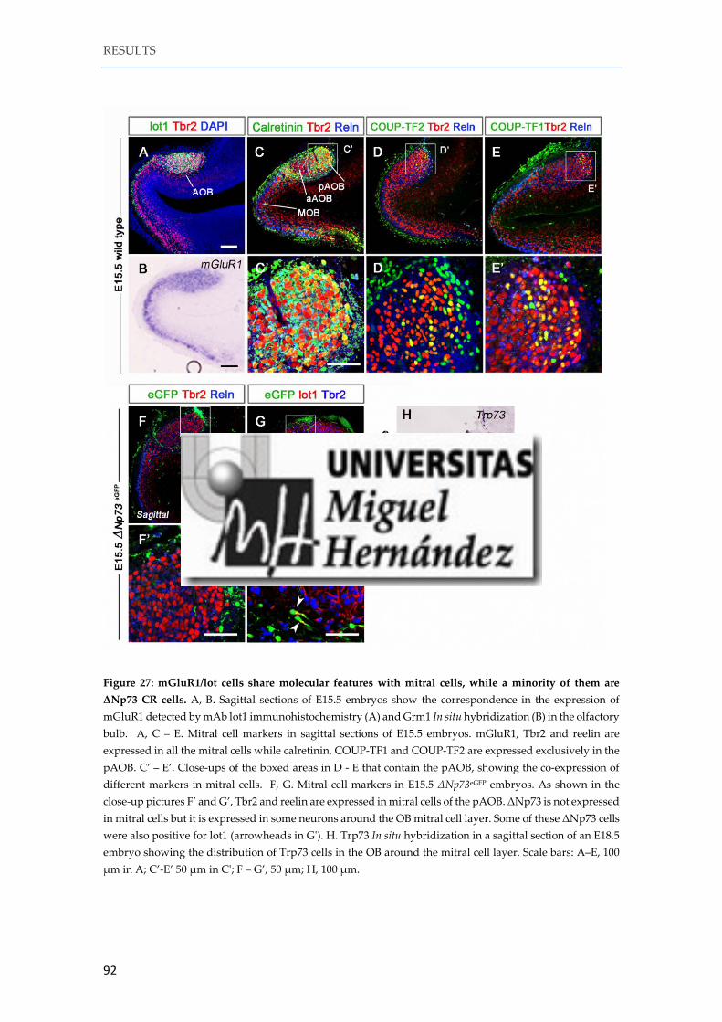
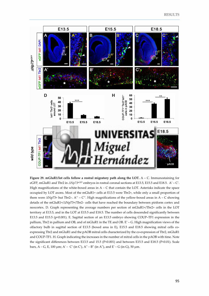
1. 9. mGluR1/lot cells follow a rostral migratory path to populate the pAOB ........ 93
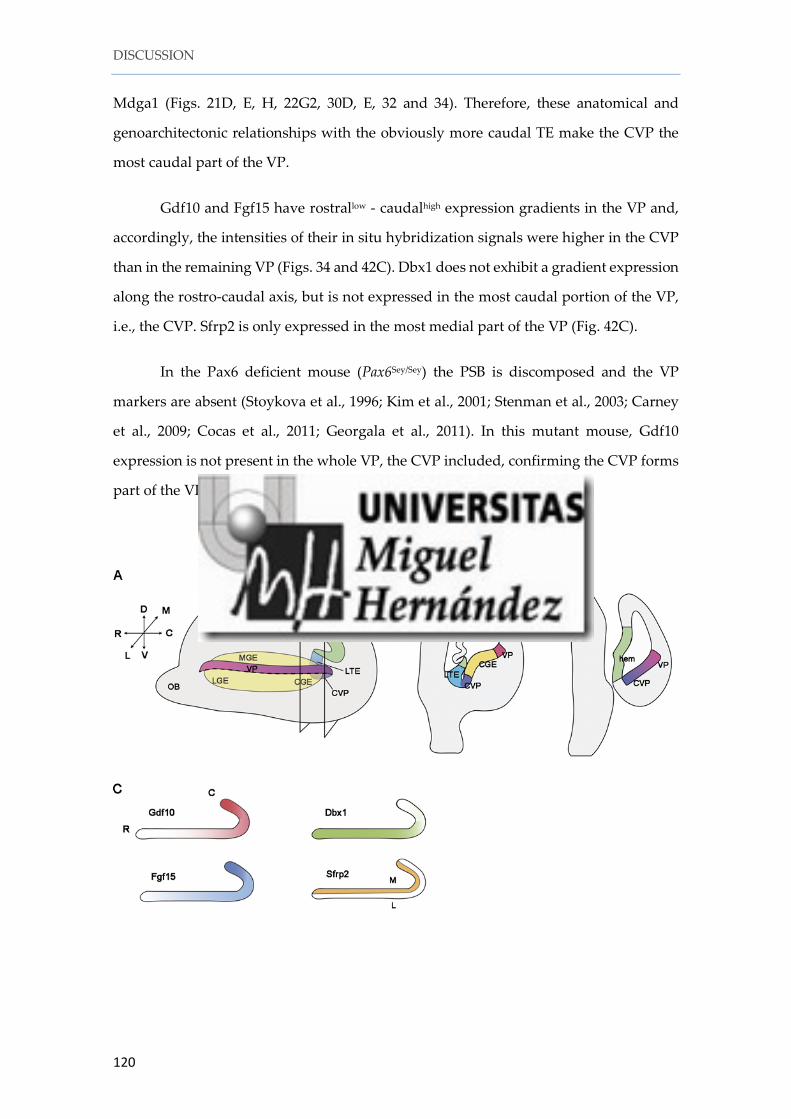
SECTION 2. The caudoventral pallium (CVP) as a putative origin of amygdaloid glutamatergic neurons ..................................................................................................... 97
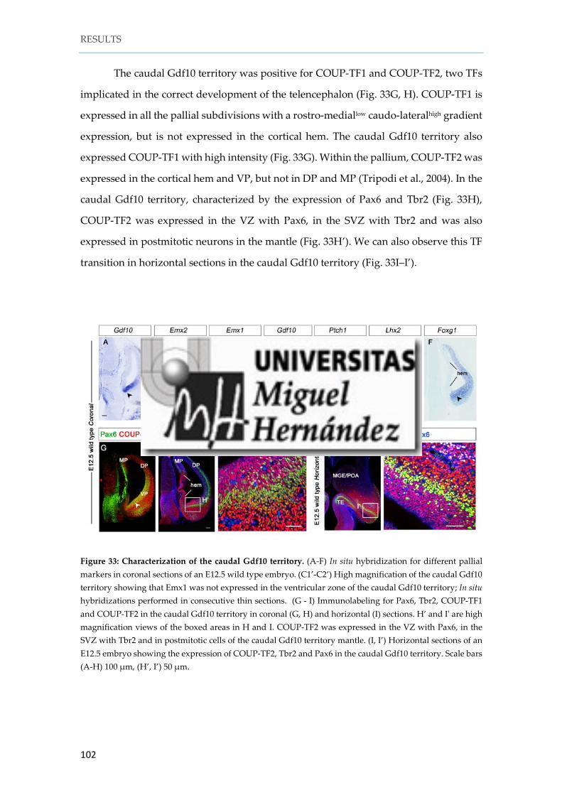
2. 1. Expression of Gdf10 in the forebrain ..................................................................... 97
2. 2. The caudal Gdf10 territory: a pallial or a subpallial region? .............................. 98
2. 3. Characterization of the caudal Gdf10 territory .................................................. 101
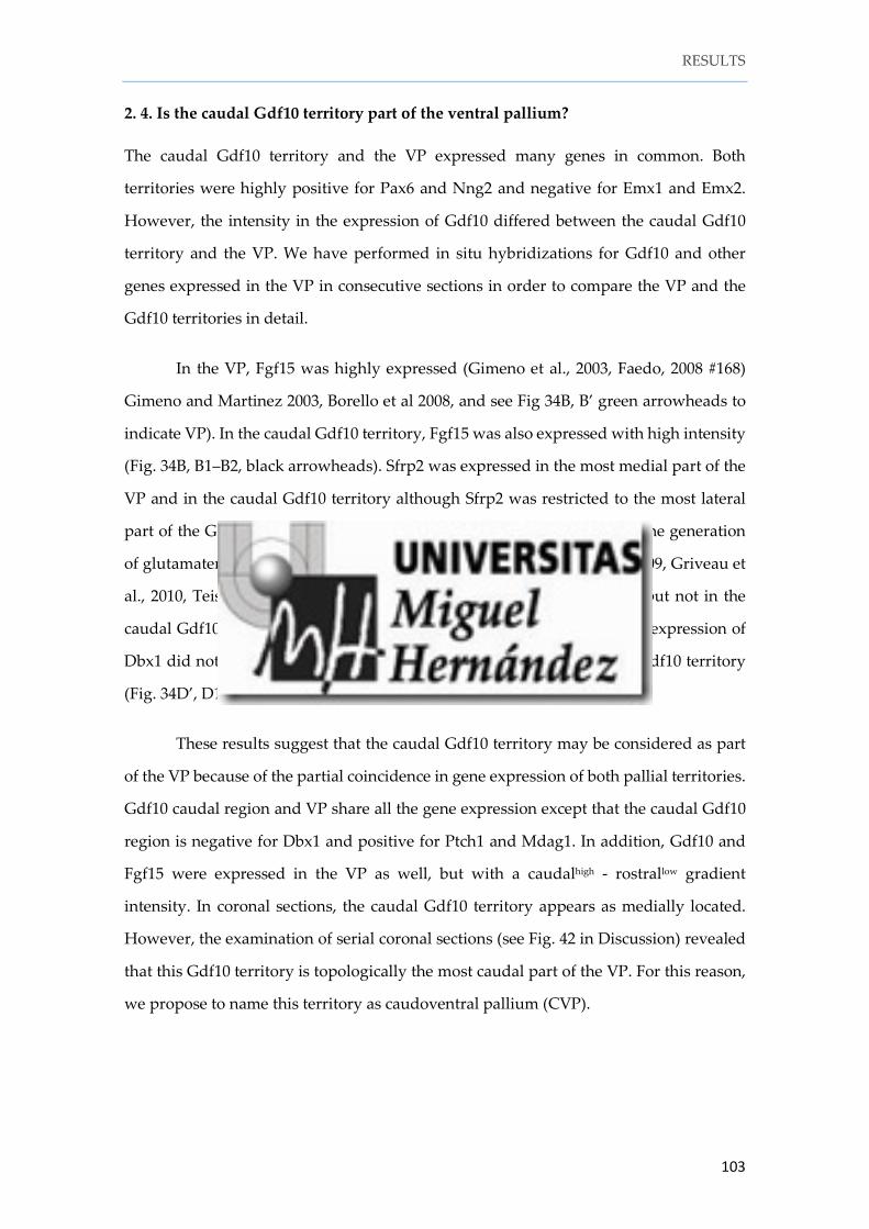
2. 4. Is the caudal Gdf10 territory part of the ventral pallium? ................................ 103
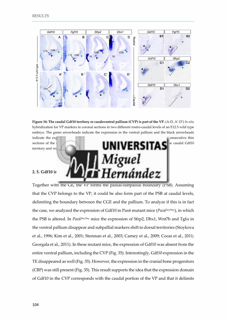
2. 5. Gdf10 in the CVP corresponds with the caudal portion of the VP .................. 104
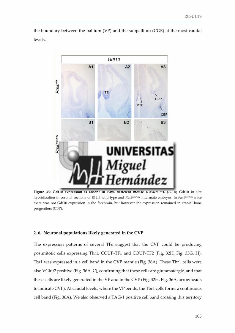
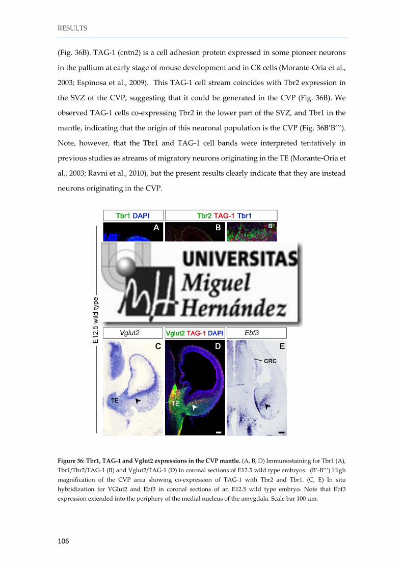
2. 6. Neuronal populations likely generated in the CVP.......................................... 105
2. 7. A number of Me nucleus cells may originate in the CVP ................................. 107
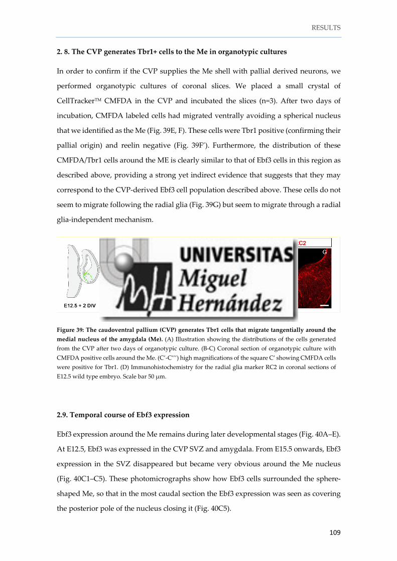
2. 8. The CVP generates Tbr1+ cells to the Me in organotypic cultures .................. 109
2. 9. Temporal course of Ebf3 expression .................................................................... 109
SECTION 1. The lateral thalamic eminence is the main origin of lot cells .................. 113
Which is the source of mGluR1/lot cells: the pallium or the thalamic eminence? ............ 113
mGluR1/lot cells: prospective mitral cells or Cajal-Retzius cells? .................................... 116
SECTION 2. The caudoventral pallium (CVP) as a putative origin of amygdaloid glutamatergic neurons ................................................................................................... 119
CVP contributions to the medial amygdala ....................................................................... 121
Role of COUP-TF2 in amygdala formation. ..................................................................... 122
During embryonic development, the brain organizers secrete signaling molecules that
are necessary for the correct patterning of the different subdivisions of the nervous
system. In the case of the telencephalon, the organizers secrete morphogens such as bone
morphogenetic proteins (BMPs), fibroblast growth factors (FGFs), and Wnt proteins. In
particular, growth differentiation factor Gdf10 (Bmp-3b) is a morphogen expressed in
two discrete regions of the forebrain: the thalamic eminence (TE) and the caudoventral
pallium (CVP). The TE and the CVP share the expression of many other morphogens
besides Gdf10 and generate glutamatergic neurons. These two regions localize at the
caudal end of the pallium, forming the limit between diencephalon and telencephalon.
Taken together, these properties make them putative forebrain signaling centers. In this
thesis, we perform a genetic and anatomical characterization of the TE and the CVP,
introduce a new model of the diencephalic-telencephalic boundary, and describe for the
first time certain neural populations generated from the TE and the CVP.
This thesis has been divided into two sections:
SECTION 1: The lateral thalamic eminence is the main origin of lot cells
The thalamic eminence (TE) is a transitory structure localized in the diencephalic
prosomere 3. We show here that the TE is integrated by two subregions, the lateral TE
(LTE) and the medial TE (MTE). The LTE, localized next to the cortical hem, is
continuous with this structure and it is genetically related to the telencephalon. We
demonstrate here that the LTE is a source of mGluR1+ cells (also called lot cells) that
migrate through the lateral olfactory tract (LOT) territory before the arrival of mitral cell
axons. Our results also reveal that the mGluR1/lot cells are not a unique population.
Instead, they are a mixture of at least two different set of neurons: (i) mitral cell
precursors that will populate the posterior accessory olfactory bulb (pAOB) and, (ii) a
small population of p73+ mGluR1+ Cajal-Retzius cells localized around the LOT axons
that spread over the piriform cortex.
ABSTRACT
12
SECTION 2: The caudoventral pallium (CVP) as a putative origin of amygdaloid
glutamatergic neurons.
The CVP is a ventral pallial (VP) territory situated at the caudalmost part of the VP and
anatomically continuous to the thalamic eminence (TE). As the VP, the CVP territory
expresses markers such as Gdf10, Sfrp2 and Fgf15 but is negative for Dbx1, a TF
implicated in glutamatergic neurogenesis in the ventral pallium. The CVP generates
glutamatergic neurons that express Ebf3 during embryonic stages and will populate the
shell of the medial nucleus of the amygdala.
RESUMEN
13
Durante el desarrollo embrionario, los organizadores cerebrales secretan moléculas
señales necesarias para el correcto modelaje de las diferentes subdivisiones del sistema
nervioso. En el caso del telencéfalo, los organizadores secretan morfógenos tales como
proteínas morfogenéticas del hueso (bone morphogenetic proteins; BMP), factores de
crecimiento de fibroblastos (fibroblast growth factors; FGF) y proteínas Wnt. En
particular, el factor de diferenciación de crecimiento Gdf10 (Bmp-3b) es un morfógeno
expresado en dos regiones discretas del prosencéfalo, la eminencia talámica (ET) y el
palio caudoventral. La ET y el palio caudoventral comparten la expresión de muchos
otros morfógenos a parte de Gdf10 y ambos territorios generan neuronas
glutamatérgicas. Estas dos regiones se localizan en la parte más caudal del palio,
formando el límite entre diencéfalo y telencéfalo. Estas propiedades le hacen ser centros
de señalización putativos para el prosencéfalo. En esta tesis, realizamos una
caracterización genética y anatómica de la ET y del palio caudoventral, introducimos un
nuevo modelo del límite diencefálico-telencefálico y describimos por primera vez las
poblaciones neuronales generadas por la ET y el palio caudoventral.
Esta tesis ha sido dividida en dos secciones:
SECTION 1: La eminencia talámica lateral es el origen principal de las células lot
La eminencia talámica (ET) es una estructura transitoria localizada en el prosómero
diencefálico 3. Aquí mostramos que la ET está integrada en dos subregiones, la ET lateral
y la medial. La ET lateral, se localiza seguida del hem cortical, forma un continuo con
esta estructura y es genéticamente relacionada con el telencéfalo. En esta tesis
demostraremos que la ET lateral es una fuente de células mGluR1 positivas (también
llamadas células lot) que migran a través del territorio del tracto olfativo lateral antes de
la llegada de los axones de las células mitrales. Nuestros resultados también revelan que
las células mGluR1/lot no son una única población. En su lugar se trata de una mezcla
de al menos dos conjuntos de neuronas diferentes. (i) Las precursoras de las células
mitrales que poblarán el bulbo olfativo accesorio posterior y, (ii) una pequeña población
de células de Cajal-Retzius positivas para mGluR1+ y p73+ que se localizan alrededor de
los axones del tracto olfativo lateral y esparcidas por la corteza piriforme.
RESUMEN
14
SECTION 2: El palio caudoventral como origen putativo de neuronas glutamatergicas
amigdalinas.
The palio caudoventral es un territorio palial situado en la parte más caudal del palio
ventral y que se continua anatómicamente con la eminencia talámica. Al igual que el
palio ventral, el territorio del palio caudoventral expresa marcadores tales como Gdf10,
Sfrp2 y Fgf15, pero es negativo para Dbx1, un factor de transcripción implicado en
neurogénesis glutamatérgica en el palio ventral. El palio caudoventral genera células
glutamatérgicas que expresan Ebf3 durante estadios embrionarios y que poblarán el
revestimiento del núcleo medial de la amígdala.
INTRODUCTION
INTRODUCTION
17
The nervous system develops from a simple neural tube to a highly sophisticated
network system. This is possible due to critical events that occur during development
ranging from neural induction, proliferation, differentiation, migration and neuronal
connectivity. One of the first critical events during embryogenesis is gastrulation.
During this period, the polarity of the embryo is established (anterior versus posterior,
medial versus lateral) and part of the ectoderm differentiates to form the neural plate
(neuroectoderm) thanks to action of the notochord. The signaling molecules implicated
in neural induction include bone morphogenetic proteins (BMPs), fibroblast growth
factors (FGFs), vertebrate orthologs of Drosophila wingless (Wnts), retinoic acid and
Sonic hedgehog (Shh), among others. During neural induction, the neural plate closes
itself forming the neural tube in a process called neurulation. The rostral part of the
neural tube will differentiate into prosencephalon, mesencephalon and
rhombencephalon while the caudal region will form the spinal cord. At this time,
progenitor cells, localized in the ventricular zone (VZ) of the neuroepithelium divide
symmetrically and asymmetrically to increase the pool of progenitors and neurons in
order to generate neural diversity. Then, newborn neurons undergo a migratory process
to reach their final positions in the prospective brain. Newly differentiated neurons then
follow two migratory strategies to disperse in the brain, radial or tangential migration.
Neurons migrating radially move perpendicular to the ventricular surface and they use
the radial glia as scaffold. In contrast, neurons migrating tangentially progress
orthogonal to the radial glia palisade. During the development of the cerebral cortex
both types of migration are combined. Pyramidal neurons are born in the VZ of the
pallium to then migrate radially to the cortical plate. In contrast, Cajal-Retzius cells and
cortical interneurons migrate tangentially long distances from their neurogenic places in
the telencephalon to their final positions in the cortex.
After the migration process neurons start to establish neural connectivity. Some
neurons form local circuits while some others will connect with structures that are
spatially far away, and therefore, they have to extend their axons long distances to reach
their final targets. One of the neuronal populations that need to extend very long axons
to reach the final targets are, for instance, the mitral cells of the olfactory bulb. These
neurons extend their axons to connect with the piriform cortex and the amygdala
INTRODUCTION
18
forming what is known as the lateral olfactory tract (LOT). However, many of the neural
connections established during development finally disappear in a process called
pruning, which is an important mechanism that shapes the final connectivity of the
brain.
1. The development of the prosencephalon
One of the most popular models to explain how morphogenesis and patterning takes
place during development is the so-called prosomeric model, as opposed to the
traditional columnar model by Herrick and Kuhlenbeck and actualized by Swanson. The
columnar model defends that the developing brain is subdivided into longitudinal
columns that represent histogenic compartments (Herrick, 1910; Kuhlenbeck, 1973;
Alvarez-Bolado et al., 1995; Swanson, 2003). By contrast, the prosomeric model proposed
by Puelles and Rubenstein more than twenty years ago (Puelles and Rubenstein, 1993)
and updated in successive versions (Rubenstein et al., 1998; Puelles, 2001; Puelles and
Rubenstein, 2003; Puelles et al., 2004; Puelles, 2009; Puelles and Rubenstein, 2015), states
that the brain is formed by an uninterrupted series of transverse subunits along the
rostro-caudal axis of the neural tube, called neuromeres. The different neuromeres are
distinguished on the basis of gene expression patterns inside the neuromeres or in their
limits.
For example, the rhombomers, i.e., the neuromeres of the rhombencephalon, are
characterized by the differential expression of homeobox genes (Marin et al., 2008;
Chambers et al., 2009). Besides the transverse subdivisions, or neuromeres, the neural
tube is also divided along the dorsoventral axis into four different longitudinal
compartments: the dorsal midline (or roof plate), which is continuous with the alar plate,
then the basal plate and finally the most ventral one that is called the floor plate. Each
plate has several parallel longitudinal progenitor microzones with different molecular
specifications that will generate diverse neuronal populations.
INTRODUCTION
19
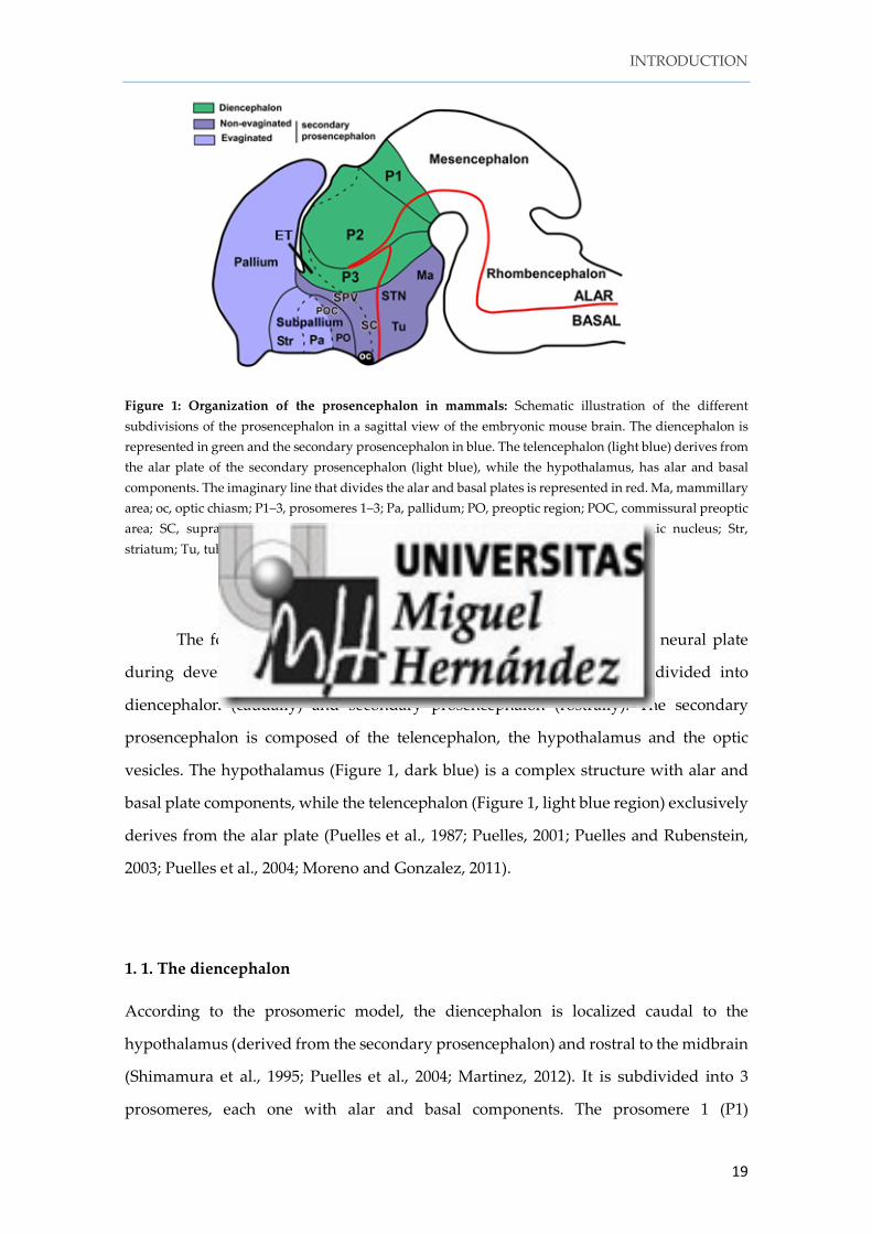
Figure 1: Organization of the prosencephalon in mammals: Schematic illustration of the different subdivisions of the prosencephalon in a sagittal view of the embryonic mouse brain. The diencephalon is represented in green and the secondary prosencephalon in blue. The telencephalon (light blue) derives from the alar plate of the secondary prosencephalon (light blue), while the hypothalamus, has alar and basal components. The imaginary line that divides the alar and basal plates is represented in red. Ma, mammillary area; oc, optic chiasm; P1–3, prosomeres 1–3; Pa, pallidum; PO, preoptic region; POC, commissural preoptic area; SC, suprachiasmatic area; SPV, supraoptic-paraventricular area; STN, subthalamic nucleus; Str, striatum; Tu, tuberal hypothalamus (Modified from Moreno and Gonzalez 2011).
The forebrain (or prosencephalon) is the most rostral part of the neural plate
during development and, according to the prosomeric model, is subdivided into
diencephalon (caudally) and secondary prosencephalon (rostrally). The secondary
prosencephalon is composed of the telencephalon, the hypothalamus and the optic
vesicles. The hypothalamus (Figure 1, dark blue) is a complex structure with alar and
basal plate components, while the telencephalon (Figure 1, light blue region) exclusively
derives from the alar plate (Puelles et al., 1987; Puelles, 2001; Puelles and Rubenstein,
2003; Puelles et al., 2004; Moreno and Gonzalez, 2011).
1. 1. The diencephalon
According to the prosomeric model, the diencephalon is localized caudal to the
hypothalamus (derived from the secondary prosencephalon) and rostral to the midbrain
(Shimamura et al., 1995; Puelles et al., 2004; Martinez, 2012). It is subdivided into 3
prosomeres, each one with alar and basal components. The prosomere 1 (P1)
INTRODUCTION
20
corresponds to the pretectum (PT) in the alar region. The alar domain of prosomere 2
(P2) is subdivided into epithalamus (ETh, habenula and pineal gland) and thalamus
(Th). The prosomere 3 (P3) is composed of the prethalamus (pTh) and the thalamic
eminence (TE)1.
1. 2. The thalamic eminence
In mammals, the thalamic eminence (TE) (also called eminentia thalami or prethalamic
eminence) is a transient structure that, according to the prosomeric model, localizes in
the most rostral part of the diencephalic P3 (Keyser, 1972; Abbott and Jacobowitz, 1999;
Puelles et al., 2000). The TE is continuous with the hypothalamus and the telencephalon
in its rostral aspect and with the prethalamus in its caudal part (Shimamura et al., 1995;
Puelles et al., 2004; Martinez, 2012). The most lateral part of the thalamic eminence
reaches the choroid plexus and this territory is considered the limit between the
diencephalon and the telencephalic vesicles (Puelles and Rubenstein, 2003). In coronal
sections, this lateral extension is localized ventral to the choroid plexus and its VZ faces
the lateral ventricle of the telencephalon. This territory was called strionuclear
neuroepithelium or amygdalar hem (Altman and Bayer, 1995; Siegenthaler and Miller,
2008; Meyer, 2010; Puelles, 2011). The medial TE (MTE), so called because it is localized
in a medial position in coronal sections, is continuous with the prethalamus and it shares
gene expression patterns with the remainder of the TE, especially growth differentiation
factor Gfd10 (Shimogori et al., 2010) but also T-box brain 1 and 2 (Tbr1 and Tbr2),
calretinin, and LIM homeobox 1, 5 and 9 (Lhx1, Lhx5 and Lhx9) (Abbott and Jacobowitz,
1999; Abellan et al., 2010; Roy et al., 2013; Roy et al., 2014).
1 We prefer the name "thalamic eminence" (or eminentia thalami) instead of prethalamic eminence since the latest term could generate confusion regarding a possible relation with the prethalamus. The term "prethalamic eminence" was proposed by Ted Jones (Hayes et al., 2003) with a totally different purpose: to reflect the observed fact that the thalamic eminence has no participation in thalamic development.
INTRODUCTION
21
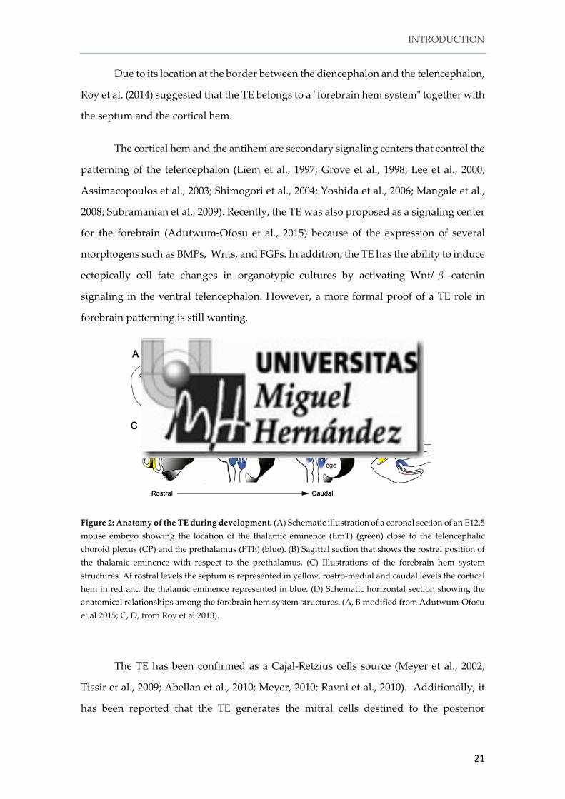
Due to its location at the border between the diencephalon and the telencephalon,
Roy et al. (2014) suggested that the TE belongs to a "forebrain hem system" together with
the septum and the cortical hem.
The cortical hem and the antihem are secondary signaling centers that control the
patterning of the telencephalon (Liem et al., 1997; Grove et al., 1998; Lee et al., 2000;
Assimacopoulos et al., 2003; Shimogori et al., 2004; Yoshida et al., 2006; Mangale et al.,
2008; Subramanian et al., 2009). Recently, the TE was also proposed as a signaling center
for the forebrain (Adutwum-Ofosu et al., 2015) because of the expression of several
morphogens such as BMPs, Wnts, and FGFs. In addition, the TE has the ability to induce
ectopically cell fate changes in organotypic cultures by activating Wnt/β -catenin
signaling in the ventral telencephalon. However, a more formal proof of a TE role in
forebrain patterning is still wanting.
Figure 2: Anatomy of the TE during development. (A) Schematic illustration of a coronal section of an E12.5 mouse embryo showing the location of the thalamic eminence (EmT) (green) close to the telencephalic choroid plexus (CP) and the prethalamus (PTh) (blue). (B) Sagittal section that shows the rostral position of the thalamic eminence with respect to the prethalamus. (C) Illustrations of the forebrain hem system structures. At rostral levels the septum is represented in yellow, rostro-medial and caudal levels the cortical hem in red and the thalamic eminence represented in blue. (D) Schematic horizontal section showing the anatomical relationships among the forebrain hem system structures. (A, B modified from Adutwum-Ofosu et al 2015; C, D, from Roy et al 2013).
The TE has been confirmed as a Cajal-Retzius cells source (Meyer et al., 2002;
Tissir et al., 2009; Abellan et al., 2010; Meyer, 2010; Ravni et al., 2010). Additionally, it
has been reported that the TE generates the mitral cells destined to the posterior
INTRODUCTION
22
accessory olfactory bulb (pAOB) that follow the lateral olfactory tract as a migratory path
(Huilgol et al., 2013). According to these authors, this migratory stream seems to be
evolutionary conserved since this phenomenon also occurs in Xenopus.
1. 3. The telencephalon
In the prosomeric model, the telencephalon is defined as a dorsal evagination of the alar
plate of the secondary prosencephalon (Fig. 1). The telencephalon is divided into dorsal
and ventral telencephalon. The dorsal telencephalon (or pallium) is the primordium of
the cerebral cortex, which is the brain structure responsible for the highest levels of
neural function, and the olfactory bulbs. The ventral telencephalon (or subpallium) gives
rise to the basal ganglia.
The pallium and the subpallium express different TFs implicated in neurogenesis
that will determine the phenotype of the cells generated in each compartment. The
pallium expresses TFs implicated in glutamatergic neurogenesis, while the subpallium
produces mainly GABAergic neurons (destined to the basal ganglia, but also to the
cerebral cortex and the olfactory bulb), cholinergic interneurons to the striatum and the
dopaminergic olfactory interneurons (Olsson et al., 1998; Corbin et al., 2000).
1.4. The pallium: the primordium of the cerebral cortex
The pallium, located in the dorsal part of the telencephalon, is the primordium of the
cerebral cortex and the pallial amygdala. The majority of the neurons in these structures
are glutamatergic projecting neurons. The mammalian cerebral cortex is commonly
subdivided into neocortex and allocortex. The neocortex (or isocortex) is integrated by
six different neuronal layers, each one with specific gene expression patterns and
connectivity. The allocortex includes structures with less than six layers, and comprise
the parahippocampal gyrus, the hipocampus, the olfactory bulbs, the piriform cortex,
the periamygdalar area and the entorhinal cortex.
INTRODUCTION
23
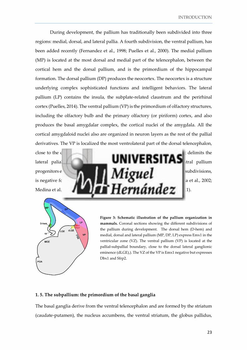
During development, the pallium has traditionally been subdivided into three
regions: medial, dorsal, and lateral pallia. A fourth subdivision, the ventral pallium, has
been added recently (Fernandez et al., 1998; Puelles et al., 2000). The medial pallium
(MP) is located at the most dorsal and medial part of the telencephalon, between the
cortical hem and the dorsal pallium, and is the primordium of the hippocampal
formation. The dorsal pallium (DP) produces the neocortex. The neocortex is a structure
underlying complex sophisticated functions and intelligent behaviors. The lateral
pallium (LP) contains the insula, the subplate-related claustrum and the perirhinal
cortex (Puelles, 2014). The ventral pallium (VP) is the primordium of olfactory structures,
including the olfactory bulb and the primary olfactory (or piriform) cortex, and also
produces the basal amygdalar complex, the cortical nuclei of the amygdala. All the
cortical amygdaloid nuclei also are organized in neuron layers as the rest of the pallial
derivatives. The VP is localized the most ventrolateral part of the dorsal telencephalon,
close to the dorsal portion of the lateral ganglionic eminence (dLGE) that delimits the
lateral palial-subpalial boundary (PSB) (Yun et al., 2001). The ventral pallium
progenitors express Dbx1 and Sfrp2 and, in contrast to the rest of the pallial subdivisions,
is negative for Emx1 in the ventricular zone (Fernandez et al., 1998; Hirata et al., 2002;
Medina et al., 2004; Carney et al., 2009; Cocas et al., 2009; Medina et al., 2011).
Figure 3: Schematic illustration of the pallium organization in mammals. Coronal sections showing the different subdivisions of the pallium during development. The dorsal hem (D-hem) and medial, dorsal and lateral pallium (MP, DP, LP) express Emx1 in the ventricular zone (VZ). The ventral pallium (VP) is located at the pallial-subpallial boundary, close to the dorsal lateral ganglionic eminence (dLGE),). The VZ of the VP is Emx1 negative but expresses Dbx1 and Sfrp2.
1. 5. The subpallium: the primordium of the basal ganglia
The basal ganglia derive from the ventral telencephalon and are formed by the striatum
(caudate-putamen), the nucleus accumbens, the ventral striatum, the globus pallidus,
INTRODUCTION
24
the ventral pallidus and the subpallial amygdala. The embryonic primordia of the basal
ganglia are the so-called ganglionic eminences (GEs), subdivided into lateral (LGE),
medial (MGE) and caudal (CGE) ganglionic eminences, and also includes the subpallial
septum and the preoptic area (POA). In the last decade, multiple subpallial domains
have been identified according to the combinatorial expression patterns of several TFs
and it has been proposed that each of these domains generates different types of
GABAergic neurons (Flames et al., 2007). The subpallium produces GABAergic
progenitors that will either generate interneurons migrating tangentially to the cerebral
cortex, olfactory bulb, pallial amygdala and hippocampus, or projection neurons of the
striatum, pallidus and subpallial amygdala. The principal neurons of the striatum are
the so-called medium spiny neurons, which derive exclusively from the LGE (Waclaw
et al., 2009). The CGE generates amygdalar GABAergic neurons (Nery et al., 2002), and
the MGE and the POA produce the globus pallidus projection neurons and the striatal
interneurons (Marin et al., 2000; Fragkouli et al., 2009; Nobrega-Pereira et al., 2010). The
cortical interneurons are generated mainly in the MGE (60%) but also in the CGE (30%)
and a small population in the POA (10%) (Marin and Rubenstein, 2001; Marin et al., 2002;
Wonders and Anderson, 2006; Gelman et al., 2009; Miyoshi et al., 2010). The olfactory
bulb interneurons are generated in the dorsal LGE (dLGE) during development and in
the subventricular zone (SVZ) in adult stages.
Figure 4: Radial and tangential migration of the telencephalic neurons: Schematic illustration of the origin and migration of the telencephalic neurons in coronal sections of E14.5 mouse embryos. Glutamatergic neurons (green) are generated in the pallium while GABAergic neurons (blue) are generated in the subpallium, the ganglionic eminences: (predominantly the MGE) and the anterior preoptic area, POa –formerly named AEP, anterior entopeduncular area. In addition, the POa generates acetylcholine neurons (red). (Modified from Rubenstein and Campbell, 2013).
INTRODUCTION
25
1. 6. The patterning of the telencephalon
1. 6. 1. Signaling centers
For the proper development of the initial neuroepithelium that will give rise to the
mature telencephalon, the expression of specific signals that guide its correct
development is essential (O'Leary and Sahara, 2008). These signals are called
morphogens and are secreted from the signaling centers localized either within the
telencephalon or surrounding it (Subramanian et al., 2009; Subramanian and Tole, 2009).
The morphogens are released from their sources in a diffusible manner, producing
concentration gradients. Neuroepithelial cells express the receptors for these
morphogens. Depending of the amount of signaling molecules the cells in the
neuroepithelium respond by expressing unique combinations of TFs. The differential
expression of TFs produces different kinds of progenitors that will generate specific
neuronal subtypes. The absence of morphogens, of their receptors or of the TFs directly
related to morphogens leads to defects in the specification of the different telencephalic
subdivisions.
There are four main signaling centers involved in telencephalic patterning which
are localized at the edges of the telencephalon:
- The commissural plate (CoP) or anterior neural ridge (ANR) is localized in the
rostral neural tube between the neural and the non-neural ectoderm. The CoP is
responsible for inducing the expression of Foxg1 in the telencephalic precursors. Foxg1
is a TF member of the forkhead family whose expression is turned on specifically in
telencephalic progenitors. Therefore, the expression of Foxg1 delimits the rostral
embryonic telencephalon (Shimamura and Rubenstein, 1997). The CoP expresses, at
least, five FGFs ligands (Fgf3, Fgf8, Fgf15, Fgf17, and Fgf18) that are important for the
rostralization of the telencephalon (Iwata and Hevner, 2009). A decrease in the
expression of Fgf8 and Fgf17 caudalizes the cortex (Garel et al., 2003; Storm et al., 2006;
Cholfin and Rubenstein, 2007, 2008), while the reduction of Fgf15 expression produces
the opposite phenotype (Borello et al., 2008). Some of the FGFs effects on the
regionalization of the cortex are mediated by the regulation of the expression of COUP-
INTRODUCTION
26
TF1 and Emx2, two TFs implicated in cortical arealization, in dorsal neural cells
(Hamasaki et al., 2004; Armentano et al., 2007). The absence of the FGF receptors, fgfr1,
fgfr2 and fgfr3 produces the loss of Foxg1 positive cells and therefore the lack of
differentiation of the entire telencephalon. However, the lack of two or only one of these
receptors leads to defects in telencephalon patterning but the structure is still present,
suggesting a functional compensation for each other (Gutin et al., 2006).
-The roof plate (RP) is located in the dorsal midline of the telencephalic vesicles,
whereas the cortical hem is in the dorsomedial edge of the pallium. The RP is a secondary
organizer for the development of the hippocampus and other caudomedial cortical area
(Liem et al., 1997; Grove et al., 1998; Lee et al., 2000; Assimacopoulos et al., 2003;
Shimogori et al., 2004; Yoshida et al., 2006; Mangale et al., 2008; Subramanian et al., 2009).
The hem secretes BMPs and Wnts signals, which affect pallium specification. The genetic
ablation of the cortical hem leads to the loss of the hippocampus (Lee et al., 2000; Yoshida
et al., 2006), while the transplantation of the hem in an ectopic position in the dorsal
telencephalon induces the formation of a hippocampus close to the hem patches
(Mangale et al., 2008; Subramanian and Tole, 2009).
BMPs and Wnts repress the expression of FGFs in the CoP. To maintain the
correct expression of FGF, the CoP expresses BMPs and Wnt inhibitors. The CoP
expresses Chordin and Noggin, two BMP inhibitors that are required to maintain the
correct level of expression of Fgf8 (O'Leary et al., 2007). The Wnt inhibitor Tlc, a secreted
frizzled-related Wnt antagonist, is expressed in the CoP and promotes Fgf8 expression,
which is necessary and sufficient to induce the telencephalon (Houart et al 2002).
-The antihem (which anatomically corresponds to the VP) is considered another
signaling center because it produces some morphogens such as Tgfα, Neuregulin1,
Neuregulin3, Fgf7, and the Wnt antagonist secreted frizzled related protein, Sfrp2
(Assimacopoulos et al., 2003; Subramanian et al., 2009). The antihem has been suggested
to cooperate with the cortical hem to stabilize the latero-medial axis of the brain.
Although the role of the antihem in telencephalon patterning and/or cortical arealization
has not yet been demonstrated, the defects in the patterning of the telencephalon
INTRODUCTION
27
observed in Pax6Sey/Sey mice may be due to the absence of the antihem in these animals
(Assimacopoulos et al., 2003).
-The floor plate, locates at the ventral aspect of the developing neural tube,
expresses Shh. Shh is considered a morphogen implicated in the dorsoventral patterning
that confers ventral identity. In Shh-/- mice, there is a reduction in the size of the
telencephalon and a loss of ventral markers such as Gsx2, Dlx2, Nkx2.1 and therefore the
ventral cell types are missing (Corbin et al., 2003). Shh has a function opposite to that of
Gli3 (a zinc-finger transcription factor), which is implicated in the dorsalization of the
telencephalon. In mice, two transmembrane proteins, patched (Ptc) and smoothened
(Smo), act as Shh receptors. In the absence of Shh, Ptch1 inhibits Smo function allowing
the proteolytic processing of Gli3 to the active form (Motoyama, 2006)
Figure 5: Signaling centers and graded expression of transcription factors drive telencephalon patterning. Representative illustration of the patterning centers in the telencephalon and these principal morphogens. The commissural plate (CoP), or anterior neural ridge (ANR), is localized in the most rostral end of the telencephalon and secretes FGF molecules. Wnts and BMPs proteins are expressed mainly in the cortical hem at the dorsal midline and Shh is expressed in the floor plate. The anti-hem in the ventral pallium is suggested to be an additional signaling center for the medial telencephalic areas (Modified from O’Leary and Sahara 2008).
Shh/Gli3 double mutant mice show the same phenotype of Gli3 null mice
extratoes (Gli3Xt/Xt) in the dorsal telencephalon. These mice lack dorsal midline
structures, including the hippocampus; the expression of Emx1 and Wnt8b in the
pallium is missing but there is ectopic expression of Fgf8 and Fgf15. Contrary to the
dorsal telencephalon, the organization of the ventral telencephalon is rescued, indicating
that other additional factors are implicated to generate ventral precursor cells (Rallu et
al., 2002; Rash and Grove, 2007). One such candidate is Fgfs signaling, which induces
ventral identity independently of Shh or downstream to the Shh pathway (Gutin et al.,
INTRODUCTION
28
2006). In the Fgf8 null mice there is a progressive loss of ventral progenitors and
associated structures (Storm et al., 2006).
1. 6. 2. Transcription factors responsible for the correct patterning of the telencephalon
The expression of different TFs in progenitor cells in the VZ of the telencephalic
neuroepithelium is the result of the action of morphogens released from the signaling
centers and it is essential for the correct subdivisions and regionalization of the
telencephalon.
A number of TFs have been reported to be essential for the patterning of the
telencephalon :
-Lhx2 and Lhx5. The LIM homeodomain proteins Lhx2 and Lhx5 seem to be
implicated in cortical hem development. Lhx2 is expressed in the entire telencephalic
VZ, excepting the cortical hem and the choroid plexus. Lhx2 represses BMP and Wnt
signaling from the cortical hem. In the Lhx2 deficient mice (Lhx2-/-), the hem, the ventral
pallium and the thalamic eminence, are expanded and, the dorsal pallium is absent
(Bulchand et al., 2001; Monuki et al., 2001; Vyas et al., 2003; Mangale et al., 2008;
Subramanian and Tole, 2009; Roy et al., 2013; Roy et al., 2014). Lhx5, opposite to Lhx2, is
expressed in the cortical hem. In Lhx5-/- mutant mice, the choroid plexus and the cortical
hem are missing and as a consequence the hippocampus does not develop (Zhao et al.,
1999b).
-Foxg1 is expressed in all the telencephalic progenitors except those in the cortical
hem. Foxg1 and Fgf8 promote the expression of each other forming a positive feedback
loop that promotes ventral development (Hebert and Fishell, 2008). Foxg1 is mainly
implicated in the establishment of the ventral identity, so that the ventral structures are
missing in the Foxg1 null mice (Foxg1-/-) (Xuan et al., 1995; Dou et al., 1999; Martynoga et
al., 2005). However, in these mice the pallium is also affected: it is reduced in size, the
cortical hem expands, the medial pallium is present but a normal hippocampus cannot
INTRODUCTION
29
develop, and the dorsal and ventral pallial markers are not present (Xuan et al., 1995;
Hanashima et al., 2004; Muzio and Mallamaci, 2005).
-Pax6 is a paired box domain TF whose expression pattern in dorsal pallium
progenitors (rostrolateralhigh caudomediallow gradient) is opposite to that of Emx2. Pax6
is implicated in cortical arealization (Warren et al., 1999; Bishop et al., 2000). Although
the Pax6sey/sey deficient mice die before birth, the analysis of the cortical areas at E18.5
reveal that Pax6 has a role in the patterning of rostral areas of the DP (Bishop et al., 2000;
Bishop et al., 2002; Li et al., 2006). Pax6 is also very important for the establishment of
the PSB. The PSB is delimited by the expression of Pax6 in the pallium and Gsh2 in the
subpallium, and their mutual antagonism is necessary for the positioning of the
boundary (Cocas et al., 2011). In Pax6sey/sey mice, the ventral pallium becomes dLGE and,
on the contrary, Gsh2-/- mouse the dLGE becomes ventral pallium (Corbin et al., 2000;
Stoykova et al., 2000; Toresson et al., 2000; Yun et al., 2001).
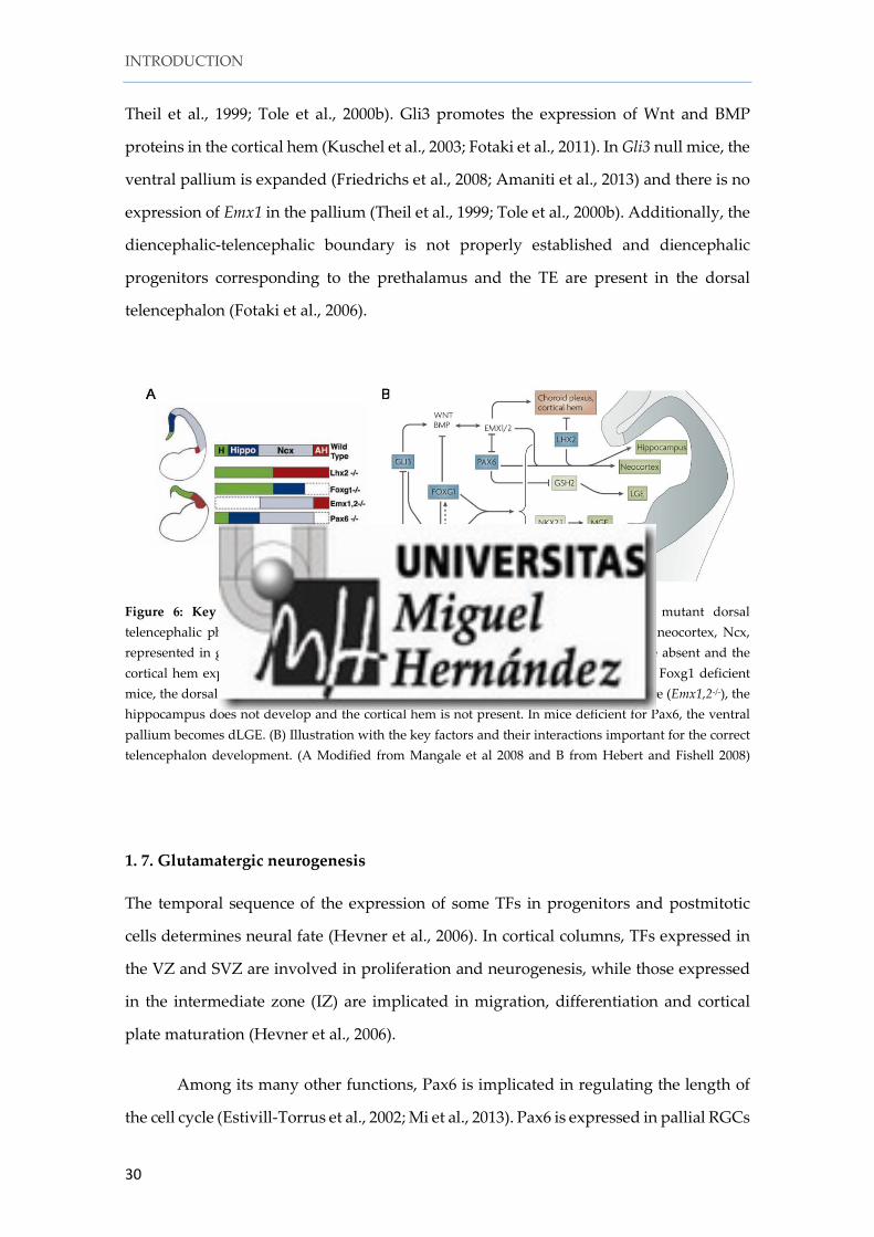
-Emx1/Emx2 are homeodomain TFs related to Drosophila empty spiracles (ems).
In the Gli3 deficient mouse extratoes (Gli3Xt/Xt), the expression of Emx1 is completely lost
while the expression of Emx2 is missing only partially (Theil et al., 1999; Tole et al.,
2000b). However, the role of Gli3 in promoting Emx2 expression is likely indirect
through the activation of BMPs and Wnts (Theil et al., 2002; Hasenpusch-Theil et al.,
2015). In the Emx2 null mice, dorsal progenitors acquire ventral phenotypes (Muzio et
al., 2002). Although both Emx1 and Emx2 are related and share a similar expression
gradient (caudomedialhigh - rostrolaterallow), only Emx2 is implicated in the specification
of the caudal cortical areas (Bishop et al., 2002; Muzio et al., 2002; Muzio and Mallamaci,
2003; Hamasaki et al., 2004; Kimura et al., 2005). However, Emx1 and Emx2 cooperate in
the initial phase of archipallium development. In mice double KO for Emx1 and Emx2,
the hem and the hippocampal formation are missing, but the rest of the pallium is
present (Fig 6, (Shinozaki et al., 2004)).
-Gli3: During forebrain development, Gli3 is strongly expressed in the dorsal
forebrain. In the Gli3 null mouse extratoes (Gli3Xt/Xt), which carries a spontaneous
deletion of Gli3 protein, the dorsal telencephalic midline does not evaginate and the
choroid plexus, the cortical hem and the hippocampus do not develop (Grove et al., 1998;
INTRODUCTION
30
Theil et al., 1999; Tole et al., 2000b). Gli3 promotes the expression of Wnt and BMP
proteins in the cortical hem (Kuschel et al., 2003; Fotaki et al., 2011). In Gli3 null mice, the
ventral pallium is expanded (Friedrichs et al., 2008; Amaniti et al., 2013) and there is no
expression of Emx1 in the pallium (Theil et al., 1999; Tole et al., 2000b). Additionally, the
diencephalic-telencephalic boundary is not properly established and diencephalic
progenitors corresponding to the prethalamus and the TE are present in the dorsal
telencephalon (Fotaki et al., 2006).
Figure 6: Key factors implicated in telencephalon patterning. (A) Schematics of mutant dorsal telencephalic phenotypes. In Lhx2 mutant mice the dorsal pallium (the primordium of neocortex, Ncx, represented in gray) and the medial pallium (the primordium of hippocampus, blue) are absent and the cortical hem expands (H, green color) and the ventral pallium (or antihem, AH, red). In Foxg1 deficient mice, the dorsal and ventral pallia are missing. In mice double KO for Emx1 and Emx2 mice (Emx1,2-/-), the hippocampus does not develop and the cortical hem is not present. In mice deficient for Pax6, the ventral pallium becomes dLGE. (B) Illustration with the key factors and their interactions important for the correct telencephalon development. (A Modified from Mangale et al 2008 and B from Hebert and Fishell 2008)
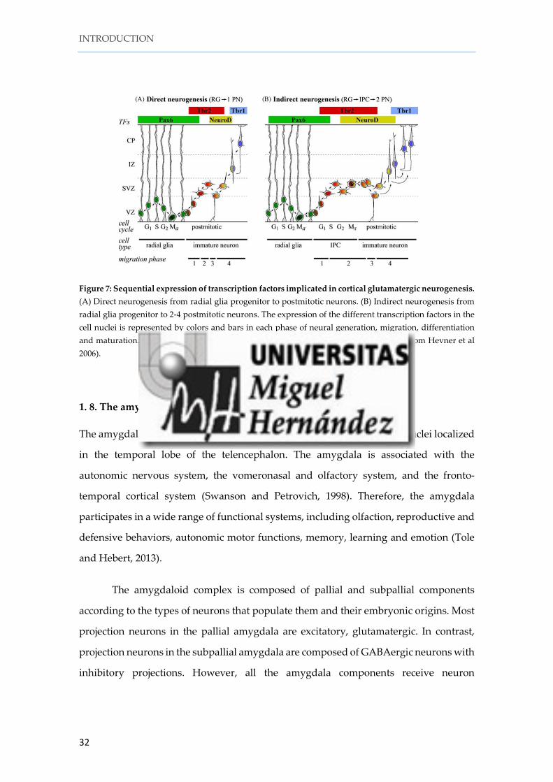
1. 7. Glutamatergic neurogenesis
The temporal sequence of the expression of some TFs in progenitors and postmitotic
cells determines neural fate (Hevner et al., 2006). In cortical columns, TFs expressed in
the VZ and SVZ are involved in proliferation and neurogenesis, while those expressed
in the intermediate zone (IZ) are implicated in migration, differentiation and cortical
plate maturation (Hevner et al., 2006).
Among its many other functions, Pax6 is implicated in regulating the length of
the cell cycle (Estivill-Torrus et al., 2002; Mi et al., 2013). Pax6 is expressed in pallial RGCs
INTRODUCTION
31
(Gotz et al., 1998; Englund et al., 2005) and to a minor extent, in the VZ of the dorsal
lateral ganglionic eminence (dLGE) (Georgala et al., 2011). Pax6 promotes the expression
of Ngn2 and represses Gsh2, and is required to specify pallial glutamatergic identities
(Kroll and O'Leary, 2005). In the absence of Pax6 there is a loss of dorsal fate, concomitant
with the acquisition of ventral telencephalic features (Quin et al 2007).
The basic-helix-loop-helix (bHLH) TF neurogenin2 (Ngn2) is expressed in pallial
progenitors and plays an important role in glutamatergic specification by repressing the
transcription factor Mash1 (Parras et al., 2002; Schuurmans and Guillemot, 2002; Mattar
et al., 2004). Neurogenin 1 (Ngn1) is also expressed in pallial progenitors. It promotes
the RGC identity and suppresses the differentiation of RGCs into astrocytes (Schmid et
al., 2003).
The T-box TF Tbr2 (Eomes) is expressed in intermediate progenitor cells (IPCs)
of the cortical SVZ and it is involved in the generation and maturation of the IPCs. A
recent paper revealed the 65.7% of the glutamatergic neurons in the cortex, including the
subplate, are generated from IPC progenitors (Vasistha et al., 2015).
NeuroD is a TF expressed in the upper part of the SVZ and lower intermediate
zone (IZ). NeuroD progenitors in the upper SVZ express Tbr2 as well (Hevner et al.,
2006). At early stages of development, the T-box TF Tbr1 is expressed in virtually all
postmitotic glutamatergic neurons in the pallium (Bulfone et al., 1995), in intermediate
zone (IZ), subplate, cortical plate (Layer 6) and marginal zone (MZ) (Englund et al.,
2005).
As shown by Hevner and his group, all these TFs are expressed sequentially to
generate glutamatergic neurons not only in the pallium but also in other regions of the
central nervous system such as the cerebellum and the adult hippocampus (Hevner et
al., 2006). Pax6 is first expressed in primary progenitors of the VZ. Tbr2 is then expressed
after Pax6 in intermediate progenitors in the SVZ. Some cells co-express both TFs in the
upper part of the VZ/lower SVZ suggesting a sequentially in the expression of these
transcription factors. Tbr2 and NeuroD are then expressed in late intermediate
progenitors, while early postmitotic cells express Tbr1 (Englund et al., 2005; Figure 7).
INTRODUCTION
32
Figure 7: Sequential expression of transcription factors implicated in cortical glutamatergic neurogenesis. (A) Direct neurogenesis from radial glia progenitor to postmitotic neurons. (B) Indirect neurogenesis from radial glia progenitor to 2-4 postmitotic neurons. The expression of the different transcription factors in the cell nuclei is represented by colors and bars in each phase of neural generation, migration, differentiation and maturation. G1, S, G2 M s are the cell cycle phases during neurogenesis (Modified from Hevner et al 2006).
1. 8. The amygdala
The amygdala (or amygdaloid complex) is a heterogeneous collection of nuclei localized
in the temporal lobe of the telencephalon. The amygdala is associated with the
autonomic nervous system, the vomeronasal and olfactory system, and the fronto-
temporal cortical system (Swanson and Petrovich, 1998). Therefore, the amygdala
participates in a wide range of functional systems, including olfaction, reproductive and
defensive behaviors, autonomic motor functions, memory, learning and emotion (Tole
and Hebert, 2013).
The amygdaloid complex is composed of pallial and subpallial components
according to the types of neurons that populate them and their embryonic origins. Most
projection neurons in the pallial amygdala are excitatory, glutamatergic. In contrast,
projection neurons in the subpallial amygdala are composed of GABAergic neurons with
inhibitory projections. However, all the amygdala components receive neuron
INTRODUCTION
33
subpopulations from pallial and or non-pallial subdivisions (Garcia-Moreno et al., 2010;
Bupesh et al., 2011; Medina et al., 2011).
1. 8. 1. The pallial amygdala
The pallial amygdala is composed of the basolateral complex and the cortical nuclei of
the amygdala. The ventral pallium (VP) is the major source of neurons contributing to
the basolateral complex, the anterior (ACo) and posteromedial cortical (PMCo) nuclei
and layer 1 of the nucleus of the lateral olfactory tract (nLOT1). The VP is characterized
by the ventricular expression of Dbx1 and Pax6 and by the lack of Emx1 expression in
the VZ. Tbr1, Ngn2, Lhx2, Lhx9 and Sema5A are expressed in the VP derivatives
(Medina et al., 2011).
The lateral pallium (LP) is negative for Dbx1 but expresses Emx1 in its VZ, and
has been proposed as a source of cells located in the basolateral amygdalar nucleus
(BLA) and the posterolateral cortical nucleus of the amygdala (PLCo). However, a recent
work casts doubts on the proposed LP origin of the BLA and PLCo, leaving the VP as
the only origin of neurons destined to the pallial amygdala (Puelles, 2014).
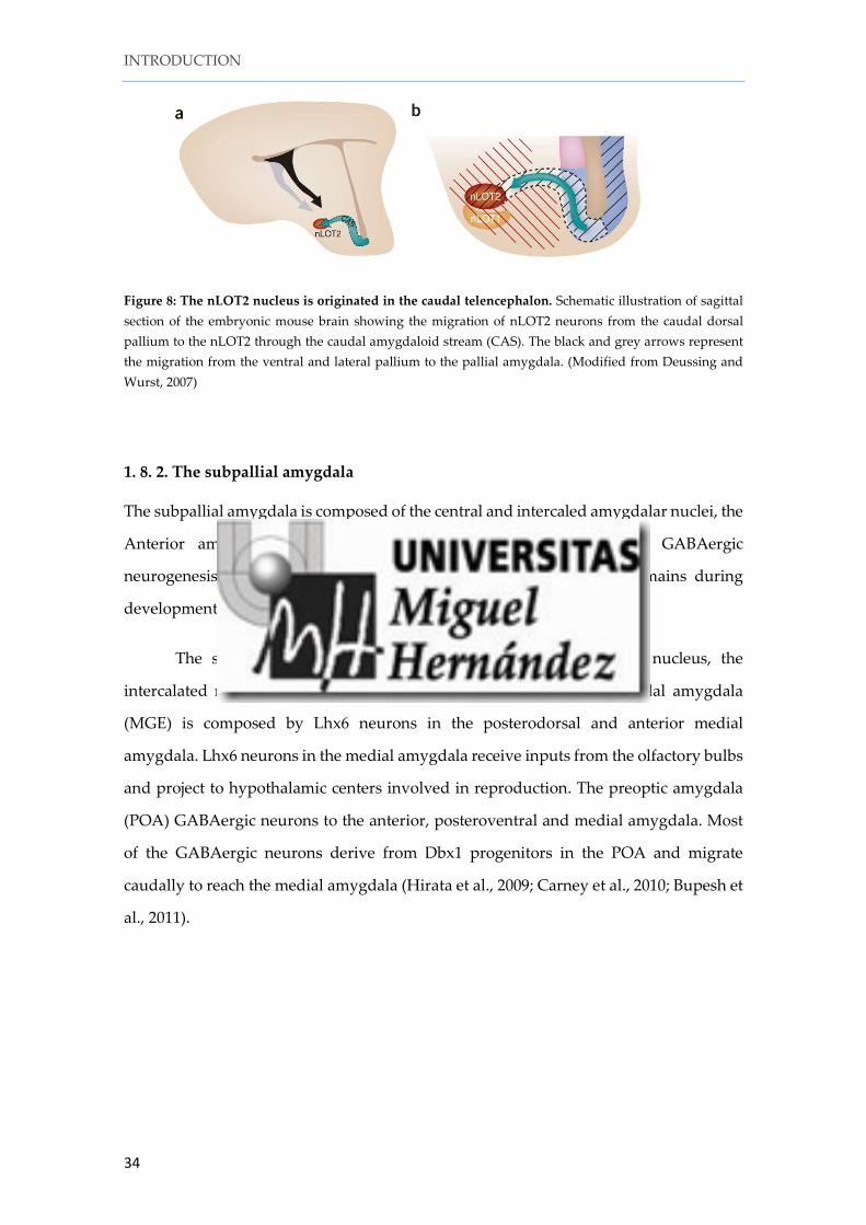
The dorsal pallium (DP) also generates neurons destined to the amygdala. In the
caudalmost pole of the telencephalic vesicle, the DP bends ventrally. This DP domain
generates Tbr1+ cells destined to layer 2 of the nucleus of the lateral olfactory tract
(nLOT2). nLOT2 neurons are generated in the VZ of this caudal region of the dorsal
pallium and then migrate rostrally through the caudal amygdaloid stream (CAS) to
finally accumulate in the nLOT2 (Remedios et al., 2007; Murillo et al., 2015).
The cortical amygdala and the LOT nucleus present laminar organization with
radially oriented pyramidal cells and receive inputs from the olfactory bulb and the
piriform cortex. In addition, the pallial amygdala also contains GABAergic interneurons
originated in the subpallium.
INTRODUCTION
34
Figure 8: The nLOT2 nucleus is originated in the caudal telencephalon. Schematic illustration of sagittal section of the embryonic mouse brain showing the migration of nLOT2 neurons from the caudal dorsal pallium to the nLOT2 through the caudal amygdaloid stream (CAS). The black and grey arrows represent the migration from the ventral and lateral pallium to the pallial amygdala. (Modified from Deussing and Wurst, 2007)
1. 8. 2. The subpallial amygdala
The subpallial amygdala is composed of the central and intercaled amygdalar nuclei, the
Anterior amygdala and the Medial amygdala. TFs implicated in GABAergic
neurogenesis, such as Mash1 and Dlx1/2/5 are expressed in these domains during
development.
The striatal amygdala is composed of the central amygdalar nucleus, the
intercalated nuclei and the dorsal part of the anterior amygdala. Pallidal amygdala
(MGE) is composed by Lhx6 neurons in the posterodorsal and anterior medial
amygdala. Lhx6 neurons in the medial amygdala receive inputs from the olfactory bulbs
and project to hypothalamic centers involved in reproduction. The preoptic amygdala
(POA) GABAergic neurons to the anterior, posteroventral and medial amygdala. Most
of the GABAergic neurons derive from Dbx1 progenitors in the POA and migrate
caudally to reach the medial amygdala (Hirata et al., 2009; Carney et al., 2010; Bupesh et
al., 2011).
INTRODUCTION
35
2. Migratory populations originated in the prosencephalon
2. 1. Cajal-Retzius cells
The Cajal-Retzius cells (CR cells) are the first neurons generated in the cortical
neuroepithelium. At early stages of mouse development (E11.5-E13.5) CR cells form part
of the preplate. From E13.5, when the preplate split, CR cells remain in the MZ, the
future layer I, covering the entire cortical surface. The first description of the CR cells
dates from 1890 by Santiago Ramon y Cajal. Studying new born small mammals (rodents
and lagomorpha) with the Golgi method, Cajal observed a novel neuron population
displaying ascending dendrites towards the pial surface and multiple axons that extend
horizontally for long distances. Because of this peculiar morphology, Cajal called them
special cells. Gustav Retzius (1893) observed similar cells in more mammalian species,
and called them Cajal cells.
CR cells are responsible of the correct lamination of the cerebral cortex via the
secretion of the extracellular matrix glycoprotein reelin (D'Arcangelo et al., 1995; Ogawa
et al., 1995). During cortical development, the pyramidal neurons, i.e., the projecting
neurons of the cortex, are generated from radial glia progenitors in the VZ of the pallium
and migrate radially to form the cortical plate (CP). Pyramidal cells become positioned
in the CP following an “inside-out” gradient, in which the youngest neurons accumulate
past the pre-existing ones (Angevine and Sidman, 1961; Berry and Rogers, 1965; Raedler
and Raedler, 1978; Rakic, 1988). Early in neocorticogenesis, the postmitotic cells migrate
independently of glia to form the subplate and layer VI (Nadarajah et al., 2001). Later
on, the cells migrate using the radial glia scaffold and then detach to move by somal
translocation to the top of the cortical plate (Borrell et al., 2006). In the “detach and go”
theory, reelin regulates both events: detachment and somal translocation. Reelin binds
to its receptors ApoER2 and VLDLR producing the phosphorylation of Dab1. Reelin
receptors are expressed in radial glia cells (Luque et al., 2003) and in the cortical plate
from E15.5 (in mouse) to postnatal stages (Perez-Garcia et al., 2004). Reelin controls the
radial migration through integrins but also controls somal translocation through the
signaling mediated by Dab1 and nectins and cadherins (Sanada et al., 2004; Franco et al.,
2011; Gil-Sanz et al., 2013).
INTRODUCTION
36
In reeler mice, (Relnrl-Alb2 or Relnrl-orl) or in the double KO mouse for reelin
receptors (VLDLR-/- ApoER2-/-), the preplate does not split into MZ and subplate, but
forms a superplate instead. The cortical plate neurons accumulate under the superplate,
in this case following an "outside-in" gradient thus producing an inverted pattern of
neuronal positioning. This happens not only in the cortex but in other laminated
structures as well, such as the hippocampus, the cerebellum and the olfactory bulb
(Tissir and Goffinet, 2003).
Figure 9: Schematic view of early cortical development in mice. At early stages (mouse: E12.5-E13.5) the preplate is composed of pioneer cells (pink) and CR cells (red). When the preplate splits, CR cells remain in the marginal zone and the pioneer cells form the subplate. At this stage, the reeler preplate does not split forming a superplate and the cortical plate (green) forms below the superplate. In wild type mice the cortical plate exhibit inside-out layering but in the reeler mice this gradient is lost. (Modified from Tissir and Goffinet 2003).
2.1.1. Origin and characteristics of Cajal-Retzius cells
CR cells are glutamatergic neurons that express the pallial marker Tbr1 (Hevner et al.,
2001; Hevner et al., 2003), the transcription factors COUP-TF2 (Tripodi et al., 2004), Lhx5
and Lhx1 (Yamazaki et al., 2004; Abellan et al., 2010). Calretinin was proposed as general
marker of CR cells (Alcantara et al., 1998) but recently has been demonstrated that only
some CR populations express it (Hevner et al., 2003; Bielle et al., 2005; Garcia-Moreno et
al., 2007). The 75% of CR cells die before the second week of postnatal mice life (del Rio
INTRODUCTION
37
et al., 1995; Soda et al., 2003), and less than the 3% remain in adulthood (Chowdhury et
al., 2010; Martinez-Galan et al., 2014). Although CR cells were initially thought to be
generated in the ventricular zone of the entire pallium (Marin-Padilla, 1998; Hevner et
al., 2003; Shen et al., 2006) nowadays there is consensus about the origin of CR cells at
discrete neurogenic sites located at the borders of the pallium (Bielle et al., 2005). The CR
cells of the neocortex are generated between E10.5 and E12.5 in mouse (Hevner et al.,
2003) and then migrate tangentially within the upper part of the preplate to cover the
entire cortical surface.
The principal CR cell sources are the cortical hem (Meyer et al., 2002; Takiguchi-
Hayashi et al., 2004; Yoshida et al., 2006; Zhao et al., 2006; Garcia-Moreno et al., 2007),
the prospective choroid plexus (Imayoshi et al., 2008), the ventral pallium (VP) at the
pallial-subpallial boundary (PSB) and the pallial septum (Bielle et al., 2005; Zimmer et
al., 2010; Griveau et al., 2010). Besides, the thalamic eminence has been postulated as an
additional source of CR cells (Meyer et al., 2002; Abellan et al., 2010; Tissir et al., 2009,
Ravni et al., 2010). Distinct programs of gene expression characterize the subpopulations
of CR cells defined according to their embryonic origins.
-The cortical hem derives from the telencephalic roof and it is a secondary
signaling center for the correct development of the hippocampus and telencephalon
(Shimogori et al., 2004). The cortical hem expresses Wnts and BMPs proteins (Grove et
al., 1998) and it is negative for Foxg1 and Lhx2 (Monuki et al., 2001; Hanashima et al.,
2004). Both transcription factors regulate the development of the cortical hem. Lack of
Foxg1 and Lhx2 leads to a bigger cortical hem and a massive production of CR cells
(Bulchand et al., 2001; Hanashima et al., 2004; Muzio and Mallamaci, 2005; Hanashima
et al., 2007; Roy et al., 2013). CR cells generated in the cortical hem derive from Wnt3a+
progenitors (Yoshida et al., 2006), express the antiapoptopic variant of tumor protein Δ
Np73 (Meyer et al., 2002, Tissir et al., 2009) and Zic1-3 (Inoue et al., 2008; Murillo et al.,
2015). Genetic ablation of Wnt3a progenitors produces a huge reduction of reelin cells in
the dorsal telencephalon that is not compensated for the other sources. Nevertheless,
surprisingly cortical lamination is not affected postnatally at rostral levels (Yoshida et
al., 2006).
INTRODUCTION
38
p73 is a transcription factor implicated in neural survival and apoptosis (Tissir et
al., 2009). p73 locus encodes two different isoforms (Tap73 and ΔNp73) depends of the
activation of different promoters and alternative polyadenylation sites. Tap73 is
expressed in the cortical hem but not in postmitotic CR cells (Meyer et al., 2004) and its
inactivation does not affect the development of CR cells. ΔNp73 is the N-terminally
truncated isoform of p73 and it is expressed in CR cells, and is implicated in their
survival. The lack of all p73 isoforms (Trp73), or the isoform ΔNp73 only, produces the
death of all the p73-derived CR cells and therefore a severe reduction of reelin cells in
the MZ (Meyer et al., 2004, Tissir et al., 2009). The cortical lamination is however not
affected probably because of a redundant production of reelin.
-The ventral pallium at the pallial-subpallial boundary is also called antihem
because it localizes in the opposite position of the cortical hem and expresses some
morphogens. For this reason, some authors consider the antihem as a secondary
signaling center (Assimacopoulos et al., 2003, Subramanian et al., 2009). Dbx1
progenitors from the ventral pallium give rise to glutamatergic cells. Some of these
glutamatergic cells are reelin and calretinin positive cells in the MZ and, therefore, are
considered CR cells. The VP CR cells are p73 negative and calretinin positive (Bielle et
al., 2005). The ablation of the glutamatergic neurons generated from Dbx1 progenitors
in the VP (Ngn2Cre-Dbx1DTA) produces reduction in the thickness of the lateral and
dorsal cortex (Teissier et al., 2010; Teissier et al., 2012).
-The pallial septum is localized in another signaling center, the commissural plate
(CoP), in the most rostral part of the telencephalon characterized for the expression by
FGF molecules (Cholfin and Rubenstein, 2007). Septal CR cells derive from Dbx1
progenitors and they are p73 positive but calretinin negative (Bielle et al., 2005). Er81, a
TF downstream of FGF signaling, is expressed exclusively in the rostral CR cells derived
from the pallial septum at early stages of development (E11.5). The ectopic expression of
Fgf8 in the rostral pallium induces the generation of Er81+ rostral CR cells (Zimmer et
al., 2010). The ablation of CR cells derived from Dbx1 progenitors in the septum by using
the animal Emx1Cre Dbx1DTA produces a reduction in the reelin cells in the rostro-
medial pallium at E11.5, but at E12.5 there is a compensation of CR cells derived from
INTRODUCTION
39
other sources. Postnatally, however, there are mild defects in the positioning of the
cortical areas suggesting that Cajal-Retzius cells have a modest role in cortical
arealization (Borello and Pierani, 2010; Griveau et al., 2010).
d) The Thalamic Eminence is localized in the diencephalic prosomere P3. It has
been proposed as a source of p73 CR cells to populate the ventral telencephalon (Meyer
et al., 2002; Tissir et al., 2009, Ravni et al., 2010, Abellan et al., 2010, Meyer 2010).
However, the characteristics and migratory pathways of TE-derived CR cells are still
undefined. The development of the TE depends on the TF Lhx2. In Lhx2 deficient mice,
the TE expands as well the remainder CR cell sources: cortical hem, septum and VP. The
TE is also a source of another population of reelin positive cells, the precursors of mitral
cells of the posterior accessory olfactory bulb (pAOB). In mice, these neurons are
generated early (E9.5) and then migrate from the TE to the olfactory bulb through the
lateral olfactory tract (LOT) territory in the piriform cortex. While migrating, these cells
express reelin but they are not Cajal-Retzius cells as they do not share common
molecular mechanisms involved in their migration (Huilgol et al., 2013).
2. 1. 2. Migration mechanisms of Cajal-Retzius cells
An initial pool of CR cells seeds the pallium MZ at the outset of corticogenesis. Later in
time, CR cells spread all over the pallial surface by migration mechanisms.
How CR cells become distributed over the entire cortical surface is not clear.
Some authors support the idea that each CR cells subpopulation occupies specific pallial
territories. So, the cortical hem-CR cells are localized in the most dorsal and caudal
sectors of the pallium, the VP-CR cells occupy the lateral pallium while the septal CR
cells populate the rostral pallial sectors (Griveau et al., 2010). Experiments in which one
of the CR cells subpopulation is ablated, or the velocity of migration of CR cells is
changed, show mild defects in cortical arealization suggesting a possible, but modest,
participation of CR cells in the establishment of the cortical areas during development
(Griveau et al., 2010; Barber et al., 2015; Barber and Pierani, 2015). However, this idea is
INTRODUCTION
40
contradicted in some way by the results published recently that demonstrate a random
distribution of CR cells over the entire pallial surface.
Figure 10: Illustration showing CR cell subtypes distribution at early stages of cortical development in mice. (A) The pallial septum (green) generates p73+ CR cells from Dbx1 progenitors that migrate to the rostro-medial cortex. The VP (orange) at the PSB, generates calretinin+ CR cells, from Dbx1 lineage, migrate to the lateral pallium. The cortical hem (blue) generates p73+, calretinin+ CR cells, from Wnt3a lineage, migrate from medial to lateral pallium. (B) Distribution in the cortical surface of the different CR cells subpopulations according with the random dispersion. (A modified from Griveau et al., 2013; B from Villar-Cervino et al., 2013).
A recent elegant report has proposed that CR cells tangentially invade the cortex
by contact-repulsion mediated by Eph/ephrin signaling (Villar-Cervino et al., 2013).
Other mechanisms have been also reported to be essential for the migration of CR cells,
such as the early B-cell factor Ebf2/Ebf3, the signaling mediated by Sema3a and its
receptor PlexinD1 (Chiara et al., 2012; Bribian et al., 2014), CXCL12/CXCR4 signaling
(Borrell and Marin, 2006) and the cannabinoid receptors CB1 and CB2 (Saez et al., 2014).
More recently, the transcription factor Zic2 has been implicated in the migration and
INTRODUCTION
41
dispersion of hem/septum CR cells (Murillo et al., 2015). In Zic2 hypomorphic mice, CR
cells migrate aberrantly forming clusters in the cortical surface and some of the CR cells
are misallocated in deep cortical layers. Zic2 mutant mice exhibit alterations in cortical
formation might due for the defects in CR cells migration and distribution (Inoue et al.,
2008, Murillo et al., 2015)
2. 2. The posterior accessory olfactory bulb (pAOB) mitral cells.
The olfactory circuit represents one of the oldest sensory modalities in the phylogenetic
history of mammals. Besides the detection of odorants, the olfactory system plays a
significant role in the regulation of behavioral responses such as sexual behavior, the
recognition of predators and the care of offspring (Halpern and Martinez-Marcos, 2003;
Ashwell, 2012). The olfactory system is the only sensory system lacking a relay station
in the thalamus (Kay and Sherman, 2007), and the information integrated by the
principal neurons in the olfactory bulb (OB) is conveyed directly to the higher olfactory
centers in the brain by axons that course along the lateral olfactory tract (LOT).
Mammals have two olfactory systems, the main olfactory system that processes
the responses to odorants, and the accessory or vomeronasal system that processes the
responses to pheromones.
-Odorant receptor neurons, localized in the chemosensory epithelium of the nasal
cavity, are responsible to receive odorant signals. These neurons send sensory
information to the main olfactory bulb (MOB) through the olfactory nerve. The olfactory
bulb (OB) in mice is a big rostral extension of the telencephalon integrated by
glutamatergic neurons, both mitral and tufted cells, and interneurons. Mitral cells extend
their axons through the lateral olfactory tract (LOT) to the major regions of the olfactory
cortex including the anterior olfactory nucleus (AON), the olfactory tubercle (OT), the
piriform cortex (PC) and the entorhinal cortex (EC) (Treloar et al., 2010). This projection
generates a complex odor-specific map in the olfactory cortex.
INTRODUCTION
42
-Pheromone receptor neurons are located in the vomeronasal organ (VNO).
There are two different classes of vomeronasal receptor neurons (V1R and V2R)
depending on their location in the VNO and on the type of G-protein that they express
(Tirindelli et al., 2009). These two families of receptor neurons send their axons to either
the anterior or the posterior portions of the accessory olfactory bulb (aAOB, pAOB)
(Martinez-Marcos, 2009). Mitral/tufted cells in the aAOB and pAOB extend their axons
through the LOT to the vomeronasal amygdala (Mucignat-Caretta, 2010). The pAOB is
a structure that resides in the dorso-caudal region of the OB and forms a parallel yet
independent pathway (Mohedano-Moriano et al., 2007) directly related to the neural
control of aggressive and defensive behaviors, just as the anterior tier of the AOB (aAOB)
serves sexual behaviors (Kumar et al., 1999).
Figure 11: The olfactory system in mice is formed by two different sub-systems; the main olfactory system and the accessory (or vomeronasal) olfactory system. The main olfactory system processes odorant information, which is conveyed from the olfactory neuroepithelium (MOE) to the main olfactory bulb (MOB) to the piriform cortex (PC), entorhinal cortex (EC), anterior olfactory nucleus (AON), olfactory tubercle (OT) and cortical amygdala (LA). The accessory olfactory system processes pheromone information from the vomeronasal nucleus (VNO), which is conveyed to the accessory olfactory bulb (AOB) and then to the vomeronasal amygdala (V. Amyg). (Modified from Dulac and Torello, 2003)
The specification of the OB depends on the expression of specific morphogens
and transcription factors (TF). The neuroepithelium of the OB is localized at the most
rostral part of the dorsal telencephalon and starts to be morphological distinguishable at
E12.5. FGF molecules are implicated in OB specification. In mice, the disruption of the
Fgf receptor Fgfr1 in the telencephalon of the conditional KO mouse line Foxg1Cre Fgfr1F/F
INTRODUCTION
43
produces an abnormal development of the OBs (Hebert et al., 2003). Another TF
implicated in OB development is the paired box protein Pax6. In Pax6 deficient mice
(Pax6Sey/Sey), an aberrant olfactory bulb-like structure (OBLS) is formed, which suggests a
role for Pax6 in the positioning of mitral cell progenitors (Jimenez et al., 2000; Nomura
and Osumi, 2004).
Additional mutant mice that exhibit an OBLS are the deficient mice for the
ciliopathy gene Ftm (Ftm-/- mice: Besse et al., 2011); the Gli3 deficient mice extratoes
(XtJ/XtJ; Franz, 1994); the conditional mice Emx1Cre-Gli3F/F (Amaniti et al., 2015); and the
Lhx2-/- mice (Saha et al., 2007). Lhx2 is expressed in mitral cells in the MOB and in the
anterior part of the AOB (aAOB). In Lhx2-/- mice, although mitral cells are specified, they
locate in an aberrant position forming an OBLS (Saha et al., 2007). The opposite scenario
is seen in the Tbr1 deficient mice, in which the mitral cells are absent although the
olfactory protrusion is present, indicating that OB morphogenesis and the specification
of mitral cells are independent events (Bulfone et al., 1998).
The lateral olfactory tract (LOT) contains the axons of the mitral and tufted
neurons from the OB projecting to the different parts of the telencephalon: olfactory
tubercle, olfactory and vomeronasal amygdala, and piriform and entorhinal cortex.
Mitral cell axons exit the olfactory bulb at E13.5, and at E15.5 a thick LOT is formed in
the piriform cortex. Around E16.5-E17.5, LOT collateral axonal branches invade the
olfactory areas to initiate synaptogenesis.
Mitral cell axons have a laminar organization in the LOT. The AOB projection
axons express neuropilin 2 (Nrp2) and are localized in the deepest layer of the LOT. The
MOB projection axons express Nrp1 and cntn2 (TAG-1) and extend within the
superficial layers of the LOT (Inaki et al., 2004). Nrp1 binds preferably to Sema3A, while
Nrp2 binds Sema3F. Although it was suggested Nrp1 and Nrp2 are implicated in the
LOT guidance (de Castro et al 1999) in the deficient mice for Nrp1 (Nrp1-/-), Nrp2 (Nrp2-
/-) or Sema3F (Sema3F-/-) not obvious defects in LOT axons guidance were detected (Ito et
al., 2008; Matsuda et al., 2010; Kitsukawa et al., 1997).
INTRODUCTION
44
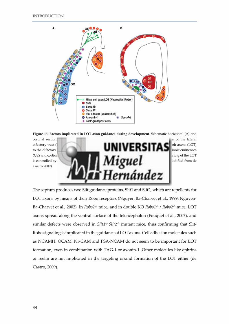
Figure 13: Factors implicated in LOT axon guidance during development. Schematic horizontal (A) and coronal section (B) showing the molecules and cells implicated in the correct formation of the lateral olfactory tract (LOT) (green). The mitral and tufted cells in the olfactory bulb (OB) send their axons (LOT) to the olfactory cortex (OC). Chemorepellents (red) are expressed in the septum (S), ganglionic eminences (GE) and cortical plate (CP) and provoke avoidance of LOT axons to those areas. The positioning of the LOT is controlled by lot cells (violet stars) and chemoattractants (blue) in the olfactory cortex (Modified from de Castro 2009).
The septum produces two Slit guidance proteins, Slit1 and Slit2, which are repellents for
LOT axons by means of their Robo receptors (Nguyen Ba-Charvet et al., 1999; Nguyen-
Ba-Charvet et al., 2002). In Robo2-/- mice, and in double KO Robo1-/- / Robo2-/- mice, LOT
axons spread along the ventral surface of the telencephalon (Fouquet et al., 2007), and
similar defects were observed in Slit1-/- Slit2-/- mutant mice, thus confirming that Slit-
Robo signaling is implicated in the guidance of LOT axons. Cell adhesion molecules such
as NCAMH, OCAM, Nr-CAM and PSA-NCAM do not seem to be important for LOT
formation, even in combination with TAG-1 or axonin-1. Other molecules like ephrins
or reelin are not implicated in the targeting or/and formation of the LOT either (de
Castro, 2009).
INTRODUCTION
45
2. 2. 1. Generation and migration of mitral cells
The OB derives from the ventral pallium and is considered as allocortex, with 4 neuron
(Price and Powell, 1970b, a; Pinching and Powell, 1971). The OB glutamatergic neurons
(mitral and tufted neurons) are generated in the local ventricular zone (VZ) and then
migrate radially toward the intermediate zone (IZ) where they differentiate. Mitral
neurons are the first being generated (E10.5-E11.5), followed by the tufted cells. The TFs
implicated in the generation of mitral/tufted cells in the OB are the same of the rest of
telencephalic glutamatergic neurons (Englund et al., 2005, Hevner et al., 2006). These TF
are Pax6 and Ngn2 in the primary progenitors, Tbr2 in intermediate progenitors and
Tbr1 in postmitotic cells (Campbell et al., 2011; Winpenny et al., 2011; Imamura and
Greer, 2013; Kahoud et al., 2014). However, unlike the cortical pyramidal cells, the
mitral/tufted cells express Tbr2 postmitotically (Bulfone et al., 1995; Bulfone et al., 1999;
Faedo et al., 2002; Mizuguchi et al., 2012), the extracellular matrix glycoprotein reelin
(Schiffmann et al., 1997), and the metabotropic glutamate receptor mGluR1 (Heinbockel
et al., 2004). Absence of either Tbr1 or Tbr2 in postmitotic mitral cell precursors causes
comparable defects in mitral cell development, indicating that both molecules are
necessary for the cells to progress toward a mitral/tufted cell phenotype (Bulfone et al.,
1998; Arnold et al., 2008; Sessa et al., 2008). Pax6 controls the expression of Tbr2 and Tbr1
in mitral cells (Imamura and Greer, 2013) and Tbr2, in intermediate progenitors in the
subventricular zone (SVZ) and in postmitotic mitral cells, are needed for the correct
development of the OB (Kahoud et al., 2014).
Although the majority of the mitral cells of the OB are born in the VZ of the
olfactory neuroepithelium, a recent work (Huilgol et al., 2013) revealed that mitral cells
of the pAOB have their origin in the thalamic eminence (TE). Accordingly, pAOB mitral
cells are generated early in the cortical development and migrate from the TE to the
pAOB through the prospective LOT territory, in the piriform cortex, between E11.5 and
E14.5. The precursors of the pAOB mitral cells express Lhx5, AP2α and reelin but,
according to these authors, they are not a subpopulation of CR cells as they do not share
the same migration mechanisms.
INTRODUCTION
46
Figure 12: A proposed TE origin of pAOB mitral cells in mice. Schemata showing the expression of Lhx5 in immature pAOB mitral cells during the time of migration. The pAOB mitral cells are generated in the TE and then migrate rostrally through the prospective LOT territory to the OB between E11.5 and E14.5. In the OB, Lhx5 is expressed exclusively in the pAOB) while Tbet is expressed in both the anterior accessory olfactory bulb (aAOB) and main olfactory bulb (MOB). (Modified from Huilgol et al 2013).
2. 2. 2. The lot cells
The lot cells are considered guidepost cells for mitral cell axons, since chemically
induced ablation of those cells prevents lateral olfactory tract formation in organotypic
cultures (Sato et al., 1998; Hirata et al., 2012). Lot cells supposedly derive from progenitor
cells in the DP (Sato et al., 1998; Tomioka et al., 2000; Kawasaki et al., 2006); for reviews
see (Marin et al., 2010; Bielle and Garel, 2011; Squarzoni et al., 2015). This DP origin was
supported by diverse sets of experiments in Tatsumi Hirata laboratory. Explant cultures
and tracing experiments in whole mount cultured embryos showed that lot cells are
generated in the entire dorsal pallium and then migrate ventrally and tangentially within
the pallial MZ towards the future LOT territory at the pallial-subpallial boundary (PSB).
Once they arrive to the LOT presumptive territory (before the arrival of LOT axons), lot
cells were supposed to turn 90º and migrate in a caudorostral direction within the LOT
territory and, later, at the periphery of the LOT axonal tract (Tomioka et al., 2000;
Kawasaki et al., 2006; Ito et al., 2008).
The lot cells were visualized for the first time using the monoclonal antibody (mAb) lot1
that recognizes the glutamate metabotropic receptor mGluR1 (Sato et al., 1998; Hirata et
al., 2012)
INTRODUCTION
47
mGluR1 expression, however, does not occur within the accepted migration
territory of lot cells within the pallial marginal zone but only once the lot cells arrive to
the LOT territory. Therefore, in the reports showing migrations of early neurons from
DP to the LOT territory (Sato et al., 1998; Tomioka et al., 2000; Kawasaki et al., 2006), the
nature of these ventrally migrating cells has remained obscure in the absence of specific
cell markers. Thus, it is unclear whether mGluR1/lot cells have a pallial origin as
previously suggested or, on the contrary, they are akin to the AP2α/Lhx5 presumptive
mitral cell destined for the posterior accessory olfactory bulb (pAOB) that arise in the TE
(Huilgol et al., 2013).
The position of the lot cells is controlled during development by multiple
guidance molecules. Lot cells express neuropilin 2 (Nrp2), the receptor for semaphorin
3F. Sema3F is expressed in the ganglionic eminences and provokes the avoidance of lot
cells to these territories. In Nrp2 or Sema3F deficient mice (Nrp2-/-, sema3F-/-) the
distribution of the lot cells is abnormal and their dorsal tangential migration is affected.
However, even if lot cells are in aberrant position, the formation of the LOT seems to be
normal (Ito et al., 2008), suggesting that this cells are not essential for LOT formation.
Netrin1 is expressed in the ganglionic eminence, while its receptor DCC is
expressed in lot cells. Netrin1-DCC signaling is implicated in the dorsal tangential
migration of the lot cells. In Netrin1 and DCC deficient mice the lot cells have mild
changes in the position in the LOT territory and the LOT axons have modest defects in
their trajectories (Kawasaki et al., 2006).
Figure 14: Illustration showing the migration and distribution of lot cells. The lot cells (pink color) migrate tangentially from the dorsal pallium to the prospective piriform cortex at the pallial subpallial boundary (PSPB). The lot cells are the guidepost cells to the correct lateral olfactory tract (LOT) formation. The LOT will project to the piriform cortex (Pir), entorhinal cortex (EC), anterior olfactory nucleus (AOB), olfactory tuberculum (Tu) and amygdala (Am). (Modified from (Bielle and Garel, 2013)).
48
HYPOTHESES AND OBJECTIVES
HYPOTHESES AND OBJECTIVES
51
During brain development, newborn, immature neurons migrate radially or/and
tangentially to reach their final destinations. Some neurons settle in places close to their
origins while some others migrate over long distances to reach their definitive locations
in the brain. In the telencephalon, Cajal-Retzius (CR) cells are generated from different
sources: the pallial septum at the rostralmost part of the telencephalic vesicle, the cortical
hem at the midline and the ventral pallium at the pallial-subpallial boundary. From these
sources, CR cells migrate tangentially to cover the entire surface of the cortex. The
thalamic eminence (TE), at the boundary between the diencephalon and the
telencephalon, has also been proposed as a source of p73 CR cells (Tissir et al., 2009), but
the characterization, migration pathways and final positioning of the TE-derived CR
cells are not well established yet.
On the other hand, Huilgol et al. (2013) have proposed that the TE is a source of
a novel population of Lhx5+ immature mitral cells that migrate along the lateral olfactory
tract (LOT) to occupy the posterior tier of the accessory olfactory bulb. These cells,
despite their expression of reelin, are not a CR cell subpopulation because they do not
share migration mechanisms with CR cells.
Additionally, an outstanding group of mGluR1+ guidepost cells for mitral cell
axons called lot cells populates the LOT during development. These cells are said to
generate in the dorsal pallium neuroepithelium (Tomioka et al., 2000). However, our
preliminary studies indicated a high level of mGluR1 expression in the TE and in a
putative migration path from the TE to the posterior end of the LOT, suggesting that the
TE could be involved in the generation of lot cells.
In our preliminary studies of gene expression in the TE and its surrounding
territories, we observed that the morphogen Gdf10 (Bmp-3b) is expressed not only in the
TE (Shimogori et al 2010) but also in the telencephalon. Thus, we found that Gdf10 was
expressed at a very low intensity in the ventral pallium, and at high intensity in a hitherto
undefined territory located between the TE and the caudal ganglionic eminence.
HYPOTHESES AND OBJECTIVES
52
On this basis, we decided to analyze in detail:
• The embryonic origins of mGluR1/lot cells and the migratory routes towards the
LOT.
• The neurochemical characteristics of lot cells.
• The final destinations of mGluR1/lot cells.
• The genoarchitectonic features of the novel Gdf10high telencephalic territory to
determine its pallial vs. subpallial character.
• The fate map of the neurons generated in the telencephalic Gdf10high territory.
MATERIAL AND METHODS
MATERIALS AND METHODS
55
Animals
Experiments were performed on different mouse strains. Mutant mice were maintained
in an inbred strain C57BL6 and wild type mice in outbred strain ICR at the animal
facilities of the Servicio de Experimentación Animal (RMG-SEA) in the Instituto de
Neurociencias de Alicante, Universidad Miguel Hernández-Consejo Superior de
Investigaciones Científicas (INA, UMH-CSIC)
The care and handling of the animals prior or during the experimental
procedures followed European Union (2010/63/UE) and current Spanish Legislation
(Real Decreto 53/2013), and were approved by the Animal Care and Use Committees of
Universidad Miguel Hernández. Animals were kept on a 12 h light/dark cycle with
constant ambient temperature and humidity. Food and water were available ad libitum.
List of mouse lines used in these thesis work:
-Grm1tm1Dgen/J mouse line (The Jackson laboratory B6.129P2-Grm1tm1Dgen/J) has
a targeted mutation in the Grm1 gene (glutamate metabotropic receptor 1, mGluR1). A
bacterial lacZ gene was inserted into the gene such that the endogenous gene promoter
drives expression of beta-galactosidase and don’t permit the correct expression of the
Grm1 gene. For the β-gal expression experiments we used heterozygous animals (Sachs
et al., 2007).
-DeltaNp73eGFP mouse line was generated by inserting an CRE-IRES-EGFP
cassette downstream from the 5’ end of the p73 locus, using standard homologous
recombination (Tissir et al., 2009). (Animal provided from F. Tissir, Institute of
Neuroscience, Université catholique de Louvain, Belgium)
-Dbx1Cre mouse line was obtained by inserting an IRES-CRE cassette into the
3’UTR of the Dbx1 gene by homologous recombination. Dbx1Cre mice were mated with
ROSA26YFP to obtain a permanent labeling of the Dbx1 cellular lineage (Bielle et al.,
2005) (Animal provided from A. Pierani, Institut Jacques Monod, Paris, France).
-ROSA26YFP mouse reporter line was generated by inserting EYFP cDNA into the
ROSA26 locus, preceded by a loxP-flanked stop sequence (Srinivas et al., 2001). Dbx1Cre
MATERIALS AND METHODS
56
animals were mated with ROSA26YFP to trace the cellular derivatives of Dbx1 to which
the Cre recombinase was attached. (Animal provided from A. Pierani, Institut Jacques
Monod, Paris, France).
-Wnt3aCre mouse line was generated by inserting an IRES-CRE cassette into the
3’ end of the Wnt3a locus, using standard homologous recombination (Yoshida et al.,
2006). (Animal provided from S. Garel, Institut de Biologie de l’École Normale
Supérieure, Paris, France)
-Ai9 td-Tomato mouse line was generated by inserting a construct containing the
strong and ubiquitous CAG promoter, followed by a floxed-Stop cassette controlled
fluorescent td-Tomato marker into the ROSA26 locus (Madisen et al., 2010). Wnt3aCre
animals were mated with Ai9 td-Tomato to trace the cellular derivatives of Wnt3a to
which the Cre recombinase was attached. (Animal provided from S. Garel, Institut de
Biologie de l’École Normale Supérieure, Paris, France).
-Lhx2-/- mouse line was generated using homologous recombination in
embryonic stem cells (ES cells). A replacement targeting vector, pEGneo, was designed
to delete the two exons encoding the LIM domains and the domain linking the LIM and
homeodomain domains of Lhx2, and replace them with a neomycin-resistance gene
(PGK-neo) (Porter et al., 1997). (Animals provided from S. Tole, TATA Institute,
Mumbai, India).
-Pax6Sey/Sey (Small eye). This mouse line carries a spontaneous semidominant
mutation which in homozygous condition results in the complete lack of eyes and nasal
primordia. The mutant mice have a base-pair change at codon 194 of the protein
producing an in frame stop codon resulting in a protein terminating before the
homeobox (Hill et al 1991). (Animals provided from D. Price, University of Edinburgh,
Scotland, UK).
MATERIALS AND METHODS
57
Embryo harvesting, dissection and fixation of the embryonic brains.
For staging of embryos, midday of the vaginal plug was considered as embryonic day
0.5 (E0.5). Timed-pregnant C57BL6/J and ICR females were killed by cervical dislocation
and the embryos were extracted by caesarean section. The uterine horns were cut and
placed on cold Krebs medium (if the embryos were destined to organotypic culture
procedures) L15 medium (the embryos used for primary cultures) or cold PBS for
immunostaining procedures in a Petri dish. The embryos were extracted from the uterus
removing all their coverings and their brains were rapidly and carefully dissected by
using two FST #5 forceps under a Leica scope. After dissection, the brains destined for
immunohistochemistry were fixed by immersion in 4% paraformaldehyde (PFA) in
0.2M phosphate buffer (PB) pH 7.4 with picric acid at room temperature (RT) with
agitation. The time depends of the embryo stage:
Fixation time
E12.5 2 hours
E13.5 3 hours
E15.5 6 hours
E18.5 overnight
After fixation, the brains were rinsed several times in phosphate saline buffer (PBS) and
stored in 0.05% sodium azide in PBS at 4ºC to preserve them of (from) fungal
contamination. Brains for In situ hybridization were fixed with PFA 4% in 0.1 M PBS
overnight at 4ºC with continuous agitation to avoid the degradation of RNA. After
fixation, the brains were rinsed in PBS. For cryostat sections the brains were transferred
to 30% sucrose in 0.1M PB. After sucrose incubation, the brains were embedded in OCT
compound and stored at -80ºC. For microtome sections the brains were transferred in
70º ethanol for one day and then processed to embed in paraffin.
MATERIALS AND METHODS
58
Perfusion and dissection of early postnatal mice
Early postnatal mice (P0-P5) were anesthetized by hypothermia and perfused
intracardially by means of a peristaltic pump (Watson Marlow 505S) at a flow rate of 6
ml/min (20 rpm) with 4% paraformaldehyde (PFA) in PBS. Perfusion was maintained
for 10 min using a total volume of 60 ml of fixative per animal. After perfusion, animals
were decapitated and the brains were dissected out. Brains were immersed in the same
fixative solution overnight at 4ºC. Finally, the brains were rinsed several times in PBS
and stored in PBS containing 0.05 % sodium azide at 4 ºC.
Immunofluorescence on 100-300 μm thick, free-floating sections
Immunohistochemistry was performed in multiwell plates. The sections were washed 3
times with Tris-buffered saline (TBS) 0.05M pH 7.4. In order to expose those specific
antigens that are hardly detectable, we first incubate the tissue with 0.1 M citrate buffer,
pH 6 during 5 minutes. Then, we boiled it in the microwave oven around 5 seconds and
shacked it until RT was reach. The sections were washed in TBS for 10 min, 3 times. After
unmasking treatment, the tissue was blocked in a solution composed of 10% normal goat
serum (NGS), 0.2% Triton X-100 in TBS for 1 hour and a half at room temperature (RT).
Next, sections were incubated with the primary antibodies diluted in a solution
composed of 4% bovine serum albumin (BSA), 3% normal horse serum (NHS) and 0.2%
Triton X-100 diluted in PBS with 0.05% azide overnight at RT. Sections were washed for
10 min, 3 times, in TBS and incubated with the corresponding secondary fluorescent
antibodies diluted a solution 1% NGS in TBS for 2h, protected from the light. The tissue
was washed 3 times more with TBS, and were incubated 5 minutes in DAPI solution and
washed again 3 times with TBS 10 minutes each time. The sections were mounted on
microscope slides and covered with AF2 (Citifluor Ltd.).
MATERIALS AND METHODS
59
Immunofluorescence in cryostat sections (12-15 μm thickness)
Sections were taken out of the -80 º C freezers and let dry for minimum 30 min or until
the sections are dry. Then, slides were placed horizontally in a humid chamber and
washed with PBS containing 0.1% Triton X-100 (PBT). Next, sections were pre-treated
for 10 min at RT with a solution of 1% NHS in PBT. Sections were incubated with the
primary antibodies in the same blocking solution overnight at 4ºC. After rinsing with
the blocking solution for 3 times, the sections were incubated with the secondary
antibodies diluted in the same blocking solution for 30 min at RT. Finally, tissue was
washed 3 times with PBS and covered with AF2 5 (Citifluor Ltd.).
Immunocytochemistry in primary culture cells
The medium of the primary culture was removed carefully and added 4% PFA for 20
minutes. Then the cells were washed with PBS and blocked I hour with PBS + 4% BSA +
0.5% Tritón X-100 solution at room temperature. The cultures were incubated overnight
at room temperature with primary antibodies diluted in PBS + 1% BSA + 0.5% Tritón X-
100. The day after the cells were washed with PBS 3 times and incubated with secondary
antibodies for 90 min in the same blocking solution used for primary antibodies. Then
the cells were washed with PBS, incubated with DAPI solution for 5 min and washed
again 3 times with PBS. To mount the samples, the crystals with the attached
Salt 10X 1X 1,5 ml 4,5 ml Formamid 50% 7,5 ml 22,5 ml Dextran Sulfate 50X 10X 3ml 9 ml tRNA 0,10% 1,5 ml 4,5 ml Denhart 50x 1X 300 μl 900 µl H2Odpc 1,2 ml 3,6 ml
Washing Solution FINAL concentration 600 ml 1L
SSC 2x 1x 300 ml 500 ml Formamid 50% 300 ml 500 ml Tween 10 0,10% 600 μl 1 ml
Blocking buffer FINAL concentration 150 ml 75 ml
MABT 1x 10⁵ ml 52,5 ml Sheep Serum 10% 15 ml 7,5 ml BBR 2% 30 ml 15 ml
MABT 1x FINAL concentration 1L
MAB 5x 1x 200 ml
Tween 20 0,10% 1 ml
H2Od 800 ml
MATERIALS AND METHODS
68
MAB 5x 1L
Ac maleico 58g
NaCL 43.5g
NaOH until ph7.4 38g
H2Od Until 1l
NTMT 1x FINAL concentration
300 ml
NaCl 5M 0,1M 6 ml Tris HCl pH 9,5 1M 0,1M 30 ml MgCl2 2M 0,1M 15 ml Tween 20 0,10% 300 µl H2Od 249 ml
SECTION 1. The lateral thalamic eminence is the main origin of lot cells
1. 1. Comparative expression of lot cells markers
To assess the origin of lot cells, we first compared the expression patterns of several
markers known to be expressed in the TE and/or the LOT areas: (i), the LIM-homeobox
transcription factor Lhx5, expressed in future mitral cells (Huilgol et al., 2013) and also
in CR cells (Abellan et al., 2010), (ii), the transcription factor Trp73, expressed by CR cells
from the hem, the pallial septum (Meyer et al., 2002, 2004), the thalamic eminence (Tissir
et al., 2009; Meyer, 2010) and in the LOT territory (Yoshida et al., 2006) and, (iii), the
metabotropic glutamate receptor mGluR1, expressed in lot cells when already located in
the LOT territory (Hirata et al., 2012).
In situ hybridization in coronal sections of E12.5 wild type embryos revealed that
Lhx5 was expressed in the TE as a whole including its ventricular zone, in the DTB and
in the LOT territory (Fig. 14 A1 – A3). Trp73 was expressed in the lateral part of the TE
(LTE), a territory otherwise called strionuclear neuroepithelium (Altman and Bayer,
1995; Meyer, 2010) that is continuous with the caudal end of the cortical hem (hereafter,
caudal hem), and showed a continuous distribution pattern from the LTE to the TE
mantle, the DTB and the posterior part of the LOT (pLOT) and along the LOT territory
(Fig. 14 B1 – B3). p73 was expressed in postmitotic cells and also in progenitor cells of
the ventricular zone of both the LTE and caudal hem. The expression of Grm1, coding
for mGluR1, showed a pattern similar to Lhx5 and Trp73 in the TE and LOT (Fig. 14 C1
– C3) with the notable exception that Grm1 was not expressed in the TE ventricular zone.
Unlike Grm1, at E12.5 Lhx5 and Trp73 were also expressed in CR cells in the
marginal zone of the cortex, as previously reported (Yamazaki et al., 2004; Tissir et al.,
2009; Abellan et al., 2010).
RESULTS
74
Figure 14. Comparing expression patterns of three genes expressed in the thalamic eminence and the LOT territory: Lhx5, Trp73 and Grm1 (coding for mGluR1). Coronal sections of E12.5 wild type embryos at three rostrocaudal levels. A1 – A3'. Lhx5 is expressed in the septum, the nuclei of the diagonal band in the basal forebrain, the whole thalamic eminence, and the SPV domain of the hypothalamus (asterisk in A3). A3' is a close-up image of the boxed area in A3 that displays Lhx5 expression in the lateral TE (LTE), including its VZ. Arrowheads point to the subpial side of the LTE. B1 – B3'. Trp73 is expressed in the ventricular zone of the pallial septum (B1, arrow), the dorsal cortical hem (B2), the choroid plexus (B2, B3), the caudal cortical hem, and the LTE (B3, B3'). In B3, the VZ of the dorsal hem, the caudal hem and the LTE display Trp73 positive progenitors, whereas the MTE VZ lacks Trp73 progenitors. Note subpial accumulations of transcripts in the preplate in the LTE (arrowheads in B3') suggesting the invasion of the TE mantle by p73 cells derived from the lateral TE (in B3). C1 – C3'. Compared to Lhx5 and Trp73, Grm1 expression is spatially much more limited, but is outstanding in the TE. Note in C3' the absence of Grm1 expression in the ventricular zone of the LTE, and the modest accumulation of transcripts in the subpial side of the LTE (arrowheads in C3'). Note, for all these three genes, the strong hybridization signal in the LOT at all rostrocaudal levels, and the presence of the respective transcripts within the space between the lateral hypothalamus and the caudal ganglionic eminence (the DTB), suggesting a putative migrating path between the TE and the posterior end of the LOT (pLOT). Scale bars: A1 – C3, 100 μm; A3' – D3', 50 μm.
RESULTS
75
At E11.5, Grm1 was already expressed in the TE, DTB and LOT territory (Fig. 15).
Figure 15: mGluR1 expression in the forebrain at E11.5. A1 – A3. Grm1 In situ hybridization in an E11.5 wild type embryo; coronal sections at three rostrocaudal levels. Expression is limited to the lateral olfactory tract at rostral levels (A1, B1). In A3, note the intense Grm1 expression in a domain that includes the TE, the posterior end of the LOT territory and the supraoptic–paraventricular region of the hypothalamus. Scale bar, 100 μm.
Monoclonal antibody (mAb) lot1 recognizes mGluR1 (Sato et al., 1998; Hirata et al
2012). We observed that Grm1 (mGluR1) In situ hybridization (Fig. 14 D1 – D3) provided
equivalent images to mAb lot1 immunostaining (Fig. 18 A, B). Even further, reporter
mGluR1 mice B6.129P2-Grm1tm1Dgen/J, where LacZ was inserted in frame with the
mGluR1 coding sequence (Sachs et al., 2007), confirmed that mAb lot1 specifically
recognizes the mGluR1 protein (Fig. 16).
Figure 16. lot1 mAb recognizes mGluR1 specifically. In this photomicrograph, the TE mantle shows mGluR1 positive cells immunostained with mAb lot1 in an E12.5 Grm1tm1Dgen/J mouse embryo. In these animals, LacZ was inserted in frame with the mGluR1 coding sequence resulting in the expression of a β-Gal fusion protein. Thus, the co-expression of lot1/mGluR1 and β-Gal indicates the specificity of mAb lot1. Scale bar, 50 μm.
RESULTS
76
1. 2. mGluR1/lot cells do not originate in the dorsal pallium
It has been previously proposed that cells located at the LOT territory originate in the
DP neuroepithelium (Tomioka et al., 2000; Kawasaki et al., 2006). We decided to re-
examine the mGluR1/lot cells pallial origin by analyzing mice lacking the transcription
factor Lhx2 (Lhx2-/-mice). In this animal, the cortical hem and the ventral pallium (VP)
enlarge considerably whereas the DP is absent (Bulchand et al., 2001; Monuki et al., 2001;
Vyas et al., 2003, Mangale et al., 2008; Subramanian and Tole, 2009), and the TE is
aberrantly expanded (Roy et al., 2014). In Lhx2-/-mice, the OB mitral cells are specified
but they reside in an ectopic olfactory bulb-like structure (OBLS) in which mitral cells
are not able to pioneer their axonal projections towards the LOT (Saha et al., 2007).
Additionally, to these previously reported observations, we found that AOB marker
neuropilin2 (NP2), the aAOB marker Tbet and the pAOB marker Lhx5 were all expressed
in the OBLS region (Fig. 17).
Figure 17. The altered OB (OBLS) in Lhx2 mice retain a normal expression of markers of Lhx5/AP2a prospective mitral cells. The expression of the AOB marker NP2 in the AOB of Lhx2+/+ or Lhx2+/- mice (a) did not differ from that found in the OBSL of Lhx2-/-mice (b) at E16.5. Likewise, Tbet expression in the aAOB of Lhx2+/+ or Lhx2+/- mice (c) remained unchanged in the OBLS of Lhx2-/- mice (d) at E14.5. A similar situation was encountered for the pAOB marker Lhx5 at E14.5 (e, f). Scale bar, 500 μm. Experiment and figure done by Dhananjay Huilgol
RESULTS
77
In contrast, the Lhx2-/- brain displayed a considerably enlarged lot1 (mGluR1)
immunoreactive territory. Compared to E12.5 wild type mice (Fig. 18A – B), mutants
showed an intensely lot1-immunoreactive TE field that expanded along the DTB, the
pLOT and the LOT territories at mid-rostrocaudal levels (Fig. 18 C – D). The expansion
of the mGluR1 expression domains remained conspicuous in E14.5 Lhx2-/- mice (Fig. 18
E, F). We based our identification of the enlarged TE in Lhx2-/- mutant mice first on the
intense calretinin immunostaining of the TE mantle (Fig. 18 C'; compare with the wild
type pattern of calretinin expression in Fig. 5G, and see Abbott and Jacobowitz, 1999)
and, second, on the presence of a Tbr2-immunostained TE subventricular zone encircling
the lot1+ TE mantle (Fig. 18 E'; see wild type pattern in Fig. 5E). Tbr2 also decorated the
subventricular zone of the VP and highlighted the enlarged cortical hem of these animals
(Fig. 18E''). Finally, Gdf10 expression, limited to the TE ventricular zone in wild type
mice (see Fig. 4), was also apparent in the mutant TE ventricular zone (not shown).
RESULTS
78
Figure 18. Absence of dorsal pallium in Lhx2-/- mice does not prevent an increased invasion of mGluR1/lot cells in the piriform cortex. A – D. mGluR1 immunohistochemistry with mAb lot1 in coronal sections of E12.5 wild type (A, B) and Lhx2 knockout littermates (C, D) at two different rostrocaudal levels. The immunohistochemical signal was more extended and more intense in the TE and LOT of the Lhx2–/– deficient mice than in the wild type embryo. Arrows in C, C', D, G indicate the abnormal optic-like vesicle located at the DTB. Asterisks signal the presence of artefactual staining over the meninges. C'. From the boxed area in C. Calretinin immunostaining in the putative migration path from the TE to the pLOT in Lhx2–/– mice. G. In situ hybridization shows Grm1 transcripts surrounding the optic-like vesicle in a section similar to that in C'. E, F. mGluR1 immunostaining in E14.5 Lhx2–/– mouse embryos shows the notable enlargement of the putative migration pathway of mGluR1 cells along the DTB to the posterior end of the LOT (pLOT, in E) and in the LOT at mid-rostrocaudal levels (in F). E'. From the boxed area E' in E. Tbr2-immunoreactive subventricular zones surround the mGluR1+ mantle of the TE and serve to corroborate its TE identity. E''. From the boxed area E'' in E. Tbr2 immunoreactive secondary progenitor cells in the expanded VP and the cortical hem, both known features of Lhx2-/- mice. A white line signals the limit between these two structures. All calibration bars, 100 μm. Bar in A applies to A – F; in E', the bar applies to E' – E''.
As recently described by Roy et al. (2013), Lhx2-/- mice display an abnormal optic-
like vesicle at the DTB (Fig. 18C, C', D, G, arrows). This vesicle was calretinin positive
(Fig 18C'), and combined lot1/calretinin immunostaining (Fig. 18C') or Grm1 (mGluR1)
In situ hybridizations (Fig. 18G) showed that the putative migration path of mGluR1 TE-
derived cells surrounded the optic-like vesicle, which in the mutants is anatomically
very close to the pLOT territory.
In conclusion, because the Lhx2-/- mice lack a DP, it is very unlikely that the
increased number of mGluR1/lot cells in the LOT territory come from the neocortex.
1. 3. mGluR1/lot cells do not originate from Wnt3a progenitors in the hem, nor from Dbx1 progenitors in the ventral pallium or the septum
Because the cortical hem, the VP and the septum are all expanded in the Lhx2-/- mice, we
wondered whether mGluR1/lot cells were coming from these origins instead of coming
from the TE. To address this issue, we used a Wnt3aCre line (Yoshida et al., 2006) to
drive the expression of the Td-Tomato reporter (Wnt3aCre; Ai9Td–Tomato mice) in Wnt3a-
lineage cells, but we did not find Wnt3a-lineage, td-Tomato cells in the TE mantle or in
the LOT territory (Fig 19A, A’). At E12.5, we observed strong expression of td-Tomato
RESULTS
79
in the cortical hem and in the marginal zone of the DP that corresponds to cortical hem
derived CR cells (Fig. 19A, B).
At rostral levels there were occasional Wnt3a/tdTomato cells in the LOT territory,
but they were not mGluR1 positive (Fig 19B, B’). This result demonstrates that Wnt3a
hem progenitors are not the precursors of mGluR1/lot cells.
Figure 19. mGluR1/lot cells do not derive from Wnt3a progenitors in the cortical hem or Dbx1 progenitors in the ventral pallium, and a small percentage of mGluR1 cells express p73. A, B. Coronal sections of an E12.5 Wnt3aCre; Ai9Td–Tomato mouse embryo. td-tom+ cells are Wnt3a-lineage neurons that concentrate in the dorsal cortical hem. Td-Tom+ cells were conspicuous in the pallial preplate, but virtually no td-Tom+ cells were found in the LOT territory. A', B'. Close-ups of the boxed areas in A, B show the absence of td-Tom+ cells in the pLOT and occasional td-tom+ (but mGluR1–) cells at more rostral levels of the LOT, thus discarding that Wnt3a progenitors in the cortical hem could generate mGluR1/lot cells. C, D. Coronal sections of an E12.5 Dbx1Cre;R26RYFP mouse embryo. Dbx1-lineage, YPF+ cells were abundant in the TE, in the basal telencephalon mantle and in the septum, but very scarce in the LOT territory. C', D'. High magnification images of the boxed areas in C, D show that occasional Dbx1-lineage cells may co-express mGluR1 (white arrowheads) both in the pLOT and at rostral levels of the LOT (see text for details). E, F. Coronal sections of an E12.5 ΔNp73eGFP embryo. ΔNp73-eGFP+ expressing cells were observed in the TE, the di-telencephalic boundary and the LOT territory. E', F'. Close-up images of the boxed areas in E. F. A small percentage of mGluR1+ cells were eGFP positive (ΔNp73+) in the LOT territory (white arrowheads). In addition, among the eGFP+ cells there were ΔNp73-expressing cells that did not express mGluR1 (hollow arrowheads). See text for details. Scale bars: A – F, 100 μm; A’ – F’, 50 μm
RESULTS
80
Dbx1 is a homeobox transcription factor expressed in progenitor cells in the VP and
pallial septum that generate glutamatergic neurons destined to the cortex and amygdala
(Bielle et al., 2005; Hirata et al., 2009; Teissier et al., 2010, Griveau et al., 2010; Zimmer et
al., 2010). In addition, the ventricular zone of the medial tier of the TE displayed strong
Dbx1 expression (Fig. 4J) We tracked Dbx1-lineage cells from septum and VP origins
using the Dbx1Cre mouse line (Bielle et al., 2005) crossed to ROSA26RYFP mice to follow
the distribution of Dbx1-lineage cells in the LOT. Dbx1-lineage cells distributed widely
in the hypothalamus and the basal telencephalon, but very few of them invaded the LOT
territory (Fig. 19C). We found that only 3.3 ± 0.85 % (mean ± s.e.m.) of mGluR1/lot cells
located in the posterior LOT displayed YFP immunoreactivity (Fig 19C, C’, white
arrowheads). Similarly, at rostral levels the percentage of mGluR1 cells expressing Dbx1-
YFP was also very low (4.16 ± 0.76 %) (Fig 19D, D’, white arrowheads), indicating that
Dbx1 progenitors do not generate mGluR1/lot cells to the LOT territory and thus
discarding the hypothesis that Dbx1 progenitors from the VP or the septum contribute
to the population of mGluR1/lot cells.
1. 4. A small percentage of mGluR1/lot cells express p73
A recent article by Dixit el al. (2014) suggests that most mGluR1/lot cells are p73
immunoreactive, and conclude that lot cells are a subset of CR cells associated to the
LOT. ΔNp73, the N-truncated isoform of Trp73 (Meyer et al., 2004; Tissir et al., 2009) is
expressed in CR cells derived from the cortical hem and septum (Meyer et al., 2002, 2004,
Hernandez-Acosta el al., 2010) and the TE (Tissir et al., 2009), while it is not expressed in
VP-derived CR cells (Hanashima et al., 2007). We first compared the expression of eGFP
in a ΔNp73Cre–IRES–eGFP reporter mouse line (Tissir et al., 2009) to that of Trp73 mRNA, and
confirmed that both methods yielded equivalent patterns of staining. So, in the TE,
ΔNp73-eGFP was expressed in the ventricular zone and in the subpial side of the lateral
TE (LTE), in the TE mantle and in the putative migration path to the pLOT along the
DTB (Fig. 19E), reflecting the distribution of the In situ hybridization signals in the same
territories (Fig. 14B3, B3'). ΔNp73-eGFP and Trp73 transcripts were also observed in the
caudal cortical hem (Figs. 14B, B', and Fig. 19).
RESULTS
81
In contrast to Dixit et al 2014, we found that only a minority of mGluR1/lot cells
co-expressed ∆Np73, indicating that most mGluR1/lot cells are not CR cells. In the pLOT
territory, only 15.36 ± 2.76 % of the mGluR1/lot cells were ∆Np73-eGFP immunoreactive,
quite similar to the 17.97 ± 3.13 % detected in the LOT territory at mid-rostrocaudal levels
(Fig 19E’, F', white arrowheads). While mGluR1/lot cells tended to locate deeper within
the LOT territory, we found that most ΔNp73-eGFP CR cells accumulated near the
subpial side of the LOT (Figs. 19E, F) and more so at more advanced stages (Fig. 8A' –
C').
Next, as we found cases of p73 cells in the LOT that did not express mGluR1 (Fig.
19E', F', hollow arrowheads), we quantified the extent of mGluR1 co-expression in
∆Np73-eGFP positive cells. We found that 47.11 ± 1.98% of the ΔNp73-eGFP positive
cells co-expressed mGluR1 (Fig. 19E', F', solid arrowheads) against the 52.98 ± 1.98 % of
the ΔNp73-eGFP positive cells that did not co-express mGluR1 (Fig. 19E', F', hollow
arrowheads) and could not be considered lot cells. p73 cells of either group, mGluR1+ or
mGluR1–, are likely CR cells.
1.5. The TE can produce lot cells
Since mGluR1/lot cells do not derive from Wnt3a hem or from Dbx1 septal or VP
territories, the TE remains their most likely site of origin. To explore the potential of the
TE to produce lot cells, we dissociated cells from TE or DP from E11.5 embryos (n = 17)
and put them in culture (n = 3). After 4 D.I.V., cells were immunostained for Tuj1 and
mGluR1 using mAb lot1 (Fig. 4A, B) and a minimum of 500 Tuj1+ newly differentiated
neurons were counted. In the case of TE-derived primary cultures, 19.5 ± 2.4 % of the
Tuj1+ cells also expressed mGluR1, while in the case of pallial cultures only 0.5% ± 0.2 %
of the Tuj1+ cells were also mGluR1+ (Fig 4A – C). This experiment demonstrates that the
E11.5 TE has the potential to generate mGluR1 cells and confirms that the neocortex lacks
this potential at this embryonic stage (Tomioka et al., 2000), agreeing with the results
obtained with the Lhx2–/– mice.
RESULTS
82
Organotypic slice cultures (Stoppini et al., 1991) provided a further line of
evidence in favor of a TE origin for mGluR1 cells in the LOT territory. We placed a
minute crystal of CellTracker CMFDA over the cross-sectioned TE in coronal slices from
E12.5 wild type embryos and cultured them for 24h. CMFDA-labeled cells were then
detected in the DTB and in the LOT (Fig. 20D), and mAb lot1 immunostaining revealed
that the CMFDA labeled cells were mGluR1+ (Fig. 20D – D''). In these experiments,
CMFDA labeled cells remained confined to the posterior LOT.
Figure 20. The E11.5 TE has the potential to generate mGluR1 cells. A, B. Primary cultures of E11.5 cells from DP (A, A') and TE (B, B') after 4 D.I.V. In both cases, we counted the percentages of cells co-expressing mGluR1+ and Tuj1+ cells over the total of Tuj1+ young neurons. C. The percentages of mGluR1-expressing neurons were much higher in TE than in DP cultures. D. Organotypic culture of a 300 μm thick coronal section at the level of the TE from one wild type embryo. A tiny crystal of CellTracker CMFDA (green) was placed on the TE. D' – D'' High magnification of the boxed area in D shows the migratory pathway of lot cells along the di-telencephalic boundary after 2 D.I.V. The cells that had incorporated CMFDA while in the TE are mGluR1+. Scale bars: A – B', 100; D, 100 μm; D’ – D'', 25 μm.
RESULTS
83
1. 6. The thalamic eminence expresses genes associated with glutamatergic neurogenesis
c site of GABAergic neurons that will populate the thalamus in the adult brain (Inamura
et al., 2010) and thus expresses genes encoding for transcription factors involved in
GABAergic neurogenesis such as Mash1 and Gsh2 (Fig. 21A, B). However, we observed
that, unlike the prethalamus, the TE is negative for genes related to GABAergic
neurogenesis (Fig. 21A – B)
Figure 21: The TE is a source of glutamatergic cells. A, B. In situ hybridizations for Mash1 and Gsh2 in coronal sections of an E12.5 wild type embryo. Both transcription factors, implicated in GABAergic neurogenesis, are expressed in the ventricular zone of the caudal ganglionic eminence and prethalamus but not in the TE. C, D. In situ hybridization for Pax6 and Neurog2 in coronal sections of an E12.5 wild type embryo. Both transcription factors, implicated in glutamatergic neurogenesis, are expressed in the ventricular zone of the TE. Note that Pax6 is also expressed in the prethalamus, and that it is expressed with low intensity in the LTE and more intensely in the MTE. E – H. Immunolabeling for different markers of glutamatergic neurons in coronal sections of E12.5 wild type embryos. Tbr2 (in E) is expressed in the subventricular zone of both the TE (asterisks) and the pallium. Vglut2 (F), calretinin (G) and Tbr1 (H), three glutamatergic neuron markers, are expressed in the TE mantle, indicating that the TE produces glutamatergic neurons. Note that VGlut2, calretinin and Tbr1 are expressed in the migration path between the TE and the pLOT along the DTB. Scale bar, 100 μm.
RESULTS
84
The TE was however positive for a number of genes that participate in
glutamatergic neurogenesis such as Pax6, Neurog2 and Tbr2 (Schuurmans and
Guillemot, 2002; Mattar et al., 2004; Englund et al., 2005; Hevner et al., 2006). The
homeobox transcription factor Pax6 was expressed in the prethalamus, MTE and, with
a lower level of expression, in the LTE; the pallial ventricular zone was also labeled (Fig.
21C). Moreover, the proneural gene Neurog2 was expressed in the TE ventricular zone
(Fig. 21D) and the T-box transcription factor Tbr2 was expressed in the TE subventricular
zone (Fig 5E). We also observed that cell populations in the TE mantle expressed Vglut2,
calretinin and Tbr1 (Fig 21F – H), three markers of glutamatergic neurons (Bulfone et al.,
1995; Abbott and Jacobowitz, 1999; Ina et al., 2007). These expression patterns are
consistent with the idea that the TE is a potential source of lot cells.
1. 7. mGluR1 cells are generated in the lateral TE
While analyzing the expression of glutamatergic markers in the TE (Fig. 21), we realized
that the TE encompasses two well-differentiated neuroepithelial compartments, i.e., the
medial TE (MTE) and the lateral TE (LTE), which surround a centrally located mantle
zone. In order to properly characterize the TE, we decided to carefully define the
expression patterns of genes that have been previously identified in this brain region.
We found that certain genes were expressed in the whole TE territory, while other
highlighted specific TE subdomains, sometimes also encompassing hem subregions (Fig.
22). In particular, Wnt3a delimited the complete cortical hem and its expression domain
ceased at the point where the caudal hem continues with the TE (Fig. 22B, D1 – D3).
Similarly, BMP4 expression highlighted the cortical hem (Fig. 22H). By contrast, the
expression of Wnt8b extended from the caudal hem to the LTE ventricular zone (Fig.
22E1 - E3). Such a genoarchitectonic caudal hem - LTE continuum occurred with Emx2
although its expression domain extended more dorsally than Wnt8b. Other genes, such
as Sfrp2, a soluble modulator of Wnt signaling (Fig. 22C, F1 – F3) and the transcription
factor Dbx1 (Fig. 22J), delimited the neuroepithelium of the MTE. Interestingly, Sfrp2
and Dbx1 also labeled the VP at the pallium-subpallium boundary (see also Fig. 19C, D).
Gdf10 (BMP3b) (Fig. 22G1 – G3; Shimogori et al., 2010) and Fgf15 (Fig. 22K) were
RESULTS
85
detected in both the LTE and the MTE ventricular zones but not so in the cortical hem.
Therefore, the differential anatomical distributions of the expression domains of these
markers indicate that the MTE and the LTE have different molecular identities and
support the hypothesis that the MTE and the LTE might be programmed to generate
distinct sets of neuronal populations.
RESULTS
86
Figure 22: Definition of the medial and lateral subdomains of the TE based on gene expression patterns A. Schematic displaying the cortical hem (green) as it appears in a hemi-dissected forebrain at E12.5, viewed from the medial side. Note the characteristically curved J-shape of the hem and the distinction we made of a dorsal hem, D-hem, and a caudal hem (hitherto named ventral hem), C-hem. The choroid plexus is colored in light grey, and the projection of the medial TE over the caudal hem is colored in red. The genes shown display expression domains that encompass different territories within the TE and the cortical hem, some of which do not respect the accepted limits between these two structures. Note that their names are color-coded according to the schematics, to remark their main expression territories. B, C. Medial views of in toto In situ hybridizations for Wnt3a and Sfrp2 in hemi-dissected forebrains at E12.5. Wnt3a is expressed in the cortical hem with a rostralhigh - caudallow gradient, while Sfrp2 labels intensely the MTE. 1 – 3. Schematized coronal sections of E12.5 wild type embryos at three successive rostrocaudal levels. The colors represent the different parts of the thalamic eminence and some adjacent structures. The ventricular zone of the LTE is labeled in blue; the ventricular zone of the MTE is labeled in red; the TE mantle in pink and the cortical hem (D-hem and C-hem) in green. Colors also highlight the gene expression transition observed between the caudal hem and the LTE, in 2. Other labeled structures are the zona limitans intrathalamica in yellow, and the prethalamus in orange. D – G. In situ hybridization in coronal sections of E12.5 embryos at the three rostro-caudal levels schematized in 1 – 3. Wnt3a labels the cortical hem including its dorsal and caudal tiers; note that the expression domain of Wnt3a does not invade the lateral LTE. On its part, the expression domain of Wnt8b in the caudal hem extends to include the LTE. Note that the caudal levels in the coronal section sequence (D3, E3) show the anatomical continuation between dorsal hem and caudal hem. The ventricular zone of the MTE corresponds to an Sfrp2 expression domain, although Sfrp2 is also expressed in the VP. Diencephalic expression of Gdf10 exclusively occurs in the ventricular zone of the whole thalamic eminence (Shimogori et al., 2010). H – K. In situ hybridizations in coronal sections of E12.5 embryos. These sections correspond to the rostro-caudal level shown in 2 and D2 – G2. Of the four genes selected here, BMP4 highlights the cortical hem and the choroid plexus, Emx2 the lateral TE and the caudal hem, Dbx1 the medial thalamic eminence, and Fgf15 the ventricular zone of the whole TE. Note that the Emx2 expression domain in the LTE extends into the caudal hem, and that Dbx1 and Sfrp2 label also the VP. Scale bars: B, C, 200 μm in B; D – K, 100 μm in D1
In utero electroporation allows the targeting of gene expression to specific
locations of the developing brain, and therefore we used this technique to introduce
plasmids encoding for electroporation (Borrell et al., 2005; García-Frígola et al., 2007)
into the developing TE of E11.5 embryos. By manipulating the position and polarity of
the electrodes, we could target progenitors residing in different subdomains of the TE
(Fig. 23 and Fig. 24). We first targeted the MTE area and found that three days after
electroporation, at E14.5, eGFP+ cells had not migrated to the LOT territory or to the
ventral telencephalon (Fig 23).
RESULTS
87
Figure 23: In Utero electroporations in the MTE A. Schemata showing in utero electroporations at E11.5 that labeled precursor cells in the MTE. Plasmid pCAG-GFP was injected in the third ventricle (green) and a current was applied that permitted electroporation of progenitor cells in the MTE. Note that cells in the prethalamus and thalamus were labeled to a minor degree. B1 – B4, C1 – C4. Coronal section series of two E14.5 embryos electroporated in the MTE at E11.5. The cells expressing eGFP remained in the TE mantle while a few of them migrated towards the hypothalamus (yellow arrowheads) but never to the telencephalon. Thalamocortical axons were eGFP labeled as a consequence of the electroporation in the thalamus. Note the TE subventricular zone highlighted by Tbr2 immunostaining, and the reelin immunostaining of the LOT. Scale bar, 100 μm in B1.
However, when electroporation targeted the LTE, we systematically found at
E14.5 groups of eGFP+ cells in the TE mantle and a larger population of labeled cells
scattered along the DTB migratory path between the TE and the pLOT and the LOT as
well (Fig 24B, C). These eGFP+ cells were localized around the LOT. Notably, LTE-
derived cells located in the DTB and in the LOT were immunoreactive for mAb lot1 and
thus mGluR1+ (Fig 24B2’, C4’). In sum, these experiments showed that cells generated in
the LTE at E11.5 migrate to the TE mantle and then ventrally to the posterior LOT to
RESULTS
88
subsequently turn rostrally along the LOT, and that these cells express the lot cell marker
mGluR1.
Figure 24. mGluR1 cells are specifically generated in the LTE. A. Schemata of the in utero electroporation experiments. Plasmid pCAG-GFP was injected in the lateral ventricle (green) of E11.5 embryos, and tweezers-type electrodes were oriented to electroporate progenitor cells in the ventricular zone of the lateral thalamic eminence (LTE), facing the lateral ventricle. Three days after electroporation, at E14.5, the embryos were removed, fixed and processed for immunohistochemistry. B1 – B4, C1 – C4. Coronal sections of E14.5 embryos in which the LTE was targeted at E11.5. The sections arranged in a caudorostral order show that the cells electroporated in the LTE express eGFP (green) and have migrated to the TE mantle, the DTB and the LOT in the piriform cortex. Note that the contralateral thalamus was also labeled (arrows) due to the direction of the current applied. B2', C3'. Close-ups of the boxed areas in B2 and C$, showing eGFP+ cells co-expressing mGluR1 (white arrowheads) while they are migrating along the DTB (B2') and also when they occupy the posterior end of the LOT (C4'). Scale bars: B1 – B4, C1 – C4, 100 μm; B2’, C4', 50 μm
1. 8. mGluR1/lot cells share molecular features with pAOB mitral cells
To further characterize the mGluR1/lot cells coming from the LTE, we explored whether
mGluR1/lot cells share neurochemical properties with AOB mitral neurons, as was the
case for the Lhx5/AP2α prospective pAOB mitral cells born in the TE (Huilgol et al., 2013).
To this end, we performed double immunostaining for mGluR1 and for different
markers of OB mitral cells that included reelin, calretinin, COUP-TF2, Tbr2 and COUP-
TF1 (Fig. 25). Coronal sections at different rostrocaudal levels of the brain showed that
reelin was strongly expressed in the LOT territory, but not along the migratory route
between the TE and the pLOT (Fig 25A). In the pLOT, 70.7 ± 1.5% of the mGluR1/lot cells
RESULTS
89
co-expressed reelin (Fig. 25A’, F) while in the aLOT the percentage reached an 89.74 ±
3.04% (Fig.26A, F). Calretinin immunostaining was intense in the TE mantle (Figs. 21G
and 25B). In the LOT territory, most mGluR1/lot cells co-expressed calretinin [92.2 ± 3.5%
of mGluR1/lot cells in the pLOT (Fig 25B’, F) and 90.30 ± 4% in the aLOT (Fig. 26B, F)].
The nuclear orphan receptor COUP-TF2, a transcription factor expressed by CR cells in
the pallium (Tripodi et al., 2004), was also expressed in the TE, in the stream that
connects the TE along the DTB to the pLOT, and further rostrally in the LOT territory
1). COUP-TF1 was expressed in the DTB and in the LOT territory (Fig. 25E). 74.25 ± 4.37%
of mGluR1/lot cells in the pLOT territory co-expressed COUP-TF1 (Fig. 25E', F) vs. 59.56
± 3.17% in the aLOT (Fig. 26E, F).
RESULTS
90
Figure 25. Immunohistochemical characterization of the mGluR1 cells in the pLOT. A, E. Immunohistochemistry of mGluR1 and additional markers of lot cells in coronal sections of E12.5 embryo. The dashed lines highlight the pathway between the TE and the pLOT through the DTB. Note that mGluR1+ cells start to co-express calretinin, COUP-TF2 and COUP-TF1 within the TE; Tbr2 is co-expressed in mGluR1+ cells along the DTB, and reelin starts its co-expression in mGluR1+ cells when in the pLOT territory A’ – E’ High magnifications of the pLOT territory (boxed areas in A - E) showing the co-expression of different markers with lot1. F. Percentages of mGluR1 cells that express additional markers in the pLOT territory. Scale bars: A – E, 100 μm in A; A' – E´, 50 μm in A'.
RESULTS
91
Figure 26: Immunohistochemical characterization of the mGluR1 cells in the aLOT. A, E. Immunohistochemistry of mGluR1 and additional markers of lot cells in coronal sections of an E12.5 embryo. mGluR1+ cells co-express reelin, calretinin, COUP-TF2, Tbr2 and COUP-TF1 in the prospective LOT territory at rostral levels. F. Percentages of mGluR1 cells that express additional markers in the pLOT territory. Scale bars: A – E, 50 μm.
Tbr2, mGluR1 and reelin are general markers for mitral cells in the OB (Bulfone
et al., 1995, 1999; Schiffmann et al., 1997, Fig. 27) and calretinin, COUP-TF2 and COUP-
TF1 are expressed in mitral cells of the pAOB (Fig. 27C – E).
This result suggests that the majority of the mGluR1/lot cells situated in the LOT
territory at E12.5 correspond to the Lhx5/AP2α mitral cell precursors migrating rostrally
to the pAOB. To explore this hypothesis, we performed In situ hybridization in
consecutive thing brain slices (10 μm) for Grm1 (mGluR1) and Lhx5 (Figure 28). Both
markers occupy the same area in the LOT territory at rostral and posterior levels (Figure
28A1’-B1’, A4’-B4’) indicating possible co-expression, and indirectly indicating that
mGluR1/lot cells and Lhx5/AP2α immature mitral cells may belong to a unique
population.
RESULTS
92
Figure 27: mGluR1/lot cells share molecular features with mitral cells, while a minority of them are ΔNp73 CR cells. A, B. Sagittal sections of E15.5 embryos show the correspondence in the expression of mGluR1 detected by mAb lot1 immunohistochemistry (A) and Grm1 In situ hybridization (B) in the olfactory bulb. A, C – E. Mitral cell markers in sagittal sections of E15.5 embryos. mGluR1, Tbr2 and reelin are expressed in all the mitral cells while calretinin, COUP-TF1 and COUP-TF2 are expressed exclusively in the pAOB. C’ – E’. Close-ups of the boxed areas in D - E that contain the pAOB, showing the co-expression of different markers in mitral cells. F, G. Mitral cell markers in E15.5 ΔNp73eGFP embryos. As shown in the close-up pictures F’ and G’, Tbr2 and reelin are expressed in mitral cells of the pAOB. ΔNp73 is not expressed in mitral cells but it is expressed in some neurons around the OB mitral cell layer. Some of these ΔNp73 cells were also positive for lot1 (arrowheads in G'). H. Trp73 In situ hybridization in a sagittal section of an E18.5 embryo showing the distribution of Trp73 cells in the OB around the mitral cell layer. Scale bars: A–E, 100 μm in A; C’-E’ 50 μm in C'; F – G’, 50 μm; H, 100 μm.
RESULTS
93
Figure 28: mGluR1 and Lhx5 expression share the same areas in the TE and the LOT territory. In situ hybridization for Grm1 (mGluR1) and Lhx5 in consecutive thin sections of E12.5 mouse embryo. The sections are arranged from rostral to caudal. A1-B1’; A4’-B4’ are close-ups of the aLOT and pLOT territories. Scale bar (A1) 100 μm, (A1’, A4’) 50 μm.
1. 9. mGluR1/lot cells follow a rostral migratory path to populate the pAOB
When we examined the numbers of mGluR1/lot cells in the LOT and the pAOB between
E13.5 and E18.5, we observed conspicuous changes in their rostrocaudal positioning. By
E13.5, when the axons of the OB mitral cells have already formed the LOT, there was an
average of 37.2 ± 2.25 mGluR1/lot/Tbr2 cells per section around this axonal tract at mid-
rostrocaudal levels (Fig 29A’, D). This number significantly dropped to 8.9 ± 1.03 (q =
20.15, P˂0.001) by E15.5. Concomitant to this decrease of mGluR1/lot/Tbr2 cells around
the LOT, there was an increase in the number of Tbr2+ mitral cells in the pAOB (Fig. 29E
– G). We have used COUP-TF1 immunolabeling to distinguish the pAOB mitral cells
from the rest of mGluR1/lot/Tbr2 cells in the AOB. At E13.5, the OB evagination was
already apparent but very few mGluR1/lot/Tbr2/COUP-TF1 cells were found in the
pAOB (Fig 29E, E’, H). At E15.5, 38.33 ± 1.87 mGluR1/lot/Tbr2 cells per section were
found in the pAOB and at E18.5 they reached an average of 50.1 ± 3.38 cells/section (E13.5
vs. E15.5, q = 18.3, p<0.001; E15.5 vs. E18.5, q = 5.5, p<0.01) (Fig. 29F – H). These results
RESULTS
94
strongly suggest the mGluR1/lot/Tbr2 cells located around the LOT at E12.5 – E13.5 are
prospective pAOB mitral cells that are migrating rostrally toward the pAOB, a structure
that they reach by E15.5 – E18.5. Our study therefore identifies an unexpected
heterogeneity in the pAOB population – an early arriving wave of Lhx5/AP2α expressing
neurons that completes its migration by E14.5 (Huilgol et al., 2013), and a second wave
of mGluR1/lot/Tbr2 expressing cells that reaches the pAOB by E18.5.
We have shown that ∆Np73-eGFP expression in the LOT identifies two CR cell
populations present in equal numbers, differing in their expression of mGluR1, and
sharing the absence of Tbr2 expression. At E13.5, most mGluR1 cells that express ΔNp73
but not Tbr2 are localized subpially (Fig 29A). At later developmental stages (E15.5 and
E18.5), mGluR1/ΔNp73 cells were still located subpially over the LOT and also extended
into the piriform cortex but, interestingly, they did not invade the neocortex primordium
(Fig 29B, C). At E15.5, some of the mGluR1/ΔNp73 cells had reached the olfactory bulb
(Fig. 27G). p73 was not expressed in the mitral cells of the OB but it was detected around
the AOB and the MOB mitral cell layer (Fig. 27F – H). Thus, ∆Np73 cells generated in the
TE migrated together with the prospective pAOB mitral cells to the AOB, but they did
not integrate in the AOB as mitral cells.
Taken together, these results demonstrate that the majority of the mGluR1/lot
cells seen in the LOT territory at E12.5 are prospective pAOB neurons. In addition, the
LOT territory also contains ∆Np73-expressing CR cell subpopulations that may co-
express mGluR1 or not. These CR cells migrate rostrally to reach the pAOB, but do not
integrate into this structure as mitral cells. Instead, these cells spread over the
paleocortical primordium by E18.5 without invading the neocortex. These results are
summarized schematically in Fig. 41, to highlight the point that mGluR1/lot cells diverge
along time to populate the pAOB or the piriform cortex.
RESULTS
95
Figure 29. mGluR1/lot cells follow a rostral migratory path along the LOT. A – C. Immunostaining for eGFP, mGluR1 and Tbr2 in ΔNp73eGFP embryos in rostral coronal sections at E13.5, E15.5 and E18.5. A’ – C’. High magnifications of the white-boxed areas in A – C that contain the LOT. Asterisks indicate the space occupied by LOT axons. Most of the mGluR1+ cells at E13.5 were Tbr2+, while only a small proportion of them were ΔNp73+ but Tbr2–. A’’ – C’’. High magnifications of the yellow-boxed areas in A – C showing details of the mGluR1+/ΔNp73+/Tbr2– cells that have reached the boundary between piriform cortex and neocortex. D. Graph representing the average numbers per section of mGluR1+/Tbr2+ cells in the LOT territory at E13.5, and in the LOT at E15.5 and E18.5. The number of cells descended significantly between E13.5 and E15.5 (p<0.001). E. Sagittal section of an E13.5 embryo showing COUP–TF1 expression in the pallium, Tbr2 in pallium and OB, and of mGluR1 in the TE and OB. E’ – G. High magnification views of the olfactory bulb in sagittal section of E13.5 (boxed area in E), E15.5 and E18.5 showing mitral cells co-expressing Tbr2 and mGluR1 and the pAOB mitral cells characterized by the co-expression of Tbr2, mGluR1 and COUP-TF1. H. Graph indicating the increases in the number of mitral cells in the pAOB with time. Note the significant differences between E13.5 and 15.5 (P<0.001) and between E15.5 and E18.5 (P<0.01). Scale bars, A – G, E, 100 μm; A' – C’ (in C'), A'' – B'' (in A''), and E' – G (in G), 50 μm.
RESULTS
96
RESULTS
97
SECTION 2. The caudoventral pallium (CVP) as a putative origin of amygdaloid glutamatergic neurons.
2. 1. Expression of Gdf10 in the forebrain
Growth differentiation factor 10 (Gdf10) is a BMP protein (BMP-3b) related to head
formation and skeletal morphogenesis. We observed an intense Gdf10 expression in
cranial bone progenitors (CBP) in E12.5 mouse embryos (Fig. 30), concomitant with the
known role of Gdf10 in osteoblast differentiation (Hino et al., 1999; Zhao et al., 1999a;
Matsumoto et al., 2012).
Expression of Gdf10 has been reported during forebrain development in the TE
(Shimogori et al 2010, and present results: Figs. 22 and 30). By performing in situ
hybridizations in coronal and horizontal sections of E12.5 wild type embryos, we
observed that Gdf10 is also expressed in the developing telencephalon. We examined
rostrocaudal series of coronal sections and found that at mid-rostrocaudal levels, Gdf10
was expressed very weakly in the ventricular zone (VZ) of ventral pallium (VP) (Fig.
30B). This expression increased in intensity and extension in more caudal sections (Fig
30B–E).
At caudal levels (Fig. 30D), Gdf10 was intensely expressed in the VZ of a territory
medial to the caudal ganglionic eminence (CGE) and this expression was continuous
with Gdf10 expression in the LTE (Fig 30D, black arrowheads). Gdf10 expression became
most obvious at even more posterior sections, past the CGE (Fig. 30E–F). This expression
was more intense than Gdf10 expression in the VP in all the cases. At even more caudal
sections, the Gdf10 expression domain expanded laterally and joined the expression
domain in the VP (Fig 30E–F).
In horizontal sections, Gdf10 expression was localized in the VZ, facing the lateral
ventricle in the posterior horn and posterior to the CGE (Fig 30G, black arrowhead). In
more ventral sections, Gdf10 was expressed (Fig 30H, black arrowhead) in a territory
that some atlases call amygdaloid neuroepithelium (e.g., Schambra, 2008).
We will show in the next Section that this Gdf10 telencephalic territory is pallial
on account of its anatomical positioning and gene expression patterns. Since it is
RESULTS
98
topologically located rostral to the LTE, it is caudal within the pallium and, accordingly,
we named it the "caudal Gdf10 territory".
Figure 30: Gdf10 expression in the telencephalon. (A-F) In situ hybridization for Gdf10 in coronal sections of E12.5 wild type mice showing that in the telencephalon, Gdf10 was expressed in the ventral pallium (VP) and in a caudal Gdf10 territory (Black arrowheads). Gdf10 was expressed also in the TE. (G-H) In situ hybridization for Gdf10 in horizontal sections of an E12.5 wild type embryo. Scale bars 100 μm.
2. 2. The caudal Gdf10 territory: a pallial or a subpallial region?
The telencephalic territory where Gdf10 is intensely expressed (the caudal Gdf10
territory) is a not-well characterized area. Therefore, we decided to analyze it in detail.
In coronal sections, this territory is localized between the most medial part of the caudal
ganglionic eminence (CGE or pLGE3) and the most caudal part of the dorsal pallium
(Flames et al., 2007, Remedios et al., 2007). The caudal Gdf10 territory is not a subregion
of the subpallial CGE, because it is negative for genes expressed in the CGE such as Gsh2
and Mash1 (Fig. 31, black arrowheads).
RESULTS
99
Figure 31: The caudal Gdf10 territory is not an extension of the caudal ganglionic eminence. In situ hybridizations for Gsh2 and Mash1 in quasi-coronal sections of an E12.5 wild type embryo. Gsh2 and Mash1 were expressed in the caudal ganglionic eminence (CGE), but not at most posterior coronal sections, in the caudal Gdf10 territory (black arrowheads). Scale bar 100 μm.
On the contrary, the caudal Gdf10 territory is a pallial territory characterized by
the expression of TFs involved in a conserved program of glutamatergic neurogenesis
(Englund et al., 2005, Hevner et al., 2006). Pax6 and Nng2 were expressed in the pallial
VZ with a latero-ventralhigh medio-dorsallow gradient (Fig. 32B, C). In the caudal Gdf10
territory, both TFs were expressed intensely (Fig. 32B’, C’ black arrowheads). Tbr2 was
expressed in intermediate progenitors in the pallial SVZ (Fig. 32D). In the caudal Gdf10
territory, Tbr2 was expressed in a territory corresponding to the SVZ (Fig. 32D’, white
arrowheads). In situ hybridization for Gdf10 combined with immunohistochemistry for
Tbr2 showed that Gdf10 was expressed in the VZ close to the lateral ventricle, while Tbr2
was expressed in the SVZ. Gdf10 co-localized with Tbr2 in the transition area between
the VZ and the SVZ (Fig. 32G).
On the other hand, Mdga1 is an IgG-CAM protein expressed in the pallial
preplate and in a subpopulation of CR cells (Takeuchi et al., 2007) at early stages of
development. Mdga1 was expressed in the SVZ of the caudal Gdf10 territory with high
intensity as compared to other pallial subdivisions (Fig. 32F, F’ black arrowheads). In a
triple immunostaining for Pax6, Tbr2 and Tbr1, we found cells that co-expressed Pax6
and Tbr2 in the upper VZ and Tbr2 and Tbr1 in the lower SVZ, implying a transition in
RESULTS
100
the expression of these TFs (Fig. 32H, H’, H’’). This TF transition suggests that the caudal
Gdf10 territory generates glutamatergic Tbr1 cells (Englund et al., 2005, Hevner et al.,
2006).
The same TFs expressed in the caudal Gdf10 territory were also observed in the
adjacent region, the LTE (Fig. 32A–F) revealing that the caudal Gdf10 territory is rostral
and anatomically continuous to the LTE.
Figure 32: The caudal Gdf10 territory is a pallial subdivision. (A-C, F; A’-C’, F’) In situ hybridization in coronal sections of E12.5 wild type embryo showing the expression of Pax6, Ngn2 and Mdag1 in the caudal Gdf10 territory (black arrowheads) at two different rostro-caudal levels. (D, D’) Tbr2 immunolabeling in coronal sections of an E12.5 embryo. Tbr2 was expressed in the SVZ of the caudal Gdf10 territory (white arrowhead). (G) High magnification of the caudal Gdf10 territory with In situ hybridization of Gdf10 plus immunohistochemistry for Tbr2. (H) immunolabeling of Pax6, Tbr2 and Tbr1 in the caudal Gdf10 territory. Scale bars (D, F) 100 μm; (H’’) 50 μm.
RESULTS
101
2.3. Characterization of the caudal Gdf10 territory
Thus, we concluded that the caudal Gdf10 territory is a subdivision of the pallial
telencephalon that produces glutamatergic Tbr1+ neurons. Then, we wondered whether
this Gdf10 territory belongs to the VP or to other pallial subdivisions, in particular the
DP or the LP or, alternatively, if it forms a different pallial subdivision. To address this
question, we analyzed the expression of some pallial genes in the caudal Gdf10 territory.
Emx1 and Emx2 are essential TFs for the development of the pallium, and Emx2 is
implicated in specification and arealization of the cerebral cortex (Muzio and Mallamaci
2003, Bishop et al., 2003, Shinozaki et al., 2004). However, it is well known that not all
the pallial subdivisions express Emx1 and Emx2 in an identical manner. Emx2 is
expressed in the pallium with a caudo-medialhigh to rostro-laterallow gradient.
Therefore, Emx2 is expressed with high intensity in the cortical hem, MP and DP, but
not in the VP. In the caudal Gdf10 territory, Emx2 was likewise not expressed (Fig. 33A,
B black arrowheads). Emx1 displays the same expression pattern as Emx2, but is not
expressed in the caudal cortical hem (Tole et al., 2000a) our Fig. 33C). Emx1 was not
expressed in the VP nor in the caudal Gdf10 territory (Medina et al 2004, 2011; our Fig.
33C, black arrowheads). When we compared the expression of Emx1 in the caudal Gdf10
territory using consecutive coronal sections we observed that Emx1 was not expressed
in the VZ, although it was weakly expressed in the SVZ (Fig 33C1’, C2’).
Another characteristic of the caudal Gdf10 territory was the expression of Ptch1
in its VZ (Fig. 33D, black arrowheads). Ptch1 is a Shh receptor expressed in the boundary
between the LGE and MGE during development. It was also expressed in the POa, in
the TE, pTh, hypothalamus, part of the CGE and in the caudal Gdf10 territory
(information available in Allen Brain Atlas: http://www.brain-map.org/).
The caudal Gdf10 territory was also Foxg1 and Lhx2 positive as the remainder of
the pallium (Fig. 33D, F, black arrowheads). Both Lhx2 and Foxg1 were expressed in the
telencephalon with the notable exception of the cortical hem. Both TFs delimit the hem
extension, and the lack of either one of these genes leads to cortical hem expansion and
massive production of CR cells (Hanashima et al., 2004, 2007, Muzio and Mallamaci 2005,
Roy et al., 2013).
RESULTS
102
The caudal Gdf10 territory was positive for COUP-TF1 and COUP-TF2, two TFs
implicated in the correct development of the telencephalon (Fig. 33G, H). COUP-TF1 is
expressed in all the pallial subdivisions with a rostro-mediallow caudo-lateralhigh gradient
expression, but is not expressed in the cortical hem. The caudal Gdf10 territory also
expressed COUP-TF1 with high intensity (Fig. 33G). Within the pallium, COUP-TF2 was
expressed in the cortical hem and VP, but not in DP and MP (Tripodi et al., 2004). In the
caudal Gdf10 territory, characterized by the expression of Pax6 and Tbr2 (Fig. 33H),
COUP-TF2 was expressed in the VZ with Pax6, in the SVZ with Tbr2 and was also
expressed in postmitotic neurons in the mantle (Fig. 33H’). We can also observe this TF
transition in horizontal sections in the caudal Gdf10 territory (Fig. 33I–I’).
Figure 33: Characterization of the caudal Gdf10 territory. (A-F) In situ hybridization for different pallial markers in coronal sections of an E12.5 wild type embryo. (C1’-C2’) High magnification of the caudal Gdf10 territory showing that Emx1 was not expressed in the ventricular zone of the caudal Gdf10 territory; In situ hybridizations performed in consecutive thin sections. (G - I) Immunolabeling for Pax6, Tbr2, COUP-TF1 and COUP-TF2 in the caudal Gdf10 territory in coronal (G, H) and horizontal (I) sections. H’ and I' are high magnification views of the boxed areas in H and I. COUP-TF2 was expressed in the VZ with Pax6, in the SVZ with Tbr2 and in postmitotic cells of the caudal Gdf10 territory mantle. (I, I’) Horizontal sections of an E12.5 embryo showing the expression of COUP-TF2, Tbr2 and Pax6 in the caudal Gdf10 territory. Scale bars (A-H) 100 μm, (H’, I’) 50 μm.
RESULTS
103
2. 4. Is the caudal Gdf10 territory part of the ventral pallium?
The caudal Gdf10 territory and the VP expressed many genes in common. Both
territories were highly positive for Pax6 and Nng2 and negative for Emx1 and Emx2.
However, the intensity in the expression of Gdf10 differed between the caudal Gdf10
territory and the VP. We have performed in situ hybridizations for Gdf10 and other
genes expressed in the VP in consecutive sections in order to compare the VP and the
Gdf10 territories in detail.
In the VP, Fgf15 was highly expressed (Gimeno et al., 2003, Faedo, 2008 #168)
Gimeno and Martinez 2003, Borello et al 2008, and see Fig 34B, B’ green arrowheads to
indicate VP). In the caudal Gdf10 territory, Fgf15 was also expressed with high intensity
(Fig. 34B, B1–B2, black arrowheads). Sfrp2 was expressed in the most medial part of the
VP and in the caudal Gdf10 territory although Sfrp2 was restricted to the most lateral
part of the Gdf10 territory (Fig. 34C1–C2). Dbx1, which is implicated in the generation
of glutamatergic neurons in the VP (Bielle et al., 2005, Shimogori et al., 2009, Griveau et
al., 2010, Teissier et al., 2010), was expressed at rostral levels of the VP but not in the
caudal Gdf10 territory (Fig. 34D). However, at more posterior levels, the expression of
Dbx1 did not correspond to the intense Gdf10 expression in the caudal Gdf10 territory
(Fig. 34D’, D1, D2).
These results suggest that the caudal Gdf10 territory may be considered as part
of the VP because of the partial coincidence in gene expression of both pallial territories.
Gdf10 caudal region and VP share all the gene expression except that the caudal Gdf10
region is negative for Dbx1 and positive for Ptch1 and Mdag1. In addition, Gdf10 and
Fgf15 were expressed in the VP as well, but with a caudalhigh - rostrallow gradient
intensity. In coronal sections, the caudal Gdf10 territory appears as medially located.
However, the examination of serial coronal sections (see Fig. 42 in Discussion) revealed
that this Gdf10 territory is topologically the most caudal part of the VP. For this reason,
we propose to name this territory as caudoventral pallium (CVP).
RESULTS
104
Figure 34: The caudal Gdf10 territory or caudoventral pallium (CVP) is part of the VP. (A-D, A’-D’) In situ hybridization for VP markers in coronal sections in two different rostro-caudal levels of an E12.5 wild type embryo. The green arrowheads indicate the expression in the ventral pallium and the black arrowheads indicate the expression in the caudal Gdf10 territory. (B1-D2) In situ hybridization in consecutive thin sections of the same brain showing the correspondence between Gdf10 expression in the caudal Gdf10 territory and ventral pallium markers. Scale bar 100 μm.
2. 5. Gdf10 in the CVP corresponds with the caudal portion of the VP
Together with the GE, the VP forms the pallial-subpallial boundary (PSB). Assuming
that the CVP belongs to the VP, it could be also form part of the PSB at caudal levels,
delimiting the boundary between the CGE and the pallium. To analyze if this is in fact
the case, we analyzed the expression of Gdf10 in Pax6 mutant mice (Pax6Sey/Sey), in which
the PSB is altered. In Pax6Sey/Sey mice the expression of Sfrp2, Dbx1, Wnt7b and Tgfα in
the ventral pallium disappear and subpallial markers shift to dorsal territories (Stoykova
et al., 1996; Kim et al., 2001; Stenman et al., 2003; Carney et al., 2009; Cocas et al., 2011;
Georgala et al., 2011). In these mutant mice, the expression of Gdf10 was absent from the
entire ventral pallium, including the CVP (Fig. 35). Interestingly, Gdf10 expression in the
TE disappeared as well (Fig. 35). However, the expression in the cranial bone progenitors
(CBP) was still present (Fig. 35). This result supports the idea that the expression domain
of Gdf10 in the CVP corresponds with the caudal portion of the VP and that it delimits
RESULTS
105
the boundary between the pallium (VP) and the subpallium (CGE) at the most caudal
levels.
Figure 35: Gdf10 expression is absent in Pax6 deficient mouse (Pax6Sey/Sey). (A, B) Gdf10 In situ hybridization in coronal sections of E12.5 wild type and Pax6Sey/Sey littermate embryos. In Pax6Sey/Sey mice there was not Gdf10 expression in the forebrain, but however the expression remained in cranial bone progenitors (CBP).
2. 6. Neuronal populations likely generated in the CVP
The expression patterns of several TFs suggest that the CVP could be producing
Tbr1 was expressed in a cell band in the CVP mantle (Fig. 36A). These Tbr1 cells were
also VGlut2 positive (Fig. 36A, C), confirming that these cells are glutamatergic, and that
these cells are likely generated in the VP and in the CVP (Fig. 32H, Fig. 36A, arrowheads
to indicate CVP). At caudal levels, where the VP bends, the Tbr1 cells forms a continuous
cell band (Fig. 36A). We also observed a TAG-1 positive cell band crossing this territory
RESULTS
106
(Fig. 36B). TAG-1 (cntn2) is a cell adhesion protein expressed in some pioneer neurons
in the pallium at early stage of mouse development and in CR cells (Morante-Oria et al.,
2003; Espinosa et al., 2009). This TAG-1 cell stream coincides with Tbr2 expression in
the SVZ of the CVP, suggesting that it could be generated in the CVP (Fig. 36B). We
observed TAG-1 cells co-expressing Tbr2 in the lower part of the SVZ, and Tbr1 in the
mantle, indicating that the origin of this neuronal population is the CVP (Fig. 36B’B’’’).
Note, however, that the Tbr1 and TAG-1 cell bands were interpreted tentatively in
previous studies as streams of migratory neurons originating in the TE (Morante-Oria et
al., 2003; Ravni et al., 2010), but the present results clearly indicate that they are instead
neurons originating in the CVP.
Figure 36: Tbr1, TAG-1 and Vglut2 expressions in the CVP mantle. (A, B, D) Immunostaining for Tbr1 (A), Tbr1/Tbr2/TAG-1 (B) and Vglut2/TAG-1 (D) in coronal sections of E12.5 wild type embryos. (B’-B’’’) High magnification of the CVP area showing co-expression of TAG-1 with Tbr2 and Tbr1. (C, E) In situ hybridization for VGlut2 and Ebf3 in coronal sections of an E12.5 wild type embryo. Note that Ebf3 expression extended into the periphery of the medial nucleus of the amygdala. Scale bar 100 μm.
RESULTS
107
2.7. A number of Me nucleus cells may originate in the CVP
Ebf transcription factors Ebf2 and Ebf3 are expressed in the septum, cortical hem,
antihem in the ventral pallium, and in postmitotic CR cells (Garel et al., 1997). Both
transcription factors are implicated in tangential migration of CR cells along the cortical
surface (Chiara et al., 2012). Additionally, we have seen that Ebf3 was expressed in the
CVP and in the adjoining ventral telencephalon.
Since Ebf3 seems not to be expressed in other pallial derivatives (with the sole
exception of CR cells), we studied its expression in the CVP in detail. Ebf3 expression
occurred in the CVP mantle and in the adjoining ventral telencephalon (Fig. 36E, 38A–
C). Ebf3 expression in the CVP corresponds to the SVZ and mantle (Fig. 38A–C black
arrowheads). In the amygdala, Ebf3 was expressed in the shell of the medial nucleus of
the amygdala (Me) (Fig. 38A–C). In consecutive coronal sections, we found that Ebf3 was
expressed in a territory located ventral to the Gdf10+ VZ of the CVP. This territory
corresponds to the CVP SVZ and contains immature neurons (Fig. 38C1’, C2’). Ventrally
to this territory, Ebf3 was expressed in a dispersed pattern around the medial nucleus of
the amygdala (Me) (Fig. 37, Fig. 38A–C). Me is generated at around E10.5 in mouse
(Soma et al., 2009) and is characterized by the postmitotic expression of Pax6 (Bupesh et
al., 2011 Fig. 37A’’) and Tbr2 (Fig. 37A’). The posteroventral subdivision of this nucleus
(MePV) is characterized also by the expression of Shh (Carney et al., 2010, and see Fig.
37B).
In situ hybridization for Ebf3 combined with Tbr2 immunostaining showed a
spatial correspondence between Ebf3 and Tbr2 expression in the SVZ of the CVP (Fig.
38D1–D4, red arrowheads). However, Ebf3 expression around the ME did not occur in
Tbr2+ cells indicating that Tbr2 and Ebf3 cells are not the same population (Fig. 38D1–
D4). In addition, these result also revealed that in the nucleus Me itself, Tbr2 cells formed
the nucleus core and Ebf3 cells are organized as a shell for the Me nucleus. Moreover,
the distribution of the Ebf3 suggested that the Ebf3 population in the pallium could
contribute to the Me nucleus having their origin in the CVP.
RESULTS
108
Fig 37: The medial nucleus of the amygdala (Me) contains Pax6 and Tbr2 postmitotic neurons and is positive for Shh. (A) Triple immunostaining for Tbr2, Pax6 and Ki67 in a coronal section of an E12.5 wild type embryo. (A’-A’’) Close-ups of the Me showing that Pax6 and Tbr2 positive cells are Ki67 negative. (B) Shh In situ hybridization in a coronal section of an E12.5 wild type embryo shows strong expression of Shh in the medial nucleus of the amygdala. Scale bars (A, B) 100 μm, (A’’) 50 μm.
Figure 38: Ebf3 expression in the ventral telencephalon. (A-C) Ebf3 In situ hybridization in coronal sections at three rostrocaudal levels of an e12.5 wild type embryo. The black arrowheads indicate the subventricular zone of the caudoventral pallium. (C1’-C2’) (C1’-C2’) Close-ups of the boxed area in C showing mRNA expression of Gdf10 and Ebf3 in two consecutive thin sections of the same brain. Gdf10 expression delimits the CVP VZ while Ebf3 expression highlights the corresponding SVZ and the putative ventral dispersion of Ebf3 cells towards the Me. (D1-D4) Ebf3 in situ hybridization plus Tbr2 immunohistochemistry label two different cell populations in the Me. Scale bar 100 μm.
RESULTS
109
2. 8. The CVP generates Tbr1+ cells to the Me in organotypic cultures
In order to confirm if the CVP supplies the Me shell with pallial derived neurons, we
performed organotypic cultures of coronal slices. We placed a small crystal of
CellTrackerTM CMFDA in the CVP and incubated the slices (n=3). After two days of
incubation, CMFDA labeled cells had migrated ventrally avoiding a spherical nucleus
that we identified as the Me (Fig. 39E, F). These cells were Tbr1 positive (confirming their
pallial origin) and reelin negative (Fig. 39F’). Furthermore, the distribution of these
CMFDA/Tbr1 cells around the ME is clearly similar to that of Ebf3 cells in this region as
described above, providing a strong yet indirect evidence that suggests that they may
correspond to the CVP-derived Ebf3 cell population described above. These cells do not
seem to migrate following the radial glia (Fig. 39G) but seem to migrate through a radial
glia-independent mechanism.
Figure 39: The caudoventral pallium (CVP) generates Tbr1 cells that migrate tangentially around the medial nucleus of the amygdala (Me). (A) Illustration showing the distributions of the cells generated from the CVP after two days of organotypic culture. (B-C) Coronal section of organotypic culture with CMFDA positive cells around the Me. (C’-C’’’) high magnifications of the square C’ showing CMFDA cells were positive for Tbr1. (D) Immunohistochemistry for the radial glia marker RC2 in coronal sections of E12.5 wild type embryo. Scale bar 50 μm.
2.9. Temporal course of Ebf3 expression
Ebf3 expression around the Me remains during later developmental stages (Fig. 40A–E).
At E12.5, Ebf3 was expressed in the CVP SVZ and amygdala. From E15.5 onwards, Ebf3
expression in the SVZ disappeared but became very obvious around the Me nucleus
(Fig. 40C1–C5). These photomicrographs show how Ebf3 cells surrounded the sphere-
shaped Me, so that in the most caudal section the Ebf3 expression was seen as covering
the posterior pole of the nucleus closing it (Fig. 40C5).
RESULTS
110
Postnatally, the intensity of Ebf3 expression decreased, and at P3 it was virtually
inexistent (Fig. 40E), so either Ebf3 expression is transient, or instead the Ebf3 Me shell
disappears along postnatal development. This is an intriguing question that we intend
to answer in the next future.
Figure 40: Temporal course of Ebf3 expression in the shell of the Me nucleus. (A-E) In situ hybridization in coronal sections showing Ebf3 expression around the medial nucleus of the amygdala (Me) at E12.5, E13.5, E15.5, P0 and P3. (C1-C5) Ebf3 In situ hybridization at five selected rostro-caudal levels. Scale bar: 100 μm
DISCUSSION
DISCUSSION
113
SECTION 1. The lateral thalamic eminence is the main origin of lot cells
As proposed by Sato et al. (1998), an early-generated population of neurons, the lot cells,
act as guidepost cells for mitral cell axons along the LOT. Here we demonstrate that, in
contrast to the current belief, the lot cells predominantly come from the TE, a transient
structure localized in the boundary between the telencephalon and diencephalon, and
specifically from its lateral region, the lateral thalamic eminence (LTE). The present
findings highlight the TE as a neurogenic hub of diverse neuronal sets destined to
different telencephalic domains that include CR cells (Meyer et al., 2002; Takiguchi-
Hayashi et al., 2004; Tissir et al., 2009; Roy et al., 2014; present results), Lhx5/AP2α
prospective pAOB mitral cells (Huilgol et al., 2013), and mGluR1/lot cells that also
invade the pAOB (present results). Further, that mGluR1/lot cells are akin to Lhx5/AP2
α cells in that both sets of cells share molecular markers of pAOB mitral cells, and
incorporate into this structure.
Which is the source of mGluR1/lot cells: the pallium or the thalamic eminence?
Tomioka et al. (2000) and Kawasaki et al. (2006) cultured pallial and subpallial
progenitor cells at E10.5 and found that the pallial progenitors, unlike the subpallial
ones, could differentiate into the lot cells described previously by Sato et al. (1998). Lot
cells are immunoreactive to mAb lot1, later characterized as an mGluR1 antibody (Hirata
et al., 2012). In addition, migration experiments (Tomioka et al., 2000; Kawasaki et al.,
2006; Ito et al 2008) supported the view that mGluR1/lot cells are generated in the DP
(the primordium of neocortex) and then migrate tangentially within the pallial marginal
zone to reach the LOT territory in the piriform cortex, prior to the arrival of mitral cell
axons. However, although a ventral tangential migration was clearly demonstrated over
the pallium surface at E10.5 – E11.5, the exact nature of the migrating cells could not be
ascertained in these experiments because lot cells were supposed to only express
mGluR1 once they reach the LOT territory at later stages (~ 12.5). We report here mGluR1
expression in a continuous domain that comprises the TE, the DTB and the LOT territory
as early as
E11.5 (Suppl. Fig. 1). This strongly suggests a TE origin for these cells. The principal
evidence used by Tomioka et al. (2000) and Kawasaki et al. (2006) in favor of a pallial
DISCUSSION
114
origin of lot cells was based on in vitro cell culture experiments in which a proportion of
cells isolated from different regions of the pallium expressed the lot1 epitope (mGluR1)
after several days in culture. We replicated these experiments at E11.5 including cells
derived from the TE. Similarly, as described by Tomioka et al. (2000), we detected a small
proportion of cells derived from the E11.5 pallium that expressed mGluR1 in culture.
However, the percentages of mGluR1 cells derived from the TE were almost 20-fold
higher than those derived from the pallium. Thus, our results show that the E11.5 TE has
a much higher potential to produce mGluR1 cells than the pallium.
The phenotype of Lhx2 null mice further supports our hypothesis of a TE origin
for lot cells. These mice lack a DP and display a clearly enlarged TE and paleocortex
(Vyas et al., 2003; Roy et al., 2014), making it a very useful model to provide the proof of
principle that mGluR1/lot cells are TE-derived similar to the prospective mitral cells
directed to the pAOB (Huilgol et al., 2013). Indeed, the spread of mGluR1/lot cells in the
piriform cortex was considerably larger than normal in Lhx2–/– mice and, therefore, it is
unlikely that these cells come from the DP and not from the TE. In a similar vein, it has
been shown that in Lhx2–/– mice the OB is specified as an olfactory bulb like structure
(OBLS), whose mitral cell axons failed to pioneer a LOT projection. This anomaly was
compounded by defects in axon guidance associated to the described mislocalization of
the lot guidepost cells (Saha et al., 2007). In view of the present findings, however, it
seems that lot cells instead form a rather exuberant cell population in Lhx2–/– mice.
Intriguingly, Saha et al. (2007) detected in these mutants a notable overexpression of
Sema6A in a rostrolateral region of the telencephalon that topographically corresponds
to the expanded mGluR1 immunoreactive domain described in the present report, and
we found that lot cells express Sema6A in wild type mice (not shown). It remains to be
studied how lot cells exuberance, the overexpression of Sema6A or other anomalies
affect axon guidance along the LOT in Lhx2–/– mice.
Another argument that has been used to suggest a dorsal pallium origin for the
lot cells was the phenotype exhibited by the Gli3 null mice Gli3Xt/Xt. At E13.5, these
animals show subpial clusters of mGluR1/lot cells limited to the DP (Tomioka et al., 2000)
together with a convincing reduction of tangential cell migrations in the pallial surface
DISCUSSION
115
attributed to the absence of Gli3 function (Kawasaki et al., 2006). The authors proposed
that the lack of Gli3 function arrested the migration of lot cells originated in the pallium,
causing these cells to pack together abnormally within the pallium itself instead of
reaching the LOT territory. However, it has been demonstrated that the choroid plexus,
cortical hem and medial pallium are missing in Gli3 null mice (Grove et al., 1998, Theil
et al., 1999, Tole et al., 2000) and that the DP is poorly developed containing a mixture of
dorsal pallial, ventral pallial and TE cells (Fotaki et al., 2006; Friedrichs et al., 2008). In
addition, in a conditional Gli3 KO mouse (Emx1Cre-Gli3F/F) where Gli3 is absent from the
Emx1 pallium, lot cells are in the correct position at the piriform cortex. (Amaniti et al
2015). This finding gives further support to the interpretation that lot cells are originated
from outside the Emx1 positive dorsal pallium.
Even further, our own unpublished data indicate that the mGluR1 clusters in the
pallium of Gli3 null mice correspond to TE ectopias resulting from a faulty specification
of the TE (NR, TT, AF, ms. in preparation). Thus, these new findings strongly exclude
the cortical hem and the medial pallium, but also the DP, as possible origins of
mGluR1/lot cells.
Although results from Gli3 null mice discard the cortical hem as a putative source
of lot cells, the cortical hem itself, the VP, and the pallial septum were all enlarged in
Lhx2–/– mice and could contribute to the enlarged lot cell population observed in these
animals. We explored here such potential sources of lot cells using two reporter mouse
lines in which we followed cells derived from Wnt3a progenitors in the cortical hem and
from Dbx1 progenitors at the VP and septum, and demonstrated that mGluR1 cells do
not arise from these progenitors, strongly supporting an extra-pallial origin for most of
the mGluR1/lot cells. The use of in utero electroporations have added a further, firm
evidence that the origin of mGluR1/lot cells is the TE and, more specifically, that the LTE
is the most plausible site of origin for these cells.
DISCUSSION
116
mGluR1/lot cells: prospective mitral cells or Cajal-Retzius cells?
The identity of the mGluR1/lot cells has recently become a controversial issue. Lot cells
were first described as glutamatergic neurons expressing mGluR1 and calretinin but not
reelin (Hirata et al., 2012). However, a recent work describes the lot cells as reelin and
p73 positive and suggests that mGluR1/lot cells are a subset of CR cells (Dixit et al 2014;
reviewed in Squarzoni et al 2015). Preplate neurons and CR cells migrate ventrally
within the pallial marginal zone (MZ) to reach the LOT territory, which is the external
pallial-subpallial boundary. As described, lot cells share the same migratory territory
with preplate cells from the rostro-medial telencephalic wall, or CR cells from cortical
hem or the pallial septum (Yoshida et al., 2006; Zhao et al., 2006; Garcia-Moreno et al.,
2007; Zimmer et al., 2010; Pedraza et al., 2014). Obviously, the absence of a specific
marker of lot cells during their pallial migration makes interpretations controversial.
Our results clearly show that mGluR1/lot cells do not derive from the accepted CR cell
progenitors such as Dbx1 or Wnt3a progenitors, discarding the septum, the VP and the
cortical hem as origins. Additionally, the results of our in utero electroporation
experiments also make it highly improbable that the MTE might generate mGluR1/lot
cells (see also, de Carlos et al., 2010). Using a ΔNp73 knock-in reporter mouse line, we
observed that 15 – 18 % of the mGluR1/lot cells were ΔNp73eGFP positive. This result
differs from that of Dixit et al. (2014). In this article the authors propose that almost all
the mGluR1/lot cells are p73+ and therefore lot cells are a subset of CR cells. However,
the evidence shown by these authors was exclusively based on p73
immunohistochemical experiments performed with a p73 antibody whose specificity
was not adequately tested. In contrast, we have performed cell lineage experiments
using the ∆Np73eGFP mice and shown that eGFP labeling in these mice closely matched
the In situ hybridization images for Trp73. On the other hand, previous studies defining
the expression and functional roles of p73 in CR cell biology (Meyer et al., 2002, 2004)
have noted that the expression of p73 is a characteristic of most subtypes of CR cells, and
only CR cells derived from the VP lack p73 expression (Hanashima et al., 2007). In our
experiments some mGluR1/lot cells did actually co-express p73 and therefore it was
important to systematize the extent of that co-expression. Our results suggest that lot
DISCUSSION
117
cells form a heterogeneous population (Fig. 9) that may comprise lot cells destined to
differentiate as mitral cells in the AOB together with certain subsets of CR cells.
In situ hybridizations for Lhx5 and Grm1 (mGluR1) showed both markers occupy the
same territory in the LOT area at the piriform cortex, suggesting that Lhx5 pAOB mitral
cells and mGluR1 cells may correspond to the same population. In addition, one of the
neurochemical characteristics of the mGluR1/lot cells that are presumptive pAOB mitral
cells is their postmitotic expression of Tbr2, which we never found in ΔNp73-eGFP
expressing cells. Tbr2 is not expressed in CR cells in other locations (Yamazaki et al.,
2004), with the sole exception of CR cells from the cortical hem that are destined to the
dentate gyrus (Hodge et al., 2013). At E12.5 we found ∆Np73-eGFP expressed in cells
located first inside the LOT territory. From E13.5 on, these cells were mostly detected
near the subpial side of the LOT. Among the ∆Np73-eGFP cells, roughly a 50% of them
did not express mGluR1 (and thus they should be considered CR cells beyond any
doubts). At rostral levels, ∆Np73-eGFP+/mGluR1– CR cells might come from the cortical
hem (because the hem-derived CR cells express p73 as shown in Meyer et al., 2002).
However, at caudal levels Wnt3a-lineage cells were not detected in the pLOT, suggesting
that these ΔNp73 cells generate in the caudal cortical hem or the LTE from a Wnt3a-
independent lineage (see Yoshida et al., 2006).
The other 50 % of the ∆Np73-eGFP cells in the LOT were mGluR1+ and Tbr2–. On
the basis of this neurochemical profile, we consider these cells as a separated population
from the ∆Np73-eGFP+/mGluR1– CR cells and believe that they are a subset of CR cells
that express mGluR1. Our in utero electroporation experiments suggest that they are
generated in the LTE along with the mGluR1/lot/Tbr2 cells. Our results about the fate of
these mGluR1+ CR cells suggest that they reach the pAOB but do not differentiate as
mitral cells. At E18.5, mGluR1/p73 cells extended into the marginal zone of the piriform
cortex but not into the neocortex. It remains to be determined whether these mGluR1+
putative CR cells migrate later into the neocortex, where mGluR1+ CR cells have already
been identified (Martinez-Galan et al., 2001; Lopez-Bendito et al., 2002; Boer et al., 2010).
Lineages of CR cells originating from different sources at the edges of the telencephalon
Finally, the numbers of mGluR1/lot cells that are positive for Tbr2 diminish
around the LOT over time, concomitantly with an increase in the number of pAOB mitral
cells, indicating that this population corresponds to the pAOB mitral cell precursors that
are migrating through the piriform cortex.
In summary, we conclude that the mGluR1/lot cells are a compound of two
different cell populations: first, Tbr2+/mGluR1+ cells, those corresponding to the pAOB
mitral cell precursors that migrate to the OB and second, ΔNp73+/mGluR1+ cells
positioned around the LOT territory and in the marginal zone of the piriform cortex that,
together with the ΔNp73+/mGluR1– cells, likely are CR cells (Fig. 9).
Figure 41. Temporal evolution of early-generated cells in the LOT: mGluR1/lot cells and CR cells. Schemata summarizing the spatiotemporal changes in the distribution of early-generated cells associated to the LOT and generated in the TE. At E12.5, most mGluR1/lot cells (blue-red dots) are mGluR1+, Tbr2+ and ∆Np73–. These mGluR1/lot cells concentrate in the TE and extend rostrally along the LOT territory. Less abundant are the mGluR1+/Tbr2–/DNp73+ CR cells (Green-red dots) of LTE origin distributed within the LOT territory. Green dots represent mGluR1–/Tbr2/∆Np73+ CR cells, possibly generated in the LTE and in the caudal hem. At E18.5, the mGluR1/lot cells have advanced rostrally to be conspicuous in the pAOB, while mGluR1+/∆Np73+ CR cells have extended dorsally within the paleocortex (PCx) and rostrally to invade the AOB, where they do not differentiate as mitral cells. Note that mGluR1–/∆Np73+ CR cells (green dots) could not be safely identified outside the LOT in the absence of reliable additional markers. Mitral cell axons were depicted in blue.
DISCUSSION
119
SECTION 2. The caudoventral pallium (CVP) as a putative origin of amygdaloid glutamatergic neurons
In this thesis we have studied the expression of Gdf10 in the telencephalon during
development and describe, for the first time, a new pallial region belonging to the ventral
pallium, located in the most caudal region of the dorsal telencephalon.
The rostrocaudal polarity of the neural tube is lost in the telencephalon, which is
a huge expansion of the alar plate of the secondary prosencephalon. (Puelles and
Rubenstein, 2003). Due to the expansion of the telencephalon, the spatial relationships
among its constituent elements are in certain cases difficult to define. For instance,
Remedios et al (2007) described a pallial territory located behind the ventral
telencephalon at the caudal part of the telencephalic vesicle, which was shown to belong
to the dorsal pallium. Another example is the cortical hem (see Fig. 22). The cortical hem
is localized close to the roof plate and the midline, in the most medial and dorsal part of
the dorsal telencephalon. As discussed in Section 1 of this Dissertation, the cortical hem
folds at posterior levels (Grove et al., 1998; Yoshida et al 2006) and what seems to be
ventral in coronal sections (the so-called ventral hem) is actually caudal, the caudal hem.
The ventral pallium (VP) is localized at the most lateroventral subdivision of the
dorsal telencephalon, at the pallial-subpallial boundary (PSB). In common with other
pallial borders, the VP expresses signaling molecules that contribute to delimit the
boundaries and play a role in the establishment of the lateromedial axis of the
telencephalon (Subramanian and Tole, 2009). One of these signaling molecules is Gdf10.
We have shown that Gdf10 is expressed with low intensity in the ventral pallium and
with a higher intensity in the CVP (Fig. 34A’-D’), where the VP expands considerably.
At these levels, the ventral pallium surrounds the caudal ganglionic eminence in such a
way that the expression domains of Gdf10 and some other VP markers appear in coronal
sections as located at the two sides of the CGE (Fig. 42B1). The examination of coronal
sections taken at more posterior levels (Fig. 42B2) revealed the anatomical continuity
between VP and CVP. Fig. 42A also shows that the CVP is topologically the most caudal
tier of the VP. Moreover, the CVP continues anatomically with the LTE (Fig. 42A, B1).
CVP and LTE share the expression of genes such as Gdf10, Fgf15, Pax6, Tbr2, Ngn2 and
DISCUSSION
120
Mdga1 (Figs. 21D, E, H, 22G2, 30D, E, 32 and 34). Therefore, these anatomical and
genoarchitectonic relationships with the obviously more caudal TE make the CVP the
most caudal part of the VP.
Gdf10 and Fgf15 have rostrallow - caudalhigh expression gradients in the VP and,
accordingly, the intensities of their in situ hybridization signals were higher in the CVP
than in the remaining VP (Figs. 34 and 42C). Dbx1 does not exhibit a gradient expression
along the rostro-caudal axis, but is not expressed in the most caudal portion of the VP,
i.e., the CVP. Sfrp2 is only expressed in the most medial part of the VP (Fig. 42C).
In the Pax6 deficient mouse (Pax6Sey/Sey) the PSB is discomposed and the VP
markers are absent (Stoykova et al., 1996; Kim et al., 2001; Stenman et al., 2003; Carney
et al., 2009; Cocas et al., 2011; Georgala et al., 2011). In this mutant mouse, Gdf10
expression is not present in the whole VP, the CVP included, confirming the CVP forms
part of the VP.
DISCUSSION
121
Figure 42: The CVP is localized in the most caudal portion of the VP and is anatomically continuous with the LTE. (A) Schematic illustration of a telencephalic hemisphere showing the position of the ventral pallium (pink-violet band) at the most lateroventral part of the pallium. The VP and the ganglionic eminences (represented in yellow), form the PSB. At posterior levels, the VP bends surrounding the CGE and continues anatomically with the LTE. The caudal portion of the VP is the CVP, represented in violet. (B) Schematic coronal sections taken at levels 1 and 2 (corresponding to frames 1 and 2 in A). At level 1, the CVP and the VP are separated by the CGE as if they were two different structures. At more posterior levels (2), the anatomical continuation between VP and the CVP became evident. (C) Expression gradients of VP markers represented as in a dorsal view. Gdf10 and Fgf15 have a rostral (R)low caudal (C)high expression gradient. Thus when the VP bends at the level of the CVP, the expression intensity of both genes increase. Dbx1 is expressed in the VP but is not expressed in the CVP. Sfrp2 is expressed in the most medial (M) part of the VP.
CVP contributions to the medial amygdala
The amygdaloid complex is composed of pallial and subpallial divisions depending on
where the projecting neurons of each pallial or subpallial nuclei are generated during
development. However, both pallium and subpallium contribute to the entire amygdala
because pallial components receive cells from subpallial subdivision and vice versa.
The ventral pallium contributes projecting neurons to the basolateral complex of
the amygdala and the posterior cortical amygdala (Puelles et al., 2000, Stenman et al.,
2003, Remedios et al., 2004, Puelles et al., 2004). The medial nucleus of the amygdala
(Me) is a subpallial component of the amygdala that belongs to the extended amygdala.
The projecting neurons of the Me are generated in the anterior entopeduncular area and
the commissural preoptic area (Garcia-Lopez et al 2008, Bupesh et al 2010). The medial
amygdala is subdivided into posterodorsal (MePD) and posteroventral (MePV)
subnuclei that regulate reproductive and defense behaviors, respectively (Swanson and
Petrovich 1991, Canteras 2002). The CVP, like the remainder of the VP, generates
glutamatergic neurons destined to the amygdala. The CVP generates neurons
characterized by the expression of Ebf3 that are localized at the periphery (the shell) of
the Me. Coherent with the subpallial nature of the Me, the glutamatergic Ebf3 neurons
from the CVP are not projecting neurons of the Me.
This pallial contribution from the CVP seems to be transient since the Ebf3
neurons are not detected beyond the first postnatal week. However, we have not yet
DISCUSSION
122
determined if this is because the Ebf3 neuron population of the Me shell dies or if,
alternatively, the expression of Ebf3 is switched off at postnatal ages. The possibility
exists that the Ebf3 cells might play a role in the development of Me nucleus, so that the
disappear after the nucleus is mature. That is the case of CR cells (that also express Ebf3):
when the process of cortical lamination is finished at early postnatal stages, the CR cells
die (e.g., Chowdhury et al., 2010). The opposite scenario (i.e., Ebf3 expression ceases after
the first postnatal week) would be a possible consequence of the Ebf3 implications in cell
migration: the expression of the gene could become off after the neuron migration is
complete.
Role of COUP-TF2 in amygdala formation.
The nuclear orphan receptor COUP-TF2 is a TF implicated in neural cell migration in the
basal forebrain (Tripodi et al 2004). During mouse development, COUP-TF2 has a
rostrallow-caudalhigh expression gradient in the telencephalon and is expressed in the VP,
CGE, preoptic area (POA), the boundary between MGE and LGE, the cortical hem and
also in CR cells (Tripodi et al 2004). However, it is not expressed in the progenitors and
postmitotic cells in the DP and MP. In the amygdala, COUP-TF2 is expressed in Tbr1
positive cells of the basolateral amygdala (BLA) and cortical amygdala (CoA) and in
Pax6 positive cells of the Me (Tang et al 2012), thus both in pallial and subpallial
subdivisions of the amygdala. We found expression of COUP-TF2 in the VP as well but
this expression was notably higher in the CVP (Fig. 33), reflecting the known COUP-TF2
expression gradient.
Tang et al (2012) showed that the loss of COUP-TF2 in the ventral telencephalon
produces a morphological reduction of the pallial and subpallial amygdaloid
compartments. In particular, these authors found a huge reduction of Tbr1,
glutamatergic neurons in Me, while the numbers of GAD67 positive cells of this nucleus
remained unaltered. It is to note that Tang et al. (2012) interpreted as the CGE the pallial
region that we have defined here as CVP. Thus, the authors interpreted that the CGE
generates GABAergic and glutamatergic neurons to the medial amygdala depending on
DISCUSSION
123
the timing of neurogenesis and the differential combinatorial gene expression in the
cells. According to the interpretation of the CGE and CVP territories presented in this
Thesis, we propose that the elimination of COUP-TF2 might affect the CVP and also the
VP proper, based on the present evidence on COUP-TF2 expression patterns. The lack
of COUP-TF2 in the CVP and VP could thus affect the glutamatergic neuron contribution
to the medial amygdala.
Tang et al (2012) used a conditional mutant mouse (RxCre - COUP-TF2Flox/Flox) to
remove COUP-TF2 from the ventral telencephalon, but the effects are not specific for
pallial of subpallial subdivisions (Swindell et al., 2006). To study the role of COUP-TF2
in the development of the pallial amygdala and the Ebf3 shell of the medial amygdala,
it would be needed a conditional COUP-TF2 KO mouse to remove COUP-TF2 from the
ventral pallium but not from subpallial compartments. The conditional COUP-TF2
mutants for pallial derivatives are usually based in mating floxed COUP-TF2 mice with
Emx1Cre mice. However, this strategy cannot be recommended since Emx1 is absent from
the VP VZ (Fig. 33C1’-C2’), or expressed in low levels (Hirata et al 2002, Medina et al
2004, Carney et al 2009, Cocas et al 2009, Medina et al 2011).
The morphological malformations in the amygdala in the COUP-TF2 mutant
mice are produced probably because of defects in neural migrations, since COUP-TF2 is
implicated in migrations in the basal forebrain (Tripodi et al 2004) and control the
expression of Neuropilin 1 (Nrp1) and Neuropilin 2 (Nrp2) (Tang et al., 2012), two
semaphorin receptors implicated in neural migration and axon guidance.
124
CONCLUSIONS
CONCLUSIONS
127
SECTION 1. The lateral thalamic eminence is the main origin of lot cells.
1. In contrast to what it was previously thought, the main origin of the mGluR1/lot cell
population is not the dorsal pallium.
2. mGluR1/lot cells are not coming from Wnt3a progenitors from the cortical hem,
neither from Dbx1 progenitors from de ventral pallium or the septum.
3. The thalamic eminence has the potential to generate mGluR1+ cells in primary cell
cultures, with an output ten-fold higher than that of the pallium.
4. The thalamic eminence is subdivided into two different regions according to their
anatomical relationships and gene expression, the lateral and the medial thalamic
eminence. Only the lateral thalamic eminence is able to generate mGluR1+ cells destined
to the telencephalon.
5. From the lateral thalamic eminence, mGluR1/lot cells migrate ventrally through the
diencephalic-telencephalic boundary to invade the posterior end of the lateral olfactory
tract. After that, these cells travel along the prospective territory of this tract to finally
occupy the posterior tier of the accessory olfactory bulb and the piriform cortex.
6. Around an 80% of mGluR1/lot cells express mitral cell markers such as the
transcription factors Tbr2 and COUP-TF1. This cell subpopulation corresponds with
mitral cell precursors going to the posterior tier of the accessory olfactory bulb.
7. Only a small percentage of mGluR1/lot cells is negative for Tbr2 and positive for the
Cajal-Retzius cell marker ∆Np73. These cells are a subpopulation of Cajal-Retzius cells
destined to the piriform cortex.
CONCLUSIONS
128
SECTION 2. The caudoventral pallium (CVP) as a putative origin of amygdaloid
glutamatergic neurons
8. On genoarchitectonic grounds, the telencephalic territory of high Gdf10 expression is
a novel subdomain of the ventral pallium that we named caudoventral pallium
according to its topological position with respect to the rest of the pallium.
9. The caudoventral pallium generates glutamatergic neurons that express Ebf3 during
embryonic stages.
10. The Ebf3 population generated in the caudoventral pallium migrates in a radial-glia
independent manner to finally locate in an area that surrounds the medial nucleus of the
amygdala.
CONCLUSIONES
129
SECCIÓN 1. La eminencia talámica lateral es el origen principal de las células lot
1. En contra de lo que previamente se pensaba, el principal origen de la población de
células mGluR1/lot no es el palio dorsal.
2. Las células mGluR1/lot no derivan de progenitores Wnt3a del hem cortical, tampoco
de progenitores Dbx1 en el palio ventral o septum.
3. La eminencia talámica tiene el potencial de generar células mGluR1+ en cultivos
primarios, con un potencial diez veces mayor que el palio.
4. La eminencia talámica está subdividida in dos diferentes regiones de acuerdo con las
relaciones anatómicas y la expresión de genes, la eminencia talámica lateral y la medial.
Solo la eminencia talámica lateral es capaz de generar células mGluR1+ al telencéfalo.
5. Desde la eminencia talámica lateral, las células mGluR1/lot migran ventralmente a
través del límite entre diencéfalo y telencéfalo para invadir la parte posterior del tracto
olfativo lateral. Después, estas células viajan a través del territorio prospectivo de este
tracto para ocupar finalmente la porción posterior del bulbo olfativo accesorio y la
corteza piriforme.
6. Cerca del 80% de las células mGluR1/lot expresan marcadores de células mitrales tales
como los factores de transcripción Tbr2 y COUP-TF1. Esta subpoblación celular
corresponde con los precursores de las células mitrales que poblarán la parte posterior
del bulbo olfativo accesorio.
7. Solo un pequeño porcentaje de las células mGluR1/lot son negativas para Tbr2 y
positivas para el marcador de células de Cajal-Retzius ∆Np73, y por lo tanto es una
subpoblación de células Cajal-Retzius destinadas a la corteza piriforme.
CONCLUSIONES
130
SECTION 2. El palio caudoventral como origen putativo de células glutamatergicas
amigdalinas.
8. Por razones genoarquitectonicas, el territorio telencefálico con una alta expresión de
Gdf10 es un subdominio del ventral palio al que nosotros llamamos palio caudoventral
de acuerdo con posición topológica con respecto al resto del palio.
9. El palio caudoventral genera neuronas glutamatérgicas que expresan Ebf3 durante
estadíos embrionarios.
10. La población Ebf3 que se genera en palio caudoventral migra de manera glia radial
independiente para localizarse en un área que rodea el núcleo medial de la amígdala.
BIBLIOGRAPHY
BIBLIOGRAPHY
133
Abbott, L.C. & Jacobowitz, D.M. (1999) Developmental expression of calretinin-immunoreactivity in the thalamic eminence of the fetal mouse. International journal of developmental neuroscience : the official journal of the International Society for Developmental Neuroscience, 17, 331-345.
Abellan, A., Menuet, A., Dehay, C., Medina, L. & Retaux, S. (2010) Differential expression of LIM-homeodomain factors in Cajal-Retzius cells of primates, rodents, and birds. Cerebral cortex, 20, 1788-1798.
Adutwum-Ofosu, K.K., Magnani, D., Theil, T., Price, D.J. & Fotaki, V. (2015) The molecular and cellular signatures of the mouse eminentia thalami support its role as a signalling centre in the developing forebrain. Brain structure & function.
Alcantara, S., Ruiz, M., D'Arcangelo, G., Ezan, F., de Lecea, L., Curran, T., Sotelo, C. & Soriano, E. (1998) Regional and cellular patterns of reelin mRNA expression in the forebrain of the developing and adult mouse. The Journal of neuroscience : the official journal of the Society for Neuroscience, 18, 7779-7799.
Altman, J. & Bayer, S. (1995) Atlas of the prenatal rat brain development. CRC Press Inc.
Alvarez-Bolado, G., Rosenfeld, M.G. & Swanson, L.W. (1995) Model of forebrain regionalization based on spatiotemporal patterns of POU-III homeobox gene expression, birthdates, and morphological features. The Journal of comparative neurology, 355, 237-295.
Amaniti, E.M., Fu, C., Lewis, S., Saisana, M., Magnani, D., Mason, J.O. & Theil, T. (2013) Expansion of the Piriform Cortex Contributes to Corticothalamic Pathfinding Defects in Gli3 Conditional Mutants. Cerebral cortex, 25, 460-471
Amaniti, E.M., Kelman, A., Mason, J.O. & Theil, T. (2015) Cerebral Cortex Expression of Gli3 Is Required for Normal Development of the Lateral Olfactory Tract. PloS one, 10, e0141525.
Angevine, J.B., Jr. & Sidman, R.L. (1961) Autoradiographic study of cell migration during histogenesis of cerebral cortex in the mouse. Nature, 192, 766-768.
Armentano, M., Chou, S.J., Tomassy, G.S., Leingartner, A., O'Leary, D.D. & Studer, M. (2007) COUP-TFI regulates the balance of cortical patterning between frontal/motor and sensory areas. Nature neuroscience, 10, 1277-1286.
Arnold, S.J., Huang, G.J., Cheung, A.F., Era, T., Nishikawa, S., Bikoff, E.K., Molnar, Z., Robertson, E.J. & Groszer, M. (2008) The T-box transcription factor Eomes/Tbr2 regulates neurogenesis in the cortical subventricular zone. Genes & development, 22, 2479-2484.
Ashwell, K.W. (2012) Development of the olfactory pathways in platypus and echidna. Brain, behavior and evolution, 79, 45-56.
Assimacopoulos, S., Grove, E.A. & Ragsdale, C.W. (2003) Identification of a Pax6-dependent epidermal growth factor family signaling source at the lateral edge of
BIBLIOGRAPHY
134
the embryonic cerebral cortex. The Journal of neuroscience : the official journal of the Society for Neuroscience, 23, 6399-6403.
Barber, M., Arai, Y., Morishita, Y., Vigier, L., Causeret, F., Borello, U., Ledonne, F., Coppola, E., Contremoulins, V., Pfrieger, F.W., Tissir, F., Govindan, S., Jabaudon, D., Proux-Gillardeaux, V., Galli, T. & Pierani, A. (2015) Migration Speed of Cajal-Retzius Cells Modulated by Vesicular Trafficking Controls the Size of Higher-Order Cortical Areas. Current biology : CB, 25, 2466-2478.
Barber, M. & Pierani, A. (2015) Tangential migration of glutamatergic neurons and cortical patterning during development: Lessons from Cajal-Retzius cells. Developmental neurobiology. ‘Accepted Article’, doi: 10.1002/dneu.22363
Berry, M. & Rogers, A.W. (1965) The migration of neuroblasts in the developing cerebral cortex. Journal of anatomy, 99, 691-709.
Besse, L., Neti, M., Anselme, I., Gerhardt, C., Ruther, U., Laclef, C. & Schneider-Maunoury, S. (2011) Primary cilia control telencephalic patterning and morphogenesis via Gli3 proteolytic processing. Development, 138, 2079-2088.
Bielle, F. & Garel, S. (2011) [Connecting the neocortex with cell migration during development and evolution]. Medecine sciences : M/S, 27, 802-804.
Bielle, F. & Garel, S. (2013) Neuronal Migration of Guidepost Cells. In Rubenstein, J.L., Rakic, P. (eds) Cellular Migration and Formation of Neuronal Connections: Comprehensive Developmental Neuroscience. Academic Press, pp. 457-479.
Bielle, F., Griveau, A., Narboux-Neme, N., Vigneau, S., Sigrist, M., Arber, S., Wassef, M. & Pierani, A. (2005) Multiple origins of Cajal-Retzius cells at the borders of the developing pallium. Nature neuroscience, 8, 1002-1012.
Bishop, K.M., Goudreau, G. & O'Leary, D.D. (2000) Regulation of area identity in the mammalian neocortex by Emx2 and Pax6. Science, 288, 344-349.
Bishop, K.M., Rubenstein, J.L. & O'Leary, D.D. (2002) Distinct actions of Emx1, Emx2, and Pax6 in regulating the specification of areas in the developing neocortex. The Journal of neuroscience : the official journal of the Society for Neuroscience, 22, 7627-7638.
Borello, U., Cobos, I., Long, J.E., McWhirter, J.R., Murre, C. & Rubenstein, J.L. (2008) FGF15 promotes neurogenesis and opposes FGF8 function during neocortical development. Neural development, 3, 17.
Borello, U. & Pierani, A. (2010) Patterning the cerebral cortex: traveling with morphogens. Current opinion in genetics & development, 20, 408-415.
Borrell, V., Kaspar, B.K., Gage, F.H. & Callaway, E.M. (2006) In vivo evidence for radial migration of neurons by long-distance somal translocation in the developing ferret visual cortex. Cerebral cortex, 16, 1571-1583.
BIBLIOGRAPHY
135
Borrell, V. & Marin, O. (2006) Meninges control tangential migration of hem-derived Cajal-Retzius cells via CXCL12/CXCR4 signaling. Nature neuroscience, 9, 1284-1293.
Bribian, A., Nocentini, S., Llorens, F., Gil, V., Mire, E., Reginensi, D., Yoshida, Y., Mann, F. & del Rio, J.A. (2014) Sema3E/PlexinD1 regulates the migration of hem-derived Cajal-Retzius cells in developing cerebral cortex. Nature communications, 5, 4265.
Bulchand, S., Grove, E.A., Porter, F.D. & Tole, S. (2001) LIM-homeodomain gene Lhx2 regulates the formation of the cortical hem. Mechanisms of development, 100, 165-175.
Bulfone, A., Martinez, S., Marigo, V., Campanella, M., Basile, A., Quaderi, N., Gattuso, C., Rubenstein, J.L. & Ballabio, A. (1999) Expression pattern of the Tbr2 (Eomesodermin) gene during mouse and chick brain development. Mechanisms of development, 84, 133-138.
Bulfone, A., Smiga, S.M., Shimamura, K., Peterson, A., Puelles, L. & Rubenstein, J.L. (1995) T-brain-1: a homolog of Brachyury whose expression defines molecularly distinct domains within the cerebral cortex. Neuron, 15, 63-78.
Bulfone, A., Wang, F., Hevner, R., Anderson, S., Cutforth, T., Chen, S., Meneses, J., Pedersen, R., Axel, R. & Rubenstein, J.L. (1998) An olfactory sensory map develops in the absence of normal projection neurons or GABAergic interneurons. Neuron, 21, 1273-1282.
Bupesh, M., Legaz, I., Abellan, A. & Medina, L. (2011) Multiple telencephalic and extratelencephalic embryonic domains contribute neurons to the medial extended amygdala. The Journal of comparative neurology, 519, 1505-1525.
Canteras, N.S. (2002) The medial hypothalamic defensive system: hodological organization and functional implications. Pharmacology, biochemistry, and behavior, 71, 481-491.
Campbell, G.R., Baudhuin, A., Vranizan, K. & Ngai, J. (2011) Transcription factors expressed in olfactory bulb local progenitor cells revealed by genome-wide transcriptome profiling. Molecular and cellular neurosciences, 46, 548-561.
Carney, R.S., Cocas, L.A., Hirata, T., Mansfield, K. & Corbin, J.G. (2009) Differential regulation of telencephalic pallial-subpallial boundary patterning by Pax6 and Gsh2. Cerebral cortex, 19, 745-759.
Carney, R.S., Mangin, J.M., Hayes, L., Mansfield, K., Sousa, V.H., Fishell, G., Machold, R.P., Ahn, S., Gallo, V. & Corbin, J.G. (2010) Sonic hedgehog expressing and responding cells generate neuronal diversity in the medial amygdala. Neural development, 5, 14.
Cocas, L.A., Georgala, P.A., Mangin, J.M., Clegg, J.M., Kessaris, N., Haydar, T.F., Gallo, V., Price, D.J. & Corbin, J.G. (2011) Pax6 is required at the telencephalic pallial-
BIBLIOGRAPHY
136
subpallial boundary for the generation of neuronal diversity in the postnatal limbic system. The Journal of neuroscience : the official journal of the Society for Neuroscience, 31, 5313-5324.
Cocas, L.A., Miyoshi, G., Carney, R.S., Sousa, V.H., Hirata, T., Jones, K.R., Fishell, G., Huntsman, M.M. & Corbin, J.G. (2009) Emx1-lineage progenitors differentially contribute to neural diversity in the striatum and amygdala. The Journal of neuroscience : the official journal of the Society for Neuroscience, 29, 15933-15946.
Corbin, J.G., Gaiano, N., Machold, R.P., Langston, A. & Fishell, G. (2000) The Gsh2 homeodomain gene controls multiple aspects of telencephalic development. Development, 127, 5007-5020.
Corbin, J.G., Rutlin, M., Gaiano, N. & Fishell, G. (2003) Combinatorial function of the homeodomain proteins Nkx2.1 and Gsh2 in ventral telencephalic patterning. Development, 130, 4895-4906.
Chambers, D., Wilson, L.J., Alfonsi, F., Hunter, E., Saxena, U., Blanc, E. & Lumsden, A. (2009) Rhombomere-specific analysis reveals the repertoire of genetic cues expressed across the developing hindbrain. Neural development, 4, 6.
Chiara, F., Badaloni, A., Croci, L., Yeh, M.L., Cariboni, A., Hoerder-Suabedissen, A., Consalez, G.G., Eickholt, B., Shimogori, T., Parnavelas, J.G. & Rakic, S. (2012) Early B-cell factors 2 and 3 (EBF2/3) regulate early migration of Cajal-Retzius cells from the cortical hem. Developmental biology, 365, 277-289.
Cholfin, J.A. & Rubenstein, J.L. (2007) Patterning of frontal cortex subdivisions by Fgf17. Proceedings of the National Academy of Sciences of the United States of America, 104, 7652-7657.
Cholfin, J.A. & Rubenstein, J.L. (2008) Frontal cortex subdivision patterning is coordinately regulated by Fgf8, Fgf17, and Emx2. The Journal of comparative neurology, 509, 144-155.
Chowdhury, T.G., Jimenez, J.C., Bomar, J.M., Cruz-Martin, A., Cantle, J.P. & Portera-Cailliau, C. (2010) Fate of cajal-retzius neurons in the postnatal mouse neocortex. Frontiers in neuroanatomy, 4, 10.
D'Arcangelo, G., Miao, G.G., Chen, S.C., Soares, H.D., Morgan, J.I. & Curran, T. (1995) A protein related to extracellular matrix proteins deleted in the mouse mutant reeler. Nature, 374, 719-723.
de Castro, F. (2009) Wiring Olfaction: The Cellular and Molecular Mechanisms that Guide the Development of Synaptic Connections from the Nose to the Cortex. Frontiers in neuroscience, 3, 52.
de Castro, F., Hu, L., Drabkin, H., Sotelo, C. & Chedotal, A. (1999) Chemoattraction and chemorepulsion of olfactory bulb axons by different secreted semaphorins. The
BIBLIOGRAPHY
137
Journal of neuroscience : the official journal of the Society for Neuroscience, 19, 4428-4436.
del Rio, J.A., Martinez, A., Fonseca, M., Auladell, C. & Soriano, E. (1995) Glutamate-like immunoreactivity and fate of Cajal-Retzius cells in the murine cortex as identified with calretinin antibody. Cerebral cortex, 5, 13-21.
Deussing, J.M. & Wurst, W. (2007) Amygdala and neocortex: common origins and shared mechanisms. Nature neuroscience, 10, 1081-1082..
Dixit R, Wilkinson G, Cancino GI, Shaker T, Adnani L, et al. 2014. Neurog1 and Neurog2 control two waves of neuronal differentiation in the piriform cortex. The Journal of neuroscience : the official journal of the Society for Neuroscience 34: 539-53
Dou, C.L., Li, S. & Lai, E. (1999) Dual role of brain factor-1 in regulating growth and patterning of the cerebral hemispheres. Cerebral cortex, 9, 543-550.
Dulac, C. & Torello, A.T. (2003) Molecular detection of pheromone signals in mammals: from genes to behaviour. Nature reviews. Neuroscience, 4, 551-562.
Englund, C., Fink, A., Lau, C., Pham, D., Daza, R.A., Bulfone, A., Kowalczyk, T. & Hevner, R.F. (2005) Pax6, Tbr2, and Tbr1 are expressed sequentially by radial glia, intermediate progenitor cells, and postmitotic neurons in developing neocortex. The Journal of neuroscience : the official journal of the Society for Neuroscience, 25, 247-251.
Espinosa, A., Gil-Sanz, C., Yanagawa, Y. & Fairen, A. (2009) Two separate subtypes of early non-subplate projection neurons in the developing cerebral cortex of rodents. Frontiers in neuroanatomy, 3, 27.
Estivill-Torrus, G., Pearson, H., van Heyningen, V., Price, D.J. & Rashbass, P. (2002) Pax6 is required to regulate the cell cycle and the rate of progression from symmetrical to asymmetrical division in mammalian cortical progenitors. Development, 129, 455-466.
Faedo, A., Ficara, F., Ghiani, M., Aiuti, A., Rubenstein, J.L. & Bulfone, A. (2002) Developmental expression of the T-box transcription factor T-bet/Tbx21 during mouse embryogenesis. Mechanisms of development, 116, 157-160.
Faedo, A., Tomassy, G.S., Ruan, Y., Teichmann, H., Krauss, S., Pleasure, S.J., Tsai, S.Y., Tsai, M.J., Studer, M. & Rubenstein, J.L. (2008) COUP-TFI coordinates cortical patterning, neurogenesis, and laminar fate and modulates MAPK/ERK, AKT, and beta-catenin signaling. Cerebral cortex, 18, 2117-2131.
Fernandez, A.S., Pieau, C., Reperant, J., Boncinelli, E. & Wassef, M. (1998) Expression of the Emx-1 and Dlx-1 homeobox genes define three molecularly distinct domains in the telencephalon of mouse, chick, turtle and frog embryos: implications for the evolution of telencephalic subdivisions in amniotes. Development, 125, 2099-2111.
BIBLIOGRAPHY
138
Flames, N., Pla, R., Gelman, D.M., Rubenstein, J.L., Puelles, L. & Marin, O. (2007) Delineation of multiple subpallial progenitor domains by the combinatorial expression of transcriptional codes. The Journal of neuroscience : the official journal of the Society for Neuroscience, 27, 9682-9695.
Fotaki, V., Price, D.J. & Mason, J.O. (2011) Wnt/beta-catenin signaling is disrupted in the extra-toes (Gli3(Xt/Xt) ) mutant from early stages of forebrain development, concomitant with anterior neural plate patterning defects. The Journal of comparative neurology, 519, 1640-1657.
Fotaki, V., Yu, T., Zaki, P.A., Mason, J.O. & Price, D.J. (2006) Abnormal positioning of diencephalic cell types in neocortical tissue in the dorsal telencephalon of mice lacking functional Gli3. The Journal of neuroscience : the official journal of the Society for Neuroscience, 26, 9282-9292.
Fouquet, C., Di Meglio, T., Ma, L., Kawasaki, T., Long, H., Hirata, T., Tessier-Lavigne, M., Chedotal, A. & Nguyen-Ba-Charvet, K.T. (2007) Robo1 and robo2 control the development of the lateral olfactory tract. The Journal of neuroscience : the official journal of the Society for Neuroscience, 27, 3037-3045.
Fragkouli, A., van Wijk, N.V., Lopes, R., Kessaris, N. & Pachnis, V. (2009) LIM homeodomain transcription factor-dependent specification of bipotential MGE progenitors into cholinergic and GABAergic striatal interneurons. Development, 136, 3841-3851.
Franco, S.J., Martinez-Garay, I., Gil-Sanz, C., Harkins-Perry, S.R. & Muller, U. (2011) Reelin regulates cadherin function via Dab1/Rap1 to control neuronal migration and lamination in the neocortex. Neuron, 69, 482-497.
Franz, T. (1994) Extra-toes (Xt) homozygous mutant mice demonstrate a role for the Gli-3 gene in the development of the forebrain. Acta anatomica, 150, 38-44.
Friedrichs, M., Larralde, O., Skutella, T. & Theil, T. (2008) Lamination of the cerebral cortex is disturbed in Gli3 mutant mice. Developmental biology, 318, 203-214.
Garcia-Moreno, F., Lopez-Mascaraque, L. & De Carlos, J.A. (2007) Origins and migratory routes of murine Cajal-Retzius cells. The Journal of comparative neurology, 500, 419-432.
Garcia-Moreno, F., Pedraza, M., Di Giovannantonio, L.G., Di Salvio, M., Lopez-Mascaraque, L., Simeone, A. & De Carlos, J.A. (2010) A neuronal migratory pathway crossing from diencephalon to telencephalon populates amygdala nuclei. Nature neuroscience, 13, 680-689.
Garel, S., Huffman, K.J. & Rubenstein, J.L. (2003) Molecular regionalization of the neocortex is disrupted in Fgf8 hypomorphic mutants. Development, 130, 1903-1914.
Garel, S., Marin, F., Mattei, M.G., Vesque, C., Vincent, A. & Charnay, P. (1997) Family of Ebf/Olf-1-related genes potentially involved in neuronal differentiation and
BIBLIOGRAPHY
139
regional specification in the central nervous system. Developmental dynamics : an official publication of the American Association of Anatomists, 210, 191-205.
Gelman, D.M., Martini, F.J., Nobrega-Pereira, S., Pierani, A., Kessaris, N. & Marin, O. (2009) The embryonic preoptic area is a novel source of cortical GABAergic interneurons. The Journal of neuroscience : the official journal of the Society for Neuroscience, 29, 9380-9389.
Georgala, P.A., Carr, C.B. & Price, D.J. (2011) The role of Pax6 in forebrain development. Developmental neurobiology, 71, 690-709.
Gil-Sanz, C., Franco, S.J., Martinez-Garay, I., Espinosa, A., Harkins-Perry, S. & Muller, U. (2013) Cajal-Retzius cells instruct neuronal migration by coincidence signaling between secreted and contact-dependent guidance cues. Neuron, 79, 461-477.
Gimeno, L., Brulet, P. & Martinez, S. (2003) Study of Fgf15 gene expression in developing mouse brain. Gene expression patterns : GEP, 3, 473-481.
Gotz, M., Stoykova, A. & Gruss, P. (1998) Pax6 controls radial glia differentiation in the cerebral cortex. Neuron, 21, 1031-1044.
Griveau, A., Borello, U., Causeret, F., Tissir, F., Boggetto, N., Karaz, S. & Pierani, A. (2010) A novel role for Dbx1-derived Cajal-Retzius cells in early regionalization of the cerebral cortical neuroepithelium. PLoS biology, 8, e1000440.
Griveau, A., Borello, U. & Pierani, A. (2013) Neuronal Migration and Brain Patterning. Patterning and Cell Type Specification in the Developing CNS and PNS: Comprehensive Developmental Neuroscience, Volume 1; Editors: Rubenstein & Rakic.
Grove, E.A., Tole, S., Limon, J., Yip, L. & Ragsdale, C.W. (1998) The hem of the embryonic cerebral cortex is defined by the expression of multiple Wnt genes and is compromised in Gli3-deficient mice. Development, 125, 2315-2325.
Gutin, G., Fernandes, M., Palazzolo, L., Paek, H., Yu, K., Ornitz, D.M., McConnell, S.K. & Hebert, J.M. (2006) FGF signalling generates ventral telencephalic cells independently of SHH. Development, 133, 2937-2946.
Halpern, M. & Martinez-Marcos, A. (2003) Structure and function of the vomeronasal system: an update. Progress in neurobiology, 70, 245-318.
Hamasaki, T., Leingartner, A., Ringstedt, T. & O'Leary, D.D. (2004) EMX2 regulates sizes and positioning of the primary sensory and motor areas in neocortex by direct specification of cortical progenitors. Neuron, 43, 359-372.
Hanashima, C., Fernandes, M., Hebert, J.M. & Fishell, G. (2007) The role of Foxg1 and dorsal midline signaling in the generation of Cajal-Retzius subtypes. The Journal of neuroscience : the official journal of the Society for Neuroscience, 27, 11103-11111.
BIBLIOGRAPHY
140
Hanashima, C., Li, S.C., Shen, L., Lai, E. & Fishell, G. (2004) Foxg1 suppresses early cortical cell fate. Science, 303, 56-59.
Hasenpusch-Theil, K., Watson, J.A. & Theil, T. (2015) Direct Interactions Between Gli3, Wnt8b, and Fgfs Underlie Patterning of the Dorsal Telencephalon. Cerebral cortex.
Hebert, J.M. & Fishell, G. (2008) The genetics of early telencephalon patterning: some assembly required. Nature reviews. Neuroscience, 9, 678-685.
Hebert, J.M., Lin, M., Partanen, J., Rossant, J. & McConnell, S.K. (2003) FGF signaling through FGFR1 is required for olfactory bulb morphogenesis. Development, 130, 1101-1111.
Heinbockel, T., Heyward, P., Conquet, F. & Ennis, M. (2004) Regulation of main olfactory bulb mitral cell excitability by metabotropic glutamate receptor mGluR1. Journal of neurophysiology, 92, 3085-3096.
Hernández-Acosta NC, Cabrera-Socorro A, Morlans MP, Delgado FJ, Suárez-Solá ML, Sottocornola R, Lu X, González-Gómez M, Meyer G. 2011. Dynamic expression of the p53 family members p63 and p73 in the mouse and human telencephalon during development and in adulthood. Brain Res. 1372:29-40.
Herrick, C.J. (1910) The Evolution of Intelligence and Its Organs. Science, 31, 7-18.
Hevner, R.F., Hodge, R.D., Daza, R.A. & Englund, C. (2006) Transcription factors in glutamatergic neurogenesis: conserved programs in neocortex, cerebellum, and adult hippocampus. Neuroscience research, 55, 223-233.
Hevner, R.F., Neogi, T., Englund, C., Daza, R.A. & Fink, A. (2003) Cajal-Retzius cells in the mouse: transcription factors, neurotransmitters, and birthdays suggest a pallial origin. Brain research. Developmental brain research, 141, 39-53.
Hevner, R.F., Shi, L., Justice, N., Hsueh, Y., Sheng, M., Smiga, S., Bulfone, A., Goffinet, A.M., Campagnoni, A.T. & Rubenstein, J.L. (2001) Tbr1 regulates differentiation of the preplate and layer 6. Neuron, 29, 353-366.
Hino, J., Matsuo, H. & Kangawa, K. (1999) Bone morphogenetic protein-3b (BMP-3b) gene expression is correlated with differentiation in rat calvarial osteoblasts. Biochemical and biophysical research communications, 256, 419-424.
Hirata, T., Kumada, T., Kawasaki, T., Furukawa, T., Aiba, A., Conquet, F., Saga, Y. & Fukuda, A. (2012) Guidepost neurons for the lateral olfactory tract: expression of metabotropic glutamate receptor 1 and innervation by glutamatergic olfactory bulb axons. Developmental neurobiology, 72, 1559-1576.
Hirata, T., Li, P., Lanuza, G.M., Cocas, L.A., Huntsman, M.M. & Corbin, J.G. (2009) Identification of distinct telencephalic progenitor pools for neuronal diversity in the amygdala. Nature neuroscience, 12, 141-149.
BIBLIOGRAPHY
141
Hirata, T., Nomura, T., Takagi, Y., Sato, Y., Tomioka, N., Fujisawa, H. & Osumi, N. (2002) Mosaic development of the olfactory cortex with Pax6-dependent and -independent components. Brain research. Developmental brain research, 136, 17-26.
Huilgol, D., Udin, S., Shimogori, T., Saha, B., Roy, A., Aizawa, S., Hevner, R.F., Meyer, G., Ohshima, T., Pleasure, S.J., Zhao, Y. & Tole, S. (2013) Dual origins of the mammalian accessory olfactory bulb revealed by an evolutionarily conserved migratory stream. Nature neuroscience, 16, 157-165.
Imamura, F. & Greer, C.A. (2013) Pax6 regulates Tbr1 and Tbr2 expressions in olfactory bulb mitral cells. Molecular and cellular neurosciences, 54, 58-70.
Imayoshi, I., Shimogori, T., Ohtsuka, T. & Kageyama, R. (2008) Hes genes and neurogenin regulate non-neural versus neural fate specification in the dorsal telencephalic midline. Development, 135, 2531-2541.
Inaki, K., Nishimura, S., Nakashiba, T., Itohara, S. & Yoshihara, Y. (2004) Laminar organization of the developing lateral olfactory tract revealed by differential expression of cell recognition molecules. The Journal of comparative neurology, 479, 243-256.
Ina A, Sugiyama M, Konno J, Yoshida S, Ohmomo H, Nogami H, Shutoh F, Hisano S. 2007. Cajal-Retzius cells and subplate neurons differentially express vesicular glutamate transporters 1 and 2 during development of mouse cortex. Eur J Neurosci. 26:615-623.
Inamura N, Ono K, Takebayashi H, Zalc B, Ikenaka K. 2011. Olig2 lineage cells generate GABAergic neurons in the prethalamic nuclei, including the zona incerta, ventral lateral geniculate nucleus and reticular thalamic nucleus. Dev Neurosci. 33:118-129.
Inoue, T., Ogawa, M., Mikoshiba, K. & Aruga, J. (2008) Zic deficiency in the cortical marginal zone and meninges results in cortical lamination defects resembling those in type II lissencephaly. The Journal of neuroscience : the official journal of the Society for Neuroscience, 28, 4712-4725.
Ito, K., Kawasaki, T., Takashima, S., Matsuda, I., Aiba, A. & Hirata, T. (2008) Semaphorin 3F confines ventral tangential migration of lateral olfactory tract neurons onto the telencephalon surface. The Journal of neuroscience : the official journal of the Society for Neuroscience, 28, 4414-4422.
Iwata, T. & Hevner, R.F. (2009) Fibroblast growth factor signaling in development of the cerebral cortex. Development, growth & differentiation, 51, 299-323.
Jimenez, D., Garcia, C., de Castro, F., Chedotal, A., Sotelo, C., de Carlos, J.A., Valverde, F. & Lopez-Mascaraque, L. (2000) Evidence for intrinsic development of olfactory structures in Pax-6 mutant mice. The Journal of comparative neurology, 428, 511-526.
BIBLIOGRAPHY
142
Kahoud, R.J., Elsen, G.E., Hevner, R.F. & Hodge, R.D. (2014) Conditional ablation of Tbr2 results in abnormal development of the olfactory bulbs and subventricular zone-rostral migratory stream. Developmental dynamics : an official publication of the American Association of Anatomists, 243, 440-450.
Kawasaki, T., Ito, K. & Hirata, T. (2006) Netrin 1 regulates ventral tangential migration of guidepost neurons in the lateral olfactory tract. Development, 133, 845-853.
Kay, L.M. & Sherman, S.M. (2007) An argument for an olfactory thalamus. Trends in neurosciences, 30, 47-53.
Keyser, A. (1972) The development of the diencephalon of the Chinese hamster. Acta morphologica Neerlando-Scandinavica, 9, 379.
Kim, A.S., Anderson, S.A., Rubenstein, J.L., Lowenstein, D.H. & Pleasure, S.J. (2001) Pax-6 regulates expression of SFRP-2 and Wnt-7b in the developing CNS. The Journal of neuroscience : the official journal of the Society for Neuroscience, 21, RC132.
Kimura, J., Suda, Y., Kurokawa, D., Hossain, Z.M., Nakamura, M., Takahashi, M., Hara, A. & Aizawa, S. (2005) Emx2 and Pax6 function in cooperation with Otx2 and Otx1 to develop caudal forebrain primordium that includes future archipallium. The Journal of neuroscience : the official journal of the Society for Neuroscience, 25, 5097-5108.
Kitsukawa, T., Shimizu, M., Sanbo, M., Hirata, T., Taniguchi, M., Bekku, Y., Yagi, T. & Fujisawa, H. (1997) Neuropilin-semaphorin III/D-mediated chemorepulsive signals play a crucial role in peripheral nerve projection in mice. Neuron, 19, 995-1005.
Kroll, T.T. & O'Leary, D.D. (2005) Ventralized dorsal telencephalic progenitors in Pax6 mutant mice generate GABA interneurons of a lateral ganglionic eminence fate. Proceedings of the National Academy of Sciences of the United States of America, 102, 7374-7379.
Kuhlenbeck, H. (1973) The Central Nervous System of Vertebrates. Vol. 3, part II: Overall morphological pattern. Basel, Karger.
Kumar, A., Dudley, C.A. & Moss, R.L. (1999) Functional dichotomy within the vomeronasal system: distinct zones of neuronal activity in the accessory olfactory bulb correlate with sex-specific behaviors. The Journal of neuroscience : the official journal of the Society for Neuroscience, 19, RC32.
Kuschel, S., Ruther, U. & Theil, T. (2003) A disrupted balance between Bmp/Wnt and Fgf signaling underlies the ventralization of the Gli3 mutant telencephalon. Developmental biology, 260, 484-495.
Lee, K.J., Dietrich, P. & Jessell, T.M. (2000) Genetic ablation reveals that the roof plate is essential for dorsal interneuron specification. Nature, 403, 734-740.
BIBLIOGRAPHY
143
Li, H., Bishop, K.M. & O'Leary, D.D. (2006) Potential target genes of EMX2 include Odz/Ten-M and other gene families with implications for cortical patterning. Molecular and cellular neurosciences, 33, 136-149.
Liem, K.F., Jr., Tremml, G. & Jessell, T.M. (1997) A role for the roof plate and its resident TGFbeta-related proteins in neuronal patterning in the dorsal spinal cord. Cell, 91, 127-138.
Luque, J.M., Morante-Oria, J. & Fairen, A. (2003) Localization of ApoER2, VLDLR and Dab1 in radial glia: groundwork for a new model of reelin action during cortical development. Brain research. Developmental brain research, 140, 195-203.
Madisen, L., Zwingman, T.A., Sunkin, S.M., Oh, S.W., Zariwala, H.A., Gu, H., Ng, L.L., Palmiter, R.D., Hawrylycz, M.J., Jones, A.R., Lein, E.S. & Zeng, H. (2010) A robust and high-throughput Cre reporting and characterization system for the whole mouse brain. Nature neuroscience, 13, 133-140.
Marin-Padilla, M. (1998) Cajal-Retzius cells and the development of the neocortex. Trends in neurosciences, 21, 64-71.
Marin, F., Aroca, P. & Puelles, L. (2008) Hox gene colinear expression in the avian medulla oblongata is correlated with pseudorhombomeric domains. Developmental biology, 323, 230-247.
Marin, O., Anderson, S.A. & Rubenstein, J.L. (2000) Origin and molecular specification of striatal interneurons. The Journal of neuroscience : the official journal of the Society for Neuroscience, 20, 6063-6076.
Marin, O., Baker, J., Puelles, L. & Rubenstein, J.L. (2002) Patterning of the basal telencephalon and hypothalamus is essential for guidance of cortical projections. Development, 129, 761-773.
Marin, O. & Rubenstein, J.L. (2001) A long, remarkable journey: tangential migration in the telencephalon. Nature reviews. Neuroscience, 2, 780-790.
Marin, O., Valiente, M., Ge, X. & Tsai, L.H. (2010) Guiding neuronal cell migrations. Cold Spring Harbor perspectives in biology, 2, a001834.
Martínez-Galán JR, López-Bendito G, Luján R, Shigemoto R, Fairén A, Valdeolmillos M. (2001) Cajal-Retzius cells in early postnatal mouse cortex selectively express functional metabotropic glutamate receptors. Eur J Neurosci. , 13, 1147-1154.
Martinez-Galan, J.R., Moncho-Bogani, J. & Caminos, E. (2014) Expression of calcium-binding proteins in layer 1 reelin-immunoreactive cells during rat and mouse neocortical development. The journal of histochemistry and cytochemistry : official journal of the Histochemistry Society, 62, 60-69.
BIBLIOGRAPHY
144
Martinez-Marcos, A. (2009) On the organization of olfactory and vomeronasal cortices. Progress in neurobiology, 87, 21-30.
Martinez, S., Puelles, E., Puelles, L., Echevarria, D., (2012) Molecular regionalization of the developing neural tube. Watson, C., Paxinos, G., Puelles, L. (Eds.), The Mouse Nervous System. Elsevier Academic Press, London, pp. 2–18.
Martynoga, B., Morrison, H., Price, D.J. & Mason, J.O. (2005) Foxg1 is required for specification of ventral telencephalon and region-specific regulation of dorsal telencephalic precursor proliferation and apoptosis. Developmental biology, 283, 113-127.
Matsuda, I., Fukaya, M., Nakao, H., Nakao, K., Matsumoto, H., Mori, K., Watanabe, M. & Aiba, A. (2010) Development of the somatosensory cortex, the cerebellum, and the main olfactory system in Semaphorin 3F knockout mice. Neuroscience research, 66, 321-329.
Matsumoto, Y., Otsuka, F., Hino, J., Miyoshi, T., Takano, M., Miyazato, M., Makino, H. & Kangawa, K. (2012) Bone morphogenetic protein-3b (BMP-3b) inhibits osteoblast differentiation via Smad2/3 pathway by counteracting Smad1/5/8 signaling. Molecular and cellular endocrinology, 350, 78-86.
Mattar, P., Britz, O., Johannes, C., Nieto, M., Ma, L., Rebeyka, A., Klenin, N., Polleux, F., Guillemot, F. & Schuurmans, C. (2004) A screen for downstream effectors of Neurogenin2 in the embryonic neocortex. Developmental biology, 273, 373-389.
Medina, L., Bupesh, M. & Abellan, A. (2011) Contribution of genoarchitecture to understanding forebrain evolution and development, with particular emphasis on the amygdala. Brain, behavior and evolution, 78, 216-236.
Medina, L., Legaz, I., Gonzalez, G., De Castro, F., Rubenstein, J.L. & Puelles, L. (2004) Expression of Dbx1, Neurogenin 2, Semaphorin 5A, Cadherin 8, and Emx1 distinguish ventral and lateral pallial histogenetic divisions in the developing mouse claustroamygdaloid complex. The Journal of comparative neurology, 474, 504-523.
Meyer, G. (2010) Building a human cortex: the evolutionary differentiation of Cajal-Retzius cells and the cortical hem. Journal of anatomy, 217, 334-343.
Meyer, G., Cabrera Socorro, A., Perez Garcia, C.G., Martinez Millan, L., Walker, N. & Caput, D. (2004) Developmental roles of p73 in Cajal-Retzius cells and cortical patterning. The Journal of neuroscience : the official journal of the Society for Neuroscience, 24, 9878-9887.
Meyer, G., Perez-Garcia, C.G., Abraham, H. & Caput, D. (2002) Expression of p73 and Reelin in the developing human cortex. The Journal of neuroscience : the official journal of the Society for Neuroscience, 22, 4973-4986.
Mi, D., Carr, C.B., Georgala, P.A., Huang, Y.T., Manuel, M.N., Jeanes, E., Niisato, E., Sansom, S.N., Livesey, F.J., Theil, T., Hasenpusch-Theil, K., Simpson, T.I., Mason,
BIBLIOGRAPHY
145
J.O. & Price, D.J. (2013) Pax6 exerts regional control of cortical progenitor proliferation via direct repression of Cdk6 and hypophosphorylation of pRb. Neuron, 78, 269-284.
Miyoshi, G., Hjerling-Leffler, J., Karayannis, T., Sousa, V.H., Butt, S.J., Battiste, J., Johnson, J.E., Machold, R.P. & Fishell, G. (2010) Genetic fate mapping reveals that the caudal ganglionic eminence produces a large and diverse population of superficial cortical interneurons. The Journal of neuroscience : the official journal of the Society for Neuroscience, 30, 1582-1594.
Mizuguchi, R., Naritsuka, H., Mori, K., Mao, C.A., Klein, W.H. & Yoshihara, Y. (2012) Tbr2 deficiency in mitral and tufted cells disrupts excitatory-inhibitory balance of neural circuitry in the mouse olfactory bulb. The Journal of neuroscience : the official journal of the Society for Neuroscience, 32, 8831-8844.
Mohedano-Moriano, A., Pro-Sistiaga, P., Ubeda-Banon, I., Crespo, C., Insausti, R. & Martinez-Marcos, A. (2007) Segregated pathways to the vomeronasal amygdala: differential projections from the anterior and posterior divisions of the accessory olfactory bulb. The European journal of neuroscience, 25, 2065-2080.
Monuki, E.S., Porter, F.D. & Walsh, C.A. (2001) Patterning of the dorsal telencephalon and cerebral cortex by a roof plate-Lhx2 pathway. Neuron, 32, 591-604.
Morales, A.V., Acloque, H., Ocana, O.H., de Frutos, C.A., Gold, V. & Nieto, M.A. (2007) Snail genes at the crossroads of symmetric and asymmetric processes in the developing mesoderm. EMBO reports, 8, 104-109.
Morante-Oria, J., Carleton, A., Ortino, B., Kremer, E.J., Fairen, A. & Lledo, P.M. (2003) Subpallial origin of a population of projecting pioneer neurons during corticogenesis. Proceedings of the National Academy of Sciences of the United States of America, 100, 12468-12473.
Moreno, N. & Gonzalez, A. (2011) The non-evaginated secondary prosencephalon of vertebrates. Frontiers in neuroanatomy, 5, 12.
Motoyama, J. (2006) Essential roles of Gli3 and sonic hedgehog in pattern formation and developmental anomalies caused by their dysfunction. Congenital anomalies, 46, 123-128.
Mucignat-Caretta, C. (2010) The rodent accessory olfactory system. Journal of comparative physiology. A, Neuroethology, sensory, neural, and behavioral physiology, 196, 767-777.
Murillo, B., Ruiz-Reig, N., Herrera, M., Fairen, A. & Herrera, E. (2015) Zic2 Controls the Migration of Specific Neuronal Populations in the Developing Forebrain. The Journal of neuroscience : the official journal of the Society for Neuroscience, 35, 11266-11280.
BIBLIOGRAPHY
146
Muzio, L., Di Benedetto, B., Stoykova, A., Boncinelli, E., Gruss, P. & Mallamaci, A. (2002) Conversion of cerebral cortex into basal ganglia in Emx2(-/-) Pax6(Sey/Sey) double-mutant mice. Nature neuroscience, 5, 737-745.
Muzio, L. & Mallamaci, A. (2003) Emx1, emx2 and pax6 in specification, regionalization and arealization of the cerebral cortex. Cerebral cortex, 13, 641-647.
Muzio, L. & Mallamaci, A. (2005) Foxg1 confines Cajal-Retzius neuronogenesis and hippocampal morphogenesis to the dorsomedial pallium. The Journal of neuroscience : the official journal of the Society for Neuroscience, 25, 4435-4441.
Nadarajah, B., Brunstrom, J.E., Grutzendler, J., Wong, R.O. & Pearlman, A.L. (2001) Two modes of radial migration in early development of the cerebral cortex. Nature neuroscience, 4, 143-150.
Nery, S., Fishell, G. & Corbin, J.G. (2002) The caudal ganglionic eminence is a source of distinct cortical and subcortical cell populations. Nature neuroscience, 5, 1279-1287.
Nguyen-Ba-Charvet, K.T., Plump, A.S., Tessier-Lavigne, M. & Chedotal, A. (2002) Slit1 and slit2 proteins control the development of the lateral olfactory tract. The Journal of neuroscience : the official journal of the Society for Neuroscience, 22, 5473-5480.
Nguyen Ba-Charvet, K.T., Brose, K., Marillat, V., Kidd, T., Goodman, C.S., Tessier-Lavigne, M., Sotelo, C. & Chedotal, A. (1999) Slit2-Mediated chemorepulsion and collapse of developing forebrain axons. Neuron, 22, 463-473.
Nobrega-Pereira, S., Gelman, D., Bartolini, G., Pla, R., Pierani, A. & Marin, O. (2010) Origin and molecular specification of globus pallidus neurons. The Journal of neuroscience : the official journal of the Society for Neuroscience, 30, 2824-2834.
Nomura, T. & Osumi, N. (2004) Misrouting of mitral cell progenitors in the Pax6/small eye rat telencephalon. Development, 131, 787-796.
O'Leary, D.D., Chou, S.J. & Sahara, S. (2007) Area patterning of the mammalian cortex. Neuron, 56, 252-269.
O'Leary, D.D. & Sahara, S. (2008) Genetic regulation of arealization of the neocortex. Current opinion in neurobiology, 18, 90-100.
Ogawa, M., Miyata, T., Nakajima, K., Yagyu, K., Seike, M., Ikenaka, K., Yamamoto, H. & Mikoshiba, K. (1995) The reeler gene-associated antigen on Cajal-Retzius neurons is a crucial molecule for laminar organization of cortical neurons. Neuron, 14, 899-912.
Olsson, M., Bjorklund, A. & Campbell, K. (1998) Early specification of striatal projection neurons and interneuronal subtypes in the lateral and medial ganglionic eminence. Neuroscience, 84, 867-876.
BIBLIOGRAPHY
147
Parras, C.M., Schuurmans, C., Scardigli, R., Kim, J., Anderson, D.J. & Guillemot, F. (2002) Divergent functions of the proneural genes Mash1 and Ngn2 in the specification of neuronal subtype identity. Genes & development, 16, 324-338.
Pedraza M, Hoerder-Suabedissen A, Albert-Maestro MA, Molnar Z, De Carlos JA. 2014. Extracortical origin of some murine subplate cell populations. Proceedings of the National Academy of Sciences of the United States of America 111: 8613-8
Perez-Garcia, C.G., Tissir, F., Goffinet, A.M. & Meyer, G. (2004) Reelin receptors in developing laminated brain structures of mouse and human. The European journal of neuroscience, 20, 2827-2832.
Pinching, A.J. & Powell, T.P. (1971) The neuron types of the glomerular layer of the olfactory bulb. Journal of cell science, 9, 305-345.
Porter, F.D., Drago, J., Xu, Y., Cheema, S.S., Wassif, C., Huang, S.P., Lee, E., Grinberg, A., Massalas, J.S., Bodine, D., Alt, F. & Westphal, H. (1997) Lhx2, a LIM homeobox gene, is required for eye, forebrain, and definitive erythrocyte development. Development, 124, 2935-2944.
Price, J.L. & Powell, T.P. (1970a) The mitral and short axon cells of the olfactory bulb. Journal of cell science, 7, 631-651.
Price, J.L. & Powell, T.P. (1970b) The synaptology of the granule cells of the olfactory bulb. Journal of cell science, 7, 125-155.
Puelles, L. (2001) Thoughts on the development, structure and evolution of the mammalian and avian telencephalic pallium. Philosophical transactions of the Royal Society of London. Series B, Biological sciences, 356, 1583-1598.
Puelles, L. (2009) Contributions to Neuroembryology of Santiago Ramon y Cajal (1852-1934) and Jorge F. Tello (1880-1958). The International journal of developmental biology, 53, 1145-1160.
Puelles, L. (2011) Pallio-pallial tangential migrations and growth signaling: new scenario for cortical evolution? Brain, behavior and evolution, 78, 108-127.
Puelles, L. (2014) Development and Evolution of the Claustrum. The Claustrum; Structural, Functional, and Clinical Neuroscience Edited by:John Smythies, Larry Edelstein and Vilanayur S. Ramachandran ISBN: 978-0-12-404566-8.
Puelles, L., Domenech-Ratto, G. & Martinez-de-la-Torre, M. (1987) Location of the rostral end of the longitudinal brain axis: review of an old topic in the light of marking experiments on the closing rostral neuropore. Journal of morphology, 194, 163-171.
Puelles, L., Kuwana, E., Puelles, E., Bulfone, A., Shimamura, K., Keleher, J., Smiga, S. & Rubenstein, J.L. (2000) Pallial and subpallial derivatives in the embryonic chick and mouse telencephalon, traced by the expression of the genes Dlx-2, Emx-1, Nkx-2.1, Pax-6, and Tbr-1. The Journal of comparative neurology, 424, 409-438.
BIBLIOGRAPHY
148
Puelles, L., Martínez, S., Martínez-de-la-Torre, M. & Rubenstein, J.L.R. (2004) Gene maps and related histogenetic domains in the forebrain and midbrain. (Ed) Paxinos, G.; The Rat Nervous System, Academic Press, San Diego.
Puelles, L. & Rubenstein, J.L. (1993) Expression patterns of homeobox and other putative regulatory genes in the embryonic mouse forebrain suggest a neuromeric organization. Trends in neurosciences, 16, 472-479.
Puelles, L. & Rubenstein, J.L. (2003) Forebrain gene expression domains and the evolving prosomeric model. Trends in neurosciences, 26, 469-476.
Puelles, L. & Rubenstein, J.L. (2015) A new scenario of hypothalamic organization: rationale of new hypotheses introduced in the updated prosomeric model. Frontiers in neuroanatomy, 9, 27.
Raedler, E. & Raedler, A. (1978) Autoradiographic study of early neurogenesis in rat neocortex. Anatomy and embryology, 154, 267-284.
Rakic, P. (1988) Specification of cerebral cortical areas. Science, 241, 170-176.
Rallu, M., Machold, R., Gaiano, N., Corbin, J.G., McMahon, A.P. & Fishell, G. (2002) Dorsoventral patterning is established in the telencephalon of mutants lacking both Gli3 and Hedgehog signaling. Development, 129, 4963-4974.
Rash, B.G. & Grove, E.A. (2007) Patterning the dorsal telencephalon: a role for sonic hedgehog? The Journal of neuroscience : the official journal of the Society for Neuroscience, 27, 11595-11603.
Ravni, A., Tissir, F. & Goffinet, A.M. (2010) DeltaNp73 transcription factors modulate cell survival and tumor development. Cell cycle, 9, 1523-1527.
Remedios, R., Huilgol, D., Saha, B., Hari, P., Bhatnagar, L., Kowalczyk, T., Hevner, R.F., Suda, Y., Aizawa, S., Ohshima, T., Stoykova, A. & Tole, S. (2007) A stream of cells migrating from the caudal telencephalon reveals a link between the amygdala and neocortex. Nature neuroscience, 10, 1141-1150.
Roy, A., de Melo, J., Chaturvedi, D., Thein, T., Cabrera-Socorro, A., Houart, C., Meyer, G., Blackshaw, S. & Tole, S. (2013) LHX2 is necessary for the maintenance of optic identity and for the progression of optic morphogenesis. The Journal of neuroscience : the official journal of the Society for Neuroscience, 33, 6877-6884.
Roy, A., Gonzalez-Gomez, M., Pierani, A., Meyer, G. & Tole, S. (2014) Lhx2 regulates the development of the forebrain hem system. Cerebral cortex, 24, 1361-1372.
Rubenstein, J.L. & Campbell, K. (2013) Neurogenesis in the Basal Ganglia. Patterning and Cell Type Specification in the Developing CNS and PNS: Comprehensive Developmental Neuroscience, Volume 3; Editors: Rubenstein & Rakic.
Rubenstein, J.L., Shimamura, K., Martinez, S. & Puelles, L. (1998) Regionalization of the prosencephalic neural plate. Annual review of neuroscience, 21, 445-477.
BIBLIOGRAPHY
149
Sachs, A.J., Schwendinger, J.K., Yang, A.W., Haider, N.B. & Nystuen, A.M. (2007) The mouse mutants recoil wobbler and nmf373 represent a series of Grm1 mutations. Mammalian genome : official journal of the International Mammalian Genome Society, 18, 749-756.
Saez, T.M., Aronne, M.P., Caltana, L. & Brusco, A.H. (2014) Prenatal exposure to the CB1 and CB2 cannabinoid receptor agonist WIN 55,212-2 alters migration of early-born glutamatergic neurons and GABAergic interneurons in the rat cerebral cortex. Journal of neurochemistry, 129, 637-648.
Saha, B., Hari, P., Huilgol, D. & Tole, S. (2007) Dual role for LIM-homeodomain gene Lhx2 in the formation of the lateral olfactory tract. The Journal of neuroscience : the official journal of the Society for Neuroscience, 27, 2290-2297.
Sanada, K., Gupta, A. & Tsai, L.H. (2004) Disabled-1-regulated adhesion of migrating neurons to radial glial fiber contributes to neuronal positioning during early corticogenesis. Neuron, 42, 197-211.
Sato, Y., Hirata, T., Ogawa, M. & Fujisawa, H. (1998) Requirement for early-generated neurons recognized by monoclonal antibody lot1 in the formation of lateral olfactory tract. The Journal of neuroscience : the official journal of the Society for Neuroscience, 18, 7800-7810.
Schiffmann, S.N., Bernier, B. & Goffinet, A.M. (1997) Reelin mRNA expression during mouse brain development. The European journal of neuroscience, 9, 1055-1071.
Schmid, R.S., McGrath, B., Berechid, B.E., Boyles, B., Marchionni, M., Sestan, N. & Anton, E.S. (2003) Neuregulin 1-erbB2 signaling is required for the establishment of radial glia and their transformation into astrocytes in cerebral cortex. Proceedings of the National Academy of Sciences of the United States of America, 100, 4251-4256.
Schuurmans, C. & Guillemot, F. (2002) Molecular mechanisms underlying cell fate specification in the developing telencephalon. Current opinion in neurobiology, 12, 26-34.
Sessa, A., Mao, C.A., Hadjantonakis, A.K., Klein, W.H. & Broccoli, V. (2008) Tbr2 directs conversion of radial glia into basal precursors and guides neuronal amplification by indirect neurogenesis in the developing neocortex. Neuron, 60, 56-69.
Shen, Q., Wang, Y., Dimos, J.T., Fasano, C.A., Phoenix, T.N., Lemischka, I.R., Ivanova, N.B., Stifani, S., Morrisey, E.E. & Temple, S. (2006) The timing of cortical neurogenesis is encoded within lineages of individual progenitor cells. Nature neuroscience, 9, 743-751.
Shimamura, K., Hartigan, D.J., Martinez, S., Puelles, L. & Rubenstein, J.L. (1995) Longitudinal organization of the anterior neural plate and neural tube. Development, 121, 3923-3933.
BIBLIOGRAPHY
150
Shimamura, K. & Rubenstein, J.L. (1997) Inductive interactions direct early regionalization of the mouse forebrain. Development, 124, 2709-2718.
Shimogori, T., Banuchi, V., Ng, H.Y., Strauss, J.B. & Grove, E.A. (2004) Embryonic signaling centers expressing BMP, WNT and FGF proteins interact to pattern the cerebral cortex. Development, 131, 5639-5647.
Shimogori, T., Lee, D.A., Miranda-Angulo, A., Yang, Y., Wang, H., Jiang, L., Yoshida, A.C., Kataoka, A., Mashiko, H., Avetisyan, M., Qi, L., Qian, J. & Blackshaw, S. (2010) A genomic atlas of mouse hypothalamic development. Nature neuroscience, 13, 767-775.
Shinozaki, K., Yoshida, M., Nakamura, M., Aizawa, S. & Suda, Y. (2004) Emx1 and Emx2 cooperate in initial phase of archipallium development. Mechanisms of development, 121, 475-489.
Siegenthaler, J.A. & Miller, M.W. (2008) Generation of Cajal-Retzius neurons in mouse forebrain is regulated by transforming growth factor beta-Fox signaling pathways. Developmental biology, 313, 35-46.
Soda, T., Nakashima, R., Watanabe, D., Nakajima, K., Pastan, I. & Nakanishi, S. (2003) Segregation and coactivation of developing neocortical layer 1 neurons. The Journal of neuroscience : the official journal of the Society for Neuroscience, 23, 6272-6279.
Soma, M., Aizawa, H., Ito, Y., Maekawa, M., Osumi, N., Nakahira, E., Okamoto, H., Tanaka, K. & Yuasa, S. (2009) Development of the mouse amygdala as revealed by enhanced green fluorescent protein gene transfer by means of in utero electroporation. The Journal of comparative neurology, 513, 113-128.
Squarzoni, P., Thion, M.S. & Garel, S. (2015) Neuronal and microglial regulators of cortical wiring: usual and novel guideposts. Frontiers in neuroscience, 9, 248.
Srinivas, S., Watanabe, T., Lin, C.S., William, C.M., Tanabe, Y., Jessell, T.M. & Costantini, F. (2001) Cre reporter strains produced by targeted insertion of EYFP and ECFP into the ROSA26 locus. BMC developmental biology, 1, 4.
Stenman, J., Yu, R.T., Evans, R.M. & Campbell, K. (2003) Tlx and Pax6 co-operate genetically to establish the pallio-subpallial boundary in the embryonic mouse telencephalon. Development, 130, 1113-1122.
Stoppini, L., Buchs, P.A. & Muller, D. (1991) A simple method for organotypic cultures of nervous tissue. Journal of neuroscience methods, 37, 173-182.
Stoykova, A., Fritsch, R., Walther, C. & Gruss, P. (1996) Forebrain patterning defects in Small eye mutant mice. Development, 122, 3453-3465.
BIBLIOGRAPHY
151
Stoykova, A., Treichel, D., Hallonet, M. & Gruss, P. (2000) Pax6 modulates the dorsoventral patterning of the mammalian telencephalon. The Journal of neuroscience : the official journal of the Society for Neuroscience, 20, 8042-8050.
Subramanian, L., Remedios, R., Shetty, A. & Tole, S. (2009) Signals from the edges: the cortical hem and antihem in telencephalic development. Seminars in cell & developmental biology, 20, 712-718.
Subramanian, L. & Tole, S. (2009) Mechanisms underlying the specification, positional regulation, and function of the cortical hem. Cerebral cortex, 19 Suppl 1, i90-95.
Swanson, L.W. & Petrovich, G.D. (1998) What is the amygdala? Trends in neurosciences, 21, 323-331.
Swindell, E.C., Bailey, T.J., Loosli, F., Liu, C., Amaya-Manzanares, F., Mahon, K.A., Wittbrodt, J. & Jamrich, M. (2006) Rx-Cre, a tool for inactivation of gene expression in the developing retina. Genesis, 44, 361-363.
Takeuchi, A., Hamasaki, T., Litwack, E.D. & O'Leary, D.D. (2007) Novel IgCAM, MDGA1, expressed in unique cortical area- and layer-specific patterns and transiently by distinct forebrain populations of Cajal-Retzius neurons. Cerebral cortex, 17, 1531-1541.
Takiguchi-Hayashi, K., Sekiguchi, M., Ashigaki, S., Takamatsu, M., Hasegawa, H., Suzuki-Migishima, R., Yokoyama, M., Nakanishi, S. & Tanabe, Y. (2004) Generation of reelin-positive marginal zone cells from the caudomedial wall of telencephalic vesicles. The Journal of neuroscience : the official journal of the Society for Neuroscience, 24, 2286-2295.
Teissier, A., Griveau, A., Vigier, L., Piolot, T., Borello, U. & Pierani, A. (2010) A novel transient glutamatergic population migrating from the pallial-subpallial boundary contributes to neocortical development. The Journal of neuroscience : the official journal of the Society for Neuroscience, 30, 10563-10574.
Teissier, A., Waclaw, R.R., Griveau, A., Campbell, K. & Pierani, A. (2012) Tangentially migrating transient glutamatergic neurons control neurogenesis and maintenance of cerebral cortical progenitor pools. Cerebral cortex, 22, 403-416.
Theil, T., Alvarez-Bolado, G., Walter, A. & Ruther, U. (1999) Gli3 is required for Emx gene expression during dorsal telencephalon development. Development, 126, 3561-3571.
Theil, T., Aydin, S., Koch, S., Grotewold, L. & Ruther, U. (2002) Wnt and Bmp signalling cooperatively regulate graded Emx2 expression in the dorsal telencephalon. Development, 129, 3045-3054.
BIBLIOGRAPHY
152
Tirindelli, R., Dibattista, M., Pifferi, S. & Menini, A. (2009) From pheromones to behavior. Physiological reviews, 89, 921-956.
Tissir, F. & Goffinet, A.M. (2003) Reelin and brain development. Nature reviews. Neuroscience, 4, 496-505.
Tissir, F., Ravni, A., Achouri, Y., Riethmacher, D., Meyer, G. & Goffinet, A.M. (2009) DeltaNp73 regulates neuronal survival in vivo. Proceedings of the National Academy of Sciences of the United States of America, 106, 16871-16876.
Tole, S., Goudreau, G., Assimacopoulos, S. & Grove, E.A. (2000a) Emx2 is required for growth of the hippocampus but not for hippocampal field specification. The Journal of neuroscience : the official journal of the Society for Neuroscience, 20, 2618-2625.
Tole, S. & Hebert, J. (2013) Telencephalon Patterning. Patterning and Cell Type Specification in the Developing CNS and PNS: Comprehensive Developmental Neuroscience, Volume 3; Editors: Rubenstein & Rakic
Tole, S., Ragsdale, C.W. & Grove, E.A. (2000b) Dorsoventral patterning of the telencephalon is disrupted in the mouse mutant extra-toes(J). Developmental biology, 217, 254-265.
Tomioka, N., Osumi, N., Sato, Y., Inoue, T., Nakamura, S., Fujisawa, H. & Hirata, T. (2000) Neocortical origin and tangential migration of guidepost neurons in the lateral olfactory tract. The Journal of neuroscience : the official journal of the Society for Neuroscience, 20, 5802-5812.
Toresson, H., Potter, S.S. & Campbell, K. (2000) Genetic control of dorsal-ventral identity in the telencephalon: opposing roles for Pax6 and Gsh2. Development, 127, 4361-4371.
Treloar, H.B., Miller, A.M., Ray, A. & Greer, C.A. (2010) Development of the Olfactory System. In Menini, A. (ed) The Neurobiology of Olfaction, Boca Raton (FL).
Tripodi, M., Filosa, A., Armentano, M. & Studer, M. (2004) The COUP-TF nuclear receptors regulate cell migration in the mammalian basal forebrain. Development, 131, 6119-6129.
Vasistha, N.A., Garcia-Moreno, F., Arora, S., Cheung, A.F., Arnold, S.J., Robertson, E.J. & Molnar, Z. (2015) Cortical and Clonal Contribution of Tbr2 Expressing Progenitors in the Developing Mouse Brain. Cerebral cortex, 25, 3290-3302.
Villar-Cervino, V., Molano-Mazon, M., Catchpole, T., Valdeolmillos, M., Henkemeyer, M., Martinez, L.M., Borrell, V. & Marin, O. (2013) Contact repulsion controls the dispersion and final distribution of Cajal-Retzius cells. Neuron, 77, 457-471.
Vyas, A., Saha, B., Lai, E. & Tole, S. (2003) Paleocortex is specified in mice in which dorsal telencephalic patterning is severely disrupted. The Journal of comparative neurology, 466, 545-553.
BIBLIOGRAPHY
153
Waclaw, R.R., Wang, B., Pei, Z., Ehrman, L.A. & Campbell, K. (2009) Distinct temporal requirements for the homeobox gene Gsx2 in specifying striatal and olfactory bulb neuronal fates. Neuron, 63, 451-465.
Warren, N., Caric, D., Pratt, T., Clausen, J.A., Asavaritikrai, P., Mason, J.O., Hill, R.E. & Price, D.J. (1999) The transcription factor, Pax6, is required for cell proliferation and differentiation in the developing cerebral cortex. Cerebral cortex, 9, 627-635.
Winpenny, E., Lebel-Potter, M., Fernandez, M.E., Brill, M.S., Gotz, M., Guillemot, F. & Raineteau, O. (2011) Sequential generation of olfactory bulb glutamatergic neurons by Neurog2-expressing precursor cells. Neural development, 6, 12.
Wonders, C.P. & Anderson, S.A. (2006) The origin and specification of cortical interneurons. Nature reviews. Neuroscience, 7, 687-696.
Xuan, S., Baptista, C.A., Balas, G., Tao, W., Soares, V.C. & Lai, E. (1995) Winged helix transcription factor BF-1 is essential for the development of the cerebral hemispheres. Neuron, 14, 1141-1152.
Yamazaki, H., Sekiguchi, M., Takamatsu, M., Tanabe, Y. & Nakanishi, S. (2004) Distinct ontogenic and regional expressions of newly identified Cajal-Retzius cell-specific genes during neocorticogenesis. Proceedings of the National Academy of Sciences of the United States of America, 101, 14509-14514.
Yoshida, M., Assimacopoulos, S., Jones, K.R. & Grove, E.A. (2006) Massive loss of Cajal-Retzius cells does not disrupt neocortical layer order. Development, 133, 537-545.
Yun, K., Potter, S. & Rubenstein, J.L. (2001) Gsh2 and Pax6 play complementary roles in dorsoventral patterning of the mammalian telencephalon. Development, 128, 193-205.
Zhao, C., Guan, W. & Pleasure, S.J. (2006) A transgenic marker mouse line labels Cajal-Retzius cells from the cortical hem and thalamocortical axons. Brain research, 1077, 48-53.
Zhao, R., Lawler, A.M. & Lee, S.J. (1999a) Characterization of GDF-10 expression patterns and null mice. Developmental biology, 212, 68-79.
Zhao, Y., Sheng, H.Z., Amini, R., Grinberg, A., Lee, E., Huang, S., Taira, M. & Westphal, H. (1999b) Control of hippocampal morphogenesis and neuronal differentiation by the LIM homeobox gene Lhx5. Science, 284, 1155-1158.
Zimmer, C., Lee, J., Griveau, A., Arber, S., Pierani, A., Garel, S. & Guillemot, F. (2010) Role of Fgf8 signalling in the specification of rostral Cajal-Retzius cells. Development, 137, 293-302.