Tropical Life Sciences Research, 20(2), 1–14, 2009 Cloning and Characterisation of (R)-3-hydroxyacyl-acyl Carrier Protein- coenzyme A Transferase Gene (phaG) from Pseudomonas sp. USM 4-55 1 Hasni Arsad, 1 Kumar Sudesh, 1 Nazalan Najimudin, 2 Tengku Sifzizul Tengku Muhammad, 3 Habibah Wahab and 1 Mohd. Razip Samian * 1 School of Biological Sciences, Universiti Sains Malaysia, 11800 USM, Pulau Pinang, Malaysia 2 Department of Biological Sciences, Faculty of Science and Technology, Universiti Malaysia Terengganu, 21030 Kuala Terengganu, Malaysia 3 School of Pharmaceutical Sciences, Universiti Sains Malaysia, 11800 USM, Pulau Pinang, Malaysia Abstrak: Enzim (R)-3-hydroxyacyl-ACP-CoA transferase merupakan enzim pemangkin penukaran (R)-3-hydroxyacyl-ACP kepada terbitan (R)-3-hydroxyacyl-CoA yang berfungsi sebagai substrat untuk pempolimeran polyhydroxyalkanoat (PHA) daripada substrat tidak berkaitan dalam pseudomonads. PhaG merupakan enzim yang bertanggungjawab menyalurkan substrat untuk enzim polyhydroxyalkanoat (PHA) sintase melalui laluan biosintesis de novo asid lemak apabila karbohidrat seperti glukosa atau glukonat digunakan dalam kultur pertumbuhan. Gen phaG telah diklon daripada Pseudomonas sp. USM 4-55 menggunakan kaedah prob homolog. Gen phaG terletak di dalam rantaian DNA Sal I bersaiz 3660 bp (nombor capaian GenBank EU305558). Open reading frame (ORF) phaG ialah 885 bp DNA yang mengekod 295 asid amino. Berat molekul anggaran ialah 33251 Da dan ia menunjukkan 62% identiti terhadap PhaG daripada Pseudomonas aeruginosa. Aktiviti enzim PhaG daripada Pseudomonas sp. USM 4-55 disahkan melalui ujikaji komplementasi. Plasmid pBCS39 yang mengandungi rantaian DNA Sal I 3660 bp menunjukkan aktiviti enzim PhaG apabila dimasukkan ke dalam sel perumah phaG- mutant strain Pseudomonas putida PhaG N -21. P. putida PhaG N -21 yang membawa plasmid pBCS39 menghasilkan PHA sehingga 18% berat kering sel (CDW). P. putida PhaG N -21 yang membawa vektor (PBBR1MCS-2) hanya menghasilkan 0.6% CDW PHA. Kata kunci: Pseudomonas sp. USM-455, phaG, Polyhydroxyalkanoate Abstract: The (R)-3-hydroxyacyl-ACP-CoA transferase catalyses the conversion of (R)-3- hydroxyacyl-ACP to (R)-3-hydroxyacyl-CoA derivatives, which serves as the ultimate precursor for polyhydroxyalkanoate (PHA) polymerisation from unrelated substrates in pseudomonads. PhaG was found to be responsible for channelling precursors for polyhydroxyalkanoate (PHA) synthase from a de novo fatty acid biosynthesis pathway when cultured on carbohydrates, such as glucose or gluconate. The phaG gene was cloned from Pseudomonas sp. USM 4-55 using a homologous probe. The gene was located in a 3660 bp Sal I fragment (GenBank accession number EU305558). The open reading frame (ORF) was 885 bp long and encoded a 295 amino acid protein. The predicted molecular weight was 33251 Da, and it showed a 62% identity to the PhaG of Pseudomonas aeruginosa. The function of the cloned phaG of Pseudomonas sp. USM 4- 55 was confirmed by complementation studies. Plasmid pBCS39, which harboured the 3660 bp Sal I fragment, was found to complement the PhaG-mutant heterologous host cell, Pseudomonas putida PhaG N -21. P. putida PhaG N -21, which harboured pBCS39, accumulated PHA that accounted for up to 18% of its cellular dry weight (CDW). * Corresponding author: [email protected]1
Transcript
Tropical Life Sciences Research, 20(2), 1–14, 2009
Cloning and Characterisation of (R)-3-hydroxyacyl-acyl Carrier Protein-coenzyme A Transferase Gene (phaG) from Pseudomonas sp. USM 4-55 1Hasni Arsad, 1Kumar Sudesh, 1Nazalan Najimudin, 2Tengku Sifzizul Tengku Muhammad, 3Habibah Wahab and 1Mohd. Razip Samian* 1School of Biological Sciences, Universiti Sains Malaysia, 11800 USM, Pulau Pinang, Malaysia 2Department of Biological Sciences, Faculty of Science and Technology, Universiti Malaysia Terengganu, 21030 Kuala Terengganu, Malaysia 3School of Pharmaceutical Sciences, Universiti Sains Malaysia, 11800 USM, Pulau Pinang, Malaysia Abstrak: Enzim (R)-3-hydroxyacyl-ACP-CoA transferase merupakan enzim pemangkin penukaran (R)-3-hydroxyacyl-ACP kepada terbitan (R)-3-hydroxyacyl-CoA yang berfungsi sebagai substrat untuk pempolimeran polyhydroxyalkanoat (PHA) daripada substrat tidak berkaitan dalam pseudomonads. PhaG merupakan enzim yang bertanggungjawab menyalurkan substrat untuk enzim polyhydroxyalkanoat (PHA) sintase melalui laluan biosintesis de novo asid lemak apabila karbohidrat seperti glukosa atau glukonat digunakan dalam kultur pertumbuhan. Gen phaG telah diklon daripada Pseudomonas sp. USM 4-55 menggunakan kaedah prob homolog. Gen phaG terletak di dalam rantaian DNA Sal I bersaiz 3660 bp (nombor capaian GenBank EU305558). Open reading frame (ORF) phaG ialah 885 bp DNA yang mengekod 295 asid amino. Berat molekul anggaran ialah 33251 Da dan ia menunjukkan 62% identiti terhadap PhaG daripada Pseudomonas aeruginosa. Aktiviti enzim PhaG daripada Pseudomonas sp. USM 4-55 disahkan melalui ujikaji komplementasi. Plasmid pBCS39 yang mengandungi rantaian DNA Sal I 3660 bp menunjukkan aktiviti enzim PhaG apabila dimasukkan ke dalam sel perumah phaG-mutant strain Pseudomonas putida PhaGN-21. P. putida PhaGN-21 yang membawa plasmid pBCS39 menghasilkan PHA sehingga 18% berat kering sel (CDW). P. putida PhaGN-21 yang membawa vektor (PBBR1MCS-2) hanya menghasilkan 0.6% CDW PHA. Kata kunci: Pseudomonas sp. USM-455, phaG, Polyhydroxyalkanoate Abstract: The (R)-3-hydroxyacyl-ACP-CoA transferase catalyses the conversion of (R)-3-hydroxyacyl-ACP to (R)-3-hydroxyacyl-CoA derivatives, which serves as the ultimate precursor for polyhydroxyalkanoate (PHA) polymerisation from unrelated substrates in pseudomonads. PhaG was found to be responsible for channelling precursors for polyhydroxyalkanoate (PHA) synthase from a de novo fatty acid biosynthesis pathway when cultured on carbohydrates, such as glucose or gluconate. The phaG gene was cloned from Pseudomonas sp. USM 4-55 using a homologous probe. The gene was located in a 3660 bp Sal I fragment (GenBank accession number EU305558). The open reading frame (ORF) was 885 bp long and encoded a 295 amino acid protein. The predicted molecular weight was 33251 Da, and it showed a 62% identity to the PhaG of Pseudomonas aeruginosa. The function of the cloned phaG of Pseudomonas sp. USM 4-55 was confirmed by complementation studies. Plasmid pBCS39, which harboured the 3660 bp Sal I fragment, was found to complement the PhaG-mutant heterologous host cell, Pseudomonas putida PhaGN-21. P. putida PhaGN-21, which harboured pBCS39, accumulated PHA that accounted for up to 18% of its cellular dry weight (CDW).
P. putida PhaGN-21, which harboured the vector alone (PBBR1MCS-2), accumulated only 0.6% CDW of PHA. Keywords: Pseudomonas sp. USM-455, phaG, Polyhydroxyalkanoate INTRODUCTION Polyhydroxyalkanoates (PHA) is a kind of bioplastic that has decent potential to replace fossil-based thermoplastics, because it is biodegradable. PHAs are also used in product applications, such as latex paints (van der Walle et al. 2001) and medical applications (Williams & Martin 2005), such as scaffolding material for tissue engineering (Williams et al. 1999). PHAs accumulate in various microorganisms as intracellular carbon and energy storage material under nutrient-limiting conditions (Steinbϋchel & Fuchtenbusch 1998; Madison & Huisman 1999). For example, almost all pseudomonads synthesise mcl-PHA when cultured on alkanes, organic acids, glucose or many other carbon sources (Fiedler et al. 2000, Matsusaki et al. 2000). Steinbüchel (2001) reported that there are approximately 150 different hydroxyalkanoic acids that are known to be constituents of bacterial storage polyester (PHA). Huijberts et al. (1994) and Rehm et al. (1998) found that there are at least three different metabolic routes in P. putida for the synthesis of 3-hydroxyacyl coenzyme A, which is the substrate of the PHA synthase to synthesise PHA. They are 1) the beta oxidation pathway, 2) the fatty acid de novo biosynthesis pathway and 3) the chain elongation reaction pathway (Kessler et al. 1998). Further investigation on PHA synthesis by the fatty acid de novo pathway revealed that (R)-3-hydroxyacyl-acyl-carrier protein-Coenzyme A transferase (PhaG) was the enzyme that was responsible for channelling substrates from the fatty acid de novo biosynthesis pathway to PHA synthase (Madison & Huisman 1999; Fiedler et al. 2000) in order to accumulate PHA in P. putida (Rehm et al., 1998). The evidence shows that PhaG catalyses the conversion of (R)-3-hydroxyacyl-ACP into (R)-3-hydroxyacyl-CoA derivatives, which serve as the ultimate precursors for PHA polymerisation from unrelated substrates.
The organism used in this study is a Gram-negative soil bacterium, Pseudomonas sp. USM 4-55, which is able to accumulate two types of polymer simultaneously, which are P(3HB) and mcl-PHA (Sudesh et al. 2004). Here, we describe the cloning of phaG from Pseudomonas sp. USM 4-55 as well as its functional expression in a phaG mutant P. putida PhaGN-21. MATERIALS AND METHODS Bacterial Strains and Growth of Bacteria The bacterial strains and plasmids that were used in this study are listed in Table 1. Pseudomonads were grown at 30°C in either Luria-Bertani (LB) or E medium (Kroumova et al. 2002) with 1.5% (w/v) sodium gluconate.
2
Cloning and Characterisation of phaG Gene
When needed, kanamycin (50 mg/l) and ampicillin (50 mg/l) were added to the medium for plasmid maintenance purposes. Table 1: Bacterial strains and plasmids.
Strains and plasmid Characteristics Source or reference
S17-1 recA and tra genes of plasmid RP4 integrated into chromosome; auxotrophic for proline and thiamine
Simon et al. (1983)
Pseudomonas sp. USM4-55 Wild type Isolated in this lab Pseudomonas putida PhaGN-21
PhaG-negative mutant of P. putida KT2440
Rehm et al. (1998)
Plasmid: pBluescript II KS(+)
2961bp Phagemid, derived from pUC19, lacPOZ, Apr, T3 and T7 promoters, blue/white colour selection
Stratagene
pCE660
pBluescript II KS(+) derivative containing 660bp PCR product of phaG of P. sp USM 4-55
This study
pP1
pBluescript II KS(+) derivative containing Sal I fragment from positive plaque carrying phaG of P. sp USM 4-55
This study
pBBR1MCS-2
Kmr, broad host range, lacPOZ’
Kovach et al. (1995)
pBCS39
pBBR1MCS-2 derivative containing the Sal I fragment harbouring phaG of P. sp. USM 4-55 with putative promoter
This study
Lambda FIX®II / Xho 1 Partial Fill-in.
Vector was digested with Xho 1 and filled in with dCTP and dTTP, Spi/P2 selection, T3 and T7 promoters
Stratagene
DNA Manipulations The isolation of total genomic DNA and plasmid, the digestion of DNA with restriction endonucleases, agarose gel electrophoresis, and the transformation of E. coli JM 109 were carried out by standard procedures (Sambrook et al. 1989). Transfer of Plasmid Plasmids were transferred into E. coli according to the established heat shock method (incubate at 42°C for 90 s) (Sambrook et al. 1989). The transfer of the plasmid into the P. putida PhaGN-21 phaG-negative mutant was performed by conjugation. Conjugation was conducted as described by Simon et al. (1983) and employed E. coli S17-1 as the donor strain.
3
Hasni Arsad et al.
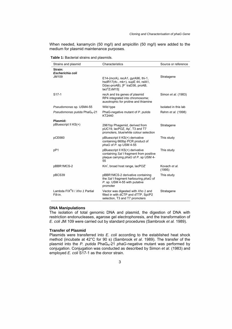
Nucleotide Sequence Analysis The DNA fragments to be sequenced were cloned into pBluescript II KS(+)
(Stratagene, California). A primer walking strategy was applied in order to get the full length sequence (Fig. 1). The sequencing reaction was performed according to the instructions in the ABI PRISM Ready Reaction DyeDeoxy Terminator Cycle Sequencing Kit (PE Applied Biosystems, California). The reaction mixture was then loaded onto the ABI Prism 310 for sequence analysis. The nucleic acid sequence was analyzed using MacDNAsis and Genbank BLAST.
The sequence data has been submitted to the GenBank nucleotide database (Accession No. EU305558 ).
Figure 1: Sequencing strategy of pP1.
Construction of Genomic Library of Pseudomonas sp. USM4-55 The genomic library of Pseudomonas sp. USM 4-55 was constructed using λ FIX 11, according to the cloning kit manufacturer’s protocol (Stratagene, California).
4
Cloning and Characterisation of phaG Gene
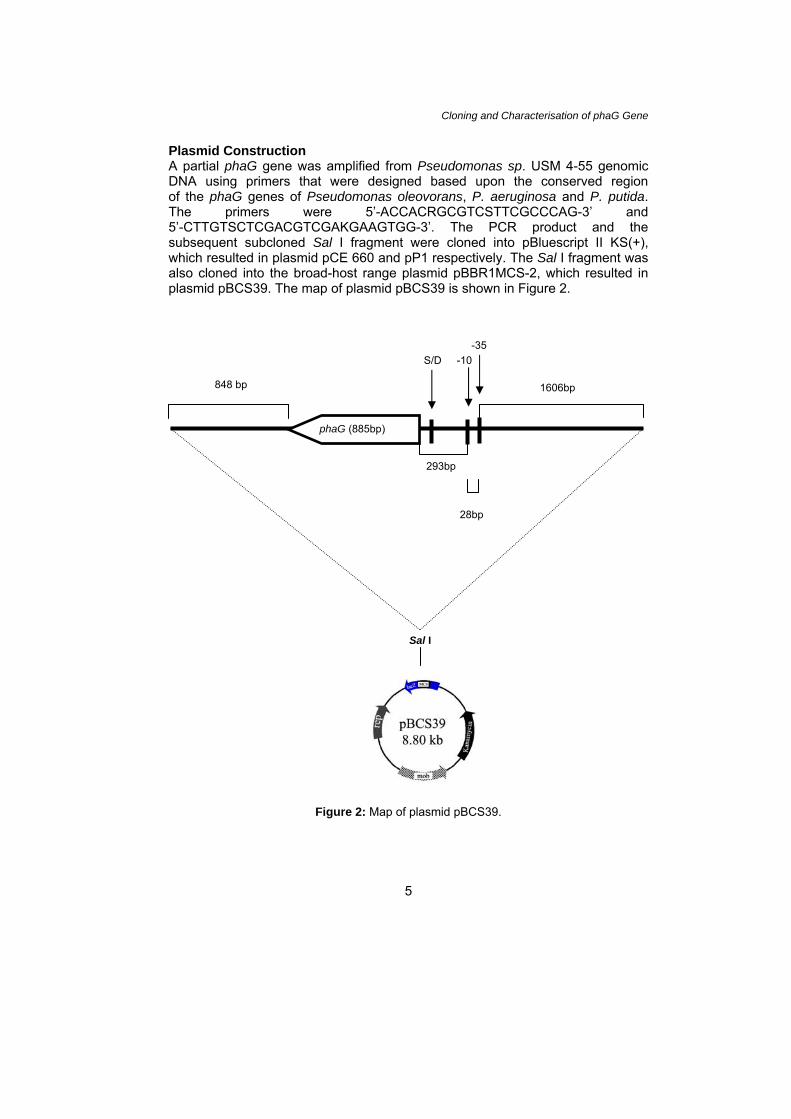
Plasmid Construction A partial phaG gene was amplified from Pseudomonas sp. USM 4-55 genomic DNA using primers that were designed based upon the conserved region of the phaG genes of Pseudomonas oleovorans, P. aeruginosa and P. putida. The primers were 5’-ACCACRGCGTCSTTCGCCCAG-3’ and 5’-CTTGTSCTCGACGTCGAKGAAGTGG-3’. The PCR product and the subsequent subcloned Sal I fragment were cloned into pBluescript II KS(+), which resulted in plasmid pCE 660 and pP1 respectively. The Sal I fragment was also cloned into the broad-host range plasmid pBBR1MCS-2, which resulted in plasmid pBCS39. The map of plasmid pBCS39 is shown in Figure 2.
Figure 2: Map of plasmid pBCS39.
28bp
293bp
phaG (885bp)
-10-35
S/D
848 bp
1606bp
Sal I
5
Hasni Arsad et al.
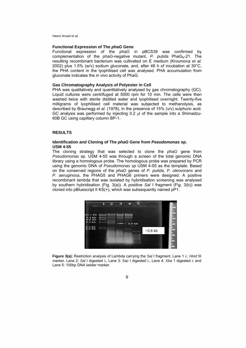
Functional Expression of The phaG Gene Functional expression of the phaG in pBCS39 was confirmed by complementation of the phaG-negative mutant, P. putida PhaGN-21. The resulting recombinant bacterium was cultivated on E medium (Kroumova et al. 2002) plus 1.5% (w/v) sodium gluconate, and, after 48 h of incubation at 30°C, the PHA content in the lyophilised cell was analysed. PHA accumulation from gluconate indicates the in vivo activity of PhaG. Gas Chromatography Analysis of Polyester in Cell PHA was qualitatively and quantitatively analysed by gas chromatography (GC). Liquid cultures were centrifuged at 5000 rpm for 10 min. The cells were then washed twice with sterile distilled water and lyophilised overnight. Twenty-five milligrams of lyophilised cell material was subjected to methanolysis, as described by Braunegg et al. (1978), in the presence of 15% (v/v) sulphuric acid. GC analysis was performed by injecting 0.2 μl of the sample into a Shimadzu-60B GC using capillary column BP-1. RESULTS Identification and Cloning of The phaG Gene from Pseudomonas sp. USM 4-55 The cloning strategy that was selected to clone the phaG gene from Pseudomonas sp. USM 4-55 was through a screen of the total genomic DNA library using a homologous probe. The homologous probe was prepared by PCR using the genomic DNA of Pseudomonas sp USM 4-55 as the template. Based on the conserved regions of the phaG genes of P. putida, P. oleovorans and P. aeruginosa, the PHAG5 and PHAG6 primers were designed. A positive recombinant lambda that was isolated by hybridisation screening was analysed by southern hybridisation (Fig. 3(a)). A positive Sal I fragment (Fig. 3(b)) was cloned into pBluescript II KS(+), which was subsequently named pP1.
~3.6 kb
Figure 3(a): Restriction analysis of Lambda carrying the Sal I fragment. Lane 1 λ: Hind III marker, Lane 2: Sal I digested λ, Lane 3: Sac I digested λ, Lane 4: Xba 1 digested λ and Lane 5: 100bp DNA ladder marker.
6
Cloning and Characterisation of phaG Gene
7
~ 3.6 kb
Figure 3(b): Autoradiograph (Southern blot analysis) of gel on Figure 3(a). The estimated size of the positive DNA fragment in Lane 2 is 3.6 kb, when compared to the λ Hind III marker, and the fragments in Lane 3 and Lane 4 were 22 kb. Nucleotide Sequence of The phaG Gene Locus Sequence analysis revealed that the size of the fragment is 3660 bp (GenBank accession number EU305558). BlastX analysis of the sequence revealed that there were five ORFs within the fragment. ORF 4 (nucleotides 1928 to 2812) contained an amino acid sequence that exhibited significant identity with the PhaG proteins from P. aeruginosa (AF209711, 62%), P. putida (AF052507, 56%), Burkholderia caryophylli (AY039841.1, 57%), Pseudomonas nitroreducens (AY039839.1, 56%), P. oleovorans (AF169252.1, 55%), Pseudomonas fluorescens (ZP_00084908.1, 53%), Pseudomonas pseudoalcaligenes (AF396832, 55%), Pseudomonas sp. 61-3 (AB047080.1, 55%) and Pseudomonas syringae (AE016853.1, 55%). A reliable Shine-Dalgarno consensus sequence was detected at base position 1915. The DNA sequence upstream of the start codon (1928 bp) was analysed by the GeneTyx software to identify any homology with unknown prokaryote control regions. A putative control sequence was detected, which included a possible σ70 promoter (TTGCAC) at base position 1607 and a possible -24/12 promoter at base position 1630 (TTGAAT). This ORF encoded a putative protein that was composed of 295 amino acid residues with a calculated molecular mass (MW) of 33251 Da. The deduced amino acid sequence of the ORF revealed high homologies (62%) to genes that encode the (R)-3-hydroxyacyl-ACP-CoA acyltransferases of P. aeruginosa (Fig. 4).
P. sp. USM 4-55 MRPETAVVEI NRKHKVHTEF YGNPAASKTI ILVNGSLATT ASFAQTVKYL QPQFNVVAFD LPYAGQSKTH NSDFTPISKE DEAAILLKLI DHYGANYLMS P. nitroreducens MRPEIAVLDI QGQYRVYTEF YRADAAENTI ILINGSLATT ASFAQTVRNL HPQFNVVLFD QPYSGKSKPH NRQERLISKE TEAHILLELI EHFQADHVTS P. putida MRPEIAVLDI QGQYRVYTEF YRADAAENTI ILINGSLATT ASFAQTVRNL HPQFNVVLFD QPYSGKSKPH NRQERLISKE TEAHILLELI EHFQADHVMS P. pseudoalcaligens MRPEIAVLDI QGQYRVYTEF YRADAAENTI ILINGSLATT ASFAQTVRNL HPQFNVVLFD QPYSGKSKPH NRQERLISKE TEAHILLELI EHFQADHVMS P. oleovoran MRPEIAVLDI QGQYRVYTGF YRADAAENTI ILINGSLATT ASFAQTVRNL HPQFNVVLFD QPYAGKSKPH NRQERFISKE TEAHILLELI EHFQADHVMS B. caryophylli MRPEIAVLDI QGQYRVYTEF YRADAAEKTI ILINGSLATT ASFAQTVRNL HPQFNVVLYD QPYSGKSKPH NRNDHLLTKE IEGQILLELI DHFAADHIMS P. sp. 61-3 MRPEIAVLDI QGQYRVYTEF YRADAAEKTI ILVNGSMATT ASFAQTVKNL HPQFNVVLYD QPYAGKSKAH NLHEKMLTKE IEGQILLELI DHFAAEHVLS P. aeruginosa MRPETAIIEI HGQYRIHTEF YGNPAAQQTI ILVNGSLSTT ASFAQTVKYL QPHYNVVLYD QPYAGQSKPH NENHTPISKE CEARILLELI ERFRAEVVMS P. stutzeri ------MTEV LGGTSGDERI VELDASEPVD IAEGAAIIEE AVLEPAKTVI IDTTLVAKLN LADYMNAVPV IRELRIRNET AEHYRSLTLS LSADPAIFKP Consensus : :: : A:. . I ..:: A : : : V. : . :: . . .: E L L . . . P. sp. USM 4-55 FSWGGVASML ALAQRPATLE KAAICSFSPI LNVPMLDYLH KGLRFLNAVD RDNIALLVNS TIGKHLPSLF KRFNHKHVST LDEHEYRQMY AHIKQVLNME P. nitroreducens FSWGGASTLL ALAHQPRYVK KAVVSSFSPV INEPMRDYLD RGCQYLAACD RYQVGNLVND TIGKHLPSLF KRFNYRHVSS LDSHEYAQMH FHINQVLEHD P. putida FSWGGASTLL ALAHQPRYVK KAVVSSFSPV INEPMRDYLD RGCQYLAACD RYQVGNLVND TIGKHLPSLF KRFNYRHVSS LDSHEYAQMH FHINQVLEHD P. pseudoalcaligenes FSWGGASTLL ALAHQPRYVK KAVVSSFSPV INEPMRVYLD RGCQYLAACD HYQVGNLVND TIGKHLPSLF KRFNYRHVSS LDSHEYAQMH FHINQVLEHD P. oleovoran FSWGGASTLL ALAHQPRGVK KAVVSSFSPV INEPMRDYLD RGCQYLAACD RYQVGNLVND TIGKHLPSLF KRFNYRHVSS LDSHEYAQMH FHINEVLQHD B. caryophylli FSWGGACTLL ALAHRPRRIE KAVISSFSPV INEPMRDYLE RGSHYLSKCD RYEVGALVND TIGKHLPSLF KRFNYRHVSS LDNHEYKQMH FHINQVLKHD Pseudomonas sp. 61-3 FSWGGAAALV ALAHRPRRIK KAVISSFSPV INEPMREYLE RGVDYLGNLD RDRVGHLVNN TIGKHLPSLF KRFNYRHVST LAEHEYGQMR FHISDVLNSD P. aeruginosa FSWGGVATLL ALAQRPGRIR RAVVNSFSPQ LNPAMLDYLH RGLDYLAACD RTQIGNLVNE TIGRYLPQLF KRYNFRHVSS LDEHEYHQMH FHIREVLRLN P. stutzeri KTWNIDYLSA NAFLQIPGLD VEVDSSLLTR LVESEYSKLS FELTAAGASD AAPRVEVAKR ELSLEMLPRN HWGGLSHIPE MTAAFVQPND PAIEILLKKA Consensus :W. : : . S: . : . L D :.: :. : : . H:. : I :L. P sp. USM 4-55 AHCRMECLQA IDIPLLFVNG E--RDEYTSV EDACLFAQHI DNAQFAVIDD AGHFLDMEHK AAWLQTQRVL LDFFN----A PSKRLQLP-T RGELQELQAI AV P. nitroreducens LERALQGARN INIPVLFING E--RDEYTTV EDARQFSKHV GRSQFSVIRD AGHFLDMENK TACENTRNVM LGFLK----- PTVREPRQRY QPVQQGQHAF AI P. putida LERALQGARN INIPVLFING E--RDEYTTV EDARQFSKHV GRSQFSVIRD AGHFLDMENK TACENTRNVM LGFLK----- PTVREPRQRY QPVQQGQHAF AI P. pseudoalcaligenes LERALQGARN INIPVLFING E--RDEYTTV EDARQFSKHV GRSQFSVIRD AGHSLDMENK TACENTRNVM LGFLK----- PTVREPRQRY QPVQQGQHAF AI P. oleovoran LERALDGARN IDIPVLFING D--RDEYTTV EDARQFSKHV GRSHFSVIRD AGHFLDMENK TACEDTRSVM LGFLK----- PTMREPRHRY QPVKQGQHAL AI B. caryophylli LDNALRSARV IDIPVLFMNG E--WDEYTTT EDAQKFSKHV RNSHFSRIES AGHFLDMEHK AACRDSRDAL LSFLT----- PSPREHRVR- TPFTLGEHAF AI Pseudomonas sp 61-3 RFCYLNAAKK IDIPVLFMNG E--WDEYTAA DDARIFADHV QHSTFSTIQA AGHFLDMEHK AACRDSRHAL LGFLK----- PAQPESRPRY QYVR-DHHAL AI P. aeruginosa ADSYTESFAG IEIPMLFMNG E--LDIYTTP HEARQFGQLI RGAEFHTIRN AGHFIDVEHK AAWQQTQDAL LAFLRPQRTQ PLNPIYRPQP NGASVPLAAL AS P. stutzeri CELLTKAGKS SSLDGYGSGS EHAWEIMSAI WNAVLAMGLD YTLPPASFEL NGQKVRSPSH IAANGLATCM DTTMLFC--- AALEQAGLNP MAIFTEGHAF AI Consensus .: .. : : :: :A : G: : : A : : . A: A
Figure 4: Alignment using Clustal (Larkin et al. 2007) of the deduced amino acid sequences of PhaG from Pseudomonas sp USM 4-55, P. nitroreducens, P. putida, P. pseudoalcaligens, P. oleovoran, P. Caryophylli, Pseudomonas sp. 61-3, P. aeruginosa and P. stutzeri. The HX4D motif is underlined.
Cloning and Characterisation of phaG Gene The conserved HX4D motif, which has been proposed to play an important role in enzymatic catalysis, was also found in the amino acid sequence that was deduced from the phaG of Pseudomonas sp. USM 4-55 (Fig. 4), and its sequence is HVSTLD. Heterologous Complementation of phaG In order to confirm the identity of the Sal I fragment, heterologous expression study was carried out in the phaG-negative mutant strain of P. putida PhaGN-21. The expression plasmid was constructed by cloning the Sal I fragment into the Sal I site of a broad-host range plasmid (pBBR1MCS-2), which was designated as pBCS39. The plasmid construct (Fig. 2) was then transformed into the phaG-negative mutant strain of P. putida PhaGN-21 by conjugation. The resulting transconjugants were grown on mineral medium that contained 1.5% sodium gluconate as the sole carbon source. GC analysis confirmed that the CS39 fragment did indeed show PhaG activity by complementing the mutated phaG gene in P. putida PhaGN-21. The GC results from four transconjugants (C3 series) were averaged and summarised in Table 2. As compared to the controls, which were the P. putida PhaGN-21 mutant and CMCS-2, the C3 cells showed an increased ability to synthesise PHA, which was, on average, 45-fold increase in the PHA content. Table 2: Complementation of P. putida mutant PhaGN-21 by pBCS39 harbouring the 3.6 kb Sal I fragment of P. sp. USM 4-55.
PHA composition (mol%) Strain DCW (g/l)
PHA content (wt% of DCW)
3HB (C4)
3HHx(C6)
3HO (C8)
3HD (C10)
3HDD (C12)
3H5DD (C12-1)
3H7TD (C14)
TOTAL %
P. utida PhaGN-21
1.0 0.3 N/D N/D 18 47 6 11 N/D 100
CMCS-2
0.8 0.6 N/D N/D 14 50 11 18 N/D 100
C3 0.7 17.7 1 9 25 61 2 2 TR 100
Cells were grown at 30°C for 48 hours in mineral medium, containing 1.5% (w/v) sodium gluconate as the carbon source. PHA monomers content were analysed by GC. 3HB: 3-hydroxybutyrate, 3HHx: 3-hydroxyhexanoate; 3HO: 3-hydroxyoctanoate, 3HD: 3-hydroxydecanoate, 3HDD: 3-hydroxydodecanoate, 3H5DD: 3-hydroxy-cis-5-dodecanoate, 3H7TD: 3-hydroxy-cis-7-tetradecenoate, N/D: None detectable, TR: trace, CMCS-2: P. putida PhaGN-21 harbouring plasmid pBBR1MCS-2, C3: P. putida PhaGN-21 harbouring pBCS39. DISCUSSION In this study, the phaG gene from Pseudomonas sp. USM 4-55 was successfully cloned and characterised. The gene was isolated by screening a genomic library of Pseudomonas sp. USM 4-55 using a homologous probe that was prepared by amplification of a section of the phaG. The primers were designed based upon the conserved regions of the phaG genes from P. putida, P. aeruginosa and
9
Hasni Arsad et al.
10
P. oleovorans. The genomic DNA of Pseudomonas sp. USM 4-55 was used as the template.
Southern analysis of gDNA of Pseudomonas sp. USM 4-55 using the homologous probe (DCE660) indicated the presence of a single copy of the phaG gene, which is consistent with the report by Hoffmann et al. (2000a). For further analysis, a 3660 bp Sal I fragment (CS39) was subcloned into pBluescript II KS vector, which formed plasmid pP1.
BLASTX analysis of the Sal I fragment revealed an 885 bp ORF that had a high similarity to the PhaG of several Pseudomonas species that were deposited in the GenBank Database. The ORF translates into a predicted polypeptide of 295 residues and is proposed to be the putative phaG of Pseudomonas sp. USM 4-55. This putative PhaG protein is most closely related to the PhaG protein of P. aeruginosa (62% identity). Other regions of the Sal I fragment, aside from the phaG gene, showed a high similarity to the published P. putida KT2440 genome.
The molecular organisation of phaG in three strains of Pseudomonas is compared in Figure 5. Overall, the organisation of phaG in Pseudomonas sp. USM 4-55 seems to be closely related to that of P. putida KT2440. BLASTX analysis showed that, besides phaG, the 3660 bp Sal I fragment could possibly encode a partial GGDEF domain protein, which is a conserved hypothetical protein of P. putida (PP1410), and the ribosomal small subunit pseudouridine synthase (RSSPS). Again, similar to phaG, the RSSPS shows the highest identity to P. aeruginosa PAO1 (61%), but its location mirrors that of P. putida KT2440 (Stover et al. 2000).
In congruence with the report by Rehm et al. (1998), this study also found that the adjacent DNA sequences of phaG in Pseudomonas sp. USM 4-55 were not related to genes involved in PHA metabolism. In P. putida KT2440, phaG is separated from phaC1 by about 2 Mbp (Nelson et al. 2002). Heterologous complementation was carried out in mineral medium that contained sodium gluconate as the sole carbon source. The provision of gluconate ensures that the substrate for PHA synthesis is derived via the fatty acid de novo synthesis pathway. P. putida PhaGN-21 (Rehm et al. 1998) is known to have a defective phaG, although it can grow normally on any carbon source. It was able to synthesise PHA from fatty acids, but gluconate or glucose as the carbon source results in either a very low or nonexistent PHA synthesis. This made it an ideal host for a complementation assay to confirm the identity of the putative phaG of Pseudomonas sp. USM 4-55. For this purpose, the 3660 bp Sal I fragment was cloned into a PBBRMCS-2 plasmid and transferred into P. putida PhaGN-21 by conjugation. The resulting transconjugant provided evidence of a functional phaG gene. The PhaG mutant, P. putida PhaGN-21, produces very little PHA, which amounts to less than 1% of DCW, and does not accumulate C4, C6 and C14 monomers. The putative phaG gene that is present in the 3660 bp Sal I fragment of Pseudomonas sp. USM 4-55 conferred upon P. putida PhaGN-21 the ability to accumulate PHA up to 17.7% of DCW when grown on gluconate as the sole carbon source. An earlier study on homologous complementation of P. putida PhaGN-21 resulted in a higher PHA accumulation of up to 50% DCW (Rehm et al. 1998). Heterologous complementation of
Figure 5: Molecular organisation of phaG in P. aeruginosa PAO1 (AE004091/GI:12057214), P. putida KT2442 (NC_002947/GI: 26986745) and P. sp USM 4-55. The phaG gene is indicated by the dark shaded arrow.
Hasni Arsad et al. P. putida PhaGN-21 has also been reported by Hoffmann et al. (2000b) using PhaG of P. oleovorans, which resulted in PHA accumulation of 36.7% of DCW. Although the accumulated PHA from this study was lower than previously mentioned complementation studies (Rehm et al. 1998; Hoffmann et al. 2000a, b), the results demonstrate that the putative phaG in the 3660 bp Sal I fragment of Pseudomonas sp. USM 4-55 was indeed the phaG gene of Pseudomonas sp. USM 4-55. Heath and Rock (1998) reported that the HX4D motif was conserved in a variety of glycerolipid acyltransferases. Matsumoto et al. (2001) also reported that the histidine is the most important residue and is essential at that position for PhaG activity. These proteins share a highly conserved domain that all contain an indispensable histidine and an aspartic acid residue that are separated by four or fewer conserved residues. This motif is also found in phaG of Pseudomonas sp. USM 4-55, located at amino acid positions 177 to 182, with the sequence HVSTLD. CONCLUSION In this study, the phaG gene from Pseudomonas sp. USM 4-55 was successfully cloned and characterised. An 885 bp ORF was identified, and it contained a predicted polypeptide of 295 amino acids and a calculated molecular mass (Mr) of 33251 Da. Functional activity of the PhaG of Pseudomonas sp. USM 4-55 was confirmed by a complementation test of the 3660 bp Sal I fragment in a phaG mutant, P. putida PhaGN-21. ACKNOWLEDGEMENT We thank Bernd H. A. Rehm for his generous gift of the mutant strain P. putida PhaGN-21 and plasmid pBBR1MCS-2, which were used in this study. This work was supported by FRGS grant 203/PBIOLOGI/671053, Universiti Sains Malaysia, Malaysia. REFERENCES Braunegg G, Sonnleitner B and Lafferty R M. (1978). A rapid gas chromatographic method
for the determination of poly-β-hydroxybutyric acid in microbial biomass. European Journal of Applied Microbiology and Biotechnology 6(1): 29–37.
Fiedler S, Steinbϋchel A and Rehm B H A. (2000). PhaG-mediated synthesis of poly(3-
hydroxyalkanoates) consisting of medium-chain-length constituents from nonrelated carbon sources in recombinant Pseudomonas fragi. Applied Environmental Microbiology 66(5): 2117–2124.
Heath R J and Rock C O. (1998). A conserved histidine is essential for glycerolipid
acyltransferase catalysis. Journal of Bacteriology 180(6): 1425–1430.
12
Cloning and Characterisation of phaG gene
Hoffmann N, Steinbüchel A and Rehm B H A. (2000a). The Pseudomonas aeruginosa phaG gene product is involved in the synthesis of polyhydroxyalkanoic acid consisting of medium-chain-length constituents from non-related carbon sources. FEMS Microbiology Letters 184(2): 253–259.
. (2000b). Homologous functional expression of cryptic phaG from Pseudomonas
oleovorans establishes the transacylase-mediated polyhydroxyalkanoate biosynthetic pathway. Applied Microbiology and Biotechnology 54(5): 665–670.
Huijberts G N M, de Rijk T C, de Waard P and Eggink G. (1994). 13C nuclear magnetic
resonance studies of Pseudomonas putida fatty acid metabolic routes involved in poly(3-hydroxyalkanoates) synthesis. Journal of Bacteriology 176(6): 1661–1666.
Kessler B, Kraak M N, Ren Q, Klinke S, Preito M and Witholt B. (1998). Enzymology and
molecular genetics of PHA mcl-biosynthesis. In A Steinbüchel (ed.). Biochemical principles and mechanisms of biosynthesis and biodegradation of polymers. Weinheim, Germany: Wiley-VCH, 48–56.
Kovach M E, Elzer P H, Hill D S, Robertson G T, Farris M A, Roop R M and Peterson K M.
(1995). Four new derivatives of the broad-host-range cloning vector pBBR1MCS, carrying different antibiotic-resistance cassettes. Gene 166(1): 175–176.
Kroumova A B, Wagner G J and Davies H M. (2002). Biochemical observations on
medium-chain-length polyhydroxyalkanoate biosynthesis and accumulation in Pseudomonas mendocina. Archives of Biochemistry and Biophysics 405(1): 95–103.
Larkin M A, Blackshields G, Brown N P, Chenna R, McGettigan P A, McWilliam H and
Valentin F et al. (2007). Clustal W and Clustal X version 2.0. Bioinformatics 23(21): 2947–2948.
Madison L L and Huisman G W. (1999). Metabolic engineering of poly (3-
hydroxyalkanoates): From DNA to plastic. Microbiology and Molecular Biology Review 63(1): 21–25.
Matsumoto K, Matsusaki H, Taguchi S, Seki M and Doi Y. (2001). Cloning and
characterization of the Pseudomonas sp. 61-3 phaG gene involved in polyhydroxyalkanoate biosynthesis. Biomacromolecules 2(1): 142–147.
Matsusaki H, Abe H, Taguchi K, Fukui T and Doi Y. (2000). Biosynthesis of poly(3-
hydroxybutyrate-co-3-hydroxyalkanoates) by recombinant bacteria expressing the PHA synthase gene phaC1 from Pseudomonas sp. 61-3. Applied Microbiology and Biotechnology 53(4): 401–409.
Nelson K E, Weinel C, Paulsen I T, Dodson R J, Hilbert H, Martins dos Santos V A,
Fouts D E et al. (2002). Complete genome sequence and comparative analysis of the metabolically versatile Pseudomonas putida KT2440. Environmental Microbiology 4(12): 799–808.
Rehm B H A, Krüger N and Steinbüchel A. (1998). A new metabolic link between fatty acid
de novo synthesis and polyhydroxyalkanoic acid synthase. The Journal of Biological Chemistry 273(37): 24044–24051.
13
Hasni Arsad et al.
14
Sambrook J, Fritsch E F and Maniatis T. (1989). Molecular cloning: A laboratory manual. New York: Cold Spring Harbor Laboratory Press, 8.30–10.70.
Simon R, Prieffer U and Pühler A. (1983). A broad host range mobilization system for in
vitro engineering: Transposon mutagenesis in Gram-negative bacteria. Biotechnology 1(9): 784–790.
Steinbϋchel A and Fuchtenbusch B. (1998). Bacterial and other biological systems for
polyester production. Trends in Biotechnology 16(10): 419–427. Steinbϋchel A. (2001). Perspectives for biotechnological production and utilization of
biopolymers: Metabolic engineering of polyhydroxyalkanoate: Biosynthesis pathway as a successful example. Macromolecular Bioscience 1: 1–24.
Stover C K, Pham X Q, Erwin A L, Mizoguchi S D, Warrener P, Hickey M J, Brinkman F S
et al. (2000). Complete genome sequence of Pseudomonas aeruginosa PA01, an opportunistic pathogen. Nature 406(6799): 959–964.
Sudesh K, Few L L, Azizan M N M, Majid M I A, Samian M R and Nazalan N. (2004).
Biosynthesis and characterization of polyhydroxyalkanoate blends accumulated by Pseudomonas sp. USM 4-55. Journal of Bioscience 15(2): 15–28.
van der Walle G A M, de Koning G J M, Weusthuis R A and Eggink G. (2001). Properties,
modifications and applications of biopolyesters. In W Babel and A Steinbϋchel (eds.). Advances in biochemical engineering/biotechnology: Biopolyester. Berlin: Springer–Verlag, 71: 263–291.
Williams S F, Martin D P, Horowitz D M and Peoples O P. (1999). PHA applications:
Addressing the price performance issue 1. Tissue engineering. International Journal of Biological Macromolecules 25(1–3): 111–121.
Williams S F and Martin D P. (2005). Applications of PHAs in medicine and pharmacy. In
A Steinbϋchel and R H Marchessault (eds.). Biopolymers for medical and pharmaceutical applications. Weinheim, Germany: Wiley-VCH Verlag, 1: 89–125.