Proc. Nati. Acad. Sci. USA Vol. 75, No. 6, pp. 2649-2653, June 1978 Biochemistry Cold pepsin digestion: A novel method to produce the Fv fragment from human immunoglobulin M (IgM cleavage/immunoglobulin domains/antigen-binding site) LIEN-CHING LIN AND FRANK W. PUTNAM* Department of Biology, Indiana University, Bloomington, Indiana 47401 Contributed by Frank W. Putnam, March 17, 1978 ABSTRACT A strategy for the proteolytic fragmentation of human IgM has been developed. This method is called "cold pepsin digestion" because of its unique feature of achieving restricted peptic cleavages at 40 and pH 4.0. Cold pepsin di- gestion has been applied successfully to produce an Fv fragment from 14 human IgM proteins. The Fv fragment consists of the heavy chain variable domain (VH) and the light chain variable domain (VL) held together by strong noncovalent interaction. Thus, each Fv fragment contains one intact antigen-binding site and represents the minimal active fragment derivable from an antibody molecule. A series of other structurally and function- ally important fragments were also isolated and characterized. Two basic digestion pathways were recognized; these mainly reflect the relative accessibility of five sets of major interdomain cleavage sites. The Fv fragment contains a single antibody-combining site and is defined as a fragment derived by cleavage of immunoglob- ulin molecules that consists of both a VH and a VL domain held together by noncovalent interactions. To generate the Fv fragment requires that both the VL-CL and the VH-CHl switch regions be selectively cleaved either chemically or enzymati- cally without irreversibly affecting the conformational integrity of the variable domains. The presence of an exposed area in the VL-CL switch region of the light chain is suggested by the observation of spontaneous VL and/or CL domain fragments in the urine of some patients with multiple myeloma. The proteolytic origin of these frag- ments is demonstrable by incubating Bence-Jones proteins and isolated light chains of both antigenic groups (K or A) either with "active" urine or with endopeptidases such as trypsin, papain, or pepsin (1). Selective cleavage in the VH-CHl switch region of the heavy chain is more difficult to achieve. The isolation of a VH domain fragment after limited papain digestion of a human IgG3 protein appears to be unique to that protein be- cause similar attempts have failed with other IgG3 or IgGl proteins (2); however, the VH domain can be produced from pooled rabbit IgG in reasonable yield by papain digestion of the isolated Fd fragment (3). Production of the Fv fragment is even more sporadic; so far, only three successful isolations have been reported, and the methods have not been generally applicable. An Fv fragment prepared from a mouse IgA (protein 315) by peptic digestion retained both the intact conformation and the original anti- gen-binding activity (4-6); hence, it represents the elementary unit of recognition and specificity in the immune system. However, this technique proved to be effective only with protein 315 and could not be applied to other mouse IgA pro- teins. A much more laborious approach was used to produce Fv fragment from another mouse IgA protein (XRPC-25) (7). An Fv fragment has been prepared from a human Waldenstr6m IgM by peptic digestion of the intact protein or its papain- derived Fab fragment. Although the yield of Fv from this particular IgM was nearly quantitative, the procedure was not applicable to other IgM proteins (8). In contrast to the above, the "cold pepsin digestion" method reported in this communication has been tried on 14 human monoclonal IgM proteins, and a significant yield of Fv frag- ments (up to 100%) was obtained from 13 of them. The Fv fragments have been characterized by chemical, physical, and immunological methods, and the underlying mechanisms of their production have been elucidated. Thus, this cold pepsin digestion method appears to be a general procedure for pro- duction of Fv fragments from human IgM proteins. The Fv fragments have potential application for structural, functional, and genetic studies of human IgM antibodies. MATERIALS AND METHODS Purification of Human IgM. Human IgM proteins were isolated from the plasma of 14 patients with Waldenstr6m disease (macroglobulinemia). Eleven (Ou, Ga, Cam, Di, Dau, Ba, Ne, Gro, Irv, HA, and Ren) were purified by repeated eu- globulin precipitation against distilled water. One pseudoglo- bulin (Web) was precipitated in 1 mM NaH2PO4, pH 7.4 so- lution, one euglobulin (Na) was used as a lyophilized powder, and one noneuglobulin (Cle) was found to be a cryoglobulin and was precipitated at -5° in the presence of 20% (vol/vol) glyc- erol. In all cases the precipitate was dissolved in 0.1 M Tris- HC1/0.15 M NaCl, pH 8.0 buffer and passed through a Sephadex G-200 or Sepharose 6B column. The purity of the IgM proteins was routinely checked by polyacrylamide gel elec- trophoresis in the presence of 0.1% sodium dodecyl sulfate (NaDodSO4) and 2-mercaptoethanol. Cold Pepsin Digestion. A 1% solution of purified human IgM was dialyzed against 0.02 M sodium acetate/0.15 M NaCl, pH 4.0 buffer. Pepsin (porcine gastric pepsin, twice crystallized, Worthington Biochemical Corp.) was added to the protein so- lution at a ratio of 1 to 25 (wt/wt), and the digestion was allowed to proceed at 40 for 24 hr. A second portion of pepsin was then added at a ratio of 1 to 50 (wt/wt) and the digestion was con- tinued for another 24 hr. To stop the reaction, the turbid di- gestion mixture was slowly titrated back to neutrality with concentrated NaOH solution. The clarified solution was Abbreviations: NaDodSO4, sodium dodecyl sulfate. Abbreviations for classes, fragments, regions, and domains of immunoglobulins accord with official World Health Organization recommendations for human immunoglobulins [(1972) Biochemistry 11, 3311-3312]. * To whom reprint requests should be addressed. 2649 The costs of publication of this article were defrayed in part by the payment of page charges. This article must therefore be hereby marked "advertiement" in accordance with 18 U. S. C. §1734 solely to indicate this fact.
Transcript
Proc. Nati. Acad. Sci. USAVol. 75, No. 6, pp. 2649-2653, June 1978Biochemistry
Cold pepsin digestion: A novel method to produce the Fv fragmentfrom human immunoglobulin M
LIEN-CHING LIN AND FRANK W. PUTNAM*Department of Biology, Indiana University, Bloomington, Indiana 47401
Contributed by Frank W. Putnam, March 17, 1978
ABSTRACT A strategy for the proteolytic fragmentationof human IgM has been developed. This method is called "coldpepsin digestion" because of its unique feature of achievingrestricted peptic cleavages at 40 and pH 4.0. Cold pepsin di-gestion has been applied successfully to produce an Fv fragmentfrom 14 human IgM proteins. The Fv fragment consists of theheavy chain variable domain (VH) and the light chain variabledomain (VL) held together by strong noncovalent interaction.Thus, each Fv fragment contains one intact antigen-binding siteand represents the minimal active fragment derivable from anantibody molecule. A series of other structurally and function-ally important fragments were also isolated and characterized.Two basic digestion pathways were recognized; these mainlyreflect the relative accessibility of five sets of major interdomaincleavage sites.
The Fv fragment contains a single antibody-combining site andis defined as a fragment derived by cleavage of immunoglob-ulin molecules that consists of both a VH and a VL domain heldtogether by noncovalent interactions. To generate the Fvfragment requires that both the VL-CL and the VH-CHl switchregions be selectively cleaved either chemically or enzymati-cally without irreversibly affecting the conformational integrityof the variable domains.The presence of an exposed area in the VL-CL switch region
of the light chain is suggested by the observation of spontaneousVL and/or CL domain fragments in the urine of some patientswith multiple myeloma. The proteolytic origin of these frag-ments is demonstrable by incubating Bence-Jones proteins andisolated light chains of both antigenic groups (K or A) either with"active" urine or with endopeptidases such as trypsin, papain,or pepsin (1). Selective cleavage in the VH-CHl switch regionof the heavy chain is more difficult to achieve. The isolation ofa VH domain fragment after limited papain digestion of ahuman IgG3 protein appears to be unique to that protein be-cause similar attempts have failed with other IgG3 or IgGlproteins (2); however, the VH domain can be produced frompooled rabbit IgG in reasonable yield by papain digestion ofthe isolated Fd fragment (3).
Production of the Fv fragment is even more sporadic; so far,only three successful isolations have been reported, and themethods have not been generally applicable. An Fv fragmentprepared from a mouse IgA (protein 315) by peptic digestionretained both the intact conformation and the original anti-gen-binding activity (4-6); hence, it represents the elementaryunit of recognition and specificity in the immune system.However, this technique proved to be effective only withprotein 315 and could not be applied to other mouse IgA pro-
teins. A much more laborious approach was used to produce Fvfragment from another mouse IgA protein (XRPC-25) (7). AnFv fragment has been prepared from a human Waldenstr6mIgM by peptic digestion of the intact protein or its papain-derived Fab fragment. Although the yield of Fv from thisparticular IgM was nearly quantitative, the procedure was notapplicable to other IgM proteins (8).
In contrast to the above, the "cold pepsin digestion" methodreported in this communication has been tried on 14 humanmonoclonal IgM proteins, and a significant yield of Fv frag-ments (up to 100%) was obtained from 13 of them. The Fvfragments have been characterized by chemical, physical, andimmunological methods, and the underlying mechanisms oftheir production have been elucidated. Thus, this cold pepsindigestion method appears to be a general procedure for pro-duction of Fv fragments from human IgM proteins. The Fvfragments have potential application for structural, functional,and genetic studies of human IgM antibodies.
MATERIALS AND METHODSPurification of Human IgM. Human IgM proteins were
isolated from the plasma of 14 patients with Waldenstr6mdisease (macroglobulinemia). Eleven (Ou, Ga, Cam, Di, Dau,Ba, Ne, Gro, Irv, HA, and Ren) were purified by repeated eu-globulin precipitation against distilled water. One pseudoglo-bulin (Web) was precipitated in 1 mM NaH2PO4, pH 7.4 so-lution, one euglobulin (Na) was used as a lyophilized powder,and one noneuglobulin (Cle) was found to be a cryoglobulin andwas precipitated at -5° in the presence of 20% (vol/vol) glyc-erol. In all cases the precipitate was dissolved in 0.1 M Tris-HC1/0.15 M NaCl, pH 8.0 buffer and passed through aSephadex G-200 or Sepharose 6B column. The purity of the IgMproteins was routinely checked by polyacrylamide gel elec-trophoresis in the presence of 0.1% sodium dodecyl sulfate(NaDodSO4) and 2-mercaptoethanol.Cold Pepsin Digestion. A 1% solution of purified human
IgM was dialyzed against 0.02 M sodium acetate/0.15 M NaCl,pH 4.0 buffer. Pepsin (porcine gastric pepsin, twice crystallized,Worthington Biochemical Corp.) was added to the protein so-lution at a ratio of 1 to 25 (wt/wt), and the digestion was allowedto proceed at 40 for 24 hr. A second portion of pepsin was thenadded at a ratio of 1 to 50 (wt/wt) and the digestion was con-tinued for another 24 hr. To stop the reaction, the turbid di-gestion mixture was slowly titrated back to neutrality withconcentrated NaOH solution. The clarified solution was
Abbreviations: NaDodSO4, sodium dodecyl sulfate. Abbreviations forclasses, fragments, regions, and domains of immunoglobulins accordwith official World Health Organization recommendations for humanimmunoglobulins [(1972) Biochemistry 11, 3311-3312].* To whom reprint requests should be addressed.
2649
The costs of publication of this article were defrayed in part by thepayment of page charges. This article must therefore be hereby marked"advertiement" in accordance with 18 U. S. C. §1734 solely to indicatethis fact.
2650 Biochemistry: Lin and Putnam
VL I CLL-Chain -]l-
H-Chain
H-Chain
ii2II/I $ I Z
_ sss-s s IslsIs ts_,SJ Ls-sJ7S
VH Co'1 CIA2 S #3 C 4 S,rs-s-i rs-4 rs si'l Irs-si rs-si I" ! " L '~~~~~~~~7M7q
N
IgM
SLSChaiS-\SL-Chain I_: I.11{1
2/c
VL C,,,L-Chainr
LS-SJs,es-S
Fd,* CM2
Fd;, C02
Is- SJ LS-SJ Is-SJ sVH Cl1 CM2 IS-Sr s-sl s-s
- i-
ES- Si rS- Si I
V, CLL-Chain r .-E,,,,,
LS- SJ LS-SJ_SS
Fd, CMA2
'old Pepsin Digestion(CPD)
%%
Hot Trypsin Digestion%% (H TD)
Z1 L
VH\
c'IJS S_ SS
S5 S
VI CLL-Chain
'7:777:L-Cain .:.. -..:. -1 1111111118
CPD S s-s/~ ~ ~ ~Fd,:
VH CS'1Cp)4 A~~s~~s~~CLSSJ LSSJ LS-SJ S-
VH Cp1 CM2S-S s
CM2 lr ;? I
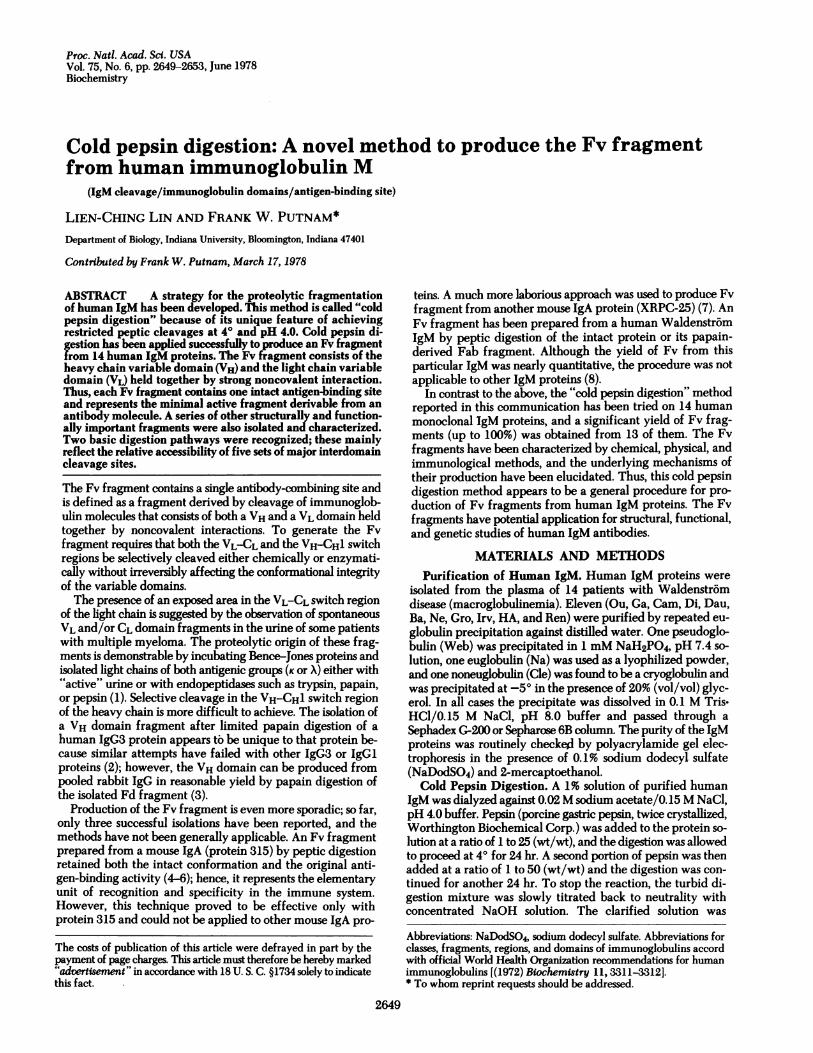
FIG. 1. Enzymatic fragmentation of human IgM proteins and the structural characteristics of the proteolytic fragments. Cold pepsin digestion(CPD) degrades the Fc region and produces the Fv, the F(ab')2,,, the Fab,,-(C,2)2, the Fab, (not shown), and the F(C.2)4 fragments via eitherpathway I or II, the details of which are described in the text. Hot trypsin digestion (HTD) degrades the C,2 domain to form the Fab, and the(Fc)s5, fragments (15, 16). The Fv fragment can also be produced from the tryptic Fab, fragment by cold pepsin digestion. In the diagram, a
schematic monomeric subunit is used to represent the actual pentameric structure of human IgM molecules. The solid arrows indicate the majorpeptic cleavage sites and the dashed arrows indicate the major tryptic cleavage sites. The diagrams for IgM, F(ab')2,, and Fab,-(C,2)2 are drawnto the same scale, those for Fv and F(C,,2)4 are magnified, and the diagram for (Fc)5, is reduced in scale. The individual domains of the H andL chains are identified by different shading.
transferred into uncooked dialysis tubing with a molecularweight cutoff of 3,500 (Spectrapor 3, Spectrum Medical In-dustries) and dialyzed against 0.1 M Tris/0.15 M NaCl, pH 8.0buffer for 24 hr to remove the dialyzable peptides. A smallamount of denatured and/or aggregated material was removedby centrifugation before the solution was subjected to gel fil-tration on a Sephadex G-100 column in the same Tris buffer.Other Methods. NaDodSO4/polyacrylamide disc-gel elec-
trophoresis was carried out essentially as described by Weberand Osborn (9) except that the Tris/acetate buffer system ofFairbanks et al. (10) was used. The sedimentation velocity ofFv fragments was determined at 200 in a Spinco model E an-
alytical ultracentrifuge. Amino acid analysis was performedon a Beckman amino acid analyzer (model 120B, 120C, or
121M); The amino-terminal sequence of Fv fragments was
determined with a Beckman sequencer (model 890B, updated)using a Quadrol protein program (060275) supplied by Beck-man Instruments. The carboxy-terminal sequence of the iso-lated VH and VL domains was determined by carboxypeptidaseA digestion and tryptic peptide mapping. The sequences ob-
tained were compared to the complete C region sequence re-
ported for the K and ,t chains of Ou IgM (11, 12).t Complete or
partial VH region sequences have been reported for the Cam(13), Dau (14), Di, and Ga (15) ,u chains.
RESULTS AND DISCUSSIONUnder the conditions used for cold pepsin digestion, the Fcregion (the C,3 and the CM,4 domains) of human IgM was
completely degraded while the rest of the molecule was cleavedinto a series of peptic fragments (Fig. 1). The lability of thepeptic Fc compared to the stability of the tryptic Fc may be dueto the fact that peptic cleavage occurs after AlaM4 carboxy-terminal to the interchain disulfide bond (CysM7), the loss ofwhich destabilizes the peptic Fc. In contrast, tryptic cleavageoccurs after Arg325, which leaves the disulfide bond intact inthe tryptic Fc.The Fv Fragment. The Fv fragment was produced in a yield
t Heavy chains of the same class (e.g., the ,u chain of human IgM) mayhave different lengths because of variation in the length of the VHregion., To facilitate comparison of the C regions, the numberingsystem of the human ,i chain Ou (12) is used herein.
Fab,,, (Ct2)2
Fab,
(Fc)5P
Proc. Natl. Acad. Sci. USA 75 (1978)
Proc. Natl. Acad. Sci. USA 75 (1978) 2651
of 85-100% from five IgM proteins (Cam, Cle, Web, Na, andBa), 50-70% from three IgM proteins (Ne, Gro, HA), and35-45% from five IgM proteins (Di, Dau, Ga, Irv, and Ren).Only one IgM (Ou) failed to produce a satisfactory yield of Fvfragment (6-10%). Upon analytical ultracentrifugation, the Fvfragment had a sedimentation coefficient of s20 = 2.5 i 0.1 S,which corresponds to an approximate molecular weight of25,000; however, it gave a single band of molecular weight13,000 + 1,000 on NaDodSO4/polyacrylamide gel electro-phoresis in the absence of reducing agent. (In the case of Cam,Cle, Web, Ba, and Di Fv fragments, two closely spaced bandswere discernible.) When the Ga Fv fragment was subjected toautomatic sequence analysis, a single sequence was found,which corresponded to the prototype sequence of human lightchain subgroup KI. These results implied that the Ga Fv frag-ment consists of two polypeptide chains with similar size; onehas a blocked amino-terminus as the Ga it chain does (15), andthe other contains the amino-terminal region of the Ga K chain.The chemical nature of the Fv fragments was further con-firmed by amino acid composition analysis, peptide mapping,cyanogen bromide fragmentation, and in one case (Cam Fv)by the actual separation of the VH domain from the VL domain.The major carboxy-terminal sequence of the isolated VL do-main was found to be -Ser-Val-Phe-; this is eight residuesinto the CL domain and is designated cleavage site A (A').t Themajor carboxy-terminal sequence of the isolated VH domainwas found to be -Pro-Thr-Leu--; this is eight residues intothe C,,1 domain and is designated cleavage site C (C'). Thus,the Fv fragment produced by cold pepsin digestion consists ofthe intact VH and VL domains held together by strong nonco-valent interactions (Fig. 1).The F(CA2)4 Fragment. The F(CMA2)4 fragment was isolated
as a by-product from the cold pepsin digest of all the humanIgM proteins in the attempt to produce the Fv fragment. TheF(C,2)4 fragment contains the intact CM2 domain; its amino-terminus extends 8-18 residues into the C,,1 domain to Val215or Asp225, which are two different cleavage points at site D (D'),the former in Cam, the latter in Ga. A carbohydrate moiety atAsn32 and a disulfide bridge at Cysas7 that links two CM2 do-mains together are located near the carboxy-terminus of theF(CM2)4 fragment. The molecular weight of F(CM2)4 deter-mined by NaDodSO4/polyacrylamide gel electrophoresis was32,000M 1,000, and it decreased to one-half after reduction inaccordance with the expected covalent dimer structure.However, in the native state the F(C,,2)4 fragment exists in atetrameric structure with a pair of disulfide-linked C,2 domaindimers interacting noncovalently (Fig. 1). The F(Cu,2)4 frag-ment is relatively stable at pH 4 and 40, but it becomes ex-
tremely susceptible to proteolytic degradation at higher tem-perature.The Three Digestion Intermediates. The F(ab')U, fragment,
the Fab,-(C,,2)2 fragment, and the Fab,,, fragment are the threedigestion intermediates from which the Fv fragment can bereleased by further degradation. The F(ab')2,, fragment has a
four-chain structure consisting of two light chains and twoFd,,.C,,2 pieces arranged symmetrically into a pair of disul-fide-linked half molecules (Fig. 1). The Fab,,,(C,,2)2 fragment
t A and A', etc., represent two identical sites on the two light chains,just as C (C'), etc., represent identical sites on each of the two ,u chainsin each symmetrical monomer. Although the sites within each pairare structurally equivalent, our experimental evidence indicates thatthey are not necessarily cleaved at the same rate. For example, asillustrated below, the Fab,,.(C,2)2 fragment can be generated fromthe F(ab')2, fragment only when either site D or D' is cleaved, butnot when both are cleaved.
1.2
1.0
0.8
0.6[
Q4[E
co
oO.00-0
.0
0.8
0.4
02
50 100 150Fraction
200 250 300
FIG. 2. Gel filtration pattern of the cold pepsin digest of (A)Cam IgM and (B) Ga IgM. Three hundred milligrams of purified IgMwas digested by pepsin under the standard condition. A 5 X 100 cmSephadex G-100 column in 0.1 M Tris/0.15M NaCl, pH 8.0 buffer wasused to separate the peptic fragments. The fraction size was controlledat 7.5 ml and the flow rate was adjusted to about 50 ml/hr.
is composed of three covalently linked polypeptide subunits:the light chain, the FdM.C,2 piece, and the CM2 domain frag-ment (Fig. 1). The Fd,.C,2 piece is about 340 amino acidresidues long and is derived from the amino-terminus of the ,uchain; it includes the VH, CAI, and CM2 domains and terminatesat Ala34. The peptic FabM fragment (not illustrated in Fig. 1)is structurally similar to the tryptic Fab, fragment except thatthere are frequent internal cleavages observed between the VHand the CM' domains, at the carboxy-terminus of the light chain,and within the CL and the CM1 domains.The Two Basic Digestion Pathways. Fig. 2A shows the gel
filtration pattern of Cam IgM on a Sephadex G-100 columnafter it was digested by pepsin under the standard conditions.Virtually all the IgM was converted into two main digestionproducts identified as the Fv fragment and the F(CM2)4 frag-ment. The yields of Cam Fv fragment and Cam F(CM2)4 frag-ment were estimated to be 103% and 70%, respectively, on thebasis that 10 Fv fragments and 2.5 F(CM2)4 fragments can beproduced from each pentameric molecule. Kinetic studies in-dicated that only a trace of the three digestion intermediatescould be detected in the cold pepsin digest of Cam IgM at anystage of digestion. Thus, the Cam Fv and F(CM2)4 fragmentsappeared to be produced directly from the Cam IgM moleculewithout formation of the three digestion intermediates (path-way I in Fig. 1).
AFv
F (C,,2)4
Subdomainfragments
1gM
50 100 150 200 250 300
B
..CN
0
-o
I U~~~ Fv'DU- ~~Subdomain
.3 ZL fragmentsLL
U- VL
_O
Biochemistry: Lin and Putnam
2
2652 Biochemistry: Lin and Putnam
The digestion pathway followed by Ga IgM was differentfrom that of CamIgM. Kinetic studies showed that the F(ab')2M,fragment was the predominant cleavage product of GaIgM atthe initial stage of cold pepsin digestion. The F(ab')u fragmentwas then amputated at one of its Fab arms, thus forming anFab/A fragment and an FabM,.(C,,2)2 fragment; the latter wasfurther cleaved into the F(CMA2)4 fragment and another FabJAfragment. The FabA, fragment appeared to be the immediateprecursor of the Ga Fv fragment. In the final digest of GaIgM,the full spectrum of peptic fragments was displayed (Fig. 2B).The yield of Ga Fv fragment (35-40%) was significantly low-ered on one hand by the resistance to cleavage of the three di-gestion intermediates, and on other hand by the vulnerabilityof the Ga Fv fragment, especially its VH domain, to secondarydegradation as evidenced by the accumulation of free VL do-main in the digest.Among the 12 otherIgM proteins studied, 3 (Na, Ba, and Cle)
followed a digestion pathway similar to that of Cam IgM(pathway I in Fig. 1), and they all gave nearly quantitativerecovery of Fv fragment. The remaining nine followed a di-gestion pathway similar to that of GaIgM (pathway II in Fig.1), and their yield of Fv fragment varied from 10% (Ou) up to100% (Web). One explanation of the differential yields of Fvand the varying susceptibility of the cleavage sites of differentIgM proteins lies in the variation in the VH and VL sequences,which in turn affects the degree of interaction of VH and VLand possibly also of the constant domains.The Five Interdomain Cleavage Sites. The two basic di-
gestion pathways (I and II) mainly reflect the relative accessi-bility of five sets of major interdomain cleavage sites: site A (A')is located between the VL and the CL domain; site B (B') is lo-cated near the carboxy-terminus of the CL domain; site C (C')is located between the VH and the C.1 domains; site D (D') islocated between the C/1 and the CA2 domains; site E (E') islocated between the CIA2 and the C ,3 domains (Fig. 3).
Sites B (B') and C (C') of Cam IgM and its digestion homo-logues were found most susceptible to peptic cleavage. Thissituation prevented the formation of any significant amountof intermediate fragments even at the beginning of the diges-tion and led to pathway I of Fig. 1. Site B (B') was not directlyinvolved in the generation of either the Fv fragment or theF(C.2)4 fragment; however, its cleavage seemed to weakeninteractions between the CL and the C/Al domains, probably bydisruption of the stabilizing effect of the H-L disulfide bond,and thus facilitated the subsequent exposure of sites A (A') andD (D').On the other hand, pepsin attacked Ga IgM and its digestion
homologues first at site E (E') to produce the F(ab')2, fragment(pathway II, Fig. 1). A subsequent differential cleavage at siteD (or D') would then generate the FabM,(CM,2)2 fragment. TheF(CM,2)4 fragment was produced upon completion of cleavageat the remaining site D' (or D). Sites A (A'), B (B'), and C (C')apparently became exposed to pepsin only after the C^,2 domainregion had been removed from the Fab region. The relativeexposure of these three interdomain sites varied significantlyamong different homologues of Ga IgM. For example, the verylow yield of Ou Fv fragment was found in part to be due to theresistance of these three interdomain cleavage sites and in partdue to the acid instability of Ou Fv fragment itself. Thus, theOu Fv fragment not only was generated with difficulty, but alsowas readily degraded.
Factors Affecting the Cold Pepsin Digestion of HumanIgM. Various experimental factors may affect the final resultof cold pepsin digestion. Among them are the temperature, pH,buffer strength, protein concentration, digestion time, and
FIG. 3. Distribution of the major interdomain cleavage sites.There are five sets of major interdomain cleavage sites distributedin each monomeric subunit of human IgM proteins. They are indi-cated by arrows whose positions are drawn approximately corre-sponding to the actual cleavage sites. The dark region (the VL, VH,and CA2 domains) is most resistant, the hatched region (the CL andC/1 domains) is less resistant, while the light region (the C^,3 and theCA4 domains) is most vulnerable to cold pepsin digestion.
pepsin-to-protein ratio. Temperature was found to be the mostcrucial factor for successful production of Fv fragment. Fvfragment is relatively unstable at acidic pH and is very sus-ceptible to peptic degradation at 37O. For example, Cam Fvfragment was produced in quantitative yield when the digestionwas done at 40, but almost no Cam Fv fragment persisted if thedigestion proceeded at 37°. This observation apparently ex-plains why the Fv fragment was not isolated by conventionalpeptic digestion at 370 except in rare cases (4, 8). Elevatedtemperature also adversely affects the integrity of the CM,2domain. No F(C,,2)4 fragment could be isolated if IgM proteinswere digested at 370. For most of the IgM proteins pH 4.0 wasfound optimal to produce a maximal yield of Fv fragment.Variation of digestion pH around 4.0 affected different IgMproteins to different degrees. Raising the buffering capacityof the digestion medium tended to accelerate the digestionprocess as well as to accentuate the peptic action; this leads torapid disappearance of intermediates without increase in theyield of Fv. Because 20-40% of the IgM molecules remainedintact or only partially cleaved after digestion for 24 hr owingto autolysis of the pepsin, it was necessary to replenish the en-zyme and continue the digestion for another 24 hr in order toachieve a higher yield of Fv. The relatively high pepsin-to-protein ratio used was found necessary because of the low di-gestion temperature and the long digestion time employed.
Structural Studies of the Antigen-Combining Site. The Fvfragment has several unique advantages over the Fab fragment,the light chain dimer, and the VL dimer to be a model forstructural studies of the antigen-binding function of antibodies:(i) The Fv fragment contains an intact antigen-binding site andrepresents the minimal active fragment derivable from anantibody molecule. (ii) The Fv fragment is completely devoidof the constant domains; this greatly simplifies the practicalassignment of many experimental observations. (hi) The lowmolecular weight plus the high aqueous solubility of Fv frag-ment permits the preparation of a protein solution of highconcentration (10 mM or 250 mg/ml), which is desirable forphysical measurements such as nuclear magnetic resonancestudies. (iv) The relatively compact globular structure of theFv fragment offers a greater chance of success in attempts atcrystallization. (v) The subunit components of the Fv fragmentcan potentially be dissociated, isolated, and reassembled without
Proc. Nati. Acad. Sci. USA 75 (1978)
;. IL II
9 B
Proc. Natl. Acad. Sci. USA 75 (1978) 2653
significant alteration in conformation. (vi)Amino acid sequenceanalysis of the variable regions, which determine the combiningsite of an antibody molecule, is much more readily done on anFv fragment than on the whole immunoglobulin molecule.An Fv fragment prepared from a single mouse IgA (myeloma
protein MOPC 315) that has anti-dinitrophenyl activity hasbeen studied extensively by circular dichroism (5), equilibriumdialysis (4), fluorescence quenching (6), and nuclear magneticand electron spin resonances (17). The topography of its bindingcavity has been elucidated from these dynamic data by com-parison with the x-ray crystallographic structure of the variableregion of a phosphocholine-binding mouse IgA (myelomaprotein MoPC 603) (18).
Similar studies on human antibodies have been hamperedby the inability to produce Fv fragment from monoclonal im-munoglobulins with proven antigen-binding function. So far,no definite antigen-binding activity has been shown to be as-sociated with the 14 human IgM proteins studied here exceptpossibly for Ga IgM, which may have a rheumatoid factor-likeantibody activity against antigen-antibody complex. However,the finding of several Waldenstrom IgM proteins with speci-ficities toward phosphocholine (19), nitrophenyl compounds(20, 21), and glycosphingolipids (22) clearly implies the po-tential antibody-like function of many human monoclonal IgMparaproteins. Evidence for the conformational integrity of theFv and of the F(C.2)4 fragments has been obtained by circulardichroism measurements (23). Cold pepsin digestion of humanIgM with proven antigen-binding activity might thereforeprovide the first human Fv fragment that is suitable for struc-tural and immunological studies of the antigen-combiningsite.
We thank L.-C. Huang, A. Galen, J. Madison, and S. Dorwin fortechnical assistance. This work was supported by Grants IM-2E of theAmerican Cancer Society and CA08497 of the National Cancer In-stitute.
1. Solomon, A. (1976) N. Engl. J. Med. 294, 91-98.
2. Dammacco, F., Franklin, E. C. & Frangione, B. (1972) J. Im-munol. 109, 565-569.
3. Mole, L. E., Geier, M. D. & Koshland, M. E. (1975) J. Immunol.114, 1442-1448.
4. Inbar, D., Hochman, J. & Givol, D. (1972) Proc. Natl. Acad. Sci.USA 69,2659-2662.
5. Inbar, D., Givol, D. & Hochman, J. (1973) Biochemistry 12,4541-4543.
6. Hochman, J., Inbar, D. & Givol, D. (1973) Biochemistry 12,1130-1135.
7. Sharon, J. & Givol, D. (1976) Biochemistry 15, 1591-1594.8. Kakimoto, K. & Onoue, K. (1974) J. Immunol. 112, 1373-
1382.9. Weber, K. & Osborn, M. (1969) J. Biol. Chem. 244, 4406-
4412.10. Fairbanks, G., Steck, T. L. & Wallach, D. F. H. (1971) Bio-
chemistry 10, 2606-2617.11. K6hler, H., Shimizu, A., Paul, C. & Putnam, F. W. (1970) Science
169,56-59.12. Putnam, F. W., Florent, G., Paul, C., Shinoda, T. & Shimizu, A.
(1973) Science 182, 287-291.13. Putnam, F. W. (1977) in The Plasma Proteins, ed. Putnam, F.
W. (Academic, New York), 2nd Ed., Vol. 3, pp. 1-153.14. Lehman, D. (1975) Dissertation (Indiana University, Bloom-
ington, IN).15. Florent, G., Lehman, D. & Putnam, F. W. (1974) Biochemistry
13,2482-2498.16. Plaut, A. G. & Tomasi, T. B., Jr. (1970) Proc. Natl. Acad. Sci. USA
65,318-322.17. Dwek, R. A. (1976) Contemp. Top. Mol. Immunol. 6, 1-53.18. Dwek, R. A., Wain-Hobson, S., Dower, S., Gettins, P., Sutton, B.,
Perkins, S. J. & Givol, D. (1977) Nature 266,31-37.19. Riesen, W. F., Schlessinger, J. & Jaton, J. C. (1976) Biochemistry
15,3391-395.20. Tolleshaug, H. & Hannestad, K. (1975) Immunochemistry 12,
173-182.21. Ashman, R. F., & Metzger, H. (1969) J. Biol. Chem. 244,
3405-3414.22. Naiki, M. & Marcus, D. M. (1977) J. Immunol. 119,537-539.23. Lin, L.-C. (1977) Dissertation (Indiana University, Bloomington,