Complex Formation of Platelet Membrane GlycoproteinsIlb and Illa with Fibrinogen
RALPHL. NACHMANand LAWRENCEL. K. LEUNG, Department of Medicine,Division of Hematology-Oncology, New York Hospital-Cornell MedicalCenter, New York 10021
A B S T R A C T Wehave recently reported the isolationof purified platelet membrane glycoproteins lIb andIIIa and the generation of monospecific antisera tothese membrane proteins. Using these monospecificantisera in an enzyme-linked immunosorbent assaysystem, it is now demonstrated that glycoprotein Ilb(GPIIb) and glycoprotein IIIa (GPIIIa) form a complexwith purified human fibrinogen. The formation of thisGPIIb-GPIIIa fibrinogen complex is calcium depen-dent, fibrinogen specific, saturable, and inhibited byspecific amino sugars and amino acids. These obser-vations suggest that the GPIIb-GPIIIa macromolecularcomplex on the platelet surface acts under the properphysiologic circumstances as the fibrinogen bindingsite required for normal platelet aggregation.
INTRODUCTION
Platelet membrane glycoprotein IIb (GPIIb)' and gly-coprotein IlIa (GPIIIa) are major components of theplatelet plasma membrane that may mediate platelet-platelet interactions (1, 2). GPIIb and GPIIIa are mark-edly diminished to absent in platelets in Glanzmann'sthrombasthenia, a hereditary bleeding disorder char-acterized by defective platelet aggregation. An IgGalloantibody isolated from a polytransf used throm-basthenic patient interacts with GPIIb and GPIIIa (3)and induces a thrombasthenia-like state in normalplatelets (4, 5). ADP, epinephrine, and thrombin in-duce fibrinogen binding to normal platelets, whichcorrelates with platelet aggregability (6-9). Throm-basthenic platelets do not bind fibrinogen (7, 9). Theseobservations suggest that GPIIb and/or GPIIIa are in-volved in platelet fibrinogen receptor function. Wehave recently reported the isolation of purified GPIIb
Received for publication 7 August 1981 and in revisedform 19 October 1981.
1 Abbreviations used in this paper: ELISA, enzyme-linkedimmunosorbent assay; GP, glycoproteins; SDS-PAGE, so-dium dodecyl sulfate-polyacrylamide gel electrophoresis.
and GPIIIa and the generation of monospecific anti-sera to these membrane proteins (10). In this report,we present evidence demonstrating complex forma-tion of GPIIb and GPIIIa with purified human fibrin-ogen.
METHODS
Materials. Lentil lectin (lens culinaris hemagglutinin),glucosamine, mannosamine, N-acetyl glucosamine, N-acetylmannosamine, arginine, glycine, p-nitrophenyl phosphate,and type VII calf mucosa alkaline phosphatase were obtainedfrom Sigma Chemical Co., St. Louis, Mo. Protein A wasobtained from Pharmacia Fine Chemicals, Div. of Phar-macia, Inc., Piscataway, N. J. Microtitration plates and aTitertek Multiscan photometer were purchased from FlowLaboratories, Inc., Rockville, Md. All reagents were of an-alytical grade.
Protein purification. Chromatographically pure peak 1human fibrinogen prepared by the method of Finlayson andMosesson (11) (kindly supplied by Dr. M. Mosesson, Down-state Medical Center, New York) was free of factor XIII,plasminogen, factor VIII antigen (VIII:AGN), and fibronec-tin as determined by lack of reactivity using monospecificantisera to these potential contaminants in an enzyme-linkedimmunosorbent assay (ELISA). The clottability of this fi-brinogen was -96%. The protein migrated as a single bandon 5% sodium dodecyl sulfate-polyacrylamide gel electro-phoresis (SDS-PAGE) with an estimated M, of 330,000. Thereduced protein in SDS-PAGErevealed a typical distributionof Aa, B,B, and y chains with absence of degradation prod-ucts. Whenchromatographed on a Sepharose CL-6B column(1.5 X 45 cm) in 0.25 M NaCl, 0.05 M Tris-HCI, pH 8.6,fibrinogen eluted as a single sharp peak indicating freedomfrom detectable quantities of fibrin monomers (6). Plasmin-ogen isolated from plasma by lysine affinity chromatography(12) and gel filtration chromatography was kindly providedby Dr. Peter Harpel, Cornell University Medical College,New York. Factor VIII:AGN and fibronectin were isolatedfrom human plasma as described (13, 14). Purified humanalbumin was obtained from Calbiochem-Behring Corp.,American Hoechst, San Diego, Calif.
Platelet membrane GPIIb and GPIIIa were isolated andpurified using lentil lectin affinity chromatography and elec-trophoretic elution from SDS-PAGEgels as described (10).Platelet GPIb was prepared as described (15). A crude prep-aration of platelet glycoprotein G or thrombospondin wasprepared according to the method of Phillips et al. (16).
Washed platelets were incubated with thrombin in the pres-ence of 1 mMEDTA without stirring. The platelets wereremoved by centrifugation at 8370 g for 3 min using a Beck-man microfuge (Beckman Instruments, Inc., Fullerton,Calif.). The supernate was incubated with 10% by volumeof insoluble antifibrinogen beads. After reduction on SDS-PAGE, this protein mixture contained major bands at Mr185,000 (thrombospondin) and at M, 68,000, probably rep-resenting GPVfragment (17). Platelet factor 4 and ,B-throm-boglobulin were also detected at the lower Mr range.
Humanerythrocyte membranes were prepared accordingto the method of Dodge et al. (18). The membrane proteinswere solubilized with 1% sodium deoxycholate, and lentillectin affinity chromatography was performed as describedfor platelet membrane proteins (10). Densitometric scans ofCoomassie Blue-stained SDS-PAGEgels were carried out ina Gilford model 240 spectrophotometer equipped with a gelscanning attachment (Gilford Instrument Laboratories, Inc.,Oberlin, Ohio) and a Densicord recorder equipped with anintegrator (Photovolt Corp., New York).
Antisera. Individual monospecific antisera have beenraised in rabbits to the separate isolated GPIIb and GPIIIa(10). The antisera were absorbed with washed platelets froma patient with classical Glanzmann's thrombasthenia. Bytwo-dimensional immunoelectrophoresis (19) anti-GPIIb re-acted with a single antigen of apparent Mr 140,000 in amixture of solubilized platelet membrane proteins. Anti-GPIIIa similarly reacted with a single antigen of apparentMr 95,000 in a mixture of solubilized platelet membraneproteins. The antisera either singly or in combination failedto react with fibrinogen, albumin, fibronectin, plasminogen,Factor VIII-AGN, platelet GPIb or a crude thrombospondinmixture. For these studies an ELISA system was used inwhich the antigen tested was coated at concentrations of 1-6 gg/ml and probed with anti-GPIIb and/or anti-GPIIIa -yglobulin solutions (1 mg/ml) in varying dilutions from 1:50to 1:1,000. Antiplasminogen, antifibrinogen, and antialbu-min sera were obtained from Calbiochem-Behring Corp.Anti-GPIb was prepared as described (20). Antierythrocytemembrane sera were obtained from Accurate Chemical andScientific Corp., Hicksville, N. Y. -y-globulin fractions andFab'2 fragments of antisera were prepared as described (20).Antifibrinogen 'y-globulin was coupled to CNBR-activatedsepharose 4B beads as described (21).
ELISA. The performance of the assay was essentially asdescribed by Voller et al. (22). Microtitration plates werecoated with purified human fibrinogen. 0.2-ml portions ofthe fibrinogen in the bicarbonate coating buffer (0.1 M so-dium carbonate, pH 9.6, 0.02% NaNs) were incubated in ahumid chamber overnight at 4°C. Optimum binding oc-curred at fibrinogen concentrations of 4-6 ,g/ml. Contentsof the microtitration plates were removed and the wellswashed three times for 3 min each in Tris-Tween buffer(0.01 MTris, 0.15 MNaCl, pH 7.4 with 0.5 mMCaCl2, con-taining 0.05% Tween 20). The solutions containing the par-tially purified platelet GPIIb and GPIIIa (lentil lectin affin-ity column eluate) (10) were diluted in Tris-Tween bufferand added in duplicate to coated wells and the plates in-cubated overnight at 4°C in a humid chamber. The washingprocedure was repeated and a mixture of anti-GPIIb andanti-GPIIIa -y-globulin (1 mg/ml), each diluted to final con-centration of 1:100 in Tris-Tween buffer, was added for 24h, 4°C. The washing procedure was repeated and the al-kaline phosphatase-labeled protein A prepared as describedbelow was added for 3-h incubation in a humid chamber at4°C. The wells were emptied, the wash step repeated, and0.2 ml of the substrate p-nitrophenyl phosphate (1 mg/mlin 10% diethanolamine buffer, pH 9.8) was added. The color
development was followed at 5-15-min intervals by repeatedreadings at 405 nm in a Titertek Multiscan photometer.Color development with time was plotted and the best fitcurve was calculated by linear regression analysis. The for-mation of the GPIIb-GPIIIa-fibrinogen complex was ex-pressed as the enzymatic activity of the bound alkaline phos-phatase (AA405 min-').
Alkaline phosphatase-labeled protein A. Protein A waslabeled with alkaline phosphatase as described by Engvall(23). The labeled protein was stored at 4°C with 1% bovineserum albumin and 0.02% NaN3 in Tris buffered saline, pH7.4. The activity of the labeled protein A was assessed bycoating the microtitration plates with fibrinogen (1-6 ,g/ml) in coating buffer for 18 h in a humid chamber at 4°C.After washing three times for 3 min each in Tris-Tween,dilutions of fibrinogen antisera in Tris-Tween were addedfor 18 h. The washing step was repeated and Protein A-alkaline phosphatase (0.125 mg/ml) in various dilutions ofTris-Tween were added for 3 h. The wash step was againrepeated and the substrate added. Maximum color developedusing protein A-alkaline phosphatase at 1:1,000 dilution.
Determination of protein coating efficiency on micro-titration plate. Purified human fibrinogen, fibronectin,plasminogen, VIII-AGN, and GPIIb-GPIIIa mixtures werelabeled with 1125 using the modified chloramine-T method(6). Microtitration plates were coated in duplicate with 0.2-ml portions of the labeled proteins at increasing concentra-tions in the bicarbonate coating buffer at 4°C overnight asdescribed above. After washing three times with Tris-Tweenbuffer, each well was cut out, radioactivity of the coatedprotein counted, and the coating efficiency for each proteinwas determined. The saturating amount of proteins thatcould be coated per well were 0.7 pm (fibrinogen), 1.2 pm(GPIIb-GPIIIa, assuming Mr of 235,000), 3.4 pm (plasmin-ogen), 1.2 pm (fibronectin), and 0.3 pm (VIII:AGN). Optimalcoating concentration was 4-6 Ag/ml for the various pro-teins. Thus the coupling efficiency for the proteins tested,i.e., fibrinogen, GPIIb-GPIIIa, plasminogen, fibronectin, andFactor VIII:AGN were comparable.
Binding of 125" fibrinogen to platelets. The method ofPeerschke et al. (9) was followed with minor modifications.Purified fibrinogen (see above) was labeled with 125I usingthe modified chloramine-T method (6). Isolated humanplatelets were gel filtered on a Sepharose 2B column in di-valent ion-free Tyrode's buffer at pH 7.35 containing 0.35%bovine serum albumin. Samples of gel-filtered platelets weremixed with labeled fibrinogen, CaCl2 (1 mMfinal concen-tration), and buffer. Anti-GPIIb and/or anti-GPIIIa FabCwere added at a final concentration of 0.5 mg/ml. The mix-ture was incubated at 37°C for 10 min. ADP (10 ,uM, finalconcentration) was then added. After an initial agitation toinsure mixing, incubation was continued at 37°C for 10 minwithout stirring. The platelet suspensions were then layeredon 0.5 ml of silicone oil in a 1.5-ml conical centrifuge tube.Free and platelet bound labeled fibrinogen was separatedby centrifugation through the silicone oil mixture at 15,600g for 5 min in an Eppendorf centrifuge. After centrifugation,the tip of the centrifuge tube containing the platelet pelletwas sliced off and counted for 125I. Each assay was done induplicate. Nonspecific fibrinogen binding was measured byperforming the binding assays in the absence of ADP stim-ulation. Specific binding was expressed as nanograms of fi-brinogen bound per 108 platelets.
RESULTS
GPIIb-GPIIIa mixture. The platelet glycoproteinswere isolated from solubilized platelet membranes
264 R. L. Nachman and L. L. K. Leung
using lentil lectin affinity chromatography. The mix-ture contained primarily two major polypeptides ofapparent Mr 140,000 and M, 95,000 (unreduced). Gelsscan analysis of Coomassie Blue-stained gels of the len-til lectin eluate showed that these major polypeptidesconstituted 87% of the protein mixture. These bandshave been characterized as GPIIb and GPIIIa (10).
ELISA of the GPIIb-GPIIIa mixture. Monospe-cific anti-GPIIb and anti-GPIIIa reacted with theGPIIb-GPIIIa mixture in an ELISA assay. The gly-coprotein mixture in increasing concentrations waspassively adsorbed to the wells of the microtitrationplate. Anti-GPIIb and anti-GPIIIa 'y-globulin wereadded. The amount of antibody bound to the adsorbedglycoproteins was monitored by incubation with al-kaline phosphatase-labeled protein A. The resultinghydrolysis of the substrate p-nitrophenyl phosphatewas linear with the concentration of platelet glyco-proteins added to the well up to 1 gg/ml.
Using 125I-labeled platelet glycoproteins, the actualamount of GPIIb and GPIIIa bound to the plastic wellwas determined at each concentration of proteinadded, and this was correlated with color generatedin the ELISA (Fig. 1). It should be noted that no dif-ferences in reactivity with specific antibody werenoted when nonlabeled glycoproteins were comparedto '25I-labeled glycoproteins.
Complex formation of GPIIb-GPIIIa with fibrin-ogen. GPIIb and GPIIIa attached and formed a com-plex with the purified fibrinogen passively adsorbed
200r
7
Eto0
x
0
15O0
1oo0
50
S
0
0
0
0 50 100 150 200ng GP bound
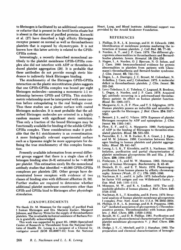
FIGURE 1 Correlation of the amount of GPIIb-GPIIIa boundand the color generated in the ELISA. The radiolabeled gly-coprotein mixture varying from 0.5 to 12 ug/ml in coatingbuffer was applied to the plastic wells for 18 h at 4°C. Afterwashing, a mixture of anti-GPIIb and anti-GPIIIa -y-globulin(10 ug/ml each) was added for 24 h at 4°C. After washing,alkaline phosphatase-labeled protein A (0.125 Mg/ml) wasadded for 3 h at room temperature. After washing the sub-strate p-nitrophenyl phosphate was added and color devel-opment followed in a Titertek Multiscan photometer. Thereaction was expressed as the enzymatic activity of the boundalkaline phosphatase (10' X AA405 min-'). The wells werethen washed, cut out, and counted and the amount of proteinin each well determined. Y=1.116x + 13.73, R=0.97.
to the wells of the plastic microtitration plate. Theformation of the GPIIb-GPIIIa complex with adsorbedfibrinogen was dependent on the presence of Ca2 . Inthe absence of added Ca2+, no significant complex for-mation was demonstrated using a mixture of anti-GPIIb and anti-GPIIIa -y-globulin (Table I). Maximumcomplex formation was demonstrated in the presenceof 0.5 mMCa2+. A mixture of anti-GPIIb and anti-GPIIIa '-globulin appeared to detect a greater degreeof membrane glycoprotein complex formation withfibrinogen than the sum detectable by using each an-tiserum alone at an identical concentration.
Specificity and stoichiometry of GPIIb-GPIIIacomplex formation with fibrinogen. Complex for-mation of GPIIb-GPIIIa with adsorbed fibrinogen wasdetermined in the presence of excess fluid phase fi-brinogen. Following the coating of fibrinogen (3 ;ig/ml) on the microtitration plate, the platelet glycopro-tein mixture (2.4 Mg/ml) was incubated with Tris-Tween buffer with 0.5 mMCa21 in the presence of 6jig/ml fibrinogen. The amount of GPIIb and GPIIIathat was detectable as a complex with the adsorbedfibrinogen was decreased practically to the controllevel (Table II). The small amount of GPIIb and GPIIIathat complexed to adsorbed fibrinogen in the presenceof soluble fibrinogen was considered "nonspecific"binding. No inhibition of GPIIb-GPIIIa complex for-mation with fibrinogen was detected when the exper-iments were repeated in the presence of comparableamounts of fluid phase purified human albumin.
Saturation of the binding or complexing of the gly-coproteins to the fibrinogen was determined by plot-ting the specific binding (total minus nonspecific) ofincremental amounts of GPIIb and GPIIIa to a fixedamount of adsorbed fibrinogen (Fig. 2). Saturation wasachieved at a GPIIb-GPIIIa concentration of 2.4 jg/ml. The color generated in the ELISA by this amountof bound GPIIb-GPIIIa (35 X 10-4 AA405 min-') cor-
TABLE IELISA Detection of GPIIb-GPIIIa-Fibrinogen Complexes
Fibrinogen (6 Ag/ml) in coating buffer was applied to the plasticwells for 18 h, 40C. After washing, the glycoprotein mixture (2.88,gg/ml) in Tris-Tween with or without added Ca21 was added for24 h at 4°C. The remaining steps were carried out as described inFig. 1. Control studies performed with antialbumin substituing foranti-GPIIb and anti-GPIIIa.
Platelet Glycoproteins IIb-IIIa Complex Formation with Fibrinogen 265
TABLE IIInhibition of Complex Formation by Fluid Phase Fibrinogen
Mixture' 104 X AA405 min'
GPIIb-GPIIIa 52.1±1.1GPIIb-GPIIIa plus fluid phase fibrinogen 10.8±1.4GPIIb-GPIIIa plus fluid phase albumin 51.8±1.6
o Fibrinogen (3 ug/ml) in coating buffer was applied to the plasticwells for 18 h, 4°C. After washing, the GPIIb-GPIIIa mixture (2.4Mg/ml) in Tris-Tween Ca2" buffer was added alone or in the pres-ence of fibrinogen (6 ,ug/ml) or albumin (12 ,ug/ml). The remainingsteps were carried out as described in Fig. 1.
responded to 19 ng GPIIb-GPIIIa bound (Fig. 1). Usingradiolabeled fibrinogen, the coating efficiency of fi-brinogen at 3 ,tg/ml was 35.8%, corresponding to 215ng bound to the well. Assuming a Mr of 235,000 fora 1:1 GPIIb-GPIIIa complex and Mr of 330,000 forfibrinogen, the data suggest a stoichiometric relation-ship of fibrinogen to GPIIb-GPIIIa of 8:1.
The specificity of the GPIIb-GPIIIa interaction withfibrinogen was further studied by incubating GPIIb-GPIIIa mixtures with adsorbed fibronectin, plasmin-ogen, or factor VIII:AGN. The coating efficiency forthese proteins to the microtitration plate was deter-mined using '25I-labeled proteins and was comparableto that of fibrinogen. No evidence of complex for-mation was detected (Table III). Platelet membraneGPIb, purified human albumin, and a mixture of hu-
40
30 *T 30
20 X
:x 20
O 10 - /
0 0.5 1 1.5 2 2.5GP*IEb-Ma (pgg/ml)
FIGURE 2 Saturation of GPIIb-GPIIIa complex formationwith adsorbed fibrinogen as measured by the ELISA. Fi-brinogen (3 Mg/ml) in coating buffer was applied to theplastic wells for 18 h, 4°C. After washing varying amountsof the GPIIb-GPIIIa mixture in Tris-Tween Ca2' buffer withor without fluid phase fibrinogen (6 ,ug/ml) was added for24 h at 4°C. The remaining steps were carried out as de-scribed in Fig. 1. The extent of specific GPIIb-GPIIIa com-plex formation with adsorbed fibrinogen (total minus non-
specific) was plotted as a function of increasing GPIIb-GPIIIa concentration.
TABLE IIIInteraction of GPIIb-GPIIIa Complexes With Other Proteins
The proteins (6 ,g/ml) in coating buffer were applied to the plasticwells for 18 h, 4°C. After washing, the GPIIb-GPIIIa mixture (2.88/g/ml) in Tris-Tween calcium buffer was added for 24 h, 4°C.The remaining steps were carried out as described in Fig. 1.
man erythrocyte membrane glycoproteins (lentil lectinaffinity column eluate) also did not show any complexformation with adsorbed fibrinogen, using monospe-cific antisera to these proteins in an ELISA assay(Table IV).
Effect of amino sugars and amino acids on GPIIb-GPIIIa complex formation with fibrinogen. Previousstudies have suggested that specific amino sugars suchas glucosamine, mannosamine, and amino acids suchas arginine block platelet lectin activity and may in-terfere with platelet fibrinogen binding (24, 25).Glucosamine, mannosamine, and arginine signifi-cantly inhibited GPIIb-GPIIIa complex formationwith fibrinogen (Table V). In contrast, N-acetyl glu-cosamine, N-acetyl mannosamine, and glycine at sim-ilar concentrations had no effect.
Lack of complex formation using purified GPIIb-GPIIIa. It has previously been demonstrated that theeluate from the lentil lectin affinity chromatgraphyof solubilized platelet membrane proteins containsother minor proteins in addition to GPIIb and GPIIIa(26). These include GPIb and thrombospondin. Anti-sera raised to the purified GPIIb and GPIIIa isolatedfrom SDS-PAGEgels appeared to be monospecific anddid not react with thrombospondin or GPIb. The SDS-PAGE gel isolated platelet GPIIb and GPIIIa were
TABLE IVInteraction of Other Proteins With Adsorbed Fibrinogen
Fibrinogen (6 Mg/ml) in coating buffer was applied to the plasticwells for 18 h, 4°C. After washing the proteins (12 Ag/ml) in Tris-
Tween calcium buffer were added for 24 h, 4°C. The remainingsteps were carried out as described in Fig. 1. The antisera to theindividual proteins were used at 1:100 dilution.
266 R. L. Nachman and L. L. K. Leung
TABLE VEffects of Aminosugars and Amino Acids on GPIIb-GPIIIa-
Fibrinogen (6 Mg/ml) in coating buffer was applied to the plasticwells for 18 h, 4°C. After washing, the GPIIb-GPIIIa mixture (2.88Mg/iml) in Tris-Tween Ca2" buffer containing the above was addedfor 24 h, 4°C. The aminosugars and amino acids were used at finalconcentration of 120 mMexcept for mannosamine and N-acetylmannosamine, which were used at 30 mM. The pH of the glyco-protein mixture in all cases was adjusted to 7.4 with HCJ. Theremaining steps were carried out as described in Fig. 1. The extentof complex formation was expressed as the enzymatic activity ofbound alkaline phosphatase (AA405 min-'). Control was considered100% complex formation.
studied for complex formation with adsorbed fibrin-ogen (Table VI). The isolated partially denatured pu-rified GPIIb and GPIIIa did not form complexes in thepresence or absence of calcium.
The effect of anti-GPIIb and anti-GPIIIa Fab'2fragments on platelet fibrinogen binding. Incuba-tion of washed human platelets with anti-GPIIbFab'2 fragments alone, anti-GPIIIa Fab'2 fragmentsalone, or a mixture of both failed to block '251-fibrin-ogen binding (Table VII). Previous studies have shownthat both anti-GPIIb and anti-GPIIIa Fab'2 fragmentscaused platelet agglutination, indicating that theywere directed against platelet surface components
TABLE VIComplex Formation of Isolated Purified GPIIb and
GPIIIa with Fibrinogen
Glycoprotein 104 X AA405 min'
GPIIb-GPIIIa, purified 7.1±0.65GPIIb-GPIIIa mixture, eluate of affinity column 112.3±2.6Control 8.1±1.4
Fibrinogen (6 ,g/ml) in coating buffer was applied to the plasticwells for 18 h, 4°C. After washing, the isolated purified GPIIb andGPIIIa (1.44 Ag/ml each) or the GPIIb-GPIIIa mixture (2.88 Ag/ml) was added for 24 h at 4°C. The remaining steps were carriedout as described in Fig. 1. Control included purified human al-bumin (12 fig/ml).
TABLE VIIEffects of Anti-GPIIb and Anti-GPIIIa Fab'2 on the Binding of
Binding assays were performed at fibrinogen concentrations of 300,ug/ml. ADPwas used in final concentration of 10 MM. Represen-tative experiment of two similar studies. Each value represents theaverage of duplicate assays.
(10). The reason for the increased binding detected inthe presence of anti-GPIIb Fab'2 was not determined.
DISCUSSIONIn this study we have demonstrated that a mixture ofundenatured platelet membrane glycoproteins elutedfrom a lentil lectin affinity column forms a complexwith human fibrinogen. Using monospecific antiserato isolated purified GPIIb and GPIIIa (10), we havedemonstrated that platelet GPIIb and GPIIIa in themembrane glycoprotein mixture are the specific mo-lecular components that complex with fibrinogen. Theformation of the GPIIb-GPIIIa fibrinogen complex iscalcium dependent, fibrinogen specific, saturable, andinhibited by specific amino sugars and amino acids.These observations suggest that GPIIb and GPIIIa,which probably exist in the platelet membrane as amacromolecular complex (3, 28, 29), act under theproper physiologic circumstances as the fibrinogenbinding site required for normal platelet aggregation.It is not clear from our studies whether GPIIb alone,GPIIIa alone, or the GPIIb-GPIIIa complex bears theactual binding site. The ELISA studies demonstratingcomplex formation were possible only using undena-tured mixtures of the membrane glycoproteins. WhenGPIIb and GPIIIa were individually isolated by elec-trophoretic elution from SDS-PAGEgels, no complexformation was demonstrable (Table VI). It is thushighly probable that the binding site(s) of the mem-brane glycoproteins was denatured in the process ofpurification. The premise is further supported by thefact that the monospecific antisera to GPIIb andGPIIIa neither inhibited platelet aggregation to phys-iologic stimuli (10) nor blocked fibrinogen binding byADP stimulated platelets (Table VII). The possibilityshould be considered that the binding of GPIIb-GPIIIa
Platelet Glycoproteins IIb-IIIa Complex Formation with Fibrinogen 267
to fibrinogen is facilitated by an additional componentor cofactor that is present in the lentil lectin eluate butis absent in the mixture of purified proteins. Korneckiet al. (27) have described a high affinity fibrinogenreceptor present on normal as well as thrombasthenicplatelets that is exposed by chymotrypsin. It is notknown how this latter activity is related to the GPIIb-GPIIIa system.
Interestingly, a recently described monoclonal an-tibody to the platelet membrane GPIIb-GPIIIa com-plex also did not interfere with ADPor thrombin-in-duced platelet aggregation (28). It is probable thatthese antibodies do not provide enough steric hin-drance to indirectly block fibrinogen binding.
The stoichiometry of the fibrinogen GPIIb-GPIIIainteraction on the plastic microtitration plates suggeststhat one GPIIb-GPIIIa complex was bound per eightfibrinogen molecules-assuming a monomeric 1:1 re-lationship between GPIIb and GPIIIa. This stoichio-metric relationship must be interpreted with some cau-tion before extrapolating to the real biologic event.Thus these studies use a plastic surface with coatedfibrinogen molecules. It is most probable that the ad-sorbed fibrinogen molecules are oriented in a highlyrandom manner with significant steric restriction.Thus only a fraction of the bound fibrinogen may befunctionally available for binding of the soluble GPIIb-GPIIIa complex. These considerations make it prob-able that the 8:1 stoichiometry is an overestimation.A more biologically relevant experimental surfacesuch as a liposome might be more appropriate for de-fining the true stoichiometry of this complex forma-tion.
Presently available information from several differ-ent groups suggest that there is a single class of fi-brinogen binding sites (6-8) estimated to be -40,000per platelet. This estimation nicely fits the monoclonalantibody studies that identified -40,000 GPIIb-GPIIIacomplexes per platelets (28). Other groups have de-monstrated fewer receptors with evidence of twoclasses of binding sites with different affinities (9, 30).Further studies are required to determine whetheradditional platelet membrane constituents other thanGPIIb and GPIIIa bind to fibrinogen after physiologicstimulation.
ACKNOWLEDGMENTSWethank Dr. M. Mosesson for the supply of purified PeakI human fibrinogen and Drs. Marjorie Zucker, MargaretJohnson, and Harvey Weiss for the supply of thrombasthenicplatelets. The invaluable technical assistance of Barbara Fer-ris is gratefully acknowledged.
This work was supported by grant HL 18828 (SpecializedCenter of Research in Thrombosis) from the National Insti-tutes of Health. Dr. Leung is a recipient of a Clinical In-vestigator award (KO8 HL00877-01) from the National
Heart, Lung, and Blood Institute. Additional support wasprovided by the Arnold Krakower Foundation.
REFERENCES
1. Phillips, D. R., L. K. Jennings, and H. H. Edwards. 1980.Identification of membrane proteins mediating the in-teraction of human platelets. J. Cell Biol. 86: 77-86.
2. Nurden, A. T., and J. P. Caen. 1979. The different gly-coprotein abnormalities in thrombasthenic and Bernard-Soulier platelets. Semin. Hematol. 16: 234-250.
3. Hagen, I., A. Nurden, 0. J. Bjerrum, N. 0. Solum, andJ. Caen. 1980. Immunochemical evidence for proteinabnormalities in platelets from patients with Glanz-mann's thrombasthenia and Bernard-Soulier syndrome.J. Clin. Invest. 65: 722-731.
4. Degos, L., A. Dautigny, J. C. Brouet, M. Colombani, N.Ardaillou, J. Caen, and J. Colombani. 1975. A moleculardeficit in thrombasthenic platelets. J. Clin. Invest. 56:235-240.
5. Levy-Toledano, S., G. Tobelem, C. Legrand, R. Bredoux,L. Degos, A. Nurden, and J. P. Caen. 1978. AcquiredIgG antibody occurring in a polytransfused thrombas-thenic patient: its effect on human platelet function.Blood. 51: 1065-1071.
6. Marguerie, G. A., E. F. Plow, and T. S. Edgington. 1979.Human platelets possess an inducible and saturable re-ceptor specific for fibrinogen. J. Biol. Chem. 254: 5357-5363.
7. Bennett, J. S., and G. Vilaire. 1979. Exposure of plateletfibrinogen receptors by ADP and epinephrine. J. Clin.Invest. 64: 1393-1401.
8. Plow, E. F., and G. A. Marguerie. 1980. Participationof ADP in the binding of fibrinogen to thrombin-stim-ulated platelets. Blood. 56: 553-555.
9. Peerschke, E. I., M. B. Zucker, R. A. Grant, J. J. Egan,and M. M. Johnson. 1980. Correlation between fibrin-ogen binding to human platelets and platelet aggrega-bility. Blood. 55: 841-847.
10. Leung, L. L. K., T. Kinoshita, and R. L. Nachman. 1981.Isolation, purification and partial characterization ofplatelet membrane glycoproteins Ilb and IIIa. J. Biol.Chem. 256: 1994-1997.
11. Finlayson, J. S., and M. W. Mosesson. 1963. Heteroge-neity of human fibrinogen. Biochemistry. 2: 42-46.
12. Deutsch, D. G., and E. T. Mertz. 1970. Plasminogen:purification from human plasma by affinity chromatog-raphy. Science (Wash., D. C.). 170: 1095-1096.
13. Nachman, R. L., and E. A. Jaffe. 1975. Subcellular plate-let factor VIII antigen and von Willebrand's factor. J.Exp. Med. 141: 1101-1113.
14. Mosesson, M. W., and R. A. Umfleet. 1970. The cold-insoluble globulin of human plasma. J. Biol. Chem. 245:5728-5736.
15. Nachman, R. L., T. Kinoshita, and B. Ferris. 1979. Struc-tural analysis of human platelet membrane glycoproteinI complex. Proc. Natl. Acad. Sci. U.S.A. 76: 2952-2954.
16. Phillips, D. R., L. K. Jennings, and H. R. Prasanna. 1980.Ca2+-mediated association of glycoprotein G (Thrombin-sensitive protein, Thrombospondin) with human plate-lets. J. Biol. Chem. 255: 11629-11632.
17. Berndt, M. C., and D. R. Phillips. 1981. Purification andpreliminary physicochemical characterization of humanplatelet membrane glycoprotein V. J. Biol. Chem. 256:59-65.
18. Dodge, J. T., C. Mitchell, and D. J. Hanahan. 1963. Thepreparation and chemical characterization of hemoglo-
268 R. L. Nachman and L. L. K. Leung
bin-free ghosts of human erythrocytes. Arch. Biochem.Biophys. 100: 119-130.
19. Converse, C. A., and D. S. Papermaster. 1975. Mem-brane protein analysis by two-dimensional immunoelec-trophoresis. Science (Wash., D. C.). 189: 469-472.
20. Nachman, R. L., E. A. Jaffe, and B. B. Weksler. 1977.Immunoinhibition of ristocetin induced platelet aggre-gation. J. Clin. Invest. 59: 143-148.
21. Cuatrecasas, P. 1970. Protein purification by affinitychromatography. J. Biol. Chem. 245: 3059-3065.
22. Voller, A., D. Bidwell, and A. Bartlett. 1976. Microplateenzyme immunoassays for the immunodiagnosis of virusinfections. In Manual of Clinical Immunology. N. R.Rose and H. Friedman, editors. American Associationfor Microbiology, Washington, D. C. 506-512.
23. Engvall, E. 1978. Preparation of enzyme-labeled Staph-ylococcal protein A and its use for detection of antibod-ies. Scan. J. Immunol. 8(Suppl. 7): 25-31.
24. Gartner, T. K., D. C. Williams, F. C. Minion, and D. R.Phillips. 1978. Thrombin-induced platelet aggregationis mediated by a platelet plasma membrane-bound lec-tin. Science (Wash., D. C.). 200: 1281-1283.
25. Kinlough-Rathbone, R. L., A. Chahil, D. W. Perry,M. A. Packham, and J. F. Mustard. 1979. Effect of aminosugars that block platelet lectin activity on fibrinogen
binding to washed rabbit or human platelets. Blood.54(Suppl. 1): 249a.
26. McGregor, J. L., K. J. Clemetson, E. James, T. Green-land, E. F. Liuscher, and M. W. Dechavanne. 1980. Stud-ies on platelet glycoproteins in Glanzmann's thrombas-thenia using 1251-labeled lectins. Br. J. Haematol. 46: 99-107.
27. Kornecki, E., S. Niewiarowski, T. A. Morinelli, and M.Kloczewiak. 1981. Effects of chymotrypsin and adeno-sine diphosphate on the exposure of fibrinogen receptorson normal human and Glanzmann's thrombasthenicplatelets. J. Biol. Chem. 256: 5696-5701.
28. McEver, R. P., N. L. Baenziger, and P. W. Majerus.1980. Isolation and quantitation of the platelet mem-brane glycoprotein deficient in thrombasthenia using amonoclonal hybridoma antibody. J. Clin. Invest. 66:1311-1318.
29. Polley, M. J., L. L. K. Leung, F. Y. Clark, and R. L.Nachman. 1981. Thrombin induced platelet membraneglycoprotein lIb and IIIa complex formation. An elec-tron microscope study. J. Exp. Med. 154: 1058-1068.
30. Niewiarowski, S., A. Z. Budzynski, T. A. Morinelli,T. M. Brudzynski, and G. J. Stewart. 1981. Exposure offibrinogen receptor on human platelets by proteolyticenzymes. J. Biol. Chem. 256: 917-925.
Platelet Glycoproteins IIb-IIIa Complex Formation with Fibrinogen 269