KnE Medicine ASPIRE Conference Proceedings The 6th Congress of the Asia Pacific Initiative on Reproduction (ASPIRE 2016), Volume 2016 Conference Paper Ovarian Tissue Vitrification as a Method for Ovarian Preservation in Women with Cancer: An Analysis of Granulose Cell Apoptosis Budi Wiweko 1 , Huthia Andriyana 2 , and Achmad Aulia 2 1 Department of Obstetrics and Gynecology, Faculty of Medicine Universitas Indonesia, Dr. Cipto Mangunkusumo General Hospital, Jakarta 10430, Indonesia 2 Department of Histology, Faculty of Medicine Universitas Indonesia, Jakarta 10430, Indonesia Abstract The aim of this study was to obtain the effective method of ovarian function preservation with granulose cell apoptosis assessment. Ovarian tissue vitrification became a method for ovarian function preservation in women with cancer. This technique can be done anytime without delay on cancer therapy, in prepubertal and unmarried patient. It also can store many primordial follicles. Ovarian tissue vitrification study is still limited to animal test and there was no data about apoptosis assessment after ovarian vitrification in human ovary. This is a quasi experimental study which was held in Department of Obstetrics and Gynecology Faculty of Medicine Universitas Indonesia - Dr. Cipto Mangunkusumo General Hospital and Fatmawati Hospital Jakarta from March 2012 to May 2015. Ovaries from thirteen women between 31-37 years of age who underwent oophorectomy with gynecological indication were examined. There were no difference morphologically between follicles from fresh and warmed-vitrified ovaries. The mean protein Bax expression on the fresh ovaries which assessed in the form of H-score was 1,66 ± 0,14 compared to 1,68 ± 0,13 on the warmed-vitrified grup (p = 0,165). The mean protein Bcl-2 expression on the fresh ovaries which assessed in the form of H-score was 1,73 ± 0,10 compared to 1,71 ± 0,10 on the warmed-vitrified grup (p = 0,068). As a conclusion, it was shown that vitrification did not affect Bax and Bcl-2 expression on human ovary. Keywords: Apoptosis, Bax, Bcl-2, ovarian tissue vitrification 1. Introduction Over the past three decades, there has been a remarkable improvement in the survival rates due to progress in cancer treatment, more than 650.000 new female cancer cases are estimated to be diagnosed in 2003 in the USA [1]. Advances in the diagnosis and treatment of childhood, adolescent and adult cancer have greatly enhanced the life expectancy of premenopausal women with cancer. As a result, there is a growing pop- ulation of adolescent and adult long-term survivors of childhood cancer [2]. Ovaries, which are endowed with an irreplaceable number of follicles, are extremely sensitive to cytotoxic drugs that induce an irreversible gonadal damage [1,3]. Ovarian cortex vitrification is one of the promising freezing techniques in order to maintain reproductive function. But freezing procedure is a process that can cause How to cite this article: Budi Wiweko, Huthia Andriyana, and Achmad Aulia, “Ovarian Tissue Vitrification as a Method for Ovarian Preservation in Women with Cancer: an Analysis of Granulose Cell Apoptosis,” KnE Medicine, vol. 2016, 7 pages. DOI 10.18502/kme.v1i1.532 Page 1 Corresponding Author: Budi Wiweko; email: [email protected]Received: 24 August 2016 Accepted: 25 September 2016 Published: 4 October 2016 Publishing services provided by Knowledge E Budi Wiweko et al. This article is distributed under the terms of the Creative Commons Attribution License, which permits unrestricted use and redistribution provided that the original author and source are credited. Selection and Peer-review under the responsibility of the ASPIRE Conference Committee.
Transcript
KnE Medicine
ASPIRE Conference ProceedingsThe 6th Congress of the Asia Pacific Initiative onReproduction (ASPIRE 2016), Volume 2016
Conference Paper
Ovarian Tissue Vitrification as a Method forOvarian Preservation in Women with Cancer:An Analysis of Granulose Cell ApoptosisBudi Wiweko1, Huthia Andriyana2, and Achmad Aulia2
1Department of Obstetrics and Gynecology, Faculty of Medicine Universitas Indonesia, Dr. CiptoMangunkusumo General Hospital, Jakarta 10430, Indonesia2Department of Histology, Faculty of Medicine Universitas Indonesia, Jakarta 10430, Indonesia
AbstractThe aim of this study was to obtain the effective method of ovarian functionpreservation with granulose cell apoptosis assessment. Ovarian tissue vitrificationbecame a method for ovarian function preservation in women with cancer. Thistechnique can be done anytime without delay on cancer therapy, in prepubertaland unmarried patient. It also can store many primordial follicles. Ovarian tissuevitrification study is still limited to animal test and there was no data about apoptosisassessment after ovarian vitrification in human ovary. This is a quasi experimentalstudy which was held in Department of Obstetrics and Gynecology Faculty of MedicineUniversitas Indonesia - Dr. Cipto Mangunkusumo General Hospital and FatmawatiHospital Jakarta from March 2012 to May 2015. Ovaries from thirteen women between31-37 years of age who underwent oophorectomy with gynecological indication wereexamined. There were no difference morphologically between follicles from freshand warmed-vitrified ovaries. The mean protein Bax expression on the fresh ovarieswhich assessed in the form of H-score was 1,66 ± 0,14 compared to 1,68 ± 0,13 onthe warmed-vitrified grup (p = 0,165). The mean protein Bcl-2 expression on the freshovaries which assessed in the form of H-score was 1,73 ± 0,10 compared to 1,71 ±0,10 on the warmed-vitrified grup (p = 0,068). As a conclusion, it was shown thatvitrification did not affect Bax and Bcl-2 expression on human ovary.
Over the past three decades, there has been a remarkable improvement in the survivalrates due to progress in cancer treatment, more than 650.000 new female cancer casesare estimated to be diagnosed in 2003 in the USA [1]. Advances in the diagnosis andtreatment of childhood, adolescent and adult cancer have greatly enhanced the lifeexpectancy of premenopausal women with cancer. As a result, there is a growing pop-ulation of adolescent and adult long-term survivors of childhood cancer [2]. Ovaries,which are endowed with an irreplaceable number of follicles, are extremely sensitiveto cytotoxic drugs that induce an irreversible gonadal damage [1,3].Ovarian cortex vitrification is one of the promising freezing techniques in order to
maintain reproductive function. But freezing procedure is a process that can cause
How to cite this article: Budi Wiweko, Huthia Andriyana, and Achmad Aulia, “Ovarian Tissue Vitrification as a Methodfor Ovarian Preservation in Women with Cancer: an Analysis of Granulose Cell Apoptosis,” KnE Medicine, vol. 2016,7 pages. DOI 10.18502/kme.v1i1.532
follicle damage due to its potential to increase apoptosis process. Assessment of apop-tosis by expression of pro and anti-apoptotic proteins can be conducted to deter-mine biomolecular processes occurring intracellular that has been indicated to precedemorphological changes. Ovarian cortex vitrification research is still limited in animalexperiments and there has been no data to assess the incidence of apoptosis of thegranulosa cells after vitrification of human ovarian cortex that can be seen from theexpression of apoptosis related genes [1,3,4,5].
2. Methods
All experimental procedures were approved by the Ethical Research Committee of theFaculty of Medicine, Universitas Indonesia After obtaining written informed consent,ovaries were obtained from six women between the ages of 30 and 37 years whounderwent oophorectomy due to cervical or breast cancer. Surgeries were performedin the Acute Tertiary Care Hospital in Jakarta during the period of March 2012 to April2015.
3. Ovarian Tissue Vitrification
Ovarian tissue was suspended in 37∘C phosphate buffered saline (PBS) and transferredto the laboratorywithin 15minutes. A tissue slicer (SquareMeasure, Kitazato, Shizuoka,Japan) was used to cut the ovarian cortex into pieces measuring 10 × 10 × 1 mm [6,7].Two chosen section of ovarian cortexeswere divided into two groups. The first group
is a control group for assessing Bax and Bcl-2 protein expression using immunohis-tochemical method. The second group is an experiment group that were frozen byvitrification technique and followed to thawing process, then Bax and Bcl-2 proteinexpression were assessed with the same immunohistochemical method to be com-pared with the first group. After being excised, ovarian cortexes were placed in 15 mLof 𝛼-MEM (minimum essential medium). Ovarian cortexes were initially equilibrated in7,5% ethylene glycol (EG); 7,5% dimethyl sulfoxide (DMSO) for 25 minutes, followed bya second equilibration in 20% EG, 20% DMSO, 0,5 mol/L sucrose liquid for 15 minutes.Ovarian cortexes were placed in a minimum volume of solution onto a thin metal stripand submerged directly into liquid nitrogen [7].
4. Immunohistochemical (IHC) Staining of Bax and Bcl-2Protein in Fresh and Vitrified Human Ovarian Tissue
Bax and Bcl-2 protein staining using IHC were done in fresh and vitrified ovarian cor-tex from each patient. IHC staining was performed using 3 steps polymer detectionsystem: Starr Trek Universal HRP Detection System (BioSB Santa Barbara, CA, USA).The negative control groups were stained using the same method, with the first anti-body substituted by PBS. Rabbit anti-human monoclonal Bax and mouse anti-humanmonoclonal Bcl-2 antibodies were used (BioSB). All paraffin-embedded samples were
DOI 10.18502/kme.v1i1.532 Page 2
KnE Medicine ASPIRE Conference Proceedings
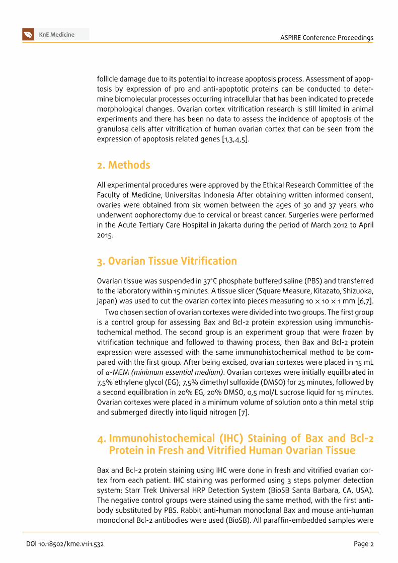
Characteristic n (%) Mean ± SD
Age (year) - 33 ± 1,6
Body mass index (kg/m2) - 23,6 ± 4,7
Parity
Nuliparity 3 (23,1%) -
Primiparity 4 (30,8%)
Multiparity 6 (46,2%)
AMH level (ng/mL) - 2,9 ± 1,6
T 1: Subject characteristics.
deparaffinized and rehydrated. Sections were incubated in 0,5%H2O2 to block endoge-nous peroxides, and then incubated with blocking background sniper to reduce non-specific binding. The primary antibody was then applied and the sections kept at roomtemperature overnight. Universal link (secondarymonoclonal antibody from rabbit andmouse) were put on for 15 minutes. After incubating with TrekAvidin-horseradish per-oxidase to bind antibody with chromogen, sections were stained with DAB (Diaminobenzidin tetraacetic acid) for 5-10 minutes.The expression of Bax and Bcl-2 proteins was identified as diffuse brown cytoplas-
mic staining. Ovarian cortex apoptosis were assessed semi-quantitativelywith Bax andBcl-2 protein expression in primordial follicle, primary follicle, and secondary follicle.The number of granulosa cells stained (Pi) in each follicle were counted and the meanPi from all follicles examinedwas calculated. The color intensity (i) was scored as weak(1), moderate (2), or strong (3) and the final H-score was calculated with the algorithm:H-score =∑ Pi (i+1).
5. Statistical Analysis
Statistical analyses were performed using Statistical Program for Social Sciences(Chicago, IL, USA) version 20.0. Saphiro-Wilk test was used to assess normality.Comparison between H-score were done using paired T-test.
6. Result
6.1. Sample Characteristics
Samples obtained from thirteen patients between 30-37 years of age who underwentoophorectomy due to cervical cancer (stage IB) or breast cancer (Table 1). The meanlevel of anti-mullerian hormone (AMH) in these patients was 2.9 ± 1,6 ng/mL.
DOI 10.18502/kme.v1i1.532 Page 3
KnE Medicine ASPIRE Conference Proceedings
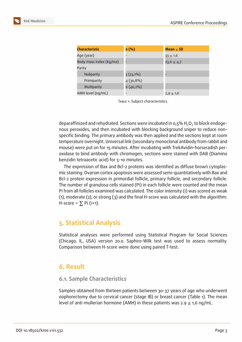
Figure 1: Follicles from fresh and warmed-vitrified ovaries. Primordial follicles (A and B), primary follicles(C and D), secondary follicles (D and E). There were no significant morphological differences betweenfollicles from fresh and warmed-vitrified ovaries based on examinatons of basal membrane, granulosacells, and oocyte. A, C, E: follicles from fresh ovaries; B, D, F: follicles from warmed-vitrified ovaries.
6.2. Evaluation of Morphology and Apoptosis of Folliclesfrom Fresh and Warmed-Vitrified Ovaries
There were no significant morphological differences between follicles from fresh andwarmed-vitrified ovaries based on examinatons of basal membrane, granulosa cells,and oocyte (Figure 1). This findings also supported by the result of IHC staining of Baxand Bcl-2 protein expression, which also showed no changes (Figure 2 and 3). Themean H-score for Bax on fresh ovaries was 1.66 ± 0.14 versus 1.68 ± 0.13 on warmed-vitrified ovaries (p = 0.165). The mean H-score for Bcl-2 on fresh ovaries was 1.73 ±0.10 versus 1.71 ± 0.10 on warmed-vitrified ovaries (p = 0.068).
7. Discussion
In this study, vitrification of human ovarian tissue was performed according to themethod by Kagawa who used DMSO and EG as a cryoprotectants [7]. No significantdifference was found in terms of follicles morphology (basal membrane integrity offollicles, and oocyte) between fresh and warmed-vitrified ovarian tissue. Apoptosiscan be initiated by the extrinsic or intrinsic pathways. Bax and Bcl-2 were expressedin granulosa cells, oocytes, and stroma of both fresh and warmed-vitrified ovaries.However, vitrification did not increase apoptosis via the intrinsic pathway in follicles.
DOI 10.18502/kme.v1i1.532 Page 4
KnE Medicine ASPIRE Conference Proceedings
Figure 2: Bax expression on ovarian tissue. A. Bax expression on breast cancer tissue (positive control).B. No visible Bax expression on stromal ovarian tissue (negative control). C. Bax expression on granulosacells and oocyte with low intensity in fresh ovarian tissue. D Bax expression on granulosa cells and oocytewith low intensity in warmed-vitrified ovaries.fpr: primordial follicles
Numerous studies also reported no ultrastructural changes in oocytes, ovarian fol-licles, and stromal cells in post-vitrification ovarian tissue. Sheiki et al found that vitri-fication is the best method for ovarian tissue cryopreservation. Vitrification liquid thatwere used on that experiment was ethylene glycol as cryoprotectant. There were alsono significant difference among oocytes ultrastructure, granulosa cells, and stromalcells from electron microscope observation [8].A non-randomized study comparing slow freezing to vitrificationwas reported on 20
ovarian biopsies. There was no significant difference in the number and morphologyof follicles between two groups [9]. Kagawa, Silber and Kuwayama also reported thesuccessful technique using Cryotissue method with the results of oocyte viability andno significant difference in morphology of ovarian cortex between fresh ovary andwarmed-vitrified ovaries. This method used ethylene glycol, DMSO and sucrose as acryopretectants in ovarian cortex vitrification technique [7].Assesment of ovarian tissue apoptosis can be performed based on morphology,
level of apoptosis and protein expression related apoptosis in granulosa cells. Witha good assesment, we can understand biomolecular process that happened intracel-lular. Protein expression changes precede morphological changes, so that not onlymorphological changes were assessed but it is also impontant to determine proteinexpression changes in ovaries after vitrification. The expression of Bax and Bcl-2 were
DOI 10.18502/kme.v1i1.532 Page 5
KnE Medicine ASPIRE Conference Proceedings
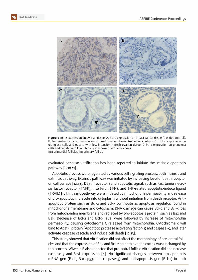
Figure 3: Bcl-2 expression on ovarian tissue. A. Bcl-2 expression on breast cancer tissue (positive control).B. No visible Bcl-2 expression on stromal ovarian tissue (negative control). C. Bcl-2 expression ongranulosa cells and oocyte with low intensity in fresh ovarian tissue. D Bcl-2 expression on granulosacells and oocyte with low intensity in warmed-vitrified ovaries.fpr: primordial follicles, fp: primary follicle
evaluated because vitrification has been reported to initiate the intrinsic apoptosispathway [6,10,11].Apoptotic processwere regulated by various cell signaling process, both intrinsic and
extrinsic pathway. Extrinsic pathwaywas initiated by increasing level of death receptoron cell surface [12,13]. Death receptor send apoptotic signal, such as Fas, tumor necro-sis factor receptor (TNFR), interferon (IFN), and TNF-related apoptotis-induce ligand(TRAIL) [12]. Intrinsic pathway were initiated by mitochondria permeability and releaseof pro-apoptotic molecule into cytoplasm without initiation from death receptor. Anti-apoptotic protein such as Bcl-2 and Bcl-x contribute as apoptosis regulator, found inmitochondria membrane and cytoplasm. DNA damage can cause Bcl-2 and Bcl-x lossfrom mitochondria membrane and replaced by pro-apoptosis protein, such as Bax andBak. Decrease of Bcl-2 and Bcl-x level were followed by increase of mitochondriapermeability, causing cytochrome C released from mitochondria. Cytochrome c willbind to Apaf-1 protein (Apoptotic protease activating factor-1) and caspase-9, and lateractivate caspase cascade and induce cell death [12,13].This study showed that vitrification did not affect the morphology of pre-antral folli-
cles and that the expression of Bax and Bcl-2 on both ovarian cortex was unchanged bythis process.Wiweko B also reported that pre-antral follicle vitrification did not increasecaspase-3 and FasL expression [6]. No significant changes between pro-apoptosismRNA gen (FasL, Bax, p53, and caspase-3) and anti-apoptosis gen (Bcl-2) in both
DOI 10.18502/kme.v1i1.532 Page 6
KnE Medicine ASPIRE Conference Proceedings
fresh and warmed-vitrified ovary also reported by Abdollahi et al [14]. In conclusion,vitrification did not increase apoptosis process in ovary, both via intrinsic and extrinsicpathways.
References
[1] M. Sommezer and K. Oktay, Fertility reservation in female patients, HumanReproduction Update, 10, no. 3, 251–266, (2004).
[2] J. Donnez and S. Bassil, Indications for cryopreservation of ovarian tissue, HumanReproduction Update, 4, no. 3, 248–259, (1998).
[3] HJ. Chang and CS. Suh, Fertility preservation for women with malignancies: currentdevelopments of cryopreservation, J Gynecol Oncol, 19, no. 2, 99–107, (2008).
[4] I. Demeestere, P. Simon, S. Emiliani, A. Delbaere, and Y. Englert, Fertilitypreservation: Successful transplantation of cryopreserved ovarian tissue in a youngpatient previously treated for Hodgkin’s disease, Oncologist, 12, no. 12, 1437–1442,(2007).
[5] P. Jadoul, M.-M. Dolmans, and J. Donnez, Fertility preservation in girls duringchildhood: Is it feasible, efficient and safe and to whom should it be proposed?Human Reproduction Update, 16, no. 6, Article ID dmq010, 617–630, (2010).
[6] B. Wiweko, Upaya preservasi fungsi ovarium dengan melakukan vitrifikasi korteks danfolikel pre-antral, Universitas Indonesia, Jakarta, 2014.
[7] N. Kagawa, S. Silber, and M. Kuwayama, Successful vitrification of bovine andhuman ovarian tissue, Reproductive BioMedicine Online, 18, no. 4, 568–577, (2009).
[8] M. Sheikhi, K. Hultenby, B. Niklasson, M. Lundqvist, and O. Hovatta, Clinical gradevitrification of human ovarian tissue: An ultrastructural analysis of follicles andstroma in vitrified tissue, Human Reproduction, 26, no. 3, 594–603, (2011).
[9] V. Keros, S. Xella, K. Hultenby, K. Pettersson, M. Sheikhi, A. Volpe, J. Hreinsson, and O.Hovatta, Vitrification versus controlled-rate freezing in cryopreservation of humanovarian tissue, Human Reproduction, 24, no. 7, 1670–1683, (2009).
[10] T. Mazoochi, M. Salehnia, S. Pourbeiranvand, M. Forouzandeh, S. J. Mowla, and E.Hajizadeh, Analysis of apoptosis and expression of genes related to apoptosis incultures of follicles derived from vitrified and non-vitrified ovaries,Molecular HumanReproduction, 15, no. 3, 155–164, (2009).
[11] H.-C. Liu, Z. He, and Z. Rosenwaks, Mouse Ovarian Tissue Cryopreservation Hasonly a Minor Effect on In Vitro Follicular Maturation and Gene Expression, Journal ofAssisted Reproduction and Genetics, 20, no. 10, 421–431, (2003).
[12] M. R. Hussein, Apoptosis in the ovary: molecular mechanisms, Hum Reprod Update,11, no. 2, 162–178, (2004).
[13] F. Lumongga, (2008)., Apoptosis. Medan: USU.[14] M. Abdollahi, M. Salehnia, S. Salehpour, and N. Ghorbanmehr, Human ovarian tissue
vitrification/warming has minor effect on the expression of apoptosis-relatedgenes, Iranian Biomedical Journal, 17, no. 4, 179–186, (2013).