Cubital tunnel syndrome due to the anconeus epitrochlearis in an amateur weight lifter In-Ho Jeon MD, Poong-Taek Kim MD , II-Hyung Park MD, Hee-Soo Kyung MD, Joo-Chul Ihn MD Department of Orthopaedic Surgery, School of Medicine, Kyungpook National University Taegu, Korea Correspondence: Poong-Taek Kim, Department of Orthopaedic Surgery Kyungpook National University Hospital 50, 2-Ga, Samduk, Chung-Gu Taegu, 700-721, KOREA (South). Tel: +82-53-420-5630 Fax: +82-53-422-6605 E-mail: [email protected]Abstract A Cubital tunnel syndrome caused by hypertrophy of the aberrant muscle, anconeus epitrochlearis in an amateur weight lifter is presented. Résumé Un syndrome de tunnel cubital causé par l'hypertrophie du muscle aberrant, anconeus epitrochlearis chez un haltérophile amateur est présenté. Sicot Case-Reports: July 2002 Page 1
Transcript
Cubital tunnel syndrome due to the anconeusepitrochlearis in an amateur weight lifter
In-Ho Jeon MD, Poong-Taek Kim MD , II-Hyung Park MD, Hee-Soo Kyung MD,Joo-Chul Ihn MD
Department of Orthopaedic Surgery, School of Medicine, Kyungpook NationalUniversity Taegu, Korea
Correspondence:Poong-Taek Kim, Department of Orthopaedic Surgery Kyungpook National UniversityHospital 50, 2-Ga, Samduk, Chung-Gu Taegu, 700-721, KOREA (South). Tel:+82-53-420-5630 Fax: +82-53-422-6605
AbstractA Cubital tunnel syndrome caused by hypertrophy of the aberrant muscle, anconeusepitrochlearis in an amateur weight lifter is presented.
RésuméUn syndrome de tunnel cubital causé par l'hypertrophie du muscle aberrant,anconeus epitrochlearis chez un haltérophile amateur est présenté.
Sicot Case-Reports: July 2002
Page 1
IntroductionWe present a case of a cubital tunnel syndrome secondary to the aberrant muscleanconeus epitrochlearis in an amateur weight lifter.
Case-ReportThis 28-year-old right-handed man had an eight-week history of tingling sensationand weakness of the grip power. He denied any trauma to his elbow and hissymptoms progressed despite the use of splints and medication for 3 months. Hewas a businessman and had to do computer works for several hours a day. Heattended classes in physical fitness and lifted weight to develop his muscles.Numbness of the 4th and 5th fingers and difficulty in fine motion like touchingcomputer key board became constant recently and weakness of flexor profundus ofulnar 2 digits was present. Examination revealed positive Tinnel sign at the cubitaltunnel with radiating pain into the ring and little fingers. However, Tinnel sign wasnegative at the Guyon's canal. There was no atrophy of the thenar, hypothenar, andinterosseous muscles. His symptom was aggravated by elbow flexion beyond 90degrees. Laboratory test was within normal limit including test for syphilis.Electromyography of the left ulnar nerve showed the prolonged distal latencycompared to the opposite (2.46msec: 2.38msec). The motor amplitude of the ulnarnerve above the elbow was 8.40 compared to 3.37 of the below elbow. Conductionvelocity above the elbow was 54.8m/sec compared to 45.9m/sec of the below elbow.Spontaneous fibrillation of left adductor digiti quinti muscle was observed. In addition,recruitment pattern of left adductor digiti quinti was decreased. Sensory nerveconduction was within normal range. Ultrasound of the cubital tunnel showed noganglion or mass like lesion, but increased diameter of the ulnar nerve compared tothe opposite. He was diagnosed as having an idiopathic cubital tunnel syndrome andplanned to surgical release of the cubital tunnel retinaculum. At exploration of the leftcubital tunnel, a group of muscle fibers approximately 3-centimeter in width and3-centimeter in length (Figure 1) crossed the ulnar nerve from olecranon to medialepicondyle, which was found to be the anconeus epitrochlearis muscle. The ulnarnerve was compressed by the aberrant muscle and a fusiform thickening andinduration of the nerve trunk were observed just proximal to the muscle. The musclewas tight in flexion and significantly compressed the ulnar nerve. The ulnar nervewas strained proximal to the muscle bulk at elbow flexion beyond 90. The aberrantmuscle was split longitudinally and flexor retinaculum as well. Splitting of the musclerevealed the aberrant muscle was hypertrophied as thick as 5 millimeter in depth andcompressed the nerve during elbow flexion. It disclosed narrowed area of ulnar nerve10mm under the muscle (Figure 2) No epineural or perineural neurolysis wasperformed. No transposition of the nerve was carried out. Three weeks after surgery,there was gradual recovery and at 6 weeks sensation in the ulnar digits weresubjectively and objectively close to normal and his symptoms almost relieved.Electromyography at this time showed improvement in motor nerve conductionvelocity and motor distal latency.
DiscussionVarious incidence of the anconeus epitrochlearis muscle has been reported in theliterature from 34% to 4% in the cadaver study [6,8]. LeDouble [5] reported the
Sicot Case-Reports: July 2002
Page 2
muscle present in 32 of 102 cadavers. However, Clemens [2] reported only fourcases out of 100 cadavers, one of which was completely developed and other threehaving only a rudimentary muscle. Although sexes and arms may have nearly equalincidences, the anomalous anconeus epitrochlearis has been reported to be morewell developed in men and in the right arm [6]. Hirasawa et al [4] reported similarfindings. They reported a weight lifter with bilateral ulnar nerve neuropathy, whichwas caused by hypertrophy of the muscle. In our study, the patient was alsomuscular and enjoyed weight lifting. The boundary for potential ulnar nervecompression begins approximately 10cm proximal to the elbow and end about 5cmdistal to the joint. One of them is olecranon groove bounded anteriorly by medialepicondyle, laterally by the olecranon and ulnohumeral ligament, and medially by afibroaponeurotic covering, which is also called cubital tunnel retinaculum (CTR). Thisretinaculum has exactly same course as did anconeus epitrochlearis. Thus,O'Driscoll et al [7] considered the CTR as a remnant of the anconeus epitrochlearismuscle and its function is to hold ulnar nerve in position. Compression at this site canbe caused by a wide variety of conditions. Aberrant anconeus epitrochlearis is onecause of lesions outside of the groove. In humans, the muscle is probably atavisticand is replaced by a band passing in the same direction as the muscle, called theepitrochleoanconeus ligament. The treatment for ulnar neuropathy at elbow joint withan associated anconeus epitrochlearis muscle varied from excision of the mass toanterior transposition. Vanderpool et a! [9] and Dahner LE [3] suggested localdecompression and excision of the muscle. Although Chalmers [1] recommendedanterior transposition of the ulnar nerve, we split the anconeus muscle and localdecompression of the ulnar nerve at the cubital tunnel was carried out withoutanterior transposition. Subluxation has not noted after splitting of the muscle in thisstudy. Masear et al. [6] presented improvement of electrophysiologic study between 8and 12 months after operation. We also aware of improvement of electrophysiologicstudy after surgical release. It is probable that the syndrome can be caused byweight lifting and other overuse condition. If isolated compression by the aberrantmuscle, in situ decompression only can achieve satisfactory result.
Sicot Case-Reports: July 2002
Page 3
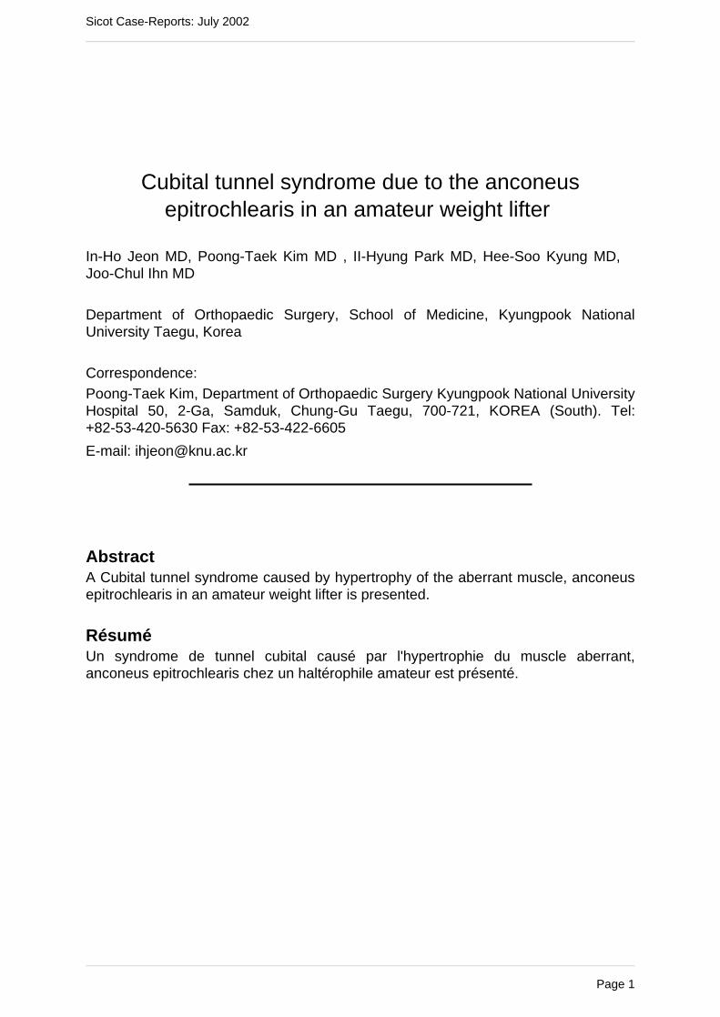
Legends
Figure 1: The anconeus epitrochlearis muscle compresses the ulnar nerve (Ulnar N.)
against the medial epicondyle. This is the left elbow with the hand being to the right
in the photograph. The anconeus epitrochlearis is seen extending from olecranon to
the inferior surface of the medial epicondyle (ME).
Sicot Case-Reports: July 2002
Page 4
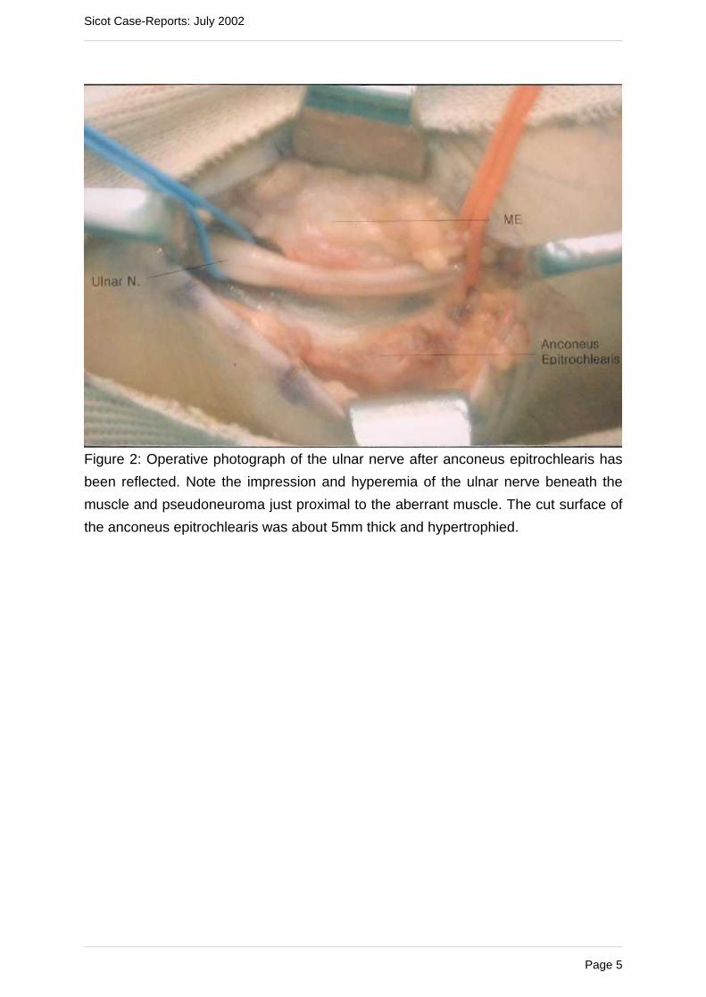
Figure 2: Operative photograph of the ulnar nerve after anconeus epitrochlearis has
been reflected. Note the impression and hyperemia of the ulnar nerve beneath the
muscle and pseudoneuroma just proximal to the aberrant muscle. The cut surface of
the anconeus epitrochlearis was about 5mm thick and hypertrophied.
Sicot Case-Reports: July 2002
Page 5
References1. Chalmer J. (1978) Unusual causes of peripheral nerve compression. Hand10:168-175
2. Clemens HJ. (1957) Zur morphologie des ligamentum epitrochleo-anconeum. AnatAnz 104:343-344
3. Dahners LE and Wood FM. (1984) Anconeus epitrochlearis, a rare cause of cubitaltunnel syndrome: A case report. J Hand Surg [Am]. 9: 579-580.
4. Hirasawa Y Sawamura H and Sakakida K. (1979) Entrapment neuropathy due tobilateral epitrochleoanconeus muscle: A case report. J Hand Surg. 4:181-184
5. LeDouble AF. (1897) Traite des variations du syteme musclulaire de I'homme etde leur significantion au pointe de vue de I'anthropologie zoologiques. 2nd ed. Paris:Schleicher Freres. : 60-75
6. Masear VR, Hill JJ and Cohen SM. (1988) Ulnar compression neuropathysecondary to the anconeus epitrochlearis muscle. J Hand Surg [Am] 13: 720-724.
7. O'Driscoll SW, Horii E, Carmichael SW and Morre BF. (1991) The cubital tunneland ulnar neuropathy. J Bone Joint Surg. [Br] 73:613-617
8. O'Hara JJ and Stone JH. (1996) Ulnar nerve compression caused by a prominentmedial head of the triceps and an anconeus epitrochlearis muscle. J Hand Surg [Br]21:133-135
9. Vanderpool DW, Chalmers J, Lamb DW, Whiston TB. (1968) Peripheralcompression lesion of the ulnar nerve. J Bone Joint Surg [Br] 50 : 792-803