13
DISSEMINATED INTRAVASCULAR COAGULATION SYNDROME (DIC) VAISHNAVI SURESH NAIR
| Date post: | 16-Jul-2015 |
| Category: |
Health & Medicine |
| Upload: | vaishnavi-s-nair |
| View: | 213 times |
| Download: | 0 times |
DISSEMINATED INTRAVASCULAR COAGULATIONSYNDROME
(DIC)
VAISHNAVI SURESH NAIR

It is an acquired syndrome. It is a complication of an underlying illness, and involves systemic activation of the
coagulation system when blood is exposed to pro-coagulants such as tissue factor.
Thrombin is produced in disproportionate amounts, leading to widespread deposition of excess fibrin. The
accumulation of intravascular fibrin impairs circulation via formation of microvascular thrombi. The end results of
which are vessel damage and tissue ischemia. The excessive amount of fibrin also causes consumption of platelets
and clotting factors. If DIC is left undiagnosed or untreated, life-threatening haemorrhage may result.
Classification
Acute (decompensated) DIC:
Occurs in a short amount of time when pro-coagulants are expelled into the blood. Thrombin is produced in vast
amounts triggering coagulation and excess deposition of intravascular fibrin, resulting in severe bleeding, tissue
injury and/or organ failure. Acute DIC is typically seen in severe infection, obstetric complications and massive
tissue injury due to trauma or burns.
Chronic (compensated) DIC:
Occurs when the blood is exposed to small amounts of pro-coagulant continuously over an extended period of time.
It is seen in conditions such as malignancy and less commonly chronic infections such as tuberculosis,
osteomyelitis and in inflammatory bowel disease. More rarely, chronic DIC can be seen in cases of aortic
aneurysm and dead fetus in utero. Although these patients are generally asymptomatic, they can manifest with
signs such as minor skin/ mucosal bleeds or thrombosis.
Disseminated intravascular coagulation (DIC)

AETIOLOGY
(1-5%)
(1-5%)
(4-12%)
(24-34%)
(31-43%)
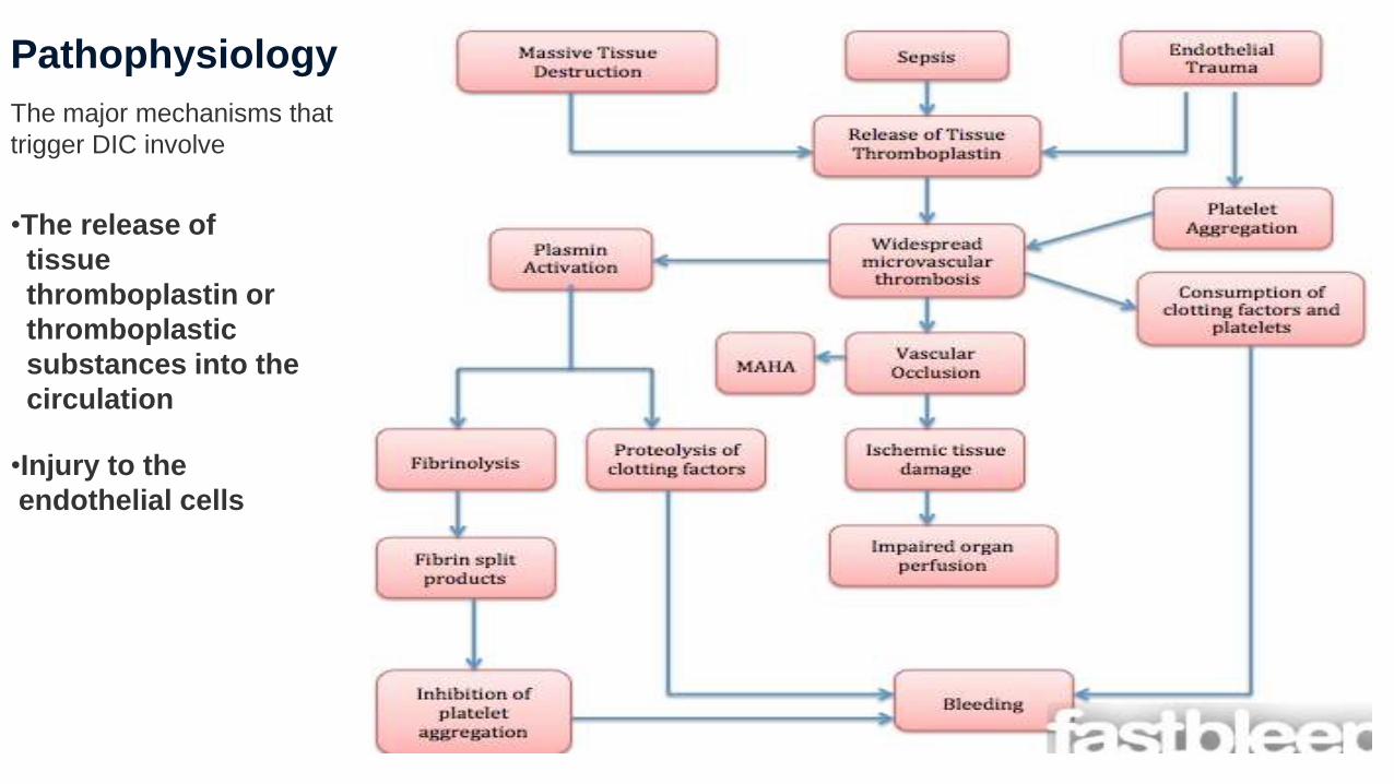
Pathophysiology
The major mechanisms that
trigger DIC involve
•The release of
tissue
thromboplastin or
thromboplastic
substances into the
circulation
•Injury to the
endothelial cells

Circulatory signs include the following:•Signs of spontaneous and life-threatening hemorrhage•Signs of sub acute bleeding•Signs of diffuse or localized thrombosis•Bleeding into serous cavities
CNS signs include the following:
•Nonspecific altered consciousness or stupor•Transient focal neurologic deficits
Cardiovascular signs include the following:
•Hypotension•Tachycardia•Circulatory collapse
Genitourinary signs include the following:•Signs of azotemia and renal failure•Acidosis•Hematuria•Oliguria•Metrorrhagia•Uterine hemorrhage
Dermatologic signs include the following:•Petechiae•Jaundice (liver dysfunction or hemolysis)•Purpura•Hemorrhagic bullae•Acral cyanosis•Skin necrosis of lower limbs (purpura fulminans)•Localized infarction and gangrene•Wound bleeding and deep subcutaneous hematomas•Thrombosis
SIGNS & SYMPTOMS

PHYSICAL EXAMINATION:
• Look for symptoms and signs of thrombosis in large vessels (eg, [DVT]) and microvascular thrombosis (as in renal failure).
• Bleeding from at least 3 unrelated sites is particularly suggestive of DIC. As many as 25% of patients present with renal failure.
• Patients with pulmonary involvement can present with dyspnoea, haemoptysis, and cough. Comorbid liver disease as well as rapid haemolytic bilirubin production may lead to jaundice.
• Neurologic changes (eg, coma, altered mental status, and paraesthesia) are also possible.
With acute DIC, the physical findings are usually those of the underlying or inciting condition; however, patients with the acute disease (i.e., the haemorrhagic variety associated with excess plasmin formation) have Petechiae on the soft palate, trunk, and extremities from thrombocytopenia and ecchymosis at venepuncture sites. These patients also manifest ecchymosis in traumatized areas.
In patients with so-called chronic or sub acute DIC, of which the primary manifestation is thrombosis from excess thrombin formation, the signs of venous thromboembolism may be present.
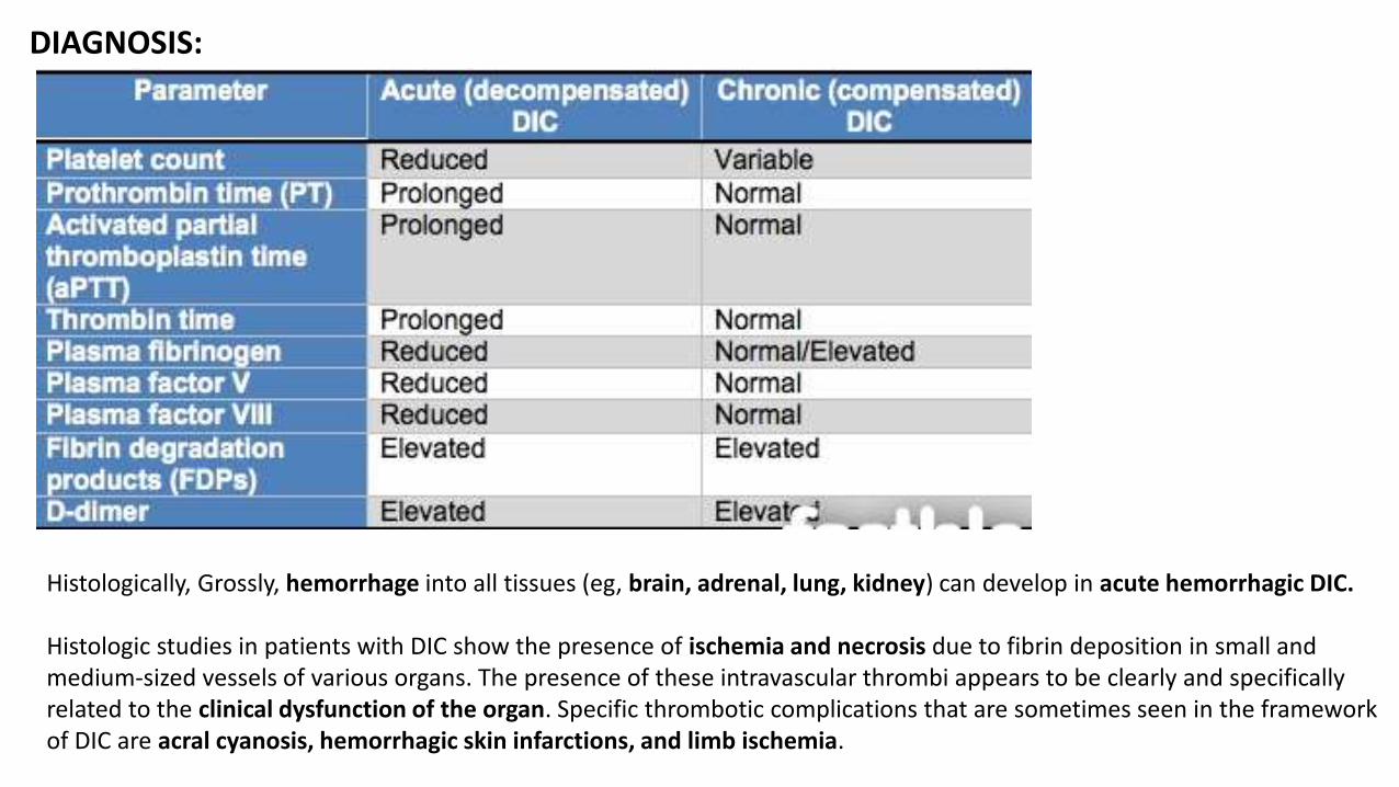
DIAGNOSIS:
Histologically, Grossly, hemorrhage into all tissues (eg, brain, adrenal, lung, kidney) can develop in acute hemorrhagic DIC.
Histologic studies in patients with DIC show the presence of ischemia and necrosis due to fibrin deposition in small and medium-sized vessels of various organs. The presence of these intravascular thrombi appears to be clearly and specifically related to the clinical dysfunction of the organ. Specific thrombotic complications that are sometimes seen in the framework of DIC are acral cyanosis, hemorrhagic skin infarctions, and limb ischemia.

The International Society on Thrombosis and Haemostasis (ISTH) Scoring System for Diagnosis of DIC is another
method used to diagnose DIC early in its course (sensitivity of 91-93% and a specificity of 97-98%).
• A score of five or more indicates a clear diagnosis
of DIC.
• A score of less than five does not rule out DIC, but
suggests that DIC is not definitive.

TREATMENT:
Treatment should primarily focus on addressing the underlying disorder.
Management of the DIC itself has the following basic features:
Monitor vital signs Assess and document the extent of haemorrhage and
thrombosis Correct hypovolemia Administer basic haemostatic procedures when indicated
First - ABCDE! When dealing with the critically ill, patients may become unresponsive and unconscious. When this type of
situation presents itself it is imperative to take the appropriate measures. The first step is to assess the patient’s Airway; if inadequate the patient may need intubation.
Next, the patient’s breathing adequacy is assessed. Consider ventilator support if necessary. In addition, if there is suspicion of a pneumothorax/haemothorax, decompression and drainage should be performed.
Thirdly, the patient’s circulation is evaluated. Administrating fluids and giving inotropes/vasopressors can aid the situation if circulation is compromised. Applying direct pressure to an external haemorrhage may also improve the patient’s circulation.

MEDICATION THERAPY:
The goals of pharmacotherapy in cases of disseminated intravascular coagulation (DIC) are to reduce morbidity and to prevent complications. Therapy should be based on aetiology and aimed at eliminating the underlying disease. Treatment should be appropriately aggressive for the patient’s age, disease, and severity and location of hemorrhage or thrombosis.
• ANTICOAGULANTS – HEPARIN , ANTITHROMBIN (ATRYN,THROMBATE III)Anticoagulants are used in the treatment of clinically evident intravascular thrombosis when the patient continues to bleed or clot 4-6 hours after initiation of primary and supportive therapy.
• RECOMBINATNT HUMAN ACTIVATED PROTEIN C Recombinant human APC inhibits factors Va and VIIIa of the coagulation cascade. It may also inhibit plasminogen activator inhibitor-1 (PAI-1)
• BLOOD COMPONENTSBlood components are used to correct abnormal hemostatic parameters(Packed red blood cells (PRBCs; washed), Platelets, Fresh frozen plasma (FFP),Cryoprecipitate or fibrinogen concentrates.

ANTI FIBRINOLYTIC AGENTS-Aminocaproic acid (Amicar),Tranexamic acid (Cyklokapron, Lysteda).
They may have a role in a local intravascular coagulation (LIC) as is seen in genitourinary bleeding after a transurethral resection for Kasabach-Merritt syndrome.In addition, in patients with trauma and massive bllood loss, in massive postpartum hemorrhage, in cases of DIC secondary to hyperfibrinolysis associated with acute promyelocytic leukemia and other forms of cancer when alpha-2-antiplasmin is uniquely decreased.
OTHERS:
Platelet transfusion may be considered in patients with DIC and severe thrombocytopenia, in particular, in patients with bleeding or in patients at risk for bleeding.
The consumption-induced deficiency of coagulation factors can be partially rectified by administering large quantities of FFP, particularly in patients with an international normalized ratio (INR) higher than 2.0, a 2-fold or greater prolongation of the aPTT, or a fibrinogen level below 100 mg/dL.The suggested starting dose is 15 mg/kg.
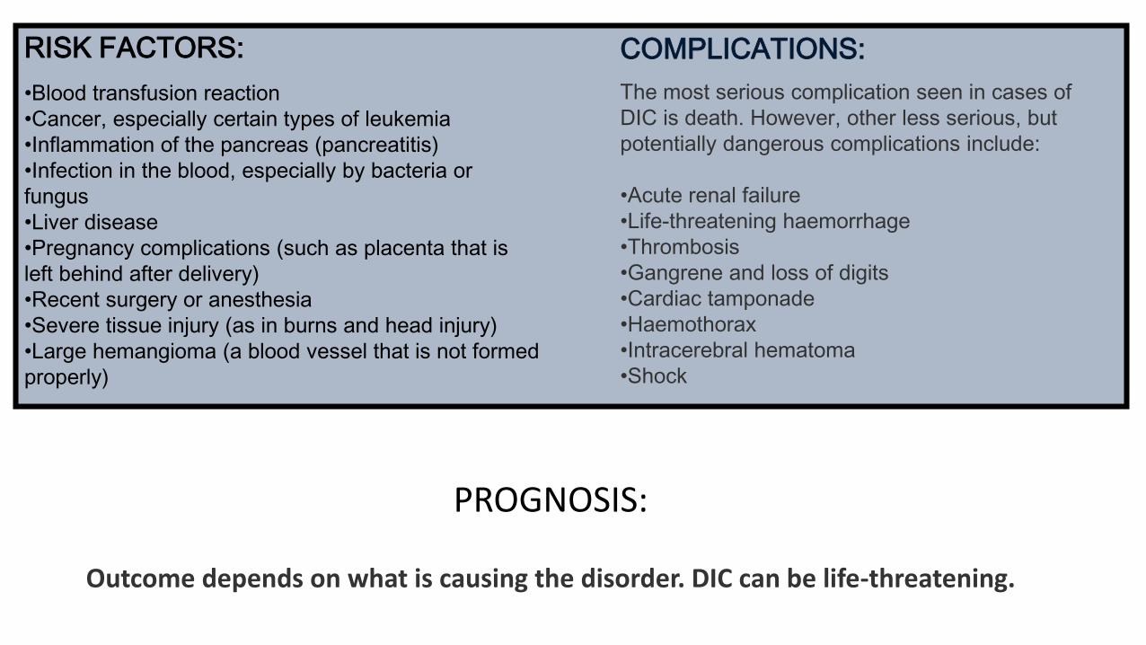
COMPLICATIONS:
The most serious complication seen in cases of
DIC is death. However, other less serious, but
potentially dangerous complications include:
•Acute renal failure
•Life-threatening haemorrhage
•Thrombosis
•Gangrene and loss of digits
•Cardiac tamponade
•Haemothorax
•Intracerebral hematoma
•Shock
PROGNOSIS:
Outcome depends on what is causing the disorder. DIC can be life-threatening.
RISK FACTORS:
•Blood transfusion reaction
•Cancer, especially certain types of leukemia
•Inflammation of the pancreas (pancreatitis)
•Infection in the blood, especially by bacteria or
fungus
•Liver disease
•Pregnancy complications (such as placenta that is
left behind after delivery)
•Recent surgery or anesthesia
•Severe tissue injury (as in burns and head injury)
•Large hemangioma (a blood vessel that is not formed
properly)

THANK YOU


