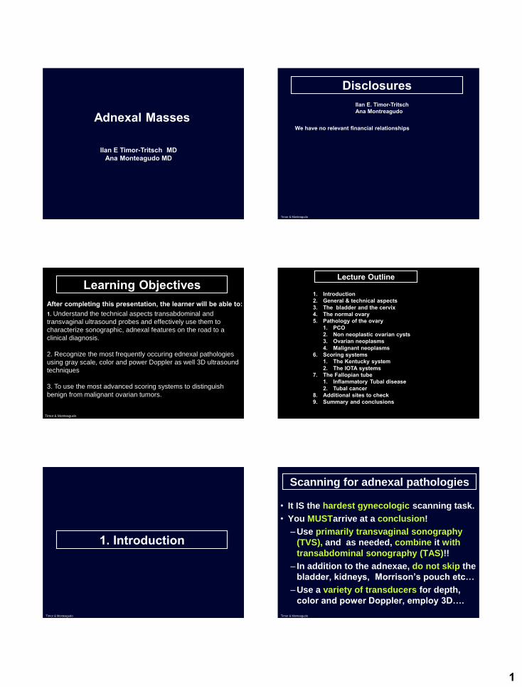
1 Adnexal Masses Ilan E Timor-Tritsch MD Ana Monteagudo MD Disclosures Ilan E. Timor-Tritsch Ana Montreagudo We have no relevant financial relationships Timor & Monteagudo Learning Objectives After completing this presentation, the learner will be able to: 1. Understand the technical aspects transabdominal and transvaginal ultrasound probes and effectively use them to characterize sonographic, adnexal features on the road to a clinical diagnosis. 2. Recognize the most frequently occuring ednexal pathologies using gray scale, color and power Doppler as well 3D ultrasound techniques 3. To use the most advanced scoring systems to distinguish benign from malignant ovarian tumors. Timor & Monteagudo Lecture Outline Timor & Monteagudo 1. Introduction 2. General & technical aspects 3. The bladder and the cervix 4. The normal ovary 5. Pathology of the ovary 1. PCO 2. Non neoplastic ovarian cysts 3. Ovarian neoplasms 4. Malignant neoplasms 6. Scoring systems 1. The Kentucky system 2. The IOTA systems 7. The Fallopian tube 1. Inflammatory Tubal disease 2. Tubal cancer 8. Additional sites to check 9. Summary and conclusions 1. Introduction Timor & Monteagudo Scanning for adnexal pathologies • It IS the hardest gynecologic scanning task. • You MUSTarrive at a conclusion! – Use primarily transvaginal sonography (TVS), and as needed, combine it with transabdominal sonography (TAS)!! – In addition to the adnexae, do not skip the bladder, kidneys, Morrison’s pouch etc… – Use a variety of transducers for depth, color and power Doppler, employ 3D…. Timor & Monteagudo
Transcript
1
Adnexal Masses
Ilan E Timor-Tritsch MDAna Monteagudo MD
DisclosuresIlan E. Timor-TritschAna Montreagudo
We have no relevant financial relationships
Timor & Monteagudo
Learning ObjectivesAfter completing this presentation, the learner will be able to:1. Understand the technical aspects transabdominal and
transvaginal ultrasound probes and effectively use them to
characterize sonographic, adnexal features on the road to a
clinical diagnosis.
2. Recognize the most frequently occuring ednexal pathologies
using gray scale, color and power Doppler as well 3D ultrasound
techniques
3. To use the most advanced scoring systems to distinguish
benign from malignant ovarian tumors.
Timor & Monteagudo
Lecture Outline
Timor & Monteagudo
1. Introduction2. General & technical aspects3. The bladder and the cervix4. The normal ovary5. Pathology of the ovary
1. PCO2. Non neoplastic ovarian cysts3. Ovarian neoplasms4. Malignant neoplasms
6. Scoring systems1. The Kentucky system2. The IOTA systems
7. The Fallopian tube1. Inflammatory Tubal disease2. Tubal cancer
8. Additional sites to check9. Summary and conclusions
1. Introduction
Timor & Monteagudo
Scanning for adnexal pathologies
• It IS the hardest gynecologic scanning task.
• You MUSTarrive at a conclusion!
– Use primarily transvaginal sonography
(TVS), and as needed, combine it with
transabdominal sonography (TAS)!!
– In addition to the adnexae, do not skip the
bladder, kidneys, Morrison’s pouch etc…
– Use a variety of transducers for depth,
color and power Doppler, employ 3D….
Timor & Monteagudo
2
Scanning for adnexal pathologies
• Remember: not all masses are ovarian.
• If you scan your own patient:
– Take a short history ; examine the patient before the scan, but do so after the scan to confirm your ultrasound findings.
• If you scan a referred patient:
– Take a short history yourself: don’t trust the referral slip; it is usually useless!!
– If in doubt: perform a bimanual exam yourself.
Timor & Monteagudo
Important!!
• In the reproductive years, physiologic as
well as pathologic processes are driven by
the menstrual cycle or by (therapeutic or
pathologic) hormonal stimulation.
• Know the day of your patients’ day of the
cycle, therefore…
Timor & Monteagudo
Important!!• ……clearly mark the LMP on the screen to
avoid erasure every time you unfreeze the
picture (type in the LMP or the letters PM 1998
[for: postmenopausal since 1998] to carry them
over to every picture).
• Judge EVERY US finding (ovarian findings, pelvic
fluid, endometrium etc) as a function of the
hormonal status (or day in cycle)
Timor & Monteagudo
2. General and Technical
Aspects
Timor & Monteagudo
Technical aspects
1. The most efficient pelvic evaluation is by using
transvaginal US probes.
(If the bladder is full you may want to do first a transabdominal scam)
2. Vaginal probes operate at frequencies of 5-9 (or 6-12) MHz.
3. Their most effective scanning depth is 2 to 10-12 cm.
What is the sliding organs sign…?Generated by the to-and-fro movement of the vaginal probe aided by the abdominal hand moving the cervix, uterine body, ovaries to evaluate their movement relative to the pelvic floor and/or each other, to diagnose or rule out pelvic adhesions.
Technical aspects
Timor & Monteagudo
3
Sliding organs sign
Useful to diagnose adhesions in the
pelvis as well as the upper abdomen.
Useful even at the time of laparoscopy
(selection of safe port placement site)
Example: If patient with infertility or
suspect for a frozen pelvis, a discrete
endometrioma on US has absent sliding
pelvic or abdominal organs, she most
probably has pelvic adhesions.
• * First described in: Transvaginal Sonography. (eds): Timor-Tritsch IE and Rottem S Elsevier Science Publishing Co. New York 1988; Pages 24,35,52,55,72,84
Timor & Monteagudo
Sliding organs sign: the ovaries
Timor & Monteagudo
Timor-Tritsch IE and Rottem S Elsevier Science Publishing Co. New York 1988; Pages 24,35,52,55,72,84
Record the mobility or fixed nature of pelvic organs
• Lately US machines are equiped with the ability to record scanning sequences using two kinds of features: “on-the-fly”(going forward) or “retro-view” (reviewing a structure just seen before)
• Use them to record the mobility (“sliding”), or fixed nature of pelvic organs.
• Add credibility to your report!• Acquire also a “sweep” of the adnexa Timor & Monteagudo
Even though it is not strictly the adnexum,* on the way in,
look at the bladder and the cervix.
* Latin: Adnexum = singular, adnexae = plural;
adnexa = grammatically incorrect but can be used, since it is
already deeply rooted in our vocabulary!Timor & Monteagudo
3. The Bladder and the cervix
4. The Normal Ovary
General
Timor & Monteagudo
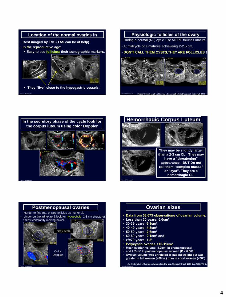
Location of the normal ovaries• Best imaged by TVS (TAS may be of help)
• In the reproductive age:
– Follicles are their sonographic markers.
– They “live” close to the hypogastric vessels.
– In the secretory phase look for the corpus luteum (CL) with color or power Doppler.
• In menopause:
– Harder to find (no, or rare follicles as markers).
– Linger on the adnexae and look for hypoechoic1-3-cm structures amidst constantly moving
bowel.
Timor & Monteagudo
4
Location of the normal ovaries in
• Best imaged by TVS (TAS can be of help)
• In the reproductive age:
• Easy to see follicles: their sonographic markers.
• They “live” close to the hypogastric vessels.
Timor & Monteagudo
Physiologic follicles of the ovary
• During a normal (NL) cycle 1 or MORE follicles mature.
• At midcycle one matures achieveing 2-2.5 cm.
• DON’T CALL THEM CYSTS,THEY ARE FOLLICLES !
Timor-Tritsch and Goldstein. Ultrasound Obstet Gynecol Editorial 2005Timor & Monteagudo
In the secretory phase of the cycle look for
the corpus luteum using color Doppler
Gray scale Color Doppler Power Doppler
Timor & Monteagudo
Hemorrhagic Corpus Luteum
They may be slightly larger
than a 2-3 cm CL. They may
have a “threatening”
appearance. BUT Do not
call them “complex masss”
or “cyst”. They are a
hemorrhagic CL!
Timor & Monteagudo
– Harder to find (no, or rare follicles as markers).
– Linger on the adnexae & look for hypoechoic 1-3 cm structures
amidst constantly moving bowel.
Postmenopausal ovaries
Gray scale
Color
Doppler
Timor & Monteagudo
Ovarian sizes• Data from 58,673 observations of ovarian volume. • Less than 30 years: 6.6cm³ • 30-39 years: 6.1cm³ • 40-49 years: 4.8cm³• 50-59 years: 2.6cm³ • 60-69 years: 2.1cm³ and • >/=70 years: 1.8³• Polycystic ovaries >10-11cm³ • Mean ovarian volume: 4.9cm³ in premenopausal• and 2.2cm³ in postmenopausal women (P < 0.001). • Ovarian volume was unrelated to patient weight but was
greater in tall women (>68 in.) than in short women (<58”)
. Pavlik EJ et al 1, Ovarian volume related to age. Gynecol Oncol. 2000 Jun;77(3):410-2. Timor & Monteagudo
5
5. Ovarian Pathology: What to look for?
Timor & Monteagudo
--Sassone AM1, Timor-Tritsch IE, Artner A, Westhoff C, Warren WB Transvaginal sonographic characterization of ovarian disease: evaluation of a new scoring system to predict ovarian
malignancy. Obstet Gynecol. 1991 Jul;78(1):70-6. --Timor-Tritsch IE, Goldstein SR: The simplicity of a simple cyst and the complexity of a complex
mass. JUM Editorial 2005--Timmerman D, Testa AC, Bourne T, et al. Simple ultrasound-based rules for the diagnosis of
ovarian cancer. Ultrasound Obstet Gynecol 2008;31(6):681-690--Testa AC et al. Ovarian cancer arising in endometrioid cysts: ultrasound findings. UOG 2011; 38: 99
--John R van Nagell Jr & John T Hoff: Transvaginal sonography in ovarian screening: current perspectives. International journal of womann’s health 2013
.
.
Ovarian lesions (findings)What do you look for?
• Internal echo structure (“echogenicity”):
– Anechoic (fluid component)
– Echogenic (solid component)
– Low-level echoes (ground glass appearance)
– Mixed echogenicity, reticular etc
• Wall structure:
– Thickness
– Internal and/or external papillae
(the moment you see papillae, apply power Doppler[not color!] and set it to the highest sensitivity to rule-in or-out blood flow). Blood vessels in a papilla is highly predictive of malignancy
SeptaeTimor & Monteagudo
Ovarian lesions (findings)What do you look for?
• Appearance:
• “Bizarre shapes”
• Mixed components
• Size
• Is it bilateral?
• Ascites
• Motion tenderness
• Vessels
• Sliding of the ovary
When these are
documented, the next
step is: LOOK AT THE
VASCULARITY.
Timor & Monteagudo
• Vascularity
– CAN ANY VESSELS BE SEEN AT ALL?
– If seen: Look for their qualitative appearance:• Location (central/peripheral)
• Amount of vascularity
• Tortuous appearance
• Caliber changes
• Anastomoses
• “Lakes”
– If seen: Measurements can be done (less used
lately, however a low RI & PI is common in cancer):
Ovarian lesions (findings)What do you look for?
Timor & Monteagudo
Ovarian lesions:What do you look for?
• General appearance
– Solid
– Cystic:
• without solid component
• With solid component
– Unilocular, Multilocular,
Timor & Monteagudo
Ovarian lesions: What do you look for?
• Internal echo structure:
– Anechoic/hypoechoic
– Echogenic (solid)
– Low-level echoes (ground glass appearance)
– Mixed echogenicity
– Reticular, etc
Timor & Monteagudo
6
Ovarian lesions (findings)What do you look for?
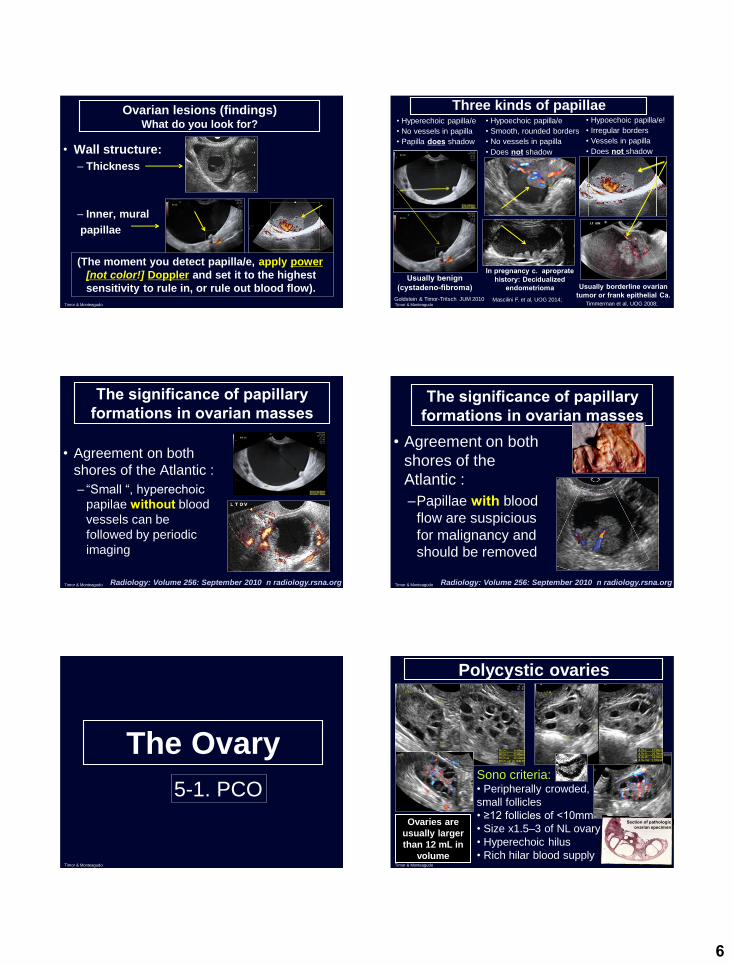
• Wall structure:
– Thickness
– Inner, mural
papillae
(The moment you detect papilla/e, apply power
[not color!] Doppler and set it to the highest
sensitivity to rule in, or rule out blood flow).
Timor & Monteagudo
Three kinds of papillae• Hyperechoic papilla/e
• No vessels in papilla
• Papilla does shadow
• Hypoechoic papilla/e!
• Irregular borders
• Vessels in papilla
• Does not shadow
Usually benign (cystadeno-fibroma)
Goldstein & Timor-Tritsch JUM 2010
Usually borderline ovarian tumor or frank epithelial Ca.
In pregnancy c. aproprate history: Decidualized
endometrioma
• Hypoechoic papilla/e
• Smooth, rounded borders
• No vessels in papilla
• Does not shadow
Mascilini F. et al, UOG 2014;Timor & Monteagudo Timmerman et al, UOG 2008;
The significance of papillary formations in ovarian masses
• Agreement on both
shores of the Atlantic :
– “Small “, hyperechoic
papilae without blood
vessels can be
followed by periodic
imaging
Radiology: Volume 256: September 2010 n radiology.rsna.orgTimor & Monteagudo
The significance of papillary formations in ovarian masses
• Agreement on both
shores of the
Atlantic :
–Papillae with blood
flow are suspicious
for malignancy and
should be removed
Radiology: Volume 256: September 2010 n radiology.rsna.orgTimor & Monteagudo
The Ovary
5-1. PCO
Timor & Monteagudo
Polycystic ovaries
Sono criteria:• Peripherally crowded,
small follicles
• ≥12 follicles of <10mm
• Size x1.5–3 of NL ovary
• Hyperechoic hilus
• Rich hilar blood supply
Ovaries are
usually larger
than 12 mL in
volume
Section of pathologic ovarian specimen
Timor & Monteagudo
7
True PCO or only “sonographic PCO,”
aka multicystic ovary?
• Not every ovary that fulfills the sono
criteria is a PCO syndrome!
• An ovary can have a PCO appearance in
the following clinical situations:
– Hyperthyroid state (36%)
– Hyperprolactinemia (50%)
– Hypothalamic hypogonadism (24%)
– Or without any known reasonTimor & Monteagudo
Pay attention! Day 10 of cycle
Paraovarian/paratubal cyst
Frequently seen, benign appearing cysts with the following sono markers: 1.Very thin,
2.smooth wall.
3. Anechoic
4. Unilociular
5. Ipsilateral ovary HAS TO BE SEEN!!!
Timor & Monteagudo
Postoperative peritoneal inclusion cysts in loculated pelvic fluid
Sohaey R, Gardner TL, Woodward PJ, Peterson CM. Sonographic diagnosis of peritoneal inclusion cysts. J Ultrasound Med 1995; 14:913-917
• The Dx should be suspected in the right clinical setting.
• Dx depends on the presence of normal ipsilateral ovary with surrounding loculated fluid conforming to the peritoneal space.
Timor & Monteagudo
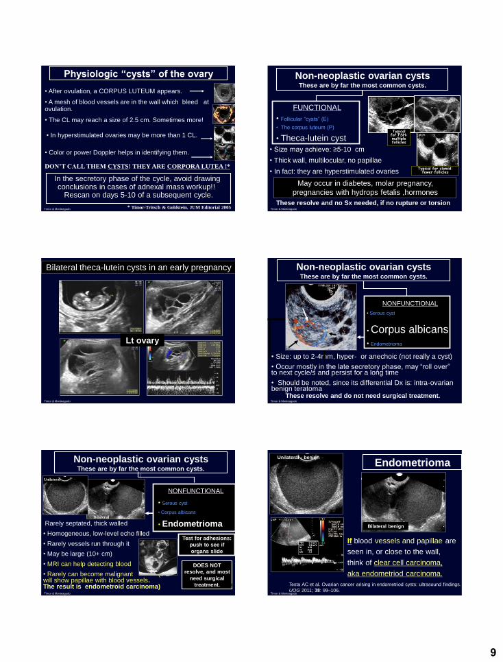
5-2. Non neoplastic ovarian cysts
Non-neoplastic ovarian cystsThese are by far the most common cystic
structures.
FUNCTIONAL
• Follicular “cysts” (E)
• Corpus luteum (P)
• Theca-lutein cyst (E)
NONFUNCTIONAL
• Serous cyst
• Corpus albicans
• Endometrioma
Except the endometrioma: most resolve and do not need
surgical treatment, provided they do not twist. If Dx. in
doubt, scan the patient in the next cycle (days 5-9).
E: estrogen secreting; P: progesteron secreting
Timor & Monteagudo
Non-neoplastic ovarian cystsThese are by far the most common cysts.
FUNCTIONAL
• Follicular “cysts” (E)
• aka SIMPLE CYSTS
• The corpus luteum (P)
• Theca-lutein cyst (E)
These resolve and no surgery (Sx) needed, provided no rupture or torsion exists.
• Size: up to 4-5 cm, sometimes more
• Smooth wall, unilocular, no papillae
• Lined with flat granulosa cells
• Circular blood flow around the wall
• Almost never malignant (<½%)
• No additional information by MRI
Merz: Ultrasound Obstet
Gynecol 1999;14:81
MRI
Timor & Monteagudo
8
--Alcázar JL et al Is expectant management of sonographically benign adnexal cysts an option in
• Translate macroscopic, clinical, and pathologic features and appearances to sonographically recognizable features.
• All or most sono-scoring systems are based upon the same building blocks:
– Wall thickness
– Septations
– Echogenicity
– Papillary formations
– Solid components
– Blood supply (vascularity)
• Some systems add: size, ascites, age, etc… Timor & Monteagudo
15
• Sassone M, Timor-Tritsch et al, AJOG 1991
• Kentucky. DePriest et al, Gynecol Oncol 1997
• 1993; Osmers, AJOG 1994
• Bromley et al, Obstet Gynecol 1994
• Lerner JP, Timor-Tritsch al, AJOG 1994
• Kurjak, UOG 1994
• Ferazzi, UOG 1998
• IOT A. Timmerman, UOG 1999 (Neural Network analysis)
You may use Morphology Scoring Systems: they are out there.
However, you do not have to apply them to the letter.
Just understand their basic idea to differentiate benign
tumor &from suspicious or malignantTimor & Monteagudo
The first
sonographic
scoring system
published
Timor & Monteagudo
Sonographic images of benign and malignant ovarian morphology. Numeric representation of increasing morphologic complexity is noted in the first column.
John R van Nagell Jr & John T Hoff: Transvaginal sonography in ovarian screening: current perspectives. International journal of womann’s health 2013Timor & Monteagudo
6-1. The Kentucky scoring system
The simple rules by the IOTA group
But first : What is the IOTA group?
Timor & Monteagudo
6-2.The IOTA scoring systems
The IOTA group• The International Ovarian Tumor Analysis (IOTA)
group was founded in 1999 by Dirk Timmerman, Lil
Valentin and Tom Bourne.
• Its first aim was to develop standardized terminology.
• In 2000, IOTA published a consensus statement on
terms, definitions and measurements to describe the
sonographic features of adnexal masses, which is
now widely used.
• IOTA now covers a multitude of studies examining
many aspects of gynecological ultrasonography
within a network of contributing centers throughout
the world that are coordinated from KU Leuven.Timor & Monteagudo
Risk assessment of adnexal masses based on the IOTA
WYNANTS, MSc6,7, Caroline VAN HOLSBEKE, MD, PhD2,8, Elisabeth EPSTEIN, MD, PhD9, Dorella FRANCHI, MD10, Jeroen KAIJSER, MD, PhD2,11, Artur CZEKIERDOWKSI, MD, PhD12,
Stefano GUERRIERO, MD, PhD13, Robert FRUSCIO, MD, PhD14, Francesco PG LEONE, MD15, Alberto ROSSI, MD16, Chiara LANDOLFO, MD1,2, Ignace VERGOTE, MD, PhD2,17, Tom
BOURNE, MD, PhD1,2,18, Lil VALENTIN, MD, PhD19AJOG 2016
* Joint first author
Timor & Monteagudo
16
Background
• Accurate methods to preoperatively characterize
adnexal tumors are pivotal for optimal patient
management.
• A recent meta-analysis* concluded that the
International Ovarian Tumor Analysis (IOTA)
algorithms such as the Simple Rules are the best
approaches to preoperatively classify adnexal
masses as benign or malignant.
(*) Kaijser J, Sayasneh A, Van Hoorde K, et al. Presurgical diagnosis of adnexal tumours using mathematical models and scoring systems: A systematic review and meta-analysis. Hum Reprod
Update 2014;20(3):449-462.
Timor & Monteagudo
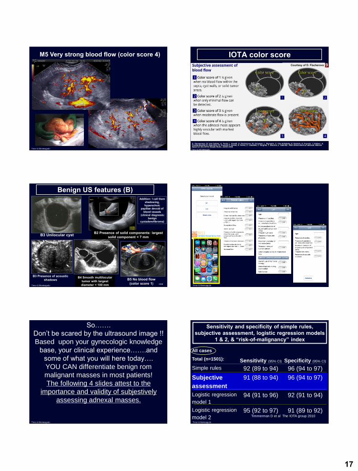
IOTA Simple RulesUltrasound features predictive for a malignant tumor (M-features)
Features predictive for a benign tumor (B-features)
M1 Irregular solid tumor B1 Unilocular
M2 Presence of ascites B2 Presence of solid components where the largest solid component has a largest diameter < 7 mm
M3 At least 4 papillary structures B3 Presence of acoustic shadows
M4 Irregular multilocular-solid tumor with largest diameter ≥ 100 mm
B4 Smooth multilocular tumor with largest diameter < 100 mm
M5 Very strong blood flow (color score 4)
B5 No blood flow (color score 1)
Timmerman D, Testa AC, Bourne T, et al. Simple ultrasound-based rules for the diagnosis of ovarian cancer. Ultrasound Obstet Gynecol 2008;31(6):681-690.
Timor & Monteagudo
IOTA Simple Rules
Timmerman D, Testa AC, Bourne T, et al. Simple ultrasound-based rules for the
diagnosis of ovarian cancer. Ultrasound Obstet Gynecol 2008;31(6):681-690.
Ultrasound features used in the International OvarianTumor Analysis (IOTA) simple rules, illustrated by ultrasoundimages. B1–B5, benign features; M1–M5, malignant features.
Timor & Monteagudo
IOTA Simple Rules
International Ovarian Tumor Analysis (IOTA)
‘easy descriptors’ illustrated by
ultrasound images. BD1–BD4, benign
descriptors;MD1–MD2, malignant
descriptors.
Timmerman D, Testa AC, Bourne T, et al. Simple ultrasound-based rules for the
diagnosis of ovarian cancer. Ultrasound Obstet Gynecol 2008;31(6):681-690.
Timor & Monteagudo
IOTA Simple Rules• If one or more M-features apply in the absence of a
B-feature, the mass is classified as malignant.
• If one or more B-features apply in the absence of an
M-feature, the mass is classified as benign.
• If both M-features and B-features apply, the mass
cannot be classified. If no feature applies, the mass
cannot be classified.
• Correct application of the Simple Rules requires the
knowledge and proper use of the ultrasound
features, as published by the IOTA group.*
• Timmerman D, Testa AC, Bourne T, et al. Simple ultrasound-based rules for the diagnosis of ovarian cancer. Ultrasound Obstet Gynecol 2008;31(6):681-690.
Timor & Monteagudo
Malignant US features (M)
M1 Irregular solid tumor M2 Presence of ascites
M4 Irregular multilocular-solid tumor: largest diameter ≥ 100 mm
D. Timmerman, B. Van Calster, A. Testa, L. Savelli, D. Fischerova, W. Froyman, L. Wynants, C. Van Holsbeke, E. Epstein, D. Franchi, J. Kaijser, A. Czekierdowski, S. Guerriero, R. Fruscio, F. Leone, A. Rossi, C. Landolfo, I. Vergote, T. Bourne, L. Valentin. Risk assessment of adnexal masses based on the IOTA Simple Rules. AJOG 2016.
2nd step in a pelvic inflammatory process: Tubo-ovarian-complex (TOC)
7-2. Tubal cancer
Timor & Monteagudo
21
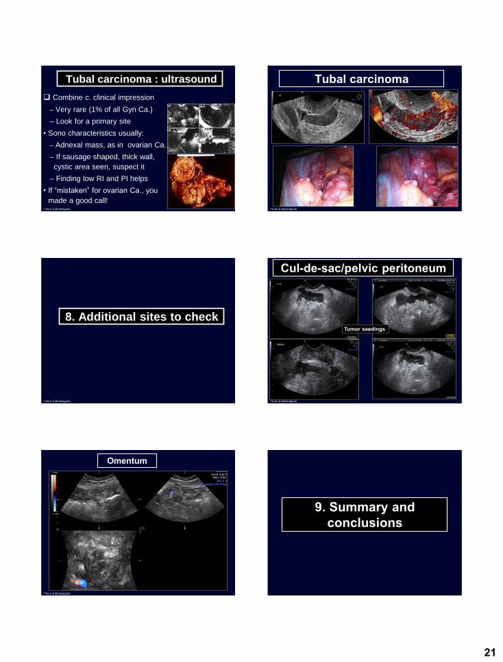
Tubal carcinoma : ultrasound
Combine c. clinical impression
– Very rare (1% of all Gyn Ca.)
– Look for a primary site
• Sono characteristics usually:
– Adnexal mass, as in ovarian Ca.
– If sausage shaped, thick wall,
cystic area seen, suspect it
– Finding low RI and PI helps
• If “mistaken” for ovarian Ca., you
made a good call!Timor & Monteagudo
Tubal carcinoma
Timor & Monteagudo
8. Additional sites to check
Timor & Monteagudo
Cul-de-sac/pelvic peritoneum
Tumor seedings
Timor & Monteagudo
Omentum
Timor & Monteagudo
9. Summary and conclusions
8/8/2017
22
Summary and conclusions• Most of the time adnexal masses carry defined
sono characteristics and pathognomonic
features (markers)
• The main sono markers of the commonly seen
adnexal masses were described to enable a
better recognition of their possible histology
• Where relevant, clinical features helping the
diagnosis were mentioned
• Where applicable, relevant articles from the
contemporary literature were quotedTimor & Monteagudo
Conclusions• Most adnexal masses can be assessed
subjectively using:
– A transvaginal US probe (TAS if large mass)
– An enhanced basic US knowledge (Reading
REVIEWS)
– Liberal use of power Doppler
– Recognizing benign and malignant sono markers
• If you like to use the term : “complex mass”,
describe the mass in terms of their sonographic
characretistics (possibly the IOTA descriptors)
Timor & Monteagudo
Conclusions• Avoid the word “cyst” referring to follicles or
corpora lutea
• Be attuned to the issues of papillae in a cyst (size, number, blood vessels in it)
• Avoid the sentence: “…malignancy can not be
ruled out”, use it when really needed
• Use the sentence: ”My suspicion of the
structure to be malignant is: high, moderate,
low, none or can not classify”
• Ask for the help of a GO when in real need
Timor & Monteagudo
Benacerraf BR, Abuhamad AZ, Bromley B, Goldstein SR, Groszman Y, Shipp TD, Timor-Trisch IE. Consider ultrasound first for imaging the female pelvis. Am J Obstet Gynecol 2015; 212: 450-5
--Sassone AM1, Timor-Tritsch IE, Artner A, Westhoff C, Warren WB Transvaginal sonographic characterization of
ovarian disease: evaluation of a new scoring system to predict ovarian malignancy. Obstet Gynecol. 1991 Jul;78(1):70-6.
--Timor-Tritsch IE, Goldstein SR: The simplicity of a simple cyst and the complexity of a complex mass. JUM Editorial 2005
--Timmerman D, Testa AC, Bourne T, et al. Simple ultrasound-based rules for the diagnosis of ovarian cancer.
Ultrasound Obstet Gynecol 2008;31(6):681-690--Testa AC et al. Ovarian cancer arising in endometrioid cysts: ultrasound findings. UOG 2011; 38: 99
--John R van Nagell Jr & John T Hoff: Transvaginal sonography in ovarian screening: current perspectives. International journal of womann’s health 2013
--Radiology: Volume 256: September 2010 n radiology
-- Sohaey R, Gardner TL, Woodward PJ, Peterson CM. Sonographic diagnosis of peritoneal inclusion cysts. J Ultrasound Med 1995; 14:913-917
--Sohaey R, Gardner TL, Woodward PJ, Peterson CM. Sonographic diagnosis of peritoneal inclusion cysts. J Ultrasound Med 1995; 14:913-917
-- Modesitt SC et al Risk of malignancy in unilocular ovarian cystic tumors less than 10 cm in diameter. Obstet Gynecol
2003;102:594–9-- Saunders BA, et al. Risk of malignancy in sonographically confirmed septated cystic ovarian tumors. Gynecol Oncol
2010;188:278–82--Fruscella E et al. Sonographic features of decidualized ovarian endometriosis suspicious for malignancy..UOG 2004;
24: 578
-- Mascilini F. et al, Imaging in gynecological disease. 10: Clinical and ultrasound characteristics of decidualizedendometriomas surgically removed during pregnancy. UOG 2014;44):354-60.
-- Monteagudo A et al. Ovarian steroid cell tumors: sonographic characteristics. UOG 1997;10:282.-- Jeong-Ah Kim et al. High-Resolution Sonographic Findings
of Ovarian Granulosa Cell Tumors JUM 2010; 29:187–19
--van Nagell JR Jr,, Miller, RW. Management of Asymptomatic Ovarian Tumors Obstet Gynecol 2016;127:848–58-- John R van Nagell Jr & John T Hoff: Transvaginal sonography in ovarian screening: current perspectives.