Page 1
University of Khartoum
Graduate College
Medical and Health Studies Board
Ultrasonographic Assessment of the Gallbladder
Motor Function
BY
Dr. Mohammed Hassan Nasr
MBBS(U of K )
A thesis Submitted in partial fulfillment for the
requirements of the Degree of Clinical M.D in Radiology
Supervisor
Prof . Mutasim Ahmed Elseed
2001
Page 2
DEDICATION
TO
MY mother , father ,
wife ,Children ,
brothers , sisters
&
friends
Page 3
الرحيم الرحمن اهللا بسم
: تعالى قال
العظيم اهللا صدق
Page 4
Table of Contents
Dedication
Table of Contents i
Acknowledgments ii
English Abstract iii
Arabic Abstract iv
List of figures v
List of Tables vi
CHAPTER ONE
INTRUDUCTION 1
LITERATURE REVIEW 3
OBJECTIVES 29
CHAPTER TWO
PATIENTS & METHODES 30
CHAPTER THREE
RESULTS 33
CHAPTER FOUR
DISCUSSION 50
CONCLUSION 52
RECOMMENDATION 53
REFERENCES 54
i
Page 5
Acknowledgement
Great thanks and good prayers for my supervisor professor Mutasim Ahmed El
seed , consultant radiologist khartoum Teaching Hospital , for his invaluable help ,
advice and close supervision .
I would also like to acknowledge Dr Osman Abdall Wahab and Dr. Tarig Ali
yousif . consultants Radiologist for their great help and advice which put me in he
first step of the study , and good prayers for them .
I would also like to thank Mr. Abdalla El Ata for his continuous encouragement
and follow up .
Thank to all senior Radiologists , to all Surgical and Medical Department in
Omdurman Military Hospital and specially for the ultrasound Department .
Also I am deeply indebted foe the skill and support of Miss . Widad A.Magsood for
her fine prints and final arrangement .
Thanks and prayers for all who gave me parte of their lives in the form of help
ii
Page 6
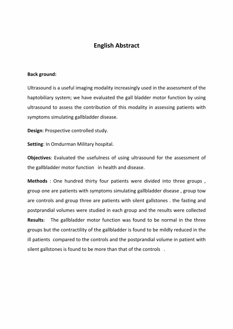
English Abstract
Back ground:
Ultrasound is a useful imaging modality increasingly used in the assessment of the
haptobiliary system; we have evaluated the gall bladder motor function by using
ultrasound to assess the contribution of this modality in assessing patients with
symptoms simulating gallbladder disease.
Design: Prospective controlled study.
Setting: In Omdurman Military hospital.
Objectives: Evaluated the usefulness of using ultrasound for the assessment of
the gallbladder motor function in health and disease.
Methods : One hundred thirty four patients were divided into three groups ,
group one are patients with symptoms simulating gallbladder disease , group tow
are controls and group three are patients with silent gallstones . the fasting and
postprandial volumes were studied in each group and the results were collected
Results: The gallbladder motor function was found to be normal in the three
groups but the contractility of the gallbladder is found to be mildly reduced in the
ill patients compared to the controls and the postprandial volume in patient with
silent gallstones is found to be more than that of the controls .
Page 7
Conclusion: There are some variations in the volumes of the gallbladders in the healthy Sudanese people .The gallbladder motor function in the diseased patients is reduced probably as other body functions are reduced and further study is needed. The gallbladder containing silent stones remains normally functioning unless complicated with infection, obstruction, etc …
iii
Page 8
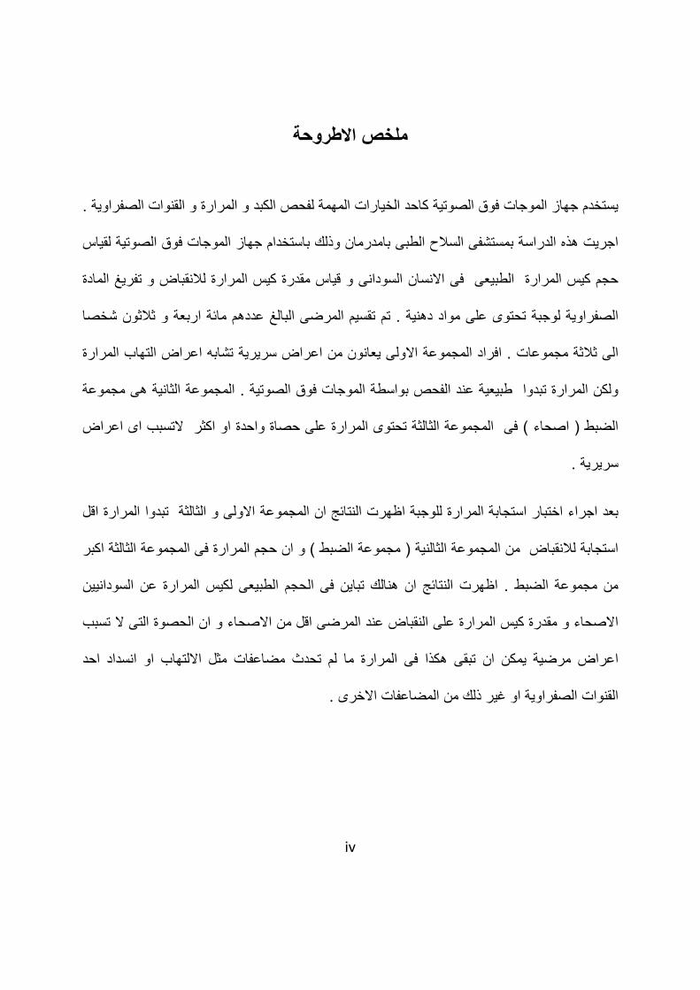
االطروحة ملخص
. الصفراوية القنوات و المرارة و الكبد لفحص المهمة الخيارات آاحد الصوتية فوق الموجات جهاز يستخدم
لقياس الصوتية فوق الموجات جهاز باستخدام وذلك بامدرمان الطبى السالح بمستشفى الدراسة هذه اجريت
المادة تفريغ و لالنقباض المرارة آيس مقدرة قياس و السودانى االنسان فى الطبيعى المرارة آيس حجم
شخصا ثالثون و اربعة مائة عددهم البالغ المرضى تقسيم تم. دهنية مواد على تحتوى لوجبة الصفراوية
المرارة التهاب اعراض تشابه سريرية اعراض من يعانون االولى المجموعة افراد. مجموعات ثالثة الى
مجموعة هى الثانية المجموعة. الصوتية فوق الموجات بواسطة الفحص عند طبيعية تبدوا المرارة ولكن
اعراض اى التسبب اآثر او واحدة حصاة على المرارة تحتوى الثالثة المجموعة فى) اصحاء( الضبط
. سريرية
اقل المرارة تبدوا الثالثة و االولى المجموعة ان النتائج اظهرت للوجبة المرارة استجابة اختبار اجراء بعد
اآبر الثالثة المجموعة فى المرارة حجم ان و) الضبط مجموعة( الثالنية المجموعة من لالنقباض استجابة
السودانيين عن المرارة لكيس الطبيعى الحجم فى تباين هنالك ان النتائج اظهرت. الضبط مجموعة من
تسبب ال التى الحصوة ان و االصحاء من اقل المرضى عند النقباض على المرارة آيس مقدرة و االصحاء
احد انسداد او االلتهاب مثل مضاعفات تحدث لم ما المرارة فى هكذا تبقى ان يمكن مرضية اعراض
. االخرى المضاعفات من ذلك غير او الصفراوية القنوات
iv
Page 9
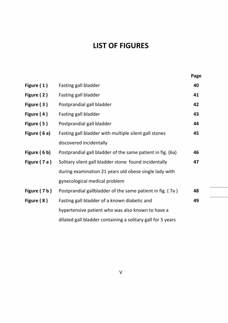
LIST OF FIGURES
Page
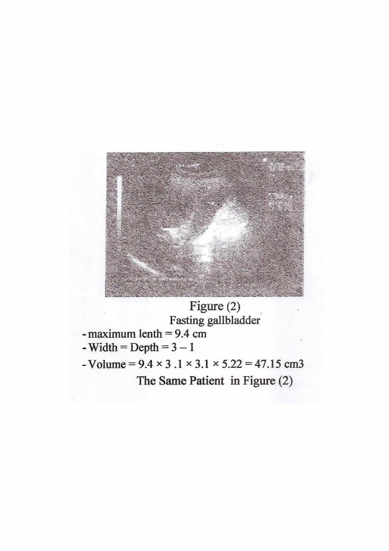
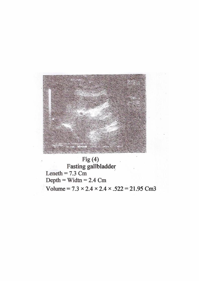
Figure ( 1 ) Fasting gall bladder 40
Figure ( 2 ) Fasting gall bladder 41
Figure ( 3 ) Postprandial gall bladder 42
Figure ( 4 ) Fasting gall bladder 43
Figure ( 5 ) Postprandial gall bladder 44
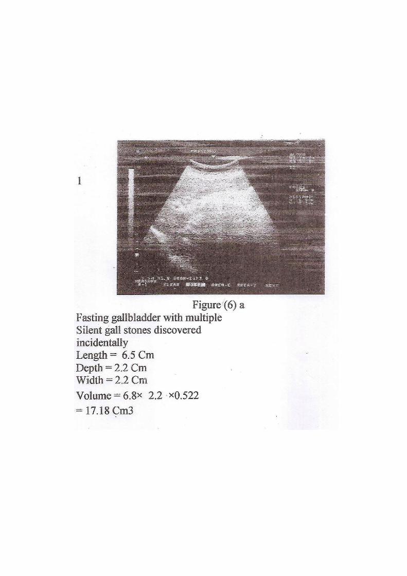
Figure ( 6 a) Fasting gall bladder with multiple silent gall stones
discovered incidentally
45
Figure ( 6 b) Postprandial gall bladder of the same patient in fig. (6a) 46
Figure ( 7 a ) Solitary silent gall bladder stone found incidentally
during examination 21 years old obese single lady with
gynecological medical problem
47
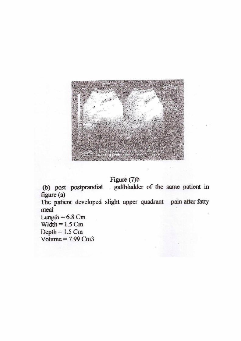
Figure ( 7 b ) Postprandial gallbladder of the same patient in fig. ( 7a ) 48
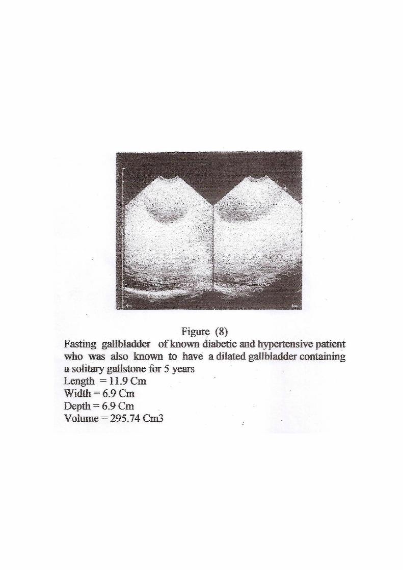
Figure ( 8 ) Fasting gall bladder of a known diabetic and
hypertensive patient who was also known to have a
dilated gall bladder containing a solitary gall for 5 years
49
V
Page 10
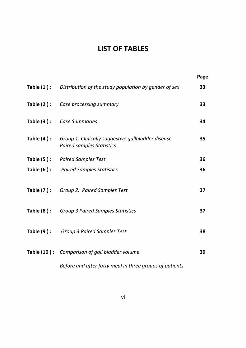
LIST OF TABLES
Page
Table (1 ) : Distribution of the study population by gender of sex
33
Table (2 ) : Case processing summary
33
Table (3 ) : Case Summaries
34
Table (4 ) : Group 1: Clinically suggestive gallbladder disease.Paired samples Statistics
35
Table (5 ) : Paired Samples Test 36
Table (6 ) : .Paired Samples Statistics
36
Table (7 ) : Group 2. Paired Samples Test
37
Table (8 ) : Group 3 Paired Samples Statistics
37
Table (9 ) : Group 3.Paired Samples Test
38
Table (10 ) : Comparison of gall bladder volume Before and after fatty meal in three groups of patients
39
vi
Page 11
Chapter one
INTRODUCTION & LITRETURE REVEW
The gall bladder is a muscular organ that serves as a reservoir for bile.
It is present in most vertebrates. In humans it is a pear shaped membranous
sac on the under-surface of the right lobe of the liver just below the ribs. It
does many important functions aiming to promote digestive process. The gall
bladder as other body organs can be abnormally located or congenitally
malformed. Also it is vulnerable to be affected by many disorders e.g. stone
formation, infectious, inflammatory and neoplastic disorders. Ultrasound,
cholescintigraphy, percutaneous transhepatic cholangiography (P.T.C),
endoscopic retrograde cholangiopancreatography (ERCP), plain radiography
and tomography are all used to investigate the gall bladder and biliary ducts
in different pathological conditions.
As imaging modalities are complementary and not competitive the
choice of the modality used depends on the clinical situation and ultrasound is
usually the primary investigation to begin with. Ultrasound is the best of all
methods of investigation of the gallbladder and biliary duct diseases, it is the
simplest and best test for showing gall stones and other diseases of the gall
bladder, and is also excellent for confirming or excluding bile ducts
dilatation(7).
Oral cholecystography has a very limited role nowadays and has been
largely abandoned as a diagnostic test. Radio-nuclide imaging using
hepatobiliary agent has an important role in excluding obstruction to the
Page 12
cystic duct. CT -scanning can demonstrate gall bladder stones, wall thickness
and dilatation of the common bile duct.
Endoscopic retrograde cholangio-pancreatogrophy
(E.R.C.P) and percutaneous transhepatic cholangiography (P.T.C) are more
Invasive techniques used for selected cases only.
The gallbladder has to contract efficiently to eject bile into the
duodenum in response to presence of food there to facilitate the digestive
process. Assessment of the gallstones motor function is of paramount
importance because of playing important role in:
1- The course and pathogenesis of cholelithiasis.
2- Dissolution of gallstones by ingestion of bile acids.
3- Lithotripsy.
4- Management of biliary dyskinesia.
Ultrasound, cholescintigraphy and oral cholecystograph are all used
and found to be reliable in assessing the gall bladder contractility and
emptying. Each imaging modality has its advantages and disadvantages, but
ultrasonography is the cheapest, simplest, non-invasive and readily available,
so it is the modality selected to assess the gallbladder motor function and
emptying in the study.
Page 13
1.2. LITERATURE REVIEW
Embryology:
The hepatic diverticulum arises from the ventral wall of the forgut and
is elongated into a stalk to form the choledocus. A lateral bud is given off,
which is distended to form the gallbladder and cystic duct. The embryonic
hepatic duct sends out many branches which join up with the canaliculi
between the liver cells. As is usual with the embryonic tubular structures,
hyperplasia obliterates the lumina of this ductal system, but normally
recanalization occurs subsequently and bile begins to flow. During early fetal
life the gallbladder is entirely intrahepatic (1).
Anatomy:
The gallbladder is a pear shaped sac lying on the undersurface of the
liver. The anatomical subdivisions are a fundus, a body and a neck, which
terminates into a narrow infundibulum . It is about 7.5 - 12.5 cm long and 2.5
- 4cm wide. It has a capacity of 30-50 ml, but capable of considerable
dialatation in certain pathological conditions. The muscle fibers in the wall of
the gallbladder are arranged in criss-cross manner, being particularly well
developed in the neck. The mucous membrane contains indentation of the
mucosa that sinks into the muscle coat; these are the crepts of Lushka.
Blood supply:
Arteries: the cystic artery, a branch of the right hepatic artery is
usually given off behind the common hepatic duct. Occasionally an accessory
cystic artery arises from the gastro duodenal artery. In 15% of cases the right
Page 14
hepatic artery and/ or the cystic artery cross in front of the common bile duct
and the cystic duct. The most dangerous anomalies are when the hepatic
artery takes a tortuous course in front of the origin of the cystic duct or the
right hepatic artery is tortuous and the cystic artery is short, because there is
a possibility of liver infarction in accidental ligation of the common hepatic
artery(1).
Lymph drainage:
The lymph vessels of the gall bladder (subserosal and submucous)
drains into the cystic lymph nodes of Lund (the sentinel lymph node) which lies
in the fork created by the junction of the cystic and common hepatic ducts.
Efferent vessels from this lymph node go to the hilum of the liver and to the
celiac lymph nodes. The subserosal lymphatic vessels of the gall bladder also
connect with the subcapsular lymph channels of the liver and this account the
frequent spread of carcinoma of the gallbladder to the liver(1).
Venous drainage:
Cystic vein drains into the portal vein.
Nerve supply:
Sympathetic and parasympathetic nerve supply is from the celiac
plexus. The gallbladder contracts in response to the hormone cholecystokinin-
panceozymin which is produced by the mucus membranes of the duodenum on
the arrival of fat from the stomach (2).
Page 15
Intrahepatic bile duct anatomy:
Bile drains from the ductular and canalicular network of the acini.
These ducts run with the branches of the portal vein and hepatic artery in the
portal triad. The smallest interlobular ducts join to form sepal bile ducts and
these finally unit to form the right and left hepatic ducts. The liver is divided
into two major parts and a caudate lobe. The left (segment 2-3) and right
(segment, 4 -8).The tow halves are divided by the principle plain that passes
through the middle of the gallbladder bed anteriorly to the left side of the
inferior vena cava posteriorly, each of these halves is then divided into two
sectors by the right and left fissures, corresponding to the line of the left and
right hepatic veins. The caudate lobe termed segment I is best considered as
an autonomous part of the liver with separate vascular and biliary apparatus,
the left hepatic duct drains the three segments of the left liver and the right
hepatic duct, the four segments of the right liver. The right hepatic duct arises
from the union of the main sectorial ducts.
Extra hepatic duct anatomy:
The right and left hepatic ducts fuse at the hilum, anterior to the
bifurcation of the portal vein to form the common hepatic duct which runs
caudally in the free edge of the omentum. The extra hepatic segment of the
right duct is short but the left duct has a much longer extra hepatic course and
hence when exposed surgically at the level of the hilar plate can facilitate a
wider biliary enteric anastomosis. The main bile duct is divided into two
segments. The common hepatic duct and the common bile duct divided by the
Page 16
cystic duct insertion whilst the cystic duct joins the common hepatic duct in its
supraduodenal segment In 80%. It may extend downward to a retro duodenal
or retro pancreatic site. The common bile duct passes inferiorly posterior to
the first part of the duodenum and in a groove in the pancreatic head. In the
majority it then forms a short common channel with the main pancreatic duct
within the posteromedial wall of the duodenum termed the ampulla of Vater.
Variance in this anatomic pathway may have pathological sequelae.
Blood supply:
Knowledge of blood supply is important because of the contribution of
ischaemia to the development of biliary strictures, now increasingly following
laparoscopic cholecystectomy and transplantation. The segments of supply are
described as hilar, supraduodenal and retro pancreatic. The supply to the retro
duodenal part is essentially axial from the retro duodenal artery, right hepatic
artery, cystic artery and gastro-duodenal artery. The majority of this supply,
60%, runs upward from the major vessels with 38% descending from the
intrahepatic divisions of the right hepatic artery. Hilar ducts recruit their
supply from the network in the continuity with the supraduodenal supply, while
the retro pancreatic common bile duct supply is derived from retro duodenal
artery.
Developmental anomalies of the biliary anatomy:
• Intrahepatic anomalies: The normal biliary confluence of left and right
hepatic ducts as described is reported in between 57-72% of individuals.
Variation described are:
Page 17
- Triple confluence of the right posterior sectoral, right anterior
sectoral, and main left hepatic duct.
- Direct insertion of the right sectoral duct into the main bile duct
(20%).
- Insertion of the right sectoral duct into the left hepatic duct (6%).
- Absence of the main hepatic confluence?
- Insertion of the right posterior sectoral duct into the cystic duct or
gallbladder
Failure to recognize these anatomic variations at cholangiography or
surgery either laparoscopic or open may result in biliary leaks or impaired
biliary drainage with its clinical sequelae of cholangitis and secondary biliary
cirrhosis.
Extra hepatic anomalies:
A number of anomalies with important radiological implication have
been described:
1- Agenesis of the gallbladder, this is rare with an incidence of less than 0.1
% of the population. Hindgut malformations of inperforate anus and
rectovaginal vistulae are documented associated abnormalities.
2- Bilobar gallbladder with a single cystic duct and two fundi.
3- Folded gallbladder, this may be retroserosal between the body and the
fundus, commonly termed the phrygian cap deformity and presents in up
to 18% of individuals, alternatively it may be serosal between the body
and infundibulum.
Page 18
4- Congenital diverticulum.
5- Duplication of the cystic duct with unilocular gall bladder.
6- Septum of the gall bladder.
The importance of the above anomalies lies in their association with
calculus formation.
7- Anomalies of the gall bladder position: A left sided gallbladder arises as
part of complete transposition of the abdominal viscera in situs inversus
as a result of abnormal migration with the gallbladder developing to the
left of falciform ligament or retro hepatic site or herniates through the
epiploic foramen. If uncomplicated by disease, these anomalies represents
interesting entities, but if pathology develops they carry a high morbidity
and present a major challenge to the surgeons and interventional
radiologists.
8- Anomalies of the cystic duct: insertion into either the left or right hepatic
duct, into the retro duodenal or retro pancreatic segment of the common
bile duct. These anomalies contribute to the complications of
laparoscopic common bile duct injury as they may be accidentally divided
resulting in postoperative biliary leak (3).
Page 19
Biliary tract physiology
• Regulation of bile flow:
- Fasting.
- Postprandial.
- Enterohepatic circulation.
• Composition of bile:
- Water.
- Bicarbonate.
- Cholesterol.
- Lecithin.
- Bile salts.
- Conjugate bilirubin.
The healthy gallbladder has many functions:
1- Reservoir for bile: during fasting the resistance to flow in the sphincter
of Oddi is high and bile excreted by the liver is diverted to the
gallbladder. After feeding tension in the sphincter is reduced, bladder
contracts and bile enters the duodenum.
2- Concentration of bile: by active reabsorption of water by the mucous
membranes into the blood, bile is concentrated 5-10 times.
3- Changing the reaction of bile: pH is changed from 8.2 to 7.6/7 in the
gallbladder.
4- Secretion of mucin: about 20ml IS secreted each 24 hours (1).
Page 20
Gallbladder and biliary duct pathology
1- Cholelithiasis:
Gallstones are common in the general population and are often found
incidentally on plain films, ultrasound and less common CT (4).
Prevalence:
It is estimated that up to 17% of the adult population have gallstones,
but a significant proportion of these are silent with no clinical sequelae.
Studies have shown that up to 50% of the detected calculi remain
asymptomatic over 10 -15 years period (3).
Risk factors:
1- Being a female.
2- Increased age.
3- Obesity.
4- Multiparity.
5- Chronic liver diseases.
6- Hemolytic disorders.
7 - Diabetes mellitus.
8- Congenital disorders of the billiary tree.
9- Sepsis.
10- Parental feeding.
11- Ileal resection.
12- Contraceptive pills.
13- Diet rich in animal fats.
Page 21
Classification:
• Cholesterol stones.
• Pigment stones.
• Mixed stones
Pathogenesis of cholesterol stones:
Cholesterol is partly derived from dietary sources. In addition it is
synthesized chiefly in the liver but also in the small intestine, skin and
adrenals. The rate limiting step in cholesterol synthesis is β-hydroxy-β-methyl
glutryl- CoA. "HMG-Co-A" reductase which catalysis the first step i.e. the
conversion of acetate to mevalonate. The cholesterol formed is co-secreted
with phospholipids into the biliary canaliculus is a unilamellar vesicles.
Cholesterol stones develop in bile that has an excess of cholesterol
relative to bile salts and phospholipids "super saturated bile". This could
occur because of excess of cholesterol or because of decrease in bile salts.
There is a reduced bile salts pool in some patients with cholesterol gall
stones and the pool circulates more frequently. This may account for the
reduction in the rate limiting cholesterol-7 -α- hydroxylase found in some
patients "feed back inhibition". Diminished bile salts synthesis is not the only
cause of super-saturated bile; there appear to be an increase in HMG
reductase with an increase in cholesterol secretion into bile in some patients.
In super saturated bile, the bile acids solubilize phospholipids from unilocular
vesicles more cholesterol. This result in unstable vesicles which are more
prone to aggregate, and form multilamellar vesicles. It is from these vesicles
Page 22
that cholesterol crystals nucleate. Factors other than cholesterol saturation
are required to form gall stones, as super-saturated bile is found in normal
subjects during an overnight fast. The rate of cholesterol crystallization and
gall bladder function also play a role. Glycoprotein in bile promote nucleation
of cholesterol crystals leading to stone formation, but why this occurs only in
bile from patients with gall stones is unclear. It may depend on presence or
absence of solubilizing factors.
Bile pigment stones:
Black pigment stones contain calcium salts of bilirubin, phosphate and
carbonate in addition to bilirubin polymers and mucin glycoproteins; the
biliary lipid are normal. These stones form in the gall bladder and are seen in
patients with chronic haemolysis e.g. hereditary sphero-cytosis and sickle cell
disease, where there is an increase in bilirubin and also in cirrhosis.
Brown pigment stones have layers of cholesterol, calcium salts of fatty
acids, mainly palpitate, and calcium bilirubinate. They tend to form in the
common bile duct after cholecystectomy and are due to precipitation of
bilirubin with calcium. They are also found with strictures, sclerosing
cholangitis and Caroli's syndrome (5).
Page 23
Clinical presentation of gallstones:
1- Asymptomatic:
The majorities of gallstones remains in the gallstones and are
asymptomatic and may only be discovered incidentally when a patient is being
investigated for some other reason. They require no treatment since it is
natural for them to remain asymptomatic with only approximately 18% of
patients having symptoms over a 15 years period or until the stone become
impacted in the neck of the gall bladder, the cystic duct or common bile(5).
2- Acute cholecystitis:
In over 90% of cases the gallbladder contains gallstones. Initially
there is obstruction to the neck of the gallbladder or the cystic duct by an
impacted stone leading to distension and inflammation. The inflammation is
usually sterile but within 24 hours gut organisms can be cultured from the gall
bladder, occasionally the inflammation may be mild and quickly subsides,
sometimes leaving a gallbladder distended with mucus "mucocele".
In this situation the patient may, only have slight abdominal pain with
palpable gallbladder. More commonly the inflammation is more severe
involving, the whole wall and giving rise to a localized peritonitis and acute
pain, occasionally the gallbladder can be distended with pus "an empyema,
and rarely an acute gangrenous cholecystitis occurs, with perforation and
more generalized peritonitis.
Page 24
Clinical presentation:
1- Right upper quadrant pain.
2- Indigestion
3- Flatulence
4- Diffuse upper abdominal pain of acute pancreatitis
5- Fever.
6- Nausea and/ or vomiting.
7- Mild jaundice "stone or edema".
8- Increased WBC.
Complications:
1- Suppurative gallbladder.
2- Emphysematous gall bladder "infection with gas
3- Forming organisms e.g. in diabetics.
4- Gallbladder perforation.
5- Fistulae formation (5).
6- Gallstones ileus
7- Pancreatitis
2- Chronic cholecystitis:
Chronic inflammatory reaction of the gallbladder is the commonest
type of gall bladder diseases (6).
Chronic ca1cular cholycystitis is the episodic symptoms in patient with
gall bladder stone.
Page 25
3- Common bile and intrahepatic duct stones:
The spectrum of presentation of common duct stones is wide ranging
from pancreatitis, septicemia resulting from untreated biliary obstruction and
cholangitis to an incidental finding in ultrasound. Coexisting stones in the
gallbladder is common.
Predisposing factors:
• Post cholecystectomy.
• Choledocal anomalies.
• Ampullary obstruction.
• Acquired disorders of the bile ducts such as, sclerosing cholangitis and
parasitic chalangiopathy.
• Hyper-concentration of the bile following surgery.
Biliary motility disorders:
1- Biliary dyskinesia "sphincter of Oddi dysfunction":
It is a clinical condition characterized by increased pressure In the
sphincter of Oddi. It may present with right upper quadrant (biliary type) pain,
usually in post cholecystectomy patients. Imaging may show dilated bile ducts
and abnormal bile emptying.
2- Gallbladder inertia:
A condition of unknown clinical significance characterized by
abnormal gall bladder emptying with stasis of contents with no crystal, sludge
or stones and may be investigated by radionuclide gall bladder emptying scan.
Page 26
3- Biliary sludge:
This is a condition characterized by viscid bile in the gallbladder,
consists of mucus, cholesterol crystals and calcium bilirubinate granules and
might be a probable precursor to gallstones formation. It is associated with
acute pancreatitis, recurrent biliary pain and recurrent jaundice.
Diagnosis:
- Ultrasound.
- Duodenal drainage after stimulation.
4- Other biliary disorders:
1- Sclerosing cholangitis: Disease of unknown etiology characterized by
an inflammatory process affecting the intra and extra hepatic ducts
causing scaring and stenosis. Biliary cirrhosis and hepatic failure
occurs with to of patients, requiring up transplantation (3).
2- Carcinoma of the gallbladder: Adenocarcinoma is associated with gall
stones in 90% of cases. Female to male ratio is 3:1. Porcelain gall
bladder and sclerosing cholangitis are predisposing factors.
Obstructive jaundice is common presenting feature. Curative surgery is
often not possible (3).
3- Carcinoma of the bile ducts "cholangiocarcinoma"
- First described by Klatskin (4).
- Sclerosing cholangitis and choledocal cyst are predisposing
factors.
- Biliary obstruction is a presenting feature.
Page 27
- Affect younger age groups.
4- Choledocal cysts: Sacular dilatation of the extra hepatic bile ducts.
Clinical significance:
• Sludge and/ or stone formation.
• High risk of cholangiocarcinoma.
• Associated with intrahepatic ductal dilatation "Caroli's disease".
• Should be surgically resected when possible.
5- Mirrizzi syndrome: This syndrome occurs when an impacted calculus
within the cystic duct causes acute cholecystitis. Extension of the local
inflammatory process involves the common hepatic or common bile
duct, this compressive effect may result in biliary obstruction and
jaundice(3).
6- Adenomyomatosis: is a condition of unknown etiology. There are fundal
nodular filling defects. Strictures can occur at any site. There are
epithelial sinuses.
7- Xanthogranulomatous cholecystitis: This condition is characterized
histologically by destructive inflammatory process with varying
proportions of fibrous tissue, inflammatory cells and lipid laden
macrophages. The presence of gall stones is variable. Its locally
invasive nature may result in biliary stricturing at intra or extra hepatic
level.
Page 28
Imaging modalities
1- Ultrasound:
As the gallbladder is a fluid filled structure it is particularly amenable
to sonographic examination. Because the gallbladder should be full of bile, the
patient is asked to fast in order to prevent gallbladder contraction, but no
other preparation is necessary. The normal gallbladder wall is so thin (<3mm)
that it is sometimes barely perceptible. Ultrasonography is also the best test
for demonstrating the bile ducts. The common bile duct can be visualized in
almost all patients, it is seen as small tubular structure lying anterior to the
portal vein in the porta hepatis and should not measure more than 7 mm in its
normal diameter. The lower end of the common bile duct is often obscured by
gas in the duodenum which lies just anterior to it. The normal intrahepatic
biliary tree is of such small calibre that only small portions few millimeters
long may be seen at porta hepatic. (7)
Sonographic findings:
1. Gallstones:
o White echogenic structure that cast a dark shadow" acoustic
shadow" behind it.
o Stones are usually mobile unless fixed to the gallbladder wall.
o Large anterior gallbladder stones may obscure the gall bladder.
o Very small stones and cholesterol stones might not cast an
acoustic shadow (4).
Page 29
2. Acute cholecystitis:
o Gallstones / s.
o Sonographic Murphy’s sign.
o Fluid around the gallbladder.(pericystic fluid)
o Gallbladder wall thickening.
o Stone impacted in the neck of the gall bladder.
o Dilated gallbladder.
3. Chronic cholecystitis:
o Contracted thick walled gallbladder± gall stone / s.
o Scarring of the gallbladder wall.
4. Biliary dyskinesia:
o Dilated bile ducts.
o Abnormal bile emptying.
5. Gallbladder inertia (Lazy gallbladder):
o Abnormal gallbladder emptying with biliary stasis.
6. Biliary sludge "viscid bile":
o Low level echoes in the dependent portion of the gallbladder.
o Sludge is unaccompanied with acoustic shadowing.
o The fluid fluid level (sludge level) is usually not entirely
horizontal.
o The sludge slope takes several minutes to re- accumulate when
patient position is changed (8).
Page 30
7. Sclerosing cholangitis:
o Segmental ductal dilatation.
o Increased periductal reflectivity (Thickening of the wall).
o Regional lymphadenopathy.
o Features of established cirrhosis and portal hypertension (3).
8. Carcinoma of the bile ducts" cholangiocarcinoma" :
o Biliary dilatation above the tumor.
o Peripheral mass at the bifurcation of the left and right hepatic
ducts (3).
9. Carcinoma of the gallbladder:
o Focal soft tissue mass.
o Focal or diffused thickening of the gallbladder wall.
o Invasion of the liver
o Evidence of metastasis.
o Associated gallstones are common (4).
10. Gallbladder polyps:
o Projecting from the wall into the echo free bile.
o Have the same texture as the wall.
o No acoustic shadowing.
o Not mobile with changing position (9).
11. Cholesterosis, adenomyomatosis and mucosal hyperplasia do not
give specific diagnostic features.
Page 31
2- Hepatobiliary radionuclide scanning:
Imino-diacetic acid (IDA) pharmaceuticals labeled with 99mTc are
excreted by the liver following intravenous injection and may be used for
imaging the bile duct system. Their main use in patients with suspected acute
cholecystitis, hepatic excretion occurs despite relatively high serum bilirubin
levels and there for these agents can be used when the patient is jaundiced
even with serum bilirubin levels of up to 250 µmol/ L (15mg%), all that is
required IS that the patient fast for four hours prior to the injection of the
radionuclide. Normally the gallbladder, common bile duct, duodenum and
small bowel are all seen within the first hour confirming the patency of both
the cystic duct and the common bile duct. If the common bile duct and
duodenum or small bowel are seen within the first hour but if the gall bladder
is not visualized the cystic duct is considered to be obstructed(7).
3- Oral cholecystography:
Now superseded by ultrasound as the primary investigation, oral
cholecystography still has a limited role in anatomic and functional
assessment of the gall bladder. The media in common use is soluble lopodate
"Biloptin" and calcium ipodate (solubiloptin). These are tri-iodinated benzene
ring compounds whose concentration in the gall bladder IS dependent upon
ingestion and adequate absorption in the gut, taken up in the liver, excreted in
bile, enterohepatic circulation and a patent cystic duct. Any factor influencing
this pathway will result in failure of opacification and a nonfunctioning gall
Page 32
bladder. There are non biliary causes of failure of opacification which need to
be considered:
1. Failure of transfer e.g. non compliant patient, oesophageal
obstruction, achalasia, pyloric stenosis.
2. Failure of absorption.
3. Parenchyma liver disease.
4. Intra or extra hepatic cholestasis.
5. Biliary enteric fistula (Surgical anastomosis).
6. Acute pancreatitis.
7. Vomiting and diarrhaea
Optimum technique include a preliminary plain radiograph, followed
by coned (low KV) films either screened or with standardized prone oblique,
supine oblique and a horizontal-ray projection of an interval of 12- 15 hours
following ingestion of 39 grams of contrast
medium. Anomalies of gallbladder position should be excluded with an
abdominal film if these standardized coned views fail to visualize the
gallbladder.
Ingestion of contrast can also be confirmed with the radiopaque medium
demonstrated within the bowel (3).
4- Endoscopic retrograde cholangiopancreatography:
Consist of injecting contrast material directly into the common bile duct
through a catheter inserted into the ampulla of Vater via an endoscope
positioned in the duodenum.
Page 33
Indications:
1- To determine the cause of jaundice in patients with large duct
obstruction and to undertake endoscopic treatment.
2- To investigate unexplained abdominal pain thought to be of biliary
origin.
3- To retrieve the common bile duct in patients undergoing laparoscopic
cholecytectomy when common bile duct stone is suspected (7).
4- Stenting of the common bile duct.
5- Percutaneous transhepatic cholangiography (PTC):
Direct puncture of the intrahepatic ducts using a fine gauge Chipa
needle allows demonstration of the biliary tree with relative safety.
Expert operators can opacity the duct system in over 98 % of cases in
both adults and children (3). Technical success is limited with undilated duct
system and in less experienced hands.
Indications:
1- Cholestatic jaundice to confirm or exclude extra hepatic bile duct
obstruction.
2- Prior to therapeutic intervention (biliary drainage procedure) (10).
3- In defining the level of the biliary leak.
4- In defining the biliary enteric or biliary cutaneous fistula (3).
5- Before constructive surgery.
6- When ERCP fails or not available.
6- Intravenous cholangiography:
Page 34
Has been replaced by endoscopic retrograde cholangiopancreatography
(ERCP) in assessment of extra hepatic biliary tree (3).
7- Magnetic resonance cholangiopancreatography (MRCP : )
Special sequence enables the biliary tree to be visualized directly without
the need for any contrast agent (7).
8- Other imaging modalities are:
Plain radiography.
Operative cholangiography.
Post operative T-tube cholangiography.
Computed tomography.
Previous studies
As mentioned in the encyclopedia that so many studies were done on the
gallbladder motor function, gallbladder emptying and other biliary disorders.
In one study the gallbladder contraction measured using
ultrasonography compared to biliary scintigraphy, results showed that,
ultrasonography can not be used routinely as a substitute for biliary
scintigraphy (11).
in another study ten balloons of various shape and size were scanned by
real-time ultrasonography in vitro and that was compared to an in vivo study
in which the gallbladder emptying was studied in 14 volunteers after ingestion
of two raw eggs, the result showed that three dimensional ultrasongraphy
methods accurately determine gallbladder volume (12).
Page 35
One hundred and fifty consecutive patients with gallbladder stones who
had undergone successful lithotripsy were followed up sonographically at
yearly intervals or whenever biliary pain was reported. The result showed
that, there is 5 years gallbladder stones recurrence interval after lithotripsy
and obesity is a predisposing factor for recurrent stones as it is for primary
stones (13).
In another study gallstones number and size as well as gallbladder
motor function were assessed by ultrasound in a population ≥60 years old. The
results showed that, in the elderly, the prevalence of gall stones disease is very
high especially in women, but gallstones size, number and pattern and
gallbladder emptying do not differ from that reported in the middle age gall
stones population. Advanced age is associated with a high rate of calcified
prop ably pigment stones (14).
The effect of long acting somatostatin agonist on gallbladder motility
and stones formation was studied in11 patients. The results showed that,
gallbladder motility is impaired in patients receiving either of somatostatin
agonist (lanreotide and octreotide) for treatment of acromegaly, and long term
follow up will be needed to establish the true incidence of stones(15).
In 13 patients postprandial gallbladder emptying was measured
sonographically before and after endoscopic sphincterotomy (EST) aiming to
study the effects of dividing the sphincter of Oddi at (EST) in filling and
emptying aspects of the gallbladder function. Results demonstrated that no
Page 36
adverse effects of dividing the sphincter of Oddi at endoscopic sphincterotomy
on the gallbladder kinetics (16).
Fasting and postprandial gallbladder volumes were studied by
ultrasonography in 49 gallstones patients with pigment (n= 14) or cholesterol
(n=35) stones and 30 healthy individuals. The results showed that, patients
with black pigment stones who do not have excess cholesterol have decreased
gall bladder emptying (17).
An experimental study was done in the guinea pig to determine the effect
of acute acalculus inflammation on the gallbladder contractility using the
common bile duct ligation model (CBDL), it was found that common bile duct
ligation in the guinea pig produces acute gallbladder inflammation and
decreased gallbladder muscle contractility. Direct inhibition of, muscle
function is indicated by impaired contractile response to potassium
depolarization. The impaired muscle contractility cause secondary
inflammation and may play a role in clinicopathology of acute calculus
cholecystitis (18).
Comparative study to determine the gallbladder emptying in non-
pregnant, pregnant and puerperal women was done and demonstrated that
fasting and postprandial residual volumes were significantly larger during
pregnancy, while the kinetics of gallbladder emptying was similar in
nulliparous and' pregnant women. During puerperum, gallbladder volume
return to the value observed in nulliparae, but the kinetics of emptying was
Page 37
significantly faster suggesting an increased sensitivity of the gallbladder
muscle to physiological stimuli (19).
Examination of inpatients with various type of biliary dyskinesia
concluded that the diagnosis of functional disorders implies combined clinical
and instrumental modalities (20).
Using ultrasound machine gallbladder contraction in response to liquid
fatty meal was studied in 100 consecutive adults resulted in. The maximum
contractions were found mostly (69%) at 60 min, ( %93 ) of subjects at 90 min.
No difference in gallbladder contraction capacity between males and females
and also among different age groups. 41.12 % contraction capacity (post
emptying volume) is considered to be the lower limit of normal(21).
In studying patients who presented with recurrent biliary type of pain in
the absence of gallstones, the study concluded that, an abnormal emptying
pattern of the gallbladder was identified(22).The question of is biliary colic is
an indication for function test when no stone seen needs to be answered.
In this study, it was found that, the volume of the gallbladder is greater
in obese subjects with no intrinsic defect in the gallbladder contractility;
therefore, this condition can be excluded from the risk factors of biliary
lithiasis in obese subjects (23).
Individuals with diabetes mellitus were reported to have 2-fold to 3-fold
increase in the incidence of cholesterol gallstones and reduced gallbladder
motility was suggested to be a predisposing factor and was studied using
real-time ultrasonography .
Page 38
The result showed that, gallbladder volume in diabetics was
significantly greater compared with that of the control. In diabetics with
autonomic neuropathy, gallbladder motility was markedly reduced in
comparison to the diabetics without autonomic neuropathy. This suggests that
impairment of the gallbladder motility complicating autonomic neuropathy
causes stasis and results in cholesterol gallstones (24).
Page 39
OBJECTIVES
1- To estimate the normal gallbladder fasting and post prandial volume in
normal Sudanese people.
2- To test the role of ultrasound in the assessment of gallbladder motor
function and emptying.
3- To find out the relationship between (right upper quadrant pain, upper
abdominal pain and dyspepsia) and abnormal gallbladder motor
function.
4- To assess the contractility of the gallbladder containing silent
gallstones.
Page 40
Chapter Tow
PATIENTS & METHODS
The study was done in the period from March 2000 to October 2001 in
Omdurman Military Hospital which is the central hospital for the Sudanese
Army and one of the major teaching hospitals in Khartoum.
One hundred and thirty-five consecutive patients presented to the
Ultrasound Department and fulfilling the following criteria were selected.
Inclusion criteria:
1- A patient with clinical conditions simulating gallbladder disease, but
the gallbladder is sonically normal.
2- Patients with clinical problems not related to gallbladder disease e.g.
infertility, but otherwise normal.
3- Healthy individuals "controls".
4- Patients with silent gall stone / s found incidentally on routine
ultrasonography.
5- Patients with known silent gall stone/s.
Exclusion criteria:
1- Symptomatic gallbladder stone/s.
2- Thick-walled gallbladder > 3mm.
3- Scaring of the gallbladder-wall.
4- Edema around the gallbladder.
5- Positive sonographic Murphy's sign.
6- Dilated intra or extra hepatic bile ducts.
Page 41
7- Gallbladder mass.
8- Gallbladder with calcified-wall (Porcelain gallbladder).
9- Congenitally malformed gallbladder.
10- Gallbladder which is abnormally located.
11- Patients with jaundice.
Technique:
Patients selected were requested to fast adequately (overnight or at
least 8 hours or more) before the examination; this is expected to
fill up the normal organ with bile.
Functional studies can be done by repeating the examination half
an hour or so after fatty meal (9).
Using Aloka 550 ultrasound machine with the 3.5MHz probe
through abdominal and pelvic ultrasound scanning done for each
patient. The gallbladder was identified and localized in two
positions, supine and left lateral decubitus.
Maximum fasting length, depth and width (cm) of the gallbladder
were measured in the optimum position for the patient.
Then the patient given two eggs to eat and drink clear water only
and asked to come back after one hour.
When he/she came back the maximum postprandial length, depth
and width (cm) of the gallbladder again measured using the same
position for the same patient.
Page 42
Then the fasting and postprandial volumes were calculated using
the formula for the ellipsoid volume:-
Length x Depth x Width x 0.522 = volume in cm3
Methods of data collection:
Questionnaire.
Methods of data analysis:
Statistical analysis.
Page 43
Chapter Three
RESULTS
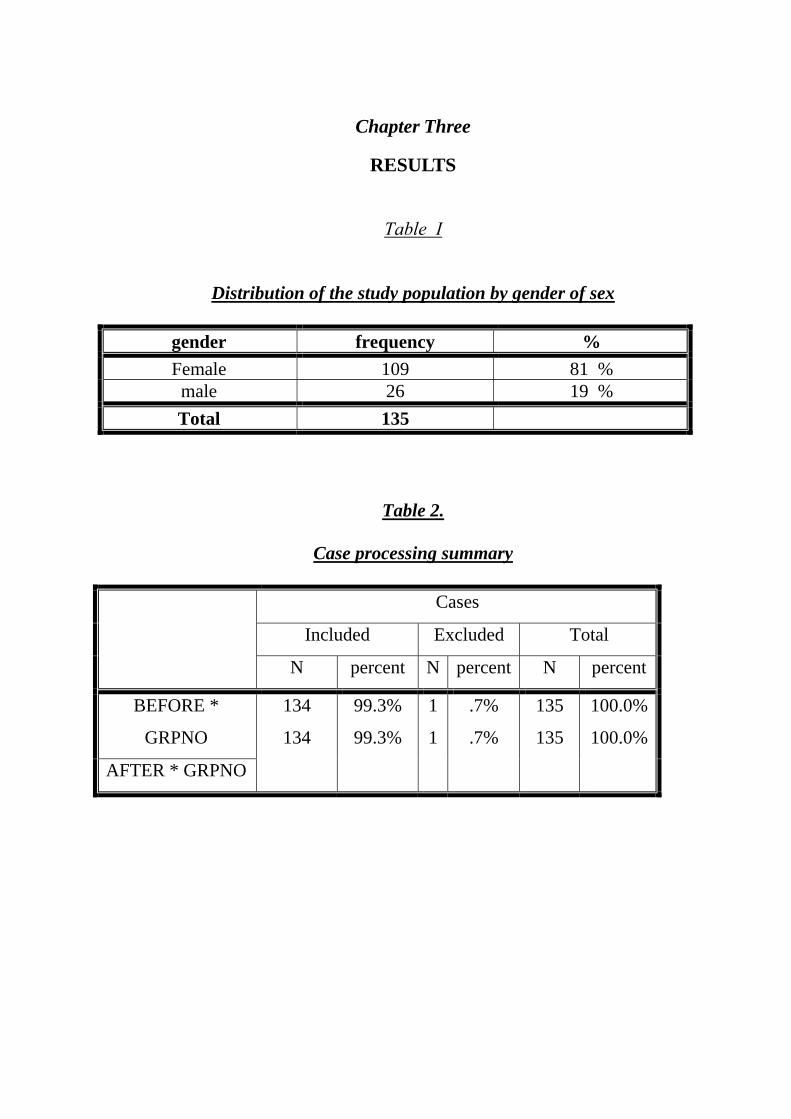
Table I
Distribution of the study population by gender of sex
gender frequency % Female 109 81 %
male 26 19 % Total 135
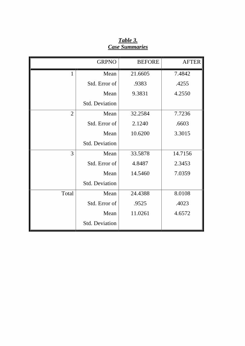
Table 2.
Case processing summary
Cases
Included Excluded Total
N percent N percent N percent
BEFORE *
GRPNO
134
134
99.3%
99.3%
1
1
.7%
.7%
135
135
100.0%
100.0%
AFTER * GRPNO
Page 44
Table 3. Case Summaries
GRPNO BEFORE AFTER
1
Mean
Std. Error of
Mean
Std. Deviation
21.6605
.9383
9.3831
7.4842
.4255
4.2550
2 Mean
Std. Error of
Mean
Std. Deviation
32.2584
2.1240
10.6200
7.7236
.6603
3.3015
3 Mean
Std. Error of
Mean
Std. Deviation
33.5878
4.8487
14.5460
14.7156
2.3453
7.0359
Total Mean
Std. Error of
Mean
Std. Deviation
24.4388
.9525
11.0261
8.0108
.4023
4.6572
Page 45
Table 4. Group 1: Clinicaly suggestive gallbladder disease.
Paired samples Statistics
Mean
N
Std
Deviation
Std. Error
of Mean
Pair BEFORE
21.6605 100 9.3831 .9383
1 AFTER 7.4842 100 402550 .4255
Table 5. Group 1
Paired Samples Test
Paired Differences
t
Mean Std.
Deviation
Std.
Error
Mean
95% Confidence
Interval of the
Difference
Lower Upper
Pair BEFORE
-AFTER
14.1763 7.1713 .7171 12.7534 15.5992 19.768
Page 46
Table 6 Group 2
.Paired Samples Statistics
Mean N Std.
Deviation
Std. Error
Mean
Pair
1
BEFORE
AFTER
32.2584
7.7036
25
25
10.6200
3.3015
2.1240
.6603
Table 7 .Group 2.
Paired Samples Test
Paired Differences
t
Mean Std.
Deviation
Std.
Error
Mean
95% Confidence
Interval of the
Difference
Lower Upper
Pair BEFORE -
AFTER
24.0422 10.0422 2.0084 20.4096 28.7000 12.226
Page 47
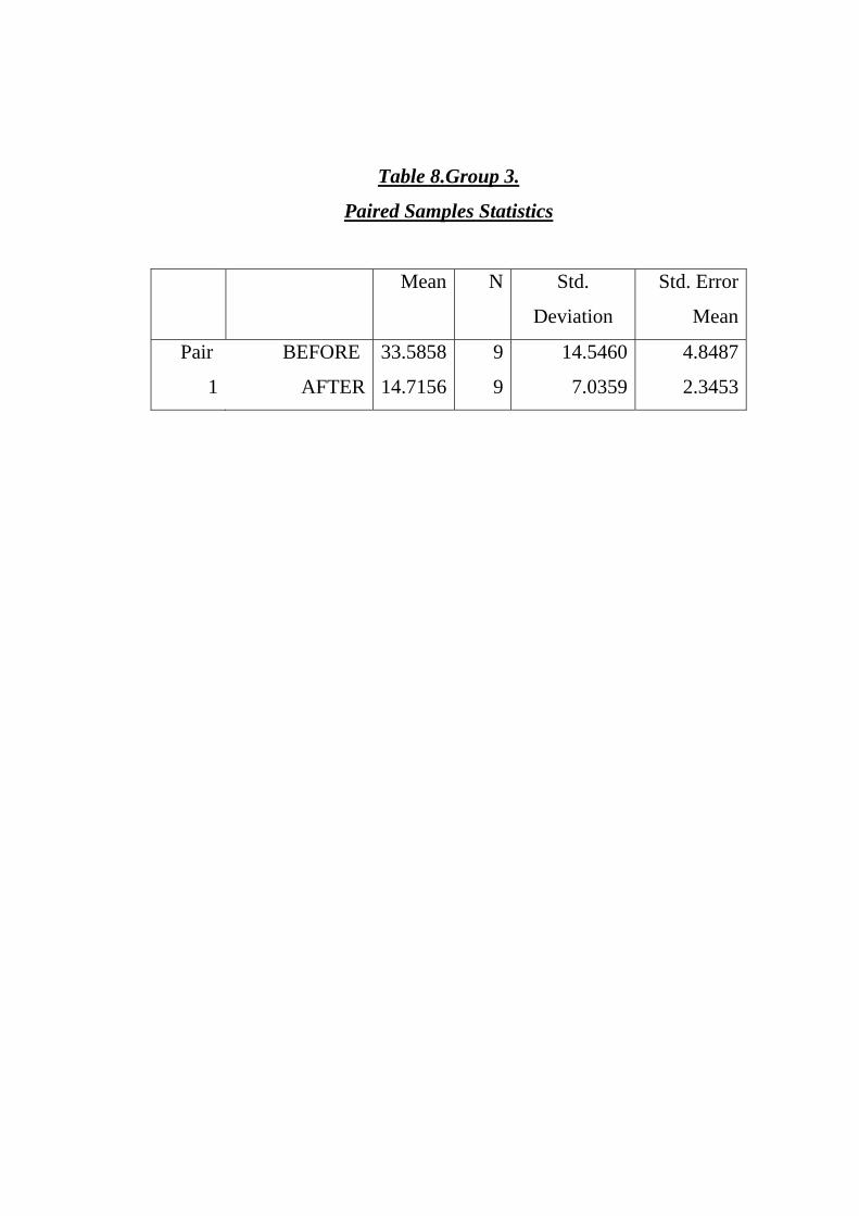
Table 8.Group 3.
sticsPaired Samples Stati
Mean N Std.
Deviation
Std. Error
Mean
Pair
1
BEFORE
AFTER
33.5858
14.7156
9
9
14.5460
7.0359
4.8487
2.3453
Page 48
Table 9. Group 3.
Paired Samples Test
Paired Differences
t
Mean Std.
Deviation
Std.
Error
Mean
95% Confidence
Interval of the
Difference
Lower Upper
Pair BEFORE -
AFTER
18.8722 7.8912 2.6304 12.8065 24.9380 7.175
Page 49
Table 10. Comparison of gall bladder volume
Before and after fatty meal in three groups of patients
Group No Statistic before After %reduction
in volume
Paired t-test
I Mean 21.6605 7.4842 56 % P < 0.001
N : 100 SD 9.3831 4.2550
C.L 21.66+1.84 80.83+7.48
II Mean 32.2584 7.7036 76 % P < 0.001
N: 25 SD 10.6200 8.3015
C.L 32.26+4.16 10.29+7.7
III Mean 33.5878 14.7156 56 % P < 0.001
N : 9 SD 14.5460 7.0359
C.L 33.58+9.5 4.6+14.72
GP I = Clinically suggestive gallbladder disease.
GP II = No gallbladder disease (controls) .
GP III = Silent gallstones.
Page 60
Chapter Four
DISCUSSION
A total number of 135 patients were studied in the period between March 200 to
October 2001 using Aloka 550 ultrasound machine with 81% female predominance
(table-1).
In the case processing ,only one case is excluded (table-2).The mean values of the
gallbladder diameters is found to be within the normal range in the literature review
(1).
Concerning group 1 patients the reduction in the gallbladder volume after fatty meal
is highly significant with 95%cofidence interval. (table- 5), and so in group 2
patients (table-7) and group 3 patients (table-9).
Comparing the gallbladder volumes before and after fatty meals in the three groups
of patients showed that the reduction in the gallbladder volume is highly significant
with p value < 0.001 in the three groups (table-10).
The mean gallbladder volume in group 1 patient is less than group 2 ones (21.66
versus 32.26) and the reduction in gallbladder volume after fatty meal is also less in
group 1 compared to group 2 (56% versus 76%) .(table -10).This difference in
gallbladder motor function exist though the gallbladder is sonically normal in both
group .But group 1 are ill patients , considering these observations I suggested that
: in the ill patient the gallbladder motor function is reduced as other body functions
are impaired and further study is needed to confirm or exclude.
The gallbladder volumes before fatty meals in group 3 patients is found to be more
than the control and the reduction in volume after fatty meal is less than the control
(56% versus 76%) ,this impaired emptying may lead to more stone formation.
Page 61
Despite the differences the values obtained in the three groups are within the normal
range.
These findings don’t imply that the gallbladder disease does not affect it is motor
function and further study is needed to answer the question.
Page 62
CONCLUSION
1. There are some variations in normal gallbladder fasting and postprandial
volumes in the Sudanese population.
2. Group 2 patients (controls) showed fasting gallbladder volumes > group 1 and
< group 3.
3. Group 1 and 3 patients showed residual postprandial gallbladder volumes >
group 2 (controls).
4. The gallbladder motor function in the ill patient decreases as other body
function decreases.
5. The presence of silent stones in the gallbladder decreases the contractility of
gallbladder but remains within normal limits.
6. Ultrasound is an initial useful method for assessing gallbladder motor function.
Page 63
Recommendations
1- Co-operation and feedback system between the clinicians and the radiologist
is of paramount importance for doctor experience and patient benefit.
2- Patients with silent gallstone/ s should be followed up by ultrasound and the
stone's should be left alone as far as the gallbladder kinetics are normal and
the stone / s remains silent (no infection).
3- Routine ultrasonography for patient with clinical condition simulating gall
bladder disease should include fasting and postprandial volume
measurements, if other signs of gallbladder disease are lacking.
4- In patient with (upper quadrant pain, dyspepsia ...etc), gallbladder disease
could be excluded sonographically if the gallbladder size, shape, wall and
function are normal.
5- Further study of the motor function of the diseased gallbladder and the
gallbladder of the ill patient after recovery from other disease is
recommended.
Page 64
REFERENCES
1- Bialy & Loves. Short Practice of Surgery, 19th ed. London H.K Lowis and Co.
Ltd., 1984; ch. 45: 871-7.
2- Richard SS. Clinical anatomy, 2nd ed. Boston, New York 1996; ch 3: 68-9.
3- David S. Textbook of radiology and imaging, 6th ed. Churchill Livingstone
1998; ch. 33: 955-78.
4- Douslas G. Katz MD, Kevin R, Math MD, Stuart A, Grolin MD. Radiology
secrets. Philadelphia Hamley & Belfus, INC 1998; ch.29: 143-49.
5- Parveen K, Michael C. Clinical medicine, 3rded. ElBS with Bailliere Tindall
1995;:ch 5 p280-1
6- Robert B. The Merk manual diagnosis and therapy. Merk show PC Dohme
Research Laboratories, 14th ed, 1994; 74: 509.
7- Pefer A, Martin LW. Diagnostic imaging. Black well Science, International, 4th
ed. 1988; 6: 204-10. :ch.6 p204
8- Roger CS, Mancy SM. Clinical sonography. Lippincoh, 3rd ed., 1988; c.h 2
p249.
9- Qurashi M. Ali. Routine ultrasonography. Vantage Press, New York, 1993; 4:
36-44.
10- Stephan C, Richard N. A guide to radiological procedures. W.B Saundars
Company Limited, 24.3.8 Oval Proad, 3rded., 1997; 10: 107.
11- Siegel A, Kuhn JC, Crow H, Holtzman S. Gall bladder ejection fraction:
Correlation of scintigraphic and ultrasonographic techniques. Clin Nuc1 Med
2000; 25(1): 1-6.
12- Hashimoto S, Goto H, Hirooka Y, Ltoh A, Ishigciro Y, Kojima S, Hirai T,
Hayakawa T, Nairoh Y. An evaluation of three dimentional ultrasonography for
Page 65
the measurement of gall bladder volume. Am J Gastroenterol 1999; 94(12):
3492-6.
13- Carrilho RL, Pinto CA, Velosa J, De Moura MC. Long- term gall bladder stone
recurrence and risk factors after successful lithotripsy. Eur J Gastroenterol
Hepatol 2000; 12(2):209-15.
14- Lirussi F, Nassuato G, Passera D, Toso S, Zalunardo B, Monica F, Virgilio C,
Frasson F, Okolics angi L. Gall stone disease In an elderly population. Eur J
Gastroenterol Hepatol 1999; 11 (5):485-91.
15- Turner HE, Lindsell DR, Vadival A, Thiuainayagam AV, Wass JA. Differing
effect on gallbladder motility of lanreotide SR and Octreotide LRA for treatment
of acromegally. Eur J Endocrinol 1999 ; 141 (6):590-4.
16- Taskin V, Ozyikan E, Sare M, Jilmioglu F. The effect of dividing the sphincter of
Oddi of endoscopic sphincterotomy on the filling and emptying as peds of
function of gallbladder. Surg Laparosc Endosc Percuta Tech 1999; 9(5): 322-5.
17- Portincasa P, Di-Ciaula A, Vendemiale G, Palmieri V, Moschetta AV,
Henegouwen GP, Palasoano G. Gallbladder motility and cholesterol
crystallization in bile from patients with pigment and cholesterol gall stones.
Eur J Clin Invest 2000; 30(4):317-24.
18- Parkman HP, Bogar LJ, Bartula LL, Pagano AP, Thomas RM, Myers SI. Effect
of experimental acalculous cholecystitis on gall bladder smooth muscle
contractility. Dig DisSci 1999; 44(11): 2235-43.
19- Valdivieso V, Severin C, Espinoza R, Orellana P, Otero CG, Huenchullan C,
Icarte G, Sepulveda R. Study of volume and kinetic of gall bladder emptying in
non pregnant and puerperal women. Rev Med Clin 1996; 124(2): 198-203.
Page 66
20- Vorobev LP, Salova LM, Mae IV, Purkhatova SI. The role of different
investigations in diagnosis of functional disorders of the biliary system. Klin
Med Mok 1996; 74(9): 35-8.
21- M ultrangura P, Siwawetkul W. Gall bladder contraction capacity in response to
liquid fatty meal: a real time ultrasonoigraphic study. J Med Assoc Thai1996;
79(10): 640-7.
22- Calabuig R, Castilla M, Pi F, Domingo J, Ramos L, Sierra E. Gall bladder
dyskinesia in a calculous biliary colic. Res Esp Enform Dig 1996 Nov; 88(11):
770-9.
23- Bonfissulo C, Soresi M, Amato D, lppolito S, Maglinarisi C, Carroccio A,
Montalto G. Ultrasonographic assessment of gallbladder motility in obese
subjects. Recenti Prog Med 1996; 87:(7- 8).
24- Hahm JS, Park JY, Rark KG, Ann YH, Lee MH, Park KN. Gallbladder motility
in diabetes mellitus using real-time ultra sonography. Am J Gastroenterol 1996;
9(11): 2391-4.